The Human Leukocyte Antigen-presented Ligandome of B Lymphocytes
Upload
khangminh22Category
view
0download
0
Retrospective Theses and Dissertations Iowa State University Capstones, Theses andDissertations
1-1-1979
Evaluation of bovine polymorphonuclearleukocyte functionJames A. RothIowa State University
Follow this and additional works at: https://lib.dr.iastate.edu/rtd
This Thesis is brought to you for free and open access by the Iowa State University Capstones, Theses and Dissertations at Iowa State University DigitalRepository. It has been accepted for inclusion in Retrospective Theses and Dissertations by an authorized administrator of Iowa State University DigitalRepository. For more information, please contact [email protected].
Recommended CitationRoth, James A., "Evaluation of bovine polymorphonuclear leukocyte function" (1979). Retrospective Theses and Dissertations. 18629.https://lib.dr.iastate.edu/rtd/18629

Evaluation of bovine polymorphonuclear
leukocyte function fS'tt ;p79 I( 7'-13 e,_, 3
by
James Allen Roth
A Thesis Submitted to the
Graduate Faculty in Partial Fulfillment of
The Requirements for the Degree of
MASTER OF SCIENCE
Department1 Veterinary Microbiology and Preventive Medicine
Major1 Veterinary Microbiology
Signatures have been redacted for privacy
Iowa State University Ames, Iowa
1979
1248794

INTRODUCTION
LITERATURE REVIEW
ii
TABLE OF CONTENTS
Polymorphonuclear Leukocytes
Basophils Eosinophils Neutrophils
Phagocytosis by Polymorphonuclear Leukocytes
Historical aspects of phagocytosis Chemotaxis Opsonization Ingestion Degranulation Energy sources Microbial killing mechanisms
Polymorphonuclear Leukocytes as Mediators of Antiviral Immunity
Virus Induced Defects in Polymorphonuclear Leukocyte Funct~on
Evaluation of Bovine Polymorphonuclear Leukocyte Function
Source of polymorphonuclear leukocytes Methods for evaluation of function
Bovine Viral Diarrhea-Mucosal Disease
PART I. EVALUATION OF BOVINE POLYMORPHONUCLEAR
Page
1
3
3 4 g 8
8 9
10 14 14 17 17
26
29
30 30 32 34
LEUKOCYTE FUNCTION 38
IODINATION
Summary
Introduction
39 39 39

iii
Materials and Methods
Results
Discussion
NITROBLUE TETRAZOLIUM REDUCTION
Summary
Introduction
Materials and Methods
Results
Discussion
CHEMILUMINESCENCE
Summary
Introduction
Materials and Methods
Results
Discussion
PART II. EXPERIMENTATION INVOLVING BOVINE VIRAL DIARRHEA VIRUS
EFFECTS OF BOVINE VIRAL DIARRHEA VIRUS INFECTION ON BOVINE POLYMORPHONUCLEAR LEUKOCYTE FUNCTION
Summary
Introduction
Materials and Methods
Results
Discussion
Page
41
46
51 57 57 57 59 62
67
71 71 71 73 76
80
85
86 86 87
88
96 106

SUMMARY
BIBLIOGRAPHY
ACKNOWLEDGMENTS
iv
Page
115
117
131

1
INTRODUCTION
Polymorphonuclear leukocytes (PMNs) are bone marrow
derived white blood cells that play a central role in the
defense of the host against infection. Through inter-
actions with antibody, complement, and chemotactic
factors, PMNs are attracted to invading microorganisms
and are stimulated to phagocytose them. A complex series
of metabolic events within the PMN leads to the intra-
cellular killing of many infectious agents. Disorders '
of PMN function may lead to severe recurrent infection.
Many pathogenic agents possess the ability to impair
or to evade host defense mechanisms; without this ability
.they presumably would not be pathogenic. When a pathogenic
agent impairs a defense mechanism of the host in order
to facilitate its own survival, it also facilitates the
survival of other infectious agents within the host.
In recent years there have been several reports of
impairment of PMN function by infectious agents.
Bovine viral diarrhea (BVD) virus is an infectious
agent which has been demonstrated to impair the host's
immune defenses, In investigations at Iowa State University
and elsewhere, the BVD virus has been demonstrated to have
a marked effect on lymphoid tissue in vivo and in vitro.
(Peter et al., 1967; Muscoplat et al., 197Ja, b; Truitt and

2
Shechmeister, 197); Reggiardo, 1975), and to inhibit the
clearance of bacterial organisms from the blood (Reggiardo,
1975). This inhibition of normal blood clearance mechanisms
suggests that the BVD virus induces a defect in phagocytic
cell function.
The purpose of the present study was to determine if
a defect in PMN function occurred in cattle following
infection with BVD virus. Techniques which had previously
been reported for the evaluation of bovine PMN function
were not felt to be adequate to screen for a defect in
bovine PMN function following BVD virus infection.
Therefore, it was necessary to adapt procedures which
had been reported for use with human PMNs to use with
bovine PMNs and to establish normal values for these
procedures in the bovine system.
The objectives of the experimentation reported in this
thesis were1 (1) to develop new techniques for the evalua-
tion of bovine PMN function and to establish normal values
using these techniques; and (2) to determine if infection
with BVD virus altered PMN function in cattle.

3
LITERATURE REVIEW
Polymorphonuclear Leukocytes
The polymorphonuclear phagocytic system is comprised
of three types of cells found in blood1 basophils,
eosinophirs, and neutrophils. These cells vary in size from
8-14 µm and are referred to collectively as polymorphonuclear
leukocytes (PMNS), PMNs are produced in the bone marrow
from a common stem cell, the myeloblast, which undergoes
a series of cell divisions and maturational changes to
eventually produce all three types of PMNs (Schalm, 1977). In response to infection, there can be a marked increase
in production of PMNs by the bone marrow and a dramatic rise
in the number of PMNs found in the blood, 1
Cells of the polymorphonucl~ar phagocytic system
comprise the first line of defense against infectious
agents. In response to infection, PMNs are rapidly
released from the bone marrow into the circulation and are
attracted to the site of microbial invasion where they
will attempt to engulf and destroy potential pathogens.
The importance of the polymorphonuclear phagocytic system
is emphasized by the observation that animals which lack
adequate numbers of· normally functioning PMNs soon die of
overwhelming bacterial infection, There are many excellent
reviews dealing with polymorphonuclear leukocytes, their

4
phagocytic activity, and their role in host defense
(Baehner, 1972; Stossel, 1974; Bellanti and Dayton, 19751
Murphy, 1976; Beeson and Bass, 1977; Babior~ 1978).
The three types of PMNs in the circulation are
distinguished on the basis of the staining properties of
their cytoplasmic granules. Neutrophils are the most
abundant and comprise about 70% of the normal circulating
white blood cell population in man, .but considerably.
less in domestic animals. Eosinophils and basophils
possess less phagocytic activity, although they participate
in other ways in the immunologic processes.
Basophils
The basophilic leukocyte is present in blood in an
almost negligible quantity, usually forming less than 0.5%
of the total number of leukocytes, The basophil has large
granules which stain deep blue-black with Wright's stain.
The granules of the basophil contain histamine and other
substances which cause contraction of smooth muscle and an
increase in the permeability of small blood vessels. The
function of the basophil is not known in detail, but it
does participate in allergic reactions and is essential in
some types of tissue damage such as serum sickness. The
basophil may be the precursor of the tissue mast cell1 it
is not tho.ught to be active in phagocytosis.

5
Eosinophils
The eosinophil has granules that stain red or orange
with Wright's stain. It has been demonstrated to have
phagocytic potential, to ingest antigen-antibody complexes,
and to have an important role in anaphylactic and allergic
phenomena (Douglas, 1976). Eosinophils are capable of
ingesting and killing bacteria in vitro, although they
are much less efficient than neutrophils or macrophages
(Cline, 1972). Eosinophils offer little or no protection
against bacterial infection even when present in great
numbers, as in children with congenital neutropenia. It
may be that eosinophil phagocytosis is an in vitro
phenomenon with limited relevance to the cell's activities
in vivo, There are some data to suggest that eosinophils
are geared more toward extracellular degranulation or
"exocytosis" than toward phagocytosis. Two functions
proposed for the eosinophil include modulation of
inflammation and defense against metazoan parasites.
These functions would be better served by a cell geared
toward exocytosis than toward phagocytosis (Beeson and
Bass, 1977).
Metabolic studies on essentially pure eosinophil
preparations obtained from the ascitic fluid of a patient
with eosinophilic gastroenteritis have demonstrated that

6
particle ingestion by eosinophiis is associated with a
marked increase in hexose monophosphate shunt activity,
H2o2 formation, superoxide anion generation, chemi-
luminescence, thyroid hormone degradation, iodination,
and estrogen binding. This postphagocytic metabolic
burst by eosinophils was qualitatively similar to that
observed in neutrophils, but for several parameters the
eosinophil response was greater than the neutrophil response
(Klebanoff et al~, 1977).
Neutrophils
The neutrophil is the most common type of PMN found
in the blood; its principal function is to combat bacterial
infection. The neutrophil contains in its cytoplasm two
types of membrane bound granules, which contain many
different subfitances; some of these substances carry
strong negative charges and others carry equally strong
posi tiv.e charges. The overall balance between the different
chemical constituents gives a net staining reaction that
is about neutral (Murphy, 1976),
Neutrophils are considered to represent a first
line of defense against bacterial infection, They arrive
at a site of bacterial invasion earlier than macrop~ages
and serve to restrict the spread of invading micro-
organisms. They adhere to particles, and when functioning
1

7
riormaliy, they ingest and destroy microorganisms by means
of the enzymic contents of their lysosomes (Tizard, 1977).
Neutrophils are much more important than eosinophils or
basophils in phagocytosis (Cline, 1972), and more is known ' about neutrophil function and metabolism,
The mature human neutrophil is an end-stage cell
which, once released from ·the bone marrow, circula.tes with
a half-life of 6-7 hours before leaving the blood stream
in a random (nonage related) fashion, In man about 108
neutrophils are turned over daily: most of these cells
leave the body through the digestive and respiratory
systems, The total blood granulocyte pool is composed of
approximately equal numbers of circulating and marginated
cells. Only the circulating pool is sampled by standard
blood collection techniques, When there is a sudden
demand for neutrophils, the marginated pool can supply
cells almost instantly to double the numbers of cells in
the circulating pool, The marginated pool is responsible
for the rise in leukocyte numbers after exercise, excitement,
fear, and feeding, If continued need for neutrophils occurs
because of tissue damage, the large reserve of cells in
the bone marrow is responsible for meeting this demand •
. Two to three times the circulating pool may be mobilized
in one hour and 8-10 times the circulating pool may be
mobilized in 6-7 hours (Medway, et al,, 1969).

8
Phagocytosis by Polymorphonuclear Leukocytes
Historical aspects of phagocytosis
The idea that bacteria are disposed of by being
taken up and digested by phagocytic cells is less than
a century old, and was first clearly formulated by the
Russian zoologist Elie Metchnikoff (Metchnikoff, 1893),
In 1882 he studied the role of motile cells of a trans-
parent starfish larvae in protection against foreign
intruders. He introduced a rose thorn into these larvae
and noted that a few hours later the rose thorn was
surrounded by motile cells, This experiment can be
considered the starting point of cellular immunology.
Koch and Neisser had previously established that bacteria
can be found in leukocytes, but they thought that this
was the result of bacterial invasion of the leukocytes,
Metchnikoff demonstrated that the leukocytes had in fact
engulfed the microorganisms and called the process
phagocytosis. He later demonstrated the existence of
two types of circulating cells capable of phagocytosis,
the polymorphonuclear leukocytes and the macrophages, and
proposed the general term "phagocytes" for these cells
(Grabar, 1976).
Phagocytosis by neutrophils is composed of four
interrelat.ed phasesa chemotaxis, opsonization, ingestion,
and degranulation (Drutz, 1976).

9
Chemotaxis
Chemotaxis is the process whereby phagocytic cells are
attracted to the vicinity of invading pathogens. Chemotactic
factors cause a change in. the direction of leukocyte
mobility but. do not cause a change in the speed of leukocyte
movement. The interaction of microorganisms and host
tissue leads to the generation of chemotactic factors by
several mechanisms (Stossel, 1974). Although some bacteria
release substances that without further alteration have
the capacity to attract phagocytes (Nelson et al., 1975; John and Sieber, 1976), most chemotactic factors are of
.host origin. The complement system appears to be the most
important source of these leukotactic factors (Ward et al.,
1965; Stossel, 1974), Complement components which are
chemotactic include CJa, C5a, and c567; they may be
generated via the classical or alternative pathway of
complement activation or by the action of nonspecific
proteases upon native complement components (Drutz, 1976; Repo, 1977). Other sequentially reacting protein systems
(kallikrein system; fibrinolysis system) also contain
factors with leukotactic activity (Drutz, 1976). Finally,
l~ukocytes themselves contain factors that are directly or
indirectly chemotactically active, Ingestion of particulate
matter by neutrophils causes them.to release a factor
that has the capacity to attract other neutrophils in the

10
absence of serum (Zigmond and Hirsch, 197J), and lymphocytes
responding to anitgens elaborate lymphokines, which, among
other properties, have chemotactic activity (Stossel,
1974), When chemotactic factors reach the surface of the
neutrophil, an esterase on the cell surface is activated,
the hexose monophosphate shunt is activated, calcium
fluxes occur in the cell, and microfilaments (composed of
actin) and microtubules (composed of tubulin) assemble,
providing the motility needed to propel the cell toward the
source of the chemotactic factor, Myosin also has been
found in phagocytic cells, and it is likely that the
motility of such cells may have a close molecular
relationship to muscle contraction (Drutz, 1976), Prior
phagocytosis of material has been shown to reduce a cell's
ability to respond to a chemotactic attractant (Mowat and
Baum, 1971).
Opsonization
Opsonins are serum components which react with micro-
organisms and make them more susceptible to ingestion by
phagocytes, This process is termed opsonization and may
occur by one of three mechanisms (Drutz, 1976).
(1) Specific antibody alone may act as an opsonin.
When specific antibody combines with antigenic sites on

11
the surface of a microorganism through antibody combining
sites located on the Fab portion of the immunoglobulin
molecule, the Fe portion of the molecule is then free to
attach to Fe receptor sites on the surface of phagocytes,
thereby completing a bridge between the microorganism and
phagocytic cell. Human PMNs are reported to have receptors
for the Fe portion of certain IgG subclasses of antibody,
but to lack an Fe.receptor for IgM (Menzel et al,, 1978),
Bovine PMNs are reported to have an Fe receptor for IgG
and for IgM (Grewal et. al., 1978). J (2) Specific antibody acting in concert with complement
via the classical complement pathway may promote opsoni-
zation. Here, a quantity of antibody apparently insufficient
to opsonize on its own may react with bacteria and activate
the classical complement sequence, resulting in the binding
of complement components to the bacterial surface. Bovine
and human PMNs have been demonstrated to have complement
receptors (Grewal et al .• , 1978; Menzel et al., 1978).
Complement component CJb is believed to be the opsonically
ac;:tive component (Gigli and Nelson, 1968). The CJb on the
bacterial surface apparently serves as a bridge between
bacteria and phagocyte, prompting ingestiQll (Drutz, 1976).
J

12
(3) Opsonization can be nonspecific and involve the
activation of complement via the alternative pathway.
This method does not require the action of specific
antibody and may play an important role in early pre-
immune stages of infection prior to the production of
specific antibody. Complement component CJb is again
believed to be the opsonically active component (Drutz,
1976).
Ingestion may occur in the absence of opsonization
by a method known as surface phagocytosis. Here, encapsu-
lated bacteria are trapped between leukocytes themselves,
between leukocytes and tissue surfaces, or along with
leukocyt"es in fibrin clots. Surface phagocytosis is much
less efficient in areas where leukocytes are not tightly
packed (pleural, pericardial, synovial, and cerebrospinal
fluids). Surface phagocytosis may also play an important
role in the preimmune stages of infection prior to the
production of specific antibody (Drutz, 1976).
The receptors for lg and complement which have been .
referred to may not be actual membrane. proteins that
specifically combine with the opsonin. It has been
postulated that the principal factor that determines
whether phagocytosis of a particle can occur is the physical
nature of the surface of the particle in comparison with

13
that of the phagocyte. Van Oss and Gillman (1972a) found
that bacteria with surfaces that were more hydrophobic
·than the ~urface of phagocytes readily became engulfed,
whereas bacteria with surfaces that were more hydrophilic
than the 13urface of phagocytes resisted engulfment. Most
nonpathogenic bacteria are hydrophobic and are readily
ingested py phagocytes, Bacteria that are highly hydro-
phobic, such as Mycobac.terium tuberculosis, are spontane-
ously ingested by phagocytic cells. These organisms owe
their patpogenicity to their resistance to digestion by
phagocyti? enzymes, Bacteria like Diplococcus pneumoniae
that possess a hydrophilic carbohydrate capsule are not
normally subject to phagocytosis. Antibody to encapsulated
organisms makes their surfaces more hydrophobic, and
simultaneously they become easier to phagocytose. The
addition of complement components Cl, C4, and C2 has no
effect, but when CJ is bound, both the hydrophobic
character of the bacterial surface and the ease of
phagocytosis increase sharply. It is, therefore, possible
that antibody and complement aid phagocytosis because
they change the character of the bacterial surface, rather
than because there are specific receptor molecules for them
on the phagocytic cell surface (Van Oss and Gilman, 1972a,
b; Carpenter, 19751 Murphy, 1976; Van Oss, 1978),

14
Ingestion
Upon particle or microbial contact, cell pseudopodia
are extended which fuse on the distal side of the material
to be ingested, The particle becomes encased within a
phagocytic vesicle, or phagosome, which is lined by
inverted plasma membrane, The phagosome buds off from
the cell periphery and moves centripetally, apparently
through the mediation of microtubules, The mechanism for
the triggering of ingestion is not clearly understood '
(Drutz, 1976),
Degranulation
The destruction of susceptible microorganisms within
neutrophils is intimately associated with the process
of degranulation, the release of granule contents into
phagosomes, The neutrophil contains two types of membrane
bound granules (lysosomes), In man, primary (azurophilic)
granules contain abundant hydrolytic lysosomal enzymes,
large amounts of myeloperoxidase, lysozyme, elastase,
and cationic proteins. Secondary (specific) granules,
which are smaller than primary granules, contain lactoferrin
and lysozyme (Drutz, 1976). Bovine neutrophils have
biochemical properties very similar to those of human 1 neutrophils, although they appear to have a lower conten

15
of pri~ary granule enzymes and to virtually lack lysozyme
(Rausch and Moore, 1975; Gennaro et al., 1978). I
As the phagosome forms during microbial engulfment,
neutrophil granules undergo violent movement in proximity
to the phagosome, fuse with the phagocytic vacuole, and
disappear from the cytoplasm (degranulate) (Stossel, 1974).
Specific granules fuse with the phagocytic vesicle first;
this process starts approximately JO seconds after ingestion.
Primary granules do not begin to discharge their contents
until J minutes after ingestion, and usually only into
the larger vesicles (Murphy, 1976). Neutrophils contain
a skeleton of microtubules, which appear to direct the
intracellular motion of the primary and secondary
granules after phagocytosis. The degranulation phenomenon
is prevented by treating neutrophils with either
colchicine or vinblastine, both of which prevent the
polymerization of microtubule subunits into functional
tubules (Zurier et al., 1973; Weissmann et al., 19751
Wilson, 1975; Hoffstein et al., 1977). Degranulation is
also prevented by agents that raise the intracellular
levels of cyclic AMP (Goldstein et al., 19731 Zurier et al.,
1973; Hawkins, 1974; Ignarro et al., 1974; Zurier et al,,
1974; Weissmann et al., 1975; Goren, 1977). Neutrophils
contain a protein kinase that can phosphorylate several
proteins, probably including tubulin. It is hypothesized

16
that cyclic AMP may stimulate this protein kinase to
phosphorylate the tubulin subunits of microtubules and
prevent their aggregation (Murphy, 1976), Agents which
are reported to raise intracellular levels of cyclic AMP
and to block degranulation include beta adrenergic drugs
(epinephrine, norepinephrine, isoproterenol), prostaglandin
E1 , histamine, cholera enterotoxin, ~· coli heat labile
enterotoxin, theophylline, and exogenous cyclic AMP itself
(Ignarro et al., 1974; Zurier et al., 1974; Ignarro and
Cech, 1975; Weissmann et al., 1975; Hoffstein et al,, 1977;
Bergman et al., 1978). Agents which increase intracellular
levels of cyclic GMP have been found to increase degranu.-
la tion by PMNs. These agents include carbamylcholine
chloride, phorbol myristate acetate, and exogenous cyclic
GMP (Zurier et al., 1974; Goldstein et al., 1975a; Weissmann
et al., 19751 Wright et al., 1977).
A low molecular weight component of complement,
similar or identical to human C5a, has been demonstrated
to be capable of interacting with human PMNs to induce
degranulation, fusion of lysosomal membranes with plasma
membranes, and transient assembly of microtubules associated
with the release of endogenous myeloperoxidase, This
component of complement has been called lysosomal enzyme-
releasing factor because it provokes secretion of lysosomal
hydrolases either into phagocytic vacuoles or into
17
extracellular spaces, Lysosomal enzyme-releasing factor
may play an important rolP in the inflammatory response
(Goldstein et al., 1973; 1975b).
Energy sources
The energy needed for PMNs to function in motility,
ingestion, and degranulation is derived from their large
reserve.of glycogen through anaerobic glycolysis. This
form of metabolism does not require oxygen, which is
important for a cell which must operate in inflammatory
sites where oxygen tension may be very low, Under normal
conditions, the neutrophil takes up glucose from its
environment, and its cellular reserve of glycogen remains
relatively constant. During phagocytosis, the rates of
glycogen breakdown, glucose uptake, and lactate output
are markedly increased, Neutrophils may also metabolize
glucose by the hexose monophosphate shunt which is important
for the oxidative killing mechanisms of the PMN (Murphy,
1976).
Microbial killing mechanisms
PMNs possess a number of mechanisms for killing
microorganisms. These mechanisms probably represent a
functional redundancy; many microorganisms may be killed
effectively by any one of the mechanisms, while some of

18
the mechanisms are likely to be active against only
specific classes of microorganisms (Drutz, 1976), Microbial killing mechanisms comprise two broad categories1
nonoxidative and oxidative.
Nonoxidative killing mechanisms Many organisms
are killed normally under anaerobic conditions (Mandell,
1974). The killing mechanisms which do not require oxygen
are very important when neutrophils must function in an
anaerobic environment. These killing mechanisms depend
upon substances present in the primary and specific granules
of the PMNs. A list of enzymes and other substances
found within neutrophil granules is presented in Table 1
(Bellanti, 1978); some of the more important bactericidal
substances are discussed below.
(1) Cationic proteins are a family of proteins
found in the primary granules. They have a positive
charge and therefore tend to stick to bacterial surfaces
which are generally negatively charged, By some unknown
mechanism, they are able to damage microbial membrane
barriers and to kill some bacterial organisms (Drutz, 19761 Murphy, 1976),
(2) Lactoferrin is an iron binding protein which is
found in the specific granules of PMNs. Most bacteria

19
Table 1. Enzymes and other substances found within neutrophils (Adapted from Cochrane, 1968.).
Acid phosphatase Acid ribonuclease Acid deoxyribonuclease Cathepsins B, C, D, E Phosphoprotein phosphatase Organophosphate-resistant
esterase 13-Glucuronidase 13-Galactosidase 13-N-acetylglucosaminase 11-fucos.idase 11-glucosidase 11-mannosidase 11-N-acetylglucosamidase .11-N-acetylgalactosaminidase Myeloperoxidase
Hyaluronidase Lysozyme Collagenase Aryl sulfatases A and B Phospholipases Acid lipase Lac'toferrin Phagocytin and other related
bactericidal proteins Endogenous pyrogen Plasmino~en activator (Puro-
kinase) Hemolysin(s) Mucopolysaccharides and
glycoproteins Basic proteins1 (a) Mast. cell-
active (b) Permeability-inducing, independent of mast cells
such as Staphylococcus aureus, E. coli, Pasteurella
multocida, and Mycobacterium tuberculosis require iron
for growth, Lactoferrin may exert its antimicrobial
function by binding and withholding required iron from
ingested bacteria. Lactoferrin may be important in
resistance to bacterial infection in the mammary gland,
In response to bacterial invasion neutrophils release
their stores of lactoferrin and, in this way, enhance
the bactericidal activity of milk (Tizard, 1977).

20
(J) Lysozyme is an enzyme which is capable of destroying
the peptidoglycan layer of the cell wall of certain
bacterial organisms by breaking the bonds which join
alterna"1>ing units of N-'acetylglucosamine and N-acetylmuramic
acid, The destruction of the peptidoglycan layer alters
the membrane barrier activity of the cell wall and leads
to bacterial death, Lysozyme is present in both the
primary and specific granules of human PMNs; it is completely
lacking in bovine, caprine, ovine, and feline PMNs (Rausch
and Moore, 1975; Gennaro et al., 1978), Lysozyme kills
those few gram-positive bacteria that do not have some
covering for their peptidoglycan layer; however, it can
kill many gram-positive and gram-negative organisms if
some other agent (antibody and complement) damages the
cell wall and allows lysozyme access to its substrate.
Because bacterial death often precedes the action of
lysozyme, this enzyme may serve in a digestive rather than
microbicidal capacity in the phagosome (Drutz, 1976;
Murphy, 1976).
(4) Elastase from primary granules attacks mucopeptides
of certain bacterial cell walls and may also be more
important in digestion Of bacterial organisms than killing,
(5) Lactic acid is generated in large quantities
during phagocytosis because of the increased glycolytic

21
activity, In some unknown way, the excess hydrogen ions
are concentrated in phagocytic vacuoles, In rat, mouse,
and rapbit neutrophils the intravacuolar pH falls to
about 4,0 following phagocytosis; in human neutrophils
the intravacuolar pH falls to only about 6,0-6,5, The
significance of the low pH is not clear. Few bacteria
can continue to grow at pH 4,0; however, the cationic
proteins and proteolytic enzymes of human neutrophil
granules have a pH optimum near neutrality and may not
function well under acidic conditions (Drutz, 1976;
Murphy, 1976),
Oxidative killing mechanisms When neutrophils
take up organisms under aerobic conditions, a series of
related changes occur1 increased oxygen consumption;
increased hexose monophosphate shunt activity; and generation
of hydrogen peroxide, superoxide anion, hydroxyl radical,
and possibly singlet oxygen (Babior, 19781 Johnston, 1978), This increased oxygen consumption is not suppressed by
cyanide, which is fairly good evidence that it is not
being mediated by cytochromes (Johnston, 1978), It appears
to be due to the activity of an oxidase enzyme, or enzyme
complex, which is located in the plasma membrane, and
therefore in the phagosomal membrane (Goldstein et al,,
1977). The most likely substrates for.the oxidase enzyme

22
in neutrophils are NADH and NADPH, There is conflicting
evidence as to which of these molecules is the substrate,
The hexose monophosphate shunt generates NADPH, but this
may undergo a transhydrogenase reaction with NAD to form
NADH, which may be the. actual substrate (Stossel, 1974),
Recent evidence strongly favors .the hypothesis that NADPH
is the electron donor in this reaction (Babior, 1978),
Oxidase enzymes do not necessarily, or even usually,
mediate two-electron transfers to form H2o2 directly.
Their reactions often proceed by one-electron transfer
steps that result in the formation of unstable and highly
reactive intermediates, In neutrophils there is evidence
for the generation of superoxide anion, singlet oxygen,
and the hydroxyl radical (Babior, 1978; Johnston, 1978),
Superoxide anion is oxygen that has accepted one
electron, It is formed either by the univalent reduction
of oxygen or by the univalent oxidation of H2o2 and is
a highly reactive radical which can act as an oxidant
or as a reductant, When it functions as a reductant, as
in the reduction of ferricytochrome C or nitroblue
tetrazolium, the superoxide anion is oxidized to oxygen.
When it acts as an oxidant, the superoxide anion is
reduced to H2o2• When two molecules interact, one is
oxidized and the other reduced as follows1 - - + o2 + o2 + 2H - o2 + H2o2 ,

23
This dismutation occurs spontaneously and is also catalyzed
by the en~yme superoxide dismutase (Rosen and Klebanoff,
1976), The growth of certain microorganisms is inhibited
by superoxide anion (Johnston et al., 1975), and this
inhibition is related to the content and distribution of
microbial superoxide dismutase (Yost and Fridovich, 1974),
Singlet oxygen is an electronically excited state of
oxygen. Ground s.tate molecular oxygen has two valence
electrons that are unpaired and have spins in the same
direction, Singlet oxygen is formed when an absorption
of energy shifts one of these electrons to an orbital
of higher energy with an inversion of spin (Rosen and
Klebanoff, 1976). The lifetime of singlet oxygen is short
with dissipation of excess energy by thermal decay,
light emi;>sion, or chemical reaction. Singlet oxygen
is thought to be responsible, either directly or indirectly
for the emission of light by PMNs during phagocytosis
(Allen et al., 1972; Andersen et al., 1977). There is
still no proof that singlet oxygen is actually formed
during phagocytosis, because the means employed for
detecting singlet oxygen in biological systems are .
nonspecific (Babior, 1978). There are several mechanisms
proposed for the formation of singlet oxygen within the
PMN (Webb et al., 1974; Rosen and Klebanoff, 19761
Babior, 1978), One proposed ·mechanism is that it is an

24
intermediate in the dismutation of superoxide anion, as
follows.·1 - - -2 1 2H+ 02 + 02 ~ 02 + 02 ~ H202'
Another proposed mechanism is by the reaction of super-
oxide anion with the hydroxyl radical, as follows1
02 + 'OH ~ OH- + 10 2 ,
The hydrogen peroxide-myeloperoxidase-halide reaction is
also proposed as a source of singlet oxygen. It is known
to display chemiluminescence under the proper conditions.
There is evidence that normal light emission by PMNs
requires both superoxide anion and myeloperoxidase1
therefore, more than one of the above mechanisms may be·
involved (Rosen and Klebanoff, 1976). It has been suggested
that singlet oxygen may function in the bactericidal activity
of PMNs by combining across the double bonds of unsaturated
fatty acids (Krinsky, 1974; Murphy, 1976),
The hydroxyl radical 'is a highly unstable oxidizing
species that reacts almost instantaneously with most
organic molecules that it encounters, It appears that this
exceedingly reactive substance can be generated biologically,
and may participate in the microbicidal activity of
phagocytes. Hydroxyl radical may b~ generated in the PMN
by the following reaction (Webb et al,, 1974; Babior, 1978)1
02 + H2o2 + H+ ~ 'OH + H20 + o2 ,

25
A potent antimicrobial agent of neutrophils is
hydrogen peroxide. Although hydrogen peroxide is
bacteri.cidal in its own right, its antibacterial, anti-
fungal, and antiviral activity is markedly potentiated by
myeloperoxidase in the presence of halide ions. The
mechanism for this bactericidal activity is not precisely
known; reactive aldehydes, which arise from the hydrogen
peroxide-myeloperoxidase-halide-microbe interaction,
may represent the actual bactericidal compounds (Stossel,
1974). The reaction C?f hydrogen peroxide and halide ions
in the presence of myeloperoxidase results in the covalent
bonding of halide anions to protein of ingested particles.
The extent of this reaction may be determined by adding
radiolabeled iodine to a phagocytic system and determining
the amount of radioactivity which is covalently bound to
protein (!'incus and Klebanoff, 1971; Simmons and Karnovsky,
1973; Klebanoff and Clark, 1977). Hydrogen peroxide for
this reaction is formed in the phagosome by the action
of the oxidase enzyme mentioned previously, Myeloperoxidase
is Tound in the primary granules and is delivered to the
phagosome by degranulation. Halide ions enter by diffusion;
iodide can even be stripped from thyroid hormones by PMNs
and subsequently fixed to bacteria (Stossel, 1974),
26
Polymorphonuclear Leukocytes as Mediators of Antiviral Immunity
P~lymorphonuclear leukocytes are considered to be
primarily involved in antibacterial rather than antiviral
defense. However, some recent observations have suggested
a role for PMNs in antiviral immunity (Rouse et al., 1978).
There are three basic mechanisms suggested for mediation
of antiviral immunity by PMNs1 1) phagocytosis and
destruction of virus particles, 2) antibody-dependent cell-
mediated cytotoxicity, and J) release of a subcellular
mediator, similar to interferon, which can render.cells
resistant to virus infection (Rouse et al., 1978).
Several viruses have been demonstrated to be engulfed
by PMNs (Baratawidjaja et al., 1965; Gresser and Lang,
1966; Sommerville, 1966; 1968; Belding and Klebanoff, 1970).
Some investigators have emphasized the protection afforded
by intraleukocytic residence of viruses against specific
antibodies and other nonspecific viral inhibitors present
in blood and have pointed to phagocytosis as a means of
disseminating viral particles (Gresser and Lang, 1966;
Smith, 1972). However, others have reported a rapid
~ecrease in the titer of certain viruses following
ingestion by leukocytes (Sommerville, 1968). The nature
of the intraleukocytic virucidal system is not known.

27
The myeloperoxidase-hydrogen peroxide-halide system has
been shown to be virucidal for poliovirus and vaccinia
virus in vitro and may have a.role in antiviral defense
in vivo (Belding and Klebanoff, 1970), If a virus is able
to interfere with degranulation in the PMN, it will prevent
the activity of this potentially virucidal system, The
virus m~y then be able to survive in the PMN and be
disseminated throughout the body,
An'!;i{lody-dependent cell-mediated cytotoxicity (ADCC)
. is the killing of antibody-coated target cells by an
effector cell, PMNs have been demonstrated to be capable
of mediating ADCC (Gale and Zighelboim, 1975; Rouse et al,,
1976; Clark and Klebanoff, 1977; Russell and Miller,
1978), and in the bovine species, PMNs have been shown
to be the most efficient of all cell types tested in
mediating antiviral ADCC (Rouse et al,, 1978), Furthermore,
PMNs, in the presence of antibody, could prevent virus
dissemination when added to virus-infected monolayers
(Wardley et al., 1976b), This PMN mediated ADCC has been '
shown to be independent of DNA, RNA, and protein synthesis
by the PMN; it is blocked·by drugs which inhibit micro-
tubule function, and it is subject to the control of cyclic
AMP and cyclic GMP. Drugs which elevate cyclic AMP
decrease cytotoxicity; drugs which decrease cyclic AMP or

28
increase cyclic GMP enhance cytotoxicity (Wardley et al,,
1976a; Clark and Klebanoff, 1977). These properties of the
regulation of ADCC are very similar to ·the regulation of
degranulation within the PMN (Weissmann et al,, 1975).
This similarity suggests that degranulation may play a role
in ADCC, Myeloperoxidase and cationic proteins, both of
which are found in PMN lysosomes, appear not to be essential
in the cytotoxicity system (Clark and Klebanoff, 1977);
however, there are many other substances in the PMN
lysosomes which may play a role,
There is recent evidence that PMNs may be involved
in antiviral immunity by the release. of subcellular
mediators which can render cells resistant to virus
infection (Rouse et al., 1977; 1978), These mediators
have properties similar to interferon and have been i
tentatively called interferon J (Rouse et al,, 1978),
The release of this material. can only be induced by
exposure of PMNs to infectious bovine rhinotracheitis
virus (IBR) infected Georgia bovine kidney cells (GBK).
Exposure of bovine PMNs to GBK cell~ infected with other
bovine viruses did not induce the release of the soluble
factors (Rouse et al,, 1978), More study is needed of this
phenomenon before its role in antiviral immunity is
elucidated,

29
Virus Induced Defects in Polymorphonuclear Leukocyte Function
There is very little known about the effects of
virus infection on polymorphonuclear leukocyte function
(Notkins, et al., 1970; Smith, 1972), There have been
studies examining the ability of PMNs to ingest bacteria
following virus infection. Kantoch et al. (1961)
demonstrated that the phagocytic activity of polymorpho-
huclear leµkOcytes,from patients with viral hepatitis was
depressed, In vitro infection of polymorphonuclear
leukocytes with mumps (Merchant and Morgan, 1950),
influenza (Merchant and Morgan, 1950; Fisher and Ginsberg,
1956; Sawyer, 1969), and Coxsackie virus (Kantoch and
Dubowska-inglot, 1960) decreased the ability of these
cells to engulf bacteria. No attempt was made to evaluate
oxidative metabolism or degranulation in these studies.
A recent report describes depressed neutrophil
motility in patients with recurrent herpes simplex virus
infections (Rabson et al., 1977). The defective chemotactic
response could be corrected in vitro by treatment of the
neutrophils with 10-3 M levamisole,
More work needs to be done utilizing new techniques
which are now available to study the interaction of viruses
and PMNs, This may lead to a better understanding of the
synergism between viral and bacterial infection (Degre, 1970),

JO
Evaluation of Bovine Polymorphonuclear Leukocyte Function
A yariety.of procedures have been described for
evaluating bovine PMN function. The most common method
reported is the microscopic enumeration of' ingested
particles, The techniques utilized vary widely between
laboratories.
Source 'of polymorphonuclear leukocytes ' . Evaluation of PMN function has been performed on
PMNs in whole blood (Banas, 19741 Guidry et al., 19741 Guidry and Paape, 19761 Guidry et al., 1976; LaMotte and
. Eberhart, 1976), on PMNs isolated from blood (Naidu and
Newbould, 197J; R.enshaw et al,, 1974; Paape and Guidry, ,
1975; Newbould, 19761 Beswick and Slater, 1977; 1978), and on PMNs collected from the bovine mammary gland
(Newbould, 19731 Naidu and.Newbould~ 1973; Paape and
Guidry, .19751 Guidry et al,·, 19761 Paape and Wergin,
1977; Jain and Lasmanis, 1978), The most common procedure reported for isolation
of PMNs from the blood was the method of' Carlson and
Kaneko (1973) involving centrifugation, removal of the
plasma and buffy coat cells, and_hypotonic lysis of the
packed red blood cells. This procedure yielded a fairly

Jl
pure preparation of PMNs with good biological activity
(Naylor and Little, 1975; Paape and Guidry, 1975; Beswick and
Slater, 1978; Jain and Lasmanis, 1978), Other procedures
reported for the isolation of PMNs from bovine blood
included ammonium chloride lysis of the erythrocytes in
whole blood (Renshaw et al., 1974), and sedimentation of
the erythrocytes by adding 3 ml of a 6% solution of
bovine fibrinogen .to 1,7 ml of whole blood. This yielded
a leukocyte rich supernatant, from which the leukocytes
were harvested (Naidu and Newbould, 197J). The isolation of PMNs from the mammary gland has been
accomplished by infusing an irritant (saline, glycogen,
or ~. coli endotoxin) into the mammary gland, then obtai~ing
milk from the gland a few hours to a few days later.
Polymorphonuclear leukocytes isolated from milk have been
found to be less active phagocytically than PMNs in the
peripheral blood (Paape and Guidry, 1975; Jain and Lasmanis,
1978; Paape and Wergin, 1977.). This depressed phagocytic
activity may be due to the previous ingestion of milk.fat
globules and casein (Paape and Guidry, 19751 Paape and
Wergin, 1977), or to the lower glycogen content of PMNs
isolat~d from the mammary gland when compared to PMNs
isolated from the peripheral blood (Naidu and Newbould,
1973).

32
Methods f2!: evaluation of function
The most common method reported for the evaluation of
bovine PMN function is the microscopic enumeration of
ingested particles (Newbould, 1973; Guidry et al., 1974;
Guidry and Paape, 1976; Guidry et al., 1976; LaMotte and
Eberhart, 1976; Newbould, 1976; Paape and Wergin, 1977;
Jain and Lasmanis, 1978), This is a time consuming,
imprecise technique which does not make possible a
distinction between particles merely attached to leukocytes
and those actually ingested (Newbould, 1973). This is the
same basic technique that was used by Metchnikoff in the
late eighteen hundreds (Metchnikoff, 1893), There is at
least one report of the evaluation of ingestion by bovine
PMNs utilizing 32P-labeled Staphylococcus aureus as the
ingested particle (Paape and Guidry, 197.5). This pr.ocedure
appeared to work fairly well; they reported a coefficient
of variation between duplicate determinations of ).6%.
However, they did have problems with leaching of the
32p from the bacteria. After a .5 minute incubation at
37° C, 33% of the 32p was lost; after 60 minutes, .51%
was lost.
Oxygen consumption by bovine PMNs following
phagocytosis has been measured using a Clarke electrode
(Beswick and Slater, 1977; 1978), This is a rapid

.3 .3
relatively simple procedure for the evaluation of one
aspect of the oxidative metabolism of the PMN.
Probably the most thorough study of bovine PMN
function was conducted by Renshaw et al. (1974). They
evaluat~d PMNs isolated from the blood of normal cattle
and cattle with the bovine homologue of the Chediak-
Higashi syndrome of man. Bactericidal activity of the PMNs
against six species of pathogenic bacteria was evaluated
by incubating PMNs with the bacteria and determining
bacterial survival utilizing standard plate counts.
Electron microscopy and cytochemical techniques were used
to detect myeloperoxidase activity in ultrastructural
studies of the PMN, and for examining the sequence of
events leading to fusion of primary granules with the
phagosomes after ingestion of bacteria. Glucose oxidation
was evaluated utilizing 14c labeled glucose. Nitroblue
tetrazolium reduction by PMNs was quantitated, They
concluded from their study that PMNs from cattle with the
Chediak-Higashi syndrome had a bactericidal defect that
was associated with abnormal intracellular killing and not
due to defective particle ingestion. The in vitro
bactericidal defect was associated with a metabolic anomaly
in the hexose monophosphate shunt, but not with an
34
alteration in the capacity to reduce nitroblue tetrazolium
dye. Ultrastructural histochemical studies of phagocytosis
and phagolysosome formation in polymorphonuclear leukocytes
suggested that the impairment in bactericidal capacity is
correlated also with either a delay or failure of primary
granules to degranulate.
The histochemical nitroblue tetrazolium reduction
test has been u.sed to evaluate bovine PMN activity (Banas,
1974). In this procedure, nitroblue tetrazolium is mixed
with a drop of whole blood on a slide and incubated for
a .defined period of time (15-30 minutes) at J7 C. The
slide is then examined microscopically, and the number of
PMNs containing intracellular deposits of formazan (formed
by the reduction of nitroblue tetrazolium) is determined.
Systemic bacterial infections resulted in an increase in
the number of cells containing formazan. This test has
been suggested for the early detection of systemic bacterial
infection; however, false negative and false positive
results do occur (Curreri et al., 1973; Stanford et al.,
1974).
Bovine Viral Diarrhea-Mucosal Disease
Virus diarrhea, as originally described by Olafson
et al. in New York in 1946, was an acute, highly contagious
disease caused by a virus. The outbreaks in affected

35
herds were explosive with high morbidity but low mortality,
Mucosal disease was described in 1953 by Ramsey and Chivers
as a disease with a morbidity rate of 2%-50% and a
mortality rate of approximately 100%. It was characterized
by an initial febrile reaction, mucoid nasal discharge,
anorexia,, constant or intermittent watery diarrhea with
feces often containing blood, rapiq dehydration, and death.
Erosions, ulcerations, and hemorrh11-ges were always found
in the alimentary canal (Jubb and Kennedy, 1970). These
two disease syndromes are related and have a common
etiologic agent (Tyler and Ramsey, 1965) called the bovine
viral diarrhea (BVD) virus. It contains RNA as its genetic
material and is ether sensitive, The virus is reported
to be approximately spherical, to possess an envelope
with few prominent surface projections, and to have a
spherically shaped central core (Pritchett and Zee, 1975).
Laboratory infections with the BVD virus in general do
not yield the complete clinical picture of the disease.
The typical response to challenge by the intravenous
route ~s severe leukopenia, diphasic temperature curve,
and decreased appetite (Tyler and Ramsey, 1965; Malmquist,
1968), After infection, an initial viremia occurs and
the virus can be found in the blood at the height of
the fever. Localization in the alimentary mucosa follows,

36
and the. local lesions and mucosal edema which develop
produce the clinical syndrome of stomatitis and enteritis.·
The grqss lesions are primarily confined to the alimentary
tract and are generally erosive in nature (Blood and '
Henderson, 1968), By far the most common form of
infection by BVD virus is the subclinical case; this
probably accounts for the high percentage of animals
with serum titers (approximately 60%) (Malmquist, 1968).
There is evidence that BVD virus may be immuno-
suppressive. A consistent finding in clinical infections
with BVD virus is lymphoid depletion to a greater or
lesser degree (Tyler and Ramsey, 1965; Malmquist, 1968) .•
The BVD virus has an apparent affinity for the cells of
the immune system and destruction of lymphoid tissues can
be observed in natural cases of the disease (Ramsey, 1956;
Pritchard, 1963; Peter et al., 1967). Replication of the
BVD virus in bovine lymphocytes and macrophages has been
demonstrated, Stimulation of infected cells with the
mitogen phytohemagglutinin (PHA) usually resulted in
increased virus titers. Cells from both immune and
susceptible cattle supported virus growth equally well
(Truitt and Shechmeister, 1973).
Inhibition of bovine lymphocyte response to PHA
after in vitro or in vivo infection has been reported "

37
(Johnsqn and Muscoplat, 19731 Muscoplat et al., 1973a;
Reggiar.do, 1975). Animals chronically infected with the
BVD virus characteristically do not produce detectable
BVD ser.um-neutralizing antibodies (Peter et al., 1967;
Johnsqn and Muscoplat, 1973: Muscoplat et al., 1973b).
Reggiardo and Kaeberle (1979) noted that BVD virus infection
resulted in inhibition of the normal blood clearance
mechanis.ms as evidenqed by the detection of an endogenous
bacteremia in up to 85% of infected calves on the first
5 days following infection. This evidence suggests that
the BVD virus is immunosuppressive and may predispose
to secondary viral and bacterial infection.

38
PART I. EVALUATION OF BOVINE fOLYMORPHONUCLEAR
LEUKOCYTE FUNCTION1
1submitted for publication to Veterinary Clinical Pathology by J. A. Roth and M. L. Kaeberle.

J9
IODINATION
Summary
Bovine polymorphonuclear leukocytes (PMNs) are capable
of converting inorganic iodide to a trichloroacetic acid
precipitable (protein bound) form during phagocytosis.
A procedure for the determination of the ability of bovine
PMNs to iodinate protein is described. The interpretation
and significance of this test as a measure of PMN phagocytic
activity is discussed and values obtained from normal
animals are reported.
Introduction
A major function of the polymorphonuclear leukocyte
(PMN) is the phagocytosis and destruction of invading
microorganisms. A congenital or acquired defect in PMN
function will result in an enhanced susceptibility to
infection with bacterial or fungal pathogens (Baehner, 1972).
When such a condition arises, it is desirable to be able
to evaluate adequately the phagocytic capability of the
circulating PMNs. The most common methods of evaluating
phagocytic capability in cattle have been either the
enumeration of ingested bacterial organisms or a determina-
tion of the ability of PMNs to kill a certain bacterial
organism. The first method evaluates only the ingestion

40
phase of phagocytosis. It is not quantitatively accurate,
it is time consuming, and it is difficult to differentiate
ingestion from adherence. The bactericidal assays
provide useful information, but their interpretation as to
the overall integrity of PMN function must be approached
cautiously. The PMN possesses a wide variety of bactericidal
mechanisms, and different.bacteria may be killed by
different mechanisms .. PMNs with a defect in a particular
bactericidal mechanism may kill one bacterial organism
normally while being totally unable to kill another
bacterium (Holmes and Good, 1972).
Iodination is a measure of the ability of the PMN to
convert inorganic iodtde to a trichloroacetic acid (TCA)
precipitable (protein-bound) form. This has been shown to
occur in both neutrophils and eosinophils during phagocytosis
under aerobic conditions (Klebanoff and Clark, 1977:
K;lebanoff et al., 1977). The iodide is covalently bound
to a suitable acceptor molecule such as the tyrosine
residues of protein in the phagocytic vacuole via the
action of hydrogen peroxide (H2o2) and myeloperoxidase.
This system has been found to exhibit a marked toxic
activity toward bacteria, fungi, and viruses (Belding and
Klebanoff, 1970: Simmons and Karnovsky, 197J). The iodina-
tion reaction is dependent upon a number of processes
(Klebanoff and Clark, 1977). Hydrogen peroxide is generated

41
by the action of a membrane bound oxidase enzyme to convert
o2 to superoxide anion which may spontaneously be reduced
to form H2o2. The myeloperoxidase needed is present in
the primary granules in the PMN cytoplasm. This enzyme
must be delivered to the phagocytic vacuole by the process
of degranulation. Inge.stion of opsonized particles will
stimulate H2o2 formation and degranulation in normal
PMNs.
The iodination reaction has been shown to be useful
for the evaluation of human PMN function and for the
measurement of opsonic activity of human serum (Klebanoff
and Clark, 1977). In this paper the use of the iodination
reaction for the evaluation of bovine PMN function is
reporteq.
Materials and Methods
Leukocyte preparation - PMNs were isolated using a
modification of the method of Carlson and Kaneko (197J),
Peripheral blood was obtained from .apparently healthy
18-24 month old Holstein-Fresian steers and bulls by
jugular venapuncture. ACD was used as the anticoagulant
at a 50% greater concentration than the standard formula A.
This was used at a ratio of anticoagulant to blood of· 1:10.

42
The blood was placed in 250 ml silicon-coated
glass centrifuge bottles and was centrifuged at 1,000 x
g for 20 minutes. The plasma, buffy coat, and top few
mm of packed red blood cells were aspirated and discarded .
. The packed red cells containing PMNs were then measured
into an Erlenmeyer flask. The. centrifuge bottle was rinsed
with phosphate buffered (pH 7.2) saline solution (PBS) to
remove PMNs which tend to pellet on the bottom. This
PBS rinse was then added to the Erlenmeyer flask. The
erythrocytes in the suspension were .lysed by adding two
volumes of cold phosphate buffered (0.0132 M, pH 7.2)
deionized water w.ith gentle mixing. Isotonicity was
restored after 45 seconds by the addition of one volume
of phosphate buffered (0.0132 M, pH 7,2) 2.7% NaCl solution.
This preparation was poured into clean centrifuge bottles
and centrifuged at 250 x g for 10 minu.tes. The supernatant
fluid was poured off, and the pellet of cells was
resuspended in 20 ml of PBS and transferred to 50 ml
siliconized screw cap tubes and again centrifuged at 200
x g for 10 minutes. The supernatant fluid was poured
off, and the cells were resuspended by vortexing in Hanks
balanced salt solution without Ca++ and Mg++ (HBSs) 1
for counting. A 11100 dilution was made and the cells
1Grand Island Biological Co., Grand Island, N.Y.
~· .

were coun:ted in a standard hemacytometer. A smear of the I
cells was made and stained with a modified Wright stain.
A differential count was performed on 100 cells. The
desired number of PMNs (neutrophils plus eosinophils)
for use in the procedure were then transferred to a
separate tube and pelleted by centrifugation. Immediately
before using the PMNs, the HBSS was poured off and Earles
balanced salt solution with Ca++ andMg++ and without
phenol red (EBSS) 1 was added to the PMNs to yield a
concentration of 5,0 x 10⁷ PMNs/ml. Holding the PMNs in ++ ++ ++ a Ca and Mg free solution minimized clumping, but Ca
and Mg++ are required in the medium for optimal phagocytosis
during the assay.
Zymosan preparation - Zymosan2 was suspended in cold
EBSS at a concentration of 10 mg/ml by homogenization for
one minute with a Potter-Elvehjem tissue grinder. This
suspension was stored at 4 C for later use. Preopsonized
zymosan was prepared by adding 100 ml of fresh bovine '•
serum to an equal volume of the zymosan suspension and
stirring for one hour at room temperature. 'l'he zymosan
was sedimented by centrifugation at 250 x g for 10 minutes.
Strong conglutination of the zymosan was reversed by washing
1Grand Island Biological Co., Grand Island, N. y·, 2sigma Chemical Co., St. Louis, Mo.

44
the zymosan pellet twice with 200 ml of 0.01 M sodium
ethylenediamine-tetraacetic acid (EDTA) with stirring
for 15 minutes .between washes. The final pellet was
resuspended in 100 ml of EBSS and frozen in aliquots at
-20 C for later use. Prior to use the preopsonized
'zymosan was thawed and homogenized with a Potter-El vehj em
tissue grinder. When other concentrations of preopsonized
zymosan were needed, this preparation was concentrated
by centrifugation or diluted with additional EBSS to
arrive at the desired concentration. Three batches of
preopsonized zymosan were used for this experimentation
with no det'ectable difference observed between them.
Serum preparations - Several apparently healthy adult
cows were bled. The blood was allowed to stand at room
temperature for 4 hours and the serum was removed and
pooled. A portion of the pooled serum was immediately
frozen in aliquots at -70 C; the remainder was heated at
56 C for 30 minutes, divided into aliquots and frozen at
-20 C. Blood collected from experimental animals was
allowed tO stand at room temperature for 4 hours; the serum I
was removed and stored at 4 C until use within 1-2 hours.
Determination of Iodination - The iodination procedure
was performed bY a modification of the procedure of
Klebanoff and Clark (1977). The standard reaction mixture

45
contained 40 runole Na!, 0.05 µCi 1251, 1 2.5 x 10⁶ PMNs,
and 0,05 ml preopsonized zyrnosan preparation (10 mg/ml)
in a total volume of 0,5 ml EBSS. Zyrnosan was deleted
for the det,ermination of resting iodination. In certain
experiments the amount· of preopsonized zymosan was varied;
in other experiments the preopsonized zymosan in the
standard reaction mixture was replaced with 0.05 ml of
unopsonized zymosan (10 mg/ml) with or without 0,05 ml
of serum. All the components except the PMNs were placed
in 12 x 75 mm polystyrene snap cap test tubes2 and allowed
to equilibrate in a 37 C incubator. The reaction was
started by the addition of PMNs, and the mixture was
incubated for 20 minutes at 37 C with end over end tumbling
approximately 20 times per minute. The reaction was
terminated by the addition of 2.0 ml of cold 10% TCA. The
precipitate was collected by centrifuRation at 1,000 x g
for 5 minutes at 4 C and washed twice with 2.0 ml of cold
10% TCA. The counts per minute (CPM) of radioactivity
remaining in the precipitate was determined in a Gamma
.Counter. 3
1carrier free in 0,1 M NaOH, New England Nuclear, Boston, Ma.
2#2058 Falcon, Oxnard, Ca.
3Autowell 11, Picker Nuclear, North Haven, Ct.

46
A' blank containing all components except serum and
leukocytes was run with each experiment and the results
subtracted from the experimental values. A standard
containing the total amount of 125r in the reaction mixture
was counted, and the nanomoles of iodide converted to a
TCA-precipitable form per 107 PMNs per hour were calculated
as follows:
CPM experimental - CPM blank x 40 x 4 x 3 CPM standard
The total amount of iodide in the reaction mixture
was 40 nmole, and the results were standardized to 107
PMNs and 60 minutes by multiplying by 4 and 3 respectively.
All reaction tubes were run in duplicate and the average
value used .. Less than 0.5% of the total added radio-
activity was TCA-precipitable in the blank. It was found
to be essential to allow the TCA solution to have no
contact with metal automatic pipettors. The metal ions
from the pipettor resulted in a 20 fold increase in the
amount of radioactivity precipitated in the blank.
Results
Leukocyte preparation - The PMN isolation procedure
performed on 50 separate blood samples from apparently
healthy animals yielded the results shown in Table 2. An
average of 1.4 x io6 PMNs were recovered per ml of blood;

47
Table 2. Yield and purity of PMNs isolated from 50 samples of blood from healthy adult cattle (Mean ± S.D.).
Percent PMNs in the final preparation 93.6 ±
PMN yield per ml of whole blood (x 106 ) 1.4 ± 6.9
0.7
Percent recovery of PMNs 45.8 t 15.5
Percent eosinophils in the PMN preparation 16.9 ± 9,0
this represented an average recovery of 45.8% of the PMNs
present in whole blood. The final PMN preparations
contained an average of 76.7% neutrophils, 16.9% eosinophils,
and 6.4% mononuclear cells.
Effect of different quantities of opsonized zymosan -
The effect of varying the quantity of opsonized zymosan
available for phagocytosis is illustrated in Figure 1.
Iodination increased with increasing quantities of
preopsonized zymosan. Nearly maximal levels of iodination
were obtained with a preopsonized zymosan preparation of
5 mg/ml; additional preopsonized zymosan beyond 5 mg/ml
did not markedly increase iodination,
Effect of method of opsonization of zymosan - The
effects of different methods of opsonization of zymosan on
iodination by normal PMNs is illustrated in Table J,

48
6QO·
.... 50.0· .c ' Ill ~ 40.0· a.. 0 ..... 30.0· ' ~ ..9l 20.0· ~ c:
10.0·
0.0 0.0 0:1 1.0 . 5.0 10.0 20.0 40.D
Amount of preopsonized zymosan in the stock solution (mg/ml)
Figure l, Influence of varying the quantities of preop-sonized zymosan available for phagocytosis on iodination by bovine PMNs, Fifty microliters of the respective stock solution was added to each reaction tube,

49
Table J. Influence of method for opsonization of zymosan on iodination by bovine PMNs. "Individual serum" refers to 50 different serum samples. "Serum from pool" refers to aliquots from one pool of bovine serum.
nmole NaI/107 PMNs/hra
PMNs (resting) 2.2 ± 0.3 (50)
PMNs + zyrnosan J,6 ± 0,6 (50)
PMNs + serum 2.4 ± 0,2 ( 50)
PMNs + individual serum + zymosan lJ.6 ± 1.0 (50)
PMNs + serum from pool + zyrnosan 15.9 ± l.J (50)
PMNs + heated serum from pool + zymosan 12.J ± 1.7 (29)
PMNs + preopsonized zymosan 48.J + 2.5 (56)
aMean ± SEM of (n) experiments
There was very little i.odination by resting PMNs or by
PMNs in the presence of either zymosan or serum alone.
Maximal iodination was attained in the absence of serum
when preopsonized zymosan was used as the ingested particle.
Heat inactivation of the serum did not abolish iodination,
Range of normal values - The points in Figure 2
illustrate the range of the data obtained. The wide
range of values from normal individuals is similar to the

Figure 2,
110.0
100.0
90.0
80.0
... .s:: 70.0 ......
"' z ~ 60.0· 0 ~ ......
"' 50.0 z .91 g 40.0 c
30.0
20.0
10.0
0.0
50
. ;: Mean .. ... ..... •• i=· .. . :
A
... .. Mean.:!!!::. ····· . .... ......
B
.. .. ... ~Mean; . .:::: -:-- Mean ... .. ...... . ... .. . . .... . ......
c D
Ra:pge of iodination values obtained using different methods for opsonization of zymosan. Each point represents a single determination, Mean values are indicated by a bar, (A) Values obtained when preopsonized zymosan was used to stimulate iodination, (B) Values obtained when 50 individual serum samples and zymosan were used to stimulate iodination, (C) Values obtained when 50 aliquots from a single pool of serum and zymosan were used to stimulate iodination, (D) Values obtained when 29 aliquots from a single pool of heat-inactivated serum were used to stimulate iodination,

51
data reported in man (Klebanoff and Clark, 1977). The
range of values obtained when a single pool of serum was
used was similar to the range of values from fifty
individual serum samples.
Discussion
The PMN isolation procedure was rapid and reproducible
and yielded a cell preparation with good biological.activity
and no observable clumping. In preliminary experimentation
clumping was observed when PMNs were held in suspension in ++ ++ the presence of Ca and Mg . This procedure worked
equally well in animals that were neutropenic; however,
the total yield of PMNs was, of course, reduced. Carlson
and Kaneko (1973) reported a PMN recovery of 90.0% ±
28.9% yielding a cell preparation of 87.8% t 7.0% PMNs.
This is a higher percent cell recovery but a lower purity
of the PMN preparation than in this report. Our reduced
recovery was probably due to the more liberal removal of
the upper portion of the packed erythrocyte layer before
lysis. This area contains a mixture of red blood cells,
PMNs, .and mononuclear cells. By liberally removing the
·upper portion of the packed red blood cells, the purity
of the PMN preparation can be increased, but many PMNs
will be lost. The percent of eosinophils in the PMN
preparations varied considerably. There was no apparent
52
correlation between the percent of eosinophils in the PMN
preparation and the i.odination value. Eosinophils have
been demonstrated to be capable of iodination in man
(Simmons and Karnovsky, 197J).
The iodinating capacity of bovine PMNs appears to
be very similar to that reported for human PMNs. Klebanoff
and Clark (1977) reported an iodination value for human.
PMNs in the presence of preopsonized zymosan of 52.9 ±
J.O (Mean± SEM, n =-4) nmole NaI/107 PMNs/hr. Bovine
PMNs in the presence of preopsonized zymosan with the same
ratio of zymosan to PMNs to total volume of reaction
mixture yielded an iodination value of 48.J t 2.5 (Mean
± SEM, n ~ 56) nmole NaI/107 PMNs/hr. When human PMNs
were incubated with zymosan in the presence of human
.serum, an iodination value was obtained higher than that
for preopso~ized zymosan (64.1 ± 2.5 nmole NaI/107 PMNs/hr)
(Mean± SEM, n = 27). In contrast, in the presence of
bovine serum,.the iodination value for bovine PMNs was
reduced by over 60% from that obtained with preopsonized
zymosan (15.9 ± l.J nmole NaI/107 PMNs/hr) (Mean± SEM,
n = 50). This lack of optimal iodination in the presence
· of serum and zymosan may·, at least partially, be due to
the presence of conglutinin in bovine serum. Conglutinin
reacts exclusively with a determinant on the third

5J
component of complement (CJb) and is responsible for the
powerfui clumping of complement coated particles in bovine
serum, The determinant is a mannose peptide and is found
not only in the fixed CJb of apparently all mammalian
species, but is also found in the cell walls of yeast,
and therefore, on zymosan. The reaction of the poly-
saccharide determinant with conglutinin requires calcium
ions but the binding of CJb to a particle is not calcium
dependent. The preopsonized zymosan was washed in EDTA to
chelate the calcium, and consequently., remove the conglu-
tinin. Following the EDTA wash CJb should still be present
on the zymosan surface for opsonization (Lachmann, 1975).
Klebanoff and Clark (1977) reported that heating
human serum yo 56 C for JO minutes reduced the mean
iodination value from 64.1to1.8 nmole NaI/107 PMNs/hr,
which is equivalent to the level of iodination by resting
PMNs. This Q.emonstrates that only heat labile factors
in human serum are able to opsonize zymosan. In the bovine
system heating of the serum to 56 C for JO minutes, reduced
the mean iodination value from 15.9 to 12.J nmole NaI/107
PMNs/hr. This indicates that in.addition to a heat labile
opsonin there is also a heat stable opsonin for zymosan
present in bovine serum.
Klebanoff and Clark (1977) concluded that when normal
cells and patient's serum are employed, the iodination

54
reaction is an indirect measure of the opsonic activity
of the patient's serum. They were able to demonstrate
decreas~d opsonic activity for zymosan of human serum
deficient in the fourth or third component of complement.
Due to the presence of conglutinin and a heat stable
opsonin for zymosan in bovine serum, the iodination reaction
as described here cannot be used to detect opsonic defects
in whole bovine serum.
The results in Figure 1 indicate that a zymosan
preparation of 5 mg/ml will yield nearly optimal values
for iodination. Additional zymosan beyond 5 mg/ml does not
markedly increase iodination. Experimental evidence
reported elsewhere utilizing nitroblue tetrazolium (NBT)
reduction and chemiluminescence indicates that this level
of zymosan does not give optimal stimulation of the
oxidative metabolism of the bovine PMN (Roth and Kaeberle,
1979b, c). A concentration of 40 mg of zymosan per ml
yielded over 150% more NBT reduction and chemiluminescence
b,ut only approximately 10% more iodination than 5 mg of
zymosan per ll)l. Chemiluminescence and NBT reduction are
measures of the oxidative metabolism of the PMN; therefore,
as the opsonized zymosan concentration is increased to·
40 'llg/ml, there is increased oxidative metabolism without
a corresponding increase in iodination.

55
Iodination has been demonstrated to occur both in the
phagocytic vacuole, and extracellularly due to release of
myeloperoxidase and H202 into the extracellular environment
(Klebanoff and Clark, 1977). The extent of the extra-
cellular iodination may depend upon the conditions under
which phagocytosis is. conducted. In an experimental
system similar to the one described here, normal iodination
w.as shown to be dependent upon ingestion (Klebanoff and
Clark, 1977).
A depressed iodination value may be due to lack of
ingestion, a lack of normal oxidative metabolism within
the PMN, a failure of degranulation, a reduced amount of
myeloperoxidase in the primary granule, destruction of
. H2o2 in the phagosome, or interference with the myelo-
peroxidase catalyzed reaction. Other experimental
procedures are requir.ed to differentiate these defects.
Because the iodination reaction is dependent upon a complex
chain of events, it is a good screening test to detect
PMN dysfunction.
A wide range of values were obtained from normal
animals. This is typical of. PMN function tests in general.
Thus, in order to demonstrate a defect in PMN function,
repeated testing is necessary and normal control PMNs must
be evaluated concurrently. The iodination reaction has
been used in man to demonstrate a defect in the PMNs from

56
patients with chronic granulomatous disease where there is
an enzymatic defect and no H2o2 generation, and in PMNs
from patients with myeloperoxidase deficiency (Pincus and
Klebanoff, 1971; Klebanoff and Clark, 1977). Both of
these conditions are characterized by severe recurrent
bacterial infections. The iodination reaction has also
been used to demonstrate a defect in PMNs from dogs with
cyclic neutropenia (Chusid et al., 1975). In work reported
elsewhere, the authors have used the iodination reaction
to demonstrate a defect in bovine PMNs following bov·ine
virus diarrhea infection in cattle (Roth et al., 1978).

57
NITROBLUE TETRAZOLIUM REDUCTION
Summary
A procedure for the quantitative determination of
nitroblue tetrazolium reduction by bovine polymorphonuclear
leukocytes is described. The interpretation and signifi-
cance of this test as a measure of polymorphonuclear
leukocyte·activity is discussed, and values obtained
from normal animals are reported.
Introduction
The ability of polymorphonuclear leukocytes (PMNs)
to reduce the yellow dye, nitroblue tetrazolium (NBT), to
an insoluble purple formazan has be!=!n used by a number of
investigators to evaluate the function of PMNs (Baehner
and Nathan, 1968; Park et al., 1968; Medway, 19731
Stossel, 1973; Banas, 1974; Renshaw et al., 1974; Stossel
and Taylor, 1976; Jarstrand, 19771 Persellin and Leibfarth,
1978). This reduction is dependent upon th.e ability of
an oxidase enzyme to catalyze the formation of superoxide
anion from molecular oxygen.· The superoxide anion is
capable of directly reducing NBT (Yost and Fridovich,,
1974). The reduction of NBT, with the formation of
intracellular deposits of formazan, is known to be increased
during phagocytosis.

58
The NBT test has been used since 1968 in two basic
procedures to evaluate PMN function. The original procedure
of Baehner· and Nathan (1968) involves the spectrophotometric
quanti ta ti on of the amount of formazan formed by the cells·.
The histochemical procedure of Park et al. (1968) involves
the microscopic observation of the proportion of cells
containidg intracellular formazan. The histochemical
procedure has been used to demonstrate increased NBT
reduction by unstimulated neutrophils during acute bacterial
infection. It has been proposed as a diagnostic test to
differentiate bacterial infection from other disease
processes. However, a number of false negative and false
positive results have been reported in both man and
domestic animals (Medway, 1973; Banas, 1974; Fuenfer et al.,
1976).
The spectrophotometric procedure quantitates NBT
reduction more accurately than the histochemical method.
It has been .used successfully to demonstrate altered PMN
fupction in chronic granulomatous disease of children
(Baehner and Nathan, 1968), in adult carriers of chronic
granulomatous disease (Baehner and Nathan, 1968), in
pa.ti.ants with Influenza A infection ( Jarstrand, 1977) ,
and in PMNs incubated in serum from pregnant women
(Persellin and Leibfarth, 1978).
59
This paper reports a' modification of the spectrophoto-
metric procedure of Baehner and Nathan (1968) for use in
the evaluation of bovine PMN function.
Materials and Methods
Leukocyte preparation ~ The PMNs were isolated from
the peripheral blood of apparently healthy 18-24 month
old Holstein-Fresian steers and bulls as previously reported
(Roth and Kaeberle, 1979a),
NBT solution - The NBT solution was prepared by
suspending NBT1 (at a concentration of 2 mg/ml) in Earles
balanced salt solution with Ca++, Mg++, and phenol red
(EBSS). The mixture was stirred for one hour at ambient
temperature. Insoluble NBT was removed and the solution
was sterilized by filtration through a 0,45 µm filter.
The NBT solution was.stored in 5 ml aliquots at 4 C since
freezing may result in precipitation of the NBT. Sterility
was maintained to prevent bacterial contamination and
prolong storage life since the NBT solution will support
bacterial growth resulting in increased NBT reduction by
"resting" PMNs.
Zymosan preparation - The zymosan suspension, serum
preparations and preopsonizaed zymosan were prepared as
previously described (Roth and Kaeberle, 1979a).
1Grade III, Sigma Chemical Co., St. Louis, Mo.

60
O.etermination of NBT reduction - Tests were conducted
in triplicate in 15 x 100 mm silicon-coated glass test
tubes. The standard reaction mixture contained 0.2 ml
of NBT solution, 5,0 x 10⁶ PMNs, 0.1 ml preopsonized
zymosan preparation (10 mg/ml), and sufficient EBSS to
bring the total volume to 1,0 ml. Zymosan was deleted
for the determination of resting NBT reduction. In certain
experiments the amount of preopsonized zymosan was varied.
In other experiments the preopsonized zymosan was replaced
with O.l ml of unopsonized zymosan (10 mg/ml) with or
without 0.1 ml of serum. All of the reactants, except
the PMNs, were added to the tubes and allowed to equilibrate
in a water bath at )7 C for 15 minutes. The tubes were left '
in the water bath, and the reaction was initiated by adding
the PMN suspension. Cells and particles were kept in
suspension by periodic shaking. After exactly 5,0 minutes,
the reaction·was stopped by adding 5,0 ml of cold lmM
N-ethylmaleimide1 in saline. The cells and precipitated
formazan were pelleted by centrifugation at 500 x g for
10 minutes. The supernatant fluid was discarded and the
pellet was suspended in 5,0 ml of pyridine. 2 The formazan
was dispersed by brief sonication of the pyridine. The.
1s · · h · igma C em1cal Co., St. Louis, Mo. 2certified ACS, Fisher Scientific Co., Pittsburgh, Pa.
61
tubes were then placed in a boiling water bath for 10
minutes in a ventilated hood to extract the formazan.
(The use of automatic pipettors with exposed metal parts
to dispense any of the reagents may result in decolorization
of the formazan upon boiling.) The pyridine-formazan
was clarified by centrifugation at 500 x g for 10 minutes
and the optical density (OD) at 580 nm was immediately
determined in a spectrophotometer1 ; using a pyridine blank.
(The formazan in pyridine is not stable and will lose
color if allowed to stand overnight before determining
the OD.) The results are reported as OD/5.0 x 106 PMNs/5
min in 5.0 ml of pyridine.
Quantitative reduction and extraction of formazan -
·Nitroblue tetrazolium was quantitatively reduced to formazan
by adding 0.5 ml of freshly prepared lmM ascorbic acid2 in
o .. 2N sodium hydroxide to 0 .1 ml of the filtered NBT
solution. Five ml of saline solution was added and the
insoluble formazan was precipitated by centrifugation at
500 x g for 10 minutes. The supernatant fluid was discarded
and the pellet was resuspended in 5.0 ml of either pyridine
or 1,4-dioxane,J The formazan was dispersed by brief
1Beckman DBG, Beckman Instruments, Inc., Irvine, Ca. 2Sigma Chemical Co., St. Louis, Mo. 3certified ACS, Fisher Scientific Co., Pittsburgh, Pa.

62
sonic treatment and extracted by heating in either a
boiling ~ater bath or in an 85 C water bath for the
indicated length of time. The solution was clarified
by centrifugation at 500 x g for 10 minutes and the optical
density at 580 nm was immediately determined using the
appropriate pyridine or dioxane blank.
Results
The results of the quantitative reduction and
extraction of NBT are shown in Figure J, Pyridine was
found to be superior to dioxane for the extraction of
formazan, Prolonged b6i·ling in either pyridine or dioxane
resulted in a decrease in optical density. Heating to 85 C
for 15 minutes or JO minutes resulted in incomplete
extraction of the formazan.
The effects of different methods of opsonization of
zymosan upon NBT reduction by normal PMNs are shown in
Table 4, There was very lit:tle NBT reduction by "resting"
PMNs or by PMNs in the presence of serum or zymosah alone.
Optimal NBT reduction occurred when preopsonized zymosan
was used as the particle for ingestion, PMNs stimulated
by zymosan in the presence of fresh serum reduced only
approximately 50% as much NBT as PMNs stimulated by
preopsonized zymosan. Heat inactivation did not markedly
diminish the opsonic activity of the serum,

1.0 0.9 0.8
.?;- 07 ·~ Q) 0.6 "C
0.5 ~ 0.4 a. 0 0.3
0.2 0.1 QO
.
..
.
.
.
.
.
.
.
.
63
Pyridine
- -f---
1,4-dioxane
- - -- ....__
Time {minutes) 10 20 30 Temperature 1oo·c
15 30 85"C
10 20 30 1oo·c
15 30 85"C
Figure J, Comparison of different methods for the extraction of formazan, The effect of heating pyridine or dioxane at different temperatures and times for the extraction of a standard amount of formazan are shown,

64
Table 4. Influence of the method for opsonization of zymosan on NBT reduction by bovine PMNs. 6 The amounts of formazan produced by 5,0 x 10 PMNs in 5 minutes are expressed as optical densities in 5,0 ml of pyridine (Mean ± SEM of 4 experiments).
PMNs (riesting)
PMNs + zymosan
PMNs + serum
PMNs + serum + zymosan
PMNs + heated serum + zymosan
PMNs + preopsonized zymosan
O.D.
0.04 ± 0.003
0,04 ± 0.014
0.02 ± 0.003
0.17 ± 0.02
0.14 ± 0.02
0.34 ± 0.02
Stimulation of PMNs with graded amounts of preopsonized
zymosan resulted in a positive correlation between the
amount of preopsonized zymosan added and NBT reduction.
The NBT reducing capacity of the PMNs was apparently
not exceeded, ev·en at high concentrations of preopsonized
zymosan (Figure 4) •
. The points in Figure 5 illustrate the mean and range
of NBT reduction obtained from 56 determinations on PMNs
from normal animals, using preopsonized zymosan as the
stimulating particle. A mean OD of o.49 with a standard

0.6
.E Q5
IO '(;; 0.4 z ~ a.. <D 0.3 0 ,.... x fa 0.2 ........ c 0 0.1
65
Amount .of preopsonized zymosan in stock solution (mg/ml)
Figure 4. Effect of varying the quantity of preopsonized zyrnosan available for phagocytosis. Fifty ul of the respective stock solution was added to each reaction tube. 6The amounts of formazan produced by 5.0 x 10 PMNs in 5 minutes are expressed as optical densities in 5 ml of pyridine. (Mean± SEM of 4 experiments, each experiment conducted in triplicate.)

Figure :5.
66
to 0.9 ••
• •• •
0.8 • c: • . E 0.7 • ..
I.{) .. ....... • en 0.6 • z • ~
• • Q. .
0.5 • Mean •• CD -~
•
x 0.4 • ·i·· Q .. ...
• 1· I() ••••••• ....... 0.3 '· c .. 0
... •
0.2
0.1
a.a Values from
normal animals
Mean and range of values obtained from normal animals, Each point represents the mean of triplicate values.·

67
deviation of 0.20 and a standard error of the mean of O.OJ
was obt~ined. Each point in Figure 5 represents the mean
of triplicate values. Agreement among triplicate values
was good; the majority of the time they differed by less
than 5% from their mean.
Discussion
The use of both pyridine and dioxane have been
reported for the extraction of formazan. It has been
recommended that the insoluble formazan be extracted
twice by boiling in pyridine (Baehner and Nathan, 1968).
This would a~pear to be unnecessary since if the formazan
is first dispersed by brief sonic treatment, a single
extraction b:f boiling is sufficient. Other workers have
recommended that the formazan be extracted with dioxane
by either heating to 85 C for 15 minutes or boiling for
15 to JO minutes. Neither of these procedures was found
to be satisfactory due to the incomplete extraction of the
formazan i'J.nd loss of color with excessive boiling.. Dioxane
is prone to the formation of peroxides. This can result
in the oxidation of formazan and the complete loss ·Of color
upon boiling. Occasionally, this tendency was observed
with pyridine also, although it was not as marked. This
can be monitored by quantitatively reducing a standard
amount of NBT with ascorbic acid, then boiling this

68
formazan in the reagent. The optical density obtained
should be similar from day to day.
Nitroblue tetrazolium is directly reduced by super-
oxide anion to an insoluble purple formazan (Yost and
Fridovich, 1974). Goldstein et al. (1977) demonstrated
that the superoxide anion generating system appears to
be associated with the outer surface of the P!ViN plasma
membrane and that superoxide generation proceeds
independently of phagocytosis and degranulation. They
found that superoxide anion recovery in the medium
surrounding PMNs was in fact enhanced when the formation
of phagocytic vacuoles.was inhibited by treatment of
cells with cytochalasin B. This superoxide anion in the
medium will reduce .extracellular· NBT to formazan. The
insoluble formazan formed in the extracellular medium
will be pelleted with the cells by centrifugation and will
contribute to the observed OD. The quantitative NBT
reduction test cannot, therefore, be interpreted to be a
valid measure of ingestion by PMNs·. In normal PMNs the
stimulation of the cell membrane by suitable particulate
material results in the ingestion of the particle.
Ingestion involves the invagination of the cell membrane
to form a phagosome containing the particle. The outer

69
surface of the cell membrane becomes the inner surface
of the phagosomal membrane; therefore, the superoxide
anion g\)11erating system will also be associated with the
inner surface of the phagosomal membrane. This allows for
the formation of superoxide anion within the phagosome.
Superoxide anion is believed to be involved in the formation
of hydroxyl radicals, hydrogen peroxide, and singlet
oxygen within the phagosome. All of these components
are believed to. play a rol.e in the killing of micro-
organisms by PMNs under aerobic conditions. The oxidative
metabolism of the PMN is an important aspect of its
bactericidal activity. The quantitative NBT reduction
test appears to be a valid measure of one aspect of the
oxidative metabolism of the bovine PMN.
A wide range of values were obtained from normal
animals, as depicted fn Figure 5, A wide range of normal
values have also been reported for human PMNs (Baehner and
Nathan, 1968; Jarstrand, 1977; Persellin and Leibfarth,
1978). Thus, in order to demonstrate a defect in PMN
function, .repeated testing is necessary and normal control
PMNs must be evaluated concurrently.
Renshaw et al. (1974) have reported the use of the
quantitative NBT reduction test to evaluate neutrophil
function in cattle with the Chediak-Higashi syndrome.
They found no significant differences in NBT reduction

70
between animals with this syndrome and control animals.
Their technique utilized 2.5 x 10⁶ PMNs, latex spheres,
and a 15 minute incubation period. They extracted the
formazan in 40 ml of pyridine and read the OD at 515 nm.
For 10 animals they reported a mean OD (± SEM) of 0.202
± 0.110, There are many differences in technique between
that report and this one. The higher value reported here
probably is due primarily to greater activity of PMNs
toward preopsonized zymosan than toward latex particles.
As seen in Table 4, maximal NBT reduction occurred
when preopsonized zymosan was used as the stimulating
particle. Zymosan in the presence of fresh serum stimulated
only approximately 50% as much NBT reduction as preopsonized
zymosan. This may have been at least partially due to
the presence of conglutinin in bovine serum as previously
discussed (Roth and Kaeberle, 1979a). Zymosan in the
p'resence of heat inactivated bovine serum stimulated
.nearly as much NBT reduction as zymosan in the presence
of fresh bovine serum. This may be due to the presence
of a heat stable opsonin for zymosan in bovine serum,
as previously discussed (Roth and Kaeberle, l979a).

71
CHEMILUMINESCENCE
Summary
Polymorphonuclear leukocytes are capable of emitting
light during phagocytosis (chemiluminescence). A procedure
for the detection of chemiluminescence by bovine polymorpho-
nuclear leukocytes is describe.d and values obtained from
normal animals are reported. The interpretation and
significance of a chemiluminescent response by polymorpho-
nuclear leukocytes is discussed.
Introduction
The emission of light by polymorphonuclear leukocytes
(PMNs) during phagocytosis was first described by Allen
et al. in 1972. They originally proposed that electronically
excited singlet oxygen is· produced during phagocytosis;
this is known to be capable of· producing chemiluminescence.
In displaying chemiluminescence, singlet oxygen may decay
to ground state oxygen, releasing its energy as light, or
it may react with other molecules, forming products that
subsequently emit light (Andersen et al., 1977). Rosen
and Klebanoff have provided experimental evidence that
singlet oxygen is produced during phagocytosis. Singlet
oxygen may be formed during phagocytosis by the spontaneous
dismutation of superoxide anion, by the reaction of

72
superoxide anion with hydrogen peroxide or the hydroxyl
radical, or by the reaction of myeloperoxidase with
hydrogen peroxide and a halide (Rosen and Klebanoff, 1976).
Which of these mechanisms occur and the extent of their
contribution to chemiluminescence by the PMN is still
unclear (Andersen and Brendzel, 1978); it may vary with 1.,,
the experimental conditions. Proof that the chemiluminescent
event is directly bactericidal is lacking, but the light
emitting reactions are related to bactericidal mechanisms
(Allen, 1977). Thus, the measurement of chemiluminescence
affords a sensitive, continuous, and nondestructive
means of quantitating one parameter of microbicidal metabolic
activity. The measurement of phagocytosis-associated
chemiluminescence has been used successfully to detect
opsonic activity of serum (Allen, 1977; Grebner et al.,
1977; Stevens and Young, 1977), virulence factors in ,
bacteria (Griffith et al., 1978), decreased metabolic
activity of myeloperoxidase deficient neutrophils (Rosen
and Klebanoff, 1976), abnormality of neutrophils from
patients with chronic granulomatous disease (Stjernholm et
al,, 1973), and increased metabolic activity in activated
peritoneal macrophages (Schleupner and Glasgow, 1978).
This paper describes a procedure for the detection of
chemiluminescence by bovine PMNs and reports normal values.
73
Materials and Methods
Leukocyte preparation - The PMNs were isolated from
the peripheral blood of apparently healthy 18-24 month
old Holstein-Fresian steers and bulls as previously
reported. (Roth and Kaeberle, 1979a).
Z,Ymosan preparation - The zymosan suspension and
preopsonized zymosan were prepared as previously described
(Roth and Kaeberle, 1979a), A preopsonized zymosan stock
solution of 10 mg/ml was used for all experimentation
except where indicated.
Determination of chemiluminescence - Chemiluminescence
was measured in a liquid scintillation spectrometer1 at
ambient temperature with the coincidence circuit switched
off, The reaction was conducted in 20ml glass scintillation
vials. 2 Test vials contained 0.5 ml of preopsonized
zymosan stock solution and l.O x 107 PMNs in a total
volume of 10 ml of Geys balanced salt solution with Ca++
and Mg++ and without phenol red) (GBSS), Zymosan was
deleted for the determination of rest.ing chemiluminescence,
1Model DPM 100, Beckman Instruments, Inc, , Irvine, Ca. 2Fisher Scientific Co., Pittsburgh, Pa. 3Grand Island Biological Co,, Grand Island, N.Y,
74
In certain experiments the amount of preopsonized zymosan
was varied, and in other experiments the preopsonized
zymosan w11s replaced with 0,5 ml of unopsonized zymosan.
The zymosan and GBSS were added to the scintillation ,,
vial; PMNs were added 1 minute before the vial was placed
in the counting chamber. The vial was capped and mixed
well before placing in the.counting chamber; it was then
left undisturbed for the remainder of. the experiment.
Each vial was counted for 1 minute at approximately 10
minute intervals. Counting was continued for at least
70 minutes. A vial containing 10 ml of GBSS with no
PMNs or zymosan was counted each cycle to determine the
background activity. The results were.graphed as the
number of counts per minute at each 10 minute interval
after the addition of PMNs, as shown in Figure 6. Due to
difr'erences in the height and shape of this graph with
different PMN preparations; it was decided to report the
majority of results as tota1 net counts per hour (cph).
This was estimated by calculating the area of the bar
graph shown in Figure 6 as follows1 The first value
obtained for a sample was multiplied by 6 minutes, the next
5 values were each multiplied by 10 minutes, the seventh
value was multiplied by 4 minutes. These products were
summed to give an estimate of the total counts for the
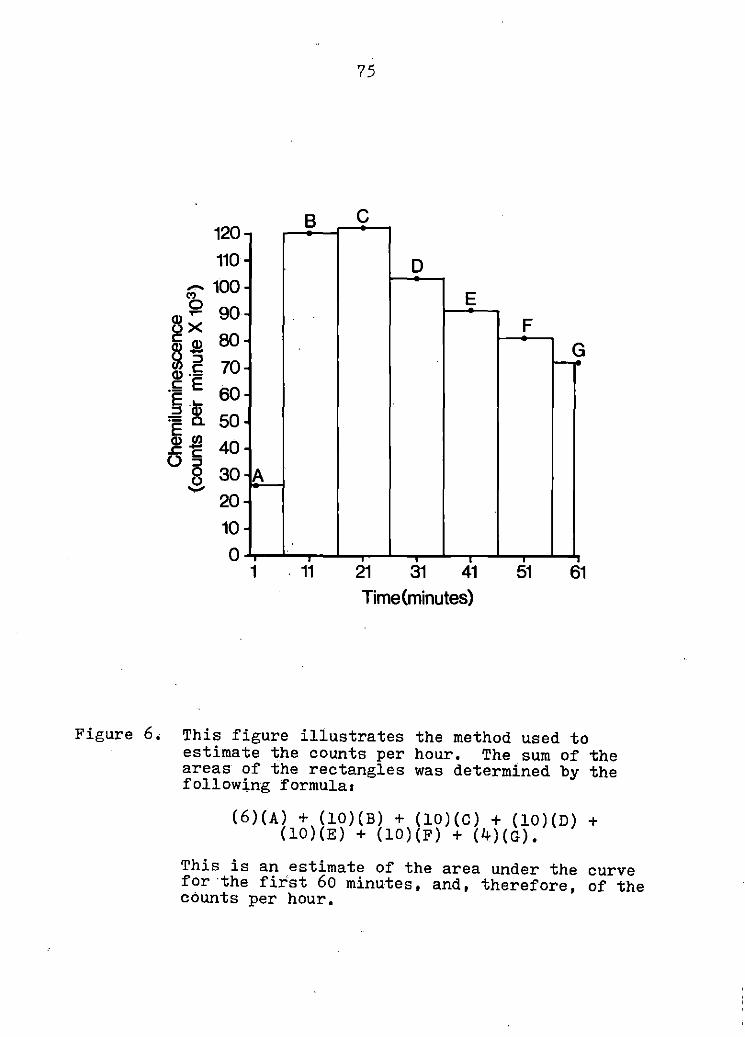
120 110
,... 100 'b g~ 90
~~ ~ . i;; E 60 § Ii>
.
.
.
.
.
.
.
.
. = c. 50 E Ill ~c: 40 () 8:i 30 '·A -..,..,
20 10 0
.
.
• 1
B
• 11
75
c
D
E F
_<}
-. ' ' -, 31 41 51 61
Time(minutes)
Figure 6, This figure illustrates the method used to estimate the counts per hour, The sum of the areas of the rectangles was determined by the following formula1
(6)(A) + (10) (B) + (10) (C) + (10) (D) + (10) (E) + (10) (F) + (4) (G),
This is an estimate of the area under the curve for ·the first 60 minutes, and, therefore, of the counts per hour,

76
first 60 minutes, or counts per hour. The net cph of a
test vial was calculated as follows1
net cph = cph of test vial - cph of background vial.
All experiments were conducted in duplicate and the average
net cph are reported.
Results
Figure 7 illustrates the effect of varying the
quantity of·preopsonized zymosan in the reaction mixture.
Increasing the amount of preopsonized zymosan increased
light emission. This did not reach an end point under
the conditions employed, Resting PMNs, in the absence of
serum or particles, did not demonstrate any significant
chemiluminescence,
Figure 8 illustrates the difference in chemiluminescence
by PMNs stimulated with either unopsonized zymosan or
preopsonized zymosan. This experiment was conducted
four times; a typical set of curves is depicted, The
preopsonized zymosan elicited a much more rapid chemi-
luminescent response than the unopsonized zymosan; however,
the unopsonized zymosan did stimulate chemiluminescence.
The range and mean of values obtained from normal
animals are illustrated in Figure 9, Each point represents
the mean of duplicate values, A wide range of values
was obtained from normal animals. Thirty-eight

-<O
§$2 Q) x ~3 Q) 0 c:.t= "§ ~ .E 2l Q) c: .z= ~ u .§
Figure 7,
10 9 8 7 6 5 4 3 2 1 0
0.0
77
0.1 1.0 5.0 10.0 20.0 Amount of preopsonizecl zymosan
in the stock solution (mg/ml)
40.0
Effect of varying the quantity of preopsonized zymosan available for phagocytosis. One half ml of the respective stock solution was added to each reaction tube, The amount of chemi-luminescence produced by 1,0 x 10⁷ PMNs in 60 minutes is expressed as counts per hour. (Mean± SEM. of 4·experiments,)

130 120 110
78
·····-··-······· .... .. ......... .... .... .... ........ .. ·· ····• ,. -·
I .-.. ... ,.. ----Resting PMNs --Opsonized Zymosan
... 20 ,.. ............. Unopsonized Zymosan
10 -------~--- ........ ------------~ o~· ~~..-~-r-~ ....... ~---.,..-~..,......~ .....
1 11 21 31 41 51 61 Time (minutes)
Figure 8. A comparison of.the chemiluminescent response of bovine PMNs to opsonized zymosan and unopsonized zymosan is illustrated. Resting values are from PMNs that were tested in the absence of particles. The experiment was conducted 4 times; typical results are shown.

...... "' Q) ~
~~ -~ _g E .._ .:: ~ j~ 09
.......
79
10
9
8 . . 7
6
5 Mean
4 i·
.. 3
. .
2
1
a ...... ________ _ Values from
normal animals
Figure 9, This figure illustrates the range and mean of chemiluminescence values obtained from·J8 evaluations of PMNs from normal animals. Each point represents the mean of duplicate values. The amount of chemiluminescence produced by 1.0 x 10⁷ PMNs in 60 minutes is expressed as· counts per hour.

80
determinations on normal PMNs yielded a mean of 5,21 x
106 cph with a standard deviation of 1.57 x 10⁶ cph, and
a standard error of the mean of 2,8 x 10⁵ cph,
Discussion
Unopsonized zymosan is reported to not stimulate
chemilui~inescence by human PMNs (Nelson et al. , 1977).
The data presented here indicates that unopsonized zymosan
does stimulate chemiluminescence by bovine PMNs, although
the kinetics of this stimulation are much different than
for opsonized zymosan. The slower chemiluminescent response
to unopsonized zymosan may reflect the necessity for the
PMNs and zymosan to settle to the bottom of the vial,
where the PMNs may be able to "trap" the unopsonized
zymosan for ingestion, The authors have previously
reported that unopsonized zymosan does not significantly
stimulate iodination (Roth and Kaeberle, 1979a) or NBT
reduction (Roth and Kaeberle, 1979b) by bovine PMNs.
However, these tests were conducted over a shorter time
period with agitation of the reaction mixture.
A wide range of values were obtained from normal
animals, This is typical of PMN function tests in general.
In order to demonstrate a defect in ·PMN function, repeated
testing is necessary and normal control PMNs must be
.evaluated concurrently,

81
Many substances have been shown to influence the
chemiluminescence response.if they are present in the
reaction medium {Nelson et al .• , 1977; Andersen and Brendzel,
1978), Phenol red, hemoglobin, or red blood cells will
decrease the amount of chemiluminescence which is detected
by the''scintillation counter, This effect may be due to
absorption of some of the light produced, These substances
were also found to decrease chemiluminescence of bovine
PMNs (data not shown), Nelson et al. (1977) demonstrated
that soluble proteins have a marked effect on chemi-
luminescence, Supplementation of the medium with gelatin
at a level of J,O gm/100 ml increased the peak counts
per minute by 150%. Addition of greater amounts of gelatin
resulted in a progressive decrease in peak chemiluminescence,
They· also de.monstrated an increase in chemiluminescence
by adding amino acids and tested 20 common amino acids
individually, Of these, the only amino acids observed to
influence significantly the amount of chemiluminescence
measured were tyrosine, tryptophane, and cysteine,
Addition of tyrosine increased the chemiluminescence
response of PMNs by more than 75%. The maximal increase
in chemiluminescence observed with tryptophane was
approximately 35%. Unlike tyrosine and tryptophane,
addition of cysteine was found to decrease the PMN

82
chemiluminescence detected by a maximum of approximately
60%. Nelson et al. (1977) also demonstrated that addition
of zymosan in excess of that required to achieve a maximal
rate of phagocytosis significantly increased the amount
of light measured. They did further studies to shciw
that unphagocytbsed extracellular zymosan will increase
the magnitude of chemiluminescence •. This is probably
partially explained by the experimentation of Goldstein
et al. (1977) who demonstrated that surface stimulation of
PMNs by opsonized particles will result in the generation
of superoxide anion by an enzyme complex on the cell
surface, and the release of superoxide anion into the
medium surrounding the cell. Superoxide anion is
theorized to form singlet oxygen by a number of possible
mechanisms (Webb et al., 1974; Rosen and Klebanoff, 1976).
Electronically excited singlet oxygen may dissipate its
energy in various ways, two of which are the direct
emission of a photon of light or the transfer of its
energy to another molecule which subsequently will emit
light in a secondary light producing reaction. Singlet
oxygen may also dissipate its energy in nonlight producing
reactions. There is good evidence to support the suggestion
that a major portion of the light measured in cellular

BJ
chemiluminescence reactions is the product of secondary
light producing reactions in which soluble protein, or
the phagocytosed particle, provides the necessary
substrate (Nelson et al., 1977). This explains the
increasing chemiluminescence measured as the level of
opsonized zymosan was increased in the reaction mixture as '!I shown in Figure 7, This is very similar to the results
obtained using high levels of preopsonized zymosan when
nitroblue tetrazolium (NBT) reduction by bovine PMNs was
measured (Roth and Kaeberle, 1979b), The high values
obtained for NBT reduction and chemiluminescence when high
levels of preopsonized zymosan were used are probably both
due to the extracellular generation of superoxide anion.
Because of this extracellular generation of superoxide
anion, chemiluminescence can proceed in the absence of
phagocytosis (Goldstein et al., 1975b; Goldstein et al.,
1977) and the chemiluminescence reaction c·annot be used as
a valid measure of ingestion. However, when normal PMNs
and the proper particle to cell ratio is used, there is
·a good correlation between chemiluminescence and ingestion
(Grebner et al,, 1977).
Since phenol red, hemoglobin, soluble proteins; certain
amino acids, and excess zymosan can all influence the
chemiluminescence measured, it is very important to

84
standardize these components in the reaction mixture.
When comparing different sera in the reaction mixture
for their effect on opsonization, caution must be used in
the interpretation of the results. All sera may not
influence the chemiluminescent response to the same extent.
However, if experimental conditions are well-controlled,
the measurement of the chemiluminescent response affords
a sensitive, continuous, and nondestructive means of
quantitating a component of bovine PMN oxidative metabolism.

85
PART II. EXPERIMENTATION INVOLVING BOVINE
VIRAL DIARRHEA VIRUS

86
EFFECTS OF BOVINE VIRAL DIARRHEA VIRUS INFECTION ON BOVINE POLYMORPHONUCLEAR
LEUKOCYTE FUNCTIONl
Summary
Four procedures were used to evaluate the function of
polymorphonuclear leukocytes (PMNs) isolated from the blood
of cattle experimentally infected with bovine viral
diarrhea (BYD) virus1 (1) uptake of an emulsion of
paraffin oil and ~· coli lipopolysaccharide, (2) nitroblue
tetrazolium reduction, (J) chemiluminescence, and (4)
iodination, or the conversion Of iodide to a trichloroacetic
acid-precipitable form. A marked impairment of iodination
was consistently observed following BYD virus infection.
A corresponding decrease in paraffin oil uptake, nitroblue
tetrazolium reduction, and chemiluminescence was not
observed. This defect was compounded by a decrease in
the number of circulating PMNs following BYD virus
infection. The impairment of PMN function may partially
explain the increased susceptibi1ity of cattle to secondary
bacterial infection during infection with BYD virus.
1submitted for publication to the American Journal of Veterinary Research by J. A. Roth, M. L. Kaeberle, and R. W. Griffith.

87
Introduction
Bovine viral diarrhea virus is the causative agent
of viral diarrhea-mucosal disease in cattle (Malmquist,
1968). The BVD virus is frequently incriminated in the
so-called "bovine shipping fever complex" along with
other viruses and bacterial organisms, Concurrent infection
with two or more infectious agents commonly occurs in
this disease syndrome. Pathogenic agents are presumably
able to evade or impair the hosts. immune defenses in order
to facilitate their own survival within the host. This
impairment of the resistance mechanisms of the host may
result in an increased susceptibility to secondary infection
with other infectious agents.
In earlier experimentation Reggiardo andKaeberle
(1979) demonstrat.ed that 1-5 days following experimental
infection with BVD virus a bacteremia could be detec.ted
(Reggiardo, 1975). This implied a defect in the bacterial
clearance mechanisms of the host. Removal of bacteria
from the blood stream is primarily the responsibility
of the polymorphonuclear phagocytes (PMNs) and the
mononuclear phagocytic system,
The purpos~ of this experimentation was to evaluate
the function of bovine peripheral PMNs to determine if
a defect in function could be demonstrated, Four procedures
were utili.zed to evaluate different aspects of PMN function1

88
(1) Ingestion was evaluated by measuring the uptake of
paraffin oil droplets which had been impregnated with oil
red O and emulsified with bacterial lipopolysaccharide.
(2) Oxidative metabolism of the PMN was evaluated by
quantitation of nitroblue tetrazolium (NBT) reduction
by PMNs when stimulated by a phagocytosable particle,
(3) An additional aspect of the oxidative metabolism of
the PMN was evaluated by the measurement of the amount
of chemiluminescence emitted by PMNs when stimulated by
a phagocytosable particle, (4) The fourth procedure,
iodination, is dependent upon ingestion, oxidative
metabolism, release of lysosomal enzymes into the phagocytic
vacuole,· and the presence of myeloperoxidase within the
lysosomes, These parameters were evaluated in animals
which were experimentally infected with BVD virus and in
control animals.
Materials and Methods
Animals - Apparently healthy 18-24 month old Holstein-
Fresian steers and bulls were used as control and challenge
animals,
BVD virus - The NADL challenge strain (lot 70-2)1 was
used for intranasal challenge, Strain 1015-74 was used
1Kiridly supplied by Biologics Virology Section, NVSL, National Animal Disease Center, Ames, Ia,

89
for intravenous challenge, This strain was isolated at
the Iowa Veterinary Diagnostic Laboratory from a typical
case of bovine viral·diarrhea-mucosal disease. The
isolate was plaque purified and used at the sixth bovine
testicle passage as a challenge strain.
Experimental procedure - Four animals were inoculated
intravenously with 10 ml of virus preparation containing 6 l.O x 10 TCID50 of the 1015-74 strain of BVD virus.
Five anima:).s were inoculated intranasally with 2.4 ml
of virus preparation containing 1 x 10⁵ TCrn50 of the
NADL challenge strain of BVD virus. Six animals were
used as controls. The research was conducted using
experimental blocks consisting of one or two infected
animals and one noninfected control. PMN function,
body temperature, and white blood cell (WBC) counts of
these animals were monitored 1-2 days prior to infection
and at nearly daily intervals for the first 10 days
following infection. For the succeeding 2 weeks the
.parameters wet"e evaluated at irregular intervals.
Hematologic studies - Total and differential WBC
counts were performed by standard procedures on heparinized
blood obtained by jugu~ar venapuncture.
PMN preparations - Blood was collected into ACD
solution by jugular venapuncture. The PMNs were isolated

90
as previously described (Roth and Kaeberle, 1979a), The
blood was centrifuged at 1,000 x g for 20 minutes; the
plasma and buffy coat material containing predominantly
mononuclear cells were removed, The red cells in the cell
sediment, which contained the. majority of the PMNs, were
lysed by treating with two volumes of cold phosphate
buffered (0,0132 M, pH 7,2) deionized water, Isotonicity
was restored after 45 seconds by the addition of one volume
of cold phosphate buffered (0,0132 M, pH 7.2) 2.7% NaCl
solution. The PMNs were pelleted by centrifugation and
washed twice with Hanks balanced salt solution without
Ca++ and Mg++ (HBSS). 1 This yielded a leukocyte preparation
of approximately 85%-100% PMNs (neutrophils plus
eosinophils).
Preopsonized zymosan - Preopsonized zymosan was
prepared as previously described (Roth and Kaeberle, 1979a).
One gram of zymosan in 100 ml of Earles balanced salt
solution with Ca++ and Mg++ and without phenol red (EBSS)1
was added to 100 ml of fresh bovine serum and incubated
at room temperature fo.r one hour, The zymosan was then
washed twice in 0,1 M EDTA solution, The pelleted zymosan
was resuspend~d in EBSS to a.concentration of 10 mg/ml and
frozen in aliquots until use.
1Grand Island Biological Co., Grand Island, N.Y.

91
Paraffin oil uptake - Paraffin oil uptake was performed
by a modification of the method of Stossel and Taylor
(1976). Heavy paraffin oil1 was impregnated with oil
red O dye2 , then emulsified with~· coli lipopolysaccharide
(cold phenol extracted) by sonic treatment. The lipopoly-
saccharide coated paraffin oil droplets were then opsonized
with complement via the alternative complement pathway
by incubating 75.0 µl of paraffin oil suspension with 0.1
ml of fres.h bovine serum at 37 C for 15 minutes. Ingestion
was initiated by adding 1.5 x 10⁷ PMNs in o.8 ml of EBSS
to the paraffin oil suspension. After exactly 5.0 minutes,
5.0 ml of c.old 1.0 mM N-ethylma.leimide2 in saline (NEM)
was added to terminate further ingestion. The cells were
separated from the uningested paraffin oil by centrifugation,
and washed with an additional 5.0 ml of NEM. The oil red
.. O in the cell button was extracted with l,4-dioxane1 ,
clarified by centrifugat1on, and the optical density (OD)
at 525 nm was determined. The OD was converted to µg of
paraffin oil/107 PMNs/min by the method of Stossel and
Taylor (1976). The procedure was performed in triplicate,
and the average value is reported.
1Fisher Scientific Co., Pittsburgh, Pa. 2Sigma Chemical Co., St. Louis, Mo.

92
Nitroblue tetrazolium reduction - Nitroblue tetrazolium
reduction by PMNs was quantitatively determined as
previously described (Roth and Kaeberle, 1979b). The
procedure was performed in 15 x 100.mm silicon-coated
glass test tubes. Each reaction tube contained 0.2 ml
of NBT solution ( 2 mg/ml)., 5,0 x 10⁶ PMNs, 0,1 ml of
prebpsonized zymosan, and sufficient EBSS to bring the
volume to-1,0 ml. All of the reactants except PMNs were
.added to the tubes and allowed to equilibrate in a water
bath at 37 C for 15 m.inutes. The reaction was started
by adding PMNs and was allowed to proceed with periodic
shaking for exactly 5,0 minutes. The reaction was stopped
by addingj,O ml of NEM. The cells and insoluble purple
formazan formed by the reduction of NBT were pelleted
by centrifugation and resuspended in 5,0 ml of pyridine.1
The pellet was dispersed by brief sonic treatment, and the
formazan was extracted by placing the test tubes in a
boiling water bath for 10 minutes, The pyridine-formazan
was clarified by centrifugation, and the OD of the super-
natant fiuid was immediately determined in a spectro-
photometer, 2 using a pyridine blank. The results are
1Fisher Scientific Co,, Pittsburgh, Pa, 2Beckman ·DBG, Beckman I t t I I . C ns rumens, nc., rvine, a,

93
reported as on/5,0 x 10 6 PMNs/5 min in 5,0 ml of pyridine.
The reported results represent the average values of
triplicate determinations.
Chemiluminescence - Chemiluminescence was measured in 1 ..
a liquid scintillation spectrometer at ambient temperature
with the coincidence circuit switched off as previously
reported (Roth and Kaeberle, 1979c), The reaction was
conducted in 20 ml glass scintillation vials. 2 Test vials
contained 0,5 ml of preopsonized zymosan suspension and 1.0
x 10 7 PMNs· in a total volume of 10 ml of Geys balanced
salt solution with Ca++ and Mg++ and without phenol red
(GBSS),3 The zymosan and GESS were added to the
scintillation vial, the PMNs were added 1 minute before
the vial was placed in the coµnting chamber, Each vial
was counted for 1 minute in each 10 minute cycle, Counting
was continued for at least 70 minutes. All samples were
run in duplicate; control vials containing 10 ml of GESS
and vials containing 10 ml of GESS and PMNs were counted
each cycle to determine the background activity and resting
chemiluminescence, The results were calculated as
1Model DPM 100, Beckman Instruments, Inc,, Irvine, Ca. 2Fisher Scientific Co., Pittsburgh, Pa.
)Grand Island Biological Co,, Grand Island, N, Y .•

94
previously described and reported as net counts per hour
(Roth and Kaeberle, 1979b),
Iodination - The procedure for the determination of
iodination by bovine PMNs has been reported elsewhere (Roth
and Kaeberle, 1979a). Each reaction tube contained 40
nmole NaI, 0,05 µCi Na 125I (carrier free in 0.1 M NaOH), 1
. 6 2.5 x 10 PMNs, and 0,05 ml Of preopsonized zymosan in a
total volume of 0,5 ml of EBSS with phenol red, All of the
components except the PMNs were placed in 12 x 75 mm
polystyrene snap cap test tubes2 and allowed to equilibrate
in a 37 C incubator. The reaction was started by the
addition of PMNs, and the mixture was incubated for 20
minutes at 37 C with end over end tumbling approximately
20 times per minute, The reaction was terminated by the
addition of 2,0 ml of cold 10% trichloroacetic acid (TCA).
The precipitate was collected by centrifugation and washed
twice with 20 ml of cold 10% TCA.. The counts per minute
of radioactivity remaining in the precipitate was determined
in a gamma counter,3 Results are expressed as nanomoles
of iodide converted to a TCA-precipitable form per 107 PMNs
1New England Nuclear, Boston, Ma, 2#2058 Falcon, Oxnard, Ca .•
3Autowell II, Picker Nuclear, North Haven, Ct.

95
per hour (nmole Nal/107 PMNs/hr) •. All assays were performed
in duplicate and the average value reported.
Determination of the effect of serum from BVD virus
infected animals ~ iodination El PMNs from control animals -
Blood was obtained from the virus-infected and control
animals by jugular venapuncture and allowed to clot at
room temperature, The serum was removed and held at 4 C
until it was used a few hours later. Normal serum for
comparison was obtained from apparently healthy Holstein-
Fresian cows. Blood was obtained from several cows and
allowed to clot at room temperature. The serum was
removed and combined into a pool of normal serum. This
was divided into 2 ml aliquots and frozen at -70 C until
use, The same pool was used throughout the experimentation.
The effect of the different sera on iodination was
determined by incubating 50.0 ul of each serum with
unopsonized zymosan in an iodination reaction tube for
15 minutes at J7 C, then performing the iodination reaction
as usual by adding 2,5 x 10⁶ PMNs obtained from a control
animal. Iodination in the presence of the serum from
control animals and virus infected animals was expressed
as a percent of the iodination in the presence of the
.pooled normal serum.

96
Statistical methods - All of the values from control
animals were averaged and a mean and standard error of the
mean were calculated. An analysis of variance procedure
was performed to determine the level of significance of
differences between the data from the control animals and
that from the infected animals. The data from the intra-
venously infected and intranasally infected animals were
considered separately. Only the data obtained from
days 1 through 14 postinfection were included in the
analysis of variance,
Results
Clincal response - Only mild clinical signs were
induced by injection of virus either intravenously or intra-
nasally. A diphasic temperature response was commonly
observed; a dry nonproductive cough, mucous nasal discharge,
and anorexia were often noted during the febrile period,
Diarrhea was not observed, All of the animals readily
recovered without treatment.
Hematologic response - The effect of BVD virus
infection on numbers of total WBCs, lymphocytes, neutrophils,
and eosinophils in peripheral blood is shown in Figures
10-13. There was a marked drop in number of all three cell
types following BVD virus infection.

9600
8800
8000 .. E E 7200 ...... "' g 54'00 ~
"' 5600 -0 I- 4800
4000.
97
! . :
--+- -----------~-------~-. l
\ ~\ \/\ i
--- Control (Mean± SEMI ( n =67) --Intranasal <P=.0001, n=48l ·········Intravenous (P=.0001, n=35l ·
3200--~ ..... _.. ..... .__.,,.....,_.,... ......... _.. ..... ._ .....
. 11 3 5 7 911.13151719212325 · Days Postinoculation
Preinoculation
Figure 10, Mean total white blood cells (WBCs) in the peripheral blood of control and BVD virus infected cattle prior to and following challenge, "n" values from each experimental group were used in the analysis of variance to arrive at the probability statement.
. I
'

6000
5500
5000
"' E 4500 E ' (/) 4000 QJ ->-(J 3500 0 ..c a. E 3000 >-_J
2500
2000
1500
~
i
98
_______ fLJ~·'"---f------t_,. .. ~ v ····· ..... . : .
' i ;\.j
Y ---Control IMean±SEMl ln=611 -- Intranasal IP=.0001, n=481 ·········Intravenous (P=.0003, n=35)
t 1 3 5 7 9 11 13 15 17 19 21 23 25 Days Postinoculation
Preinoculation
Figure 11. Mean total lymphocytes in the peripheral blood of control and BVD virus infected cattle prior to and following challenge, "n" values from each experimental group were used in the analysis of variance to arrive at the probability statement.

3500
3000
"' 2500· E E ...... II) 2000 s::. a. 0 1500 .... -:I CD 1000 z
500
0
i \ ~ ·"'· ... \ I ., : .... .. I ,, : .. :
\J
99
---Controls lMean± SEMl l n=61) - Intranasal lP=.071 n=48) ········Intravenous lP=.0001; n =35 >
t 1 3 5 7 9 111315 171921 2325 Days Postinocul~tion
Preinocula ti on
Figure 12. Mean total neutrophils in the peripheral blood of control and BVD virus infected cattle prior to and following challenge. "n" values. from each experimental group were used in the analysis of variance to arrive at the
. probability statement.

960
840
720 "' ~ 600 -rn ~ 480 c. 0 .!:: 360 rn 0 w 240
120
100
--- ------!------
\,,, _.1'· ••
: ...... ··~·· .. ····· .... ~... ~-·
--- Controls <Mean±SEM l (n=61l --· Intranasal <P=.004, n=48l ·········Intravenous <P=.0001, n =35)
o .... ~ .... ....,.....,~ ..................... ~ ................... ...., 1 3 5 7 9 11 13 15 17 19 21 23 25 f Days Postinoculation
Preinoculation
Figure 13. Mean total eosinophils in the peripheral blood of control and BVD virus infected cattle prior to and following challenge. "n" values from each experimental group were used in the analysis of variance to arrive at the probability statement.

101
Paraffin oil uptake - Figure 14 illustrates the effect
of BVD virus infection on paraffin oil uptake. Statistical
analysis indicated no significant difference in paraffin
oil uptaj{e between control and infected groups following
BVD virus infection. However, there were indications of
depressed paraffin oil uptake in three of the nine animals
followirig inoculation of BVD virus.
Nitrob1ue tetrazolium reduction - The effects of BVD
virus infection on nitroblue tetrazolium reduction by
bovine PMNs are shown in Figure 15. There was no significant
difference in nitroblue tetrazolium reduction by PMNs from
normal and infected cattle, None of the individual animals
had consistently depressed nitroblue tetrazolium reduction.
Chemiluminescence - The chemiluminescent response of
PMNs following BVD virus infection is shown in Figure 16,
There was no significant depression of chemiluminescence
by PMNs following infection. None of the individual
animals had consistently depressed chemiluminescence
.following BVD virus infection.
Iodination - The results of the iodination reaction
by PMNs from control and infected animals are shown in
Figure 17. There was a marked depression of iodination
following BVD virus infection. This· was noted consistently
in each animal infected. Following intravenous inoculation
with the 1015-74 strain of the virus, a marked depression
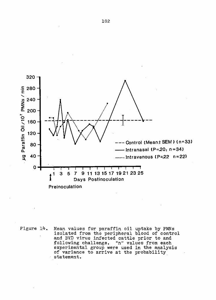
320
.!: 280 E ...... Cl) 240 z ~ a. 200 ... 0 ... 160 ' 0 c 120 --., 80 .... ., a. Cl 40 :I.
0
•.. , --\
102
. !!. ---I----- --
~:
--- Control (Mean± SEM l (n=33l - Intranasal !P=201 n =34) ......... Intravenous ( P=.22 n =22)
3 5 7 9 11 13 15 17 19 21 23 25 Days Postinoculation
Preinoculation
Figure 14. Mean values for paraffin oil uptake by PMNs isolated from the peripheral blood of control and BVD virus infected cattle prior to and following challenge, "n" values from each experimental group were used in the analysis of variance to arrive at the probability statement. ·

0.7 c: E 0.6
ll)
' 0.5 rn z ~ a. 0.4 "' 0 .... 0.3 x 0 I.Ci 0.2 ' 0 0 0.1
0
-- ---!' •• ! ·.,, ----I---. --
~ : v --- Controls (Mean± SEM > < n =56) - Intranasal <P=.681 n=38) ......... Intravenous lP=.251 n =27)
t 1 . 3 5 7 9 11 13 15 17 19 21 23 25
Days Postinoculation Preinoculation
Figure 15. Mean values for nitroblue tetrazolium reduction by PMNs isolated from the peripheral blood of control and BVD virus infected cattle prior to and following challenge. "n" values from each experimental.group were used in the analysis of variance to arrive at the probability statement.

9.6
&4
7.2 .. .c ........ 6.0 "' 0 ... )( 4.8 aJ -c 3.6 :J 0
0 2.4
12
0
104
p •.• -J~~~~---L _______ _ . .. r I ·······..... .
: : \i i --- Controls CMean±SEM > ( n""34)
-- Intranasal CP=.66, n -30) .......... Intravenous ( P=.141 n -13 >
t1 3 5 7 91113151719212325
Days Postinoculation Preinoculation
Figure 16. Mean values for chemiluminescence by PMNs isolated from the peripheral blood of control and BVD virus infected cattle prior to and following challenge. Chemiluminescence values were obtained in a liquid scintillation counter as counts per minute periodically during a 60 minute period, then extrapolated to counts per hour. "n" values from each experimental group were used in the analysis of variance to arrive at the probability statement.

64
56 ... .c ...... 48 Ill z ::!: 40 0.. .... 0 32 .,.... '\ ltl 24 z ~ 0 16 E c:
8
0
105
------~-------------
........... .. ... / 1-----
l .. ~ .. --- Controls <Mean± SEM l < n •561 -- Intranasal IP•.00011 n •45 l ......... Intravenous <P-.00011 n ·29)
1 3 5 7 9 1113151719212325 f . Days Postinoculation Preinocula ti on
Figure 17. Mean values for iodination by PMNs isolated from the peripheral blood of control and BYD virus infected cattle prior to and following challenge, "n" values from each experimental group were used in the analysis of variance to arrive at the probability statement.

106
in iodination occurred by day one or day two postinfection.
Following intranasal infection with the NADL challenge
strain a depression of iodination was not noted until day
three or day four postinfection.
Effect of serum ~ iodination - The effect of serum
from BVD infected animals on iodination by PMNs from control
animals is shown irt Figure 18. Serum from BVD infected
animals did not depress iodination by normal PMNs under
the conditions employed.
Discussion
The clinical signs, diphasic temperature response,
and severe leukopenia were typical of results reported
for experimental infection with BVD virus (Malmquist, 1968;
Tyler and Ramsey, 1965). While diarrhea has been reported
following experimental BVD infection, it was not observed
in this experimentation (Tyler and Ramsey, 1965). Bovine
viral diarrhea virus infection was confirmed by the
development of significant virus neutralizing antibody
titers in all animals following challenge,
The results of the paraffin oil uptake procedure were
equivocal. Three of the animals exhibited depressed
paraffin oil uptake. However, statistical analysis of
all data obtained from control and infected animals
indicated no significant depression of paraffin oil

140
120·
100 - 80 c Cl) () .... Cl) 60 a..
40
20
0
...... ; ...
107
; .• . _ _. _______ _]' ____ _ . . ~
.. ·····•
---Controls <Mean±SEM> <n=41) -- Intranasal <P= .30, n •37 > ......... Intravenous (P=.58, n•22)
3 5 7 9 11 13 15 17 19 21 23 25 Days Postinoculation
Preinoculation
Figure 18. Iodination values of normal .bovine PMNs in the presence of serum from control and BYD· virus infected cattle, expressed as a mean percent of iodination by normal bovine PMNs in the presence of pooled normal bovine serum, "n" values from each experimental group were used in the analysis of variance to arrive at· the probability statement.

108
uptake. This lack of significance may have been due to
problems inherent in the test itself. Difficulty was
encountered in separating the uningested paraffin oil
from the cells containing ingested paraffin oil. Another
problem was the standardization of the paraffin oil
droplets from one day to the next. Paraffin oil uptake
is a measure of the ability of the PMN to ingest particles,
Due to the weaknesses in the test, it was not possible
to conclude whether or not ingestion by PMNs was impaired
during BVD virus infection.
There was no significant depression of NBT reduction
or chemiluminescence by PMNs following BVD virus infection.
These tests are both measures of the oxidative metabolism
of the PMN (Roth and Kaeberle, 1979b, c); therefore, the
results indicate that the oxidative metabolism of the PMNs
following BVD virus infection is normal. Because oxidative
metabolism of the PMN may be stimulated in the absence of
ingestion (Goldstein et al., 1977), normal NBT reduction
and chemiluminescence cannot be interpreted t.o demonstrate
normal ingestion by PMNs. The iodination reaction was
consistently and markedly depressed following BVD virus
infection. Ingestion, the burst of oxidative metabolism,
degranulation, and myeloperoxidase in the primary granules
must all be. present to obtain a normal iodination reaction
(Klebanoff and Clark, 1977). Depressed iodination ind·icates

109
a defect in one of these parameters. The results of the
NBT reduction and chemiluminescence tests indicate that
the burst of oxidative metabolism which accompanies '
phagocyt1Jsis is normal. The depression of iodination
could not be explained on the basis of stage of maturity '
or toxic changes of the PIV!Ns. Microscopic examination
of the whole blood smears and PIV!N preparations revealed
only minor abnormalities in PMN morphology. The normal
NBT reduction, chemiluminescence, and morphology indicated
that the depression of iodination was not due to toxic
changes or to lack of viability of the PMNs. Eosinophils
are known to participate in the iodination reaction.
The percent of eosinophils in the PMN preparations varied
considerably with generally fewer eosinophils present
in the PMN preparations following BVD virus infection,
The drop in eosinophil numbers was not thought to be
involved in the depression of iodination. This is
supported by an observed lack of correlation between
eosinophil numbers and iodination in PMN preparations
from normal animals (Roth and Kaeberle, 1979a).
Depressed iodination could theoretically be due to
inadequate amounts of myeloperoxidase in the primary
granules of the PMNs. The fact that the depressed iodination
was ·observeQ. within 24 hours following intravenous infection

110
would indicate that the depressed iodination is probably
not due "j;o a lack of myeloperoxidase formation during
granulopoiesis. Granulopoiesis in the bovine takes
approximately 5-6 days {Schalm, 1977). Myeloperoxidase
in the primary granules is formed fairly early in this
period {Murphy, 1976). Inadequate amounts of myelo-
peroxidase in the primary granules could be due to previous
intracellular or extracellular degranulation. This could
be expected if significant previous contact with bacteria
had occurred. Reggiardo and Kaeberle {1979) demonstrated
that a bacteremia lasting for 1-5 days occurred following
BVD virus infection. · However, the depression of iodination
lasted for 2-3 weeks following infection, Furthermore,
ingestion, oxidative metabolism, and degranulation all
r·equire the expenditure of energy by the PMN. The normal
values for NBT reduction and chemiluminescence indicate
that the PMNs had not depleted their energy supply by the
previous ingestion of particles and degranulation.
The depressed iodination may be due to inhibition of
degranulation by the PMN following BVD virus infection.
Degranulation is thought to be dependent upon the action
of microtubules and actomyosin-like microfilaments and
upon the fusion of the lysosomal membrane with the
phagocytic vacuole {Goren, 1977). Agents which elevate
cellular cyclic AMP levels or decrease cellular cyclic GMP

111
levels interfere with degranulation (Weissmann et al, 1975).
In the case of BVD virus infection, this could theoretically
be due ti;> a direct interaction of the virus and the PMN,
or it maf be mediated secondarily through a humeral
factor which is influenced by BVD virus infection. Humeral
factors that could be responsible for an inhibition of
iodination include histamine (Zurier et al,, 1974),
13 adrenergic agents (Zurier et al .• , 1974), adrenal-
corticosteroids (Mandell et al., 1970), prostaglandins
(Smith, 1977), and toxic or inhibitory substances released
by other cells. To test for the presence of inhibitory
serum factors, serum from BVD infected animals was added
to PMNs from normal animals and their iodinating ability
was determined. Serum from animals with depressed
iodination failed to depress iodination by normal PMNs
under the conditions employed. This would indicate that
the defect is inherent in the PMNs and not due to a serum
factor.
It is not unreasonable to speculate that the depression
of PMN function may facilitate BVD virus survival in the
host animal. Bovine PMNs have·been demonstrated to be
the most active cell type tested. in mediating antibody
dependent cell mediated cytotoxicity (ADCC) against
herpes virus-infected targe~ cells (Rouse et al,, 1978).

112
The exact mechanism of this cytotoxicity is not known, but
it is not dependent upon DNA, RNA, or protein synthesis
within the PMN (Wardley et al., 1976a1 Clark and Klebanoff,
1977). The ADCC phenomenon is dependent upon microtubule
activity and is enhanced by drugs which lower cyclic AMP
or elevate cyclic GMP (Wardley et al,, 1976a; Clark and
Klebanoff, 1977). Degranulation is also dependent upon
microtubule activity and is subject to the bidirectional
control of cyclic nucleotides (Weissmann et al., 1975) •
. ':['his analogy suggests that degranulation may play a role
in ADCC. · .The PMN lysosomal enzyme myeloperoxidase appears
not to be essential in the cytotoxicity system (Clark and
Klebanoff, 1977); however, there are many other substances
in the PMN lysosome which may play a role. If the BVD virus
does indeedbloCk degranulation of the bovine PMN, it
may be able to prevent or reduce PMN mediated ADCC of
BVD virus~infected target cells. There is good evidence
that PMN mediated ADCC is important in defense against
'infectious bovine rhinotracheitis (!BR) virus infection
in cattle (Rouse et al,, 1976; Wardley et al., 1976a).
If infection with BVD virus does depress the ability of
the bovine PMN to mediate ADCC, this would facilitate
concurrent infection of the animal with !BR virus, a
situation which occurs fairly commonly in cattle,

113
PMNs could also play a role in antiviral defense by
phagocytosing virus particles, Many viruses are engulfed
by neutrophils (Sawyer, 1969; Belding and Klebanoff, 1970),
Phagocytosis normally results in a burst of oxidative
metabolism, the generation of H2o2 , and the release of
myeloperoxidase and· other lysosomal enzymes into the
phagocytic vacuole (Murphy, 1976). Myeloperoxidase and
hydrogen peroxide, in the presence of chloride ions, have
been shown to have potent virucidal activity (Belding and
Klebanoff, 1970), · This is the same system which is
responsible for the iodination reaction. The depression
of iodination by PMNs following BVD virus infection
indicates that this potentially virucidal system is
inhibited.
PMNs may also play a role in antiviral immunity by
releasing a substance with interferon-like activity (Rouse
et al., 1978), The effect of BVD virus infection upon this
activity of PMNs is not known,
The data presented here indicate that following BVD
virus infection a defect in bovine PMN function occurs.
This may be a defect in ingestion of particles, degranu-
lation and release of lysosomal enzymes into the phagosome,
or an interference with the myeloperoxidase catalyzed
reaction, The defect in PMN function is compounded by a

114
marked decrease in the number of circulating PMNs. This
combination of factors could facilitate BVD virus infection
and predispose the animal to secondary viral and bacterial
infection.
There are reports of inhibited phagocytosis (ingestion)
by PMNs following infection with mumps, influenza, and
Coxsackie viruses (Merchant and Morgan, 1950; Kantoch and
Dubowska-Inglot, 1960). Oxidative metabolism, degranulation,
or iodination were not evaluated following infection with
these viruses. It is not known if the PMN defect described
here is unique to BVD virus infection or if it may occur
with other infectious agents as well. Further study is
needed to determine the exact defect induced in the PMN and
the mechanism for the induction of this defect. Characteri-
zation of this deficiency theoretically could lead to a
therapeutic approach to normalization of PMN function
during BVD virus infection.

115
SUMIV'.ARY
New procedures for the evaluation of bovine PMN
function were evaluated, and these procedures were used
to study the effects of BVD virus infection on PMN function
in cattle,
The.· paraffin oil procedure for the evaluation of '
ingestion by PMNs was found not to be satisfactory for
use in cattle, Nitroblue tetrazolium reduction, iodination,
and chemiluminescence were all found to be satisfactory
for the evaluation of bovine PMN function, and normal
values for cattle were reported,
BVD virus infection resulted in the depression of
total numbers of lymphocytes,·neutrophils, and eosinophils
in the peripheral blood beginning at 1-3 days and lasting
for 2-3 weeks following infection, Nitroblue tetrazolium
reduction and chemiluminescence by bovine PMNs were normal
following BVD virus infection; however, iodination was
markedly depressed, The depression of .iodination was
noted as early as 24 hours following intravenous challenge
with the 1015-74 strain of the BVD virus; it occurred
2-3 days following intranasal challenge with the NADL
strain, and lasted for 2-3 weeks in both cases,

116
It was concluded that the depression of iodination
may have been due to a defect in ingestion of particles,
degranulation and release of lysosomal enzymes into the
phagosome, or an interference with the myeloperoxidase
catalyzed reaction. The defect in PMN function, compounded
by a marked decrease in the number of circulating PMNs,
could facilitate BVD virus infection and predispose
the animal to secondary viral and bacterial infection.

117
BIBLIOGRAPHY
Allen, R. C. 1977. Evaluation of serum opsonic capacity by quantitating the initial chemiluminescent response from phagocytizing polymorphonuclear leukocytes. Infect. Immun. 151828-833,
Allen, R, C,, R. L. Stjernholm, and R. H, Steele, 1972. Evidence for the generation of an electronic excitation state(s) in human polymorphonuclear leukocytes and its participation in bactericidal activity. Biochem. Biophys. Res, Comm. 471679-684.
Andersen, B. R., and A. M, Brendzel. 1978. Use of a unique chemiluminescence spectrometer in a study of factors influencing granulocyte light emission, J, Immunol. Methods 191279-287.
Andersen, B. R., A. M. Brendzel, and T. F. Lint. 1977. Chemiluminescence spectra of human myeloperoxidase and polymorphonuclear leukocytes. Infect. Immun. 17162-66,
Babior, B, M. 1978. Oxygen-dependent microbial killing by phagocytes. N, Engl. J, Med, 2981659-668, 721-725.
Baehner, R. L. to recurrent 935-956.
1972. Disorders of leukocytes leading infection, Pediatr. Clin. North Am. 191
Baehner, R, L., and D, G. Nathan. 1968, Quantitative nibroblue tetrazolium test in chronic granulomatous disease. N. Engl, J, Med. 2781971-976.
Banas, D, A. 1974. Experiences with the nitroblue tetrazoliuin test in four domestic species. Bull, Am. Soc. Vet, Clin. Pathol. 3117-19.
Baratawidjaja, R. K., L. P. Morrissey, and N. A. Labzoffsky. 1965. Demonstration of vaccinia, lymphocytic chorio-meningitis and rabies viruses in the leucocytes of experimentally infected animals. Arch. Gesamte Virusforsch, 171273-279.
Beeson, P. B,, and D. A. Bass. 1977• The eosinophil. W. B. Sa.unders Company, Philadelphia, Pa. 269 pp,

118
Belding, M. E., and s. J, Klebanoff. 1970. Peroxidase-mediated virucidal systems. Science 1671195-196.
Bellanti, J. A. 1978. Immunology II. W. B. Saunders Compapy, Philadelphia, Pa. 813 pp.
Bellanti, J. A., and D. H. Dayton. 1975. The phagocytic cell in host resistance. Raven Press, New York, N. Y. 348 pp.
Bergman, M. J,, R. L, Guerrant, F. Murad, s. H. Richardson, D, Weaver, and G. L. Mandell. 1978. Interaction of polymorphonuclear neutrophils with Escherichia coli. J, Clin. Invest. 611227-234.
Beswick, P. H., and T. F. Slater. 1977. The 'metabolic burst' in bovine blood polymorphonuclear leucocytes when stimulated by phagocytosis. Biochem. Soc, Trans. 511299-1300.
Beswick, P. H,, and T. F. Slater. 1978. Modification by metals, sulphydryl reagents and cyanide of the particle stimulated enhancement of oxygen consumption in bovine granulocytes. Chem. Biol. Interact. 201373-382.
Blood, D. C., medicine. Baltimore,
and )rd Md.
J, A. Henderson. 1968. Veterinary ed. The Williams and Wilkins Company,
927 pp.
Carlson, G. P., and J, J, Kaneko. 1973. Isolation of leukocytes from bovine peripheral blood, Proc. Soc. Exp. Biol. Med, 1421853-856.
Carpenter, P. L. 1975. Immunology and serology. W. B. Saunders Company, Philadelphia, Pa. 346 pp.
Chusid, M. J,, J; s. Bujak, and D. C. Dale, 1975. Defective polymorphonuclear leukocyte metabolism and function in canine cyclic·neutropenia. Blood 461 921-930.
Clark, R. A., and S, J, Klebanoff, 1977. Studies on the mechanism of antibody-dependent polymorphonuclear leukocyte-mediated cytotoxicity. J, Immunol. 1191 141)-1418. '

119
Cline, M. J, 1972. Microbicidal activity of human eosinophils. J. Reticuloendothel. Soc. 121332-339.
Cochrane, D. G. 1968. Immunologic tissue injury mediated by neutrophilic leukocytes. Adv. Immunol. 9197-105.
Curreri, P. W., E. L. Heck, L. Browne, and C.R. Baxter. 1973. Stimulated nitroblue tetrazolium test to assess neutrophil antibacterial function1 Prediction of wound sepsis in burned patients. Surgery 7416-13.
Degr~. M. 1970. Synergistic effect in viral-bacterial infection. Acta Pathol. Microbiol. Scand. 78141-50.
Douglas, S. D. 1976. Cells involved in the immune response. Pages 70-87 in H. H. Fudenberg, D. P. Stites, J, L. Caldwell,':8:nd J, V. Wells, eds. Basic and clinical immunology. Lange Medical Publications, Los Altos, Ca.
Drutz, D. J. 1976. Immunity and infection. Pages 182-194 in H. H. Fudenberg, D. P. Stites, J. L. Caldwell, and J:- V. Wells, eds. Basic and clinical immunology. Lange Medical Publications, Los Altos, Ca.
Fisher, T. N., and H. s. Ginsberg. 1956. The reaction of influenza viruses with guinea pig polymorphonuclear leucocytes. III.· Studies on the mechanism by which influenza viruses inhibit phagocytosis. Virology 21656-664.
Fuenfer, M. M., K. Scott, and H. C. Polk. 1976. Evaluation of the phagocytic-bactericidal capacity of neutrophils1 A current review. J. Surg. Res. 211207-214. . .
Gale, R. P., and J. Zighelboim. 1975. Polymorphonuclear leukocytes in antibody-dependent cellular cytotoxicity. J. Immunol. 11411047-1051.
Gennaro, R., C. Schneider, G. DeNicola, F. Cian, and D. Romeo. 1978. Biochemical properties of bovine granulocytes. Proc. Soc. Exp. Biol. Med. 1571342-347.
Gigli, I., and R. A. Nelson. 1968. Complement dependent immune phagocytosis. Exp. Cell Res. 51145-67.

120
Golde, D. W., and Ni. J. Cline, 1974. Regulation of granulopoiesis. N. Engl. J. Med. 29111J88-1J95.
Goldstein, I., s. Hoffstein, J. Gallin, and G. Weissmann. 197J. Mechanisms of lysosomal enzyme release from human leukocytes1 Microtubule assembly and membrane fusion induced by a component of complement. Proc. Nat. Acad. Sci. USA 7012916-2920.
Goldstein, I., S. T. Hoffstein, and G. Weissmann. 1975a. Mechanisms of lysosomal enzyme release from human polymorphonuclear leukocytes1 Effects of phorbol myristate acetate. J. Cell Biol. 661647-052. ·
Goldstein, I.., D. Roos, H. B. Kaplan, and G. Weissmann. 1975b. Complement and immunoglobulins stimulate superoxide production by human leukocytes independently of phagocytosis. J, Clin. Invest. 5611155-116J.
Goldstein, I., M. Cerqueira, S .. Lind, and H.B. Kaplan. 1977. Evidence that the superoxide-generating system of human leukocytes is associated with the cell surface. J, Clin. Invest. 591249-254.
Goren, M. B. 1977. Phagocyte lysosomes1 Interactions with infectious agents, .phagosomes, and experimental perturbations in function. Ann. Rev, Microbial, Jl1507-5JJ,
Grabar, P. 1976. The historical background of immunology, Pages J-14 in, H. H. Fudenberg, D, P, Stites, J, L .. Caldwell, and J, V, Wells, eds, Basic and clinical immunology. Lange Medical Publications, Los Altos, Ca.
Grebner, J, V., E. L. Mills, B. H. Gray, and P. G. Quie. 1977. Comparison of phagocytic and chemiluminescence response of human polymorphonuclear neutrophils. J. Lab. Clin. Med. 891153-159.
Gresser, I., and D. J, Lang, 1966, Relationships between viruses. and leucocytes, Prag. Med. Viral. 81 62-lJO.
Grewal, A. S., B. T. Rouse, and L.A. Babiuk, 1978. Characteriza,tion of surface receptors on bovine leukocytes, Int. Arch. Allergy Appl. Immunol. 561 289-JOO,

121
Griffith,.R. W., M. L. Kaeberle, and J, A. Roth. 1978. The interaction of bovine polymorphonuclear leukocytes and Pasteurella multocida. Page 15 in Abstracts of papers presented at the 59th annual meeting of the_ conference of research workers in animal disease.
Guidry, A. J,, and M. J, Paape, 1976. Effect of bovine neutrophil maturity on phagocytosis. Am. J, Vet. Res. 371703-705.
Guidry, A. J,, M. J. Paape, and R.H. Miller. 1974. In vitro procedure for measuring phagocytosis of blood neutrophils. Am. J. Vet, Res. 351705-709.
Guidry, A. J., M. J, Paape, and R, E. Pearson, 1976, Effects of parturition and lactation on blood and milk cell concentrations, corticosteroids, and neutrophil phagocytosis in the cow. Am. J, Vet. Res, 3711195-1200,
Hawkins, D. 1974. Neutrophilic leukocytes in immunologic reactions in vitro. III. Pharmacologic modulation of lysosomal constituent release, Clin, Immunol. Immunopathol, 21141-152.
Hoffstein, S., I, Goldstein, and G, Weissmann. 1977. Role of microtubule assembly in lysosomal enzyme secretion from human polymorphonuclear leukocytes1 A reevaluation. J, Cell Biol, 731242-256,
Holmes, B., and R. A. Good. 1972. Laboratory models of chronic granulomatous disease. J, Reticuloendothel. Soc, 121216-237,
Ignarro, L. J,, and S. Y. Cech. 1975. Lysosomal enzyme secretion from human neutrophils mediated by cyclic GMP1 Inhibition of cyclic GMP accumulation and neutrophil function by glucocorticosteroids, J, Cyc, Nuc. Res. 11283-292.
Ignarro, L. J,, T. F. Lint, and W. J, George, 1974. Hormonal control of lysosomal enzyme release from human neutrophils. J, Exp. Med. 13911395-1414,
Jain, N, C,, and J, Lasmanis. 1978. Phagocytosis of serum-resistant and serum-sensitive coliform bacteria (Klebsiella) by bovine neutrophils from blood and mastitic milk. Am. J, Vet. Res. 391~25-427.
Jarstrand, C. activity of influenza A
1977. The neutrophil infection.
122
NBT (nitroblue tetrazolium) granulocytes in patients with Scand. J. Infect. Dis. 915-7.
John, T. J,, and O. F. Sieber. 1976. Chemotactic migration of neutrophils under agarose. Life Sci. 18:177-182.
Johnson, D. W., and C. C, Muscoplat. 1973. Immunologic abnormalities in calves with chronic bovine viral diarrhea. Arn. J. Vet. Res. )411139-1141.
Johnston, R. B. 1978. Oxygen metabolism and the microbicidal activity of macrophages. Fed. Proc. 3712759-2764.
Johnston, R. B;, B. B. Keele, H.P. Misra, J.E. Lehrneyer, L. S. Webb, R. L. Baehner, and K. V. Rajagopalan. 1975. The role of superoxide anion generation in phagocytic bactericidal activity. J, Clin. Invest, 5511357-1372.
Jubb, K. V, F., and P. C. Kennedy. 1970. Pathology of domestic animals. Academic Press, New York, N. Y. 697 pp. .
Kantoch, M., and A. Dubowska-Inglot. 1960, Inhibition of the phagocytic activity -0f leukocytes by Coxsackie viruses. Pathol. Microbial. 23183-94.
Kantoch, M., Z. Kantoch, and H. Szalaty. 1961. Phagocytic activity of leukocytes in cases of virus hepatitis. Arch. Imrnunol. Ter. Dosw. 91757-765.
Klebanoff, S. J,, and R. A. Clark. 1977. Iodination by human polymorphonuclear leukocytes1 A re-evaluation, J, Lab. Clin. Med. 891675-686,
Klebanoff, S. J., D. T. Durack, H. Rosen, and R. A. Clark. 1977, Functional studies on human peritoneal eosinophils. Infect. Immun. 171167-173.
Krinsky, N. I. 1974. Singlet excited oxygen as a mediator of the antibacterial action of leukocytes. Science 1861363-365,
Lachrnann, P. J. 1975. Complement .. Pages 323-364 in P. G. H. Gell, R. R. A. Coombs, and P. J, Lachrnann, eds. Clinical aspects of immunology, Blackwell Scientific Publications, Philadelphia, Pa.

123
LaMotte, G. B., and R. J. Eberhart. 1976. Blood leukocytes, neutrophil phagocytosis, and plasma corticosteroids in colostrum-fed and colostrum-depri ved calves. Am. J. Vet. Res. 3711189-1193.
Malmquist, W. A. 1968. Bovine viral diarrhea-mucosal diseases etiology, pathogenesis, and applied immunity, J, Am. Vet. Med. Assoc. 1521763-770.
Mandell, G. L. 1974. Bactericidal activity of and anaerobic polymorphonuclear neutrophils. Immun. 91337-341.
aerobic Infect.
Mandell, G, L., W. Rubin, and E.W. Hook. 1970. The effect of an NADH oxidase inhibitor (hydrocortisone) on polymorphonuclear leukocyte bactericidal activity. J. Clin. Invest. 49:1381-1388,
Medway, W. 1973. The NBT test as an aid in veterinary diagnosis. Bull. Am. Soc. Vet. Clin. Pathol. 2123-24.
Medway, w., J, E. Prier, and J. s. Wilkinson, 1969. Textbook of veterinary clinical pathology. The Williams and Wilkins Company, Baltimore, Md, 522 pp.
Menzel, J,, H. Jungfer, and D. Gemsa. 1978. Contribution of immunoglobulins M and G, complement and properdin to the intracellular killing of Escherichia coli by polymorphonuclear leukocytes, Infect. Immun-:-T91659-666.
Merchant, D. J,, and H. R, Morgan. 1950. Inhibition of the phagocytic action of leucocytes by mumps and influenza viruses, Proc. Soc, Exp, Biol, Med, 741 651-653,
Metchnikoff, E. 1893, Lectures on the comparative pathology of inflammation, Translated by F. A. Starling and E, H, Starling. K. Paul, Trench, TrUbner and Company, London, England. 218 pp.
Mowat, A. G,, and J, Baum, 1971. Chemotaxis of polymorphonuclear leukocytes from patients with rheumatoid arthritis, J, Clin. Invest. 5012541-2549,
Murphy, P. 1976, The neutrophil. Plenum Publishing Corp,, New York, N. Y. 217 pp.

124
Muscoplat, c. c., D. W. Johnson, and J. Stevens. Abnormalities of in vitro lymphocyte responses bovine.viral diarrhea virus infection. Am. J, 34:753-755.
1973a• during Vet. Res.
Muscoplat, C. C., D. w. Johnson, and E. Teuscher. 1973b. Surface immunoglobulin of circulating lymphocytes in. chronic bovine diarrhea: Abnormalities in cell populations and cell function. Am. J. Vet. Res. 3411101-1104.
Naidu, T. G,, and F. H. S. Newbould. 1973. Glycogen in leukocytes from bovine blood and milk. Can. J. Comp. Med, 37:47-55.
Naylor, P. F., and T. W. A. Little. 1975. A simple method for isolating leucocytes from bovine blood and their separation into lymphocyte and granulocyte fractions. Res. Vet. Sci. 181336-337,
Nelson, R. D., P, G. Quie, and R. L. Simmons. 1975. Chemotaxis under agarose1 A new and simple method for measuring chemotaxis and spontaneous migration of human polymorphonuclear leukocytes and monocytes. J. Immunol. 115:1650-1656,
Nelson, R. D., M. J, Herron, J. R. Schmidtke, and R. L. Simmons. 1977. Chemiluminescence response of human leukocytes: Influence of medium components on light production. Infect. Immun. 17:513-520,
Newbould, F, H. S. 1973. The effect of added serum and glucose, and some inherent factors, on phagocytosis in vitro by milk leukocytes from several cows. Can. J, Comp. Med. 371189-194.
Newbould, F. H. S. 1976. Phagocytic activity of bovine leukocytes during pregnancy, Can. J, Comp. Med. 401111-116. .
Notkins, A. L., s. Effect of virus immune system.
E. Mergenhagen, and R. J, Howard. infections on the function of the Annu. Rev. Microbiol. 241525-538.
1970.
Olafson, P., A. D. Maccallum, and F. H. Fox. 1946. An apparently new transmissible disease of cattle. Cornell Vet. 361205-213.

125
Paape, M. J,, and A J, Guidry. 1975. Measurement of phagocytosis of j2P-labeled Staahyloccus aureus by bovine leukocytes1 Lysostaphinigestion and inhibitory effect of cream. Am .. J, Vet. Res. 36:1737-1743.
Paape, M, J, , and W, P, a defense mechanism. 1214-1223.
Wergin. 1977• The leukocyte as J, Am. Vet, Med. Assoc, 1701
Park, B, H., s. M. Fikrig, and E. M. Smithwick. 1968. Infection and nitroblue tetrazolium reduction by neutrophils: A diagnostic aid. Lancet 21532-534,
Persellin, R.H., and J, K, Leibfarth. 1978. Studies of the effects of pregnancy serum on polymcirphonuclear leukocyte functions, Arth. Rheum. 211316-325,
Peter, C. P., D. E, Tyler, and F. K, Ramsey, 1967. Characteristics of a condition following vaccination with bovine virus diarrhea vaccine. J, Am. Vet. Med. Assoc, 150146-52.
Pincus, S. H,, and S. J, Klebanoff, 1971. Quantitative leukocyte iodination. New Engl, J, Med, 2841744-750,
Pritchard, W. R, 1963. The bovine viral diarrhea-mucosal disease complex. Adv. Vet, Sci. 811-47,
Pritchett, R. F., and Y. C, Zee. 1975. Structural proteins of bovine viral diarrhea virus. Am. J, Vet, Res, 3611731-1734.
Rabson, A. R., D. A. Whiting, R. Anderson, A. Glover, and H. J, Koornhof, 1977. Depressed neutrophil motility in patients with recurrent herpes simplex virus infections1 In vitro restoration with levamisole, J. Infect. ~IS. 1351113-116.
Ramsey, F. cattle, Iowa.
K. 1956. Pathology of mucosal disease in Ph.D. Thesis. Iowa State University, Ames,
Ramsey, F. K., and W. H. Chivers. 1953. Mucosal disease of cattle. N, Am. Vet. 341629-633,
Rausch, ;E'. G., and T. G. Moore. 1975. Granule enzymes of polymorphonuclear neutrophils1 A phylogenetic comparison. Blood 461913-919.

Reggiardo, cattle, Iowa.
126
C. 1975. Cell-mediated immune response in Ph.D. Thesis. Iowa State University, Ames,
Reggiardo, C., and M. L. Kaeberle. 1979. Detection of bacteremia in cattle following infection with bovine virus diarrhea virus, Am. J. Vet. Res,, submitted for publication.
Renshaw, H. W., W. C. Davis, H. H. Fudenberg, and G, A. Padgett. 1974. Leukocyte dysfunction in the bovine homologue of the Chediak-Higashi syndrome of humans, Infect. Immun, 101928-937.
Repo, H. 1977. Leukocyte migration agarose test for the .assessment of human neutrophil chemotaxis. I. Effects of environmental factors on neutrophil migration under agarose. Scand. J, Immunol. 61203-218.
Rosen, H., and S, J, Klebanoff, 1976, Chemilumines.cence and superoxide production by myeloperoxidase-def icient leukocytes, J, Clin. Invest. 58150-60,
Roth, J. A., and·M. L. Kaeberle, 1979a. Evaluation of bovine polymorphonuclear leukocyte function. I. Iodination, Vet, Clin. Pathol,, submitted for publication,
Roth, J, A., and M. L. Kaeberle, 1979b. Evaluation of bovine polymorphonuclear leukocyte function, II. Nitroblue tetrazolium reduction. Vet, Clin. Pathol,, submitted for publication,
Roth, J, A., and M, L~ Kaeberle. 1979c. Evaluation of bovine polymorphonuclear leukocyte function, III. Chemiluminescence, Vet, Clin. Pathol,, submitted for publication,
Roth, J, A., M, L. Kaeberle, and R. W, Griffith. 1978. Eff.ects of BVD virus infection on bovine polymorpho-nuclear leukocyte function, Page 26 in Abstracts of papers presented at the 59th annual meeting of the conference of research workers in animal disease,
Rouse, B, T,, R. C. Wardley, and L.A. Babiuk, 1976. Anti-dependent cell-mediated cytotoxicity in cows1 Erythrocyte and herpes virus-infected bovine target cells. Infect. Immun. 1311433-1441.

127
Rouse, B. T., R. C. Wardley, L.A. Babiuk, and T. K. s. Mukkur, 1977. The role of neutrophils in antiviral defense--in vitro studies on the mechanism of antiviral inhibitioll:- J. Immunol. 11811957-1961.
Rouse, B. T., L.A. Babiuk, and P. M. Henson. 1978. Neutrophils are mediators of antiviral immunity. Experientia 341346-348.
Russell, A. S., and C. Miller. 1978. A possible role for polymorphonuclear leucocytes in the defence against recrudescent herpes simplex virus infection in man. Immunology 341371-378.
Sawyer, W. D. 1969. Interaction of influenza virus with leukocyte!! and its effect on phagocytosis. J, Infect. Dis. 1191541-545.
Schalm, O. W. 1977. Bovine hematology. Mod. Vet. Pract. 581923-930.
Schleupner, C. J,, and L.A. Glasgow. 1978. Peritoneal macrophage activation indicated by enhanced chemi-luminescence, Infect. Immun. 211886-895.
Simmons., s. R., and M. L. Karnovsky. 1973· Iodinating ability of various_ leukocytes and their bactericidal activity, J, Exp. Med. 138144-63,
Smith, H. 1972. Mechanisms of virus pathogenicity. Bacterial. Rev. 361291-310,
Smith, R. J. 1977. Modulation of phagocytosis by and lysosomal enzyme secretion from guinea-pig neutrophils1 Effect of nonsteroid anti-inflammatory agents and prostaglandins. J. Pharmacol. Exp. Ther. 2001647-657.
Sommerville, R. G. 1966. Rapid identification of neurotropic viruses by an immunofluorescent technique applied to cerebrospinal fluid cellular deposits. Arch. Gesamte Virusforsch. 19163-69,
Sommerville, R. G. 1968, Rapid diagnosis of viral infections by immunofluorescent staining of viral antigens in leucocytes and macrophages. Prag. Med, Viral. 101398-414.

128
Stanford, J. R., J, W. Kilman, and J, P. Zeiger. 1974. Use of the nitroblue tetrazolium (NBT) dye test as an indicator of infection in surgical patients. Am. J, Surg. 1281~68-671.
Stevens, P., and L. S. Young. 1977. Quantitative granu+ocyte chemiluminescence in the rapid detection of impaired opsonization of Escherichia coli. Infect. Immun, 16:796-804, --
Stjernholm, R. L., R. C. Allen, R, H. Steele, W, W. Waring, and J·, A. Harris. 1973. Impaired chemiluminescence duririg phagocytosis of opsonized bacteria. Infect. Immun. 71313-314.
Stossel, T. P. 1973. Evaluation of opsonic and leukocyte function with a spectrophotometric test in patients with infection and with phagocytic disorders. Blood 421121-130.
Stossel, T. P. 1974. Phagocytosis. N. Engl. J, Med. 2901717-723, 774-780, 833-839,
Stossel, T, P., and M, Taylor. 1976. Phagocytosis. Pages 148-154 in N. R. Rose and H. Friedman, eds. Manual of clinical immunology, American Society for Micro~ biology, Washington, D. C,
Tizard, I. R, immunology, Pa. 367 pp.
1977. An introduction to veterinary W. B. Saunders Company, Philadelphia,
Truitt, R. L., and I. L. Shechmeister. 1973. The replication of bovine viral diarrhea-mucosal disease virus in bovine leukocytes in vitro. Archiv. Gesamte Virusforsch. 42178-87. ~
Tyler, D. E., and F. K. Ramsey, 1965. Comparative pathologic, immunologic, and clinical responses produced by selected agents of the bovine mucosal disease-virus diarrhea complex. Am. J. Vet. Res. 261903-913.
Van Oss, C. J. phenomenon.
1978. Phagocytosis as a surface Annu. Rev. Microbiol. 32119-39.

129
Van Oss, C. J,, and C. F. Gillman. 1972a. Phagocytosis as a surface phenomenon. I. Contact angles and phagocytosis Of non-opsonized bacteria. J. Reticuloendothel, Soc. 12t283-292.
Van Oss, C. J., and C. F. Gillman. 1972b. Phagocytosis as a surface phenomenon. II. Contact angles and phagocytosis of encapsulated bacteria before and after opsonization by specific antiserum and complement. J. Reticuloendothel. Soc. 121497-502.
Ward, P •. A., C .. G, Cochrane, and H. J. Muller-Eberhard. 1965. The role of serum complement in chemotaxis of leukocytes in vitro. J, Exp. Med. 1221327-348.
Wardley, R. C., L.A. Babiuk, and B. T. Rouse. 1976a, Polymorph-mediated antibody-dependent cytotoxicity--modulation o~ activity by drugs and immune interferon. Can. J. Microbiol. 2211222-1228.
Wardley, R. C., B. T. Rouse, and L.A. Babiuk. 1976b, Antibody dependent cytotoxicity mediated by neutrophils1 A possible mechanism of antiviral defense. J. Reticuloendothel. Soc. 191323-332.
Webb, L. S., B. B. Keele, and R. B. Johnston. 1974. Inhibition of phagocytosis-associated chemiluminescence by superoxide dismutase. Infect. Immun. 911051-1056.
Weissmann, G., I. Goldstein, s. Hoffstein, and P. Tsu11g. 1975, Reciprocal effects of cAMP and cGMP on microtubule-dependent release of lysosomal enzymes. Ann. N, Y. Acad, Sci. 2)3:750-762. ·
Wilson, L. 1975. Action of drugs on microtubules. Life Sci. 17i303-310.
Wright, D. G., D. A. Bralove, and J. I. Gallin. 1977, The differential mobilization of human neutrophil granules. Am. J, Pathol. 871273-281.
Yost, F. J., and I. Fridovich, 1974. Superoxide radicals and phagocytosis. ·Arch. Biochem. Biophys. 1611395-401.
Zigmond, S. H., and J. G. Hirsch. 1973. Leukocyte locomotion and chemotaxis. J, Exp. Med, 1371387-410.

130
Zurier, R. B., S. Hoffstein, and G. Weissmann. 1973. Mechanisms of lysosomal enzyme release from human leukocytes. I. Effect of cyclic nucleotides and colchicine. J, Cell Biol. 58127-41.
Zurier, R. B., G. Weissmann, S. Hoffstein, s. Kammerman, and H. H. Tai. 1974. Mechanisms of lysosomal enzyme release from human leukocytes. II. Effects of cAMP and cGMP, autonomic agonists, and agents which affect microtubule function. J, Clin. Invest. 531297-309.

131
ACKNOWLEDGMENTS
I wish to express my sincere appreciation for the
valuable guidance and assistance given by Dr. M. L. Kaeberle
in the conduct of the experimentation and the preparation of
this manuscript. I am especially grateful for the foresight
with which Dr. Kaeberle guided me into a rewarding research
project and for his efforts in procuring research funds and
overcoming various obstacles to make this.research possible,
I am also grateful to Dr. D. C, Beitz and Dr. J, P.
Kluge, the other members of my graduate committee, for their
time and advice,
I wish to acknowledge the major role which Dr. R. W.
Griffith, Mr. E. C. Johnson, and Mr. R. D. Hubbard played
in the experimentation involving infection of cattle with
BVD virus; their participation and support is very much
appreciated,
Finally, my greatest expression of appreciation goes
to my wife, Jeanne, and our daughter, Claire, for their
patience and understanding during the course of the
experimentation and for my wife's assistance in the
preparation and typing of the manuscript.
This experimentation was supported by grants from the
Cooperative State Research Service of the United States
Department of Agriculture and the Iowa Beef Industry Council.
Copyright © 2022 FDOKUMEN