Heterogeneity of polymorphonuclear neutrophils in HIV-1 ...
152
HAL Id: tel-02955513 https://tel.archives-ouvertes.fr/tel-02955513 Submitted on 2 Oct 2020 HAL is a multi-disciplinary open access archive for the deposit and dissemination of sci- entific research documents, whether they are pub- lished or not. The documents may come from teaching and research institutions in France or abroad, or from public or private research centers. L’archive ouverte pluridisciplinaire HAL, est destinée au dépôt et à la diffusion de documents scientifiques de niveau recherche, publiés ou non, émanant des établissements d’enseignement et de recherche français ou étrangers, des laboratoires publics ou privés. Heterogeneity of polymorphonuclear neutrophils in HIV-1 infection. Study of SIV-infected cynomolgus macaque model. Julien Lemaitre To cite this version: Julien Lemaitre. Heterogeneity of polymorphonuclear neutrophils in HIV-1 infection. Study of SIV- infected cynomolgus macaque model.. Innate immunity. Université Paris Saclay (COmUE), 2019. English. NNT : 2019SACLS267. tel-02955513
-
Upload
khangminh22 -
Category
Documents
-
view
3 -
download
0
Transcript of Heterogeneity of polymorphonuclear neutrophils in HIV-1 ...
HAL Id: tel-02955513https://tel.archives-ouvertes.fr/tel-02955513
Submitted on 2 Oct 2020
HAL is a multi-disciplinary open accessarchive for the deposit and dissemination of sci-entific research documents, whether they are pub-lished or not. The documents may come fromteaching and research institutions in France orabroad, or from public or private research centers.
L’archive ouverte pluridisciplinaire HAL, estdestinée au dépôt et à la diffusion de documentsscientifiques de niveau recherche, publiés ou non,émanant des établissements d’enseignement et derecherche français ou étrangers, des laboratoirespublics ou privés.
Heterogeneity of polymorphonuclear neutrophils inHIV-1 infection. Study of SIV-infected cynomolgus
macaque model.Julien Lemaitre
To cite this version:Julien Lemaitre. Heterogeneity of polymorphonuclear neutrophils in HIV-1 infection. Study of SIV-infected cynomolgus macaque model.. Innate immunity. Université Paris Saclay (COmUE), 2019.English. �NNT : 2019SACLS267�. �tel-02955513�
Heterogeneity of polymorphonuclear neutrophils in HIV-1 infection
Study of SIV-infected cynomolgus macaque model
Thèse de doctorat de l'Université Paris-Saclay préparée à l’Université Paris-Sud
École doctorale n°569 Innovation thérapeutique : du fondamental à l’appliqué (ITFA)
Spécialité : Immunologie
Thèse présentée et soutenue à Fontenay-aux-Roses, le 13 septembre 2019, par
Monsieur Julien Lemaitre Composition du Jury : Sylvie Chollet-Martin Professeur–Praticien hospitalier, GHPNVS-INSERM (UMR-996) Présidente Véronique Witko-Sarsat Directeur de recherche, Institut Cochin-INSERM (UMR-1016) Rapporteur Pierre Delobel Professeur–Praticien hospitalier, CPTP-CHU de Toulouse Rapporteur INSERM (UMR-1043) Mirko Paiardini Professeur assistant, Yerkes National Primate Research Center Examinateur Emory University School of Medicine
Olivier Lambotte Professeur–Praticien hospitalier, CHU du Kremlin Bicêtre-INSERM Directeur de thèse (UMR-1184) Anne-Sophie Beignon Chargé de recherche, CEA-CNRS-INSERM (UMR-1184) Co-Directeur de thèse
NN
T :
20
19S
AC
LS2
67
1
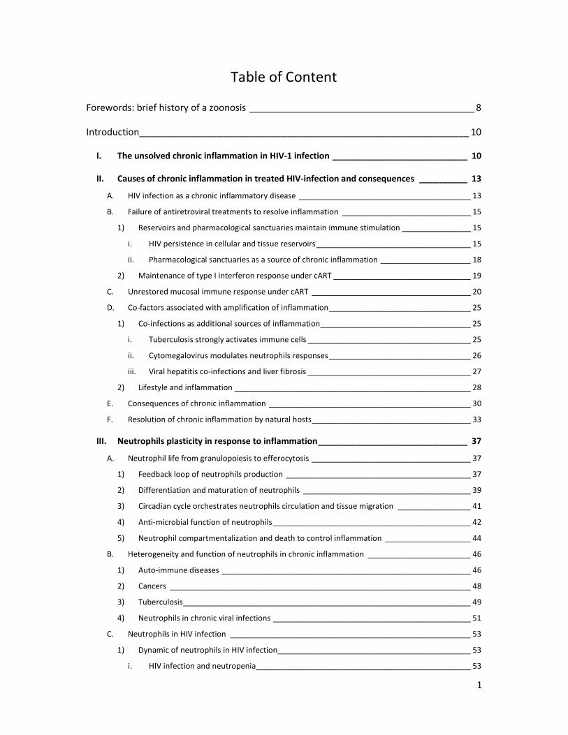
Table of Content
Forewords: brief history of a zoonosis ___________________________________________ 8
Introduction_______________________________________________________________ 10
I. The unsolved chronic inflammation in HIV-1 infection ____________________________ 10
II. Causes of chronic inflammation in treated HIV-infection and consequences __________ 13
A. HIV infection as a chronic inflammatory disease ________________________________________ 13
B. Failure of antiretroviral treatments to resolve inflammation ______________________________ 15
1) Reservoirs and pharmacological sanctuaries maintain immune stimulation ________________ 15
i. HIV persistence in cellular and tissue reservoirs ____________________________________ 15
ii. Pharmacological sanctuaries as a source of chronic inflammation _____________________ 18
2) Maintenance of type I interferon response under cART ________________________________ 19
C. Unrestored mucosal immune response under cART _____________________________________ 20
D. Co-factors associated with amplification of inflammation _________________________________ 25
1) Co-infections as additional sources of inflammation ___________________________________ 25
i. Tuberculosis strongly activates immune cells ______________________________________ 25
ii. Cytomegalovirus modulates neutrophils responses _________________________________ 26
iii. Viral hepatitis co-infections and liver fibrosis ______________________________________ 27
2) Lifestyle and inflammation _______________________________________________________ 28
E. Consequences of chronic inflammation _______________________________________________ 30
F. Resolution of chronic inflammation by natural hosts _____________________________________ 33
III. Neutrophils plasticity in response to inflammation _______________________________ 37
A. Neutrophil life from granulopoiesis to efferocytosis _____________________________________ 37
1) Feedback loop of neutrophils production ___________________________________________ 37
2) Differentiation and maturation of neutrophils _______________________________________ 39
3) Circadian cycle orchestrates neutrophils circulation and tissue migration _________________ 41
4) Anti-microbial function of neutrophils ______________________________________________ 42
5) Neutrophil compartmentalization and death to control inflammation ____________________ 44
B. Heterogeneity and function of neutrophils in chronic inflammation ________________________ 46
1) Auto-immune diseases __________________________________________________________ 46
2) Cancers ______________________________________________________________________ 48
3) Tuberculosis ___________________________________________________________________ 49
4) Neutrophils in chronic viral infections ______________________________________________ 51
C. Neutrophils in HIV infection ________________________________________________________ 53
1) Dynamic of neutrophils in HIV infection_____________________________________________ 53
i. HIV infection and neutropenia __________________________________________________ 53
2
ii. Mucosal neutrophils in HIV infection ____________________________________________ 54
2) Neutrophil function in HIV infection _______________________________________________ 54
i. Neutrophil’s antiviral response _________________________________________________ 54
ii. Dual role in HIV acquisition and protection _______________________________________ 55
iii. Neutrophils function and chronic inflammation ____________________________________ 56
Methodology ______________________________________________________________ 59
I. General objectives _________________________________________________________ 59
II. Study model ______________________________________________________________ 62
III. Principles of mass cytometry ________________________________________________ 64
Results ___________________________________________________________________ 67
I. Mass cytometry reveals the immaturity of circulating neutrophils during SIV infection __ 67
A. Summary________________________________________________________________________ 67
B. Manuscript ______________________________________________________________________ 67
II. Immature CD10- neutrophils exert immunostimulatory function during primary and chronic
SIV infection __________________________________________________________________ 80
A. Summary________________________________________________________________________ 80
B. Manuscript ______________________________________________________________________ 82
1) Abstract ______________________________________________________________________ 83
2) Introduction ___________________________________________________________________ 84
3) Material and method ___________________________________________________________ 85
4) Results _______________________________________________________________________ 88
5) Discussion ____________________________________________________________________ 94
III. Unpublished results: tissue neutrophils heterogeneity in cynomolgus macaques______ 101
A. Introduction ____________________________________________________________________ 101
B. Material and method _____________________________________________________________ 101
C. Preliminary results _______________________________________________________________ 102
Discussion and perspectives _________________________________________________ 106
I. Validation of experimental approach _________________________________________ 106
II. Discussion ______________________________________________________________ 107
III. Perspectives _____________________________________________________________ 109
References _______________________________________________________________ 113
3
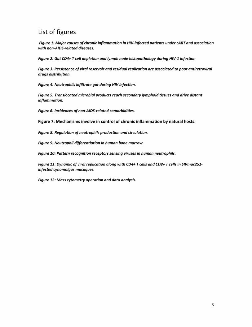
List of figures Figure 1: Major causes of chronic inflammation in HIV-infected patients under cART and association with non-AIDS-related diseases. Figure 2: Gut CD4+ T cell depletion and lymph node histopathology during HIV-1 infection Figure 3: Persistence of viral reservoir and residual replication are associated to poor antiretroviral drugs distribution. Figure 4: Neutrophils infiltrate gut during HIV infection. Figure 5: Translocated microbial products reach secondary lymphoid tissues and drive distant inflammation. Figure 6: Incidences of non-AIDS-related comorbidities. Figure 7: Mechanisms involve in control of chronic inflammation by natural hosts. Figure 8: Regulation of neutrophils production and circulation. Figure 9: Neutrophil differentiation in human bone marrow. Figure 10: Pattern recognition receptors sensing viruses in human neutrophils. Figure 11: Dynamic of viral replication along with CD4+ T cells and CD8+ T cells in SIVmac251-infected cynomolgus macaques. Figure 12: Mass cytometry operation and data analysis.
4
List of abbreviations 3TC: Lamivudine
AAV: Antibody-associated vasculitis
ACPA: Anti-citrullinated protein antibodies
ADCC: Antibody-dependent cellular cytotoxicity
ADCP: Antibody-dependent cellular phagocytosis
AGM: African green monkey
AIDS : Acquired immunodeficiency syndrome
ANRS: Agence nationale de recherche sur le sida et les hépatites virales
ARDS: Acute respiratory distress syndrome
AZT: Azidothymidine
BAFF: B-cell activating factor
C/EBP-: CCAAT/enhancer-binding protein alpha
cART: Combination antiretroviral therapy
CD: Cluster of differentiation
CLR: C-lectin receptor
CMP: Common myeloid progenitor
COPD: Chronic obstructive pulmonary disease
CRP: C-reactive protein
CTLA4: Cytotoxic T-lymphocyte-associated protein 4
DAMP: Danger-associated molecular pattern
DC: Dendritic cell
DNA: Deoxyribonucleic acid
DRV: Darunavir
DTG: Dolutegravir
Fc-R: Fragment crystallizable receptor
FDC: Follicular dendritic cell
fMLP: N-formylmethionine-leucyl-phenylalanine
FTC: Emtricitabine
G-CSF: Granulocyte-colony stimulating factor
G-MDSC: Granulocytic-myeloid-derived suppressive cell
GALT: Gut-associated lymphoid tissue
GM-CSF: Granulocyte-monocyte colony stimulating factor
GMP: Granulocyte-monocyte progenitor
HBV: Hepatitis B virus
HCMV: Human cytomegalovirus
HCV: Hepatitis C virus
HESN: HIV-exposed seronegative
HIV: Human immunodeficiency virus
HLADR: Human leukocyte antigen DR type
HSC: Hematopoietic stem cells
HTLV: Human T cell leukemia/lymphoma virus
I-FABP: Intestinal fatty-acid binding protein
ICP-MS: Inductively coupled plasma mass spectrometry
IDV: Idinavir
IFN: Interferon
Ig: Immunoglobulin
IL: Interleukin
IP-10: Interferon gamma-induced protein 10
ISG: Interferon-stimulated genes
ITAM: Immunoreceptor tyrosine-based activation motif

5
LDN: Low-density neutrophil
LN: Lymph node
LPS: Lipopolysaccharide
MALT: Mucosa-associated lymphoid tissues
MDA5: Melanoma differentiation-associated protein 5
MHC: Major histocompatibility complex
MMP: Matrix metalloproteinase
MPO: Myeloperoxidase
Mtb: Mycobacterium tuberculosis
NADPHox: Nicotinamide adenine dinucleotide phosphate oxidase
Nef: Negative regulatory factor
NET: Neutrophil extracellular trap
NK: Natural killer
NLR: Nucleotide-binding oligomerization domain-like receptor
PAMP: Pathogen-associated molecular pattern
PBMC: Peripheral blood mononuclear cell
PCR: Polyclonal chain reaction
PD-1: Programmed cell death 1
pDC: Plasmacytoid dendritic cell
PMA: Phorbol myristate acetate
PMN: Polymorphonuclear neutrophil
PPAR: Peroxisome proliferator-activated receptor gamma
QVOA: Quantitative viral outgrowth assay
RA: Rheumatoid arthritis
RIG-I: Retinoic acid-inducible gene I
RNA: Ribonucleic acid
ROS: reactive oxygen species
rRNA: Ribosomal ribonucleic acid
SIV: Simian immunodeficiency virus
SLE: Systemic lupus erythematosus
SM: Sooty mangabey
SNS: sympathetic nervous system
SPADE: Spanning-tree Progression analysis of density-normalized events
ssRNA: Single-strand ribonucleic acid
TAN: Tumor-associated neutrophil
TB: Tuberculosis
TCM: T lymphocyte central memory
TCR: T cell receptor
TDF: Tenofovir disproxil fumarate
TEM: T lymphocyte effector memory
Tfh: T follicular helper cell
TGF-: Transforming growth factor beta
Th: T helper cell
TLR: Toll-like receptor
TNF: Tumor necrosis factor
TNFR: Tumor necrosis factor receptor
Treg: T regulatory cell
WHO: World health organization
6
Acknowledgments
I would like to acknowledge Prof. Sylvie Chollet-Martin from Hôpital Bichat, for kindly accepting to preside over this thesis jury.
I would like to express my sincere gratitude to Dr. Véronique Witko-Sarsat, from Institut Cochin, and Prof. Pierre Delobel, from CHU de Toulouse, for giving their valuable time to review my thesis.
I am also very grateful to Dr. Mirko Paiardini, from Emory University School of Medicine, for kindly accepting to joint this thesis jury.
I thank sincerely Dr. Anne-Sophie Beignon, for her help and guidance throughout my PhD.
I would like to express my sincere gratitude to my advisors Dr. Roger Le Grand and Prof. Olivier Lambotte for the continuous support, motivation and guidance of my PhD, and for your immense knowledge. I could not have imagined having a better advisors and mentors for my PhD.

7
8
Forewords: brief history of a zoonosis “There will be new diseases. It is a fatal fact. Another fact, as fatal, is that we will never be able to detect them from their origin. When we have a notion of these diseases, they will all be formed, adults could we say. They will appear as Athena appeared, coming out of the brain of Zeus. How would we recognize these new diseases, how would we suspect their existence
before they donned their costume of symptoms?” – Charles Nicolle, Destin des maladies infectieuses
In 1981, first acquired immunodeficiency syndromes (AIDS) were reported among five young
men affected by pneumocystis and severe T cells depletion1. Quickly more new cases were
reported and the lentivirus involved, human immunodeficiency virus-1 (HIV-1), was identified
in a lymph node biopsy from a patient2. HIV-1 is a retroviridae lentivirus characterized by a
bilayer envelope containing glycoproteins and two single RNA strands genome. Gp120
glycoprotein determines the range of host cells and targets CD4 receptor present in CD4+
lymphocytes, macrophages and monocytes. Interaction of Gp120 with CD4 needs a co-
receptor to enter the cell, such as CXCR4 and CCR5, which also determines virus targets.
Basically, T cell tropic HIV-1 viruses use CXCR4, whereas macrophage tropic viruses use CCR5
receptor. Another important characteristic of retrovirus is the reverse transcription, allowing
HIV to integrate its genome in host cells, thus generating reservoirs and long-term infection.
Infection by HIV is divided in 3 different phases: primary infection, chronic phase and AIDS.
Only 50% of the patients present symptoms during primary infection, especially flu-like
syndrome, asthenia, lymphadenopathies and digestive symptoms3. Primary-infection lasts in
median 2 weeks with sustained viral replication and CD4+ T cells depletion. Then, with partial
control of viral replication by adaptive immune response starts the chronic phase, lasting years
in which clinical signs are reduced but transmission risk is elevated. As the CD4+ T cell count
decreases, non-AIDS defining events appear as oral candidiasis, herpes zoster. When the CD4+
T cell count is below 200/mm3, the risk of opportunistic infection strongly increases, with an
elevated risk of bacterial infection (tuberculosis), oesophageal candidiasis, pneumocystosis,
toxoplasmosis and some cancers (lymphoma, Kaposi sarcoma). AIDS is defined in the United
State by a CD4+ T cell count below 200/mm3 and in France by the occurrence of various AIDS
defining conditions. Immune restoration by antiretroviral treatment with the increase of CD4+
T cell counts is fundamental to control AIDS related diseases.
9
The emergence of HIV-1 pandemic raised question about its origin. Identification of closely
related lentivirus in African primates, Simian Immunodeficiency Viruses (SIV), supports the
hypothesis of zoonotic transmission from primate to human4. Indeed, many African monkeys
and apes harbor SIV but rarely develop AIDS, and are so called natural hosts. On the contrary,
Asian macaques are not found to be naturally infected by SIV. Experimental infection of Asian
macaques with some strains of SIV results in opportunistic infections, cancers directly related
to high viral loads and CD4 T cell depletion. Non-natural SIV hosts have been essential to better
understand HIV-1 physiopathology and for vaccine and treatment development. Besides,
natural host highlighted mechanisms involved in resistance to disease development, which
basically relies on control of immune activation. Phylogenetic analyses of SIV obtained from
40 primates species reveal that HIV-1 came from SIVcpz or SIVgor isolated from Chimpanzee
(Pan troglodytes) and Western gorilla (Gorilla gorilla) respectively4. Molecular clock analyses
had dated the beginning of HIV-1 infection (group M and O) epidemic in early twentieth
century in west central Africa5, where cross-species transmission may occurred between apes
and human6. Bushmeat hunters being exposed to apes blood and body fluids is the best
explanation of transmission to human4. Cross-species transmission was not a matter of chance
and happened independently four time for HIV-1, giving rise to the four independent groups.
HIV-1 (group M and O) spreads for 50-70 years from colonial western Africa before becoming
worldwide and its first recognition in western countries. Colonial exploitation of western
Africa’s resources and populations in early twentieth century had created perfect conditions
for HIV-1 emergence and worldwide spreading7,8. Since start of the pandemic, UNAIDS
estimated 77.3 million people have been infected with HIV and 35.4 million died from AIDS-
related illnesses9. Globally in 2017, 36.8 million people were living with HIV with 59% accessing
antiretroviral therapy.
10
Introduction
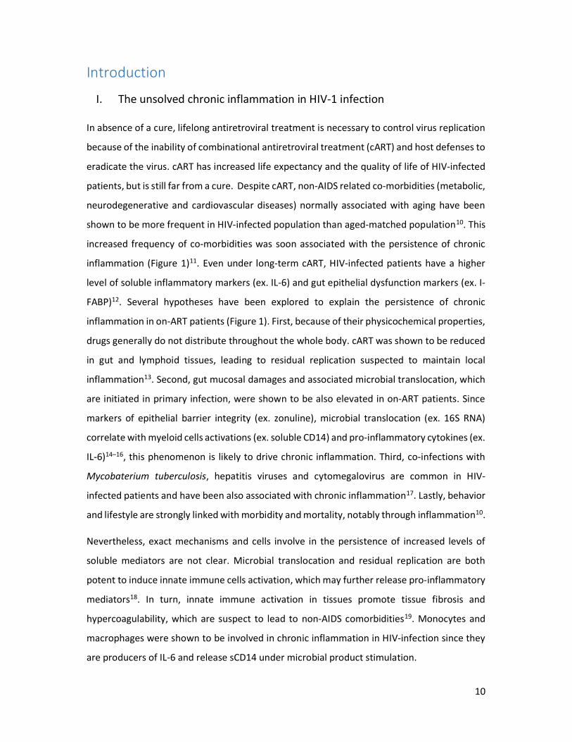
I. The unsolved chronic inflammation in HIV-1 infection
In absence of a cure, lifelong antiretroviral treatment is necessary to control virus replication
because of the inability of combinational antiretroviral treatment (cART) and host defenses to
eradicate the virus. cART has increased life expectancy and the quality of life of HIV-infected
patients, but is still far from a cure. Despite cART, non-AIDS related co-morbidities (metabolic,
neurodegenerative and cardiovascular diseases) normally associated with aging have been
shown to be more frequent in HIV-infected population than aged-matched population10. This
increased frequency of co-morbidities was soon associated with the persistence of chronic
inflammation (Figure 1)11. Even under long-term cART, HIV-infected patients have a higher
level of soluble inflammatory markers (ex. IL-6) and gut epithelial dysfunction markers (ex. I-
FABP)12. Several hypotheses have been explored to explain the persistence of chronic
inflammation in on-ART patients (Figure 1). First, because of their physicochemical properties,
drugs generally do not distribute throughout the whole body. cART was shown to be reduced
in gut and lymphoid tissues, leading to residual replication suspected to maintain local
inflammation13. Second, gut mucosal damages and associated microbial translocation, which
are initiated in primary infection, were shown to be also elevated in on-ART patients. Since
markers of epithelial barrier integrity (ex. zonuline), microbial translocation (ex. 16S RNA)
correlate with myeloid cells activations (ex. soluble CD14) and pro-inflammatory cytokines (ex.
IL-6)14–16, this phenomenon is likely to drive chronic inflammation. Third, co-infections with
Mycobaterium tuberculosis, hepatitis viruses and cytomegalovirus are common in HIV-
infected patients and have been also associated with chronic inflammation17. Lastly, behavior
and lifestyle are strongly linked with morbidity and mortality, notably through inflammation10.
Nevertheless, exact mechanisms and cells involve in the persistence of increased levels of
soluble mediators are not clear. Microbial translocation and residual replication are both
potent to induce innate immune cells activation, which may further release pro-inflammatory
mediators18. In turn, innate immune activation in tissues promote tissue fibrosis and
hypercoagulability, which are suspect to lead to non-AIDS comorbidities19. Monocytes and
macrophages were shown to be involved in chronic inflammation in HIV-infection since they
are producers of IL-6 and release sCD14 under microbial product stimulation.
11
Figure 1: Major causes of chronic inflammation in HIV-infected patients under cART and association with non-AIDS-related diseases. Unrestored gut mucosal homeostasis under cART leads to persistent microbial translocation, which maintains inflammatory state. Sustain immune activation induces tissue fibrosis especially in adipose tissue, in lymphe nodes, in the liver, which reduces clearance of microbial products and leads to liver diseases. Lifestyle influences also inflammatory state and impacts organs functions such as the liver (alcool) and the lungs (tabacco). The existence of pharmacological sanctuaries allows virus persistence and residual replication inducing local and systemic inflammation and organs dysfunction. Co-infections are common in HIV-infected patients and are contributors to chronic inflammation.
12
In the first part, I will focus on causes and consequences of chronic inflammation under cART
with the emphasis on the role of myeloid cells. But one of the most potent pro-inflammatory
myeloid cells, polymorphonuclear neutrophil (PMN), has been overlooked in HIV-infection20.
Neutrophil have been involved in most of non-AIDS-related comorbidities as an effector of
chronic inflammation21, notably potent to release IL-6 and also to modulate coagulation
cascades leading to thrombosis. In addition, PMN activation was higher in HIV-infected
patients having non-AIDS-related comorbidities, suggesting a relationship between
neutrophils and level of systemic inflammation22. Thus we hypothesize that PMN dysfunction
could be responsible of chronic inflammation in HIV infection23,24. In the last part, I will review
the literature on the role of PMN in physiology and chronic inflammatory diseases.
13
II. Causes of chronic inflammation in treated HIV-infection and
consequences
In HIV infection, chronic inflammation originates from viral persistence and inability of the
host to resolve inflammation. Natural hosts, such as African green monkeys, demonstrate that
virus-host interaction could lead to the resolution of inflammation despite viral persistence,
thus protecting against AIDS. In absence of functional cure, cART is the best option to reduce
disease progression, but inflammation remains elevated. In the first part, we will present HIV
infection as a chronic inflammatory disease. In the second part, we will describe cause and
consequences of chronic inflammation under cART.
A. HIV infection as a chronic inflammatory disease
In primary infection, immune response starts with acute phase molecules such as serum
amyloid A, detectable in plasma 3 to 5 days after transmission. Increasing of viremia is
associated with growing innate immune response and important inflammatory cytokines
release. This inflammatory cytokine burst starts with high levels of IFN-α, interleukin -15 (IL-
15), followed by a rise of IP-10, TNF-α, IL-18 and IFN-γ25. Studies conducted in both SIV-
infected macaques and HIV-infected patients showed a rapid and barely irreversible depletion
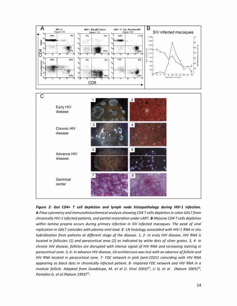
of memory CD4+ T cells in mucosa, mostly in CCR5+ cells (Figure2) 26,27. HIV-1 infection leads
to progressive depletion of memory and naïve CD4+ and CD8+ T cells via direct and indirect
mechanism28. Direct virus killing and NK cells or CD8 T cells cytotoxic activity were thought to
be the major mechanism of CD4 T cells depletion, but Quingsheng L. and colleages showed
that only 7% of lamina propria CD4+ T cells were infected by SIV29. The virus indirectly impairs
thymus output early in SIV infection, where a decrease of thymocyte number is associated
with cell apoptosis30. In addition, immune activation is an important indirect mechanism of
uninfected CD4 T cell depletion via the Fas-FasL and the TRAIL-DR5 apoptosis pathways31. The
decrease half-life of exhausted cells contributes to the reduction of the memory CD4 T cell
pool32.
14
Figure 2: Gut CD4+ T cell depletion and lymph node histopathology during HIV-1 infection. A-Flow cytometry and immunohistochemical analysis showing CD4 T cells depletion in colon GALT from chronically HIV-1 infected patients, and partial restoration under cART. B-Massive CD4 T cells depletion within lamina propria occurs during primary infection in SIV infected macaques. The peak of viral replication in GALT coincides with plasma viral load. C- LN histology associated with HIV-1 RNA in situ hybridization from patients at different stage of the disease. 1, 2- In eraly HIV disease, HIV RNA is located in follicules (1) and paracortical area (2) as indicated by white dots of silver grains. 3, 4- In chronic HIV disease, follicles are disrupted with intense signal of HIV RNA and increasing staining in paracortical zone. 5, 6- In advance HIV disease, LN architecture was lost with an absence of follicle and HIV RNA located in paracortical zone. 7- FDC network in pink (anti-CD21) coinciding with HIV RNA appearing as black dots in chronically infected patient. 8- Impaired FDC network and HIV RNA in a involute follicle. Adapted from Guadalupe, M. et al (J. Virol 2003)33, Li Q. et al . (Nature 2005)29, Pantaleo G. et al (Nature 1993)34.
15
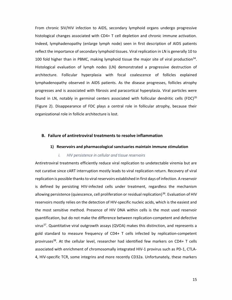
From chronic SIV/HIV infection to AIDS, secondary lymphoid organs undergo progressive
histological changes associated with CD4+ T cell depletion and chronic immune activation.
Indeed, lymphadenopathy (enlarge lymph node) seen in first description of AIDS patients
reflect the importance of secondary lymphoid tissues. Viral replication in LN is generally 10 to
100 fold higher than in PBMC, making lymphoid tissue the major site of viral production34.
Histological evaluation of lymph nodes (LN) demonstrated a progressive destruction of
architecture. Follicular hyperplasia with focal coalescence of follicles explained
lymphadenopathy observed in AIDS patients. As the disease progresses, follicles atrophy
progresses and is associated with fibrosis and paracortical hyperplasia. Viral particles were
found in LN, notably in germinal centers associated with follicular dendritic cells (FDC)35
(Figure 2). Disappearance of FDC plays a central role in follicular atrophy, because their
organizational role in follicle architecture is lost.
B. Failure of antiretroviral treatments to resolve inflammation
1) Reservoirs and pharmacological sanctuaries maintain immune stimulation
i. HIV persistence in cellular and tissue reservoirs
Antiretroviral treatments efficiently reduce viral replication to undetectable viremia but are
not curative since cART interruption mostly leads to viral replication return. Recovery of viral
replication is possible thanks to viral reservoirs established in first days of infection. A reservoir
is defined by persisting HIV-infected cells under treatment, regardless the mechanism
allowing persistence (quiescence, cell proliferation or residual replication)36. Evaluation of HIV
reservoirs mostly relies on the detection of HIV-specific nucleic acids, which is the easiest and
the most sensitive method. Presence of HIV DNA within cells is the most used reservoir
quantification, but do not make the difference between replication-competent and defective
virus37. Quantitative viral outgrowth assays (QVOA) makes this distinction, and represents a
gold standard to measure frequency of CD4+ T cells infected by replication-competent
proviruses38. At the cellular level, researcher had identified few markers on CD4+ T cells
associated with enrichment of chromosomally integrated HIV-1 provirus such as PD-1, CTLA-
4, HIV-specific TCR, some integrins and more recently CD32a. Unfortunately, these markers
16
are still unable to gather all infected cells, because none of them is universal for latently
infected CD4 T cells, and they miss infected non-CD4+ T cells.
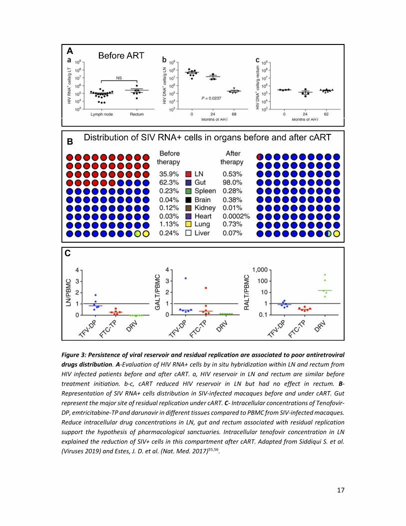
Reservoir is also defined by its anatomical localization, each tissue or organ owing its specific
cell reservoir distribution. HIV/SIV DNA has been detected in several lymphoid and non-
lymphoid tissues39. Primary and secondary lymphoid tissues are reservoir for HIV (Figure 2).
In bone marrow, HIV infects resident CD4 T cells and macrophages34,40. Studies on ART-
suppressed patients did not detected HIV DNA in CD34+ progenitor but there are evidence
that mast cell precursor could be latently infected by the virus40–42. Spleen and lymph nodes
contain huge numbers of infected cells and free virions trapped in FDC network43. Even after
years of cART HIV RNA and DNA are still detected in lymph nodes44. Germinal centers harbored
viral particles HIV p24, p17 and gp120/gp41 abundantly detected from patients under
effective cART45. In patients under ART, central memory and transitional memory represent
the largest CD4+T cells reservoir in peripheral blood, whereas effector memory CD4+ T cells
had a greater contribution in lymphoid tissues36. Recently, studies on SIV-infected macaques
and HIV-infected patients have identified another important contributor of lymphoid tissue
reservoir: CD4+ T cells follicular helpers (Tfh). Persistence of infected Tfh is due to the
impossibility of cytotoxic T lymphocytes to enter germinal centers46. Tfh were associated with
high level of HIV-1 RNA in patients after up to 12 years of treatment duration, thus
representing a source of residual replication and participating to chronic inflammation47. In
ART-suppressed patients gut, HIV-1 DNA and RNA were identified in both CD4+ T cells and
CD13+ myeloid cells, mostly macrophages48,49. Indeed, in on-cART individuals, gut is one of the
organs comprising the highest number of infected cells. By analyzing 4 different regions of the
gut from on-ART individuals, Yulk et al. showed HIV DNA levels per CD4+ T cells were 5-10 fold
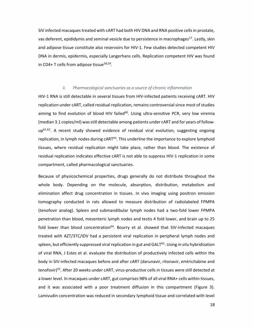
higher in gut compared to peripheral blood48(Figure3).
Myeloid cells, especially macrophages, represent an important reservoir cells in other organs.
HIV DNA and RNA were detected in lung tissue, mostly in macrophages and comparison of
HIV-1 sequence evolution in the lung showed a compartmentalization from the blood50,51. In
central nervous system from on-ART patients, HIV DNA and RNA are localized to perivascular
macrophages and microglial cells52. Male reproductive tract, especially testis is an immune-
privilege site in which specific humoral and cellular response are reduced against HIV-153,54.
17
Figure 3: Persistence of viral reservoir and residual replication are associated to poor antiretroviral drugs distribution. A-Evaluation of HIV RNA+ cells by in situ hybridization within LN and rectum from HIV infected patients before and after cART. a, HIV reservoir in LN and rectum are similar before treatment initiation. b-c, cART reduced HIV reservoir in LN but had no effect in rectum. B-Representation of SIV RNA+ cells distribution in SIV-infected macaques before and under cART. Gut represent the major site of residual replication under cART. C- Intracellular concentrations of Tenofovir-DP, emtricitabine-TP and darunavir in different tissues compared to PBMC from SIV-infected macaques. Reduce intracellular drug concentrations in LN, gut and rectum associated with residual replication support the hypothesis of pharmacological sanctuaries. Intracellular tenofovir concentration in LN explained the reduction of SIV+ cells in this compartment after cART. Adapted from Siddiqui S. et al. (Viruses 2019) and Estes, J. D. et al. (Nat. Med. 2017)55,56.
18
SIV infected macaques treated with cART had both HIV DNA and RNA positive cells in prostate,
vas deferent, epididymis and seminal vesicle due to persistence in macrophages57. Lastly, skin
and adipose tissue constitute also reservoirs for HIV-1. Few studies detected competent HIV
DNA in dermis, epidermis, especially Langerhans cells. Replication competent HIV was found
in CD4+ T cells from adipose tissue58,59.
ii. Pharmacological sanctuaries as a source of chronic inflammation
HIV-1 RNA is still detectable in several tissues from HIV-infected patients receiving cART. HIV
replication under cART, called residual replication, remains controversial since most of studies
aiming to find evolution of blood HIV failed60. Using ultra-sensitive PCR, very low viremia
(median 3.1 copies/ml) was still detectable among patients under cART and for years of follow-
up61,62. A recent study showed evidence of residual viral evolution, suggesting ongoing
replication, in lymph nodes during cART63. This underline the importance to explore lymphoid
tissues, where residual replication might take place, rather than blood. The existence of
residual replication indicates effective cART is not able to suppress HIV-1 replication in some
compartment, called pharmacological sanctuaries.
Because of physicochemical properties, drugs generally do not distribute throughout the
whole body. Depending on the molecule, absorption, distribution, metabolism and
elimination affect drug concentration in tissues. In vivo imaging using positron emission
tomography conducted in rats allowed to measure distribution of radiolabeled FPMPA
(tenofovir analog). Spleen and submandibular lymph nodes had a two-fold lower FPMPA
penetration than blood, mesenteric lymph nodes and testis 4 fold lower, and brain up to 25
fold lower than blood concentration64. Bourry et al. showed that SIV-infected macaques
treated with AZT/3TC/IDV had a persistent viral replication in peripheral lymph nodes and
spleen, but efficiently suppressed viral replication in gut and GALT65. Using in situ hybridization
of viral RNA, J Estes et al. evaluate the distribution of productively infected cells within the
body in SIV-infected macaques before and after cART (darunavir, ritonavir, emtricitabine and
tenofovir)55. After 20 weeks under cART, virus-productive cells in tissues were still detected at
a lower level. In macaques under cART, gut comprises 98% of all viral RNA+ cells within tissues,
and it was associated with a poor treatment diffusion in this compartment (Figure 3).
Lamivudin concentration was reduced in secondary lymphoid tissue and correlated with level
19
of viral RNA in tissue. Another study showed presence of HIV RNA in all lymph nodes samples
from HIV-infected patients receiving protease inhibitors66. HIV RNA was associated with lower
ARV concentration in lymphoid tissue compared to plasma. In fact all tissue reservoirs
(lymphoid tissues, central nervous system, genital tract and liver) are also suspected to host
residual replication since HIV RNA is detected13. Beside suboptimal drug distribution in tissue,
cellular mechanisms are also responsible to reduce treatment effectiveness. Transporters
expressed by monocytes and tissue macrophages could decrease intracellular drug
concentrations of ARV and substrates. Even if treatment reaches infected macrophages, efflux
transporters make tissue macrophages pharmacological sanctuaries67,68. Multidrug resistant
proteins (MRP) and P-glycoprotein were shown to be expressed by several tissue specific
macrophages, such as microglia, and decreased anti-HIV activity of lamivudine and
zidovudine.
2) Maintenance of type I interferon response under cART
Type I interferons are beneficial cytokines during viral infections by inducing hundreds of
interferon-stimulated genes (ISG) which protect other uninfected cells69. Nevertheless,
persistent HIV-1 replication maintains activation of ISG, leading to chronic immune activation.
Higher levels of IFN correlated with ISGs up-regulation in CD4+ T cells from HIV-1 infected
individuals, and was also associated with CD4+ T cell depletion70,71. Initiation of cART reduced
plasmatic inflammatory markers such as TNF- and IFN- but not IP-10 and sCD1472.
Moreover, ultrasensitive CRP (CRPus) is rarely normalized in patients on ART. Interestingly,
following cART there was a lesser reduction of plasmatic inflammatory markers in women
compared to men in a study73. Sex-related differences in term of inflammation and response
to cART in HIV infected patients have been explained by hormones and type I interferon
production74. At the same viral load, women had a higher up-regulation of ISGs compared to
men. Under TLR7 stimulation, female’s pDC express more mRNA of all IFN and IFN. HIV-1
derived ssRNA induce higher IFN production associated with CD8+ T cells activation in
women compared to men74. Hormonal cycle also influences TLR7-responsiveness and IFN
production, making more complex to analyze sex differences70.

20
C. Unrestored mucosal immune response under cART
The gastrointestinal tract is an important site for HIV pathogenesis regardless the route of
acquisition, since >40% of all body lymphocytes are located in gut associated lymphoid tissues
(GALT)75. Lamina propria contains a large population of resident memory CD4+ T cells, which
is largely depleted in primary infection, before efficient adaptive response is generated27.
Indeed, first case reports of AIDS already described gastrointestinal symptoms with diarrhea
and weigh loss76. HIV-1 preferentially depletes gut Th17 cells as shown by Paiardini, Brenchley
and colleagues77. In this study, bronchoalveolar lavages, peripheral blood and terminal ileum
biopsies obtained in HIV-1 infected patients revealed infection of Th17 cells and a tissue
dependent depletion of Th17 cells. This CD4+ T helper cells producing IL-17 was first described
in respiratory mucosal response to bacteria, with IL-17 known to promote rapid recruitment
of monocytes and neutrophils, to induce G-CSF production and IL-878,79. Indeed, Th17 cells are
enriched in human GALT and participate to gut homeostasis by inducing epithelial cells
regeneration, mucin production and by maintaining tight junction integrity80. Thus, depletion
of Th17 is thought to be responsible of epithelial barrier breakdown observed in HIV-infected
patients, leading to microbial translocation.
Microbial translocation exposes lamina propria to microbial product and fuels intestinal
inflammation, which in turn conducts to microbiota alteration81. Work conducted by Lynch S.
has shown an enrichment of Proteobacteria and a decrease abundance of Bacteriodetes in
chronically infected patients, underlying a dysbiosis associated with HIV-1 infection82. In the
phylum of Proteobacteria there was a high representation of Enterobactericeae family,
containing proinflammatory pathobionts. Dysbiosis was correlated with IL-6 and tryptophane
catabolism, both linked to inflammation and unbalance immune response. So, HIV-1 infection
induces dysbiosis because of inflammation and metabolism alteration, in turn altered
microbiota may also participate to gut inflammation as seen in inflammatory bowel diseases.
Recently, neutrophil infiltration has been associated with colonic epithelial cells apoptosis and
microbial translocation in HIV and SIV infections, especially in chronic phase83,84. Regardless
HIV-infected patients were receiving cART, there was an increase PMN infiltration in colonic
lamina propria83,85. PMN infiltration in colon was significantly higher in chronic phase compare
to primary infection86. (Figure 4) Whether neutrophils are a cause or a consequence of gut
epithelium damage remains to be determined.

21
Figure 4: Neutrophils infiltrate gut during HIV infection. A-SIV-infected RM colon stained for myeloperoxidase (brown) to quantify PMN infiltration. PMN infiltration increased with epithelial lesion and with the progression of the disease. In AIDS, neutrophils infiltration was important. Graphic showing neutrophil infiltration in few SIV infected RM. B-Neutrophil frequency in blood from HIV-infected patients under cART did not differ from healthy controls. Besides, neutrophil frequency measured in colorectal biopsies showed increased infiltration HIV-infected patients under cART. Adapted from Hensley-McBain T. et al. (PLoS Pathog. 2019).
22
In chronic inflammatory disease, increase lifespan of PMN is associated with tissue damages.
A recent study shed light on the link between PMN lifespan and gut microbiota87. Using flow
cytometry, authors confirmed the increase of neutrophils in colorectal biopsies. To evaluate
gut neutrophils survival, decrease of CD16 expression and increase of active Caspase-3 were
used and demonstrated a higher percentage of surviving PMN in HIV-infected patients,
regardless they received cART. Gut microbiota imbalance observed in HIV-infected individuals
was characterized by enrichment of Prevotella and decrease abundance of Lactobacillus.
Neutrophil stimulation with Prevotella was capable to increase PMN survival whereas
Lactobacillus species increased PMN apoptosis. Thus, neutrophil survival is associated with
pro-inflammatory microbiota and environment in HIV infection. Like inflammatory bowel
diseases, gut mucosa damage in HIV infection might be mediated by neutrophil persistence
and activation88. It will be necessary to evaluate neutrophil function in mucosae to better
understand their contribution to tissue damage and immune activation.
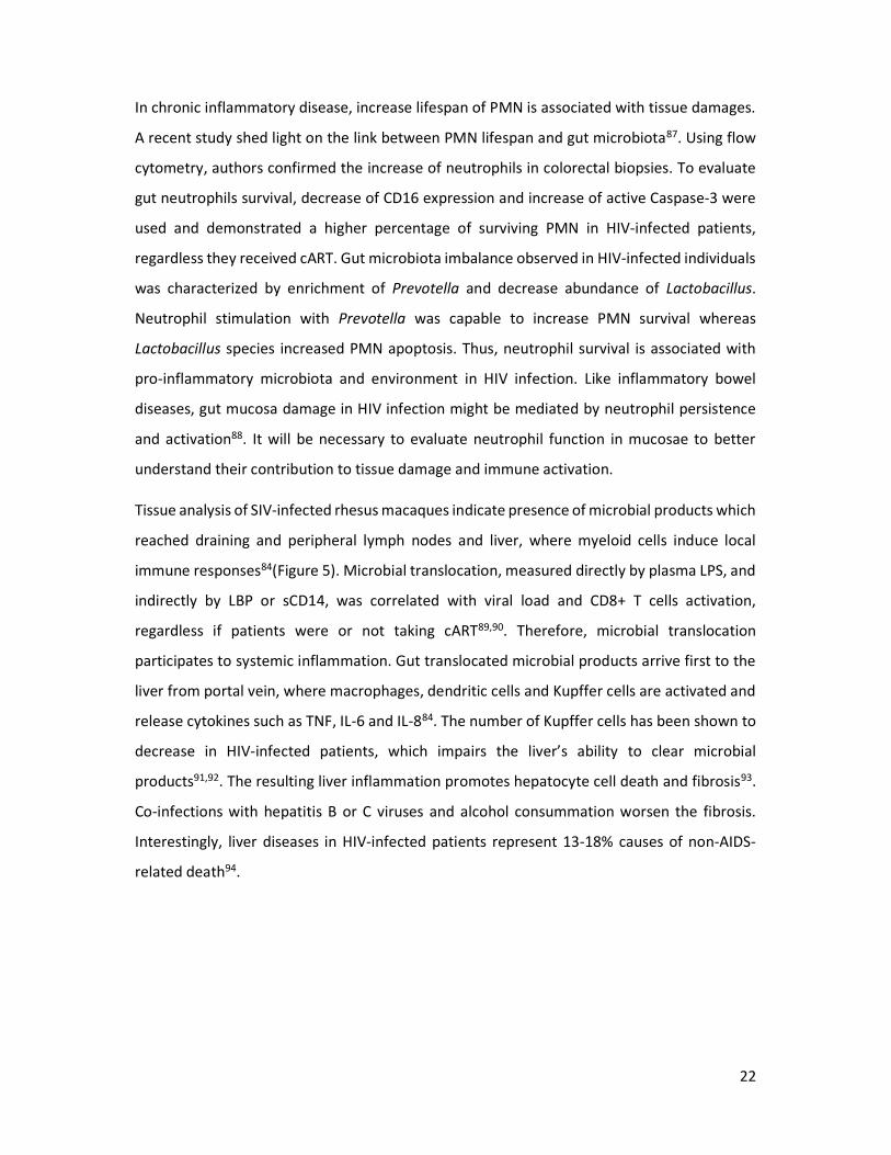
Tissue analysis of SIV-infected rhesus macaques indicate presence of microbial products which
reached draining and peripheral lymph nodes and liver, where myeloid cells induce local
immune responses84(Figure 5). Microbial translocation, measured directly by plasma LPS, and
indirectly by LBP or sCD14, was correlated with viral load and CD8+ T cells activation,
regardless if patients were or not taking cART89,90. Therefore, microbial translocation
participates to systemic inflammation. Gut translocated microbial products arrive first to the
liver from portal vein, where macrophages, dendritic cells and Kupffer cells are activated and
release cytokines such as TNF, IL-6 and IL-884. The number of Kupffer cells has been shown to
decrease in HIV-infected patients, which impairs the liver’s ability to clear microbial
products91,92. The resulting liver inflammation promotes hepatocyte cell death and fibrosis93.
Co-infections with hepatitis B or C viruses and alcohol consummation worsen the fibrosis.
Interestingly, liver diseases in HIV-infected patients represent 13-18% causes of non-AIDS-
related death94.
23
Residual replication and HIV persistence are linked with inflammation95. Despite cART, treated
HIV-infected patients have lymph nodes abnormalities such as damage of follicular structure,
poor CD4+ T cell restoration and follicular abnormalities from hyperplasia to regression96–98.
The detection of microbial products in axillary and mesenteric lymph nodes from SIV-infected
RM, suggests microbial translocation may participate to lymph nodes impairment84. Local
immune activation induced by LPS triggers TGF-β1 pathways, and thus collagen deposition in
lymphoid tissue99,100. Collagen preferentially deposits in T cell zone and correlates with disease
stage97. Collagen deposition has dual deleterious role: by reducing niches for CD4+ T cells
reconstitution, and by limiting immunological synapses formation.
In this vicious circle, generalized fibrosis disrupts normal lymphoid tissue structure and
function, and thus participates to myeloid cells activation. By inducing inflammation and
fibrosis, microbial translocation might be also a mechanism to consider to explain lymphoid
tissue damage in patient under cART. Nevertheless, it is still hard to conclude on whether
inflammation comes from previous tissue lesions acquired before treatment initiation, or
ongoing inflammatory processes occurring under cART or both.
24
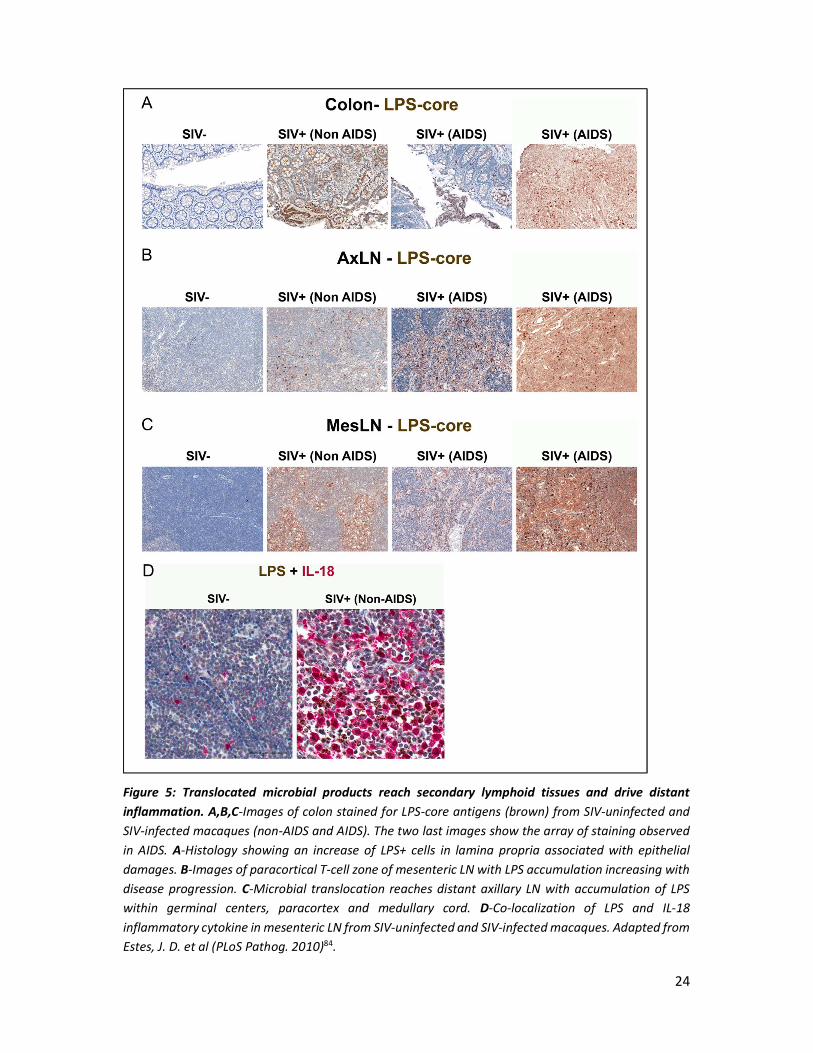
Figure 5: Translocated microbial products reach secondary lymphoid tissues and drive distant inflammation. A,B,C-Images of colon stained for LPS-core antigens (brown) from SIV-uninfected and SIV-infected macaques (non-AIDS and AIDS). The two last images show the array of staining observed in AIDS. A-Histology showing an increase of LPS+ cells in lamina propria associated with epithelial damages. B-Images of paracortical T-cell zone of mesenteric LN with LPS accumulation increasing with disease progression. C-Microbial translocation reaches distant axillary LN with accumulation of LPS within germinal centers, paracortex and medullary cord. D-Co-localization of LPS and IL-18 inflammatory cytokine in mesenteric LN from SIV-uninfected and SIV-infected macaques. Adapted from Estes, J. D. et al (PLoS Pathog. 2010)84.
25
D. Co-factors associated with amplification of inflammation
1) Co-infections as additional sources of inflammation
HIV infection, by weakening the immune system, is associated with co-infections by
reactivation of pathogens or increase susceptibility to newly acquired pathogens. Even after
initiation of cART, co-infections fuel chronic inflammation and are associated with increase
morbidity and mortality101. Tuberculosis, viral hepatitis and cytomegalovirus infection are the
most prevalent HIV-coinfections and have been studied for their impact on chronic
inflammation.
i. Tuberculosis strongly activates immune cells
Tuberculosis (TB) co-infection has been largely studied because, globally, 1 in 4 individuals are
living with latent TB infection102. In 2015, it has been estimated than 1.14 million of HIV-1
infected individuals suffer from active TB. Active TB accelerates HIV disease progression,
increasing plasma102 and cerebrospinal103 viral loads associated with increased viral
quasispecies evolution103. In active TB infection, M. tuberculosis contributes to higher immune
activation in HIV-1 co-infected patients. In TB/HIV-1 infected patients, T-cell (HLA-DR and
CD38) and macrophages activation markers (sCD163, sCD14 and IL1-RA) were elevated in both
pleural fluids and plasma104,105. These inflammatory markers in pleural fluid were also higher
in TB/HIV-1 than TB mono-infected patients, suggesting a synergy on pulmonary
inflammation. In the same studies, comparison of plasma LPS or IFABP in HIV/TB co-infected
and HIV mono-infected patients shown similar level of microbial translocation, without impact
of TB. Interestingly, even latent TB exacerbates HIV-1 associated immune activation106. TB
reactivation is increased in HIV-infected patients because of memory CD4+ T cells decline and
reduced polyfunctionality107,108. In HIV infection, tissues infected by M. tuberculosis show
depletion of CD4+ T cells and large infiltration of neutrophils109 with increase of TNF-α activity.
In active tuberculosis, patients had a distinct transcriptional signature involving interferon-
inducible genes expressed by neutrophils110. This neutrophil-driven signature was correlated
with lung radiographic severity, making neutrophils associated with tissue damages. Indeed,
neutrophils infiltration and neutrophilia are associated with poor prognosis and are correlated
with serum pro-inflammatory markers111,112. Thus, TB increases pulmonary and lymph nodes
damages and recruit PMN increasing systemic inflammation.
26
Restoration of TB immune response after cART initiation is globally associated with a
decreased risk of reactivation and primary TB108. Nevertheless, within the first 3 months of
cART incidence of TB reactivation is increased in patients113. This ART-associated TB relies on
the increase of Th1 response against M. tuberculosis, characterized by IFN-γ, CXCL10, CXCL9
and IL-18 production114,115. Indeed, cART initiation induces systemic inflammation because of
immune reconstitution inflammatory syndrome.
ii. Cytomegalovirus modulates neutrophils responses
Human cytomegalovirus (HCMV) is a common β-herpesvirus acquired during childhood or
early adulthood, with about 70% seroprevalence in adults having good socioeconomic
conditions116. Primary CMV infection induces a robust immune response and the virus persists
in latent form, or may replicate at low levels thanks to immune evasion mechanisms. HCMV is
able to stimulate and maintain high level of specific T cells, with up to 50% of circulating CD8+
T cells and 30% of CD4+ T cells117. With aging, HCMV progressively selects memory CD8 T cells
(CD28- CD57+) having a poor proliferation capacities and producing large amount of IFN-γ
,TNF-α ,IL-1β and IL-6 upon stimulation118. This effect on CD8 T cells selection may contribute
to immunoscescence observed with aging117. Most of HIV-infected patients are co-infected
with CMV, with both viruses being associated with inflammation. In a recent study, HIV/CMV
co-infected patients had a lower CD4+/CD8+ T cells ratio compared to mono-infected patients,
maybe because of deferential impact of both virus on CD4 and CD8 T cells119. Compared to
HIV mono-infection, co-infection with HCMV significantly increases inflammatory soluble
factors, such as IP-10 (CXCL10), TNF-RII and D-dimer116. Interestingly, HCMV has been shown
to modulate neutrophils at its advantage promoting viral dissemination. Production of viral
CXCL-1 by HCMV was suspected to attract neutrophils at the site of infection and further
attracting target cells, such as monocytes120. In a study, HCMV-stimulated neutrophils
released IL-8, IL-6, TNF and MIP-1, which promoted monocyte migration and activation to
a permissive HCMV phenotype121. Neutrophils had also an increase survival after HCMV
stimulation, which is frequently associated with inflammation in other diseases. In the same
study, authors hypothesize that HCMV shapes neutrophil response in a pro-inflammatory
state, promoting its own persistence and dissemination.
27
In absence of treatment, HCMV accelerates HIV disease progression and also participates to
AIDS-related infections122. For example HCMV retinitis, potentially leading to blindness, is one
of the most common manifestations in advanced HIV infection123. Nevertheless, the
inflammatory role of HCMV in HIV-infected patients under cART is not clear. Co-infection with
HCMV has been shown to increase non-AIDS related complications124. In this large study,
HIV/CMV co-infection was associated with an increased risk of cardiovascular and
cerebrovascular diseases. Treatment with valganciclovir performed in a small clinical trial
reduced CD8+ T cell activation, suggesting that treatment of herpesvirus infections can reduce
immune activation124.
iii. Viral hepatitis co-infections and liver fibrosis
Worldwide more than 30% of HIV infected patients are co-infected with hepatitis B (HBV) or
C (HCV) virus. Sharing the same route of transmission than HIV, HBV and HCV have been both
implicated to development of liver diseases, increasing morbidity and mortality. Mono-
infection with HCV or HBV are also associated with microbial translocation, showing elevated
levels of plasma LPS. In multiple studies on HIV/HCV co-infected patients, elevated levels of
plasma sCD14, LPS or 16s rRNA were associated with higher risk of severe liver disease125–127.
Similarly, HBV/HIV co-infected patients demonstrated higher LPS and sCD14 levels than HBV
mono-infected patients. Microbial translocation leads to liver fibrosis mainly through Kupffer
cells activation and depletion, especially observed in HCV/HIV co-infected patients128. Both
viruses are responsible of liver inflammation by inducing IP-10 (CXCL10) production by
hepatocytes, a cytokine which is known to correlate with liver fibrosis in HIV/HCV co-
infection129. Neutrophils participate to liver damage by secreting matrix metalloproteinase
which remodel extracellular matrix allowing mononuclear infiltration130. In a HBV mouse
model, targeting neutrophils by matrix metalloproteinase blockade, depletion of neutrophils
or neutralization of chemokines (CXCR1/2) reduce liver mononuclear infiltration and reduce
liver disease130–132. CXCR1/2 chemokine axis may be a common mechanism of pathologic
neutrophils recruitment in liver cirrhosis and failure133. Without cART, co-infections of
hepatitis viruses with HIV lead to an increase of microbial translocation, acting at the same
time on gut permeability and liver blood filtration of microbes from portal vein. Microbial
translocation participates to liver inflammation mainly through IP-10 which increase the risk
of liver disease.
28
Taken together, HBV or HCV infection in HIV-infected patients, along with aging and heavy
alcohol consumption contribute to an accelerated liver disease with progression to
cirrhosis129. HBV-active cART (tenofovir, emtricitabine) has reduced globally liver-related
mortality, but liver disease still progresses in some patients, especially when CD4 count is
low134. cART also reduces liver-related mortality in HCV/HIV co-infected patients, but they
have still a higher risk of liver disease acquisition than matched control population135. Most of
studies analyzed immune activation and inflammation in the context of untreated co-
infection136. Even if cART globally reduces immune activation, hepatitis/HIV co-infected
patients have still higher inflammatory biomarkers levels than uninfected population137. In
HIV-infected patients under cART, HCV co-infection was shown to increase T cells immune
activation and to reduce immune reconstitution, which were worsen in case of liver disease138.
Thus, liver damages induced by viral hepatitis increase chronic inflammation in HIV patients.
2) Lifestyle and inflammation
Behavior and lifestyle are strongly linked with morbidity and mortality, notably through
inflammation. Alcohol has been shown to induce Kupffer cells and liver macrophages to M2
polarization which trigger hepatocyte senescence. Besides, cigarette smoke leads to lung
inflammation and chronic lungs diseases. Deleterious lifestyles are frequently observed in
people living with HIV, with nearly 50% of which have an history or a current usage of drugs,
tobacco or alcohol disorder139. These detrimental lifestyles have been associated with chronic
inflammation in HIV-infected individuals. Methamphetamine usage in HIV-infected ART-
treated patients increased CD4+ and CD8+ T cells activation, exhaustion and proliferation
compared to non-methamphetamine users140. In the same study, the increase of immune
activation was associated with higher HIV reservoir among PBMC. In a study on 352 HIV-
infected patients, smoking was strongly correlated with an increased level of IL-6 and body
mass index was related to C-reactive protein level141. Along with chronic inflammation, HIV-
infected smokers has a higher CD4+ and CD8+ T cells activation, exhaustion and an increased
microbial translocation (sCD14 and LPS) compared to HIV non-smokers142. Maybe because of
liver toxicity, heavy alcohol consumption increases plasma sCD14, surrogate of myeloid cells
activation in case of microbial translocation143.
29
In HIV-infected patients under cART, obesity has been shown to impair glucose metabolism
and increase sensitivity to IL-6, CRP and plasma TNFR1 levels144. A recent study also
demonstrated that adipose tissue is a viral reservoir with a pro-inflammatory environment in
both SIV infected macaques and HIV infected patients under cART145. Taken together, these
studies demonstrate the importance of behavior and lifestyle in chronic inflammation
management.

30
E. Consequences of chronic inflammation
So even under long-term cART, HIV-infected patients have still higher chronic inflammation
mostly evidenced by an increase of soluble inflammatory markers (IL-6, sTNFR1/2, CRP, sCD14,
IDO) and gut epithelial dysfunction markers (zonulin and I-FABP). The increase levels of these
biomarkers are associated with non-AIDS co-morbidities11. In patients under cART, non-AIDS
related diseases normally associated with aging, have been shown to be higher in HIV-infected
population than aged matched population10. This comprises diabetes mellitus, cardiovascular
disease, arterial hypertension, non-AIDS-related cancers, neurocognitive dysfunction,
hyperlipidemia, kidney diseases and osteoporosis146. Persistent inflammation in chronic HIV-
infection has been shown to cause premature aging of immune system147. Aging of immune
system, termed immunosenescence, characterized by reduced response to vaccine and
increase sensibility to infectious diseases, was thought to be responsible of non-AIDS related
diseases148. Differentiation of CD8+ T cells from naïve to effector memory is associated with
telomere shortening and CD57 expression for highly differentiated effector memory, which
characterize senescent cell149. CD8 T cells CD28- CD57+ accumulates in HIV-infected patients,
demonstrating telomere shortening150. Telomere shortening, has been also linked to immune
activation in HIV disease151. But immunosenescence markers did not predict the development
of non-AIDS related disease152, and might be more a consequence of immune activation rather
than a cause of non-AIDS co-morbidities.
Myeloid cells and especially PMN, which are producers of inflammatory cytokines153,
modulators of coagulations cascades23 and effectors of tissue damages and fibrosis133, may
greatly participate to chronic inflammation21. Under cART, innate and myeloid markers were
strongly associated with non-AIDS related comorbidities, whereas T cells activation were
not152. In several studies, level of coagulation markers (D-dimer, tissue factor), monocyte
activation and inflammation are predictive of cardiovascular (eg. acute myocardial infraction),
thromboembolic diseases (eg. venous thromboembolism)19,154,155 and non-AIDS-defining
cancer156. Generalized tissue fibrosis observed in lymphoid tissues, adipose tissue and liver
from HIV-infected patients might be also a cause of development of non-AIDS related
diseases18. Indeed, initiation of cART slow down liver fibrosis progression notably decreasing
mortality due to liver diseases157.
31
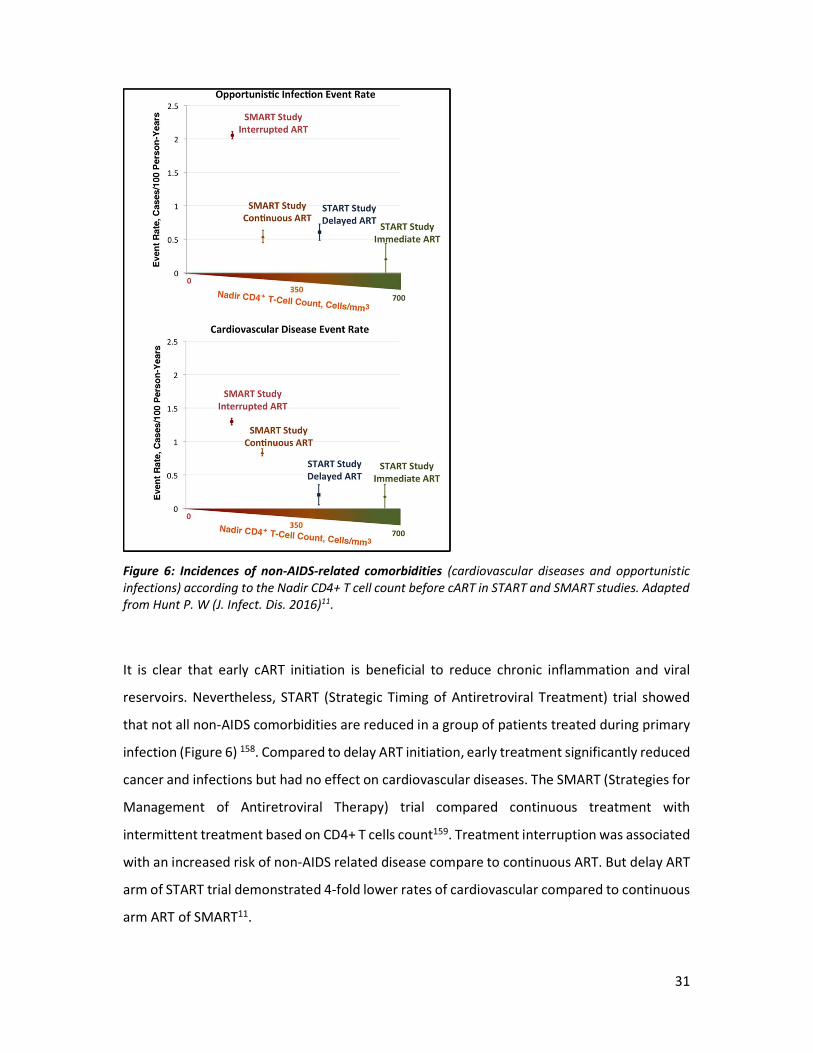
Figure 6: Incidences of non-AIDS-related comorbidities (cardiovascular diseases and opportunistic infections) according to the Nadir CD4+ T cell count before cART in START and SMART studies. Adapted from Hunt P. W (J. Infect. Dis. 2016)11.
It is clear that early cART initiation is beneficial to reduce chronic inflammation and viral
reservoirs. Nevertheless, START (Strategic Timing of Antiretroviral Treatment) trial showed
that not all non-AIDS comorbidities are reduced in a group of patients treated during primary
infection (Figure 6) 158. Compared to delay ART initiation, early treatment significantly reduced
cancer and infections but had no effect on cardiovascular diseases. The SMART (Strategies for
Management of Antiretroviral Therapy) trial compared continuous treatment with
intermittent treatment based on CD4+ T cells count159. Treatment interruption was associated
with an increased risk of non-AIDS related disease compare to continuous ART. But delay ART
arm of START trial demonstrated 4-fold lower rates of cardiovascular compared to continuous
arm ART of SMART11.
32
One explanation is maybe the follow-up duration of patients, which had not yet developed
these comorbidities. Also, there was differences in term of nadir CD4+ T cells counts between
studies. Thus, early cART initiation is not sufficient to reduce non-infectious comorbidities, and
systemic inflammatory threshold acquired in first weeks of infection may determine long term
organ diseases.
33
F. Resolution of chronic inflammation by natural hosts
HIV-1 came from zoonotic transmission occurred in the beginning of twentieth century from
chimpanzee infected with SIVcpz. Chimpanzees were thought to be resistant to SIVcpz, but
natural infection leads to immunodeficiency in this specie, as observed in human160. In fact,
SIVcpz was not found in all four subspecies of chimpanzees, suggesting that SIV acquisition was
a recent event, occurring before subspecies divergence161. SIVs have been detected in most
of African primates from genus of Ceropithecus, African green monkeys (AGM, Chlorocebus),
mandrills and drills (Mandrillus), the mangabeys (Cercocebus), which are natural host of their
respective SIV162. For natural hosts, homologous SIV isolate lacks of virulence and the infection
rarely progress to AIDS162. Nevertheless, they are still virulent for other species and SIVsmm is
an interesting example, since it gave rise to human HIV-2 and Asian macaques SIVmac163,164.
Depending on the strain, SIV infection of Asian macaques leads to high viral load, development
of progressive CD4+ T cell depletion and opportunistic infection165. Comparison of pathogenic
with nonpathogenic primate models had permit to better understand mechanisms of
protection, acquired thanks to thousands years of virus-host co-evolution166. Sooty
mangabeys (SMs) and African green monkeys (AGMs) were intensively studied to understand
mechanisms of nonpathogenic SIV infection, so I will mainly focus on these models.
As pathogenic infection in human and Asian macaques, natural hosts demonstrate a high
viremia in primary infection and chronic phase, and depletion of CD4+ T cells occurring only in
primary infection in which immune activation level is high167,168. Interestingly, AIDS resistance
mechanisms of natural hosts do not rely on immune suppression of viral replication, meaning
that host evolution selected another way for protection. First, they keep healthy levels of
peripheral CD4+ T cells on the long term particularly by preserving central memory CD4+ T
cells (TCM)169, T cell regeneration170, and lymph node architecture and function171–173. Second,
mucosal immunity is long term preserved with normal ratio between Th17 and Treg in GALT,
explaining the absence of microbial translocation in AGM and SM174,175. Lastly, despite ongoing
viral replication there is a lack of chronic immune activation173,176,177.
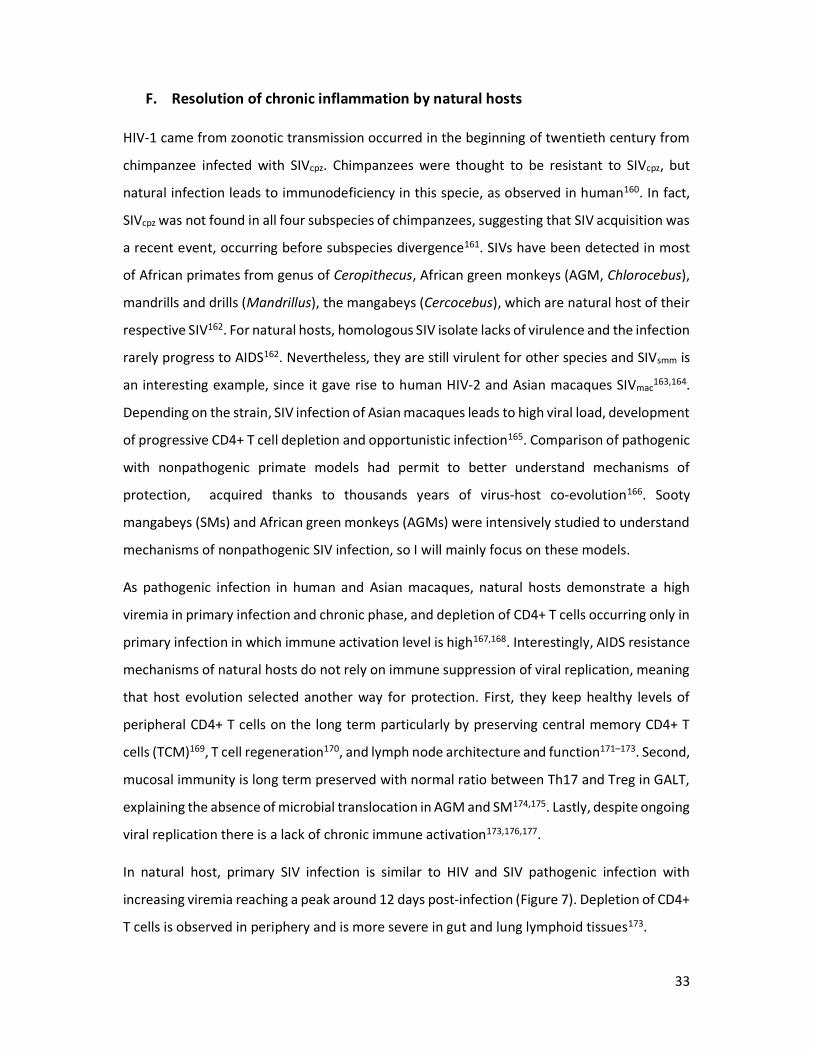
In natural host, primary SIV infection is similar to HIV and SIV pathogenic infection with
increasing viremia reaching a peak around 12 days post-infection (Figure 7). Depletion of CD4+
T cells is observed in periphery and is more severe in gut and lung lymphoid tissues173.
34
Figure 7: Mechanisms involve in control of chronic inflammation by natural hosts. A-Percentage of survival of SIVsmm-infected AGMs and SIVmac251-infected RMs of Indian and Chinese origins. Despite persistent viral replication, AGM did not develop AID-related symptoms. B-In situ hybridization of SIV RNA in LN from RMs or AGMs. In the peak of replication, AGMs and RMs demonstrated similar SIV RNA+ cells in T-cell zone, but at day 60 viral replication in LN was extremely low in AGMs, despite similar levels of plasma viral load. C- S. E. Bosinger et al. model of immunomodulation during SIV infection in natural and pathogenic hosts. Despite persistent viral replication, natural host demonstrated reduce ISG expression, T-cell proliferation and elevated immunoregulatory genes expression protecting from CD4+ T cells depletion and AIDS. Adapted from Cumont M.-C. et al (J. Virol. 2008) and Bosinger S. E. et al. (J. Clin. Invest. 2009)173,178. D-Mechanisms of control of chronic inflammation and consequences for the physiopathology of SIV infection in natural hosts. Adapted from Chahroudi A. et al. (Science 2012) and Ploquin M. J. et al. (Current Opinion in HIV and AIDS 2016)12,179.
35
Cellular immune response, especially CD8+ T cells, allows a control of the viremia which is
maintain during chronic phase180. In fact, strong innate and adaptive immune response are
initiated in primary infection, mostly characterized by type I interferon signatures, T cells
proliferation and pro-inflammatory cytokines178. Despite ongoing viral replication mucosal
CD4+ T cells stabilize or recover in chronic phase. One explanation is the resolution of
inflammation observed from 4 to 8 weeks post-infection, which protects the natural host
against all immune mediated pathologies, such as T cell apoptosis, lymph node fibrosis and
impaired immune functions181. Four mechanisms have been described to explain the control
of chronic inflammation: reduced microbial translocation, reduce ISG signature, down-
regulation of CD3-T cell receptor, reduction of immune response against the virus and low
level of replication in lymphoid tissues (Figure 7).
In natural host reduction of chronic inflammation could be explain by protection of mucosal
immune response, avoiding microbial translocation observed in pathogenic infection. Even if
there is a significant depletion of mucosal CD4+ T cells in acute infection77, Th17 cells are
protected and intestinal epithelial integrity is preserved in natural host84. In acute SIV infection
both natural and pathogenic host demonstrated elevated ISG signatures182. However, in
natural hosts (AGM and SM) ISG expression levels return to baseline 30 days post-infection,
showing a resolution of acute inflammation. By limiting type I interferon, AGM and SM may
reduce detrimental chronic immune activation. Another factor reducing immune activation
relies on the capacity of Nef protein from SIVsmm and SIVagm to down-regulate CD3-TCR
expression on T cell surface181. The hypothesis is that reduction CD3-TCR expression may
reduce further T cells activations. But Nef protein from SIVmac pathogenic infection is also
able to down-regulate CD3-TCR, so this mechanism alone is not sufficient to explain the
control of immune activation. AGM and SM have a reduced immune response to SIV infection,
contributing also to limited inflammation and immune activation. Limiting viral replication in
LN is another protective mechanism of natural host. The ability of NK cells to infiltrate
germinal centers in primary infection, thanks to IL-15 expression by FDC, is a good evidence
for the effective innate immune response183. Thus, killing CD4+ T follicular helper cells infected
by SIV reduce viral reservoir, participating to control of immune activation. The implication of
neutrophils in the resolution of inflammation in natural hosts has not been studied yet.
36
Myeloid cells are important effectors of chronic inflammation and are involved in all
mechanisms at its origin. Neutrophils are of particular interest in chronic inflammatory
diseases since they are the largest circulating leukocyte population, equipped with deadly
weapons and arriving first to injury site. Our understanding of neutrophils has been restricted
for a long time by their fragility and dogma defining them as expendable phagocytic cells. As
we are going to see, abundant recent studies demonstrate plasticity and several new functions
carried by neutrophils.
37
III. Neutrophils plasticity in response to inflammation
For long, neutrophil function in the immune response was restricted to defense against
invading pathogens using efficient killing weapons. Indeed, in case of neutropenia or
leukocyte adhesion deficiency syndrome, patients are more prone to bacterial and fungal
infections. Recently, this dogma has been challenged by the demonstration that neutrophils
lifespan in circulation reaches 5.4 days184. Neutrophils live enough to respond to their
environment thus creating phenotypical heterogeneity depending on the disease. Soon this
phenotypic diversity was associated with unexpected function of neutrophils, modulating
adaptive immune response and promoting resolution of inflammation. In the first part, we
will present the life cycle of neutrophils from bone marrow production to controlled
apoptosis, while passing by physiological functions. But in unresolved inflammation,
neutrophils homeostasis is disrupted. As we are going to see in the second part, neutrophil
has a pathogenic role by enhancing inflammation and leading to tissue damages.
A. Neutrophil life from granulopoiesis to efferocytosis
1) Feedback loop of neutrophils production
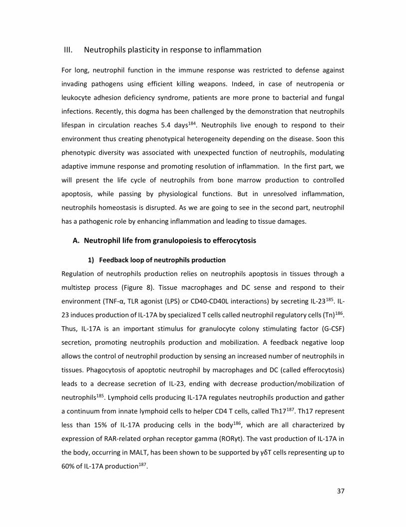
Regulation of neutrophils production relies on neutrophils apoptosis in tissues through a
multistep process (Figure 8). Tissue macrophages and DC sense and respond to their
environment (TNF-α, TLR agonist (LPS) or CD40-CD40L interactions) by secreting IL-23185. IL-
23 induces production of IL-17A by specialized T cells called neutrophil regulatory cells (Tn)186.
Thus, IL-17A is an important stimulus for granulocyte colony stimulating factor (G-CSF)
secretion, promoting neutrophils production and mobilization. A feedback negative loop
allows the control of neutrophil production by sensing an increased number of neutrophils in
tissues. Phagocytosis of apoptotic neutrophil by macrophages and DC (called efferocytosis)
leads to a decrease secretion of IL-23, ending with decrease production/mobilization of
neutrophils185. Lymphoid cells producing IL-17A regulates neutrophils production and gather
a continuum from innate lymphoid cells to helper CD4 T cells, called Th17187. Th17 represent
less than 15% of IL-17A producing cells in the body186, which are all characterized by
expression of RAR-related orphan receptor gamma (RORγt). The vast production of IL-17A in
the body, occurring in MALT, has been shown to be supported by γδT cells representing up to
60% of IL-17A production187.
38
Figure 8: Regulation of neutrophils production and circulation. Neutrophils are produced in endosteal niches within bone mainly under G-CSF signaling. In steady state, G-CSF, TLR signaling, CXCR2 agonists, and SNS mediate mature neutrophils release from bone marrow pool. Neutrophils blood mobilization and tissue migration follows circadian oscillation mediated by CXCL12/CXCR4 expression, under control of SNS. Circulating aged-neutrophils migrates back to bone marrow and tissue neutrophils are both clear by mononuclear phagocyte system. IL-17A targets numerous cells, such as endothelial cells, inducing G-CSF secretion. Neutrophils phagocytosis negatively regulates IL-23/IL-17A/G-CSF axis, which decrease neutrophils production and mobilization. In case of inflammation, macrophages or DC activate IL-23/IL-17A/G-CSF axis, which, along with TLR agonist and pro-inflammatory chemokines, increase neutrophils production and mobilization. MP: macrophage, DC: dendritic cell, Tn: neutrophil regulatory cells, EndoT: endothelial cell.
39
IL-17A maintains mucosal barrier integrity through enhancing tight junction synthesis and by
inducing microbicidal agent synthesis187. IL-17A also induces G-CSF secretion by epithelial
cells, bone marrow macrophages, endothelial cells, fibroblasts and mesothelial cells. G-CSF
binding to its receptor on neutrophils precursors induces proliferation, differentiation and
activation in bone marrow188. Nevertheless, in steady state other factors participate to
neutrophils production since in G-CSF deficient mice residual granulopoeisis was observed189.
Cell retention in the bone marrow relies on the chemokine receptor CXCR4, express by both
stem cells and neutrophils190. CXCR4 binds CXCL12 (SDF1, stromal derived factor 1) which is
expressed on bone marrow stromal cells: vascular endothelial cells, mesenchymal stem cells,
fibroblasts, adipocytes and osteoblasts191. Neutrophils form a pool of cells maintained by
CXCR4, which could be released on demand by CXCR2, G-CSFR or TLR signaling192. G-CSF
induces neutrophils mobilization to circulation by reducing CXCL12 expression on endothelial
cells, which reduce adherence and promote signaling by CXCR2.
2) Differentiation and maturation of neutrophils
Production of myeloid cells within bone marrow represent two-thirds of hematopoietic
activity, with a major part devoted to neutrophils and each day in a healthy adult, around 1 to
2 x10¹¹ neutrophils are produced193. Granulopoiesis starts from hematopoietic stem cells
(HSCs) which are located close to osteoblasts, forming a niche along the endosteal border
where blood flow and oxygen level are reduced194. A variety of osteoblast adherence factors
are expressed to maintains HSCs quiescent in endosteal niche195. Commitment and
differentiation of HSCs to granulocytes relies on HSC homing to vascular niches in vascular
sinuses of central bone marrow. Expression and secretion of CXCL12 by endothelial cells
attract HSC to vascular niches where they are bound to vascular niche via P and E-selectin196.
This environment facilitates differentiation to CD34+ multipotent progenitors (MPs).
Subsequent to this, expression of PU.1 transcription factor commit MMPs to common myeloid
progenitors (CMPs) potent to give rise to myeloid cells, megakaryocyte and erythroid
lineage197. Transition from CMPs to granulocyte/monocyte progenitors (GMPs) depends on
co-expression of PU.1 and C/EBP-α transcription factors. Further commitment to neutrophil
precursors is based on decrease PU.1 expression along with maintenance of C/EBP-α
expression.
40
Stimulation by IL-3, GM-CSF and IL-6 allow differentiation from CMPs to GMPs. G-CSF drives
commitment of GMPs to neutrophils production by proliferation and maturation of
myeloblast to segmented neutrophil.
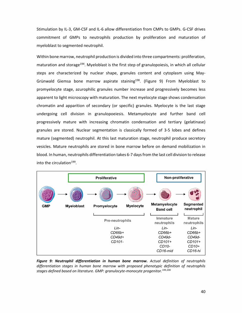
Within bone marrow, neutrophil production is divided into three compartments: proliferation,
maturation and storage194. Myeloblast is the first step of granulopoiesis, in which all cellular
steps are characterized by nuclear shape, granules content and cytoplasm using May-
Grünwald Giemsa bone marrow aspirate staining198. (Figure 9) From Myeloblast to
promyelocyte stage, azurophilic granules number increase and progressively becomes less
apparent to light microscopy with maturation. The next myelocyte stage shows condensation
chromatin and apparition of secondary (or specific) granules. Myelocyte is the last stage
undergoing cell division in granulopoeiesis. Metamyelocyte and further band cell
progressively mature with increasing chromatin condensation and tertiary (gelatinase)
granules are stored. Nuclear segmentation is classically formed of 3-5 lobes and defines
mature (segmented) neutrophil. At this last maturation stage, neutrophil produce secretory
vesicles. Mature neutrophils are stored in bone marrow before on demand mobilization in
blood. In human, neutrophils differentiation takes 6-7 days from the last cell division to release
into the circulation199.
Figure 9: Neutrophil differentiation in human bone marrow. Actual definition of neutrophils differentiation stages in human bone marrow with proposed phenotypic definition of neutrophils stages defined based on literature. GMP: granulocyte-monocyte progenitor.199,200
41
3) Circadian cycle orchestrates neutrophils circulation and tissue migration
Several immune parameters have a circadian dependent variation, and neutrophil blood count
is one of those201,202. Beside mechanisms I presented before, sympathetic nervous system
(SNS) plays a central role in coordinating immune cells oscillation observed within 24 hours201.
Briefly, light is the principal synchronizing stimulus captured by retina and is transmitted
through retinohypothalamic tract to hypothalamus, in which suprachiasmatic nuclei form the
central clock. SNS innervates most of organs, especially lymphoid organs, where locally it
synchronizes immune cells using noradrenaline203. Through hypothalamic-pituitary-adrenal
axis, SNS has also a systemic synchronization of immune response by secretion of
glucocorticoids and catecholamines by adrenal glands. Oscillation of circulating neutrophils
are based on activity phase which is diurnal for human. At night during resting phase,
neutrophils peak because of a release of mature cells from bone marrow. SNS innervation
induces a rhythmic decrease of CXCL12 expression on endothelial cells at night204. During
active phase, circulating neutrophils decrease because of a reduction of bone marrow
liberation and also by migration to tissue (Figure 8). Sympathetic nerves modulate expression
of adhesion molecules and chemokines on endothelial cells, guiding neutrophils to tissues205.
Cathecholamines are known to increase circulating neutrophils, predominantly released from
marginated pool present in capillaries of lung, liver, spleen and bone marrow203. Marginated
pools have been estimated to represent 51% of all blood granulocytes, representing a large
inaccessible neutrophil compartment by blood drawn206. Maturation and activation state of
neutrophils seem to influence their body distribution. Studies performed in mouse showed an
increase expression of CXCR4 before neutrophil apoptosis. It is proposed that increase CXCR4
expression leads neutrophils to bone marrow where macrophages phagocytosis takes place.
A recent study using plerixafor (CXCR4 agonist) demonstrated in both mice and non-human
primate models the existence of a large marginated pool of neutrophils in lungs207. Lung
endothelial cells express CXCL12 and trap aged neutrophils.
To enter the tissue, neutrophils interact with endothelial cells at post-capillary venules.
Classically, leukocytes recruitment is divided in multiple steps: tethering, rolling, adhesion,
crawling and transmigration. Endothelial cells determine the attachment of neutrophil by
expression of E and P-selectin, in response to TNF-α, IL-1β and IL-17 or by direct pathogen
recognition by pattern-recognition receptors (PRR)208,209. Neutrophils constrictively express
42
PSGL-1 and L-selectin (CD62L) allowing interaction with E and P-selection on endothelial
surface. CD44 and PSGL-1 interaction slower rolling and mediate PLC-γ and PI3K activation
leading to neutrophil cytoskeletal rearrangement. Firm adhesion to endothelium is mediated
by CD18 β integrin on neutrophils. Transmigration could be paracellular (between endothelial
cells) or transcellular depending on endothelium-neutrophils interactions. Tissue migration
increase neutrophil transcriptional activity, NADPH oxidase activity and susceptibility to
degranulation190,208,209. This state of partial activation, termed priming, prepares neutrophil to
respond rapidly to invaded pathogen present in tissue.
4) Anti-microbial function of neutrophils
During acute inflammation neutrophils accumulates in injury sites thanks to diapedesis, and
following chemokine gradients. Environment along with endothelium interaction prime and
activate neutrophils making them more potent to respond to infection. Human neutrophils
express most of Toll-like receptors (TLR), with the exception of TLR3 and TLR7, leading to pro-
inflammatory cytokines secretion210. Depending on the environment, neutrophils have been
shown to produce multiples molecules (anti-/pro-inflammatory cytokines, chemokines, TNF
superfamily and angiogenic/fibrogenic factors) thus modulating immune response211.
Phagocytosis is the most recognized antimicrobial function of neutrophils. Neutrophils are
consider to be “professional phagocytes” since they can engulfed up to 10-12 particles212.
Detection of pathogens is performed by receptors detecting microbial polysaccharides
(Dectin-1) or by sensing opsonized particles with Fc-receptors (CD89, CD16, CD32, CD64) or
complement receptor (eg. CD35). Activation of these receptors induce membrane
invagination forming cytoplasmic vesicle called phagosome212. Fusion with granules later form
phagolysosome, a deadly compartment full of proteolytic enzymes and antimicrobial
molecules. The NADPH oxidase assembly and activation are essential in microbial killing by
producing ROS within phagolysosome. Briefly, NADPH oxidase is a tertiary structure composed
of cytoplasmic p47phox ,p67phox p40phox and membranous NOX2, p22phox , acting together as
an electron transferase from NADPH to oxygen (O2), generating superoxide anion (O2-) within
the phagolysosome213. Granules proteins convert O2- to hydrogen peroxide (superoxide
dismutase) then metabolize into hypochlorous acid (myeloperoxidase). Patient with chronic
granulomatous disease, characterized by a defective mutation on NADPH subunit, have
43
neutrophils unable to generate ROS and to kill bacteria. So, ROS production is a central
mechanism in pathogen killing. Granules produced during granulopoesis are subdivided in 4
types: primary (or azurophil) granule, secondary (specific) granule, tertiary (gelatinase)
granule and secretory granules. Granules are released depending on the intensity and the type
of stimulus in a hierarchical order, called graded exocytosis214. Secretory granules are formed
by endocytosis during nucleus segmentation in bone marrow and are the first mobilized to
the plasma membrane. Secretory granules contains NOX2/p22phox NAPDH oxidase making
neutrophils more prone to produce ROS, activation state called priming213. They contain also
numerous membrane receptors (eg. CD14, CD16, CD65) and adhesion molecules (eg.
CD11b/CD18, CD67) allowing neutrophils to better sense pathogens212. Tertiary granules are
the second most easily released granules, containing matrix degrading enzymes (eg. arginase
1, gelatinase) and adhesion molecules involved in diapedesis. Gelatinase are also potent to
destroy tissue during neutrophil response. Specific granules are full of antibacterial protein
such as lactoferin, haptoglobin, lysozyme, which target different essential pathways for
microbial survival214. Matrix metalloproteinase are also overrepresented in specific granules
and act on structural components of pathogens, leading to bacterial killing. Azurophil granules
have a limited exocytosis and contribute mostly to phagolysosome formation. They contain
metalloproteinases, forming hypochlorous acid, matrix metalloproteinase and serine
proteases (eg. elastase, cathepsine G) which degrade a variety of extracellular matrix212.
Antimicrobial peptides are also found in azurophilic granules, such as α-defensin, active on
bacteria, fungus and some enveloped viruses.
First described as a new microbicidal mechanism capable to capture an kill microorganisms,
neutrophil extracellular traps (NETs) were formed by chromatin extrusion decorated with
granules protein by neutrophils215. After 2h hours of incubation with PMA, authors showed
liberation of NETs associated with necrotic cell death of neutrophils, a mechanism called
NETosis216. Further research using PMA stimulation showed that NETs are formed under
NADPH oxidase activation cascade, leading to chromatin decondensation by conversion of
arginine to citrulline on histones217. Azurophilic granules are then translocated to nucleus,
associating neutrophil elastase and myeloperoxidase with chromatin, further release into
extracellular space. Nevertheless, the use of a non-physiologic stimuli (PMA) raised questions
about the mechanism of NET formation in vivo and NETosis cell death process218–221.
44
Observation of NET liberation without cell death, using GM-CSF primed neutrophils further
stimulated by TLR4 agonists or C5a, suggested that NET production and NETosis necrotic cell
death were independent mechanisms221. In addition, NET were formed by mitochondrial DNA
rather than nuclear DNA, in a mechanism dependent on ROS activation and glycolytic ATP
production allowing cytoskeletal rearrangement220. Liberation of mitochondrial DNA may also
play a role as a danger associated molecular pattern, potentially modulating inflammation221.
Besides, killing capacities of NETs have been also questioned. NETs are capable to trap
microorganism, limiting dissemination but live organism were still recover from NETs219. The
importance of NETs as an antimicrobial mechanism was also challenged by study of Papillon-
Lefèvre syndrome (PLS), involving a lack of elastase in neutrophils, an enzyme required for
NET production222. Neutrophil from PLS patient were potent to kill K. pneumonia without
forming NETs and PLS patient did not shown elevated frequency of infections. NETs formation
is of particular interest in auto-immune diseases, in which they may provide a source of auto-
antigens218.
Neutrophils contribute also to resolution of inflammation by switching their pro-inflammatory
cytokines secretion to lipoxine and resolvines which inhibit transendothelial migration and
chemotaxis. They also produce anti-inflammatory IL1-ra and TGF-β. Neutrophils elimination
by efferocytosis also generate anti-inflammatory M2 macrophages, producing IL-10 and TGF-
β223.
5) Neutrophil compartmentalization and death to control inflammation
Classical model of neutrophil circulation in the body postulated that tissue was the final
destination. This has been challenged by number of studies showing the ability of neutrophils
to go back to circulation after migration to tissue in zebra fish and in mouse models224,225. In
this phenomenon, called reverse transmigration, neutrophils have an increase life time, are
more potent to produce ROS but cannot transmigrate again to tissue. Intravital microscopy on
a ischemia reperfusion model showed the importance of JAM-C (endothelial cell junction
molecule) degradation by neutrophils under Leukotriene B4 activation225. These reverse-
transmigrated neutrophils harbor an activated profile potentially involved in systemic
inflammation. Neutrophils are also able to emigrate from tissue using lymphatic vessels.
Indeed in several bacterial infections, pathologists have identified neutrophil infiltration in
45
draining lymph nodes226. In mouse models neutrophils have been shown to infiltrate lymph
nodes using both blood and lymph and expressed a specific activated phenotype (CD11bhigh
CD62Llow CXCR2low)227. In the same study, they demonstrated that trafficking to LN only
occurred under inflammation and neutrophils stimulation by immune complexes. In lymph
nodes, neutrophils can shape immune responses by promoting T cells proliferation and
undergoing antigen presentation227.
Since neutrophils are involved in tissue damage by releasing enzymes and pro-inflammatory
components, clearance of apoptotic neutrophils is essential. Phagocytosis of apoptotic
neutrophils by tissue macrophages, termed efferocytosis, participates to resolution of
inflammation and wound healing. Macrophages undergoing efferocytosis produce anti-
inflammatory mediators TGF-β and PGE2 . Efferocytosis has been demonstrated in vivo to
occur in lung, spleen, liver and bone marrow228–231. Bone marrow and spleen are important
compartments performing efferocytosis in steady state228,232. Aged neutrophils expressing
CXCR4 and SDP-1α home back to bone marrow to be phagocytosed by macrophages229. Under
inflammatory conditions, liver has a major role in neutrophil clearance as shown in a
endotoxemia model and patients with inflammatory diseases230,232. In that study, gadolinium
chloride blockade of Kupffer cells induced a neutrophilia and a relocation of neutrophils in
lung and spleen. Liver is an important organ in elimination of blood-borne bacteria coming
from the gut. Kupffer cells were shown to phagocyte neutrophils which have engulfed
bacteria, leading to resolution of inflammation in a listeriosis model233. As explain above,
efferocytosis leads to a negative feed-back loop, which reduces neutrophil production by
reducing IL-23 production by macrophages.
46
B. Heterogeneity and function of neutrophils in chronic inflammation
In acute inflammation, neutrophils production and functions are adapted to both respond to
the invader and resolve inflammation to avoid tissue damages. Remodeling of stromal cells
and endosteal niches under inflammation modulate myelopoiesis, thus producing neutrophils
heterogeneity234,235. In numerous chronic diseases, such as atherosclerosis, myocardial
infarction or neurodegenerative disorders, neutrophils have been involved in tissue damages
and maintenance of inflammation199. Neutrophils heterogeneity in cancers, autoimmune
diseases and infectious diseases have been intensively studied in recent years, which are
inspiring to study neutrophils in HIV infection. In the first part, we will present the impact of
neutrophils on chronic inflammation observed in autoimmune diseases. Neutrophils
heterogeneity has been demonstrated in autoimmune diseases, in which NETosis has a central
role in physiopathology236. In the second part, we will present neutrophil heterogeneity in
cancer in which MDSC have been demonstrated to suppress T cell responses since 1984237.
The third part is dedicated to neutrophils and tuberculosis. Tuberculosis is a particular chronic
infection in which recent studies have demonstrated a role of neutrophils in immune
regulation as well as granuloma reaction238. Neutrophils phenotypes and functions are less
understood in chronic viral infections. Trying to make a parallel with HIV infection, in the last
part we will present the current knowledge on neutrophils heterogeneity in chronic viral
infections.
1) Auto-immune diseases
Autoimmune disease are multifactorial diseases characterized by a loss of tolerance against
autoantigens. Environmental factors are suspected to trigger autoimmune diseases in
genetically predisposed individuals, leading to abnormal innate and adaptive immune
responses. Neutrophils have been involved in various aspects of autoimmune diseases,
participating to chronic inflammation and tissue lesions. Rheumatoid arthritis (RA) is a
destructive joint inflammation characterized by synovitis, systemic inflammation and
presence of autoantibodies, such as rheumatoid factor (IgA/IgM against IgG) and
anticitrullinated protein antibodies (ACPA)239. Pannus represents the active lesion in RA, a
synovial invasive tissue formed by activated fibroblasts, lymphocytes, macrophages and
neutrophils240. TNF is a central cytokine in RA driving systemic inflammation. As consequence,
sustained exposure to TNF leads to abnormal neutrophil functions in RA, inducing priming and
47
genes transcription241. Circulating neutrophils from RA patient showed a higher capacity to
produce ROS, compare to healthy individuals242. Within joints, neutrophil degranulation and
ROS production are mediated by immune complexes, targeting articular cartilage243. In RA
synovial fluid, neutrophils survive for several days because of hypoxic environment and anti-
apoptotic cytokines (TNF, GM-CSF and IL-8)244,245. Neutrophils amplify lesions and
inflammation by releasing numerous pro-inflammatory mediators, comprising RANK-L and
BAFF , which activate respectively osteoclasts and B lymphocytes240. Neutrophils have been
shown to release NET in RA, with blood and synovial fluid neutrophils more prone to release
NETs. Interestingly, NETs contain citrullinated histones H4 which are consider as auto-
antigenes218. Neutrophils sustains ACPA production and may initiate production of new auto-
antibodies241. Anti-TNF therapy is efficient to reduce disease activity and inflammation, and
TNF inhibitors were shown to decrease neutrophils activation notably by downregulating NF-
Kb pathways246.
Association between neutrophils and systemic lupus erythematous (SLE) has been made in
1986 with the first description of low-density neutrophils (LDN) in peripheral blood
mononuclear cells (PBMC), obtained after density gradient centrifugation246. In SLE, B and T
cells responses have a prominent role with production of immune complexes and
autoantibodies against nuclear antigens247. The role of neutrophils in SLE have been suggested
since patients had abnormal interferon and neutrophil signatures in blood248. LDN represent
a pathogenic subset of neutrophils producing pro-inflammatory cytokines, with enhanced
capacity to form NETs and activate endothelial cells249. Neutrophils participate to tissue
damages and were observed to infiltrate and to form NETs in skin and kidneys249. The release
of NET participates actively to type I interferon production by stimulating pDC, and amplifies
inflammation in tissues250. As in RA, LDN maintain inflammation and auto-antibody production
through NETs which are composed of potent nuclear auto-antigens.
Anti-neutrophil cytoplasmic antibody (ANCA)-associated vasculitis (AAV) is characterized by
vascular inflammation with or without granulomatosis251. AAV have polymorphic
manifestations, affecting vessels from kidney, skin, respiratory tract and peripheral nerves.
The presence of MPO and PR3 auto-antibodies have been identified in vasculitis patients, and
are important to the diagnosis of these diseases. These auto-antibodies underline the role of
neutrophils which are source of auto-antibodies and induce endothelial cells dammage249,252.
48
These auto-antibodies were shown to activate neutrophils previously primed by inflammatory
stimuli such as TNF or G-CSF253. NETs formation have been involved in pathologic mechanism
of ANCA vasculitis, with neutrophils more prone to release NETs249. Also, sera from patients
contained circulating MPO-DNA and calprotectin, which were consider as markers of NETs
production254,255. In renal biopsy obtained from patients demonstrating necrotizing
glomerulonephritis, DNA and granules proteins are located in inflammatory foci or around
necrotizing area, suggesting a relationship between NETs and tissues damages256.
Nevertheless, most of studies identified NETs using DNA in combination granule proteins,
which could be also the results of cell necrosis combined with neutrophils degranulation221.
MPO and PR3 are key proteins with pro-inflammatory properties218. Disease activity is
positively correlated with serum PR3 and neutrophils count in patients257. In association with
ANCA neutrophils induce vascular damages and granuloma, perpetuating inflammation and
auto-immunity.
2) Cancers
Cancer are chronic inflammatory disease among which neutrophils diversity has been widely
studied. Inflammatory environment, partially driven by cancer cells, promotes tumor
initiation, growth, angiogenesis and metastasis258. Neutrophils were shown to favor all these
steps of tumorigenesis, but also demonstrated antitumoral function. Depending on the stage
the tumor and the compartment analyzed, different subsets of neutrophils have been
identified according to their function, phenotype and sedimentation after density gradient258.
Ligands for CXCR2 were observed early in human tumor development, leading to neutrophils
recruitment. In early stage of lung cancer, tumor-associated neutrophils (TAN) were activated
and express different chemokine receptors (CXCR3, CXCR4 and CCR7) compared to peripheral
neutrophils259. TAN produced proinflammatory factors (IL-8, TNF- and IL-6) and were able to
stimulate T cell proliferation and IFN- production. Referred as N1, these TAN have a
hypersegmented nucleus with a more mature phenotype. But in advanced carcinoma or in
head and neck cancers, elevated levels of TAN increased the risk of recurrence or death by
66%260. From early stage antitumoral to late stage pro-tumorigenic TAN, there is a switch of
functions apparently induced by tumor environment261. TAN promoting tumor growth, called
N2, displayed an immature phenotype characterized by increase arginase activity.
Demonstrated in a breast cancer mouse model, tumor cells activate IL-17/G-CSF axis using T
49
cells262. This mechanism increases TAN infiltration, promoting tumor progression and
metastasis. Interestingly, polarization to pro-tumorigenic N2 relies on TGF- anti-
inflammatory cytokine present in tumor environment263. One mechanism by which TAN
participate to tumor growth and metastasis is by their angiogenic capacities, normally
observed in resolution of inflammation and wound healing264. MMP9 secreted by TAN
degrade extracellular matrix and thereby releases VEGF and FGF2265.
Another populations described in cancer are myeloid derived suppressor cells (MDSCs), having
phenotype related to monocytes (M-MDSC) or granulocytes (PMN-MDSC) and capable to
suppress T cell responses266. There is still not a clear phenotypical definition of MDSC and true
definition relies on demonstration of suppressive activity. A transcriptomic analysis of naïve
neutrophil, TAN and PMN-MDSC demonstrate these 3 populations were different, but naïve
neutrophil being more closely related to PMN-MDSC267. TAN had downregulated genes
associated with cell-cytotoxicity compared to other populations, whereas PMN-MDSC had
increased genes expression of MHC class II and cytokines. In tumor and peripheral blood from
cancer patients, LDN are also heterogenous neutrophil populations overlapping with PMN-
MDSC and TAN, with immunosuppressive capacities268,269. LDN contain both immature and
mature cells but with different origins. Like PMN-MDSC, mature LDN seems to originate from
normal density neutrophils, acquiring suppressive function after TGF- stimulation269 and
activation-induced degranulation270. Immature LDN are also suspected to originate from an
incomplete maturation within bone marrow268. Globally tumor environment and chronic
inflammation lead to bone marrow and tumor neutrophil reprogramming, protecting from
efficient immune response and promoting development of tumor.
3) Tuberculosis
Infection by Mycobacterium tuberculosis (Mtb) is a leading cause of death worldwide, with 10
million people declaring an active tuberculosis in 2017271. Despite tuberculosis is a bacterial
infection, most of studies on innate immune response have been focused on macrophages,
NK cells and DC. Neutrophil has a dual role in tuberculosis with a protective or detrimental
role, depending on the stage of infection238. Since fundamental Koch’s work in 19th century
on guinea pig, several animal models have been used to study tuberculosis with different
inoculation routes, strains and disease evolution, conducting to misleading interpretation of
the role of neutrophils in physiopathology238. Of note mouse model fails to reproduce latent

50
infection and show different pathology compare to human. In fact, neutrophils seem to have
a differential role depending on the stage of the infection. Neutrophils are first responders,
along with alveolar macrophages, accumulating within 1st day to 1st week in lungs following
intranasal or intratracheal inoculation. Elimination of Mtb or induction of latent infection
relies on early immune response and clearance by phagocytes. Neutrophil priming seems to
influence their capacity to phagocyte and kill bacteria. Repeated Mtb challenges lead to
increase infiltrating neutrophils thank to IL-17 producing T cells. Capacity to kill Mtb has been
shown to be variable among human, thus explaining elimination capacities in early infection
by some individuals272. Resistant individuals may have circulating primed neutrophils, more
prone to rapidly respond to mycobacterial infection. If neutrophils fail to kill Mtb they may
undergo apoptosis followed by macrophages phagocytosis, necrosis or migrate to draining
lymph nodes where they disseminate bacilli. Neutrophils also modulate immune response
against Mtb notably by secretion of IL-12, IP-10, MIP-1α and MCP-1, attracting and polarizing
T cells. Neutrophils also promote T cell proliferation and IFN-, IL-5, IL-6 and IL-10 through
HNPs secretion. Neutrophil-DC interactions have been suspected since neutrophils increased
DC maturation and further T cell activation. In some circumstances, neutrophils were shown
to present antigen, especially to memory T cells requiring a lower activating signal than naïve
T cells273. This antigen presenting function was suspected in tuberculosis, with the
identification of neutrophils expressing MHC class II and CD83 and CD86 in patients’ pleura
effusions274. These neutrophils expressed also more TLR2 and CD64 but were more prone to
apoptosis compare to circulating neutrophils, making questionable their antigen presenting
capacity, which remain to be demonstrated.
Compared to uninfected and latently infected patients, active tuberculosis was characterized
by distinct transcriptional signature involving interferon-inducible genes expressed by
neutrophils110. This neutrophil-driven signature was correlated with lung radiographic
severity, making neutrophils associated with tissue damages. Indeed, neutrophils infiltration
and neutrophilia are associated with poor prognosis and correlated with serum pro-
inflammatory markers111,112. Less is known about neutrophils heterogeneity tuberculosis, but
some authors suggested that epigenetic reprogramming of neutrophils may explained
detrimental neutrophil functions, whereas other suggest that neutrophilia only reflect failure
of Th1 immunity to control Mtb238,275.
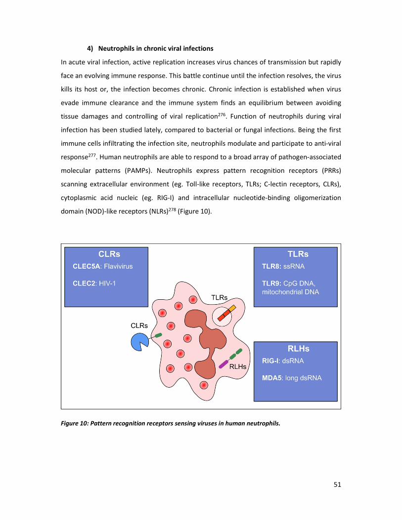
51
4) Neutrophils in chronic viral infections
In acute viral infection, active replication increases virus chances of transmission but rapidly
face an evolving immune response. This battle continue until the infection resolves, the virus
kills its host or, the infection becomes chronic. Chronic infection is established when virus
evade immune clearance and the immune system finds an equilibrium between avoiding
tissue damages and controlling of viral replication276. Function of neutrophils during viral
infection has been studied lately, compared to bacterial or fungal infections. Being the first
immune cells infiltrating the infection site, neutrophils modulate and participate to anti-viral
response277. Human neutrophils are able to respond to a broad array of pathogen-associated
molecular patterns (PAMPs). Neutrophils express pattern recognition receptors (PRRs)
scanning extracellular environment (eg. Toll-like receptors, TLRs; C-lectin receptors, CLRs),
cytoplasmic acid nucleic (eg. RIG-I) and intracellular nucleotide-binding oligomerization
domain (NOD)-like receptors (NLRs)278 (Figure 10).
Figure 10: Pattern recognition receptors sensing viruses in human neutrophils.
52
Human neutrophils express almost all TLR except TLR7 and TLR3, but they can respond to
imiquimod (R837, TLR7 agonist) and polyI:C (TLR3 agonist) in TLR-independent
pathways279,280. Stimulation of human neutrophils with ligand of TLR8 (ssRNA) and TLR9 (CpG
DNA), present in viruses, induces broad responses such as degranulation, cytokine production,
phagocytosis and respiratory burst278. In addition, all TLR agonist have been shown to promote
neutrophils survival, through NF-KB and MAPKs activation, which participate to neutrophil-
associated immunopathology in chronic inflammation281. CLRs are a family of diverse proteins
recognizing carbohydrate moieties, with some acting as PRRs thanks to immunoreceptor
tyrosine-based activation-like motif (ITAM). Some CLRs express in neutrophils sense
flaviviruses (CLEC5A) or HIV-1 (CLEC2) eliciting proinflammatory cytokines production and
phagocytosis respectively278.
Other PRRs express by neutrophils are intracellular sensors such as retinoic acid-inducible
gene I (RIG-I) and melanoma differentiation-associated protein 5 (MDA5), which detect
cytoplasmic double-stranded RNA (dsRNA). Neutrophil stimulation by polyI:C induces
expression of type I IFN, cytokines (TNF-, IL-12p40) and chemokines (CXCL10, CXCL8, CCL4
and CCL20)279. Chronic viral infections induce neutrophils activation and survival leading to
tissue damages. Human neutrophils were shown to be primed in HSV-1, hCMV, HCV and HTLV-
1 infections, associated with increase production of pro-inflammatory cytokines282–285.
Another common phenomenon in chronic viral infection is the increase neutrophils survival
observed in hCMV and HCV infections121,133. In HCV and HBV infections, neutrophils participate
to liver damage leading to cirrhosis by secreting matrix metalloproteinase and by recruiting
more immune cells 130,131.
53
C. Neutrophils in HIV infection
1) Dynamic of neutrophils in HIV infection
i. HIV infection and neutropenia
Neutropenia is a common hematological abnormality seen in early study in 1987, and was
presented in 30% of seropositive asymptomatic males286. A more recent study showed in HIV-
infected women 44% of neutropenia prevalence (<2000 cells/µl) in a 7.5 years survey287.
Neutrophil count correlated with disease progression, CD4+ T cells loss and was inversely
correlated to HIV viral load288. However, in pediatric patients neutropenia appears even earlier
in HIV infection, and is also observed in patient during primary infection288. Left-shift (reduce
neutrophil’s nuclear segmentation) is common in neutropenic HIV-infected patients,
indicating redistribution of neutrophil population289. Globally, cART reduces incidence of
neutropenia. Many mechanisms have been proposed to explain neutropenia in HIV-infected
patients, notably bone marrow impairment induced by Nef and inflammation, along with
neutrophil apoptosis. Nef protein was shown to inhibit proliferation of granulo-monocytic
precursor cells and activated transcriptional suppressor PPARγ in CD34+ cells290,291. Indeed,
HIV-infected individuals and SIV-infected macaques both demonstrated a reduced number of
bone-marrow CD34+ progenitors with decreased differentiation capacity292,293. In multiple
studies, there was no evidence of CD34+ progenitors infection by HIV or SIV292–294. Pro-
inflammatory bone marrow environment (TNFα, MIP-1α and MIP-1β) may decrease
clonogenic activity and progenitors maintenance295. The ability of marrow stromal cells in HIV
infection to release G-CSF was also reduced after stimulation296. In addition, growth factors
(IL-3, GM-CSF and SCF), which are in reduced level in HIV-patients sera, also participate to
neutropenia297,298. Antibodies against MAC-1 (CD11b/CD18), myeloperoxidase and elastase
have been detected in HIV patients, with up-to 41% of HIV individuals having anti-MAC-1 in a
study299,300. These antibodies might be the result of NET production presenting new auto-
antigens to adaptive immune cells. Neutropenia has been linked to increased susceptibility to
apoptosis of circulating neutrophils in chronically SIV-infected rhesus macaques301. In this
study, increased percentage of apoptotic neutrophils was associated with disease progression
and neutrophils priming (CD11b upregulation and ROS production). As we are going to see,
neutropenia has been also attributed to neutrophil mobilization to tissues, especially in
mucosa.
54
ii. Mucosal neutrophils in HIV infection
Mucosal dysfunction, especially in gut, is an important characteristic of HIV infection which is
associated to focal breach in the gastro-intestinal epithelial barrier leading to microbial
translocation. Neutrophil infiltration has been used as a marker of gastrointestinal epithelium
damage. Immunohistochemistry analysis of recto-sigmoid biopsies taken from HIV-infected
patients (treated and untreated) reveals an increased neutrophil infiltration in lamina propria,
even under cART83,85 (Figure 4). In the same study, early cART (initiated in Fiebig I/II) improved
GI tract integrity after 96 weeks follow-up. When patients discontinued their treatment, PMN
infiltration increased along with gastrointestinal damage. Whether neutrophils are a cause or
a consequence of gut epithelium damage remain to be determined. Relationship between
microbiota and neutrophils in HIV infection has been recently highlighted87. In this study, they
showed an increase of PMN survival in lamina propria associated with microbiota, which may
potentially lead to mucosal damages (see part I-C.).
2) Neutrophil function in HIV infection
i. Neutrophil’s antiviral response
Human neutrophil peptides (HNP), also called α-defensins, were widely studied because of
their antiviral activity, capable to inactivate HIV-1 or to block virus entry and replication in
vitro. HNP activity were first identified as a CD8+ T cells soluble antiviral factors in long-term
HIV nonprogressors302. The publication was further retracted because HNP production came
from neutrophil contamination, but this work was still interesting by showing the importance
of PMN in control of HIV infection 303. Besides HNP, release of MPO by degranulation and ROS
production was shown to inactivate HIV-1304. Neutrophils from HIV-infected patients were
able to kill infected cells using antibody-dependent cell-mediated cytotoxicity (ADCC),
mediated by antibody to env glycoprotein305. Killing mechanism is still unknown but did not
rely on ROS formation. Compared to monocytes and NK cells, neutrophils were less effective
to mediate ADCC killing of HIV–infected cells306, but were much faster to respond307. Since
neutrophils are more numerous than monocytes and NK cells, this killing mechanism might be
underestimate. In addition, NK cells from gastrointestinal tract and female reproductive tract
express negligible Fc-receptors compare to macrophages and neutrophils. Antibody-
dependent cellular phagocytosis (ADCP) of infected cells or immune complexes is part of
55
antibody response observed in elite controllers and can be performed by neutrophils308.
Neutrophils mediated higher phagocytosis of gp120 coated beads or immune complexes,
suggesting that inducing ADCP would be an interesting correlate of protection in vaccine
development309.
NET formation in HIV/SIV infection was also explored, since these structures concentrate toxic
neutrophils enzymes and peptides. HIV-1 can stimulate NET production by human neutrophils
through TLR8 signaling, leading to capture and inactivation of the virus310. In acute SIVsab-
infected pigtailed macaques (14 dpi) neutrophils had an increase of NET production, which
was related to intense viral replication prior to alteration in gut integrity311. In chronic phase
(50 dpi), there was an increase NETosis of unstimulated neutrophils, which was associated
with microbial translocation. In the same animals, co-localization of MPO and neutrophil
elastase in tissue revealed the presence of NET in gut, lung, heart and kidney. Like in ulcerative
colitis, there were crypt abscesses in colon with the presence of NETs capturing CD3+
lymphocytes and CD68+ macrophages. Around cryptic abscesses, neutrophils infiltrated
lamina propria and were in close interaction with T cells. In the same study, in vitro analysis
using PMN and PBMC co-cultures demonstrated that NETs trapped T cells, B cells and
macrophages, leading to cells apoptosis. In glomerular capillaries and other kidney’s small
blood vessels, NETs were also detected and were suspected to participate to microthrombi
formation. Thus, neutrophil infiltration in HIV/SIV infection induce tissue damage and immune
cell depletion.
ii. Dual role in HIV acquisition and protection
The role of neutrophils in term of protection versus acquisition is not well understood and
may depend on the tissue and the context studied. Neutropenia (<2500 cells/mm3) was
associated with a 3-fold increased risk of HIV acquisition in South African female sex
workers312. In female reproductive tract neutrophils are associated with local inflammation
and increase risk of HIV acquisition. In cervicovaginal secretion collected from Kenyan sex
workers, higher -defensins and LL-37 levels were associated with increased risk of HIV
acquisition313. In CAPRISA 004 microbicide trial, elevated level of IL-8 in cervicovaginal
secretion was associated with genital inflammation and increased risk of HIV acquisition314. IL-
8 is highly produced by activated neutrophils, which are important effectors in genital
inflammation. In the same cohort, neutrophil proteases were increased and were associated
56
with an increase of endocervical CD4+T cells and IL-17 expression315. Neutrophils may increase
HIV acquisition by recruiting target cells through inflammatory cytokines and participate to
mucosal damage in case of dysbiosis or genital infection.
In contrast, neutrophils were associated indirectly with protection against HIV acquisition. In
foreskin of HIV-exposed but seronegative individuals (HESN), levels of neutralizing Ig-A and -
defensins were elevated compared to unexposed controls316. In fact, neutrophil response to
inflammatory stimulus was an important characteristic of HESN. Circulating neutrophils from
HESN presented reduced expression of PRR (TLR4, TLR9, NOD1, NOD2, NLCR4 and RIG-I) along
with reduced cytokine production (IL-1, IL-6, IL-18 and TNF-). When stimulated with HIV-1,
TLR2 and TLR4 ligands neutrophils from HESN produced lower levels of ROS compared to
seropositive individuals317.
iii. Neutrophils function and chronic inflammation
In chronically HIV-infected patients, circulating neutrophils demonstrated CD62L shedding
and up-regulation of CD11b, suggesting neutrophils priming22. Chronic exposure to pro-
inflammatory mediators in HIV-infection may also explain neutrophil priming and impairment.
In patients under cART, soluble pro-inflammatory markers persist. Neutrophil priming
measured by steady state ROS production and surface expression of CD62L, CD11b and CD16
were associated with IL-18 serum level in HIV infected patients, which is known to prime
neutrophils22. In addition, PMNs priming was more elevated in patients having non-AIDS-
related comorbidities. In this study, circulating IL-17+ lymphocytes were increased in HIV-
infected group and correlated with PMN priming. Thus, neutrophil priming may be driven by
IL-17-producing cells. By measuring CD62L shedding and CD11b translocation, authors also
shown normal PMN responses to TLR agonists and TNF- in these patients compare to healthy
donors22. But in other studies, neutrophil priming was associated with impaired antimicrobial
function. Elbim C. et al. shown also an increase of ROS production by unstimulated neutrophils
and ,using DCFH-DA as a specific fluorescent marker of H2O2 production, observed an impaired
oxidative burst in response to fMLP, IL-8 and TNF318. Authors also separated patients according
clinical stage or presence of opportunistic infection, and correlated disease progression with
PMN impairment. In chronically infected children fMLP chemotaxis and S. aureus bactericidal
activity were also impaired319. Under cART, PMN phagocytosis and oxidative burst were shown
also to be impaired and correlated negatively with viral load, and positively CD4 T cell count,
57
and treatment success320. In this study, treatment initiated in advance chronic infection did
not restored phagocytosis function. Impairment of phagocytosis and bacterial killing have
been demonstrated for S. aureus, M. tuberculosis, C. albicans and E. coli, explaining partially
the increase susceptibility of HIV-infected individuals to those pathogens 319,321–323. Exact
mechanisms responsible of phagocytosis impairment are unknown, but abnormal NADPHox
complex distribution within PMN have been proposed to explain reduce ROS production and
bacterial killing324.
Few studies have evaluated neutrophil immunomodulatory functions in HIV infection. First
observation of immunomodulatory neutrophils was made on HIV-infected individuals with
low CD4+ count showing abnormally high arginase activity in PBMC325. Arginase is produced
by neutrophils and capable to inhibit T cell function by degrading L-arginine, so the authors
looked for an immunomodulatory PMN population within PBMC. Absent in healthy donors,
LDN were significantly increased in chronically HIV infected patients (CD4 count below
350/mm3), but represented less than 5% of PBMC. They were segmented CD33high CD16low
CD11bhigh neutrophils demonstrating a phenotype close to G-MDSC. Demonstration of
suppressive activity of both low-density and normal density neutrophils in HIV-infection were
made by Bowers N.L. et al326. In this study, neutrophils from HIV-infected individuals
expressed PD-L1, which correlated with T cell immune activation and plasma markers of
degranulation (arginase 1, NGAL and MPO). Both NDN and LDN from chronically HIV-infected
patients were potent to suppress T cells IFN-γ production in PD1/PD-L1 and ROS dependent
mechanisms. Since plasma markers of degranulation were higher, they hypothesize that
neutrophil priming and degranulation under inflammatory environment promote LDN/G-
MDSC function. Neutrophils heterogeneity is a matter of definition and latter description of
suppressive neutrophils were termed G-MDSC in HIV/SIV infection. In primary HIV infected
patients, G-MDSC were increased in PBMC compared to healthy controls, but representing
less than 1% of PBMC327. Functional analysis demonstrated suppressive activity of G-MDSC
against CD8+ T cell proliferation and IFN- production, which was in part mediated by PD-L1.
Another study confirms the increased frequency of G-MDSC in primary HIV infection (Fiebig
II/III)328. Authors showed a higher level in chronic phase correlating with TRAIL plasma level
but without correlation with viral load, suggesting an association with chronic inflammation.
In chronically SIV infected rhesus macaques, G-MDSC were also increased in blood but
58
depleted in bone marrow whereas animals controlling viral replication had normal levels329.
Nevertheless, they did not demonstrate suppressive activity of G-MDSC, but still showed that
chronic infection changed granulocytes population in bone marrow. Taken together these
results indicates circulation of suppressive LDN, especially when CD4 count is low. Bowers N.L.
et al326 suggested that generation of suppressive LDN is a physiological process occurring in
case of chronic inflammation and which may participate to T cells immune exhaustion using
PD-L1/PD-1 axis.
But LDN represented a small population of PBMC in HIV-infected patients compare to systemic
lupus erythematosus (SLE), in which LDN represent up to 15% of PBMC330. In autoimmune
diseases, neutrophils are pro-inflammatory cells and they participate to chronic
inflammation249,331,332. Pro-inflammatory functions of neutrophils in HIV infection remain to
be determine, since other subpopulations have not been explored yet and could demonstrate
opposite immunomodulatory functions333. Thus, neutrophils could be also involved in HIV
chronic inflammation. Recently, Hensley-McBain T. et al. reported an accumulation of
neutrophils in colonic lamina propria from chronically HIV infected patients87. Increased
neutrophils survival in the gut was associated with changes in microbiota, such as
Lactobacillus: Prevotella ratio. Since neutrophils were associated with gut epithelial damage,
microbial translocation in SIV/HIV infections84 and increase of neutrophil survival is associated
with pro-inflammatory function in inflammatory bowel diseases334, authors hypothesizes that
neutrophils could participate to gut inflammation in HIV infection.
In sum, neutrophils seem to be involved in HIV-1 acquisition and progression. Less is known
about neutrophil heterogeneity and their functions in the course of HIV-1 infection. LDN were
shown to be immunosuppressive cells in chronic HIV infection. But there are also evidence
that neutrophils participate to chronic inflammation and tissue damages. Neutrophil is the
most abundant circulating leukocyte with efficient pro-inflammatory and potentially
destructive functions, which could participate to HIV-1 chronic inflammation. Thus targeting
neutrophils could also give rise to new therapeutic strategies to resolve chronic inflammation.
59
Methodology
I. General objectives
The infected host fails to eradicate HIV-1, despite significant control of viral replication by
combinational antiretroviral therapy. HIV persistence is associated with chronic inflammation
in many infected patients, leading to an increased risk of comorbidities, such as cardiovascular
diseases, neurocognitive diseases, and cancers. In addition, events occurring during HIV
primary infection determine the progression of the disease and notably innate myeloid
response is the first to sense and respond to the virus. Monocytes, dendritic cells and
macrophages have been widely studied especially because they are antigen presenting cells
initiating adaptive immune response and because they are productively infected. Neutrophils
are the first myeloid cells arriving in the site of any infection and they are also potent
proinflammatory cell in chronic diseases, which participate to tissue damages. Nevertheless,
PMN have been overlooked in the context of HIV infection, but there are few evidence of their
role during chronic inflammation. The lack of information about neutrophils in HIV
physiopathology is partially explained by their fragility. Polymorphonuclear are sensitive cells
needed to be analyzed within hours to avoid priming and cell death, furthermore
cryopreservation is impossible and lead to cells death.
Our hypothesis is that events occurring during primary HIV infection shape neutrophil
function and differentiation, which in turn lead to their participation to chronic inflammation
and immune dysfunction, even under cART.
It is still difficult to study fist days of HIV infection and to perform tissue analysis in patients
because of limited sample availability. In this project we performed all analysis in non-human
primate models of AIDS. Cynomolgus macaques infected with SIVmac251 are a suitable model
to overcome such limitations, because many aspects of this model parallel those of HIV-1
physiopathology and the hematopoietic system of macaques is similar to humans.
60
What is the impact of chronic SIV infection on myeloid cells heterogeneity and neutrophils
function?
We first wanted to evaluate the diversity of myeloid cells during late chronic SIV infection. We
analyzed blood and bone marrow using multidimensional mass cytometry to maximize the
chance of unraveling changes induced by SIV infection to myeloid population. This analysis
highlighted neutrophils heterogeneity in blood and bone marrow. In late chronic SIV infection,
blood neutrophils had a phenotype close to bone marrow, suggesting circulation of immature
neutrophils. Based on markers identified by mass cytometry, we looked for circulating
immature neutrophils in early chronic SIV-infected macaques, receiving or not cART. Based on
available marker at that time, we could not identify immature neutrophils but we
demonstrated that SIV infection leads to neutrophil priming and impairment of phagocytosis.
Under cART, neutrophil priming was partially decreased but phagocytosis remained impaired.
What is the dynamic of neutrophils during primary and chronic SIV infection? Do neutrophils
modulate T cells responses during SIV infection?
We first wanted to determine when immature neutrophils are circulating during SIV infection
and if it is associated with changes in bone marrow granulocytes differentiation. To follow
neutrophil differentiation in cynomolgus macaque, we combined flow cytometry cell sorting
with cytology to validate in our model markers newly described in human. Thereby, we
developed two flow cytometry panels, the first one making differences between pre-
neutrophils (myeloblast, promyelocyte and myelocyte), immature neutrophils
(metamyelocyte and band cell) and segmented neutrophils. The second one was dedicated to
evaluate relationships between neutrophil priming, viral replication, CD4+ T cell depletion and
T cells activation. Since LDN have been detected in people living with HIV, we followed LDN
frequencies in PBMC in our model. To evaluate if neutrophils can modulate T cell proliferation
and cytokine production during SIV infection, we developed a co-culture assay between PBMC
and neutrophils.
In primary SIV infection, there was a decrease of circulating segmented neutrophils replaced
by immature neutrophils having a primed phenotype. Changes in neutrophil maturation stage
and level of activation matched with dynamic of viral replication in primary infection. In
chronic phase, these phenotypes were maintained to a lesser extent and were correlated with
T cell immune activation. Blood neutrophils were also capable to modulate T cell response and
61
demonstrated pro-inflammatory function. Early initiation of cART partially restored neutrophil
phenotype.
Which tissues are infiltrated by neutrophils during SIV infection? Is there a tissue related
phenotype of neutrophils?
Lymphoid and mucosal tissues are major site of immune dysfunction and HIV reservoir, in
which there are few evidence of neutrophil involvement. Phenotype and function of
neutrophils in tissue during HIV infection have not been explored yet. So, we evaluate in the
cynomolgus model, neutrophil infiltration and phenotype in blood, bone marrow, spleen,
mesenteric lymph nodes, ileum, colon and bronchoalveolar lavages using flow cytometry and
minimal tissue preparation. We first analyzed tissues from healthy cynomolgus macaques and
we plan to analyze tissue from chronically infected animals. In addition, we collected tissues
from previous studies to perform multiparametric immunohistofluorenscence in order to
localize PMN, T cells and productively infected cells. Here, we will present unpublished
analysis made on tissues, showing a phenotype related to each tissue.

62
II. Study model
Cynomolgus macaques (Macaca fascicularis) are Asian primates, which are not natural host
of lentivirus, and develop AIDS after exposure to SIVmac251. Physiopathology of SIV infection
in cynomolgus macaques is close to HIV infection in humans regarding the dynamic of viral
replication, CD4 T cells depletion and chronic immune activation335,336. As in HIV-1 infected
human, SIVmac251-infected macaques slowly progress to AIDS when compared to rhesus
macaque (Macaca mulatta) and pig-tailed macaques (Macaca nemestrina), which develop
AIDS in 70 weeks and 42 weeks respectively165. Furthermore, cynomolgus macaques from
Mauritius Island demonstrate a limited MHC diversity (7 MHC haplotypes), allowing selection
or exclusion of haplotypes associated with control of viral replication337. Cynomolgus
macaques cannot be productively infected by HIV-1 because of TRIM5 genes polymorphisms,
which restrict the viral cycle338.
In 1985, SIV was isolated from rhesus macaques, from New England Primate Research Center
(USA), presenting transmissible immunodeficiency characterize by opportunistic infections
and lymphoma339. In fact, SIVsmm from captive mangabeys, housed in United States primate
centers, was unintentionally transmitted to rhesus macaques in mid-1960339,340. Thus, first
descriptions of immunodeficiency in macaques preceded HIV-1 emergence. SIVmac251 (name
from rhesus macaque from which it was isolated) is a virus isolate with genetic heterogeneity
(viral quasispecies), which use CCR5 as a co-receptor but rarely evolve to use CXCR4341. Thus,
replication predominate in memory CD4+ T cells present in mucosal tissues, as seen in most
HIV-1 isolates336. So, even if SIVmac251 is genetically related to HIV-2, host-virus interaction
leads to physiopathology mimicking HIV-1 infection such as CD4 T cells depletion, dynamic of
viral replication and cellular and humoral responses (Figure 11).
In cynomolgus macaques, pharmacodynamics / pharmacokinetic of integrase inhibitors
(dolutegravir) and NRTIs (emtricitabine, and tenofovir), administered per os or
subcutaneously, are similar to human (Figure 11)342,343. These drugs are commonly used in
human against HIV-1 infection, and also efficiently inhibit integrase and reverse transcriptase
from SIVmac251. Therefore, cynomolgus macaques infected by SIVmac251 is suitable model
to study the impact of long-term treatment on reservoirs, residual replication and chronic
immune activation.
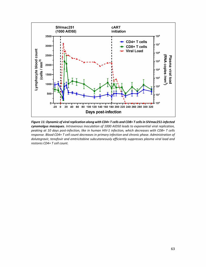
63
Figure 11: Dynamic of viral replication along with CD4+ T cells and CD8+ T cells in SIVmac251-infected cynomolgus macaques. Intravenous inoculation of 1000 AID50 leads to exponential viral replication, peaking at 10 days post-infection, like in human HIV-1 infection, which decreases with CD8+ T cells response. Blood CD4+ T cell count decreases in primary infection and chronic phase. Administration of dolutegravir, tenofovir and emtricitabine subcutaneously efficiently suppresses plasma viral load and restores CD4+ T cell count.
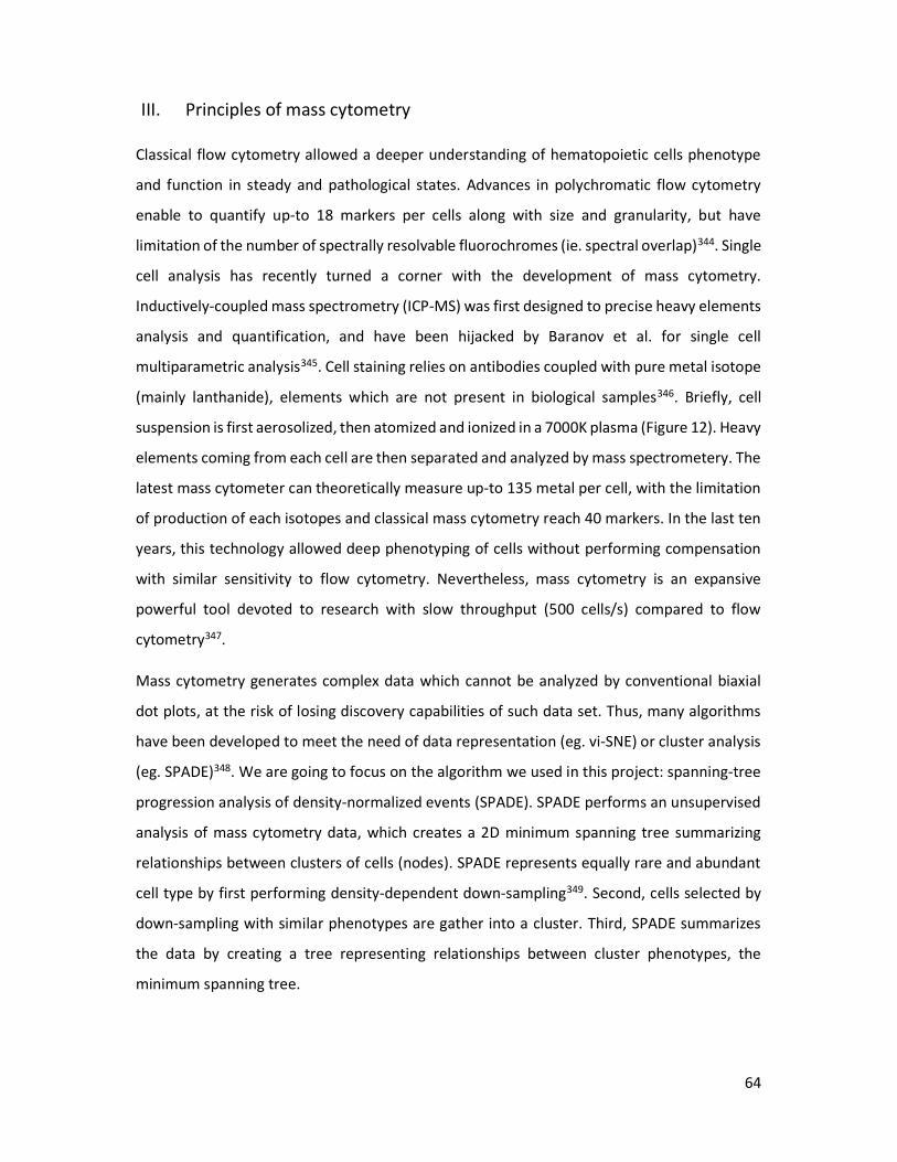
64
III. Principles of mass cytometry
Classical flow cytometry allowed a deeper understanding of hematopoietic cells phenotype
and function in steady and pathological states. Advances in polychromatic flow cytometry
enable to quantify up-to 18 markers per cells along with size and granularity, but have
limitation of the number of spectrally resolvable fluorochromes (ie. spectral overlap)344. Single
cell analysis has recently turned a corner with the development of mass cytometry.
Inductively-coupled mass spectrometry (ICP-MS) was first designed to precise heavy elements
analysis and quantification, and have been hijacked by Baranov et al. for single cell
multiparametric analysis345. Cell staining relies on antibodies coupled with pure metal isotope
(mainly lanthanide), elements which are not present in biological samples346. Briefly, cell
suspension is first aerosolized, then atomized and ionized in a 7000K plasma (Figure 12). Heavy
elements coming from each cell are then separated and analyzed by mass spectrometery. The
latest mass cytometer can theoretically measure up-to 135 metal per cell, with the limitation
of production of each isotopes and classical mass cytometry reach 40 markers. In the last ten
years, this technology allowed deep phenotyping of cells without performing compensation
with similar sensitivity to flow cytometry. Nevertheless, mass cytometry is an expansive
powerful tool devoted to research with slow throughput (500 cells/s) compared to flow
cytometry347.
Mass cytometry generates complex data which cannot be analyzed by conventional biaxial
dot plots, at the risk of losing discovery capabilities of such data set. Thus, many algorithms
have been developed to meet the need of data representation (eg. vi-SNE) or cluster analysis
(eg. SPADE)348. We are going to focus on the algorithm we used in this project: spanning-tree
progression analysis of density-normalized events (SPADE). SPADE performs an unsupervised
analysis of mass cytometry data, which creates a 2D minimum spanning tree summarizing
relationships between clusters of cells (nodes). SPADE represents equally rare and abundant
cell type by first performing density-dependent down-sampling349. Second, cells selected by
down-sampling with similar phenotypes are gather into a cluster. Third, SPADE summarizes
the data by creating a tree representing relationships between cluster phenotypes, the
minimum spanning tree.
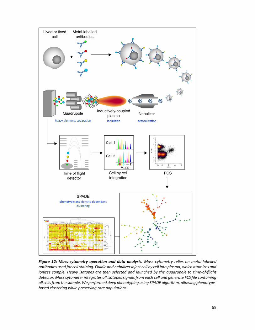
65
Figure 12: Mass cytometry operation and data analysis. Mass cytometry relies on metal-labelled antibodies used for cell staining. Fluidic and nebulizer inject cell by cell into plasma, which atomizes and ionizes sample. Heavy isotopes are then selected and launched by the quadrupole to time-of-flight detector. Mass cytometer integrates all isotopes signals from each cell and generate FCS file containing all cells from the sample. We performed deep phenotyping using SPADE algorithm, allowing phenotype-based clustering while preserving rare populations.
66
Finally, SPADE locates each cell from the data set in the cluster it belongs, which is a process
called up-sampling. So, this approach reveals cell diversity and does not loose rare population,
giving a new way to analyze hematopoietic populations. To better analyze SPADE results such
as cluster phenotypes, differences between groups and perform follow-up of cluster
abundance in multiple time-points, bioinformatics team from the lab developed a R-packages,
called SPADE-VizR350.
Research Article
J Innate Immun
Mass Cytometry Reveals the Immaturity of Circulating Neutrophils during SIV Infection
Julien Lemaitre
a Antonio Cosma
a Delphine Desjardins
a Olivier Lambotte
a, b
Roger Le Grand
a a
INSERM U1184, Immunology of Viral Infections and Autoimmune Diseases, IDMIT Department, IBFJ, CEA – Université Paris-Sud 11, Fontenay-aux-Roses/Le Kremlin-Bicêtre, France; b Service de Médecine Interne et Immunologie Clinique, Hôpital Bicêtre, Assistance Publique – Hôpitaux de Paris, Le Kremlin-Bicêtre, France
Received: October 29, 2018Accepted after revision: March 21, 2019Published online: June 21, 2019
Journal of InnateImmunity
Prof. Roger Le GrandImmunology of Viral Infections and Autoimmune Diseases, CEA – Université Paris-Sud 11 – INSERM U118418, route du PanoramaFR–92265 Fontenay-aux-Roses (France)E-Mail roger.le-grand @ cea.fr
© 2019 The Author(s)Published by S. Karger AG, Basel
E-Mail [email protected]/jin
DOI: 10.1159/000499841
KeywordsAIDS · Antiretroviral treatment · Mass cytometry · Neutrophils · Nonhuman primate · Phagocytosis
AbstractThe infected host fails to eradicate HIV-1, despite significant control of viral replication by combinational antiretroviral therapy. Here, we assessed the impact of HIV infection on immune-cell compartments in a SIVmac251 nonhuman pri-mate infection model, which allowed the choice of contam-ination route, time of infection, and treatment follow-up. We performed high-throughput multiparameter single-cell phenotyping by mass cytometry to obtain a global vision of the immune system in blood and bone marrow. Circulating polymorphonuclear neutrophils (PMNs) with impaired phagocytosis had altered surface expression of CD62L and CD11b during early chronic infection. The initiation of com-binational antiretroviral treatment during primary infection did not restore PMN function. The maturation state of PMNs was highly altered during late chronic SIV infection, showing a primarily immature phenotype. Our results provide new
insights into PMN involvement in the pathogenesis of HIV infection and may play a role in the establishment and main-tenance of chronic immune activation.
© 2019 The Author(s)Published by S. Karger AG, Basel
Introduction
Combinational antiretroviral treatment (cART) has in-creased life expectancy and the quality of life of HIV-infect-ed patients. Nevertheless, lifelong treatment is necessary to control virus replication because of the inability of cART and host defenses to eradicate the virus. HIV persistence is associated with chronic inflammation in many infected pa-tients, leading to an increased risk of comorbidities, such as cardiovascular diseases [1], neurocognitive disease [2], kid-ney disease [3], and cancer [3]. The proinflammatory cyto-kine IL-6 and soluble (s)CD14 and sCD163 levels are asso-ciated with an increased risk of mortality for both treated and untreated HIV-infected patients [4–6]. Inflammatory markers can also be directly linked to the monocyte and
This article is licensed under the Creative Commons Attribution-NonCommercial-NoDerivatives 4.0 International License (CC BY-NC-ND) (http://www.karger.com/Services/OpenAccessLicense). Usage and distribution for commercial purposes as well as any dis-tribution of modified material requires written permission.
Lemaitre/Cosma/Desjardins/Lambotte/Le Grand
J Innate Immun2DOI: 10.1159/000499841
macrophage activation associated with microbial translo-cation that occurs in HIV infection [7]. Finally, chronic in-flammation contributes to exhaustion of the adaptive im-mune system and T-cell activation. Polymorphonuclear neutrophils (PMNs) are myeloid cells that are also associ-ated with chronic inflammation in autoimmune diseases, such as rheumatoid arthritis [8]. However, few studies have been conducted on their role in HIV infection. In chronic HIV-1 or SIV infection, neutrophils show an activated pro-file [9], increased sensitivity to apoptosis [10], and impaired phagocytosis [11, 12]. Neutrophils have been shown to sup-press interferon-γ production by CD8+ T cells in chronic HIV-1 infection via the PD-L1/PD-1 axis and were more prone to degranulation, contributing to chronic immune activation in patients [13].
It is currently difficult to obtain a global view of the impact of HIV infection on the immune system, espe-cially on myeloid cells. Available studies have several lim-itations. Bone marrow, which is the differentiation and maturation compartment of myeloid cells, is still poorly characterized in HIV infection. In addition, innate im-munity plays a major role in the first steps of any infec-tion, but the study of the first days of infection is rare in humans because of limited sample availability. Cynomol-gus macaques infected with SIVmac251 are a suitable model to overcome such limitations, because many as-pects of this model parallel those of HIV-1 physiopathol-ogy [14–17] and the hematopoietic system of macaques is similar to that of humans [18, 19].
We characterized myeloid-cell heterogeneity in cyno-molgus macaque blood and bone marrow using recently developed mass cytometry technology, based on metal iso-tope-conjugated antibodies, that enables the detection of more than 40 markers per cell [20], providing major ad-vantages over conventional flow cytometry. The back-ground is very low because heavy metals are not present in biological specimens and there is no need for channel com-pensation, as required for laser-based flow cytometry. Our analysis suggests significant modification of the myeloid-cell compartment, especially circulating PMNs. During the early chronic phase, PMNs had surface expression of PMN CD62L decreased and that for CD11b increased, suggest-ing that PMNs were primed in SIV infection. In parallel, circulating PMNs demonstrate a mature phenotype but phagocytosis was impaired. In late chronic SIV infection, blood PMNs consisted of mostly immature neutrophils. These results highlight the need for further study of tissue myeloid-cell dynamics in an HIV infection model to better understand chronic inflammation and design new treat-ments for HIV remission and a functional cure.
Materials and Methods
Nonhuman Primates and Whole-Blood CollectionTwenty-nine cynomolgus macaques (Macaca fascicularis), im-
ported from Mauritius, were housed at the IDMIT infrastructure animal facility at the CEA, Fontenay-aux-Roses, France. Animals ex-pressing the H6 MHC haplotype were excluded because of their in-creased natural capacity to control SIVmac251 infection [21]. Seven-teen animals were infected by intravenous inoculation with 1,000 animal infectious dose 50% (AID50) of pathogenic SIVmac251 iso-late and followed for 18 months [22]. Six SIV-infected animals were subcutaneously treated daily, starting from 28 days after infection, with a combination of two nucleoside reverse-transcriptase inhibi-tors, emtricitabine (40 mg/kg) and tenofovir disoproxil fumarate (5.1 mg/kg), and one integrase inhibitor, dolutegravir (2.5 mg/kg) [23]. Twelve healthy macaques were used as an uninfected control group.
Three healthy animals and 2 SIV-infected animals, which were sampled 18 months after inoculation, were used for mass cytometry analysis. In addition, 15 animals in the early chronic phase of SIV infection (120–288 days after inoculation) were used, along with 9 uninfected macaques (online suppl. Table 1; for all online suppl. material, see www.karger.com/doi/10.1159/000499841), for flow cytometry analysis. Blood was drawn, and bone marrow aspirates were collected under anesthesia, consisting of an intramuscular in-jection of 10 mg/kg ketamine (Rhone-Mérieux, Lyon, France). Blood was collected into lithium-heparin tubes (Vacutainer BD, USA) and bone marrow aspirates from the iliac crest using a 10-mL 18-G syringe containing citrate dextrose (10 mg/mL).
The macaque study was approved by the Ministère de l’Education Nationale, de l’Enseignement Supérieur et de la Recherche (France) and the ethics committee Comité d’éthique en expérimentation an-imale No. 44 under reference 2015102713323361.02 (APAF-IS#2453). Animals were handled in accordance with national regu-lations (CEA accreditation No. D92-032-02) and European Direc-tive (2010/63, recommendation No. 9) in compliance with the Standards for Human Care of the Office for Laboratory Animal Welfare (OLAW, USA) under OLAW Assurance No. A5826-01.
Reagents and Cell Preparation and Storage for Mass CytometryBlood and bone marrow samples were processed using a previ-
ously described procedure, consisting of fixation, red blood cell lysis, and freezing [19], that allows the recovery of all blood leukocyte pop-ulations, especially PMNs, which are highly labile and cryosensitive. Briefly, 1 mL of whole blood or bone marrow aspirate was mixed with 10 mL fixation mixture and incubated on ice for 10 min. After centrifugation, red blood cells were lysed by adding 10 mL Milli-Q water at room temperature (RT) and incubating the samples for 20 min. After two washes with 1× modified Dulbecco’s phosphate-buff-ered saline (DPBS), the cells were counted and stored at –80 ° C in fixation mixture at a final concentration of 15 × 106 fixed leukocytes/mL and distributed into aliquots containing 3 × 10⁶ cells.
Staining and CyTOF AcquisitionThree million fixed cryopreserved cells were thawed at 37 ° C,
washed twice with PBS/0.5% bovine serum albumin and incubated on ice for 30 min with the metal-labeled surface antibodies listed in online supplementary Table 2. All antibodies were previously tested for cross-reactivity against cynomolgus cells [19]. After two washes with 1× modified DPBS, the cells were fixed with PBS/1.6% paraformaldehyde (PFA) at RT for 20 min and permeabilized with
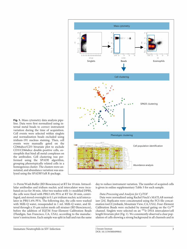
Immature Neutrophils in SIV Infection 3J Innate ImmunDOI: 10.1159/000499841
1× Perm/Wash Buffer (BD Biosciences) at RT for 10 min. Intracel-lular antibodies and iridium nucleic acid intercalator were incu-bated on ice for 30 min. After two washes with 1× modified DPBS, the cells were fixed with PBS/1.6% PFA at RT for 20 min, centri-fuged, and stored overnight in 0.1 µM iridium nucleic acid interca-lator in PBS/1.6% PFA. The following day, the cells were washed with Milli-Q water, resuspended in 1 mL Milli-Q water, and fil-tered through a 35-µm nylon mesh cell strainer (BD Biosciences), before the addition of EQTM Four-Element Calibration Beads (Fluidigm, San Francisco, CA, USA), according to the manufac-turer’s instructions. Each sample was split in half and run the same
day to reduce instrument variation. The number of acquired cells is given in online supplementary Table 3 for each sample.
Data Processing and Analysis for CyTOFData were normalized using Rachel Finck’s MATLAB normal-
izer [24]. Replicates were concatenated using the FCS file concat-enation tool (Cytobank, Mountain View, CA, USA). Four-Element Calibration Beads were excluded by manual gating on the Ce140 channel. Singlets were selected on an 191Ir DNA intercalator/cell length bivariate plot (Fig. 1). We consistently observed a clear pop-ulation of cells showing a strong background in all channels and in
Mass cytometry
Cell clustering
Phenotypic clustering
SPADE clustering
Cell population identification
Abundance analysis
EosinophilsBeadsSinglets
104
103
102Ir191
101
–100100
20 40 60 80 100 120
Cell length
140 160 180 200 220 240
104
103
102
Ir191
101
–100100
–100100 101 102 103 104
(Ce140)Di
104
103
102
CD66
101
–100100
–100100 101 102 103 104
CD3
relative.expression
1 2 3 4 5
mar
kers
CD45CD66
CD11bCD32
CD11aCD8a
GranzymeBCD28
CD4CD11c
CD14CD141
HLADRCD1cCD20
CD205CD162
CD34CD25
CD5CD16CD23CD86CCR7CD39
CD123CD64CCR5
CXCR4CD3
CX3CR1CD38
Perforine
3 26 12 13 2 41 52 45 18 66 5 21 23 85 95 97 40 75 16 71 20 54 29 32 60 84 94 10 99 96 76 90 98 64 58 7 92 46 57 44 24 79 37 87 77 4 8 62 31 35 70 14 39 49 42 56 27 25 73 36 43 47 88 93 89 22 51 80 81 59 11 30 34 33 74 1 15 17 6 28 38 100 82 63 19 53 83 68 72 78 67 91 9 50 65 48 61 86 55 69
clusters
Heatmap Viewer
Fig. 1. Mass cytometry data analysis pipe-line. Data were first normalized using in-ternal metal beads to correct instrument variation during the time of acquisition. Cell events were selected within singlets and normalization beads excluded using iridium-191 nucleus staining. Then, cell events were manually gated on the CD66abce/CD3 bivariate plot to exclude CD3/CD66abce double-positive cells, eo-sinophils that bind all metal complexes on the antibodies. Cell clustering was per-formed using the SPADE algorithm, grouping phenotypically related cells in a homogenous cluster. The clusters were an-notated, and abundance variation was ana-lyzed using the SPADRVizR R-package.
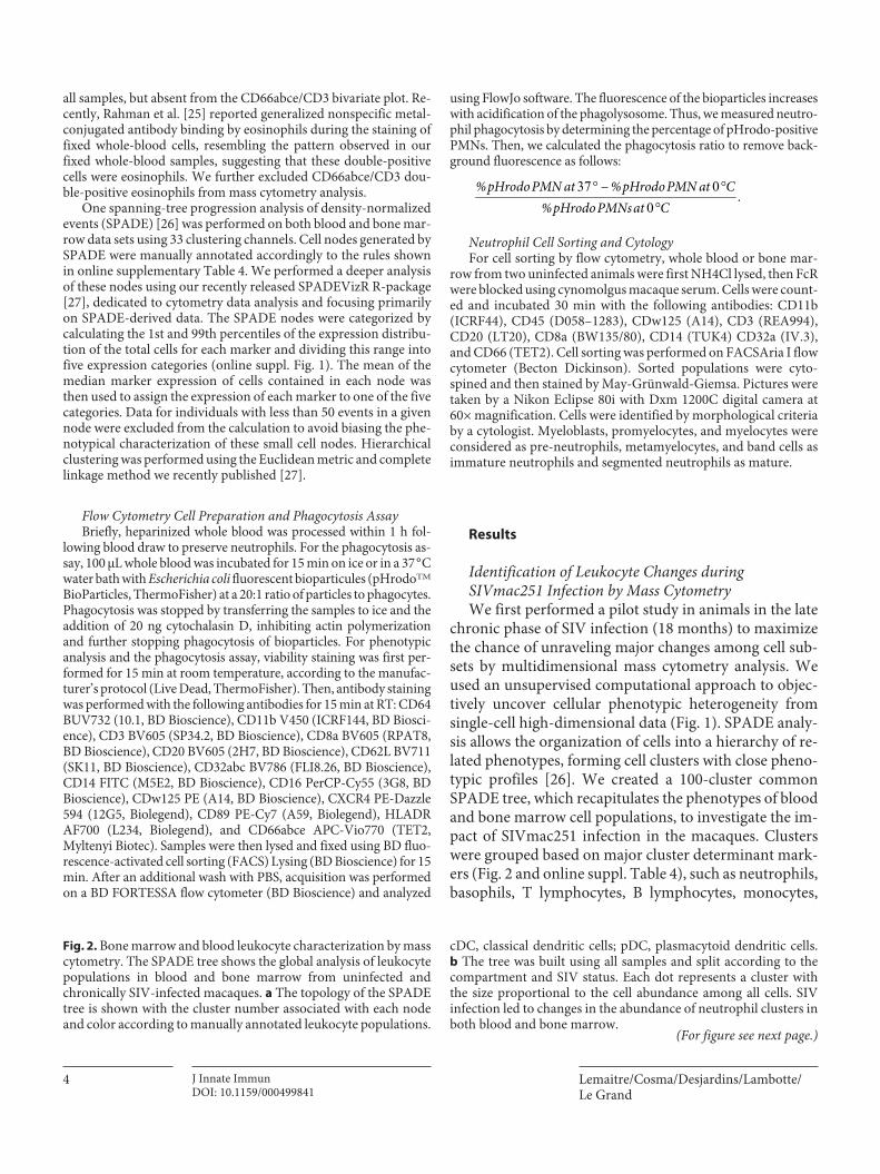
Lemaitre/Cosma/Desjardins/Lambotte/Le Grand
J Innate Immun4DOI: 10.1159/000499841
all samples, but absent from the CD66abce/CD3 bivariate plot. Re-cently, Rahman et al. [25] reported generalized nonspecific metal-conjugated antibody binding by eosinophils during the staining of fixed whole-blood cells, resembling the pattern observed in our fixed whole-blood samples, suggesting that these double-positive cells were eosinophils. We further excluded CD66abce/CD3 dou-ble-positive eosinophils from mass cytometry analysis.
One spanning-tree progression analysis of density-normalized events (SPADE) [26] was performed on both blood and bone mar-row data sets using 33 clustering channels. Cell nodes generated by SPADE were manually annotated accordingly to the rules shown in online supplementary Table 4. We performed a deeper analysis of these nodes using our recently released SPADEVizR R-package [27], dedicated to cytometry data analysis and focusing primarily on SPADE-derived data. The SPADE nodes were categorized by calculating the 1st and 99th percentiles of the expression distribu-tion of the total cells for each marker and dividing this range into five expression categories (online suppl. Fig. 1). The mean of the median marker expression of cells contained in each node was then used to assign the expression of each marker to one of the five categories. Data for individuals with less than 50 events in a given node were excluded from the calculation to avoid biasing the phe-notypical characterization of these small cell nodes. Hierarchical clustering was performed using the Euclidean metric and complete linkage method we recently published [27].
Flow Cytometry Cell Preparation and Phagocytosis AssayBriefly, heparinized whole blood was processed within 1 h fol-
lowing blood draw to preserve neutrophils. For the phagocytosis as-say, 100 µL whole blood was incubated for 15 min on ice or in a 37 ° C water bath with Escherichia coli fluorescent bioparticules (pHrodoTM BioParticles, ThermoFisher) at a 20: 1 ratio of particles to phagocytes. Phagocytosis was stopped by transferring the samples to ice and the addition of 20 ng cytochalasin D, inhibiting actin polymerization and further stopping phagocytosis of bioparticles. For phenotypic analysis and the phagocytosis assay, viability staining was first per-formed for 15 min at room temperature, according to the manufac-turer’s protocol (Live Dead, ThermoFisher). Then, antibody staining was performed with the following antibodies for 15 min at RT: CD64 BUV732 (10.1, BD Bioscience), CD11b V450 (ICRF144, BD Biosci-ence), CD3 BV605 (SP34.2, BD Bioscience), CD8a BV605 (RPAT8, BD Bioscience), CD20 BV605 (2H7, BD Bioscience), CD62L BV711 (SK11, BD Bioscience), CD32abc BV786 (FLI8.26, BD Bioscience), CD14 FITC (M5E2, BD Bioscience), CD16 PerCP-Cy55 (3G8, BD Bioscience), CDw125 PE (A14, BD Bioscience), CXCR4 PE-Dazzle 594 (12G5, Biolegend), CD89 PE-Cy7 (A59, Biolegend), HLADR AF700 (L234, Biolegend), and CD66abce APC-Vio770 (TET2, Myltenyi Biotec). Samples were then lysed and fixed using BD fluo-rescence-activated cell sorting (FACS) Lysing (BD Bioscience) for 15 min. After an additional wash with PBS, acquisition was performed on a BD FORTESSA flow cytometer (BD Bioscience) and analyzed
using FlowJo software. The fluorescence of the bioparticles increases with acidification of the phagolysosome. Thus, we measured neutro-phil phagocytosis by determining the percentage of pHrodo-positive PMNs. Then, we calculated the phagocytosis ratio to remove back-ground fluorescence as follows:
37 00
%pHrodoPMNat %pHrodoPMNat C.
%pHrodoPMNsat C°− °
°
Neutrophil Cell Sorting and CytologyFor cell sorting by flow cytometry, whole blood or bone mar-
row from two uninfected animals were first NH4Cl lysed, then FcR were blocked using cynomolgus macaque serum. Cells were count-ed and incubated 30 min with the following antibodies: CD11b (ICRF44), CD45 (D058–1283), CDw125 (A14), CD3 (REA994), CD20 (LT20), CD8a (BW135/80), CD14 (TUK4) CD32a (IV.3), and CD66 (TET2). Cell sorting was performed on FACSAria I flow cytometer (Becton Dickinson). Sorted populations were cyto-spined and then stained by May-Grünwald-Giemsa. Pictures were taken by a Nikon Eclipse 80i with Dxm 1200C digital camera at 60× magnification. Cells were identified by morphological criteria by a cytologist. Myeloblasts, promyelocytes, and myelocytes were considered as pre-neutrophils, metamyelocytes, and band cells as immature neutrophils and segmented neutrophils as mature.
Results
Identification of Leukocyte Changes during SIVmac251 Infection by Mass CytometryWe first performed a pilot study in animals in the late
chronic phase of SIV infection (18 months) to maximize the chance of unraveling major changes among cell sub-sets by multidimensional mass cytometry analysis. We used an unsupervised computational approach to objec-tively uncover cellular phenotypic heterogeneity from single-cell high-dimensional data (Fig. 1). SPADE analy-sis allows the organization of cells into a hierarchy of re-lated phenotypes, forming cell clusters with close pheno-typic profiles [26]. We created a 100-cluster common SPADE tree, which recapitulates the phenotypes of blood and bone marrow cell populations, to investigate the im-pact of SIVmac251 infection in the macaques. Clusters were grouped based on major cluster determinant mark-ers (Fig. 2 and online suppl. Table 4), such as neutrophils, basophils, T lymphocytes, B lymphocytes, monocytes,
Fig. 2. Bone marrow and blood leukocyte characterization by mass cytometry. The SPADE tree shows the global analysis of leukocyte populations in blood and bone marrow from uninfected and chronically SIV-infected macaques. a The topology of the SPADE tree is shown with the cluster number associated with each node and color according to manually annotated leukocyte populations.
cDC, classical dendritic cells; pDC, plasmacytoid dendritic cells. b The tree was built using all samples and split according to the compartment and SIV status. Each dot represents a cluster with the size proportional to the cell abundance among all cells. SIV infection led to changes in the abundance of neutrophil clusters in both blood and bone marrow.
(For figure see next page.)
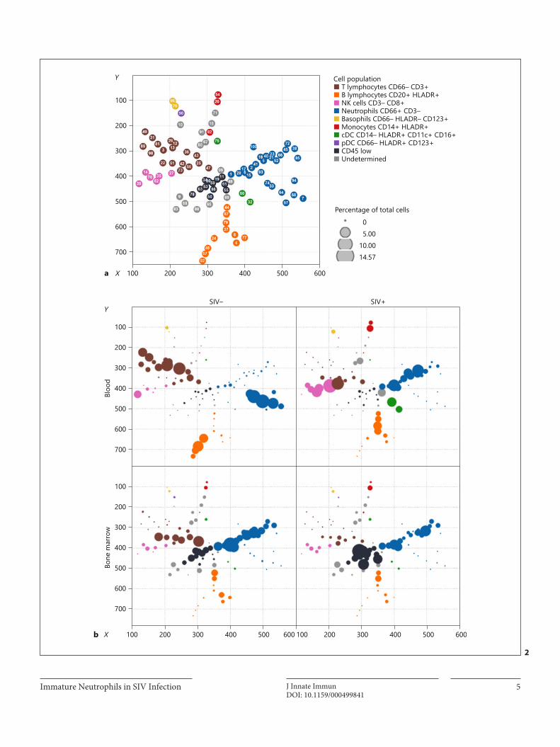
Immature Neutrophils in SIV Infection 5J Innate ImmunDOI: 10.1159/000499841
100
200
300
400
500
600
700
100 200 300 400 500 600
Cell population■ T lymphocytes CD66– CD3+■ B lymphocytes CD20+ HLADR+■ NK cells CD3– CD8+■ Neutrophils CD66+ CD3–■ Basophils CD66– HLADR– CD123+■ Monocytes CD14+ HLADR+■ cDC CD14– HLADR+ CD11c+ CD16+■ pDC CD66– HLADR+ CD123+■ CD45 low■ Undetermined
Percentage of total cells
05.00
10.0014.57
100
SIV– SIV+
Bloo
d
Y
X
X
Y
Bone
mar
row
200
300
400
500
600
700
100
200
300
400
500
600
700
100 200 300 400 500 600 100 200 300 400 500 600b
a
2
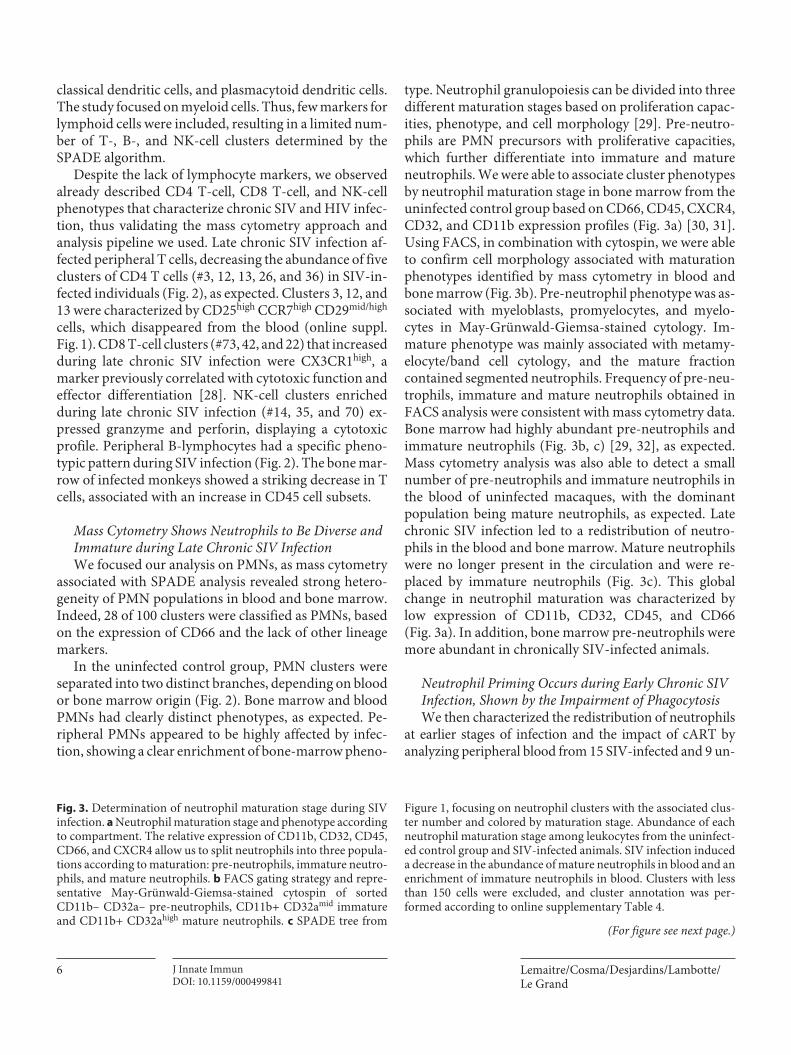
Lemaitre/Cosma/Desjardins/Lambotte/Le Grand
J Innate Immun6DOI: 10.1159/000499841
classical dendritic cells, and plasmacytoid dendritic cells. The study focused on myeloid cells. Thus, few markers for lymphoid cells were included, resulting in a limited num-ber of T-, B-, and NK-cell clusters determined by the SPADE algorithm.
Despite the lack of lymphocyte markers, we observed already described CD4 T-cell, CD8 T-cell, and NK-cell phenotypes that characterize chronic SIV and HIV infec-tion, thus validating the mass cytometry approach and analysis pipeline we used. Late chronic SIV infection af-fected peripheral T cells, decreasing the abundance of five clusters of CD4 T cells (#3, 12, 13, 26, and 36) in SIV-in-fected individuals (Fig. 2), as expected. Clusters 3, 12, and 13 were characterized by CD25high CCR7high CD29mid/high
cells, which disappeared from the blood (online suppl. Fig. 1). CD8 T-cell clusters (#73, 42, and 22) that increased during late chronic SIV infection were CX3CR1high, a marker previously correlated with cytotoxic function and effector differentiation [28]. NK-cell clusters enriched during late chronic SIV infection (#14, 35, and 70) ex-pressed granzyme and perforin, displaying a cytotoxic profile. Peripheral B-lymphocytes had a specific pheno-typic pattern during SIV infection (Fig. 2). The bone mar-row of infected monkeys showed a striking decrease in T cells, associated with an increase in CD45 cell subsets.
Mass Cytometry Shows Neutrophils to Be Diverse and Immature during Late Chronic SIV InfectionWe focused our analysis on PMNs, as mass cytometry
associated with SPADE analysis revealed strong hetero-geneity of PMN populations in blood and bone marrow. Indeed, 28 of 100 clusters were classified as PMNs, based on the expression of CD66 and the lack of other lineage markers.
In the uninfected control group, PMN clusters were separated into two distinct branches, depending on blood or bone marrow origin (Fig. 2). Bone marrow and blood PMNs had clearly distinct phenotypes, as expected. Pe-ripheral PMNs appeared to be highly affected by infec-tion, showing a clear enrichment of bone-marrow pheno-
type. Neutrophil granulopoiesis can be divided into three different maturation stages based on proliferation capac-ities, phenotype, and cell morphology [29]. Pre-neutro-phils are PMN precursors with proliferative capacities, which further differentiate into immature and mature neutrophils. We were able to associate cluster phenotypes by neutrophil maturation stage in bone marrow from the uninfected control group based on CD66, CD45, CXCR4, CD32, and CD11b expression profiles (Fig. 3a) [30, 31]. Using FACS, in combination with cytospin, we were able to confirm cell morphology associated with maturation phenotypes identified by mass cytometry in blood and bone marrow (Fig. 3b). Pre-neutrophil phenotype was as-sociated with myeloblasts, promyelocytes, and myelo-cytes in May-Grünwald-Giemsa-stained cytology. Im-mature phenotype was mainly associated with metamy-elocyte/band cell cytology, and the mature fraction contained segmented neutrophils. Frequency of pre-neu-trophils, immature and mature neutrophils obtained in FACS analysis were consistent with mass cytometry data. Bone marrow had highly abundant pre-neutrophils and immature neutrophils (Fig. 3b, c) [29, 32], as expected. Mass cytometry analysis was also able to detect a small number of pre-neutrophils and immature neutrophils in the blood of uninfected macaques, with the dominant population being mature neutrophils, as expected. Late chronic SIV infection led to a redistribution of neutro-phils in the blood and bone marrow. Mature neutrophils were no longer present in the circulation and were re-placed by immature neutrophils (Fig. 3c). This global change in neutrophil maturation was characterized by low expression of CD11b, CD32, CD45, and CD66 (Fig. 3a). In addition, bone marrow pre-neutrophils were more abundant in chronically SIV-infected animals.
Neutrophil Priming Occurs during Early Chronic SIV Infection, Shown by the Impairment of Phagocytosis We then characterized the redistribution of neutrophils
at earlier stages of infection and the impact of cART by analyzing peripheral blood from 15 SIV-infected and 9 un-
Fig. 3. Determination of neutrophil maturation stage during SIV infection. a Neutrophil maturation stage and phenotype according to compartment. The relative expression of CD11b, CD32, CD45, CD66, and CXCR4 allow us to split neutrophils into three popula-tions according to maturation: pre-neutrophils, immature neutro-phils, and mature neutrophils. b FACS gating strategy and repre-sentative May-Grünwald-Giemsa-stained cytospin of sorted CD11b– CD32a– pre-neutrophils, CD11b+ CD32amid immature and CD11b+ CD32ahigh mature neutrophils. c SPADE tree from
Figure 1, focusing on neutrophil clusters with the associated clus-ter number and colored by maturation stage. Abundance of each neutrophil maturation stage among leukocytes from the uninfect-ed control group and SIV-infected animals. SIV infection induced a decrease in the abundance of mature neutrophils in blood and an enrichment of immature neutrophils in blood. Clusters with less than 150 cells were excluded, and cluster annotation was per-formed according to online supplementary Table 4.
(For figure see next page.)
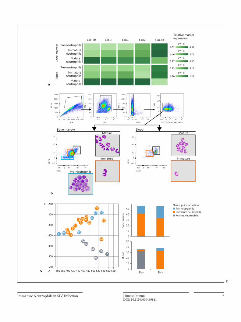
Immature Neutrophils in SIV Infection 7J Innate ImmunDOI: 10.1159/000499841
Relative markerexpression
CD11bCXCR4CD66CD45CD32CD11b
Pre-neutrophils
Bone
mar
row
Bloo
d
Immatureneutrophils
Mature neutrophils
Pre-neutrophilsImmature
neutrophilsMature
neutrophils
Bone marrowMature Mature
Immature
Pre-Neutrophils
Immature
Blood
4.35
3.710.46
2.77
3.93
0.30
0.02
4.30
5.11
1.24
CD11b
CD11b
CD11b
CD11b
250Y
X
300
350
400
450
500
550360 380 400 420 440 460 480 500 520 540 SIV–
50
Bloo
dBo
ne m
arro
w
40
30
20
10
0
50
40
30
20
10
0
SIV+560 580
Neutrophil maturation■ Pre-neutrophils■ Immature neutrophils■ Mature neutrophils
105
104
103
0
–103
–103
CD32a
CD11
b
0 103 104 105
105
104
103
0
–103
–103
CD32a
CD11
b
0 103 104 105
105
104
103CD66
0
–103
250 K
150 K
200 K
100 KSSC-
A
50 K
0–103 0 103
Lin CD3/CD20/CD8a/CDw125
104 105–103 0 103
CD14
104 105
250 K
150 K
200 K
100 KFSC-
A
50 K
00 103
CD45
104
250 K
150 K
200 K
100 KFSC-
H
50 K
00 50 K 100 K 150 K 200 K 250 K
FSC-A
c
a
b
2
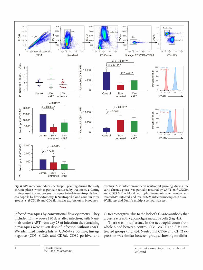
Lemaitre/Cosma/Desjardins/Lambotte/Le Grand
J Innate Immun8DOI: 10.1159/000499841
infected macaques by conventional flow cytometry. They included 12 macaques 120 days after infection, with 6 ani-mals under cART from day 28 of infection; the remaining 3 macaques were at 288 days of infection, without cART. We identified neutrophils as CD66abce positive, lineage negative (CD3, CD20, and CD8a), CD89 positive, and
CDw125 negative, due to the lack of a CD66b antibody that cross-reacts with cynomolgus macaque cells (Fig. 4a).
There was no difference in the neutrophil count from whole blood between control, SIV+ cART and SIV+ un-treated groups (Fig. 4b). Neutrophil CD66 and CD32 ex-pression was similar between groups, showing no differ-
FSC-
H
FSC-
A
SSC-
A
FSC-
W
CD89
FSC-A0
250 K 250 K
200 K
150 K
100 K
50 K
0
Singlets
50 K 100 K 150 K 200 K 250 K –103 0 103 104 105
200 K
150 K
100 K
50 K
0
Live
250 K
–103 0 103 104 105
200 K
150 K
100 K
50 K
0
Granulocytes
250 K
–103 0 103 104 105
200 K
150 K
100 K
50 K
0
Lin–
–103 0 103 104 105
–103
0
103
104
105 Neutrophils
Eosinophils
–103 0 103 104 1050
60
40
20
80
100
–103 0 103 104 1050
60
40
20
80
100
Live/dead CD66abce Lineage: CD3/CD8a/CD20 CDw125a
b
15
10
5
0
Neu
troph
il co
unt,
×103
/µL
Control SIV+cART
SIV+untreated
Perc
ent o
f max
.
CD62L
UninfectedSIV-infected
UninfectedSIV-infected
p = 0.0011**
c
10,000
5,000
0Neu
troph
ils C
D62
L M
FI
Control SIV+cART
SIV+untreated
p = 0.031*
p < 0.0001***
Perc
ent o
f max
.
CD11b
p = 0.004*
d
10,000
5,000
0Neu
troph
ils C
D11
b M
FI
Control SIV+cART
SIV+untreated
p = 0.014**p = 0.0304*
e
15,000
10,000
20,000
5,000
0Neu
troph
ils C
D89
MFI
Control SIV+cART
SIV+untreated
p = 0.0192*
p = 0.0432
f
2,000
3,000
1,000
0Neu
troph
ils C
XCR4
MFI
Control SIV+cART
SIV+untreated
p = 0.0073
Fig. 4. SIV infection induces neutrophil priming during the early chronic phase, which is partially restored by treatment. a Gating strategy used in cynomolgus macaques to isolate neutrophils from eosinophils by flow cytometry. b Neutrophil blood count in three groups. c, d CD11b and CD62L marker expression in blood neu-
trophils. SIV infection-induced neutrophil priming during the early chronic phase was partially restored by cART. e, f CXCR4 and CD89 MFI of blood neutrophils from uninfected control, un-treated SIV-infected, and treated SIV-infected macaques. Kruskal-Wallis test and Dunn’s multiple comparison test.
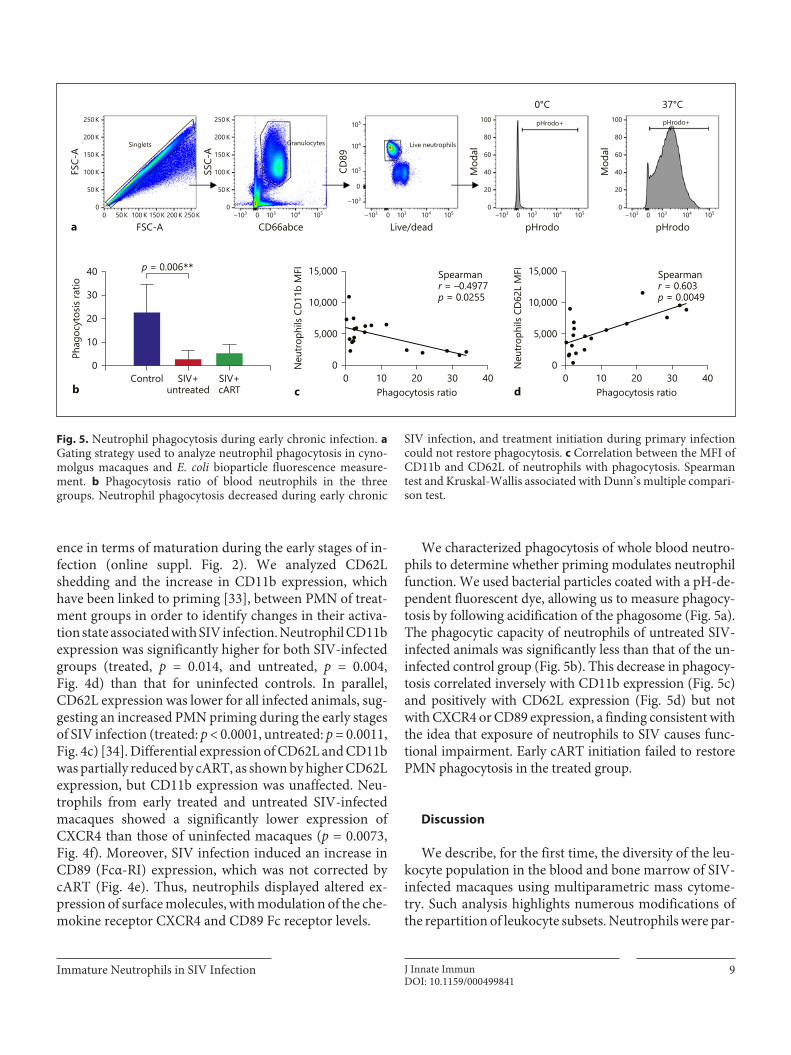
Immature Neutrophils in SIV Infection 9J Innate ImmunDOI: 10.1159/000499841
ence in terms of maturation during the early stages of in-fection (online suppl. Fig. 2). We analyzed CD62L shedding and the increase in CD11b expression, which have been linked to priming [33], between PMN of treat-ment groups in order to identify changes in their activa-tion state associated with SIV infection. Neutrophil CD11b expression was significantly higher for both SIV-infected groups (treated, p = 0.014, and untreated, p = 0.004, Fig. 4d) than that for uninfected controls. In parallel, CD62L expression was lower for all infected animals, sug-gesting an increased PMN priming during the early stages of SIV infection (treated: p < 0.0001, untreated: p = 0.0011, Fig. 4c) [34]. Differential expression of CD62L and CD11b was partially reduced by cART, as shown by higher CD62L expression, but CD11b expression was unaffected. Neu-trophils from early treated and untreated SIV-infected macaques showed a significantly lower expression of CXCR4 than those of uninfected macaques (p = 0.0073, Fig. 4f). Moreover, SIV infection induced an increase in CD89 (Fcα-RI) expression, which was not corrected by cART (Fig. 4e). Thus, neutrophils displayed altered ex-pression of surface molecules, with modulation of the che-mokine receptor CXCR4 and CD89 Fc receptor levels.
We characterized phagocytosis of whole blood neutro-phils to determine whether priming modulates neutrophil function. We used bacterial particles coated with a pH-de-pendent fluorescent dye, allowing us to measure phagocy-tosis by following acidification of the phagosome (Fig. 5a). The phagocytic capacity of neutrophils of untreated SIV-infected animals was significantly less than that of the un-infected control group (Fig. 5b). This decrease in phagocy-tosis correlated inversely with CD11b expression (Fig. 5c) and positively with CD62L expression (Fig. 5d) but not with CXCR4 or CD89 expression, a finding consistent with the idea that exposure of neutrophils to SIV causes func-tional impairment. Early cART initiation failed to restore PMN phagocytosis in the treated group.
Discussion
We describe, for the first time, the diversity of the leu-kocyte population in the blood and bone marrow of SIV-infected macaques using multiparametric mass cytome-try. Such analysis highlights numerous modifications of the repartition of leukocyte subsets. Neutrophils were par-
SSC-
A
CD89
Mod
al
Mod
al
CD66abce Live/dead pHrodo
0°C 37°C
pHrodoa
p = 0.006**
b
30
20
40
10
0
Phag
ocyt
osis
ratio
Control SIV+cART
SIV+untreated c
15,000
10,000
5,000
0Neu
troph
ils C
D11
b M
FI
0 10 20 30 40Phagocytosis ratio
Spearmanr = –0.4977p = 0.0255
d
15,000
10,000
5,000
0Neu
troph
ils C
D62
L M
FI
0 10 20 30 40Phagocytosis ratio
Spearmanr = 0.603p = 0.0049
FSC-
A
250 K
200 K
150 K
100 K
50 K
0
250 K
200 K
150 K
100 K
50 K
0
100
80
60
40
20
0
100
80
60
40
20
0
Singlets Granulocytes
0 50 K 100 K 150 K 200 K 250 K
FSC-A–103 0 103 104 105
Live neutrophils
pHrodo+ pHrodo+
–103 0 103 104 105 –103 0 103 104 105 –103 0 103 104 105
–103
0
103
104
105
Fig. 5. Neutrophil phagocytosis during early chronic infection. a Gating strategy used to analyze neutrophil phagocytosis in cyno-molgus macaques and E. coli bioparticle fluorescence measure-ment. b Phagocytosis ratio of blood neutrophils in the three groups. Neutrophil phagocytosis decreased during early chronic
SIV infection, and treatment initiation during primary infection could not restore phagocytosis. c Correlation between the MFI of CD11b and CD62L of neutrophils with phagocytosis. Spearman test and Kruskal-Wallis associated with Dunn’s multiple compari-son test.
Lemaitre/Cosma/Desjardins/Lambotte/Le Grand
J Innate Immun10DOI: 10.1159/000499841
ticularly heterogeneous, with distinct complex pheno-types in blood and bone marrow. We used myeloid mark-ers and published phenotypes [35–38] to differentiate pre-neutrophils, with proliferative capacities, from immature and mature neutrophils in a cynomolgus macaque model of HIV infection. During late chronic SIV infection, im-mature bone marrow neutrophils were found in the blood, showing mobilization of the medullary neutrophil pool, as described in emergency granulopoiesis and sepsis [37]. Immature neutrophils were the most abundant circulat-ing granulocyte populations found in SIV-infected ani-mals. Moreover, mature neutrophils almost completely disappeared from the blood and bone marrow, probably undergoing apoptosis or tissue mobilization [39]. It will be necessary for future studies to determine whether func-tions differ between mature and immature PMN subpop-ulations, and if so, whether they are functionally different populations during SIV infection.
Mass cytometry showed elevated levels of the chemo-kine receptor CXCR4 in bone marrow neutrophils and subsets of peripheral neutrophils from chronically SIV-infected macaques. At steady state, CXCR4 maintains neutrophils in the bone marrow by interacting with CXCL12 expressed on endothelial cells [30]. CXCL12 is downregulated when inflammation occurs or upon G-CSF signaling in endothelial cells, resulting in neutro-phil mobilization. CXCR4 expression decreases in young circulating neutrophils [40] and aged neutrophils re-ex-press CXCR4, allowing them to home to specific sites and be cleared by macrophages in the spleen and bone mar-row [41]. It is difficult to determine whether neutrophils are immature or aged based on CXCR4 expression alone. However, the association of strong CXCR4 expression with CD66, CD32, and CD45 suggests that the circulating neutrophils were immature, just having been mobilized from the bone marrow. Mature neutrophils were no lon-ger present in the blood of chronically infected macaques, indicating apoptosis of aged neutrophils or mobilization to tissues where viral replication sustains inflammation.
Reducing the number of markers in flow cytometry abolished the ability to differentiate immature from ma-ture neutrophils in blood as the neutrophil population was homogeneous in terms of CD66 and CD32 expres-sion. Other markers are necessary to better delineate neu-trophil maturation, such as CD49d, CD101, and CD10, recently described to distinguish between pre-neutro-phils, immature neutrophils, and mature neutrophils in human blood and bone marrow [32].
The combination of mass cytometry and flow cytom-etry results suggests that CXCR4 expression on neutro-
phils evolves, depending on the stage of SIV infection and the neutrophil subset: CXCR4 downregulation during early phases, probably due to the presence of young primed neutrophils, followed by an increase in CXCR4 expression during the late stages of infection to normal levels or higher, due to the massive egress of immature neutrophils from the bone marrow and the presence of rare aged mature neutrophils.
Neutrophils observed during the early chronic phase of SIV infection had changes in surface expression of CD62L and CD11b, which have been previously associ-ated with priming [33]. There is a continuum of PMN activation from priming to full activation depending on the stimulus intensity [42]. Priming is a complex mecha-nism in which the environment induces changes in neu-trophil responsiveness to stimuli. Priming relies on mo-bilization of secretory vesicles and gelatinase granules, whereas fully activated neutrophils are characterized by NAPDH oxidase activation along with azurophilic gran-ule release [43]. In this study, the analysis of CD62L and CD11b alone did not allow conclusions to be drawn about the extent of PMN activation following exposure to SIV.
In the early chronic phase, PMN phagocytic capacity was also impaired. The oxidative burst in neutrophils and phagocytosis correlated with viral load, CD4 T-cell count, and treatment success, as reported in a recent study [12]. In the same study, the phagocytic activity in HIV-infected pa-tients was significantly less than that of healthy individuals, but without a beneficial effect of cART. However, patients from this cohort had advanced chronic HIV infection, and treatment was initiated long after transmission. We dem-onstrated that neutrophil phagocytosis was impaired from early chronic SIV infection using E. coli bioparticles. Treat-ment initiated 1 month after inoculation did not restore neutrophil phagocytosis in SIV-infected animals. Early cART partially reduced the impact on CD62L and CD11b expression, but neutrophil phagocytosis was still impaired, probably because of chronic immune activation. Since the phagocytosis assay is based on acidification, it is also pos-sible that neutrophils lost their capacities to acidify their vacuoles during SIV infection, making them less potent pathogen killers. Neutrophil priming must be explored in further detail, to understand the inflammatory environ-ment associated with SIV infection which shapes their functionality and in turn fuels chronic immune activation.
The use of mass cytometry allowed us to identify numer-ous neutrophil subsets and show the presence of immature populations during the chronic phases of SIV infection. The monitoring of immature circulating neutrophils is per-formed for the diagnosis and prognosis of patients with sys-
Immature Neutrophils in SIV Infection 11J Innate ImmunDOI: 10.1159/000499841
temic inflammatory response syndrome [44]. Patients with high levels of myelocytes and metamyelocytes (called my-elocytosis) had a poor outcome, regardless of whether the origin was infectious or noninfectious. Whether myelocy-tosis is associated with HIV progression or treatment es-cape could be determined, as a blood smear could be a sim-ple and valuable bedside prognostic tool. Moreover, we demonstrated that cART initiated during primary infection could not reduce neutrophil priming and function. Wheth-er neutrophils play a role in the establishment and mainte-nance of chronic immune activation should be explored, as neutrophils represent 40–60% of circulating leukocytes.
Acknowledgments
This work was supported by the “Programme Investissements d’Avenir” (PIA), managed by the ANR under reference ANR-11-INBS-0008, funding the Infectious Disease Models and Inno-vative Therapies (IDMIT, Fontenay-aux-Roses, France) infra-structure, and ANR-10-EQPX-02-01, funding the FlowCyTech
facility (IDMIT, Fontenay-aux-Roses, France). We thank Gilead and ViiV for providing the antiretroviral drugs. Animal studies were supported by the ANRS and MSD Avenir, as part of the pVIS-CONTI research program, and the SIVART ANRS-IDMIT CO1 research program. We thank all members of the FlowCyTech, An-imal Science and Welfare, and Infectiology Immunology Labora-tory core facilities of the IDMIT infrastructure for their excellent expertise and outstanding contribution.
Disclosure Statement
The authors have no conflicts of interest to declare.
Author Contributions
R.L.G., O.L., and J.L. designed and supervised the study. J.L. performed experiments and discussed the data. R.L.G., O.L., D.D., and A.C. discussed the data and read the paper. J.L. wrote the pa-per.
References
1 Freiberg MS, Chang CC, Kuller LH, Skander-son M, Lowy E, Kraemer KL, et al. HIV infec-tion and the risk of acute myocardial infarc-tion. JAMA Intern Med. 2013 Apr; 173(8):
614–22. 2 Nasi M, Pinti M, De Biasi S, Gibellini L, Fer-
raro D, Mussini C, et al. Aging with HIV in-fection: a journey to the center of inflamm-AIDS, immunosenescence and neuroHIV. Immunol Lett. 2014 Nov; 162(1 1 Pt B): 329–33.
3 Deeks SG, Tracy R, Douek DC. Systemic ef-fects of inflammation on health during chron-ic HIV infection. Immunity. 2013 Oct; 39(4):
633–45. 4 Burdo TH, Lo J, Abbara S, Wei J, DeLelys ME,
Preffer F, et al. Soluble CD163, a novel mark-er of activated macrophages, is elevated and associated with noncalcified coronary plaque in HIV-infected patients. J Infect Dis. 2011 Oct; 204(8): 1227–36.
5 Kelesidis T, Kendall MA, Yang OO, Hodis HN, Currier JS. Biomarkers of microbial translocation and macrophage activation: as-sociation with progression of subclinical ath-erosclerosis in HIV-1 infection. J Infect Dis. 2012 Nov; 206(10): 1558–67.
6 So-Armah KA, Tate JP, Chang CH, Butt AA, Gerschenson M, Gibert CL, et al.; VACS Proj-ect Team. Do biomarkers of inflammation, monocyte activation, and altered coagulation explain excess mortality between hiv infected and uninfected people? J Acquir Immune Defic Syndr. 2016 Jun; 72(2): 206–13.
7 Brenchley JM, et al. Microbial translocation is a cause of systemic immune activation in chron-ic HIV infection. Nat Med. 2006; 12: 1365–71.
8 Wright HL, Moots RJ, Edwards SW. The mul-tifactorial role of neutrophils in rheumatoid arthritis. Nat Rev Rheumatol. 2014 Oct;
10(10): 593–601. 9 Campillo-Gimenez L, Casulli S, Dudoit Y, Seang
S, Carcelain G, Lambert-Niclot S, et al. Neutro-phils in antiretroviral therapy-controlled HIV demonstrate hyperactivation associated with a specific IL-17/IL-22 environment. J Allergy Clin Immunol. 2014 Nov; 134(5): 1142–52.e5.
10 Elbim C, Monceaux V, François S, Hurtrel B, Gougerot-Pocidalo MA, Estaquier J. In-creased neutrophil apoptosis in chronically SIV-infected macaques. Retrovirology. 2009 Mar; 6(1): 29.
11 Pos O, Stevenhagen A, Meenhorst PL, Kroon FP, Van Furth R. Impaired phagocytosis of Staphylococcus aureus by granulocytes and monocytes of AIDS patients. Clin Exp Immu-nol. 1992 Apr; 88(1): 23–8.
12 Michailidis C, Giannopoulos G, Vigklis V, Ar-menis K, Tsakris A, Gargalianos P. Impaired phagocytosis among patients infected by the human immunodeficiency virus: implication for a role of highly active anti-retroviral therapy. Clin Exp Immunol. 2012 Mar; 167(3): 499–504.
13 Bowers NL, Helton ES, Huijbregts RP, Goep-fert PA, Heath SL, Hel Z. Immune suppres-sion by neutrophils in HIV-1 infection: role of PD-L1/PD-1 pathway. PLoS Pathog. 2014 Mar; 10(3):e1003993.
14 Matusali G, Dereuddre-Bosquet N, Le Tor-torec A, Moreau M, Satie AP, Mahé D, et al. Detection of Simian Immunodeficiency Virus in Semen, Urethra, and Male Reproductive Organs during Efficient Highly Active Anti-retroviral Therapy. J Virol. 2015 Jun; 89(11):
5772–87.15 Mannioui A, Bourry O, Sellier P, Delache B,
Brochard P, Andrieu T, et al. Dynamics of vi-ral replication in blood and lymphoid tissues during SIVmac251 infection of macaques. Retrovirology. 2009 Nov; 6(1): 106.
16 Bruel T, Dupuy S, Démoulins T, Rogez-Kreuz C, Dutrieux J, Corneau A, et al. Plas-macytoid dendritic cell dynamics tune inter-feron-alfa production in SIV-infected cyno-molgus macaques. PLoS Pathog. 2014 Jan;
10(1):e1003915.17 Bruel T, Hamimi C, Dereuddre-Bosquet N,
Cosma A, Shin SY, Corneau A, et al. Long-term control of simian immunodeficiency vi-rus (SIV) in cynomolgus macaques not asso-ciated with efficient SIV-specific CD8+ T-cell responses. J Virol. 2015 Apr; 89(7): 3542–56.
18 Policicchio BB, Pandrea I, Apetrei C. Animal models for HIV cure research. Front Immu-nol. 2016 Jan; 7: 12.
19 Elhmouzi-Younes J, Palgen JL, Tchitchek N, Delandre S, Namet I, Bodinham CL, et al. In depth comparative phenotyping of blood innate myeloid leukocytes from healthy humans and macaques using mass cytometry. Cytometry A. 2017 Oct; 91(10):
969–82.
Lemaitre/Cosma/Desjardins/Lambotte/Le Grand
J Innate Immun12DOI: 10.1159/000499841
20 Bandura DR, Baranov VI, Ornatsky OI, An-tonov A, Kinach R, Lou X, et al. Mass cytom-etry: technique for real time single cell multi-target immunoassay based on inductively coupled plasma time-of-flight mass spec-trometry. Anal Chem. 2009 Aug; 81(16):
6813–22.21 Pereyra F, Jia X, McLaren PJ, Telenti A, de
Bakker PI, Walker BD, et al.; International HIV Controllers Study. The major genetic de-terminants of HIV-1 control affect HLA class I peptide presentation. Science. 2010 Dec;
330(6010): 1551–7.22 Karlsson I, Malleret B, Brochard P, Delache B,
Calvo J, Le Grand R, et al. Dynamics of T-cell responses and memory T cells during primary simian immunodeficiency virus infection in cynomolgus macaques. J Virol. 2007 Dec;
81(24): 13456–68.23 Del Prete GQ, Smedley J, Macallister R, Jones
GS, Li B, Hattersley J, et al. Short Communi-cation: Comparative Evaluation of Coformu-lated Injectable Combination Antiretroviral Therapy Regimens in Simian Immunodefi-ciency Virus-Infected Rhesus Macaques. AIDS Res Hum Retroviruses. 2016 Feb; 32(2):
163–8.24 Finck, R. et al. Normalization of mass cytom-
etry data with bead standards. Cytometry A. 2013; 83(5): 483–94.
25 Rahman AH, Tordesillas L, Berin MC. Hepa-rin reduces nonspecific eosinophil staining artifacts in mass cytometry experiments. Cy-tometry A 2016; 89(6): 1–7.
26 Qiu P, Simonds EF, Bendall SC, Gibbs KD Jr, Bruggner RV, Linderman MD, et al. Extract-ing a cellular hierarchy from high-dimension-al cytometry data with SPADE. Nat Biotech-nol. 2011 Oct; 29(10): 886–91.
27 Gautreau G, Pejoski D, Le Grand R, Cosma A, Beignon AS, Tchitchek N. SPADEVizR: an R package for visualization, analysis and inte-gration of SPADE results. Bioinformatics. 2017 Mar; 33(5): 779–81.
28 Gerlach C, Moseman EA, Loughhead SM, Alva-rez D, Zwijnenburg AJ, Waanders L, et al. The Chemokine Receptor CX3CR1 Defines Three Antigen-Experienced CD8 T Cell Subsets with Distinct Roles in Immune Surveillance and Ho-meostasis. Immunity. 2016 Dec; 45(6): 1270–84.
29 Bolliger AP, Fontaine M. Cytological exami-nation and cellular composition of bone mar-row in healthy, adult, cynomolgus monkeys (Macaca fascicularis). Comp Haematol Int. 1998; 8(4): 183–90.
30 Borregaard N. Neutrophils, from marrow to microbes. Immunity. 2010 Nov; 33(5): 657–70.
31 Elghetany MT. Surface antigen changes dur-ing normal neutrophilic development: a criti-cal review. Blood Cells Mol Dis. 2002 Mar-Apr; 28(2): 260–74.
32 Evrard M, Kwok IW, Chong SZ, Teng KW, Becht E, Chen J, et al. Developmental analysis of bone marrow neutrophils reveals popula-tions specialized in expansion, trafficking, and effector functions. Immunity. 2018 Feb;
48(2): 364–79.e8.33 Condliffe AM, Chilvers ER, Haslett C, Drans-
field I. Priming differentially regulates neu-trophil adhesion molecule expression/func-tion. Immunology. 1996 Sep; 89(1): 105–11.
34 Ivetic A. A head-to-tail view of L-selectin and its impact on neutrophil behaviour. Cell Tis-sue Res. 2018 Mar; 371(3): 437–53.
35 Blazkova J, Gupta S, Liu Y, Gaudilliere B, Ganio EA, Bolen CR, et al. Multicenter Sys-tems Analysis of Human Blood Reveals Imma-ture Neutrophils in Males and During Preg-nancy. J Immunol. 2017 Mar; 198(6): 2479–88.
36 Otten MA, Rudolph E, Dechant M, Tuk CW, Reijmers RM, Beelen RH, et al. Immature neutrophils mediate tumor cell killing via IgA but not IgG Fc receptors. J Immunol. 2005 May; 174(9): 5472–80.
37 Drifte G, Dunn-Siegrist I, Tissières P, Pugin J. Innate immune functions of immature neu-trophils in patients with sepsis and severe sys-temic inflammatory response syndrome. Crit Care Med. 2013 Mar; 41(3): 820–32.
38 Marini O, et al. Mature CD10+ and immature CD10– neutrophils present in G-CSF-treated donors display opposite effects on T cells. Blood. 2017; 129: 1343–57.
39 Greenlee-Wacker MC. Clearance of apop-totic neutrophils and resolution of inflam-mation. Immunol Rev. 2016 Sep; 273(1):
357–70.40 Martin C, Burdon PC, Bridger G, Gutierrez-
Ramos JC, Williams TJ, Rankin SM. Chemo-kines acting via CXCR2 and CXCR4 control the release of neutrophils from the bone mar-row and their return following senescence. Immunity. 2003 Oct; 19(4): 583–93.
41 Rankin SM. The bone marrow: a site of neu-trophil clearance. J Leukoc Biol. 2010 Aug;
88(2): 241–51.42 Sengeløv H, Kjeldsen L, Borregaard N. Con-
trol of exocytosis in early neutrophil activa-tion. J Immunol. 1993 Feb; 150(4): 1535–43.
43 Miralda I, Uriarte SM, Mcleish KR. Multiple phenotypic changes define neutrophil pri-ming. Front Cell Infect Microbiol. 2017; 7:
217.44 Mare TA, Treacher DF, Shankar-Hari M,
Beale R, Lewis SM, Chambers DJ, et al. The diagnostic and prognostic significance of monitoring blood levels of immature neutro-phils in patients with systemic inflammation. Crit Care. 2015 Feb; 19(1): 57.

80
II. Immature CD10- neutrophils exert immunostimulatory function during
primary and chronic SIV infection
A. Summary
In chronic HIV infection, neutrophils have been classically described as homogenous cell
population impaired by the infection. In the first article, using multidimensional analysis of all
leukocytes, we observed globally neutrophils heterogeneity with circulation of immature
neutrophils in late chronic SIV infection (>1 year of infection). But the moment in which
neutrophils diversification occurs during HIV/SIV infection is unknown. In this second article
in preparation, the objective was to characterize neutrophil heterogeneity in primary and
chronic SIV infection. Using conventional flow cytometry and flow cytometry cell sorting, we
first developed tools to differentiate neutrophils according to their maturation stage in
cynomolgus macaque: pre-neutrophils (promyelocyte and myelocyte), immature neutrophils
(metamyelocyte and band cell) and mature neutrophils200. In parallel, we also evaluate
neutrophil priming using CD62L expression.
Neutrophil heterogeneity is a question of phenotype, but above all a question of function.
There are few descriptions of functional heterogeneity in HIV infected patients, where authors
described low-density neutrophils with immunosuppressive functions against T cells IFN-γ
production325,326. But less is known about the impact of other neutrophils subsets, gathered
in normal-density neutrophil population. Recent studies demonstrated the co-existence of
neutrophils subpopulation with opposite immunomodulatory function against T cells
responses333,351. Indeed, G-CSF-treated donors demonstrated circulation of both mature and
immature neutrophils having respectively immunosuppressive and immunostimulatory
effects on T cells proliferation and IFN-γ production. This study underlines the need to study
function with phenotype of all subpopulations to comprehend the impact of neutrophils in
diseases: suppressive or pro-inflammatory. Immunostimulatory function of neutrophils has
not been studied in HIV/SIV infection, while it has been identified in autoimmune diseases and
cancers352. In this study, we used an autologus co-culture between normal density neutrophils
and T cells to evaluate the impact of neutrophils on T cells proliferation and cytokine
production during SIV infection.
81
In primary infection, even if total neutrophil count was not affected, sub-population of
neutrophils changed, with an increase of primed CD62L-low and immature CD10- neutrophils.
This acute response was associated with immunostimulatory functions evidenced by increase
CD4 T cell proliferation, along with CD4 and CD8 T cells IL-2 and IFN-γ production. But
interestingly, neutrophils demonstrated an opposite role on CD8 T cells by suppressing
proliferation. In chronic SIV infection, the frequency of primed and immature neutrophils was
still significantly elevated. Neutrophils had no impact on autologous T cells proliferation, IL-2
and IFN-γ production. But interestingly, neutrophils increased TNF-α production by CD4 T
cells. We also evaluate the frequency of LDN in the course of SIV infection and we observed
only a slight increase in primary infection but not in chronic phase.
This suggest a potent immunostimulatory function of normal-density neutrophils against T
cells in SIV infection, which could in turn participate to immune activation. Initiation of early
treatment within first month of SIV infection reduced frequencies of primed and immature
neutrophils to basal levels, whereas delayed treatment (6 months post-infection) had limited
impact on these subpopulations’ frequencies. Thus, our hypothesis is that chronic
inflammation shaped neutrophil functions, and maintained pathologic pro-inflammatory
immune modulation.
To better characterize immunomodulatory functions of mature and immature neutrophils
against T cells we are currently developing a cell sorting assay, using magnetic beads coated
with antibodies to negatively sort neutrophils. This technic will allow multiple functional
analysis of subpopulations, without disadvantage of density gradient separation. This last
experiment for this study will draw conclusion on whether CD10+ or CD10- neutrophils have
similar or different immunomodulation properties.
82
B. Manuscript
Title: Immature CD10- neutrophils are pro-inflammatory cells during
primary and early chronic SIV infection
Julien Lemaitre1, Delphine Desjardins1, Mario Gomez1, Christine Bourgeois1,2, Francis
Relouzat1, Anne-Sophie Gallouët1, Benoît Favier1, Roger Le Grand1* and Olivier Lambotte1,2
1CEA – Université Paris Sud 11 – INSERM U1184, Immunology of Viral Infections and
Autoimmune Diseases, IDMIT Department, IBFJ, Fontenay-aux-Roses & Le Kremlin-Bicêtre,
France
2Assistance Publique - Hôpitaux de Paris, Hôpital Bicêtre, Service de Médecine Interne et
Immunologie Clinique, F-94275 Le Kremlin-Bicêtre, France
Short title: Neutrophil heterogeneity in SIV infection.
*Corresponding Author
Olivier Lambotte
Assistance Publique - Hôpitaux de Paris, Hôpital Bicêtre, Service de Médecine Interne et
Immunologie Clinique, F-94275 Le Kremlin-Bicêtre, France
Tel:
E-mail: [email protected]
Key words: AIDS, antiretroviral treatment, neutrophils, immunomodulation, non-human
primate
83
1) Abstract
Even under combinational antiretroviral treatments (cART), HIV-1 persistence is associated
with chronic inflammation in infected patients, leading to an increased risk of comorbidities,
such as cardiovascular diseases, neurocognitive disease, and cancer 11,353. Under cART,
occurrence of non-AIDS-related comorbidities is associated with increased levels of innate
immune markers, notably myeloid cells activation, whereas T cells activation was not
correlated to outcomes152. Myeloid cells, mainly monocytes and macrophages, have been
involved in immune activation observed in HIV-1 infection. But neutrophil (PMN), the most
abundant circulating myeloid cell, have been less studied in HIV infection 20 whereas they were
associated with chronic inflammation in autoimmune diseases, such as rheumatoid
arthritis354. Most available studies shown an impact of HIV infection on PMNs function with
decrease of microbicidal functions318,355. PMNs are also primed in chronic phase with CD62L
shedding, CD11b translocation and increased ROS production in unstimulated cells22. In this
study, authors shown neutrophils activation in HIV-infected individuals having a non-AIDS-
related comorbidity. Thus, HIV-1 infection has a deleterious effect on neutrophils but less is
known about the role of neutrophils on chronic inflammation. To evaluate if PMNs are
associated with HIV chronic inflammation, we characterized neutrophils heterogeneity in
blood and bone marrow from SIV-infected cynomolgus macaques. In primary infection, even
if PMN count was not significantly changed, frequency of circulating mature CD10+ PMN was
decreased, and they were replaced by immature CD10- PMN. At the same time, PMN
demonstrated a CD62L-low CD64+ HLADR+ primed phenotype. After 6 months of infection,
CD62L-low primed and CD10- immature PMN were significantly higher in bone marrow and
blood. Depending on the time of infection, PMN demonstrated variable immunomodulatory
function against T cells proliferation and cytokine production. Notably, neutrophils were
capable to increase CD4 and CD8 T cells production IL-2, IFN- and CD4 T cells proliferation,
suggesting an immunostimulatory function. But in chronic phase, PMN increased TNF-
production by CD4 T cells. In this study, we provide unprecedented insight into PMN
heterogeneity in the course of SIV infection. Since PMN represent 40-70% of circulating
leukocytes and primed PMN are more potent to release pro-inflammatory cytokines and to
transmigrate, they should be considered as a new player in HIV chronic inflammation.
84
2) Introduction
In absence of a cure for HIV infection, lifelong antiretroviral treatment is necessary to control
virus replication because of the inability of combinational antiretroviral treatment (cART) and
host defenses to eradicate the virus. cART has increased life expectancy and the quality of life
of HIV-infected patients, but is still far from a cure13,356. Despite cART, non-AIDS related co-
morbidities (metabolic, neurodegenerative and cardiovascular diseases) normally associated
with aging have been shown to be higher in HIV-infected population than aged-matched
population10. This increased frequency of co-morbidities was soon associated with the
persistence of chronic inflammation11. Even under long-term cART, HIV-infected patients have
a higher level of soluble inflammatory markers (ex. IL-6) and gut epithelial dysfunction markers
(ex. I-FABP)12.
Nevertheless, exact mechanisms and cells involve in the persistence of increased levels of
soluble mediators are still not clear. Residual replication and microbial translocation are
potent to activate monocytes, dendritic cells and macrophages, leading to chronic
inflammation357–359. But one of the most potent pro-inflammatory myeloid cells,
polymorphonuclear neutrophil (PMN), has been overlooked in HIV-infection20. Consider as
short-lived phagocytic cells, new studies demonstrate immunomodulatory functions of PMNs
and a life expectancy reaching 5,4 days in certain conditions184,191,199,333. In addition, PMN have
been involved in pathogenesis of metabolic, cardiovascular and neurological diseases which
are also non–AIDS related comorbidities21. Recently, PMN infiltration have been shown in
patients under cART and was associated with colonic epithelial cells apoptosis and microbial
translocation in SIV-infected macaques83–85. PMN survival in gut have been shown to be
increase in chronic HIV infection, which is associated with pro-inflammatory function in other
diseases87. Beside, both normal density and low-density neutrophils expressing PD-L1 were
potent to suppress specific T cells IFN-γ production in PD1/PD-L1 and ROS dependent
mechanisms. Depending on the disease studied, PMN may has a pro-inflammatory or
suppressive function, which immunomodulatory function is impossible to predict based on
currently available phenotyping approaches 352.
Here we analyzed PMN heterogeneity in a non-human primate model infected by SIVmac251.
We demonstrated that circulating PMNs in SIV primary infection were mostly constituted of
primed (CD62L-low) immature (CD10-) PMNs. This immature phenotype was associated with
85
a positive pro-inflammatory function of PMNs against CD4+ and CD8+ T cells IL-2 and IFN-g
production but suppressed CD8+ T cells proliferation. In chronic SIV infection, percentage of
PMN of immature phenotype was lower compare to acute phase but still elevated with the
regard to non-infected animals, and PMN demonstrated positive modulation of CD4+ T cells
TNF-a production. After 3 months under cART, immunostimulatory role of PMN was return to
basal levels. Thus, our results suggest the implication of immature PMN on T cells activation
in SIV infection.
3) Material and method
Nonhuman primates and sampling Twelve cynomolgus macaques (Macaca fascicularis), imported from Mauritius, were housed
at the IDMIT infrastructure animal facility at the CEA, Fontenay-aux-Roses, France. Animals
expressing the H6 MHC haplotype were excluded because of their increased natural capacity
to control SIVmac251 infection360. All animals were infected by intravenous inoculation with
1,000 animal infectious dose 50% (AID50) of pathogenic SIVmac251 isolate and followed for
more than 12 months361. Six SIV-infected animals were subcutaneously treated daily, starting
from 28 days after infection, called group 1, with a combination of two nucleoside reverse-
transcriptase inhibitors, emtricitabine (40 mg/kg) and tenofovir disoproxil fumarate (5.1
mg/kg), and one integrase inhibitor, dolutegravir (2.5 mg/kg)362. Six remaining animals, called
group 2, were treated with same route and posology 6 months after infection. Blood was
drawn and bone marrow aspirates collected under anesthesia, consisting of an intramuscular
injection of 10 mg/kg zolazepam/tiletamine (Virbac, Carros, France). Blood was collected into
lithium-heparin tubes (Vacutainer BD, USA) and bone marrow aspirates from the iliac crest or
humeral head using a 10-ml 18G-syringe containing citrate -dextrose (10 mg/ml). The
macaque study was approved by the « Ministère de l’Education Nationale, de l’Enseignement
Supérieur et de la Recherche » (France) and the ethics committee « Comité d'éthique en
expérimentation animale n°44 » under reference 2015102713323361.02 (APAFIS#2453).
Animals were handled in accordance with national regulations (CEA accreditation number
D92-032-02) and European Directive (2010/63, recommendation Nº9) and in compliance with
the Standards for Human Care of the Office for Laboratory Animal Welfare (OLAW, USA) under
OLAW Assurance number #A5826-01.
86
Reagent and cell preparation for f low cytometry Briefly, heparinized whole blood and citrated bone marrow were processed within 1 h
following blood draw to preserve neutrophils. For phenotypic analysis viability staining was
first performed for 15 min at room temperature, according to the manufacturer’s protocol
(Live Dead, Thermofisher). Then, antibody staining was performed with the following
antibodies for 15 min at room temperature. Samples were then lysed and fixed using BD FACS
Lysing (BD Bioscience) for 15 min. After an additional wash with PBS, acquisition was
performed on a BD FORTESSA flow cytometer (BD Bioscience) and analyzed using FlowJo
software. Gating strategies are detailed in supplementary figure 1.
Cell isolation Mononuclear cells and granulocytes were isolated by density gradient centrifugation (Ficoll-
Paque; GE Healthcare Life Sciences) of blood. Using this technic, we obtained 95% of
neutrophil purity in globular fraction. Isolation was further shown to not induce PMN
activation using CD62L and CD11b expression.
Neutrophils cells sorting and cytology For cell sorting by flow cytometry, whole blood or bone marrow from two uninfected animals
were first NH4Cl lysed, then FcR were blocked using cynomolgus macaque serum. Cells were
counted and incubated 30min with the following antibodies. Cell sorting was performed on
FACSAria I flow cytometer (Becton Dickinson). Sorted population were cytospined and then
stained by May-Grünwald Giemsa. Picture were taken by Nikon Eclipse 80i with Dxm 1200C
digital camera at 60x magnification. Cell were identified by morphological criteria by a
cytologist. Promyelocytes and myelocytes were considered as pre-neutrophils,
metamyelocytes and band cells as immature neutrophils, and segmented neutrophils as
mature.
Neutrophils / PBMC co-culture for cytokine and proliferation assay TCR-stimulated (Cytostim NHP, Miltenyi) T-cells were cultured in presence or absence of
autologous PMN at 1:1 or 5:1 PMN to PBMC ratio. Concerning intracellular cytokines, after 1
hour of stimulation brefeldin A (Sigma) was added to block protein transport to Golgi. For
proliferation assay, PBMC were stained with 1µM CFSE (Cell Trace, Invitrogen) for 5 min and
reaction was stopped using cold FCS. Co-culture were maintained 16 hours for cytokine assay
and 96 hours for proliferation assay at 37°C 5% CO2, before flow cytometry analyses.
87
For both assays, 20 min of viability staining at 4°C (Live Dead, Thermofisher) was done, and
for cytokine assay, cells were permeabilized and fixed (Cytofix/cytoperm, BD Biosecience).
Then cell staining was performed for 30 min at 4°C with the following antibodies.
Statistical analysis
Statistical analyses were carried out with GraphPad Prism 8 software (GraphPad Software
Inc.). We used Friedman test with Dunn’s multiple comparison for statistical analyses of
multiple time points from the same animal. A Mann-Whitney non-parametric test was used
to compare data from Group 1 and Group 2. We performed a Wilcoxon matched-pair signed
rank test to analyze the impact of PMNs on T cells proliferation and cytokine production. In
graphs, the thresholds for statistical significance are indicated as follows: * p<0.05; ** p<0.01,
*** p<0.001.
88
4) Results
Neutrophil priming in acute and chronic SIV infection Neutrophil priming have been identified in chronic SIV and HIV infections 22,318,363, but the
dynamic of neutrophil in the course of infection is poorly defined. Therefore, we characterized
the dynamic of neutrophils priming in SIV-infected non-human primates in acute, chronic
phase and under cART. We analyzed both fresh blood and bone marrow with multiparameter
flow cytometry to explore these two important neutrophil compartments (Figure 1 A-B). We
used several surface markers expression as surrogate of priming, including CD62L shedding
along with upregulation of CD11b, CD64 and HLA-DR. To evaluate if early cART initiation could
affect PMNs priming, animals were separated in two groups accordingly to time of treatment
initiation (Group 1 cART initiated at 28 days p.i.; Group 2 cART initiated at 180 days p.i.). Taking
all time points together, there was no significant differences of neutrophils blood count
between groups and time point (p=0.915) (Figure 1C). Nevertheless, we could identify
neutrophils subpopulation on the basis of CD62L, CD64 and HLADR expression. In primary
infection, both groups demonstrated a significant increase of CD62L-low neutrophil frequency
in association with increasing viral replication, and reaching its maximum at the peak of viral
replication (Figure 1 A, B). Previous study have demonstrated an association between CD62L-
shedding and neutrophils priming in HIV-1 infection318, we therefore considered CD62L-low
population as primed neutrophils. In absence of cART, primed neutrophil population is
maintained significantly elevated in group 2 during chronic phase (Figure 1 B). In addition,
CD64 and HLADR surface expression on neutrophils was significantly elevated only at the peak
of viral replication, and showed non-significant fluctuation during the follow-up,
independently from cART time of initiation (Figure 1 D, E). In group 1, percentage of CD62L-
low neutrophils (primed) was reduced almost to basal levels after 6 months of cART. (Figure
1 F). When initiated in chronic phase (6 months p.i.), cART reduced primed PMNs frequency
after one month of treatment (Figure 1B). However, in the same group neutrophil priming
increased thereafter, being significantly elevated 6 months after cART initiation. In bone
marrow, frequency of CD62L-low neutrophils was significantly increased only during chronic
phase (Figure 1G). In group 1, cART completely restore normal values after six months of
treatment, but we are currently evaluating bone marrow from group 2. Changes in CD62L
expression among bone marrow may indicates priming or increase of CD62L-negative
neutrophil precursors, affecting neutrophils maturation.
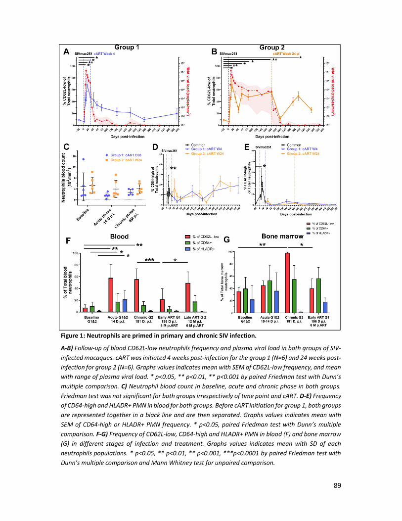
89
Figure 1: Neutrophils are primed in primary and chronic SIV infection.
A-B) Follow-up of blood CD62L-low neutrophils frequency and plasma viral load in both groups of SIV-infected macaques. cART was initiated 4 weeks post-infection for the group 1 (N=6) and 24 weeks post-infection for group 2 (N=6). Graphs values indicates mean with SEM of CD62L-low frequency, and mean with range of plasma viral load. * p<0.05, ** p<0.01, ** p<0.001 by paired Friedman test with Dunn’s multiple comparison. C) Neutrophil blood count in baseline, acute and chronic phase in both groups. Friedman test was not significant for both groups irrespectively of time point and cART. D-E) Frequency of CD64-high and HLADR+ PMN in blood for both groups. Before cART initiation for group 1, both groups are represented together in a black line and are then separated. Graphs values indicates mean with SEM of CD64-high or HLADR+ PMN frequency. * p<0.05, paired Friedman test with Dunn’s multiple comparison. F-G) Frequency of CD62L-low, CD64-high and HLADR+ PMN in blood (F) and bone marrow (G) in different stages of infection and treatment. Graphs values indicates mean with SD of each neutrophils populations. * p<0.05, ** p<0.01, ** p<0.001, ***p<0.0001 by paired Friedman test with Dunn’s multiple comparison and Mann Whitney test for unpaired comparison.
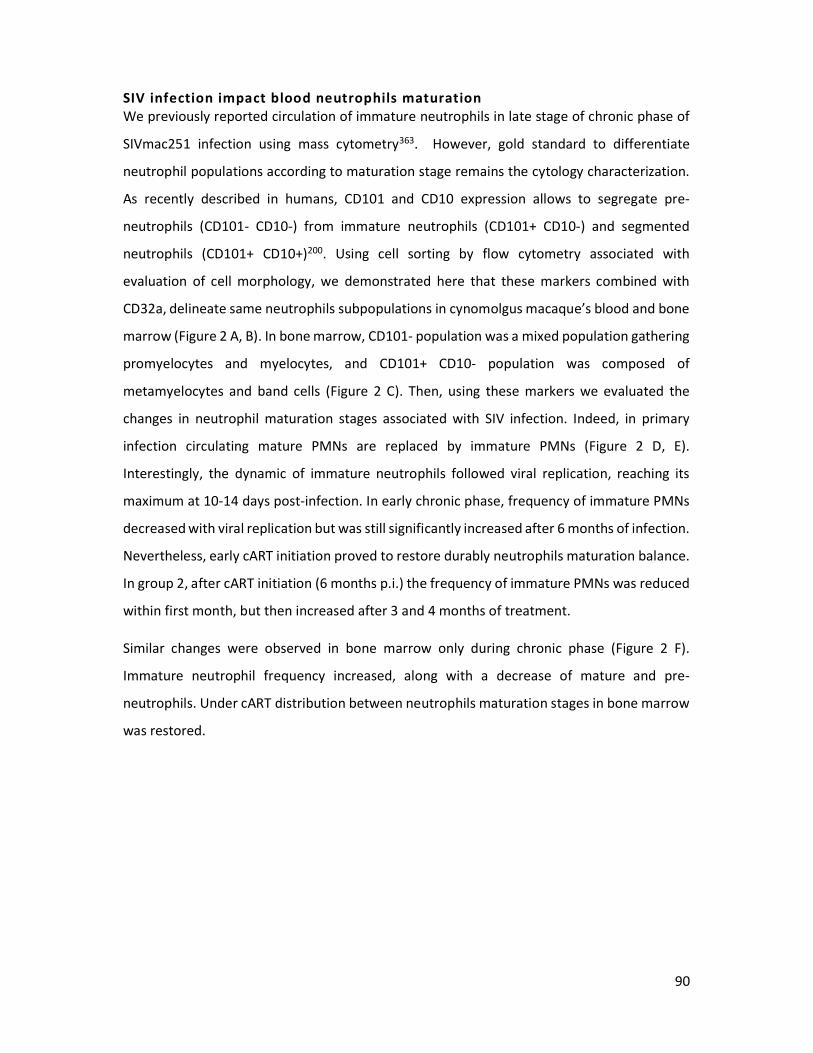
90
SIV infection impact blood neutrophils maturation We previously reported circulation of immature neutrophils in late stage of chronic phase of
SIVmac251 infection using mass cytometry363. However, gold standard to differentiate
neutrophil populations according to maturation stage remains the cytology characterization.
As recently described in humans, CD101 and CD10 expression allows to segregate pre-
neutrophils (CD101- CD10-) from immature neutrophils (CD101+ CD10-) and segmented
neutrophils (CD101+ CD10+)200. Using cell sorting by flow cytometry associated with
evaluation of cell morphology, we demonstrated here that these markers combined with
CD32a, delineate same neutrophils subpopulations in cynomolgus macaque’s blood and bone
marrow (Figure 2 A, B). In bone marrow, CD101- population was a mixed population gathering
promyelocytes and myelocytes, and CD101+ CD10- population was composed of
metamyelocytes and band cells (Figure 2 C). Then, using these markers we evaluated the
changes in neutrophil maturation stages associated with SIV infection. Indeed, in primary
infection circulating mature PMNs are replaced by immature PMNs (Figure 2 D, E).
Interestingly, the dynamic of immature neutrophils followed viral replication, reaching its
maximum at 10-14 days post-infection. In early chronic phase, frequency of immature PMNs
decreased with viral replication but was still significantly increased after 6 months of infection.
Nevertheless, early cART initiation proved to restore durably neutrophils maturation balance.
In group 2, after cART initiation (6 months p.i.) the frequency of immature PMNs was reduced
within first month, but then increased after 3 and 4 months of treatment.
Similar changes were observed in bone marrow only during chronic phase (Figure 2 F).
Immature neutrophil frequency increased, along with a decrease of mature and pre-
neutrophils. Under cART distribution between neutrophils maturation stages in bone marrow
was restored.
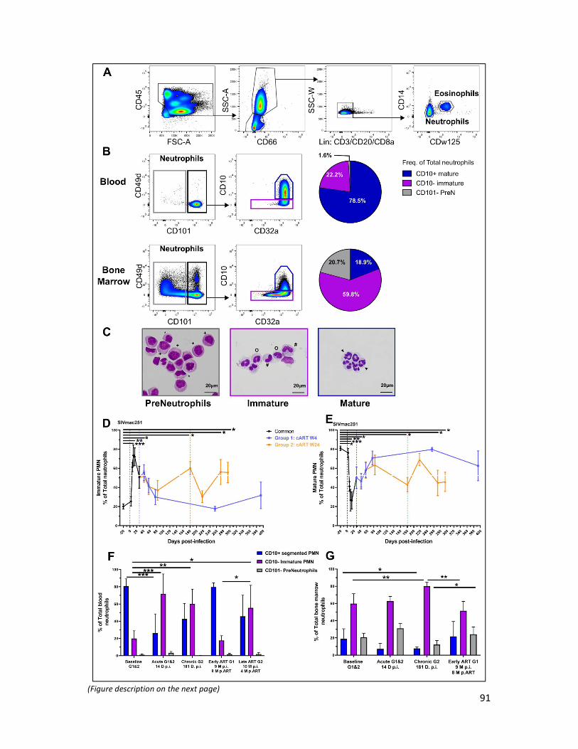
91
(Figure description on the next page)
92
Figure 2: Circulation of immature neutrophils in primary and chronic SIV infection. A) Gating strategy used to identify PMNs in blood and bone marrow from cynomolgus macaques. B) Identification of PMN’s maturation stages, corresponding distribution and cytology in blood and bone marrow. CD101- Pre-neutrophils population is composed of promyelocyte (+) and myelocytes (*). CD101+ fraction is composed of immature CD10- (#: metamyelocytes, o: band cell) and mature CD10+ (arrow: segmented PMN). D-E) Frequency of immature and mature PMNs in blood during SIV infection and treatment. Before cART initiation for group 1, both groups are represented together in a black line and are then separated. Graphs values indicates mean with SEM for both groups, with * p<0.05, ** p<0.01, ** p<0.001, ***p<0.0001 by paired Friedman test with Dunn’s multiple comparison. F-G) Frequency in blood and bone marrow of each PMN’s maturation stages in acute, chronic untreated and treated SIV infection. * p<0.05, ** p<0.01, ** p<0.001 by paired Friedman test with Dunn’s multiple comparison and Mann Whitney test for unpaired comparison between groups.
Neutrophils stimulated T-cells cytokine production and modulates T-cells proliferation in acute and chronic SIV infection As reported for several distinct diseases in human, PMN can have pro-inflammatory or
immunosuppressive function. In order to evaluate T cells immunomodulation by PMN in SIV
infection, we analyzed cytokine production by flow cytometry of TCR-activated T cells, in
presence or absence of autologous PMNs. We used density gradient centrifugation to isolate
neutrophils from blood. This approach, however, do not allow to retain low-density
neutrophils (LDN), which remain in the mononuclear fraction. Nevertheless, we did not
observe significant changes in LDN frequency in the PBMC fraction of SIV infected macaques.
We therefore surmise that impact of LDN on T cells function may be minimal, although this
hypothesis remains to be confirmed (Supplementary Fig. 2). Before infection, PMN had no
effect on CD4+ and CD8+ T cells IL-2, IL-17A, TNFa production, but were able to significantly
increase IFN- production by CD8+ T cells (Figure 3 A-F). In primary infection, neutrophils
significantly increased IL-2 and IFN- production by both CD4+ and CD8+ T cells (Figure 3 A, B,
C, D). Interestingly, PMN also increased IL-17A production by CD4+ T cells in acute phase. This
immunomodulatory function is associated with high frequency of primed immature PMN
observed in acute phase. In chronic phase, PMN did not modulate IL-2 production by T cells,
but they induced a significant increase of TNF- production by CD4+ T cells, with a trend to
increase IFN- production by CD8+ T cells (Figure 3 C-D). This suggest a switch in PMN
immunomodulatory function from acute to chronic phase, with globally, a pro-inflammatory
effect on CD4+ and CD8+ T cells cytokine production.

93
Figure 3: T cells immunomodulation by neutrophils during SIV infection. A-F) TCR-stimulated T cells were cultured 16h in presence or absence of PMN at a 5:1 PMN to T-cell ratio. T cell cytokines production was measured by intracellular cytokine staining of IL-2, IFN-g, TNFa and IL-17A after co-culture. Graphs represent frequency of CD4+ T cells or CD8+ T cells positive for each cytokine in absence (black) and presence of PMN (purple). G-H) TCR-stimulated T cells were cultured 96h in presence or absence of PMN at a 5:1 PMN to T-cell ratio. T-cell proliferation was measured by CFSE dilution and populations identified by flow cytometry. Graphs represent frequency of CD69+ CD4+ T cells or CD8+ T cells with CFSE dilution. * p<0.05, ** p<0.01, ** p<0.001 by Wilcoxon or Mann-Whitney tests depending on pairing. Baseline: group 1&2, Acute week 2: group 1, Chronic: group 2 (6M p.i.), Chronic cART: Group 1 (7M p.i., 6M pART).
94
We also measured proliferation of TCR-activated T-cells in presence or absence of PMN using
CFSE dilution after 96 hours of ex-vivo stimulation (Figure G, H). During acute phase, PMN
stimulated CD4+ T cells proliferation but had no significant effect during chronic phase and
under cART. Before infection, PMNs stimulated CD8+ T cells proliferation, whereas in acute
phase they demonstrated a suppressive effect on proliferation. Interestingly, in non-treated
animals, during chronic infection (6 months p.i.), this effect on proliferation was lost, whereas
basal proliferation of CD8+ T cells without PMNs remain elevated compare to pre-infection
level. In cART treated animals (group 1, 9M p.i., 8M p.ART) CD8+ T cells proliferation is
suppressed by PMNs, although the low number of studied animals may be insufficient to reach
statistically significant values (p=0,0625). These results suggest a T cells-dependent
immunomodulatory function of PMNs in SIV infection which evolves in the course of infection
and with a differential effect on CD4+ and CD8+ T cells.
5) Discussion
Description of neutrophils heterogeneity in peripheral blood, such as different maturation and
activation status, have been made in cancers and autoimmune diseases199. In these chronic
inflammatory diseases, new neutrophil functions have been described such as modulation of
T-cell responses. Neutrophils are also equipped with mediators potent to induce tissue
damages and maintain chronic inflammation. Chronic HIV-1 infection, even under cART, could
be considered as a chronic inflammatory disease, in which neutrophils had received little
attention. A recent study has demonstrated the link between neutrophils and microbial
translocation, a source of HIV-associated chronic inflammation87. Thus, characterization of
neutrophils heterogeneity and immunomodulatory function in HIV-1 infection would help to
better understand their roles in the maintenance of inflammation.
Here we provide the first description of neutrophil’s heterogeneity associated with T-cells
immunomodulatory functions during SIV infection. Using the association of CD101, CD10 and
CD32a expression, we were able to distinguished precursors, immature and mature
neutrophils in blood and bone marrow from SIV-infected macaques. In primary infection,
circulating PMNs were predominantly composed of immature neutrophils. This immature
phenotype was associated with an increased level of priming markers HLADR, CD64 and CD62L
shedding. Definition of PMN priming relies on partial activation of NADPH-oxidase associated
95
with an increase responsiveness to stimulus. We evaluated PMN priming using CD62L-low
shedding since it have been associated with NADPH-oxidase activation364,365, but we did not
directly evaluated NADPH-oxidase function. In peripheral blood, CD62L shedding has been
classically associated with primed hypersegmented neutrophils, notably in acute bacterial
infection366. Here immature PMNs had similar dynamic than CD62L-low PMNs, and to confirm
priming it would be necessary to evaluate NADPH-oxidase function in the course of SIV
infection. HIV infection has also a different physiopathology compare to bacterial infections
which may explain major differences in term of neutrophils heterogeneity. Changes in
peripheral blood neutrophil populations observed in primary infection partially resolved in
early chronic phase, but frequency of CD62L-low PMN and immature PMN increased in late
chronic phase. These results are consistent with bone marrow neutrophils analysis in which
primed immature PMN are increased in chronic SIV infection. Early initiation of cART (4 weeks
post-infection) protected against long term PMN changes observed in untreated chronic SIV
infection. Indeed, after 6 months under cART frequency of immature and primed PMN was
back to basal levels. Neutrophils are also potent to store and produce several cytokines under
stimulation24. As an immune cell involves in chronic inflammation, it will be interesting to
evaluate their cytokine production capacities in the course of SIV-infection and under cART.
We showed that neutrophils could differentially modulates CD4+ and CD8+ T cells. Primed
CD62L-low immature CD10- neutrophils demonstrated an immunostimulatory function
against T cells in primary infection. Notably, PMN increased IL-2 and IFN- production by CD4+
and CD8+ T cells, and increased CD4+ T cells IL-17A production. PMNs differentially modulates
T cells by increasing CD4+ T cells proliferation, but interestingly decreased CD8+ T cells
proliferation. It will be necessary to evaluate the impact of PMNs on SIV-specific CD8+ T cells
function, considering that PMNs could impaired antiviral response. In chronic SIV infection,
we observed immunostimulatory function restricted to CD4 T cells, in which PMN increased
TNF- production. Thus, depending on the phase of infection and which cell PMN interact
with (CD4 vs CD8 T cell), neutrophils shown different immunomodulation.
Acute bacterial and viral infections or LPS and G-CSF administration induce all circulation of
immature neutrophils, known as “left shift”, and PMN priming 333,367–369. Primary SIV infection
mimic this phenomenon with mobilization apparently from bone marrow mobilization. But
persistent inflammation in chronic SIV infection have been shown to alter hematopoiesis 370,371
96
and may lead to neutrophils impairment. Another hypothesis is that chronic viral infection
lead to reprogramming of neutrophil precursors, producing different granulocytes compare
to steady state. Indeed, SIV/HIV chronic inflammation affects neutrophils function, as it has
been demonstrated for phagocytosis, ROS production, and in this study, for T-cells
immunostimulatory properties 363372319. In group 1, we also explored the impact of cART on
immunomodulatory properties of PMNs. Nevertheless, due to the number of animals we lack
of statistical power, but identified an interesting trend to suppressive function against CD8+ T
cells proliferation. Under cART, PMNs did not demonstrated significant immunomodulatory
properties against T cells. We hypothesize that early cART restored PMN function by reducing
systemic inflammation.
Here, we used a co-culture assay thanks density-based neutrophil sorting because of the lack
of available sorting methods in cynomolgus macaques. This method retrieves normal-density
neutrophils (NDN) and had no impact in term of PMN priming, but it missed the effect of LDN
which are located in PBMC fraction. Bower NL. , Helton ES. et al have shown the capacity of
LDN to suppress CD8+ T cells proliferation and IFN-g production in chronically HIV-infected
patients, which was mediated by PD-L1326. In our study we did not showed significant increase
of LDN in the course of SIV infection. In addition, we did not observe nor CD8+ T cells
suppression of IFN- production in chronic SIV infection, neither PD-L1 expression on
neutrophils (supplementary Fig3). In this study HIV-infected individuals were chronically
infected for years, and authors selected patients on the basis of PD-L1 expression to perform
co-culture assay. Here, we followed only for 6 months SIV infection and PD-L1 expression on
neutrophils may necessitate a longer time of infection to be up-regulated. In G-CSF-treated
patients, Marini O. et al demonstrated opposite immunomodulatory function of immature
CD10- and mature CD10+ neutrophils, being immunostimulatory and immunosuppressive
respectively 333. In our study we identified significant PMN immunostimulatory function in
primary and chronic SIV infection, when frequency of immature CD10- and primed CD62L-low
PMN were significantly increased. To confirm that immunostimulatory function is restricted
to immature CD10- PMN in SIV infection, it will be necessary to evaluate separately the
function of CD10- immature and CD10+ mature PMN.
In sum we demonstrated neutrophils heterogeneity in SIV infection and its impact on T cells
function. Primed CD62L-low and immature CD10- neutrophils were increased in primary and
97
chronic SIV infection. This phenotype was associated with immunostimulatory function
against T-cells proliferation and cytokine production. In turn, PMNs could participate to
chronic inflammation by modulating T cells response. But deep functional characterization is
needed in both macaque models and patients to better understand the role of immature
neutrophils in HIV-1 infection. If they turn to be important as pro-inflammatory cells,
treatments aiming to modulate neutrophils could be used as strategies to reduce chronic
inflammation patients.
Acknowledgements This work was supported by the “Programme Investissements d’Avenir” (PIA), managed by
the ANR under reference ANR-11-INBS-0008, funding the Infectious Disease Models and
Innovative Therapies (IDMIT, Fontenay-aux-Roses, France) infrastructure, and ANR-10-EQPX-
02-01, funding the FlowCyTech facility (IDMIT, Fontenay-aux-Roses, France). We thank Gilead
and ViiV for providing the antiretroviral drugs. Animal studies were supported by the ANRS
and MSD Avenir, as part of the pVISCONTI research program, and the SIVART ANRS-IDMIT CO1
research program. We thank all members of the FlowCyTech, Animal Science and Welfare,
and Infectiology Immunology Laboratory core facilities of the IDMIT infrastructure for their
excellent expertise and outstanding contribution. We thank Gabrielle Carvalho, Julien Mafille
and Shahul Mouhamad from Miltenyi biotech for their help on cell sorting.
We thank Dr V. Witko-Sarsat for her help and outstanding expertise on neutrophils, which
increased our understanding of neutrophils biology.

98
Supplementary Figure 1: Gating strategies used to identify neutrophils in cynomolgus macaques. Gating strategy used in conventional flow cytometry to identify neutrophils according to their maturation stage (A) and priming (B).
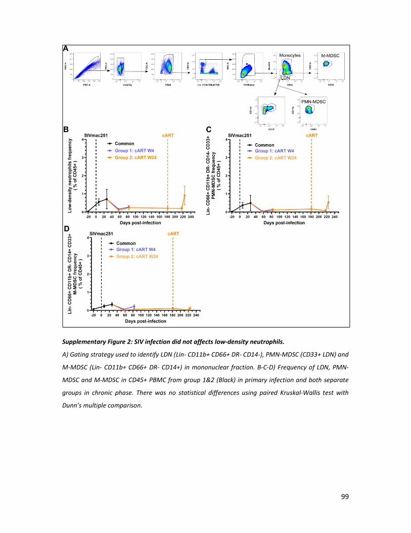
99
Supplementary Figure 2: SIV infection did not affects low-density neutrophils.
A) Gating strategy used to identify LDN (Lin- CD11b+ CD66+ DR- CD14-), PMN-MDSC (CD33+ LDN) and
M-MDSC (Lin- CD11b+ CD66+ DR- CD14+) in mononuclear fraction. B-C-D) Frequency of LDN, PMN-
MDSC and M-MDSC in CD45+ PBMC from group 1&2 (Black) in primary infection and both separate
groups in chronic phase. There was no statistical differences using paired Kruskal-Wallis test with
Dunn’s multiple comparison.
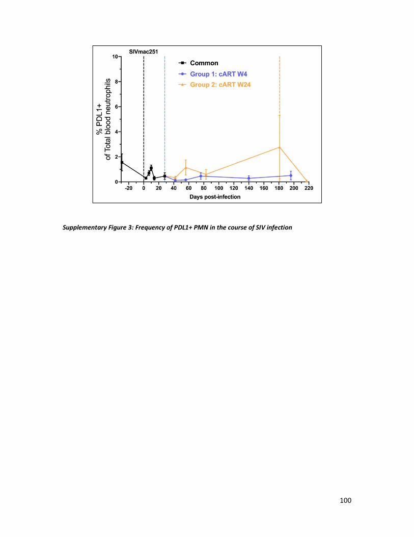
100
Supplementary Figure 3: Frequency of PDL1+ PMN in the course of SIV infection
101
III. Unpublished results: tissue neutrophils heterogeneity in cynomolgus
macaques
A. Introduction
HIV-1 infection takes place mainly in lymphoid tissues where the virus replicates, even under
antiretroviral treatment. Residual replication, microbial translocation and co-infections are
major sources of chronic inflammation observed in HIV-1 infected patients under cART373.
Thus, to better understand causes and consequences of chronic inflammation it is necessary
to explore at the tissue level. Myeloid cells, especially macrophages, have been implicated in
the setting and the maintenance of chronic immune activation. Since neutrophils are potent
pro-inflammatory cells, it is important to better characterize their phenotype and function in
tissues during HIV infection. Nevertheless, neutrophil heterogeneity has been studied mainly
in blood and bone marrow but less is known about tissues. Recently, neutrophil heterogeneity
was associated with tissue-related neutrophil function and depends on inflammatory
environment352. This have been demonstrated in inflammatory bowel disease, in which there
is an increase of circulating and lamina propria CD177+ neutrophils, releasing more ROS, MPO
and NETs under stimulation compare to CD177-374.
To study phenotype of tissues neutrophils from cynomolgus macaque, we used flow
cytometry associated with fast and gentle digestion protocol to retrieve leucocytes. Here we
present preliminary data from healthy animals, which validate the methodology used and
gives for the first time distribution and phenotype of tissue’s neutrophils from cynomolgus
macaque. In the future, we will use this approach along with histology to characterize
neutrophils in SIV infected macaques.
B. Material and method
We analyzed samples from five cynomolgus macaques (Macaca fascicularis), imported from
Mauritius, housed at the IDMIT infrastructure animal facility at the CEA, Fontenay-aux-Roses,
France. Blood was drawn and bone marrow aspirates collected under anesthesia, consisting
of an intramuscular injection of 10 mg/kg zolazepam/tiletamine (Virbac, Carros, France).
Blood was collected into lithium-heparin tubes (Vacutainer BD, USA) and bone marrow
aspirates from the iliac crest or humeral head using a 10-ml 18G-syringe containing citrate -
102
dextrose (10 mg/ml). Bronchoalveolar lavages were collected using 20ml of NaCl injected
using endotracheal tube and were kept at 4°C with RPMI. At necropsy, spleen was kept on
RPMI at 4°C, processed using gentleMACS dissociator, and cell suspension was filtered with
70µm mesh. Red blood cell lysis was performed using NH4Cl hypotonic lysis. For the gut, 3cm
of distal ileum and descendant colon were opened and washed to remove fecal content. To
focus on lamina propria and Payer patches, epithelial layer was removed for 20 min using 6.4
mM EDTA. Tissue digestion was performed using DNAse I and Collagenase D for 20 min at
37°C. Cell suspension obtained from tissues were resuspended at 3 million cells per 100µL
before staining.
Viability staining was first performed for 15 min at room temperature, according to the
manufacturer’s protocol (Live Dead, Thermofisher). Then, antibody staining was performed
with the following antibodies for 30 min at 4°C Bone marrow and blood samples were then
lysed and fixed using BD FACS Lysing (BD Bioscience) for 15 min. After an additional wash with
PBS, acquisition was performed on a BD FORTESSA flow cytometer (BD Bioscience) and
analyzed using FlowJo software.
C. Preliminary results
Flow cytometry allowed to delineate neutrophils from all tissue samples (Figure 1A-B), except
from mesenteric lymph nodes (draining ileum) in which we identified less than 100 cells. We
first evaluated neutrophils frequency among leucocytes from bone marrow, blood,
bronchoalveolar lavages (BAL), ileum and colon (Figure 1B). In the spleen and BAL, neutrophils
represented 11.9% and 7.8% of total CD45+ leucocytes respectively. We also identified
significant neutrophil population in ileum and colon, representing approximately 1% of total
leucocytes. We also evaluated maturation stages in blood, bone marrow and spleen (Figure
1C). In the blood and bone marrow we obtained a distribution of maturation stages equivalent
to previous studies.
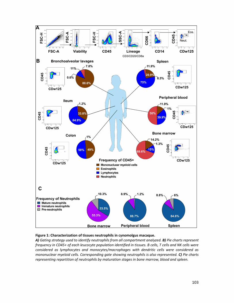
103
Figure 1: Characterization of tissues neutrophils in cynomolgus macaque. A) Gating strategy used to identify neutrophils from all compartment analyzed. B) Pie charts represent frequency in CD45+ of each leucocyte population identified in tissues. B cells, T cells and NK cells were considered as lymphocytes and monocytes/macrophages with dendritic cells were considered as mononuclear myeloid cells. Corresponding gate showing neutrophils is also represented. C) Pie charts representing repartition of neutrophils by maturation stages in bone marrow, blood and spleen.
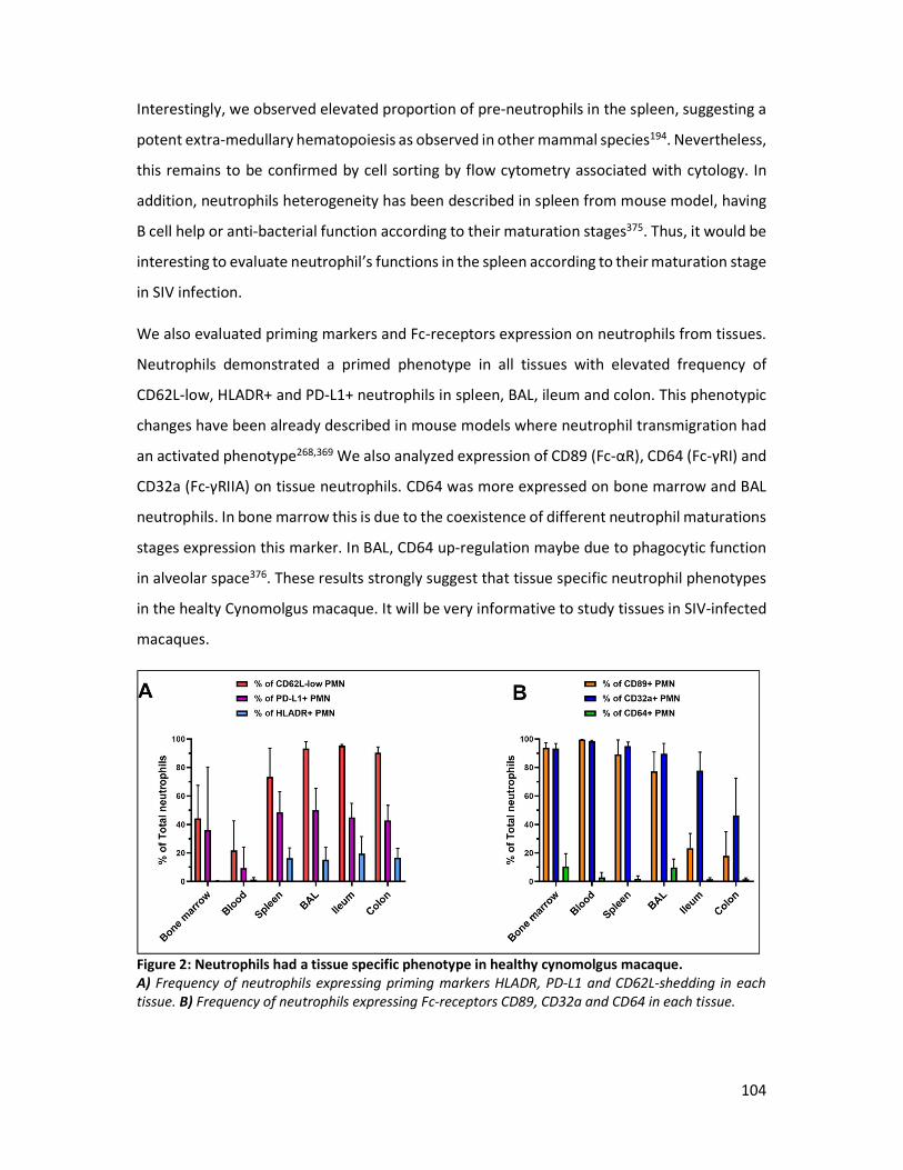
104
Interestingly, we observed elevated proportion of pre-neutrophils in the spleen, suggesting a
potent extra-medullary hematopoiesis as observed in other mammal species194. Nevertheless,
this remains to be confirmed by cell sorting by flow cytometry associated with cytology. In
addition, neutrophils heterogeneity has been described in spleen from mouse model, having
B cell help or anti-bacterial function according to their maturation stages375. Thus, it would be
interesting to evaluate neutrophil’s functions in the spleen according to their maturation stage
in SIV infection.
We also evaluated priming markers and Fc-receptors expression on neutrophils from tissues.
Neutrophils demonstrated a primed phenotype in all tissues with elevated frequency of
CD62L-low, HLADR+ and PD-L1+ neutrophils in spleen, BAL, ileum and colon. This phenotypic
changes have been already described in mouse models where neutrophil transmigration had
an activated phenotype268,369 We also analyzed expression of CD89 (Fc-αR), CD64 (Fc-γRI) and
CD32a (Fc-γRIIA) on tissue neutrophils. CD64 was more expressed on bone marrow and BAL
neutrophils. In bone marrow this is due to the coexistence of different neutrophil maturations
stages expression this marker. In BAL, CD64 up-regulation maybe due to phagocytic function
in alveolar space376. These results strongly suggest that tissue specific neutrophil phenotypes
in the healty Cynomolgus macaque. It will be very informative to study tissues in SIV-infected
macaques.
Figure 2: Neutrophils had a tissue specific phenotype in healthy cynomolgus macaque. A) Frequency of neutrophils expressing priming markers HLADR, PD-L1 and CD62L-shedding in each tissue. B) Frequency of neutrophils expressing Fc-receptors CD89, CD32a and CD64 in each tissue.

105
106
Discussion and perspectives
I. Validation of experimental approach
In these studies, we used cynomolgus macaques infected by SIVmac251 as a model for HIV-1
infection. We injected intravenously 1000 animal infectious dose 50% (AID50) allowing to
infect all animals, and reproducing physiopathology observed in HIV-1 infected patients, with
progression from primary infection to long-term chronic infection, and lastly AIDS165,336.
Cynomolgus macaques from Mauritius have the advantage to show a limited genetic diversity,
but possess multiples MHC haplotypes associated with differential progression to AIDS337.
SIVmac251 is phylogenetically related to HIV-2, but in cynomolgus macaques notably because
of restriction factors virus-host interaction leads to a dynamic of infection close to human.
This model is also useful to study pharmacological sanctuaries and reservoirs because most of
antiretroviral treatment used in patients suppress SIVmac251 replication377. Lastly, compared
to mouse models, immune system of cynomolgus macaque is closer to human, making
nonhuman primates more suitable models for infectious diseases pre-clinical research378,379.
In this study, we also performed a long-term follow-up of immunological parameters from
primary infection to advanced chronic infection. Therefore, we reduced interindividual
heterogeneity and sample size by using paired statistical analyses.
Our work was focused on macaques neutrophils, which demonstrated similar differentiation
in bone marrow compare to human380. But compared to human, cynomolgus macaque’s
neutrophils lack CD16b expression which may lead to different responses to antibodies such
as ADCC381. To analyze neutrophil immunomodulatory functions, we used density gradient
sedimentation, in the absence of validated kit to specifically purify these cells. Thus, globular
fraction used was contaminated by few basophils and by 3-10% of eosinophils depending on
the animal. For this reason, we are currently developing a negative sorting protocol based on
magnetic microbeads. This will permit to work on highly purified neutrophils as well as
neutrophils subpopulations from cynomolgus macaque for functional analysis. Furthermore,
this method will allow a fast recovery of neutrophils, which are fragile cells.
In sum, cynomolgus macaque is a relevant preclinical model to better understand
physiopathology of HIV-1 infection and response to treatments. Existence of few differences
in term of leukocytes population’s markers is one limitation of this model. In addition,
107
functional analyses of neutrophils were limited by available sorting procedure for non-human
primates. For this reason, we are developing a purification method to refine our
understanding of neutrophils.
II. Discussion
Here we provided the first description of neutrophil heterogeneity in SIV infected macaques.
Taking advantage of multiparametric mass cytometry, we identified circulating immature
neutrophils in advanced chronic SIV infection, underlying mobilization of bone marrow cells
as described in sepsis382. Based on human markers delineating maturation stages, mass
cytometry analysis was able to identify pre-neutrophils, immature neutrophils and mature
neutrophils. In early chronic phase, reducing the number of markers for flow cytometry, we
abolished the ability to differentiate immature from mature neutrophils in blood as the
neutrophil population was homogeneous in terms of CD66 and CD32 expression. Other
markers were necessary to better delineate neutrophil maturation, such as CD49d, CD101,
and CD10, recently described in human blood and bone marrow200. We used these markers in
our second work. In advanced HIV-1 infection, neutrophils were already known to be
impaired, showing priming, reduce phagocytosis and oxidative burst in correlation with viral
load and CD4 T cell count372,383. Our analysis also showed that neutrophils were primed earlier
in chronic infection, with reduced phagocytic capacities. These results suggest that changes of
neutrophils phenotype and functions occur earlier in the course of infection.
To better characterize neutrophil priming and maturation in the course of SIV infection, we
tested new markers and combined classical flow cytometry cell sorting with cytology in our
second study. We had the opportunity to study animals in a longitudinal study, called p-
VISCONTI, on SIV infection and impact of early or delay treatment on immune response. The
p-VISCONTI project aimed also to induce post-treatment control of infection in cynomolgus
macaques, as observed in VISCONTI cohort384. Interestingly, SIV infection had no significant
impact on neutrophil blood count, which was highly variable among animals and time points.
But when analyzing neutrophil subpopulations, we revealed the impact of SIV infection. In
primary infection, CD10- immature and CD62L-low primed neutrophils were increased in
peripheral blood, following the dynamic of viral replication. Despite a partial resolution after
108
primary infection, frequencies of immature and primed neutrophils were maintained elevated
after 6 months of infection. We observed also a global decrease of mature neutrophils in bone
marrow and blood suggesting a mobilization to tissues, an increase of apoptosis or a lack of
differentiation. Indeed, the increase of neutrophil apoptosis has been shown in SIV primary
infection, but other mechanisms may explain what we observed in chronic phase. Indeed, we
observed increased frequency of immature neutrophils in bone marrow only in chronic phase,
suggesting an impact on bone marrow production. In sum, primary SIV infection mimic a
classical acute infection with elevated activation of neutrophils, mobilization of mature to
tissues and emergency granulopoiesis liberating immature cells into the circulation21. But
persistence of viral replication and inability of immune system to clear the virus in chronic
infection was associated with maintenance of phenotypic changes, notably by impacting bone
marrow production.
Immunomodulatory function of neutrophils is a recent discovery which has been linked to
maturation stage by Marini O. et al 333. In G-CSF treated donors, they demonstrated opposite
role of CD10+ mature and CD10- immature neutrophils with respectively immunosuppressive
and immunostimulatory effect on T cells. Here we have also seen an association with CD10-
immature neutrophils and immunostimulatory function on T cells cytokine production and
CD4 T cells proliferation. This effect was more pronounced in primary infection than in chronic
phase notably because of a higher frequency of CD10- immature neutrophils at this time point.
By using density-based sorting, we retrieved only normal-density neutrophils (NDN) and we
missed the effect of low-density neutrophils (LDN) which are located in the PBMC fraction.
Brower NL. , Helton ES. et al have shown the capacity of LDN and NDN to suppress T cells
proliferation and IFN-γ production in chronically HIV-infected patients, which was mediated
by PD-L1326. In our study we did not showed significant increase of LDN in the course of SIV
infection. In addition, we did not observe nor suppression of IFN-γ production by T cells in
chronic SIV infection, neither PD-L1 expression on neutrophils. Nevertheless, in Brower’s
study HIV-infected individuals were chronically infected for years whereas we analyzed
neutrophil functions at only 6 months post-infection. In advanced HIV-1 infection, neutrophil
alteration may be exacerbated and LDN circulation may increase with specific
immunomodulatory properties.

109
When cART was initiated early (4 weeks post-infection), neutrophil phenotype progressively
returns to basal level. On the contrary, initiation of cART after 6 months of infection did not
fully restored phenotype. The later the treatment is initiated, the more persistent is chronic
inflammation and its impact158. So, two mechanisms may explain the impact of delay cART on
neutrophils phenotype and function: 1- SIV infection and chronic inflammation had increased
bone marrow production and neutrophils turnover leading to circulation of immature cells, 2-
persistence of inflammation change neutrophils precursors, giving rise to functionally
different cells. Both mechanisms may also coexist, but bone marrow analysis showed elevated
proportion of immature neutrophils and reduction of pre-neutrophils in chronic phase,
supporting the first hypothesis. We are currently analyzing bone marrow from animals treated
after 6 months of infection to evaluate if these changes are maintained under cART. To explore
the second hypothesis, it would be necessary to evaluate epigenetic modifications of
neutrophils precursors, which could explain the generation of functionally different
neutrophils385.
III. Perspectives
Chronic inflammation occurs when the host is unable to eliminate the initiating stimuli, or
when the host fails to resolve inflammation. Neutrophils have been associated with metabolic,
cardiovascular and neurological diseases, which are all chronic inflammatory diseases21.
Interestingly, these diseases constitute non-AIDS related co-morbidities. Our work has
highlighted a role of neutrophils in modulation of T cell response in SIV-infected macaques,
leading to an increase of pro-inflammatory secretion. We have shown neutrophil
heterogeneity, but we did not analyze function of each subpopulation. For this reason, we are
developing new tools to better sort neutrophils in cynomolgus macaque. Thus, to confirm that
immunostimulatory function is restricted to immature CD10- PMN in SIV infection, we plan to
evaluate separately the function of CD10- immature and CD10+ mature PMN. Using this
sorting tool, phagocytosis, ROS production, cytokine production, ADCC, NETosis, DC
immunomodulation are potential neutrophils function we could analyze in these
subpopulations. But we must learn and develop most of these functional assays we did not
used yet. In parallel, to obtain a precise definition of mature and immature PMN during SIV

110
infection, we will perform a transcriptomic analysis on sorted cells from blood and bone
marrow. This will increase our knowledge on neutrophils in the course of infection, and it will
reveal pathways to focus our work on specific functions.
In addition, our results suggest mobilization of mature neutrophils in tissues, and except for
the gut, the role of tissue neutrophils has not been explored in HIV infection. With cynomolgus
macaque model, we plan to first explore neutrophils phenotype in tissues using the
combination of flow cytometry and multiparametric immunohistology. To exert
immunomodulation of T cells, neutrophils must be present in the same tissue than T cells.
White pulp of the spleen and GALT are of particular interest because neutrophils were shown
to infiltrate these lymphoid tissues. Thus, immunohistofluorescence will provide in-situ
colocalization and neutrophils phenotype. Collecting neutrophils from tissue is still a difficult
task to harvest enough cells, so we will focus on spleen, bone marrow and broncholaveolar
lavages for functional analyzes. Taking advantage of positron-emission tomography
associated to tomodensitometry present in our facility, it will be interesting to use neutrophil-
specific radiotracer recently developed in order to identify tissues infiltrated by neutrophils in
the course of SIV infection386. This whole-body imaging technology will give access to tissue
neutrophils dynamic, allowing to visualize marginated pool, lung, spleen and liver neutrophils.
More importantly, it will be also necessary to confirm these results in HIV-infected patients at
different stages of the disease. For this purpose, thanks to the access to ANRS cohorts of
patients, we planned to analyze blood samples from HIV-1 infected patient in primary
infection, chronic phase and under cART to evaluate neutrophils phenotype and function.
Neutrophils appear to participate to chronic inflammation. If indeed they are important
modulators of inflammation it would be interesting to analyze PMN in natural host, such as
African green monkey, in which resolution of inflammation seems to be the answer against
AIDS. Tools we developed for cynomolgus macaques could be adapted for this specie.
Pathogenic models and natural hosts could be used to evaluate new therapeutic strategies
targeting neutrophils to reduce chronic inflammation.
111
If neutrophils represent an important contributor in HIV chronic inflammation, targeting them
could reduce inflammation. Research to modulate neutrophil activation in other chronic
inflammatory diseases may inspire new therapeutic strategies for HIV infection, since they are
sharing common mechanisms. Few drugs modulating neutrophils activity have been
developed recently. In mouse models of atherosclerosis or lupus, peptidyl arginine deiminase
4 (PAD4) inhibition (Cl-amidine) reduced NET formation and related tissue damages387,388.
Blocking leukocytes recruitment is another strategy targeting mainly adhesion molecule, but
they are not neutrophil specific21. Reducing neutrophil infiltration in tissues to resolve
inflammation is another interesting strategy, which could be interesting for HIV-infection.
Inducing neutrophil apoptosis would decrease tissue damages and phagocytosis of apoptotic
neutrophils by macrophages (efferocytosis) would induce resolution of inflammation. R-
Roscovitine (inhibitor of cyclin-dependent kinase) was shown to induce caspase-dependent
neutrophil apoptosis in mice389. It would be interesting to test these new drugs in preclinical
models of HIV-infection to reduce chronic inflammation and restore immune function.
For long, neutrophils have not been studied for their immunomodulatory properties,
considered as a homogenous phagocyte population. Modulation of the immune system by
neutrophils must be explored in other infectious diseases to better understand the link
between their heterogeneity and immune response against pathogens. Acute and chronic
respiratory diseases are one of the most important causes of severe illness and death
worldwide, with chronic obstructive pulmonary disease (COPD), acute lower respiratory tract
infections and tuberculosis are top three390. Interestingly, there are relationships between
neutrophils, lungs, and respiratory diseases. Tuberculosis in an interesting example of chronic
respiratory disease, but only few studies have demonstrated in tuberculosis a cross-talk
between neutrophils, DC and T cells238. Beside, acute respiratory distress syndrome (ARDS) is
a fatal complication of influenza infection due to epithelial cells damage and exaggerated
innate immune response, especially neutrophils391. In ARDS, neutrophils have been shown to
release NETs, ROS and pro-inflammatory cytokines leading to lung damage. But less is known
about their immunomodulatory properties, which could also participate to uncontrolled
immune response to influenza. Tuberculosis and ARDS are examples among respiratory
infections where neutrophil heterogeneity could participate to impair immune response. We
are currently developing programs to study physiopathology and vaccines against respiratory
112
infectious diseases in our facilities. Particularly, non-human primate models are available to
study tuberculosis, influenza infection and respiratory syncytial virus infection, which will be
used for these purposes. Taking advantage of these models, tools we developed to study
neutrophils in SIV infection could be used to explore neutrophil heterogeneity in these
diseases.
Neutrophil heterogeneity may originate from inflammation diversity, inducing modulation of
cell ontogeny and leading to different cellular functions. A better understanding of neutrophils
heterogeneity in infectious diseases will be necessary to better understand immunopathology
and to design new therapeutic strategies to block or enhance functions of this abundant and
efficient immune cell.

113
References 1. Centers for Disease Control (CDC). Pneumocystis pneumonia--Los Angeles. MMWR.
Morb. Mortal. Wkly. Rep. 30, 250–2 (1981).
2. Barre-Sinoussi, F. et al. Isolation of a T-lymphotropic retrovirus from a patient at risk for acquired immune deficiency syndrome (AIDS). Science (80-. ). 220(4599), 868–71 (1983).
3. Simon, V., Ho, D. D. & Abdool Karim, Q. HIV/AIDS epidemiology, pathogenesis, prevention, and treatment. Lancet (London, England) 368, 489–504 (2006).
4. Sharp, P. M. & Hahn, B. H. Origins of HIV and the AIDS pandemic. Cold Spring Harb. Perspect. Med. 1, a006841 (2011).
5. Worobey, M. et al. Direct evidence of extensive diversity of HIV-1 in Kinshasa by 1960. Nature 455, 661–4 (2008).
6. Peeters, M. et al. Risk to Human Health from a Plethora of Simian Immunodeficiency Viruses in Primate Bushmeat. Emerg. Infect. Dis. 8, 451–457 (2002).
7. Pepin, J. The origins of AIDS. (Cambridge University Press, 2011).
8. Quammen, D. The chimp and the river : how AIDS emerged from an African forest.
9. Unaids. Fact sheet - Latest global and regional statistics on the status of the AIDS epidemic.
10. Lagathu, C. et al. Basic science and pathogenesis of ageing with HIV. AIDS 31, S105–S119 (2017).
11. Hunt, P. W., Lee, S. A. & Siedner, M. J. Immunologic Biomarkers, Morbidity, and Mortality in Treated HIV Infection. J. Infect. Dis. 214, S44–S50 (2016).
12. Ploquin, M. J., Silvestri, G. & Müller-Trutwin, M. Immune activation in HIV infection: What can the natural hosts of simian immunodeficiency virus teach us? Current Opinion in HIV and AIDS 11, 201–208 (2016).
13. Cory, T. J., Schacker, T. W., Stevenson, M. & Fletcher, C. V. Overcoming pharmacologic sanctuaries. Curr Opin HIV AIDS 8, 190–195 (2013).
14. Sandler, N. G. et al. Plasma Levels of Soluble CD14 Independently Predict Mortality in HIV Infection. J. Infect. Dis. 203, 780–790 (2011).
15. Marchetti, G. et al. Microbial translocation predicts disease progression of HIV-infected antiretroviral-naive patients with high CD4+ cell count. AIDS 25, 1385–1394 (2011).
16. Kelesidis, T., Kendall, M. A., Yang, O. O., Hodis, H. N. & Currier, J. S. Biomarkers of microbial translocation and macrophage activation: association with progression of subclinical atherosclerosis in HIV-1 infection. J. Infect. Dis. 206, 1558–67 (2012).
17. Boulougoura, A. & Sereti, I. HIV infection and immune activation: the role of coinfections. Curr. Opin. HIV AIDS 11, 191–200 (2016).
18. Deeks, S. G., Tracy, R. & Douek, D. C. Systemic Effects of Inflammation on Health during
114
Chronic HIV Infection. Immunity 39, 633–645 (2013).
19. Musselwhite, L. W. et al. Markers of endothelial dysfunction, coagulation and tissue fibrosis independently predict venous thromboembolism in HIV. AIDS 25, 787–795 (2011).
20. Hensley-McBain, T. & Klatt, N. R. The Dual Role of Neutrophils in HIV Infection. Curr. HIV/AIDS Rep. 15, 1–10 (2018).
21. Soehnlein, O., Steffens, S., Hidalgo, A. & Weber, C. Neutrophils as protagonists and targets in chronic inflammation. Nat. Rev. Immunol. 17, 248–261 (2017).
22. Campillo-Gimenez, L. et al. Neutrophils in antiretroviral therapy-controlled HIV demonstrate hyperactivation associated with a specific IL-17/IL-22 environment. J. Allergy Clin. Immunol. 134, 1142–1152.e5 (2014).
23. Noubouossie, D. F., Reeves, B. N., Strahl, B. D. & Key, N. S. Neutrophils: back in the thrombosis spotlight. Blood 133, 2186–2197 (2019).
24. Tecchio, C., Micheletti, A. & Cassatella, M. A. Neutrophil-derived cytokines: Facts beyond expression. Frontiers in Immunology (2014). doi:10.3389/fimmu.2014.00508
25. Stacey, A. R. et al. Induction of a striking systemic cytokine cascade prior to peak viremia in acute human immunodeficiency virus type 1 infection, in contrast to more modest and delayed responses in acute hepatitis B and C virus infections. J. Virol. 83, 3719–33 (2009).
26. Mehandru, S. et al. Primary HIV-1 Infection Is Associated with Preferential Depletion of CD4 + T Lymphocytes from Effector Sites in the Gastrointestinal Tract. J. Exp. Med. 200, 761–770 (2004).
27. Guadalupe, M. et al. Severe CD4+ T-cell depletion in gut lymphoid tissue during primary human immunodeficiency virus type 1 infection and substantial delay in restoration following highly active antiretroviral therapy. J. Virol. 77, 11708–17 (2003).
28. Zhang, Z. Q. et al. Kinetics of CD4+ T cell repopulation of lymphoid tissues after treatment of HIV-1 infection. Proc. Natl. Acad. Sci. U. S. A. 95, 1154–9 (1998).
29. Li, Q. et al. Peak SIV replication in resting memory CD4+ T cells depletes gut lamina propria CD4+ T cells. Nature 434, 1148–1152 (2005).
30. Wykrzykowska, J. J. et al. Early regeneration of thymic progenitors in rhesus macaques infected with simian immunodeficiency virus. J. Exp. Med. 187, 1767–78 (1998).
31. Herbeuval, J.-P. et al. Differential expression of IFN- and TRAIL/DR5 in lymphoid tissue of progressor versus nonprogressor HIV-1-infected patients. Proc. Natl. Acad. Sci. 103, 7000–7005 (2006).
32. Douek, D. C., Roederer, M. & Koup, R. A. Emerging concepts in the immunopathogenesis of AIDS. Annu. Rev. Med. 60, 471–84 (2009).
33. Guadalupe, M. et al. Severe CD4+ T-cell depletion in gut lymphoid tissue during primary human immunodeficiency virus type 1 infection and substantial delay in restoration following highly active antiretroviral therapy. J. Virol. 77, 11708–17 (2003).
115
34. Pantaleo, G. et al. HIV infection is active and progressive in lymphoid tissue during the clinically latent stage of disease. Nature 362, 355–358 (1993).
35. Estes, J. D. Pathobiology Of HIV/SIV-Associated Changes In Secondary Lymphoid Tissues. Immunol. Rev. 254, 65–77 (2013).
36. Wong, J. K. & Yukl, S. A. Tissue reservoirs of HIV. Curr. Opin. HIV AIDS 11, 362–370 (2016).
37. Massanella, M., Fromentin, R. & Chomont, N. Residual inflammation and viral reservoirs: Alliance against an HIV cure. Curr. Opin. HIV AIDS 11, 234–241 (2016).
38. Massanella, M. & Richman, D. D. Measuring the latent reservoir in vivo. J. Clin. Invest. 126, 464–472 (2016).
39. Mavigner, M. et al. Simian Immunodeficiency Virus Persistence in Cellular and Anatomic Reservoirs in Antiretroviral Therapy-Suppressed Infant Rhesus Macaques. J. Virol. 92, (2018).
40. Durand, C. M. et al. HIV-1 DNA Is Detected in Bone Marrow Populations Containing CD4+ T Cells but Is not Found in Purified CD34+ Hematopoietic Progenitor Cells in Most Patients on Antiretroviral Therapy. J. Infect. Dis. 205, 1014–1018 (2012).
41. McNamara, L. A. et al. CD133+ hematopoietic progenitor cells harbor HIV genomes in a subset of optimally treated people with long-term viral suppression. J. Infect. Dis. 207, 1807–16 (2013).
42. Josefsson, L. et al. Hematopoietic Precursor Cells Isolated From Patients on Long-term Suppressive HIV Therapy Did Not Contain HIV-1 DNA. J. Infect. Dis. 206, 28–34 (2012).
43. Pantaleo, G. et al. HIV infection is active and progressive in lymphoid tissue during the clinically latent stage of disease. Nature 362, 355–358 (1993).
44. Günthard, H. F. et al. Residual Human Immunodeficiency Virus (HIV) Type 1 RNA and DNA in Lymph Nodes and HIV RNA in Genital Secretions and in Cerebrospinal Fluid after Suppression of Viremia for 2 Years. J. Infect. Dis. 183, 1318–1327 (2001).
45. Popovic, M. et al. Persistence of HIV-1 structural proteins and glycoproteins in lymph nodes of patients under highly active antiretroviral therapy. Proc. Natl. Acad. Sci. 102, 14807–14812 (2005).
46. Fukazawa, Y. et al. B cell follicle sanctuary permits persistent productive simian immunodeficiency virus infection in elite controllers. Nat. Med. 21, 132–139 (2015).
47. Banga, R. et al. PD-1+ and follicular helper T cells are responsible for persistent HIV-1 transcription in treated aviremic individuals. Nat. Med. 22, 754–761 (2016).
48. Yukl, S. A. et al. A Comparison of Methods for Measuring Rectal HIV Levels Suggests that HIV DNA Resides in Cells other than CD4+T Cells, Including Myeloid Cells. AIDS 28, 439 (2014).
49. Yukl, S. A. et al. The Distribution of HIV DNA and RNA in Cell Subsets Differs in Gut and Blood of HIV-Positive Patients on ART: Implications for Viral Persistence. J. Infect. Dis. 208, 1212–1220 (2013).
116
50. Madero, J. G. S. et al. Relationship between Load of Virus in Alveolar Macrophages from Human Immunodeficiency Virus Type 1-Infected Persons, Production of Cytokines, and Clinical Status. J. Infect. Dis. 169, 18–27 (1994).
51. Costiniuk, C. T. et al. HIV persistence in mucosal CD4+ T-cells within the lungs of adults receiving long-term suppressive antiretroviral therapy. AIDS 32, 1 (2018).
52. Churchill, M. J. et al. Use of laser capture microdissection to detect integrated HIV-1 DNA in macrophages and astrocytes from autopsy brain tissues. J. Neurovirol. 12, 146–152 (2006).
53. Winnall, W. R. et al. Simian immunodeficiency virus infection and immune responses in the pig-tailed macaque testis. J. Leukoc. Biol. 97, 599–609 (2015).
54. Wolff, H. et al. A comparison of HIV-1 antibody classes, titers, and specificities in paired semen and blood samples from HIV-1 seropositive men. J. Acquir. Immune Defic. Syndr. 5, 65–9 (1992).
55. Estes, J. D. et al. Defining total-body AIDS-virus burden with implications for curative strategies. Nat. Med. 23, 1271–1276 (2017).
56. Siddiqui, S. et al. Persistent Viral Reservoirs in Lymphoid Tissues in SIV-Infected Rhesus Macaques of Chinese-Origin on Suppressive Antiretroviral Therapy. Viruses 11, (2019).
57. Matusali, G. et al. Detection of Simian Immunodeficiency Virus in Semen, Urethra, and Male Reproductive Organs during Efficient Highly Active Antiretroviral Therapy. J. Virol. 89, 5772–5787 (2015).
58. Damouche, A. et al. Adipose Tissue Is a Neglected Viral Reservoir and an Inflammatory Site during Chronic HIV and SIV Infection. PLOS Pathog. 11, e1005153 (2015).
59. Dusserre, N. et al. In vitro HIV-1 entry and replication in Langerhans cells may clarify the HIV-1 genome detection by PCR in epidermis of seropositive patients. J. Invest. Dermatol. 99, 99S–102S (1992).
60. Martinez-Picado, J. & Deeks, S. G. Persistent HIV-1 replication during antiretroviral therapy. Curr. Opin. HIV AIDS 11, 417–23 (2016).
61. Palmer, S. et al. Low-level viremia persists for at least 7 years in patients on suppressive antiretroviral therapy. Proc. Natl. Acad. Sci. 105, 3879–3884 (2008).
62. Maldarelli, F. et al. ART Suppresses Plasma HIV-1 RNA to a Stable Set Point Predicted by Pretherapy Viremia. PLoS Pathog. 3, e46 (2007).
63. Lorenzo-Redondo, R. et al. Persistent HIV-1 replication maintains the tissue reservoir during therapy. Nature 530, 51–56 (2016).
64. North, T. W. et al. Viral Sanctuaries during Highly Active Antiretroviral Therapy in a Nonhuman Primate Model for AIDS. J. Virol. 84, 2913–2922 (2010).
65. Bourry, O. et al. Effect of a short-term HAART on SIV load in macaque tissues is dependent on time of initiation and antiviral diffusion. Retrovirology 7, 78 (2010).
66. Solas, C. et al. Discrepancies between protease inhibitor concentrations and viral load in reservoirs and sanctuary sites in human immunodeficiency virus-infected patients.
117
Antimicrob. Agents Chemother. 47, 238–43 (2003).
67. Dallas, S., Schlichter, L. & Bendayan, R. Multidrug Resistance Protein (MRP) 4- and MRP 5-Mediated Efflux of 9-(2-Phosphonylmethoxyethyl)adenine by Microglia. J. Pharmacol. Exp. Ther. 309, 1221–1229 (2004).
68. Jorajuria, S. et al. ATP binding cassette multidrug transporters limit the anti-HIV activity of zidovudine and indinavir in infected human macrophages. Antivir. Ther. 9, 519–28 (2004).
69. Odorizzi, P. M. & Wherry, E. J. An Interferon Paradox. Science (80-. ). 340, 155–156 (2013).
70. Ziegler, S. M. & Altfeld, M. Human immunodeficiency virus 1 and type I interferons-where sex makes a difference. Frontiers in Immunology (2017). doi:10.3389/fimmu.2017.01224
71. Fernandez, S. et al. CD4+ T-Cell Deficiency in HIV Patients Responding to Antiretroviral Therapy Is Associated With Increased Expression of Interferon-Stimulated Genes in CD4+ T Cells. J. Infect. Dis. 204, 1927–1935 (2011).
72. Teigler, J. E. et al. Distinct biomarker signatures in HIV acute infection associate with viral dynamics and reservoir size. JCI Insight 3, (2018).
73. Mathad, J. S. et al. Sex-Related Differences in Inflammatory and Immune Activation Markers Before and After Combined Antiretroviral Therapy Initiation. JAIDS J. Acquir. Immune Defic. Syndr. 73, 123–129 (2016).
74. Meier, A. et al. Sex differences in the Toll-like receptor–mediated response of plasmacytoid dendritic cells to HIV-1. Nat. Med. 15, 955–959 (2009).
75. Hein, W. R. Organization of mucosal lymphoid tissue. Curr. Top. Microbiol. Immunol. 236, 1–15 (1999).
76. Kotler, D. P., Gaetz, H. P., Lange, M., Klein, E. B. & Holt, P. R. Enteropathy associated with the acquired immunodeficiency syndrome. Ann. Intern. Med. 101, 421–8 (1984).
77. Brenchley, J. M. et al. Differential Th17 CD4 T-cell depletion in pathogenic and nonpathogenic lentiviral infections. Blood 112, 2826–35 (2008).
78. Kolls, J. K. & Lindén, A. Interleukin-17 Family Members and Inflammation. Immunity 21, 467–476 (2004).
79. Annunziato, F. et al. Phenotypic and functional features of human Th17 cells. J. Exp. Med. 204, 1849–1861 (2007).
80. Klatt, N. R. & Brenchley, J. M. Th17 cell dynamics in HIV infection. Curr. Opin. HIV AIDS 5, 135–40 (2010).
81. George, M. D. & Asmuth, D. M. Mucosal immunity in HIV infection. Curr. Opin. Infect. Dis. 27, 275–281 (2014).
82. Vujkovic-Cvijin, I. et al. Dysbiosis of the Gut Microbiota Is Associated with HIV Disease Progression and Tryptophan Catabolism. Sci. Transl. Med. 5, 193ra91-193ra91 (2013).
118
83. Somsouk, M. et al. Gut epithelial barrier and systemic inflammation during chronic HIV infection. AIDS 29, 43–51 (2015).
84. Estes, J. D. et al. Damaged Intestinal Epithelial Integrity Linked to Microbial Translocation in Pathogenic Simian Immunodeficiency Virus Infections. PLoS Pathog. 6, e1001052 (2010).
85. Sereti, I. et al. Decreases in Colonic and Systemic Inflammation in Chronic HIV Infection after IL-7 Administration. PLoS Pathog. 10, e1003890 (2014).
86. Deleage, C. et al. Impact of early cART in the gut during acute HIV infection. JCI Insight 1, (2016).
87. Hensley-McBain, T. et al. Increased mucosal neutrophil survival is associated with altered microbiota in HIV infection. PLoS Pathog. 15, e1007672 (2019).
88. Muthas, D. et al. Neutrophils in ulcerative colitis: a review of selected biomarkers and their potential therapeutic implications. Scand. J. Gastroenterol. 52, 125–135 (2017).
89. Baroncelli, S. et al. Microbial translocation is associated with residual viral replication in HAART-treated HIV+ subjects with <50copies/ml HIV-1 RNA. J. Clin. Virol. 46, 367–370 (2009).
90. d’Ettorre, G. et al. HIV persistence in the gut mucosa of HIV-infected subjects undergoing antiretroviral therapy correlates with immune activation and increased levels of LPS. Curr. HIV Res. 9, 148–53 (2011).
91. Hufert, F. T. et al. Human Kupffer cells infected with HIV-1 in vivo. J. Acquir. Immune Defic. Syndr. 6, 772–7 (1993).
92. Balagopal, A. et al. Kupffer cells are depleted with HIV immunodeficiency and partially recovered with antiretroviral immune reconstitution. AIDS 23, 2397–404 (2009).
93. Sacchi, P. et al. Liver fibrosis, microbial translocation and immune activation markers in HIV and HCV infections and in HIV/HCV co-infection. Dig. Liver Dis. 47, 218–225 (2015).
94. Data Collection on Adverse Events of Anti-HIV drugs (D:A:D) Study Group et al. Factors associated with specific causes of death amongst HIV-positive individuals in the D:A:D Study. AIDS 24, 1537–48 (2010).
95. J Buzón, M. et al. HIV-1 replication and immune dynamics are affected by raltegravir intensification of HAART-suppressed subjects. Nat. Med. 16, 460–465 (2010).
96. Schacker, T. W. et al. Persistent Abnormalities in Lymphoid Tissues of Human Immunodeficiency Virus–Infected Patients Successfully Treated with Highly Active Antiretroviral Therapy. J. Infect. Dis. 186, 1092–1097 (2002).
97. Schacker, T. W. et al. Lymphatic tissue fibrosis is associated with reduced numbers of naive CD4+ T cells in human immunodeficiency virus type 1 infection. Clin. Vaccine Immunol. 13, 556–60 (2006).
98. Estes, J. et al. Collagen deposition limits immune reconstitution in the gut. J. Infect. Dis. 198, 456–64 (2008).
99. Theron, A. J., Anderson, R., Rossouw, T. M. & Steel, H. C. The Role of Transforming
119
Growth Factor Beta-1 in the Progression of HIV/AIDS and Development of Non-AIDS-Defining Fibrotic Disorders. Front. Immunol. 8, 1461 (2017).
100. Seki, E. et al. TLR4 enhances TGF-β signaling and hepatic fibrosis. Nat. Med. 13, 1324–1332 (2007).
101. Chang, C. C. et al. HIV and co-infections. Immunol. Rev. 254, 114 (2013).
102. Bell, L. C. K. & Noursadeghi, M. Pathogenesis of HIV-1 and Mycobacterium tuberculosis co-infection. Nat. Rev. Microbiol. 16, 80–90 (2017).
103. Marais, S., Meintjes, G., Lesosky, M., Wilkinson, K. A. & Wilkinson, R. J. Interleukin-17 mediated differences in the pathogenesis of HIV-1-associated tuberculous and cryptococcal meningitis. AIDS 30, 395–404 (2016).
104. Toossi, Z. et al. Systemic Immune Activation and Microbial Translocation in Dual HIV/Tuberculosis-Infected Subjects. J. Infect. Dis. 207, 1841–1849 (2013).
105. Meng, Q. et al. Immune Activation at Sites of HIV/TB Co-Infection Contributes to the Pathogenesis of HIV-1 Disease. PLoS One 11, e0166954 (2016).
106. Sullivan, Z. A., Wong, E. B., Ndung’u, T., Kasprowicz, V. O. & Bishai, W. R. Latent and Active Tuberculosis Infection Increase Immune Activation in Individuals Co-Infected with HIV. EBioMedicine 2, 334 (2015).
107. Geldmacher, C. et al. Preferential infection and depletion of Mycobacterium tuberculosis –specific CD4 T cells after HIV-1 infection. J. Exp. Med. 207, 2869–2881 (2010).
108. Sutherland, J. S. et al. Polyfunctional CD4+ and CD8+ T Cell Responses to Tuberculosis Antigens in HIV-1-Infected Patients before and after Anti-Retroviral Treatment. J. Immunol. 184, 6537–6544 (2010).
109. Smith, M. B., Boyars, M. C., Veasey, S. & Woods, G. L. Generalized tuberculosis in the acquired immune deficiency syndrome. Arch. Pathol. Lab. Med. 124, 1267–74 (2000).
110. Berry, M. P. R. et al. An interferon-inducible neutrophil-driven blood transcriptional signature in human tuberculosis. Nature 466, 973–977 (2010).
111. Choi, H. et al. Clinical and Laboratory Differences between Lymphocyte- and Neutrophil-Predominant Pleural Tuberculosis. PLoS One 11, e0165428 (2016).
112. CONDOS, R., ROM, W. N., LIU, Y. M. & SCHLUGER, N. W. Local Immune Responses Correlate with Presentation and Outcome in Tuberculosis. Am. J. Respir. Crit. Care Med. 157, 729–735 (1998).
113. del Amo, J. et al. Impact of Antiretroviral Therapy on Tuberculosis Incidence Among HIV-Positive Patients in High-Income Countries. Clin. Infect. Dis. 54, 1364–1372 (2012).
114. Elliott, J. H. et al. Immunopathogenesis and Diagnosis of Tuberculosis and Tuberculosis-Associated Immune Reconstitution Inflammatory Syndrome during Early Antiretroviral Therapy. J. Infect. Dis. 200, 1736–1745 (2009).
115. Oliver, B. G. et al. Mediators of innate and adaptive immune responses differentially affect immune restoration disease associated with Mycobacterium tuberculosis in HIV
120
patients beginning antiretroviral therapy. J. Infect. Dis. 202, 1728–37 (2010).
116. Gianella, S. & Letendre, S. Cytomegalovirus and HIV: A Dangerous Pas de Deux. J. Infect. Dis. 214, S67–S74 (2016).
117. Sylwester, A. W. et al. Broadly targeted human cytomegalovirus-specific CD4 + and CD8 + T cells dominate the memory compartments of exposed subjects. J. Exp. Med. 202, 673–685 (2005).
118. Fülöp, T., Larbi, A. & Pawelec, G. Human T cell aging and the impact of persistent viral infections. Front. Immunol. 4, 271 (2013).
119. Freeman, M. L. et al. CD8 T-Cell Expansion and Inflammation Linked to CMV Coinfection in ART-treated HIV Infection. Clin. Infect. Dis. 62, 392–396 (2016).
120. Lüttichau, H. R. The cytomegalovirus UL146 gene product vCXCL1 targets both CXCR1 and CXCR2 as an agonist. J. Biol. Chem. 285, 9137–46 (2010).
121. Pocock, J. M. et al. Human cytomegalovirus delays neutrophil apoptosis and stimulates the release of a prosurvival secretome. Frontiers in Immunology 8, (2017).
122. Detels, R. et al. Persistent cytomegalovirus infection of semen increases risk of AIDS. J. Infect. Dis. 169, 766–8 (1994).
123. Springer, K. L. & Weinberg, A. Cytomegalovirus infection in the era of HAART: fewer reactivations and more immunity. J. Antimicrob. Chemother. 54, 582–586 (2004).
124. Lichtner, M. et al. Cytomegalovirus Coinfection Is Associated With an Increased Risk of Severe Non–AIDS-Defining Events in a Large Cohort of HIV-Infected Patients. J. Infect. Dis. 211, 178–186 (2015).
125. Sandler, N. G. et al. Host Response to Translocated Microbial Products Predicts Outcomes of Patients With HBV or HCV Infection. Gastroenterology 141, 1220–1230.e3 (2011).
126. García-Álvarez, M. et al. Bacterial DNA Translocation and Liver Disease Severity Among HIV-Infected Patients With Chronic Hepatitis C. JAIDS J. Acquir. Immune Defic. Syndr. 61, 552–556 (2012).
127. Balagopal, A. et al. Human Immunodeficiency Virus-Related Microbial Translocation and Progression of Hepatitis C. Gastroenterology 135, 226–233 (2008).
128. Balagopal, A. et al. Kupffer cells are depleted with HIV immunodeficiency and partially recovered with antiretroviral immune reconstitution. AIDS 23, 2397–404 (2009).
129. Harvey, C. E. et al. Expression of the chemokine IP-10 (CXCL10) by hepatocytes in chronic hepatitis C virus infection correlates with histological severity and lobular inflammation. J. Leukoc. Biol. 74, 360–9 (2003).
130. Sitia, G. et al. MMPs are required for recruitment of antigen-nonspecific mononuclear cells into the liver by CTLs. J. Clin. Invest. 113, 1158–67 (2004).
131. Sitia, G. et al. Depletion of neutrophils blocks the recruitment of antigen-nonspecific cells into the liver without affecting the antiviral activity of hepatitis B virus-specific cytotoxic T lymphocytes. Proc. Natl. Acad. Sci. U. S. A. 99, 13717–22 (2002).
121
132. Khanam, A. et al. Blockade of neutrophil’s chemokine receptors CXCR1/2 abrogate liver damage in acute-on-chronic liver failure. Front. Immunol. 8, (2017).
133. Xu, R., Huang, H., Zhang, Z. & Wang, F.-S. The role of neutrophils in the development of liver diseases. Cell. Mol. Immunol. 11, 224–231 (2014).
134. Martín-Carbonero, L. et al. Clinical and virological outcomes in HIV-infected patients with chronic hepatitis B on long-term nucleos(t)ide analogues. AIDS 25, 73–79 (2011).
135. Llewellyn, A., Simmonds, M., Irving, W. L., Brunton, G. & Sowden, A. J. Antiretroviral therapy and liver disease progression in HIV and hepatitis C co-infected patients: a systematic review and meta-analysis. Hepatol. Med. Policy 1, 10 (2016).
136. Chew, K. W. & Bhattacharya, D. Virologic and immunologic aspects of HIV-hepatitis C virus coinfection. AIDS 30, 2395–2404 (2016).
137. Boulougoura, A. & Sereti, I. HIV infection and immune activation. Curr. Opin. HIV AIDS 11, 191–200 (2016).
138. Badano, M. N. et al. Influence of Hepatitis C virus coinfection on immune reconstitution in HIV subjects. Med. Microbiol. Immunol. (2019). doi:10.1007/s00430-019-00619-4
139. Rabkin, J. G., McElhiney, M. C. & Ferrando, S. J. Mood and substance use disorders in older adults with HIV/AIDS: methodological issues and preliminary evidence. AIDS 18 Suppl 1, S43-8 (2004).
140. Massanella, M. et al. Methamphetamine Use in HIV-infected Individuals Affects T-cell Function and Viral Outcome during Suppressive Antiretroviral Therapy. Sci. Rep. 5, 13179 (2015).
141. Bastard, J.-P. et al. Increased systemic immune activation and inflammatory profile of long-term HIV-infected ART-controlled patients is related to personal factors, but not to markers of HIV infection severity. J. Antimicrob. Chemother. 70, 1816–24 (2015).
142. Valiathan, R., Miguez, M. J., Patel, B., Arheart, K. L. & Asthana, D. Tobacco Smoking Increases Immune Activation and Impairs T-Cell Function in HIV Infected Patients on Antiretrovirals: A Cross-Sectional Pilot Study. PLoS One 9, e97698 (2014).
143. Carrico, A. W. et al. Unhealthy Alcohol Use is Associated with Monocyte Activation Prior to Starting Antiretroviral Therapy. Alcohol. Clin. Exp. Res. 39, 2422–2426 (2015).
144. Koethe, J. R., Grome, H., Jenkins, C. A., Kalams, S. A. & Sterling, T. R. The metabolic and cardiovascular consequences of obesity in persons with HIV on long-term antiretroviral therapy. AIDS 30, 1 (2015).
145. Damouche, A. et al. Adipose Tissue Is a Neglected Viral Reservoir and an Inflammatory Site during Chronic HIV and SIV Infection. PLOS Pathog. 11, e1005153 (2015).
146. Serrão, R. et al. Non-AIDS-related comorbidities in people living with HIV-1 aged 50 years and older: The AGING POSITIVE study. Int. J. Infect. Dis. 79, 94–100 (2019).
147. APPAY, V., ALMEIDA, J., SAUCE, D., AUTRAN, B. & PAPAGNO, L. Accelerated immune senescence and HIV-1 infection. Exp. Gerontol. 42, 432–437 (2007).
148. Pinti, M. et al. Aging of the immune system: Focus on inflammation and vaccination.
122
Eur. J. Immunol. 46, 2286–2301 (2016).
149. Fali, T. et al. New Insights into Lymphocyte Differentiation and Aging from Telomere Length and Telomerase Activity Measurements. J. Immunol. 202, 1962–1969 (2019).
150. Appay, V. & Sauce, D. Assessing immune aging in HIV-infected patients. Virulence 8, 529–538 (2017).
151. Jiménez, V. C. et al. T-Cell Activation Independently Associates With Immune Senescence in HIV-Infected Recipients of Long-term Antiretroviral Treatment. J. Infect. Dis. 214, 216–225 (2016).
152. Tenorio, A. R. et al. Soluble Markers of Inflammation and Coagulation but Not T-Cell Activation Predict Non–AIDS-Defining Morbid Events During Suppressive Antiretroviral Treatment. J. Infect. Dis. 210, 1248–1259 (2014).
153. Tecchio, C., Micheletti, A. & Cassatella, M. A. Neutrophil-Derived Cytokines: Facts Beyond Expression. Front. Immunol. 5, 508 (2014).
154. Ford, E. S. et al. Traditional risk factors and D-dimer predict incident cardiovascular disease events in chronic HIV infection. AIDS 24, 1509–1517 (2010).
155. Funderburg, N. T. et al. Increased tissue factor expression on circulating monocytes in chronic HIV infection: relationship to in vivo coagulation and immune activation. Blood 115, 161–167 (2010).
156. Borges, Á. H. et al. Predicting risk of cancer during HIV infection. AIDS 27, 1433–1441 (2013).
157. Justice, A. C. et al. Does an Index Composed of Clinical Data Reflect Effects of Inflammation, Coagulation, and Monocyte Activation on Mortality Among Those Aging With HIV? Clin. Infect. Dis. 54, 984–994 (2012).
158. Group, T. I. S. S. Initiation of Antiretroviral Therapy in Early Asymptomatic HIV Infection. N. Engl. J. Med. 373, 795–807 (2015).
159. Strategies for Management of Antiretroviral Therapy (SMART) Study Group et al. CD4+ Count–Guided Interruption of Antiretroviral Treatment. N. Engl. J. Med. 355, 2283–2296 (2006).
160. Keele, B. F. et al. Increased mortality and AIDS-like immunopathology in wild chimpanzees infected with SIVcpz. Nature 460, 515–519 (2009).
161. Sharp, P. M. & Hahn, B. H. Origins of HIV and the AIDS Pandemic. Cold Spring Harb. Perspect. Med. 1, a006841–a006841 (2011).
162. Klatt, N. R., Silvestri, G. & Hirsch, V. Nonpathogenic Simian Immunodeficiency Virus Infections. Cold Spring Harb. Perspect. Med. 2, a007153–a007153 (2012).
163. Letvin, N. L. et al. Induction of AIDS-like disease in macaque monkeys with T-cell tropic retrovirus STLV-III. Science 230, 71–3 (1985).
164. Hirsch, V. M., Olmsted, R. A., Murphey-Corb, M., Purcell, R. H. & Johnson, P. R. An African primate lentivirus (SIVsm) closely related to HIV-2. Nature 339, 389–92 (1989).
123
165. Hatziioannou, T. & Evans, D. T. Animal models for HIV/AIDS research. Nat. Rev. Microbiol. 10, 852–867 (2012).
166. Worobey, M. et al. Island biogeography reveals the deep history of SIV. Science 329, 1487 (2010).
167. Jacquelin, B. et al. Nonpathogenic SIV infection of African green monkeys induces a strong but rapidly controlled type I IFN response. J. Clin. Invest. 119, 3544–55 (2009).
168. Silvestri, G. et al. Nonpathogenic SIV infection of sooty mangabeys is characterized by limited bystander immunopathology despite chronic high-level viremia. Immunity 18, 441–52 (2003).
169. Paiardini, M. et al. Low levels of SIV infection in sooty mangabey central memory CD4+ T cells are associated with limited CCR5 expression. Nat. Med. 17, 830–836 (2011).
170. Paiardini, M. et al. Bone marrow-based homeostatic proliferation of mature T cells in nonhuman primates: implications for AIDS pathogenesis. Blood 113, 612–21 (2009).
171. Beer, B. et al. Lack of dichotomy between virus load of peripheral blood and lymph nodes during long-term simian immunodeficiency virus infection of African green monkeys. Virology 219, 367–75 (1996).
172. CHAKRABARTI, L. et al. Limited Viral Spread and Rapid Immune Response in Lymph Nodes of Macaques Inoculated with Attenuated Simian Immunodeficiency Virus. Virology 213, 535–548 (1995).
173. Cumont, M.-C. et al. Early Divergence in Lymphoid Tissue Apoptosis between Pathogenic and Nonpathogenic Simian Immunodeficiency Virus Infections of Nonhuman Primates. J. Virol. 82, 1175–1184 (2008).
174. Pandrea, I. V et al. Acute loss of intestinal CD4+ T cells is not predictive of simian immunodeficiency virus virulence. J. Immunol. 179, 3035–46 (2007).
175. Favre, D. et al. Critical Loss of the Balance between Th17 and T Regulatory Cell Populations in Pathogenic SIV Infection. PLoS Pathog. 5, e1000295 (2009).
176. Estes, J. D. et al. Early resolution of acute immune activation and induction of PD-1 in SIV-infected sooty mangabeys distinguishes nonpathogenic from pathogenic infection in rhesus macaques. J. Immunol. 180, 6798–807 (2008).
177. Silvestri, G. et al. Nonpathogenic SIV Infection of Sooty Mangabeys Is Characterized by Limited Bystander Immunopathology Despite Chronic High-Level Viremia. Immunity 18, 441–452 (2003).
178. Bosinger, S. E. et al. Global genomic analysis reveals rapid control of a robust innate response in SIV-infected sooty mangabeys. J. Clin. Invest. 119, 3556–72 (2009).
179. Chahroudi, A., Bosinger, S. E., Vanderford, T. H., Paiardini, M. & Silvestri, G. Natural SIV Hosts: Showing AIDS the Door. Science (80-. ). 335, 1188–1193 (2012).
180. Wang, Z., Metcalf, B., Ribeiro, R. M., McClure, H. & Kaur, A. Th-1-Type Cytotoxic CD8+ T-Lymphocyte Responses to Simian Immunodeficiency Virus (SIV) Are a Consistent Feature of Natural SIV Infection in Sooty Mangabeys. J. Virol. 80, 2771–2783 (2006).
124
181. Liovat, A.-S., Jacquelin, B., Ploquin, M., Barre-Sinoussi, F. & Muller-Trutwin, M. African Non Human Primates Infected by SIV - Why Dont they Get Sick? Lessons from Studies on the Early Phase of Non-Pathogenic SIV Infection. Curr. HIV Res. 7, 39–50 (2009).
182. Bosinger, S. E., Jacquelin, B., Benecke, A., Silvestri, G. & Müller-Trutwin, M. Systems biology of natural SIV infections. Curr Opin HIV AIDS 7, 71–78 (2012).
183. Huot, N. et al. Natural killer cells migrate into and control simian immunodeficiency virus replication in lymph node follicles in African green monkeys. Nat. Med. 23, 1277–1286 (2017).
184. Pillay, J. et al. In vivo labeling with 2H2O reveals a human neutrophil lifespan of 5.4 days. Blood 116, 625–7 (2010).
185. Stark, M. A. et al. Phagocytosis of Apoptotic Neutrophils Regulates Granulopoiesis via IL-23 and IL-17. Immunity 22, 285–294 (2005).
186. Ley, K., Smith, E. & Stark, M. A. Neutrophil-Regulatory Tn Lymphocytes. Immunol. Res. 34, 229–242 (2006).
187. Cua, D. J. & Tato, C. M. Innate IL-17-producing cells: The sentinels of the immune system. Nat. Rev. Immunol. 10, 479–489 (2010).
188. Schwarzenberger, P. et al. Requirement of endogenous stem cell factor and granulocyte-colony-stimulating factor for IL-17-mediated granulopoiesis. J. Immunol. 164, 4783–9 (2000).
189. Lieschke, G. J. et al. Mice lacking granulocyte colony-stimulating factor have chronic neutropenia, granulocyte and macrophage progenitor cell deficiency, and impaired neutrophil mobilization. Blood 84, 1737–46 (1994).
190. Summers, C. et al. Neutrophil kinetics in health and disease. Trends Immunol. 31, 318–24 (2010).
191. Borregaard, N. Neutrophils, from Marrow to Microbes. Immunity 33, 657–670 (2010).
192. Theilgaard-Monch, K. et al. The transcriptional program of terminal granulocytic differentiation. Blood 105, 1785–1796 (2005).
193. Nauseef, W. M. & Borregaard, N. Neutrophils at work. Nat. Immunol. 15, 602–11 (2014).
194. Grindem, C. B. Schalm’s Veterinary Hematology, 6th edition. Editors: Douglas J. Weiss, K. Jane Wardrop. Vet. Clin. Pathol. 40, 270–270 (2011).
195. Wilson, A. & Trumpp, A. Bone-marrow haematopoietic-stem-cell niches. Nat. Rev. Immunol. 6, 93–106 (2006).
196. Lévesque, J.-P., Winkler, I. G., Larsen, S. R. & Rasko, J. E. J. Mobilization of Bone Marrow-Derived Progenitors. in Bone Marrow-Derived Progenitors 3–36 (Springer Berlin Heidelberg, 2007). doi:10.1007/978-3-540-68976-8_1
197. Friedman, A. D. Transcriptional control of granulocyte and monocyte development. Oncogene 26, 6816–6828 (2007).
198. Hoffman, R. et al. Hematology : basic principles and practice.

125
199. Ng, L. G., Ostuni, R. & Hidalgo, A. Heterogeneity of neutrophils. Nat. Rev. Immunol. (2019). doi:10.1038/s41577-019-0141-8
200. Evrard, M. et al. Developmental Analysis of Bone Marrow Neutrophils Reveals Populations Specialized in Expansion, Trafficking, and Effector Functions. Immunity 48, 364–379.e8 (2018).
201. Scheiermann, C., Kunisaki, Y. & Frenette, P. S. Circadian control of the immune system. Nat. Rev. Immunol. 13, 190–198 (2013).
202. Adrover, J. M. et al. A Neutrophil Timer Coordinates Immune Defense and Vascular Protection. Immunity 50, 390–402.e10 (2019).
203. Elenkov, I. J., Wilder, R. L., Chrousos, G. P. & Vizi, E. S. The sympathetic nerve--an integrative interface between two supersystems: the brain and the immune system. Pharmacol. Rev. 52, 595–638 (2000).
204. Méndez-Ferrer, S., Lucas, D., Battista, M. & Frenette, P. S. Haematopoietic stem cell release is regulated by circadian oscillations. Nature 452, 442–7 (2008).
205. Scheiermann, C. et al. Adrenergic nerves govern circadian leukocyte recruitment to tissues. Immunity 37, 290–301 (2012).
206. ATHENS, J. W. et al. Leukokinetic studies. IV. The total blood, circulating and marginal granulocyte pools and the granulocyte turnover rate in normal subjects. J. Clin. Invest. 40, 989–995 (1961).
207. Devi, S. et al. Neutrophil mobilization via plerixafor-mediated CXCR4 inhibition arises from lung demargination and blockade of neutrophil homing to the bone marrow. J. Exp. Med. 210, 2321–2336 (2013).
208. Borregaard, N. Neutrophils, from Marrow to Microbes. Immunity 33, 657–670 (2010).
209. Kolaczkowska, E. & Kubes, P. Neutrophil recruitment and function in health and inflammation. Nat. Rev. Immunol. 13, 159–175 (2013).
210. Thomas, C. J. & Schroder, K. Pattern recognition receptor function in neutrophils. Trends Immunol. 34, 317–328 (2013).
211. Jorch, S. K. & Kubes, P. An emerging role for neutrophil extracellular traps in noninfectious disease. Nature Medicine 23, 279–287 (2017).
212. Paul, W. E. Fundamental immunology. (Wolters Kluwer Health/Lippincott Williams & Wilkins, 2013).
213. Nguyen, G. T., Green, E. R. & Mecsas, J. Neutrophils to the ROScue: Mechanisms of NADPH Oxidase Activation and Bacterial Resistance. Front. Cell. Infect. Microbiol. 7, (2017).
214. Borregaard, N. & Cowland, J. B. Granules of the human neutrophilic polymorphonuclear leukocyte. Blood 89, 3503–21 (1997).
215. Brinkmann, V. et al. Neutrophil Extracellular Traps Kill Bacteria. Science (80-. ). 303, 1532–1535 (2004).
126
216. Fuchs, T. A. et al. Novel cell death program leads to neutrophil extracellular traps. J. Cell Biol. 176, 231–241 (2007).
217. Yipp, B. G. & Kubes, P. NETosis: How vital is it? Blood 122, 2784–2794 (2013).
218. Thieblemont, N., Wright, H. L., Edwards, S. W. & Witko-Sarsat, V. Human neutrophils in auto-immunity. Semin. Immunol. 28, 159–173 (2016).
219. Malachowa, N., Kobayashi, S. D., Quinn, M. T. & DeLeo, F. R. NET Confusion. Front. Immunol. 7, 259 (2016).
220. Yousefi, S. et al. Untangling ‘NETosis’ from NETs. European Journal of Immunology 49, 221–227 (2019).
221. Nauseef, W. M. & Kubes, P. Pondering neutrophil extracellular traps with healthy skepticism. Cell. Microbiol. 18, 1349–57 (2016).
222. Nauseef, W. M. Proteases, neutrophils, and periodontitis: the NET effect. J. Clin. Invest. 124, 4237–4239 (2014).
223. Greenlee-Wacker, M. C. Clearance of apoptotic neutrophils and resolution of inflammation. Immunol. Rev. 273, 357–370 (2016).
224. Buckley, C. D. et al. Identification of a phenotypically and functionally distinct population of long-lived neutrophils in a model of reverse endothelial migration. J. Leukoc. Biol. 79, 303–311 (2006).
225. Woodfin, A. et al. The junctional adhesion molecule JAM-C regulates polarized transendothelial migration of neutrophils in vivo. Nat. Immunol. 12, 761–769 (2011).
226. Kumar, V., Abbas, A. K., Aster, J. C. & Perkins, J. A. Robbins and Cotran pathologic basis of disease.
227. Gorlino, C. V. et al. Neutrophils Exhibit Differential Requirements for Homing Molecules in Their Lymphatic and Blood Trafficking into Draining Lymph Nodes. J. Immunol. 193, 1966–1974 (2014).
228. Løvås, K., Knudsen, E., Iversen, P. O. & Benestad, H. B. Sequestration patterns of transfused rat neutrophilic granulocytes under normal and inflammatory conditions. Eur. J. Haematol. 56, 221–9 (1996).
229. Suratt, B. T. et al. Neutrophil maturation and activation determine anatomic site of clearance from circulation. Am. J. Physiol. Cell. Mol. Physiol. 281, L913–L921 (2001).
230. Shi, J., Gilbert, G. E., Kokubo, Y. & Ohashi, T. Role of the liver in regulating numbers of circulating neutrophils. Blood 98, 1226–30 (2001).
231. Thakur, M. L., Coleman, R. E. & Welch, M. J. Indium-111-labeled leukocytes for the localization of abscesses: preparation, analysis, tissue distribution, and comparison with gallium-67 citrate in dogs. J. Lab. Clin. Med. 89, 217–28 (1977).
232. Saverymuttu, S. H., Peters, A. M., Keshavarzian, A., Reavy, H. J. & Lavender, J. P. The kinetics of 111indium distribution following injection of 111indium labelled autologous granulocytes in man. Br. J. Haematol. 61, 675–85 (1985).
127
233. Holub, M. et al. Neutrophils Sequestered in the Liver Suppress the Proinflammatory Response of Kupffer Cells to Systemic Bacterial Infection. J. Immunol. 183, 3309–3316 (2009).
234. Anzai, A. et al. The infarcted myocardium solicits GM-CSF for the detrimental oversupply of inflammatory leukocytes. J. Exp. Med. 214, 3293–3310 (2017).
235. Schultze, J. L., Mass, E. & Schlitzer, A. Emerging Principles in Myelopoiesis at Homeostasis and during Infection and Inflammation. Immunity 50, 288–301 (2019).
236. Jorch, S. K. & Kubes, P. An emerging role for neutrophil extracellular traps in noninfectious disease. Nat. Med. 23, 279–287 (2017).
237. Buessow, S. C., Paul, R. D. & Lopez, D. M. Influence of Mammary Tumor Progression on Phenotype and Function of Spleen and In Situ Lymphocytes in Mice.
238. Lowe, D. M., Redford, P. S., Wilkinson, R. J., O’Garra, A. & Martineau, A. R. Neutrophils in tuberculosis: Friend or foe? Trends Immunol. 33, 14–25 (2012).
239. Scott, D. L., Wolfe, F. & Huizinga, T. W. Rheumatoid arthritis. Lancet 376, 1094–1108 (2010).
240. Wright, H. L., Moots, R. J. & Edwards, S. W. The multifactorial role of neutrophils in rheumatoid arthritis. Nat. Publ. Gr. 10, 593–601 (2014).
241. Pratesi, F. et al. Antibodies from patients with rheumatoid arthritis target citrullinated histone 4 contained in neutrophils extracellular traps. Ann. Rheum. Dis. 73, 1414–22 (2014).
242. Eggleton, P., Wang, L., Penhallow, J., Crawford, N. & Brown, K. A. Differences in oxidative response of subpopulations of neutrophils from healthy subjects and patients with rheumatoid arthritis. Ann. Rheum. Dis. 54, 916–23 (1995).
243. Pitrak, D. L., Bak, P. M., DeMarais, P., Novak, R. M. & Andersen, B. R. Depressed neutrophil superoxide production in human immunodeficiency virus infection. J Infect Dis 167, 1406–1410 (1993).
244. Raza, K. et al. Synovial fluid leukocyte apoptosis is inhibited in patients with very early rheumatoid arthritis. Arthritis Res. Ther. (2006). doi:10.1186/ar2009
245. Weinmann, P. et al. Delayed neutrophil apoptosis in very early rheumatoid arthritis patients is abrogated by methotrexate therapy. Clin. Exp. Rheumatol. (2007).
246. Wright, H. L., Chikura, B., Bucknall, R. C., Moots, R. J. & Edwards, S. W. Changes in expression of membrane TNF, NF-{kappa}B activation and neutrophil apoptosis during active and resolved inflammation. Ann. Rheum. Dis. 70, 537–43 (2011).
247. Kyttaris, V. C., Juang, Y.-T. & Tsokos, G. C. Immune cells and cytokines in systemic lupus erythematosus: an update. Curr. Opin. Rheumatol. 17, 518–22 (2005).
248. Bennett, L. et al. Interferon and Granulopoiesis Signatures in Systemic Lupus Erythematosus Blood. J. Exp. Med. (2003). doi:10.1084/jem.20021553
249. Villanueva, E. et al. Netting Neutrophils Induce Endothelial Damage, Infiltrate Tissues, and Expose Immunostimulatory Molecules in Systemic Lupus Erythematosus. J.
128
Immunol. (2011). doi:10.4049/jimmunol.1100450
250. Garcia-Romo, G. S. et al. Netting neutrophils are major inducers of type I IFN production in pediatric systemic lupus erythematosus. Sci. Transl. Med. (2011). doi:10.1126/scitranslmed.3001201
251. Yates, M. & Watts, R. ANCA-associated vasculitis. Clin. Med. (Northfield. Il). 17, 60–64 (2017).
252. WITKO-SARSAT, V., DANIEL, S., NOËL, L.-H. & MOUTHON, L. Neutrophils and B lymphocytes in ANCA-associated vasculitis. APMIS 117, 27–31 (2009).
253. Falk, R. J., Terrell, R. S., Charles, L. A. & Jennette, J. C. Anti-neutrophil cytoplasmic autoantibodies induce neutrophils to degranulate and produce oxygen radicals in vitro. Proc. Natl. Acad. Sci. U. S. A. 87, 4115–9 (1990).
254. Pepper, R. J. et al. Leukocyte and serum S100A8/S100A9 expression reflects disease activity in ANCA-associated vasculitis and glomerulonephritis. Kidney Int. 83, 1150–8 (2013).
255. Kessenbrock, K. et al. Netting neutrophils in autoimmune small-vessel vasculitis. Nat. Med. 15, 623–5 (2009).
256. Yoshida, M., Sasaki, M., Sugisaki, K., Yamaguchi, Y. & Yamada, M. Neutrophil extracellular trap components in fibrinoid necrosis of the kidney with myeloperoxidase-ANCA-associated vasculitis. Clin. Kidney J. 6, 308–12 (2013).
257. Söderberg, D. et al. Increased levels of neutrophil extracellular trap remnants in the circulation of patients with small vessel vasculitis, but an inverse correlation to anti-neutrophil cytoplasmic antibodies during remission. Rheumatology (Oxford). 54, 2085–94 (2015).
258. Mantovani, A., Allavena, P., Sica, A. & Balkwill, F. Cancer-related inflammation. Nature 454, 436–444 (2008).
259. Eruslanov, E. B. et al. Tumor-associated neutrophils stimulate T cell responses in early-stage human lung cancer. J. Clin. Invest. 124, 5466–5480 (2014).
260. Shen, M. et al. Tumor-Associated Neutrophils as a New Prognostic Factor in Cancer: A Systematic Review and Meta-Analysis. PLoS One 9, e98259 (2014).
261. Mishalian, I. et al. Tumor-associated neutrophils (TAN) develop pro-tumorigenic properties during tumor progression. Cancer Immunol. Immunother. 62, 1745–1756 (2013).
262. Coffelt, S. B. et al. IL-17-producing γδ T cells and neutrophils conspire to promote breast cancer metastasis. Nature 522, 345–348 (2015).
263. Fridlender, Z. G. et al. Polarization of Tumor-Associated Neutrophil Phenotype by TGF-β: ‘N1’ versus ‘N2’ TAN. Cancer Cell 16, 183–194 (2009).
264. Singel, K. L. & Segal, B. H. Neutrophils in the tumor microenvironment: trying to heal the wound that cannot heal. Immunol. Rev. 273, 329–343 (2016).
265. Nozawa, H., Chiu, C. & Hanahan, D. Infiltrating neutrophils mediate the initial
129
angiogenic switch in a mouse model of multistage carcinogenesis. Proc. Natl. Acad. Sci. 103, 12493–12498 (2006).
266. Veglia, F., Perego, M. & Gabrilovich, D. Myeloid-derived suppressor cells coming of age. Nat. Immunol. 19, 108–119 (2018).
267. Fridlender, Z. G. et al. Transcriptomic analysis comparing tumor-associated neutrophils with granulocytic myeloid-derived suppressor cells and normal neutrophils. PLoS One 7, e31524 (2012).
268. Silvestre-Roig, C., Hidalgo, A. & Soehnlein, O. Neutrophil heterogeneity: Implications for homeostasis and pathogenesis. Blood 127, 2173–2181 (2016).
269. Sagiv, J. Y. et al. Phenotypic Diversity and Plasticity in Circulating Neutrophil Subpopulations in Cancer. Cell Rep. 10, 562–573 (2015).
270. Rodriguez, P. C. et al. Arginase I–Producing Myeloid-Derived Suppressor Cells in Renal Cell Carcinoma Are a Subpopulation of Activated Granulocytes. Cancer Res. 69, 1553–1560 (2009).
271. WHO | Global tuberculosis report 2018. WHO (2019).
272. Kisich, K. O., Higgins, M., Diamond, G. & Heifets, L. Tumor necrosis factor alpha stimulates killing of Mycobacterium tuberculosis by human neutrophils. Infect. Immun. 70, 4591–9 (2002).
273. Lin, A. & Loré, K. Granulocytes: New members of the antigen-presenting cell family. Front. Immunol. 8, 1–8 (2017).
274. Alemán, M. et al. In Tuberculous Pleural Effusions, Activated Neutrophils Undergo Apoptosis and Acquire a Dendritic Cell–Like Phenotype. J. Infect. Dis. 192, 399–409 (2005).
275. Lyadova, I. V. Neutrophils in Tuberculosis: Heterogeneity Shapes the Way? Mediators Inflamm. 2017, 1–11 (2017).
276. Camp, J. V. & Jonsson, C. B. A role for neutrophils in viral respiratory disease. Front. Immunol. 8, (2017).
277. Galani, I. E. & Andreakos, E. Neutrophils in viral infections: Current concepts and caveats. J. Leukoc. Biol. 98, 1–8 (2015).
278. Thomas, C. J. & Schroder, K. Pattern recognition receptor function in neutrophils. (2013). doi:10.1016/j.it.2013.02.008
279. Tamassia, N. et al. Activation of an immunoregulatory and antiviral gene expression program in poly(I:C)-transfected human neutrophils. J. Immunol. 181, 6563–73 (2008).
280. van Bruggen, R. et al. Toll-like receptor responses in IRAK-4-deficient neutrophils. J. Innate Immun. 2, 280–7 (2010).
281. Sabroe, I. et al. Selective roles for Toll-like receptor (TLR)2 and TLR4 in the regulation of neutrophil activation and life span. J. Immunol. 170, 5268–75 (2003).
282. Savard, M. & Gosselin, J. Epstein-Barr virus immunossuppression of innate immunity
130
mediated by phagocytes. Virus Res. 119, 134–145 (2006).
283. Roberge, C. J., Larochelle, B., Rola-Pleszczynski, M. & Gosselin, J. Epstein–Barr Virus Induces GM-CSF Synthesis by Monocytes: Effect on EBV-Induced IL-1 and IL-1 Receptor Antagonist Production in Neutrophils. Virology 238, 344–352 (1997).
284. Guerreiro, J. B. et al. Spontaneous neutrophil activation in HTLV-1 infected patients. Brazilian J. Infect. Dis. 9, 510–514 (2006).
285. Irvine, K. M., Ratnasekera, I., Powell, E. E. & Hume, D. A. Causes and Consequences of Innate Immune Dysfunction in Cirrhosis. Front. Immunol. 10, 293 (2019).
286. Choi, S. Y. et al. Hematological manifestations of human immunodeficiency virus infection and the effect of highly active anti-retroviral therapy on cytopenia. Korean J. Hematol. 46, 253–7 (2011).
287. Levine, A. M. et al. Neutropenia in Human Immunodeficiency Virus Infection. Arch. Intern. Med. 166, 405 (2006).
288. Shi, X. et al. Neutropenia during HIV infection: Adverse consequences and remedies. Int. Rev. Immunol. 33, 511–536 (2014).
289. Spivak, J. L., Bender, B. S. & Quinn, T. C. Hematologic abnormalities in the acquired immune deficiency syndrome. Am. J. Med. 77, 224–8 (1984).
290. Calenda, V., Graber, P., Delamarter, J. F. & Chermann, J. C. Involvement of HIV nef protein in abnormal hematopoiesis in AIDS: in vitro study on bone marrow progenitor cells. Eur. J. Haematol. 52, 103–7 (1994).
291. Prost, S. et al. Human and simian immunodeficiency viruses deregulate early hematopoiesis through a Nef/PPARgamma/STAT5 signaling pathway in macaques. J. Clin. Invest. 118, 1765–75 (2008).
292. Hillyer, C. D. et al. CD34+ and CFU-GM progenitors are significantly decreased in SIVsmm9 infected rhesus macaques with minimal evidence of direct viral infection by polymerase chain reaction. Am. J. Hematol. 43, 274–8 (1993).
293. Marandin, A. et al. Loss of primitive hematopoietic progenitors in patients with human immunodeficiency virus infection. Blood 88, 4568–78 (1996).
294. Thiebot, H. et al. Impact of bone marrow hematopoiesis failure on T-cell generation during pathogenic simian immunodeficiency virus infection in macaques. Blood 105, 2403–2409 (2005).
295. Isgrò, A. et al. Altered clonogenic capability and stromal cell function characterize bone marrow of HIV-infected subjects with low CD4+ T cell counts despite viral suppression during HAART. Clin. Infect. Dis. 46, 1902–10 (2008).
296. Moses, A. V et al. Human immunodeficiency virus infection of bone marrow endothelium reduces induction of stromal hematopoietic growth factors. Blood 87, 919–25 (1996).
297. Re, M. C., Zauli, G., Furlini, G., Ranieri, S. & La Placa, M. Progressive and selective impairment of IL-3 and IL-4 production by peripheral blood CD4+ T-lymphocytes during
131
the course of HIV-1 infection. Viral Immunol. 5, 185–94 (1992).
298. Herold, M., Meise, U., Günther, V., Rössler, H. & Zangerle, R. Serum concentrations of circulating endogenous granulocyte-macrophage colony-stimulating factor in HIV-1-seropositive injecting drug users. Presse Med. 23, 1854–8 (1994).
299. Savige, J. A., Chang, L., Horn, S. & Crowe, S. M. Anti-nuclear, anti-neutrophil cytoplasmic and anti-glomerular basement membrane antibodies in HIV-infected individuals. Autoimmunity 18, 205–11 (1994).
300. Rubinstein, D. B., Farrington, G. K., O’Donnell, C., Hartman, K. R. & Wright, D. G. Autoantibodies to leukocyte alphaMbeta2 integrin glycoproteins in HIV infection. Clin. Immunol. 90, 352–9 (1999).
301. Elbim, C. et al. Increased neutrophil apoptosis in chronically SIV-infected macaques. Retrovirology 6, 29 (2009).
302. Zhang, L. et al. Contribution of human alpha-defensin 1, 2, and 3 to the anti-HIV-1 activity of CD8 antiviral factor. Science 298, 995–1000 (2002).
303. Mackewicz, C. E. et al. alpha-Defensins can have anti-HIV activity but are not CD8 cell anti-HIV factors. AIDS 17, F23-32 (2003).
304. Klebanoff, S. J. & Coombs, R. W. Viricidal effect of polymorphonuclear leukocytes on human immunodeficiency virus-1. Role of the myeloperoxidase system. J. Clin. Invest. 89, 2014–7 (1992).
305. Baldwin, G. C., Fuller, N. D., Roberts, R. L., Ho, D. D. & Golde, D. W. Granulocyte- and granulocyte-macrophage colony-stimulating factors enhance neutrophil cytotoxicity toward HIV-infected cells. Blood 74, 1673–7 (1989).
306. Smalls-Mantey, A., Connors, M. & Sattentau, Q. J. Comparative Efficiency of HIV-1-Infected T Cell Killing by NK Cells, Monocytes and Neutrophils. PLoS One 8, e74858 (2013).
307. Worley, M. J. et al. Neutrophils mediate HIV-specific antibody-dependent phagocytosis and ADCC. J. Immunol. Methods 457, 41–52 (2018).
308. Ackerman, M. E. et al. Polyfunctional HIV-Specific Antibody Responses Are Associated with Spontaneous HIV Control. PLOS Pathog. 12, e1005315 (2016).
309. Sips, M. et al. Fc receptor-mediated phagocytosis in tissues as a potent mechanism for preventive and therapeutic HIV vaccine strategies. Mucosal Immunol. 9, 1584–1595 (2016).
310. Saitoh, T. et al. Neutrophil Extracellular Traps Mediate a Host Defense Response to Human Immunodeficiency Virus-1. Cell Host Microbe 12, 109–116 (2012).
311. Sivanandham, R. et al. Neutrophil extracellular trap production contributes to pathogenesis in SIV-infected nonhuman primates. J. Clin. Invest. 128, 5178–5183 (2018).
312. Ramsuran, V. et al. Duffy-Null–Associated Low Neutrophil Counts Influence HIV-1 Susceptibility in High-Risk South African Black Women. Clin. Infect. Dis. 52, 1248–1256
132
(2011).
313. Levinson, P. et al. Levels of innate immune factors in genital fluids: association of alpha defensins and LL-37 with genital infections and increased HIV acquisition. AIDS 23, 309–17 (2009).
314. Masson, L. et al. Genital Inflammation and the Risk of HIV Acquisition in Women. Clin. Infect. Dis. 61, 260–269 (2015).
315. Arnold, K. B. et al. Increased levels of inflammatory cytokines in the female reproductive tract are associated with altered expression of proteases, mucosal barrier proteins and an influx of HIV-susceptible target cells. Mucosal Immunol. 9, 194–205 (2016).
316. Prodger, J. L. et al. Immune correlates of HIV exposure without infection in foreskins of men from Rakai, Uganda. Mucosal Immunol. 7, 634–644 (2014).
317. Hernandez, J. C., Giraldo, D. M., Paul, S. & Urcuqui-Inchima, S. Involvement of Neutrophil Hyporesponse and the Role of Toll-Like Receptors in Human Immunodeficiency Virus 1 Protection. PLoS One 10, e0119844 (2015).
318. Elbim, C. et al. Polymorphonuclear neutrophils from human immunodeficiency virus-infected patients show enhanced activation, diminished fMLP-induced L-selectin shedding, and an impaired oxidative burst after cytokine priming. Blood 84, 2759–66 (1994).
319. Roilides, E. et al. Impairment of neutrophil chemotactic and bactericidal function in children infected with human immunodeficiency virus type 1 and partial reversal after in vitro exposure to granulocyte-macrophage colony-stimulating factor. J. Pediatr. 117, 531–40 (1990).
320. Michailidis, C. et al. Impaired phagocytosis among patients infected by the human immunodeficiency virus: Implication for a role of highly active anti-retroviral therapy. Clin. Exp. Immunol. 167, 499–504 (2012).
321. Schaumann R1, Krosing J, S. P. Phagocytosis of Escherichia coli and Staphylococcus aureus by neutrophils of human immunodeficiency virus-infected patients. - PubMed - NCBI. Eur J Med Res. (1998).
322. Mastroianni, C. M. et al. Improvement in neutrophil and monocyte function during highly active antiretroviral treatment of HIV-1-infected patients. AIDS 13, 883–90 (1999).
323. Pitrak, D. L., Bak, P. M., DeMarais, P., Novak, R. M. & Andersen, B. R. Depressed neutrophil superoxide production in human immunodeficiency virus infection. J. Infect. Dis. 167, 1406–10 (1993).
324. Salmen, S., Montilla, D., London, M., Velázquez, D. & Berrueta, L. [Analysis of p22-phox and p47-phox subcellular localization and distribution in neutrophils from human immunodeficiency virus (HIV) infected patients]. Rev. Invest. Clin. 64, 40–51
325. Cloke, T., Munder, M., Taylor, G., Müller, I. & Kropf, P. Characterization of a Novel Population of Low-Density Granulocytes Associated with Disease Severity in HIV-1
133
Infection. PLoS One 7, 1–7 (2012).
326. Bowers, N. L. et al. Immune Suppression by Neutrophils in HIV-1 Infection: Role of PD-L1/PD-1 Pathway. PLoS Pathog. 10, e1003993 (2014).
327. Zhang, Z.-N. et al. Myeloid-Derived Suppressor Cells Associated With Disease Progression in Primary HIV Infection. JAIDS J. Acquir. Immune Defic. Syndr. 76, 200–208 (2017).
328. Tumino, N. et al. Granulocytic Myeloid–Derived Suppressor Cells Increased in Early Phases of Primary HIV Infection Depending on TRAIL Plasma Level. JAIDS J. Acquir. Immune Defic. Syndr. 74, 575–582 (2017).
329. Sui, Y. et al. Paradoxical myeloid-derived suppressor cell reduction in the bone marrow of SIV chronically infected macaques. PLoS Pathog. 13, e1006395 (2017).
330. Hacbarth, E. & Kajdacsy-Balla, A. Low density neutrophils in patients with systemic lupus erythematosus, rheumatoid arthritis, and acute rheumatic fever. Arthritis Rheum. 29, 1334–42 (1986).
331. Denny, M. F. et al. A Distinct Subset of Proinflammatory Neutrophils Isolated from Patients with Systemic Lupus Erythematosus Induces Vascular Damage and Synthesizes Type I IFNs. J. Immunol. 184, 3284–3297 (2010).
332. Wright, H. L., Moots, R. J. & Edwards, S. W. The multifactorial role of neutrophils in rheumatoid arthritis. Nat. Rev. Rheumatol. 10, 593–601 (2014).
333. Marini, O. et al. Mature CD10 + and immature CD10 - neutrophils present in G-CSF – treated donors display opposite effects on T cells. 129, 1343–1357 (2017).
334. Wéra, O. et al. The Dual Role of Neutrophils in Inflammatory Bowel Diseases. J. Clin. Med. 5, 118 (2016).
335. Mannioui, A. et al. Dynamics of viral replication in blood and lymphoid tissues during SIVmac251 infection of macaques. Retrovirology 6, 106 (2009).
336. Mannioui, A. et al. Dynamics of viral replication in blood and lymphoid tissues during SIVmac251 infection of macaques. Retrovirology 6, 106 (2009).
337. Aarnink, A. et al. Influence of the MHC genotype on the progression of experimental SIV infection in the Mauritian cynomolgus macaque. Immunogenetics 63, 267–274 (2011).
338. Dietrich, E. A. et al. Variable prevalence and functional diversity of the antiretroviral restriction factor TRIMCyp in Macaca fascicularis. J. Virol. 85, 9956–63 (2011).
339. Daniel, M. D. et al. Isolation of T-cell tropic HTLV-III-like retrovirus from macaques. Science 228, 1201–4 (1985).
340. Mansfield, K. G., Lerch, N. W., Gardner, M. B. & Lackner, A. A. Origins of simian immunodeficiency virus infection in macaques at the New England Regional Primate Research Center. J. Med. Primatol. 24, 116–22 (1995).
341. Riddick, N. E. et al. A novel CCR5 mutation common in sooty mangabeys reveals SIVsmm infection of CCR5-null natural hosts and efficient alternative coreceptor use in vivo.
134
PLoS Pathog. 6, e1001064 (2010).
342. Bourry, O. et al. Effect of a short-term HAART on SIV load in macaque tissues is dependent on time of initiation and antiviral diffusion. Retrovirology 7, 78 (2010).
343. Benlhassan-Chahour, K. et al. Kinetics of lymphocyte proliferation during primary immune response in macaques infected with pathogenic simian immunodeficiency virus SIVmac251: preliminary report of the effect of early antiviral therapy. J. Virol. 77, 12479–93 (2003).
344. Bendall, S. C., Nolan, G. P., Roederer, M. & Chattopadhyay, P. K. A deep profiler’s guide to cytometry. Trends Immunol. 33, 323–332 (2012).
345. Bandura, D. R. et al. Mass Cytometry: A Novel Technique for Real-Time Single Cell Multi-Target Immunoassay Based on Inductively Coupled Plasma Time of Flight Mass Spectrometry. Anal. Chem. 81, 6813–6822 (2009).
346. Ornatsky, O. et al. Highly multiparametric analysis by mass cytometry. J. Immunol. Methods 361, 1–20 (2010).
347. Behbehani, G. K. Applications of Mass Cytometry in Clinical Medicine. Clin. Lab. Med. 37, 945–964 (2017).
348. Chester, C. & Maecker, H. T. Algorithmic Tools for Mining High-Dimensional Cytometry Data. J. Immunol. 195, 773–9 (2015).
349. Qiu, P. et al. Extracting a cellular hierarchy from high-dimensional cytometry data with SPADE. Nat. Biotechnol. 29, 886–891 (2011).
350. Gautreau, G. et al. Systems biology SPADEVizR : an R package for visualization , analysis and integration of SPADE results. 33, 779–781 (2017).
351. Pillay, J. et al. A subset of neutrophils in human systemic inflammation inhibits T cell responses through Mac-1. J. Clin. Invest. 122, 327–336 (2012).
352. Silvestre-Roig, C., Fridlender, Z. G., Glogauer, M. & Scapini, P. Neutrophil Diversity in Health and Disease. Trends Immunol. (2019). doi:10.1016/J.IT.2019.04.012
353. Younas, M., Psomas, C., Reynes, J. & Corbeau, P. Immune activation in the course of HIV-1 infection: Causes, phenotypes and persistence under therapy. HIV Med. 1–17 (2015). doi:10.1111/hiv.12310
354. Thieblemont, N., Wright, H. L., Edwards, S. W. & Witko-Sarsat, V. Human neutrophils in auto-immunity. Semin. Immunol. 28, 159–173 (2016).
355. Lowe, D. M. et al. Effect of Antiretroviral Therapy on HIV-mediated Impairment of the Neutrophil Antimycobacterial Response. Ann. Am. Thorac. Soc. 12, AnnalsATS.201507-463OC (2015).
356. Deeks, S. G. et al. Towards an HIV cure: a global scientific strategy. Nat. Rev. Immunol. 12, 607–614 (2012).
357. Herbein, G., Coaquette, A., Perez-Bercoff, D. & Pancino, G. Macrophage activation and HIV infection: can the Trojan horse turn into a fortress? Curr. Mol. Med. 2, 723–38 (2002).
135
358. Burdo, T. H. et al. Soluble CD163 Made by Monocyte/Macrophages Is a Novel Marker of HIV Activity in Early and Chronic Infection Prior to and After Anti-retroviral Therapy. J. Infect. Dis. 204, 154–163 (2011).
359. Almeida, M., Cordero, M., Almeida, J. & Orfao, A. Different subsets of peripheral blood dendritic cells show distinct phenotypic and functional abnormalities in HIV-1 infection. AIDS 19, 261–71 (2005).
360. International HIV Controllers Study et al. The major genetic determinants of HIV-1 control affect HLA class I peptide presentation. Science 330, 1551–7 (2010).
361. Karlsson, I. et al. Dynamics of T-cell responses and memory T cells during primary simian immunodeficiency virus infection in cynomolgus macaques. J. Virol. 81, 13456–68 (2007).
362. Del Prete, G. Q. et al. Short Communication: Comparative Evaluation of Coformulated Injectable Combination Antiretroviral Therapy Regimens in Simian Immunodeficiency Virus-Infected Rhesus Macaques. AIDS Res. Hum. Retroviruses 32, 163–168 (2016).
363. Lemaitre, J., Cosma, A., Desjardins, D., Lambotte, O. & Le Grand, R. Mass Cytometry Reveals the Immaturity of Circulating Neutrophils during SIV Infection. J. Innate Immun. 1–12 (2019). doi:10.1159/000499841
364. Miralda, I., Uriarte, S. M. & Mcleish, K. R. Multiple Phenotypic Changes Define Neutrophil Priming. 7, 1–13 (2017).
365. Ivetic, A. A head-to-tail view of L-selectin and its impact on neutrophil behaviour. Cell Tissue Res. 371, 437–453 (2018).
366. Tak, T. et al. Regular Article Human CD62L dim neutrophils identi fi ed as a separate subset by proteome pro fi ling and in vivo pulse-chase labeling. 129, 3476–3486 (2019).
367. Pillay, J. et al. Functional heterogeneity and differential priming of circulating neutrophils in human experimental endotoxemia. J. Leukoc. Biol. 88, 211–220 (2010).
368. Manz, M. G. & Boettcher, S. Emergency granulopoiesis. Nat. Rev. Immunol. 14, 302–314 (2014).
369. Cortjens, B. et al. Neutrophil subset responses in infants with severe viral respiratory infection. Clin. Immunol. 176, 100–106 (2017).
370. Thiebot, H. et al. Impact of bone marrow hematopoiesis failure on T-cell generation during pathogenic simian immunodeficiency virus infection in macaques. Blood 105, 2403–2409 (2005).
371. Prost, S. et al. Human and simian immunodeficiency viruses deregulate early hematopoiesis through a Nef/PPARγ/STAT5 signaling pathway in macaques. J. Clin. Invest. 118, 1765–75 (2008).
372. Michailidis, C. et al. Impaired phagocytosis among patients infected by the human immunodeficiency virus: implication for a role of highly active anti-retroviral therapy. Clin. Exp. Immunol. 167, 499–504 (2012).
373. Younas, M., Psomas, C., Reynes, J. & Corbeau, P. Immune activation in the course of
136
HIV-1 infection: Causes, phenotypes and persistence under therapy. HIV Med. 17, 89–105 (2016).
374. Zhou, G. X. & Liu, Z. J. Potential roles of neutrophils in regulating intestinal mucosal inflammation of inflammatory bowel disease. J. Dig. Dis. 18, 495–503 (2017).
375. Deniset, J. F., Surewaard, B. G., Lee, W.-Y. & Kubes, P. Splenic Ly6Ghigh mature and Ly6Gint immature neutrophils contribute to eradication of S. pneumoniae. J. Exp. Med. 214, 1333–1350 (2017).
376. Skirecki, T., Mikaszewska-Sokolewicz, M., Hoser, G. & Zielińska-Borkowska, U. The Early Expression of HLA-DR and CD64 Myeloid Markers Is Specifically Compartmentalized in the Blood and Lungs of Patients with Septic Shock. Mediators Inflamm. 2016, 1–8 (2016).
377. Witvrouw, M. et al. Susceptibility of HIV-2, SIV and SHIV to various anti-HIV-1 compounds: implications for treatment and postexposure prophylaxis. Antivir. Ther. 9, 57–65 (2004).
378. Elhmouzi-Younes, J. et al. In depth comparative phenotyping of blood innate myeloid leukocytes from healthy humans and macaques using mass cytometry. Cytom. Part A 91, 969–982 (2017).
379. Masopust, D., Sivula, C. P. & Jameson, S. C. Of Mice, Dirty Mice, and Men: Using Mice To Understand Human Immunology. J. Immunol. 199, 383–388 (2017).
380. Bolliger, A. P. & Fontaine, M. Cytological examination and cellular composition of bone marrow in healthy, adult, cynomolgus monkeys (Macaca fascicularis). Comp. Haematol. Int. 8, 183–190 (1998).
381. Rogers, K. A., Scinicariello, F. & Attanasio, R. IgG Fc receptor III homologues in nonhuman primate species: genetic characterization and ligand interactions. J. Immunol. 177, 3848–56 (2006).
382. Drifte, G., Dunn- Siegrist, I., Tissières, P. & Pugin, J. Innate immune functions of immature neutrophils in patients with sepsis and severe systemic inflammatory response syndrome. Crit. Care Med. 41, 820–832 (2013).
383. POS, O., Stevenhagen, A., MEENHORST, P. L., Kroon, F. P. & Van Furth, R. Impaired phagocytosis of Staphylococcus aureus by granulocytes and monocytes of AIDS patients. Clin. Exp. Immunol. 88, 23–28 (1992).
384. Saez-Cirion, A. et al. Post-Treatment HIV-1 Controllers with a Long-Term Virological Remission after the Interruption of Early Initiated Antiretroviral Therapy ANRS VISCONTI Study. PLoS Pathog. 9, (2013).
385. Ostuni, R., Natoli, G., Cassatella, M. A. & Tamassia, N. Epigenetic regulation of neutrophil development and function. Semin. Immunol. 28, 83–93 (2016).
386. Pellico, J. et al. In vivo imaging of lung inflammation with neutrophil-specific 68Ga nano-radiotracer. Sci. Rep. 7, 13242 (2017).
387. Knight, J. S. et al. Peptidylarginine deiminase inhibition reduces vascular damage and modulates innate immune responses in murine models of atherosclerosis. Circ. Res.
137
114, 947–56 (2014).
388. Lood, C. et al. Neutrophil extracellular traps enriched in oxidized mitochondrial DNA are interferogenic and contribute to lupus-like disease. Nat. Med. 22, 146–53 (2016).
389. Rossi, A. G. et al. Cyclin-dependent kinase inhibitors enhance the resolution of inflammation by promoting inflammatory cell apoptosis. Nat. Med. 12, 1056–1064 (2006).
390. Forum of International Respiratory Societies The Global Impact of Respiratory Disease. (2012).
391. Short, K. R., Kroeze, E. J. B. V., Fouchier, R. A. M. & Kuiken, T. Pathogenesis of influenza-induced acute respiratory distress syndrome. Lancet Infect. Dis. 14, 57–69 (2014).
Hétérogénéité des polynucléaires neutrophiles dans l’infection par le
VIH-1. Etude du modèle macaque cynomolgus infecté par le SIV.
Introduction
Persistance de l’inflammation chronique chez les patients recevant une thérapie antirétrovirale
combinée
Les thérapies antirétrovirales disponibles ne permettent pas d’éradiquer le VIH-1 de
l’organisme et nécessitent une prise quotidienne, afin de réduire la réplication virale jusqu’à
des niveaux indétectables. Les thérapies antirétrovirales combinées (TARC) ont permis
d’augmenter l’espérance de vie et la qualité de vie des personnes vivant avec le VIH-1, mais
ne sont toujours pas curatives. En cas d’interruption thérapeutique, la réplication reprend
grâce à l’existence de réservoirs viraux établis dès la primo-infection1. Le réservoir est défini
comme étant l’ensemble des cellules et tissus infectées par le VIH-1, qui persistent malgré le
traitement2. La persistance de l’infection malgré le traitement est associée à un risque accru
de développer des comorbidités non associées au SIDA, telles que des maladies métaboliques,
neurodégénératives et cardiovasculaires3. Ainsi, comparée à une population séronégative
d’âge équivalent, la fréquence de ces maladies normalement associées au vieillissement est
plus élevée chez les individus infectés par le VIH-1 et recevant un traitement. Chez ces
patients, plusieurs études ont montré une élévation du niveau de biomarqueurs
inflammatoires (CRP, IL-6 et IL-18) et de dysfonctionnement de la muqueuse digestive (I-FABP,
Zonuline)4. Le niveau de ces biomarqueurs est directement corrélé avec le risque de
développer des comorbidités non associées au SIDA, ce qui suggère une persistance
pathologique de l’inflammation même avec la TARC5. L’inflammation chronique,
caractéristique majeure de l’infection par le VIH-1, persiste donc à bas niveau chez les
individus sous TARC et serait associée au développement de comorbidités. Plusieurs
mécanismes expliquent la persistance de l’inflammation chez les individus recevant une TARC.
Premièrement, du fait de leurs propriétés physicochimiques, les molécules antirétrovirales ne
sont généralement pas distribuées de façon homogène dans l’ensemble du corps6. Les
organes dans lesquels la concentration des antirétroviraux est réduite (intestin, organes
lymphoïdes) sont appelés sanctuaires pharmacologiques et sont le siège d’une réplication
virale résiduelle alimentant l’inflammation. Deuxièmement, les lésions de la muqueuse
digestive induites par l’infection par le VIH-1 persistent sous traitement7–9. Cette atteinte de
la barrière intestinale entraîne la translocation de produits microbiens (ARN 16S) dans la
circulation sanguine, provocant l’activation des cellules myéloïdes (sCD14) et la production de
cytokines pro-inflammatoires (IL-6 et IL-18). Troisièmement, chez les personnes infectées par
le VIH-1, les co-infections par Mycobacterium tuberculosis, par les virus hépatiques et par le
cytomégalovirus sont fréquentes et participent au maintien de l’inflammation chronique10.
Enfin, le mode de vie est fortement associé à la morbidité et la mortalité, en particulier la
consommation excessive d’alcool et de drogues, qui sont impliquée dans l’inflammation3.
Les mécanismes exacts et les cellules impliquées dans la persistance de l’inflammation sont
mal compris. L’élévation de biomarqueurs inflammatoires, tels que sCD14, IL-6, IL-18 et CRP,
suggèrent que l’activation de l’immunité innée joue un rôle majeur11. En effet, la translocation
microbienne, les co-infections et la réplication résiduelle induisent l’activation des cellules
myéloïdes, qui libèrent en retour des cytokines pro-inflammatoires. Ce mécanisme est
suspecté de promouvoir la fibrose tissulaire, ainsi qu’un état d’hypercoagulabilité12. Il a été
décrit que les monocytes et les macrophages, en réponse à la translocation microbienne,
participent à l’inflammation associé au VIH-1, notamment en produisant de l’IL-613. Ainsi,
mieux comprendre les cellules et les mécanismes impliqués permettrait de développer de
nouvelles stratégies thérapeutiques visant à réduire l’inflammation chronique chez les
patients sous traitement. Mais les cellules myéloïdes ne désignent pas uniquement les
macrophages et monocytes, et le rôle des granulocytes dans la persistance de l’inflammation
reste peu étudié. Les polynucléaires neutrophiles (ou neutrophiles) sont les granulocytes les
plus abondants de la circulation sanguine (40-70% des leucocytes circulants), aux capacités
pro-inflammatoires exceptionnelles dont l’implication dans l’inflammation associé au VIH-1
est mal compris14.
Hétérogénéité des polynucléaires neutrophiles au cours de l’inflammation et implication dans
l’infection par le VIH-1
Pendant longtemps, les neutrophiles ont été considérés comme une population homogène de
cellules phagocytaires, ayant une courte durée de vie et dont le rôle était restreint à la défense
contre les microorganismes pathogènes15. Les neutrophiles sont des cellules immunitaires
complexes à étudier du fait de leur fragilité et doivent être analysées dans les heures suivant
le prélèvement, ce qui a été longtemps un frein à leur caractérisation. Récemment, de
nombreux dogmes ont été contestés avec des études montrant que les neutrophiles sont
formés de différentes sous-populations, qui participent à la modulation des lymphocytes T, la
résolution de l’inflammation et la cicatrisation16. De plus, une autre étude suggère que
l’espérance de vie d’un neutrophile peut atteindre 5 jours en conditions inflammatoires,
soutenant l’hypothèse de la plasticité des granulocytes.
En cas d’inflammation aiguë, la production et la fonction des neutrophiles s’adapte pour
éliminer l’envahisseur tout en permettant de résoudre l’inflammation, afin d’éviter tout
dommage tissulaire17. Si le système immunitaire échoue à éliminer l’agent pathogène, il doit
trouver un équilibre entre le contrôle de l’envahisseur et la réduction des dommages
tissulaires. Dans ce contexte, l’inflammation chronique a des conséquences pathologiques
importantes. Dans de nombreuses maladies inflammatoires chroniques, les neutrophiles
semblent jouer un rôle fondamental. Les maladies auto-immunes sont caractérisées par une
perte de tolérance envers les antigènes du soi, induisant une inflammation chronique. Dans
la polyarthrite rhumatoïde, le lupus érythémateux systémique ou encore les vascularites auto-
immunes, les neutrophiles participent aux lésions tissulaires et perturbent la réponse
immunitaire18. La découverte de neutrophiles de faible densité (LDN, pour « low-density
neutrophils ») fut un des premiers éléments démontrant l’existence de sous-populations de
neutrophiles. Les LDN sont obtenus après centrifugation du sang par gradient de densité et se
retrouvent avec les cellules mononuclées du sang périphérique. La fréquence des LDN
augmente dans le sang des patients atteint de lupus où ils présentent des capacités pro-
inflammatoires, supportant la formation d’auto-antigènes en libérant des « pièges
extracellulaires » (NET, pour « neutrophils extracellular traps »). Plus récemment, les travaux

de Marini et al montrent que la diversité fonctionnelle des neutrophiles est fonction de leur
stade de maturation19. En étudiant l’impact d’un traitement au G-CSF, utilisé pour mobiliser
les cellules souche hématopoïétiques, cette équipe a identifié des sous-population de
neutrophiles pouvant moduler la réponse des lymphocytes T. Chez les donneurs, le G-CSF
induit la circulation de neutrophiles matures et immatures possédant des nouvelles propriétés
immunomodulatrices des lymphocytes T. Les neutrophiles matures sont alors capables
d’augmenter la prolifération et la production d’IFN-γ des lymphocytes T, alors que les
neutrophiles immatures montraient un effet opposé. La fonction immunomodulatrice des
neutrophiles a été mise en évidence par dans différent contextes pathologiques comme
certains cancers, maladies infectieuses et auto-immunes. Néanmoins, l’impact d’une sous-
population de neutrophile sur la fonction des lymphocytes T peut différer d’une maladie à
l’autre, ce qui démontre la grande plasticité des granulocytes et souligne notre
méconnaissance de ce mécanisme.
Peu d’informations sont disponibles sur le rôle des neutrophiles dans l’infection par le VIH-
114. Des analyses immunohistologiques ont montré une augmentation du nombre de
neutrophiles dans le colon des individus infectés chroniquement par le VIH-1, recevant ou non
une TARC20. Cette infiltration neutrophilique est associée à des lésions de l’épithélium
intestinal, suggérant un rôle des neutrophiles dans l’inflammation digestive. Plus récemment,
Lynch S. et al ont montré que les neutrophiles qui infiltrent le colon voient leur survie
augmentée. Les neutrophiles présentent alors une dérégulation de leur apoptose, qui est
fondamentale pour la résolution de l’inflammation. Dans la même étude, les auteurs
observent également un lien entre la perturbation du microbiote digestif, la translocation
microbienne et l’infiltration de neutrophiles dans le colon. Les neutrophiles sont donc des
nouvelles cellules effectrices de l’inflammation chronique associée au VIH-1. D’autres études
montrent également une activation des neutrophiles circulants, avec des capacités
antimicrobiennes réduites chez des individus chroniquement infectés par le VIH-1. Il est donc
probable que, lors de l’infection par le VIH-1, les neutrophiles infiltrent des tissus comme
l’intestin où ils ne peuvent éliminer les bactéries intestinales qui traversent la muqueuse.
Enfin, Bower et al démontrent pour la première fois le rôle immunomodulateur des
neutrophiles chez des patients infectés chroniquement par le VIH-121. Les neutrophiles
seraient capables de réduire la production d’IFN- par les lymphocytes T, participant au
dysfonctionnement de la réponse immunitaire antivirale.
Méthodologie Le système immunitaire de l’hôte échoue à éradiquer le VIH-1 malgré le contrôle de la
réplication virale par la TARC. Cette persistance est associée au maintien de l’inflammation
chronique, qui entraîne un risque accru de développement de comorbidités.
Notre hypothèse est que les événements précoces suivant l’infection par le VIH-1 ont un
impact à un long terme sur la fonction et la différenciation des neutrophiles, qui vont
participer et entretenir l’inflammation chronique, et ce malgré le traitement antirétroviral.
Néanmoins, il est toujours difficile d’étudier les premiers jours de l’infection par le VIH-1 et de
réaliser des analyses tissulaires chez les personnes vivant avec le VIH-1, pour des raisons
éthiques et d’accès aux prélèvements. Dans ce projet nous avons donc réalisé ces analyses
grâce à un modèle primate non-humain, possèdant un système hématopoïétique similaire à
celui de l’homme. Le modèle du primate non-humain a permis de grandes avancés dans notre
compréhension de la physiopathologie de l’infection par le VIH-1, dans le développement des
outils de diagnostic et la mise au point des stratégies thérapeutiques et préventives22. Le
macaque cynomolgus (Macaca fascicularis) infecté par le SIVmac251 est l’un des modèles le
plus pertinent, car il reproduit de nombreux aspects de la physiopathologie du VIH-123,24. De
plus, la plupart des traitements antirétroviraux utilisés chez les patients sont efficaces contre
le SIV, ce qui permet d’étudier la persistance des réservoirs viraux et de l’inflammation. Pour
ces raisons nous avons utilisé ce modèle de l’infection par le VIH-1.
Quel est l’impact de l’infection chronique par le SIV sur la diversité des populations myéloïdes
et la fonction des neutrophiles ?
Dans un premier temps, nous avons analysé le sang et la moelle osseuse pour évaluer la
diversité des cellules myéloïdes au stade chronique tardif de l’infection par le SIVmac251. Pour
maximiser nos chances de découvrir des changements dans les populations myéloïdes nous
avons utilisé la cytométrie de masse25. Pour ce faire, nous avons développé un panel de 35
anticorps ciblant des marqueurs d’intérêt et possédant une réactivité croisée pour les cellules
du macaque cynomolgus. L’analyse multidimensionnelle non supervisée des données
obtenues par cytométrie de masse nous a permis d’identifier la circulation de neutrophiles
immatures durant la phase chronique tardive de l’infection26. Cette découverte surprenante
nous a amené a nous focalisé sur la caractérisation des neutrophiles. Avec la cytométrie en
flux nous avons évalué le niveau d’activation et la fonction phagocytaire des neutrophiles au
stade chronique précoce de l’infection par le SIV sur 18 macaques cynomolgus. En absence de
TARC, neutrophiles était activés avec une fonction phagocytaire réduite. La mise en place de
la TARC n’a pas permis de rétablir la fonction et le phénotype des neutrophiles.
Quel est la dynamique des neutrophiles durant les phase aiguë et chronique de l’infection par
le SIV ? Les neutrophiles ont-ils la capacité de moduler la fonction des lymphocytes T au cours
de l’infection ?
Nous avons décrit pour la première fois la circulation de neutrophiles immatures en phase
chronique de l’infection par le SIV. Nous souhaitions donc déterminer à quel moment de
l’infection se produisait ces changements de répartition des sous-population neutrophiles.
La cytométrie de masse nous a permis d’identifier les différents stades de maturation des
neutrophiles sur la base de nombreux marqueurs. Néanmoins cette approche reste complexe
et couteuse, nous souhaitions donc pouvoir identifier les stades de maturation des
neutrophiles plus simplement. Le travail fondamental de Evrard M. et al, récemment publié,
montre que la combinaison de CD10, CD16 et CD101 permet de séparer les neutrophiles
humains en trois populations : pré-neutrophiles (promyélocytes et myélocytes), neutrophiles
immatures (métamyélocytes et cellules en bande) et neutrophiles matures27.
En associant tri cellulaire assisté par cytométrie en flux et cytologie, nous avons pour la
première fois démontré que les marqueurs découverts chez l’homme délimitent les même
sous-populations de neutrophiles chez le macaque cynomolgus, exception faite de CD16 qui
fut remplacé par le CD32a. Ensuite à l’aide de la cytométrie en flux, nous avons suivi
longitudinalement la fréquence des neutrophiles matures et immatures, ainsi que leur niveau
d’activation, dans le sang et la moelle osseuse de 12 macaques cynomolgus infectés par le
SIVmac251. Afin d’évaluer l’impact d’une TARC (ténofovir, emtricitabine et dolutégravir) initié
précocement sur le phénotype et la fonction des neutrophiles, les animaux ont été séparés en
deux groupes : le premier groupe traité après 28 jours d’infection et le second traité après 6
mois d’infection.
Afin de caractériser la fonction immunomodulatrice des neutrophiles sur les lymphocytes T,
nous avons développé un test de co-culture entre des cellules mononuclées du sang
périphérique et des neutrophiles. A l’aide de ce test et après 16 heures de co-culture avec des
neutrophiles autologues, nous avons évalué la production de cytokines des lymphocytes T
CD4+ et T CD8+ par marquage intracellulaire. La capacité de prolifération des lymphocytes T
a été également mesuré par dilution du CFSE après 96 heures de co-culture avec des
neutrophiles autologues. Par ailleurs, les paramètres biologiques caractérisant l’infection par
le SIV, tels que la charge virale, le comptage des lymphocytes T CD4+ et l’activation
lymphocytaire ont également été mesurés au cours de cette étude.
Quels tissus sont colonisés par les neutrophiles durant l’infection par le SIV ? Existe-il un
phénotype de neutrophile associé au tissu ?
Les tissus lymphoïdes et muqueux sont des sites majeurs du dysfonctionnement du système
immunitaire, où s’établissent les réservoirs du VIH-1. La distribution et la fonction des
neutrophiles dans ces tissus est encore mal connue, seulement une étude récente montre un
lien entre inflammation, colonisation du colon par les neutrophiles et translocation
microbienne au cours de l’infection par le VIH-1. Dans le but de mieux comprendre la
dynamique et la distribution tissulaire des neutrophiles au cours de l’infection par le SIV, nous
avons développé des outils pour isoler et caractériser les neutrophiles tissulaires (issus de la
rate, des nœuds lymphatiques mésentériques, de l’iléon, du colon, de lavage
bronchoalvéolaire et des poumons) en associant cytométrie en flux et immunohistologie.
Dans ce manuscrit, nous présentons également des résultats préliminaires sur la répartition
des neutrophiles dans les tissus de macaques cynomolgues sains.
Résultats
La cytométrie de masse révèle l’immaturité des neutrophiles circulants au cours de l’infection
par le SIV
Notre but premier était de comprendre l’impact de l’infection sur l’ensemble des populations
de cellules myéloïdes. Afin d’obtenir une vision globale des populations leucocytaires du sang
et de la moelle osseuse, nous avons utilisé la cytométrie de masse. L’analyse non supervisée
par l’algorithme SPADE des données obtenues a mis en évidence à la diversité des populations
de neutrophiles26. L’infection chronique avancée par le SIV entraine des changements majeurs
des neutrophiles sanguins, qui sont principalement constitués de sous-populations
phénotypiquement immatures (CXCR4+, CD32alow et CD11blow). Néanmoins, l’identification
précise du stade de maturation des neutrophiles est effectuée classiquement en analysant
leur morphologie. Pour confirmer que ces sous-population correspondent à des neutrophiles
immatures, nous avons combiné le tri cellulaire assisté par cytométrie avec la cytologie. Cette
analyse nous a permis de valider que les neutrophiles CD32alow et CD11blow identifiés par
cytométrie de masse correspondent à des neutrophiles immatures.
Ces observations ont été effectués à un stade avancé de l’infection chronique par le SIV, nous
souhaitions donc identifier si ces modifications apparaissaient plus précocement. Nous nous
sommes donc focalisés sur la caractérisation phénotypique et fonctionnelle des neutrophiles
au début de la phase chronique de l’infection par le SIV. De plus, les patients infectés par le
VIH-1 sont traités précocement grâce aux outils diagnostiques disponibles. Nous avons donc
analysé deux groupes d’animaux infectés par le SIV, recevant, ou non, une TARC. Sur la base
des marqueurs identifiés en cytométrie de masse, nous avons développé un panel simplifié
de cytométrie en flux afin d’analyser les neutrophiles sanguins. En parallèle, nous avons utilisé
une méthode de mesure des capacités phagocytaires des neutrophiles. Nous avons observé
une augmentation de l’expression de CD11b et une diminution de CD62L à la surface des
neutrophiles chez les animaux non traités, ce qui suggèrent un état d’activation supérieur. Cet
état d’activation était également associé à une réduction des capacités phagocytaires des
neutrophiles. Chez les animaux traités par la TARC, l’état d’activation des neutrophiles était
limité mais persistant et les capacités phagocytaires n’étaient pas restaurées.
Dans cette étude nous avons montré que l’initiation du TARC ne permet pas de réduire le
niveau d’activation des neutrophiles et de restaurer leur fonction phagocytaire. Nous avons
également mis en évidence la circulation de neutrophiles immatures au cours de l’infection
chronique avancée par le SIV. Sachant que les neutrophiles représentent 40 à 70% des
leucocytes circulants, il apparaît primordial de déterminer si les neutrophiles jouent un rôle
dans l’établissement et le maintien de l’inflammation chronique.
Les neutrophiles possèdent une fonction immunomodulatrice durant la phase aiguë et
chronique de l’infection par le SIV
Il existe peu d’articles décrivant l’impact de l’infection par le VIH-1 sur les neutrophiles. Dans
toutes ces publications, les neutrophiles sont analysés durant la phase chronique de l’infection
et sont décrit comme une population homogène. Nous avons précédemment démontré, grâce
à une analyse multidimensionnelle, que les neutrophiles forment en réalité une population
hétérogène, et qu’une population de neutrophiles immatures est mobilisée au cours de la
phase chronique avancée de l’infection par le SIV. Cependant, le stade précis de l’infection par
le SIV/VIH-1 auquel se produit la diversification des neutrophiles reste indéterminé. Dans
cette seconde étude, notre objectif était de caractériser la diversité des neutrophiles durant
les phases aiguë et chronique de l’infection par le SIV. En utilisant la cytométrie en flux et le
tri cellulaire, nous avons tout d’abord développé une méthode de suivi des sous-populations
de neutrophiles suivant leur stade de maturation chez le macaque cynomolgus : les pre-
neutrophiles CD101- (promyelocytes et myélocytes), les neutrophiles immatures CD10-
(metamyelocytes et cellules en bande) et les neutrophiles matures CD10+. En parallèle, nous
avons également évalué le niveau d’activation des neutrophiles en mesurant l’expression des
marqueurs CD62L, CD11b et HLA-DR.
La diversité des neutrophiles est une question de phénotype, mais surtout de fonction. Il existe
peu de description de la diversification fonctionnelle des neutrophiles au cours de l’infection
par le VIH-1. Dans la littérature, les auteurs décrivent des neutrophiles de faible densité
possédant une fonction immunosuppressive sur la production d’IFN- par les lymphocytes T.
Mais on ne connaît pas les fonctions des autres sous-populations de neutrophiles. De récentes
études montrent que les donneurs de cellules souches traitées au G-CSF possèdent à la fois
des neutrophiles matures et immatures dans leur sang. Ces deux stades de maturation qui
coexistent démontrent respectivement des fonctions immunosuppressives et
immunostimulatrices envers les lymphocytes T. Cette étude souligne la nécessité d’étudier
simultanément le phénotype et la fonction des sous-populations pour comprendre le rôle des
neutrophiles dans une maladie. La fonction immunostimulatrice des neutrophiles au cours de
l’infection par le VIH/SIV n’a pas encore été étudié, alors qu’elle a été mise en évidence dans
d’autres maladies inflammatoires chroniques comme les maladies auto-immunes et certains
cancers. Dans cette étude, nous avons utilisé une technique de co-culture entre des
neutrophiles purifiés par gradient de densité et des lymphocytes T autologues, afin d’évaluer
l’impact des neutrophiles sur la prolifération et la production de cytokines par les lymphocytes
T durant l’infection par le SIV.
En primo-infection, même si le comptage des neutrophiles sanguins n’est pas modifié, la
répartition des sous-populations de neutrophiles a changé, avec une augmentation de la
fréquence des neutrophiles activés CD62Llow et immatures CD10-. La circulation de
neutrophiles immatures et activés en phase aiguë est associée à une fonction
immunostimulatrice, induisant une augmentation de la prolifération des lymphocytes T CD4+,
ainsi qu’une augmentation de la production d’IL-2 et d’IFN- par les lymphocytes T CD4+ et
CD8+. Néanmoins, les neutrophiles ont également montré une capacité à réduire la
prolifération des lymphocytes T CD8+. Lors de la phase chronique de l’infection par le SIV, la
fréquence des neutrophiles activés et immatures a toujours été significativement élevée.
Fonctionnellement, les neutrophiles n’ont pas eu d’impact sur la prolifération des
lymphocytes T autologues et la production d’IL-2 et d’IFN-. Néanmoins, les neutrophiles ont
contribué à l’augmentation de la production de TNF- par les lymphocytes T CD4+.
Parallèlement, nous avons également mesuré la fréquence des neutrophiles de faible densité
au cours de l’infection. Celle-ci n’a pas varié significativement au cours du suivi. Ces résultats
suggèrent que les neutrophiles possèdent une activité immunostimulatrice envers les
lymphocytes T au cours de l’infection par le SIV, ce qui pourrait contribuer à l’activation
immunitaire.
L’initiation de la TARC un mois après l’infection a permis de réduire la fréquence des
neutrophiles activés et immatures. Cependant, un traitement initié 6 mois après l’infection a
eu un effet limité sur ces fréquences, ce qui suggère que la persistance de l’inflammation
chronique à long terme pourrait irréversiblement modifier les neutrophiles. Ainsi, notre
hypothèse est que l’infection par le VIH-1/SIV façonne les fonctions des neutrophiles, qui en
retour participent au maintien de l’état inflammatoire.
Diversité des neutrophiles tissulaires et impact de l’infection par le SIV
Afin de mieux comprendre la dynamique et l’impact des neutrophiles sur l’inflammation
chronique au cours de l’infection par le VIH-1, il est nécessaire d’explorer les tissus. Pour ce
projet, nous avons développé des méthodes d’analyse par cytométrie en flux et par
immunohistologie, permettant la caractérisation phénotypique et la numération des
neutrophiles dans les tissus.
Les résultats préliminaires obtenus à partir de prélèvements sur animaux sains ont. Dans la
rate et les lavages bronchoalvéolaires, les neutrophiles représentent respectivement 11,9% et
7,8% des leucocytes de ces compartiments. De plus, nous avons observé une fréquence élevée
de pré-neutrophiles dans la rate, suggérant une capacité d’hématopoïèse extra-médullaire
chez le macaque cynomolgus. En se basant sur l’analyse de l’expression des marqueurs CD62L,
HLA-DR et PD-L1, nous avons pu mettre en évidence que les neutrophiles tissulaires ont un
profil plus activé que les neutrophiles du sang périphérique. Ces changements phénotypiques
ont été décrit dans des modèles murins où les neutrophiles ayant transmigrés montrent un
profil activé. Enfin, l’expression des récepteurs au Fc (CD64, CD32a et CD89) est dépendante
du tissu étudié. Par exemple, les neutrophiles intestinaux expriment plus faiblement le
marqueur CD89. Ces résultats démontrent la diversité des neutrophiles tissulaires et souligne
la nécessité d’approfondir cette analyse afin de mieux comprendre le rôle des neutrophiles
tissulaires dans l’infection par le VIH-1.
Discussion Avec ce travail nous apportons la première description de la diversité des neutrophiles au
cours de l’infection par le SIV. Grâce à une approche d’analyse multiparamétrique et non
supervisée, nous avons mis en évidence la présence de neutrophiles immatures dans le sang
périphérique à un stade avancé de l’infection chronique par le SIV. Ces résultats suggèrent la
mobilisation de neutrophiles immatures depuis la moelle osseuse, tel qu’observé durant le
sepsis. Les neutrophiles de patients à un stade avancé de l’infection par le VIH-1 étaient
connus pour être altérés et activés, avec une réduction des capacités phagocytaires et de la
production d’espèces réactives de l’oxygène. Notre analyse démontre que les neutrophiles
sont activés dès le début de la phase chronique, avec des capacités phagocytaires réduites.
Ces résultats suggèrent aussi que ces changements phénotypiques et fonctionnels se
produisent précocement lors de l’infection. Afin de mieux caractériser la dynamique des
neutrophiles au cours de l’infection par le SIV, nous avons exploré de nouveaux marqueurs et
combiné le tri cellulaire par cytométrie en flux avec la cytologie dans notre seconde étude.
Nous avions l’opportunité de suivre longitudinalement 12 macaques cynomolgus infectés par
le SIV. L’infection n’a pas eu d’impact significatif sur la numération des neutrophiles, mais a
eu un impact majeur sur la répartition des sous-populations de neutrophiles. Dès la primo-
infection, les fréquences des neutrophiles immatures CD10- et activés CD62Llow sont
augmentés dans le sang périphérique, et deviennent maximales 14 jours post-infection,
simultanément au pic de la réplication virale. La fréquence des neutrophiles immatures et
activés reste élevée 6 mois après l’infection. La présence de neutrophiles immatures dans la
circulation peut être expliquée par une augmentation de l’apoptose des neutrophiles ou une
mobilisation de ceux-ci vers les tissus. En effet, il a été montré que lors de la primo-infection
la fréquence des neutrophiles apoptotiques est augmentée. Mais d’autres mécanismes
peuvent expliquer ce que nous observons en phase chronique. En résumé, la primo-infection
s’apparente à une infection aiguë avec une activation et une mobilisation des neutrophiles
matures vers les tissus, sièges de la réplication virale. Une hématopoïèse d’urgence se met en
place pour compenser les pertes, libérant des cellules immatures dans le sang. La persistance
virale et l’incapacité du système immunitaire à éliminer le virus lors de la phase chronique
entraîne la persistance des changements phénotypiques des neutrophiles.
La fonction immunomodulatrice des neutrophiles est une découverte récente. Le travail de
Marini O. et al a mis en évidence le lien entre stade de maturation des neutrophiles et
orientation de la fonction immunomudulatrice19. Ainsi, chez des donneurs sains traités avec
du G-CSF, ils démontrent que les neutrophiles matures CD10+ et immatures CD10- possèdent
des rôles opposés sur la modulation de la réponse T. Notre travail suggère que les neutrophiles
immatures ont une fonction immunostimulatrice au cours de l’infection par le SIV, à la fois sur
la production de cytokines et sur la prolifération des lymphocytes T CD4+. Cette fonction est
plus forte en primo-infection, au moment où la réplication virale est intense et la fréquence
des neutrophiles immatures est élevée. Du fait du l’absence de méthodes de purification des
neutrophiles chez le macaque cynomolgus, nous avons trié les neutrophiles en utilisant une
centrifugation par gradient de densité. Grâce à cette méthode nous avons isolé les
neutrophiles de densité normale, mais pas les neutrophiles de faible densité (LDN). Bower NL.
et al ont démontré chez des patients infectés chroniquement par le VIH-1, que les LDN
peuvent réduire la prolifération et la production d’IFN- par les lymphocytes T21. Dans cette
étude, la fonction suppressive des neutrophiles est médiée par PD-L1. Dans notre étude, nous
n’avons pas observé d’augmentation significative de la fréquence des LDN. De plus, les
neutrophiles ne présentaient pas d’augmentation d’expression de PD-L1 et ne possédaient
pas de fonction suppressive sur la réponse T. Cette différence de résultats s’explique
certainement par le fait que la phase chronique de l’infection par le SIV que nous avons
analysé est plus précoce comparée à celle de patients infectés par le VIH-1 depuis des années.
Une inflammation chronique persistante pourrait induire de nouveaux mécanismes de
diversification des neutrophiles avec l’apparition de LDN aux propriétés immunosuppressives.
L’initiation précoce de la TARC permet un retour progressif du phénotype des neutrophiles au
phénotype initial. Au contraire, l’initiation du traitement en phase chronique 6 mois après
l’infection a eu un effet limité sur la restauration du phénotype des neutrophiles. Plus le
traitement est initié tardivement, plus l’inflammation chronique est persistante et elle modifie
durablement cette population cellulaire. Deux mécanismes pourraient expliquer l’impact du
traitement tardif sur le phénotype et la fonction des neutrophiles. La première hypothèse est
que l’infection entraînent une hématopoïèse d’urgence, qui produit et libère dans la
circulation un nombre important de neutrophiles immatures. La seconde hypothèse est que
l’infection et l’inflammation, même chez les patients sous TARC, agissent sur l’ensemble des
étapes de l’hématopoïèse des neutrophiles, conduisant à la production de neutrophiles
fonctionnellement différents. Ainsi, Les neutrophiles matures et immatures produits lors de
l’infection seraient différents de ceux générés à l’état basal. Pour confirmer cette seconde
hypothèse, il serait intéressant d’évaluer les modifications épigénétiques des précurseurs de
neutrophiles.
Perspectives Nous avons donc mis en évidence une diversification des neutrophiles induite lors de
l’infection par le SIV. Néanmoins, nous n’avons pas exploré la fonction immunomodulatrice
de chacune des sous-populations de neutrophiles. Pour cette raison, nous développons
actuellement une méthode de séparation des neutrophiles chez le macaque cynomolgus.
Cette approche nous permettra d’étudier séparément la fonction des neutrophiles matures
et immatures au cours de l’infection par le SIV. En effet, Il serait important d’avoir une vision
complète des fonctions supportées par ces sous-populations, telles que la production de
molécules réactives de l’oxygène, la sécrétion de cytokines, ainsi que la modulation de la
fonction d’autres cellules immunitaires. L’analyse transcriptomique des sous-populations de
neutrophiles est l’approche que nous avons sélectionnée pour explorer l’impact de l’infection
par le SIV sur les fonctions et le métabolisme des neutrophiles. Nous procédons actuellement
à cette analyse, qui nous permettra d’accroître nos connaissances sur la biologie du
neutrophile au cours de l’infection par le SIV.
Nos résultats, associés aux quelques études disponibles dans littérature, suggèrent que les
neutrophiles sont mobilisés dans les tissus lors de l’infection par le VIH-1. A l’exception de
l’intestin, le rôle des neutrophiles dans les tissus n’a pas encore été exploré. En combinant la
cytométrie en flux à l’immunohistologie, nous prévoyons de caractériser le phénotype et la
localisation des neutrophiles dans les tissus. De plus, en mettant à profit la tomographie à
émission positron couplé à la tomodensitométrie, (technologie à disposition au sein de notre
institut), nous serons capables d’évaluer la distribution des neutrophiles tissulaires au cours
de l’infection par le SIV, en utilisant un radio-traceur spécifique de cette population28.
L’imagerie corps entier, rendu possible grâce à cette technique d’imagerie, permettrait
d’obtenir une vision globale de la répartition des neutrophiles dans l’ensemble des organes
simultanément et de manière non invasive.
Il sera également important de confirmer ces résultats chez des patients infectés par le VIH-1
et ce, à différents stades de l’infection. Dans ce but, nous planifions une étude pilote afin de
caractériser la diversité des neutrophiles sanguins chez des patients infectés par le VIH-1.
Le singe vert d’Afrique (AGM) est un hôte naturel du virus SIV, contrairement au macaque
cynomolgus, qui est un modèle pathologique de l’infection reproduisant la physiopathologie
de l’infection humaine par le VIH-129. En effet, l’AGM est capable de résoudre la réponse
inflammatoire induite par la persistance du virus, ce qui le protège d’une évolution vers un
stade SIDA. Il serait donc intéressant de caractériser les neutrophiles au cours de l’infection
de l’AGM par le SIVagm. Pour cela, les outils développés pour l’étude des neutrophiles chez le
macaque cynomolgus pourraient être adaptés à cette espèce. Si les neutrophiles s’avèrent
participer activement à l’inflammation chronique, l’étude comparative de l’hôte naturel et du
modèle pathologique de l’infection par SIV pourrait être très informatrice. Cette analyse
permettrait potentiellement d’élaborer des stratégies thérapeutiques ciblant les
neutrophiles.
Notre travail a démontré l’importante diversité phénotypique et fonctionnelle des
neutrophiles, dont le rôle dans l’infection par le SIV/VIH-1 a été longtemps sous-estimé. Ce
travail ouvre la voie vers l’étude des neutrophiles dans de nombreuses autres maladies
infectieuses chroniques, où ils sont encore peu étudiés. La tuberculose, le paludisme ou
encore les infections à Chlamydia sont des infections chroniques dans lesquelles les
neutrophiles sont impliqués mais dont le rôle est mal compris30–32. Une analyse comparative
permettrait d’appréhender l’hétérogénéité des neutrophiles et leurs fonctions modulatrices
de la réponse immunitaire lors d’une infection. La caractérisation des neutrophiles dans des
modèles expérimentaux de la tuberculose, du paludisme ou de l’infection à Chlamydia
permettrait une meilleure compréhension du rôle des neutrophiles dans la physiopathologie
de ces infections.
De la diversité des réponses inflammatoires semble émerger la diversité des neutrophiles. Une
meilleure compréhension de cette diversification dans les maladies infectieuses est nécessaire
pour comprendre la physiopathologie des infections et élaborer des stratégies thérapeutiques
innovantes, capables de moduler la fonction de cette cellule immunitaire abondante et
performante.
Bibliographie 1. Barton, K., Winckelmann, A. & Palmer, S. HIV-1 Reservoirs During Suppressive Therapy. Trends
in Microbiology 24, 345–355 (2016).
2. Wong, J. K. & Yukl, S. A. Tissue reservoirs of HIV. Current Opinion in HIV and AIDS 11, 362–370
(2016).
3. Lagathu, C. et al. Basic science and pathogenesis of ageing with HIV. AIDS 31, S105–S119
(2017).
4. Justice, A. C. et al. Can Biomarkers Advance HIV Research and Care in the Antiretroviral
Therapy Era? The Journal of Infectious Diseases 217, 521–528 (2018).
5. Serrão, R. et al. Non-AIDS-related comorbidities in people living with HIV-1 aged 50 years and
older: The AGING POSITIVE study. International journal of infectious diseases : IJID : official
publication of the International Society for Infectious Diseases 79, 94–100 (2019).
6. Cory, T. J., Schacker, T. W., Stevenson, M. & Fletcher, C. v. Overcoming pharmacologic
sanctuaries. Curr Opin HIV AIDS 8, 190–195 (2013).
7. Marchetti, G. et al. Microbial translocation predicts disease progression of HIV-infected
antiretroviral-naive patients with high CD4+ cell count. AIDS 25, 1385–1394 (2011).
8. Sandler, N. G. et al. Plasma Levels of Soluble CD14 Independently Predict Mortality in HIV
Infection. The Journal of Infectious Diseases 203, 780–790 (2011).
9. Kelesidis, T., Kendall, M. A., Yang, O. O., Hodis, H. N. & Currier, J. S. Biomarkers of microbial
translocation and macrophage activation: association with progression of subclinical
atherosclerosis in HIV-1 infection. The Journal of infectious diseases 206, 1558–67 (2012).
10. Boulougoura, A. & Sereti, I. HIV infection and immune activation: the role of coinfections.
Current opinion in HIV and AIDS 11, 191–200 (2016).
11. Younas, M., Psomas, C., Reynes, J. & Corbeau, P. Immune activation in the course of HIV-1
infection: Causes, phenotypes and persistence under therapy. HIV Medicine 17, 89–105
(2016).
12. So-Armah, K. A. et al. Do biomarkers of inflammation, monocyte activation, and altered
coagulation explain excess mortality between hiv infected and uninfected people? Journal of
Acquired Immune Deficiency Syndromes 72, 206–213 (2016).
13. Herbein, G., Coaquette, A., Perez-Bercoff, D. & Pancino, G. Macrophage activation and HIV
infection: can the Trojan horse turn into a fortress? Current molecular medicine 2, 723–38
(2002).
14. Hensley-McBain, T. & Klatt, N. R. The Dual Role of Neutrophils in HIV Infection. Current
HIV/AIDS Reports 15, 1–10 (2018).
15. Hidalgo, A., Chilvers, E. R., Summers, C. & Koenderman, L. The Neutrophil Life Cycle. Trends in
immunology 40, 584–597 (2019).
16. Silvestre-Roig, C., Fridlender, Z. G., Glogauer, M. & Scapini, P. Neutrophil Diversity in Health
and Disease. Trends in Immunology 40, 565–583 (2019).
17. Borregaard, N. Neutrophils, from Marrow to Microbes. Immunity 33, 657–670 (2010).
18. Thieblemont, N., Wright, H. L., Edwards, S. W. & Witko-Sarsat, V. Human neutrophils in auto-
immunity. Seminars in Immunology 28, 159–173 (2016).
19. Marini, O. et al. Mature CD10+ and immature CD10- neutrophils present in G-CSF-treated
donors display opposite effects on T cells. Blood 129, 1343–1356 (2017).
20. Hensley-McBain, T. et al. Increased mucosal neutrophil survival is associated with altered
microbiota in HIV infection. PLoS pathogens 15, e1007672 (2019).
21. Bowers, N. L. et al. Immune Suppression by Neutrophils in HIV-1 Infection: Role of PD-L1/PD-1
Pathway. PLoS Pathogens 10, e1003993 (2014).
22. Hatziioannou, T. & Evans, D. T. Animal models for HIV/AIDS research. Nature Reviews
Microbiology 10, 852–867 (2012).
23. Mannioui, A. et al. Dynamics of viral replication in blood and lymphoid tissues during
SIVmac251 infection of macaques. Retrovirology 6, 106 (2009).
24. Bourry, O. et al. Effect of a short-term HAART on SIV load in macaque tissues is dependent on
time of initiation and antiviral diffusion. Retrovirology 7, 78 (2010).
25. Elhmouzi-Younes, J. et al. In depth comparative phenotyping of blood innate myeloid
leukocytes from healthy humans and macaques using mass cytometry. Cytometry Part A 91,
969–982 (2017).
26. Lemaitre, J., Cosma, A., Desjardins, D., Lambotte, O. & Le Grand, R. Mass Cytometry Reveals
the Immaturity of Circulating Neutrophils during SIV Infection. Journal of Innate Immunity 1–
12 (2019) doi:10.1159/000499841.
27. Evrard, M. et al. Developmental Analysis of Bone Marrow Neutrophils Reveals Populations
Specialized in Expansion, Trafficking, and Effector Functions. Immunity 48, 364-379.e8 (2018).
28. Pellico, J. et al. In vivo imaging of lung inflammation with neutrophil-specific 68Ga nano-
radiotracer. Scientific Reports 7, 13242 (2017).
29. Liovat, A.-S., Jacquelin, B., Ploquin, M., Barre-Sinoussi, F. & Muller-Trutwin, M. African Non
Human Primates Infected by SIV - Why Dont they Get Sick? Lessons from Studies on the Early
Phase of Non-Pathogenic SIV Infection. Current HIV Research 7, 39–50 (2009).
30. Lowe, D. M., Redford, P. S., Wilkinson, R. J., O’Garra, A. & Martineau, A. R. Neutrophils in
tuberculosis: Friend or foe? Trends in Immunology 33, 14–25 (2012).
31. Aitken, E. H., Alemu, A. & Rogerson, S. J. Neutrophils and Malaria. Frontiers in Immunology 9,
3005 (2018).
32. Ho, J. L. et al. Neutrophils from human immunodeficiency virus (HIV)-seronegative donors
induce HIV replication from HIV-infected patients’ mononuclear cells and cell lines: an in vitro
model of HIV transmission facilitated by Chlamydia trachomatis. The Journal of experimental
medicine 181, 1493–505 (1995).
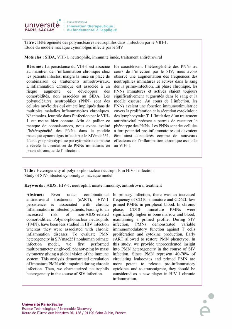
Université Paris-Saclay Espace Technologique / Immeuble Discovery Route de l’Orme aux Merisiers RD 128 / 91190 Saint-Aubin, France
Titre : Hétérogénéité des polynucléaires neutrophiles dans l'infection par le VIH-1. Etude du modèle macaque cynomolgus infecté par le SIV Mots clés : SIDA, VIH-1, neutrophile, immunité innée, traitement antirétroviral
Résumé : La persistance du VIH-1 est associée au maintien de l’inflammation chronique chez les patients infectés, malgré la mise en place de combinaison de traitements antirétroviraux. L’inflammation chronique est associée à un risque augmenté de développer des comorbidités, non associées au SIDA. Les polynucléaires neutrophiles (PNN) sont des cellules myéloïdes qui ont été impliqués dans de multiples maladies inflammatoires chroniques. Néanmoins, leur rôle dans l’infection par le VIH-1 est moins bien connue. Afin de pallier ce manque de connaissances, nous avons évalué l’hétérogénéité des PNNs dans le modèle macaque cynomolgus infecté par le SIVmac251. L’analyse phénotypique par cytométrie de masse a révélé la circulation de PNNs immatures en phase chronique de l’infection.
En caractérisant l’hétérogénéité des PNNs au cours de l’infection par le SIV, nous avons observé une augmentation des fréquences des neutrophiles immatures et activés dans le sang dès la primo-infection. En phase chronique, les PNNs immatures et activés étaient toujours significativement augmentés dans le sang et la moelle osseuse. Au cours de l’infection, les PNNs avaient une fonction immunostimulatrice envers la prolifération et la sécrétion cytokinique des lymphocytaire T. L’initiation d’un traitement antirétroviral précoce a permis de restaurer le phénotype des PNNs. Les PNNs sont des cellules à fort potentiel pro-inflammatoire qui devraient être ainsi considérés comme de nouveaux effecteurs de l’inflammation chronique associée au VIH-1.
Title : Heterogeneity of polymorphonuclear neutrophils in HIV-1 infection. Study of SIV-infected cynomolgus macaque model. Keywords : AIDS, HIV-1, neutrophil, innate immunity, antiretroviral treatment
Abstract: Even under combinational antiretroviral treatments (cART), HIV-1 persistence is associated with chronic inflammation in infected patients, leading to an increased risk of non-AIDS-related comorbidities. Polymorphonuclear neutrophils (PMN), have been less studied in HIV infection whereas they were associated with chronic inflammation diseases. To evaluate PMN heterogeneity in SIVmac251 nonhuman primate infection model, we first performed multiparameter single-cell phenotyping by mass cytometry giving a global vision of the immune system. This analysis demonstrated circulation of immature PMN with impaired during chronic infection. Then, we characterized neutrophils heterogeneity in the course of SIV infection.
In primary infection, there was an increased frequency of CD10- immature and CD62L-low primed PMNs in peripheral blood. In chronic phase, CD10- immature PMNs were significantly higher in bone marrow and blood, maintaining a primed profile. During SIV infection, PMNs demonstrated variable immunomodulatory function against T cells proliferation and cytokine production. Early cART allowed to restore PMN phenotype. In this study, we provide unprecedented insight into PMN heterogeneity in the course of SIV infection. Since PMN represent 40-70% of circulating leukocytes and primed PMN are more potent to release pro-inflammatory cytokines and to transmigrate, they should be considered as a new player in HIV-1 chronic inflammation.