Functional Heterogeneity of
11
of June 5, 2013. This information is current as CD8+ T Cells Functional Heterogeneity of Vaccine-Induced Francesco M. Marincola Provenzano, Mark E. Dudley, Steven A. Rosenberg and Vladia Monsurrò, Dirk Nagorsen, Ena Wang, Maurizio http://www.jimmunol.org/content/168/11/5933 2002; 168:5933-5942; ; J Immunol References http://www.jimmunol.org/content/168/11/5933.full#ref-list-1 , 27 of which you can access for free at: cites 49 articles This article Subscriptions http://jimmunol.org/subscriptions is online at: The Journal of Immunology Information about subscribing to Permissions http://www.aai.org/ji/copyright.html Submit copyright permission requests at: Email Alerts http://jimmunol.org/cgi/alerts/etoc Receive free email-alerts when new articles cite this article. Sign up at: Print ISSN: 0022-1767 Online ISSN: 1550-6606. Immunologists All rights reserved. Copyright © 2002 by The American Association of 9650 Rockville Pike, Bethesda, MD 20814-3994. The American Association of Immunologists, Inc., is published twice each month by The Journal of Immunology by guest on June 5, 2013 http://www.jimmunol.org/ Downloaded from
-
Upload
independent -
Category
Documents
-
view
1 -
download
0
Transcript of Functional Heterogeneity of

of June 5, 2013.This information is current as
CD8+ T CellsFunctional Heterogeneity of Vaccine-Induced
Francesco M. MarincolaProvenzano, Mark E. Dudley, Steven A. Rosenberg and Vladia Monsurrò, Dirk Nagorsen, Ena Wang, Maurizio
http://www.jimmunol.org/content/168/11/59332002; 168:5933-5942; ;J Immunol
Referenceshttp://www.jimmunol.org/content/168/11/5933.full#ref-list-1
, 27 of which you can access for free at: cites 49 articlesThis article
Subscriptionshttp://jimmunol.org/subscriptions
is online at: The Journal of ImmunologyInformation about subscribing to
Permissionshttp://www.aai.org/ji/copyright.htmlSubmit copyright permission requests at:
Email Alertshttp://jimmunol.org/cgi/alerts/etocReceive free email-alerts when new articles cite this article. Sign up at:
Print ISSN: 0022-1767 Online ISSN: 1550-6606. Immunologists All rights reserved.Copyright © 2002 by The American Association of9650 Rockville Pike, Bethesda, MD 20814-3994.The American Association of Immunologists, Inc.,
is published twice each month byThe Journal of Immunology
by guest on June 5, 2013http://w
ww
.jimm
unol.org/D
ownloaded from

Functional Heterogeneity of Vaccine-Induced CD8� T Cells1
Vladia Monsurro,* Dirk Nagorsen,* Ena Wang,* Maurizio Provenzano,* Mark E. Dudley, †
Steven A. Rosenberg,† and Francesco M. Marincola2*
The functional status of circulating vaccine-induced, tumor-specific T cells has been questioned to explain their paradoxicalinability to inhibit tumor growth. We enumerated with HLA-A*0201/peptide tetramers (tHLA) vaccine-elicited CD8 � T cellprecursor frequency among PBMC in 13 patients with melanoma undergoing vaccination with the HLA-A*0201-associated gp100:209–217(210 M) epitope. T cell precursor frequency increased from undetectable to 12,400� 3,600� 106 CD8� T cells aftervaccination and appeared heterogeneous according to previously described functional subtypes: CD45RA�CD27� (14 � 2.6% oftHLA-staining T cells), naive; CD45RA�CD27� (14 � 3.2%), memory; CD45RA�CD27� (43 � 6%), effector; andCD45RA�CD27� (30 � 4.1%), memory/effector. The majority of tHLA �CD8� T cells displayed an effector, CD27� phenotype(73%). However, few expressed perforin (17%). Epitope-specific in vitro stimulation (IVS) followed by 10-day expansion in IL-2reversed this phenotype by increasing the number of perforin� (84 � 3.6%; by paired t test, p < 0.001) and CD27� (from 28 to67%; by paired t test,p � 0.01) tHLA� T cells. This conversion probably represented a change in the functional status of tHLA�
T cells rather than a preferential expansion of a CD27� (naive and/or memory) PBMC, because it was reproduced after IVS ofa T cell clone bearing a classic effector phenotype (CD45RA�CD27�). These findings suggest that circulating vaccine-elicited Tcells are not as functionally active as inferred by characterization of IVS-induced CTL. In addition, CD45RA/CD27 expressionmay be more informative about the status of activation of circulating T cells than their status of differentiation. The Journal ofImmunology, 2002, 168: 5933–5942.
A ctive-specific vaccination against melanoma using pep-tide epitopes derived from melanoma Ags (MAGE-1,MAGE-3, NY-ESO-1, MART-1, and gp100) demon-
strated effectiveness in inducing vaccine-specific T cells that canrecognize HLA-matched melanoma cells (1, 2). However, theseimmune responses are only sporadically associated with clinicalregression (3). In addition, no correlation between clinical re-sponse and T cell precursor frequency (Tc-pf)3 could be identifiedby direct ex vivo HLA/epitope tetrameric complexes (tHLA) enu-meration of vaccine-induced lymphocytes (2, 4).
Because tHLA-based phenotyping of T cells does not yield in-formation about their effector function, it has been postulated thatthe lack of function of tumor Ag-specific T cells could be due toa suboptimal status of activation (5). Pittet et al. (6) noted thattumor Ag-specific T cells in patients with melanoma are, contraryto those analyzed in healthy individuals, CD45RAlow and can se-crete IFN-� upon cognate stimulation compatible with a functionaldifferentiation into competent effector/memory T cells in vivo. Wehave previously argued that vaccine-induced T cells should be atleast partially functional based on the fact that they can express
IFN-� mRNA and protein ex vivo upon cognate stimulation (7).Stimulation with vaccine-related epitopes induced IFN-� expres-sion detectable by intracellular cytokine analysis and quantitativereal-time PCR, suggesting that vaccine-elicited CTL retain Ag re-sponsiveness (4). However, recent studies suggest that IFN-� ex-pression by T cells may not be the functional parameter that mightbest describe their potential as effector cells. Appay et al. (8) ob-served in patients with chronic HIV infection that CMV-specificdiffer from HIV-specific circulating CD8� T cells because theyexpress significantly lower levels of perforin despite retaining theability to secrete IFN-� in response to epitope stimulation. In ad-dition, these lymphocytes retained the expression of CD27, whichwas interpreted by the authors as a sign of impaired maturationtoward fully developed effector cells. It is therefore possible thatAg-experienced vaccine-induced T cells may be in an incompletestage of differentiation, which explains their limited effect on tu-mor growth. Functionally distinct phenotypes of CD8� T cellsspanning from a naive to an effector and/or memory stage of dif-ferentiation have been described by several groups (9, 10). Ac-cording to Hamman et al. (9), memory-type CD8� T cells areCD45RA�CD27�, can express a broad range of cytokines (IL-2,IFN-�, TNF-�, and IL-4) in response to cognate stimulation, and donot display cytolytic activity without prior in vitro stimulation (IVS).In contrast, effector T cells have a CD45RA�C27� phenotype, re-lease a more stringent array of cytokines upon stimulation (IFN-� andTNF-�), and display strong cytolytic activity with high expression ofperforin and granzyme without the need for in vitro prestimulation.Thus, combining phenotypic with functional proprieties, the authorssuggested four distinct populations of CD8� T cells depending ontheir CD45RA and CD27 expression levels: naive T cells(CD45RA�CD27�), effector T cells (CD45RA�CD27�), memory Tcells (CD45RA�CD27�), and effector/memory T cells (CD45RA�
CD27�). This classification has been extensively used subsequentlyin the context of various infectious diseases and other immune pa-thologies (11–13).
*Immunogenetics Section, Department of Transfusion Medicine, Clinical Center, and†Surgery Branch, National Cancer Institute, National Institutes of Health, Bethesda,MD 20892
Received for publication January 4, 2002. Accepted for publication March 27, 2002.
The costs of publication of this article were defrayed in part by the payment of pagecharges. This article must therefore be hereby marked advertisementin accordancewith 18 U.S.C. Section 1734 solely to indicate this fact.1 D.N. was supported by Mildred Scheel Stiftung fur Krebsforschung (DeutscheKrebshilfe).2 Address correspondence and reprint requests to Dr. Francesco M. Marincola, Im-munogenetics Section, Department of Transfusion Medicine, Clinical Center, Na-tional Institutes of Health, Building 10, Room 1C-711, 10 Center Drive, MSC 1502,Bethesda, MD 20892-1502. E-mail address: [email protected] Abbreviations used in this paper: Tc-pf, T cell precursor frequency; g209, gp100:209–217; g209-2M, g209(210 M); IVS, in vitro stimulation; tHLA, HLA/epitopetetrameric complex.
The Journal of Immunology
Copyright © 2002 by The American Association of Immunologists 0022-1767/02/$02.00
by guest on June 5, 2013http://w
ww
.jimm
unol.org/D
ownloaded from

Independent of their intrinsic status of activation and/or differ-entiation, circulating lymphocytes need to be able to localize intarget organs to exert their effector function. It has been suggestedthat two subsets of memory T lymphocytes can be identified withdistinct homing potentials and effector functions based on theirlevel of expression of the chemokine receptor CCR7 (14). CCR7�
memory cells express receptors for migration to target tissues andare ready to exert their effector function. Conversely, CCR7�
memory cells express lymph node-homing receptors and lack im-mediate effector function. Champagne et al. (15) suggested thatCD8� T lymphocytes follow a consistent differentiation pat-tern that flows from CD45RA�CCR7�3CD45RA�CCR7�3CD45RA�CCR7�3CD45RA�CCR7�. In HIV patients, HIV-specific CD8� T cell populations appear to be composed predom-inantly of the preterminally differentiated CD45RA�CCR7� sub-set, while, at the same time, CMV-specific CD8� T cells displayin large majority a terminally differentiated phenotype (CD45RA�
CCR7�). Thus, it appears from the literature that T cells differ-entiate upon Ag exposure through a continuum display of evolvingfunctions that might, in turn, affect the overall effectiveness of agiven immune response.
Epitope-specific vaccination offers the unique opportunity ofevaluating the immune response in relation to a well-defined timeof Ag exposure. In particular, immunization with the HLA-A*0201-associated gp100:209–217(210M) epitope (g209-2M)dramatically converts a largely undetectable T cell response invaccine-naive patients to the frequently observable induction ofvaccine-specific CD8� T cells documentable by tHLA phenotyp-ing (2). Thus, subsets of Ag-experienced/memory T cells can becharacterized in their phenotypic and functional properties. There-fore, in this study we determined the expression of CD45RA andCD27 in epitope-specific CD8� T cells. Expression was deter-mined from the PBMC of patients undergoing vaccination withg209-2M modified from the wild-type epitope of the melanoma Aggp100/Pmel17 by substitution of a methionine in position 2 (16).This peptide has been previously shown to efficiently induceepitope-specific CD8� T cell responses in most patients when ad-ministered emulsified in IFA (17). These responses could be easilyidentified using tHLA complexes reconstituted with either theg209-2M or the wild-type gp100:209–217 (g209) peptide (2).Thus, the goal of this study was to characterize the CD45RA CD27profile of tHLA� vaccine-induced CD8� T cells with the assump-tion that these cells are Ag-experienced, because none was detect-able before vaccination. CCR7 expression was also evaluated in asubpopulation of patients because of its predictive significanceconcerning the ability of T cells to localize and exert effector func-tion in the target organ. In addition, perforin expression was in-cluded in the phenotypic characterization as a marker of “ready foraction” effector activity. More specifically, in this study we ques-tioned whether these molecules could be regarded as informativemarkers of the level of differentiation and/or function of immuni-zation-induced T cells.
Materials and MethodsPatients’ selection
In this study we randomly selected 13 patients with metastatic melanomaamong 20 who demonstrated a specific immune response to vaccinationwith a gp100 peptide. These 20 immunization-responsive patients wereidentified among 35 HLA-A*0201 patients with melanoma. These 35 pa-tients had received repeated s.c. injections of the g209-2M peptide in IFAin a protocol approved by the institutional review board of the NationalCancer Institute. No concomitant treatment, including IL-2, was adminis-tered. The HLA class I phenotype of patients was determined in PBMCusing sequence-specific primer-PCR (18). All patients had been surgicallydetermined to be without evidence of disease before enrollment in the
vaccination protocol that was administered with adjuvant purposes. Thus,no correlation with clinical outcome could be evaluated in this study.
Peptide
The g209-2M (IMDQVPFSV) peptide used for vaccination was preparedaccording to good manufacturing practice by Multiple Peptide Systems(San Diego, CA). The g209-2M peptide used for tHLA synthesis and IVSstudies was commercially synthesized by Princeton Biomolecules (Colum-bus, OH). The peptides were purified by gel filtration to �95% purity, andtheir identities were confirmed by mass spectral analysis.
Cells and culture conditions
PBMC were obtained by leukapheresis both before the patients receivedtheir first vaccine and 3 wk after vaccination. As discussed in Results,samples were obtained from patients who had received different numbersof vaccinations administered according to various time schedules. PBMCwere isolated by Ficoll gradient separation and frozen until analysis. Anal-ysis of vaccine-specific T cells was performed after overnight resting ofthawed PBMC in complete medium consisting of Iscove’s medium(Biofluids, Rockville, MD) supplemented with 10 mM HEPES buffer, 100U/ml penicillin-streptomycin (Biofluids), 10 �g/ml ciprofloxacin (Bayer,West Haven, CT), 0.03% L-glutamine (Biofluids), 0.5 mg/ml amphotericinB (Biofluids), and 10% heat-inactivated human AB serum (Gemini Bio-products, Calabasas, CA). This step allowed depletion of adherentmonocytes.
The PBMC were also analyzed 10 days following IVS with 1 �Mg209-2M peptide, followed by culture in IL-2 (300 IU/ml)-containing me-dium that was added on the day following stimulation and every 2–3 daysthereafter. Bulk CTL cultures were cloned by limiting dilution according toa modification of Riddell’s technique (19, 20). Briefly, bulk CTL cultureswere plated in 96-well plates at 0.6 cell/well with OKT3 (30 ng/ml),50,000/well irradiated allogeneic PBMC (3,000 rad), and IL-2 (300 IU/ml).The clone used for this study (P1G9) was selected according to its reac-tivity against HLA-matched, gp100-expressing tumor targets as previouslydescribed (21). For stimulation of T cells, two melanoma cell lines wereused characterized by expression (624.38 MEL) or lack of expression(624.28 MEL) of HLA-A*0201.
Kinetics of surface marker expression in response to Ag-specificstimulation
The g209- and g209-2 M-specific CTL clone P1G9 was cocultured in thepresence of the HLA-A*0201/gp100-expressing melanoma cell line 624.38MEL or a sister clone (624.28 MEL) that lacked expression of HLA-A*0201 but retained expression of gp100 (22, 23). Coculture was per-formed at a 1:4 cancer cell:CTL ratio in a six-well Costar plate (CorningGlass, Corning, NY) in complete medium at a concentration of 106 CTL/ml. Twenty-four hours after starting the coculture IL-2 (300 IU/ml) wasadded and was replenished every 2–3 days. Cocultured cells were har-vested at various time intervals (as described for individual experiments)and analyzed by FACS analysis. To simulate the conditions of high epitopedensity used for IVS of PBMC, P1G9 clone was also stimulated in vitrowith 1 �M g209-2M peptide. However, this stimulation could not be per-formed by directly adding soluble peptide to the medium as was done forthe PBMC because this caused extensive apoptotic death of the T cellclone. To avoid this, g209-2M peptide was pulsed on 624.38 MEL and624.28 MEL for 1 h and then washed away. The melanoma cell lines werethen used for epitope presentation in conditions identical with those de-scribed previously.
T cell staining
Tetrameric peptide-HLA-A*0201 complexes were produced as describedpreviously (2, 24). Recombinant HLA-A*0201 H chain containing a bi-otinylation site and recombinant �2-microglobulin were synthesized andused for refolding of soluble HLA molecules in the presence of g209-2M.Monomeric HLA/peptide complexes were biotinylated with BirA (Avidity,Denver, CO) and tetramerized by adding avidin-PE (Pierce, Rockford, IL).
Purified anti-CD27 mAb was obtained from BD PharMingen (San Di-ego, CA) and was used with the secondary PE-conjugated rat anti-mouseIgG1 from BD Biosciences (San Jose, CA). PE-conjugated anti-CD27mAb was obtained from BD PharMingen. FITC-conjugated mAb againstCD45RA was obtained from Caltag Laboratories (Burlingame, CA). Pu-rified CCR7-specific mAb was obtained from BD PharMingen and wasused with the secondary CY5.5-conjugated rat anti-mouse IgM from BDBiosciences. Perforin staining was performed using the BD Bioscienceskit. CD8 mAb was obtained from BD Biosciences.
5934 FUNCTIONAL STATUS OF VACCINE-INDUCED T CELLS
by guest on June 5, 2013http://w
ww
.jimm
unol.org/D
ownloaded from

After overnight depletion of monocytes (or 10–11 days following IVS),nonadherent PBMC were resuspended at 106 cells/50 �l FACS buffer(phosphate buffer plus 5% FCS; Biofluids). Cells were incubated at 4°Cwith 1 �l tHLA for 15 min, and then incubation was continued for 30 minwith the specific mAb. A similar staining procedure was applied for stain-ing CTL cultures and the CTL clone P1G9. After tHLA staining, cells weredirectly stained with Abs for surface markers (CD27, CD8, CD45, CCR-7)and kept for 45 min at 4°C or fixed in 4% paraformaldehyde and thenpermeabilized with acetone as previously described (25) for perforin stain-ing. The totally CD8� P1G9 clone was stained with CD27-PE, CD45RA-FITC, tHLA-PE, CD8-PerCP, and V�17-FITC (Endogen, Woburn, MA).After staining, cells were washed in 2 ml FACS buffer and analyzed byFACS (BD Biosciences). The acquisition data were analyzed after pre-gating for lymphocyte size and tetramer positivity. The CTL clone P1G9was discriminated from cocultured cancer cells based on forward scatter asdescribed in Results. Twenty thousand events were acquired for analysis ofthe T cell clone, and 200,000 were used for PBMC or CTL after IVS.
Statistical analysis
Vaccine-specific Tc-pf was calculated as the number of g209 tHLA-stain-ing CD8� T cells per 106 CD8� cells adopting the following formula: f �URQ/(URQ � LRQ) � 106 CD8� cells, with URQ (upper right quadrant)containing the tHLA�CD8� cells and LRQ (lower right quadrant) con-taining all other CD8� cells. From these frequencies the background withCD8� staining only was subtracted for each sample to obtain the correctedfrequency. The corrected Tc-pf is presented as the number of vaccine-specific T cells per 106 CD8� T cells.
Comparison of values between pre- and postimmunization specimens orcomparing identical specimens before and after IVS was performed usinga paired sample t test. The p values are reported as the level of significancefor a two-tailed analysis.
ResultsPriming of circulating CD8� T cells by epitope-basedimmunization
PBMC from 13 HLA-A*0201-expressing patients with metastaticmelanoma undergoing repeated immunization with s.c. injection ofg209-2M peptide emulsified in IFA were phenotypically charac-terized by g209-2M tHLA staining of CD8� T cells. For this studyPBMC were selected from patients who had developed detectablevaccine-specific Tc-pf after immunization (Fig. 1). Tc-pf was con-sidered detectable when �1,000 g209-2M tHLA-staining T cellswere counted per 106 CD8� T cells. This value, according to ourprevious experience, corresponded to evidence of immunizationand correlated with results obtained by IVS (2) or functional ex
vivo assays (7). In none of these patients could �1,000 g209-2MtHLA staining T cells/106 CD8� T cells be detected in vaccinenaive settings, a finding consistent with previous reports (2, 4, 7).After immunization, Tc-pf ranged between 1,600 and 46,100 �106 CD8� T cells. This broad range in Tc-pf is not uncharacteristicof vaccine-induced immune responses. In this particular case thedifferent schedule of administration and the different number ofvaccinations received by individual patients (Table I) might haveaffected Tc-pf (4). In fact, the patients included in this study wereamong those participating in a randomization protocol in whichdifferent schedules of administration of the g209-2M in IFA werecompared. According to the arm assigned, the patients received thepeptide injection weekly or at 3-wk intervals or for 4 consecutivedays at 3-wk intervals. In each arm of the study the patients re-ceived four cycles of the assigned schedule and were then immu-nologically reassessed 3 wk after administration of the last peptideinjection by obtaining a leukapheresis to compare vaccine-inducedTc-pf with pretreatment samples. This study was designed to com-pare the effectiveness of different administration schedules, and itis still under investigation. Therefore, the results are beyond thepurpose of this report. However, PBMC were randomly sampledfrom individuals who were treated in different arms of this protocoland who had received a different number of vaccinations to addresswhether any of these factors may bear any effect on the phenotypiccharacteristics of vaccine-induced T cells and deserve further in-vestigation. Thus, the only constant parameter studied here is thephenotypic and functional portrait of vaccine-induced T cells ob-tained after different vaccination schedules 3 wk after the last ad-ministration of vaccine. Results obtained from the 13 patients stud-ied suggested, as shown in the next section, that this factorsignificantly affected the CD45RA CD27 CCR7 phenotypic profile
FIGURE 1. Induction of vaccine-specific T cells by repeated immuni-zation with g209-2M peptide. Vaccine-specific Tc-pf are shown in PBMCobtained before (f) and after (u) vaccination in 13 patients undergoingimmunization with g209-2M peptide administered s.c. emulsified inIFA. Tc-pf is shown as the number of tHLA-staining T cells per 106
CD8� T cells.
Table I. Schedule and number of administrations and correspondingvaccine-induced T cell precursor frequencies in PBMC of patientswith melanoma receiving immunization with g209-2M epitopeemulsified in IFAa
Patients ScheduleNo. of Immunizations
Receivedb Tc-pfc
1 Every 3 wk 12 46,1002 Weekly 4 1,6003 4 consecutive days every 3 wk 4 13,5004 4 consecutive days every 3 wk 4 1,8005 Every 3 wk 4 4,3006 Weekly 16 14,3007 Weekly 8 6,0008 Every 3 wk 8 4,9009 Every 3 wk 8 28,700
10 4 consecutive days every 3 wk 4 3,10011 4 consecutive days every 3 wk 8 18,00012 Weekly 8 15,30013 Every 3 wk 8 3,400
a PBMC were randomly obtained before and after various numbers of immuni-zations from patients who belonged to various arms of a randomized clinical studywhere the length and frequency of immunization schedule is under investigation. Thethree randomization arms include one immunization every 3 wk, one immunizationevery week, and immunization for four consecutive days followed by 3 wk of rest.
b The number of injections of vaccine received by the patients at the time when thespecimen presented in this study was collected.
c Tc-pf was calculated as number of g209 tHLA-staining CD8� T cells per 106
CD8� cells adopting the following formula: f � URQ/(URQ � LRQ) � 106 CD8�
cells, with URQ (upper right quadrant) containing the tHLA�CD8� cells and LRQ(lower right quadrant) containing all other CD8� cells. From these frequencies, thebackground with CD8� staining only was subtracted for each sample to obtain thecorrected frequency. The corrected Tc-pf includes this correction and is presented asthe number of vaccine-specific T cells � 106 CD8� T cells.
5935The Journal of Immunology
by guest on June 5, 2013http://w
ww
.jimm
unol.org/D
ownloaded from

of vaccine-induced T cells and, therefore, the diversity of treat-ment schedule and the number of vaccinations were not investi-gated further. Thus, in all patients selected for this study Ag-ex-perienced circulating CD8� T cells were detected 3 wk after thelast immunization that could be further characterized (9).
Phenotypic characterization of vaccine-induced circulatingCD8� T cell subsets based on CD45RA and CD27 expression
Vaccine-induced tHLA�CD8� T cells were further characterizedaccording to the classification suggested by Hamann et al. (9)based on CD45RA and CD27 expression (Fig. 2). The followingsubtypes were considered: CD45RA�CD27� (naive), CD45RA�
CD27� (memory), CD45RA�CD27� (effector), and CD45RA�
CD27� (effector/memory) (Table II). Circulating vaccine-inducedT cells analyzed ex vivo were characterized by a small size, asdemonstrated by side and forward scatter, suggesting a small sizecharacteristic of resting T cells. In this respect they did not displayany morphological feature significantly different from the remain-ing circulating CD8� T cells. In the majority of cases vaccine-induced T cells belonged to effector (43 � 6% of tHLA-stainingCD8� T cells) and effector/memory (30 � 4.1%) subsets, whileonly a small proportion displayed naive or memory markers. Be-cause Hamann et al. (9) had previously reported that memory typeCD8� T cells are characterized by a high expression of perforin,we tested the expression of this cytotoxin in tHLA�CD8� T cells.Surprisingly, only a small proportion of these cells expressed de-tectable amounts of perforin (17 � 4.5%). In addition, to furthercharacterize vaccine-induced T cells, we tested PBMC from sixpatients for CCR7 expression that differentiates between centralmemory and effector memory T cells characterized, respectively,by the presence or the absence of expression of this marker (14).Sixty-three percent of vaccine-induced T cells did not expressCCR7. Of them the majority (73%; 46% of all vaccine-induced Tcells) were CD27�, while a second smaller subpopulation wasCCR7�CD27� (Table III). This observation suggested a spectrumof vaccine-induced T cells of which the preponderance appeared tobear two phenotypic characteristics associated with effector(CD27�CCR7�) function. Thus, vaccine-induced T cells appear to
belong in large majority to an effector phenotype. However, con-trary to other reports these effector cells appear deprived ofperforin.
Phenotypic characterization of vaccine-induced CD8� T cellsubsets based on CD45RA and CD27 expression after cognateIVS, followed by expansion in IL-2
CTL cultures from postvaccination PBMCs were expanded in 300IU/ml IL-2 for 10 days after IVS with 1 �M g209-2M. Forwardand side scatter analysis demonstrated that CD8� T cells (whethervaccine-specific or not) were condensed in a population of T cellsof a larger size than that observed in the generating PBMC (insetin scatter plot, Fig. 2). This increase in size was seen in all culturestested (P3–P13) and was consistent with the typical morphology ofIL-2 and/or stimulation of activated T cells after in vitro culture.Phenotyping of these cultures demonstrated a significant increasein CD27� cells independent of the expression of CD45RA (from28 to 67%; by paired t test, p � 0.01; Table II). In particular, 23%of IVS-derived CTL were CD45RA�CD27�, corresponding to anaive phenotype. However, their naive status was considered un-likely, first because no evidence of tHLA staining T cells could beidentified before vaccination, and second because, as we have re-peatedly shown, IVS does not induce g209-2M CTL in vaccina-tion-naive circulating cells (2, 4). This suggests that it is unlikelythat the CD8� tHLA-staining T cells expressing both CD45RAand CD27 resulted from the in vitro expansion of a subliminalnumber of Ag-inexperienced T cells undetectable by tHLA, be-cause this does not occur in PBMC obtained before vaccination. Inaddition, CD45 RA positivity significantly decreased in responseto IVS from 54 to 32% of vaccine-specific T cells (P3–P13; bypaired t test p � 0.02). Interestingly, together with changes inCD27 status, a dramatic increase in perforin expression was notedamong the tHLA-staining postvaccination CTL cultures (from17 � 4.5% to 84 � 3.6%; by paired t test for P3–P13, p � 0.001).Similarly, an almost complete loss of expression of CCR7 wasnoted, with 93% of vaccine-induced T cells being CCR7 negative(Table III). Taken together, these data suggest that upon IVS thevaccine-elicited T cells undergo a further step in the process of
FIGURE 2. Phenotyping of vaccine-in-duced CD8� T cells according to CD45RA,CD27, and perforin expression. PBMC ob-tained before and after vaccination were gatedaccording to g209-2M tHLA staining and phe-notyped according to their level of expressionof CD27, CD45RA, and perforin. In addition,CTL cultures from postvaccination PBMCwere expanded in 300 IU/ml IL-2 after IVSwith 1 �M g209-2M peptide. A representativepatient sample (patient 7 in Table I) is pre-sented. Percentages refer to the frequency oftHLA staining T cells over CD8� T cells. Inthe inset of each scatter plot the side and for-ward scatter of the monocyte population ana-lyzed is shown with a white gate representingthe area of higher density of CD8� T cells.The size of CD8� T cells increases after IVScompared with circulating T cells.
5936 FUNCTIONAL STATUS OF VACCINE-INDUCED T CELLS
by guest on June 5, 2013http://w
ww
.jimm
unol.org/D
ownloaded from

activation/differentiation that appears to increase their effector po-tential, which includes larger cellular size, decreased expression ofCCR7, and increased expression of perforin. In contrast, smallcellular size and lack of perforin expression in circulating PBMCin the absence of IVS and expansion suggest a resting status ofvaccine-induced CD8� T cells. Puzzling, however, remained theinterpretation of the increased frequency of CD27� vaccine-spe-cific T cells after IVS. It is not clear whether the phenotypic
changes observed after IVS were due to the epitope-specific stimula-tion rather than to the presence of IL-2 (300 IU/ml) in the culturemedium. Analysis of tHLA-negative cells after IVS (presumably non-stimulated by the epitope and, therefore, responding predominantly toIL-2 stimulation) did not show the changes noted in tHLA� cells(data not shown). In addition, in vitro culture of PBMC obtained fromthree non-tumor-bearing healthy donors in the presence or the absenceof IL-2 (300 IU/ml) and no epitope-specific stimulation did not induce
Table III. Percentage of CCR7 and CD27 coexpression in tHLA-staining CD8� T cells from fresh PBMCor 10 days following IVS
Patients CCR7�CD27� CCR7�CD27� CCR7�CD27� CCR7�CD27�
PBMC8 37 9 26 279 13 4 14 69
10 32 5 28 3411 15 3 12 7012 28 12 8 5213 52 12 10 26Average 30 7 17 46SEM 5.9 1.6 3.5 8.2
After 10 days of IVS (209-2M)8 7 2 70 219 1 5 12 82
10 6 1 83 1011 4 3 43 5112 2 1 14 8313 7 2 60 30Average 5 2 55 38SEM 1.0 0.6 12.0 12.7
Table II. Percentage of CD45RA CD27 coexpression in tHLA-staining CD8� T cells from fresh PBMC or10 days following IVSa
PatientsCD45RA�CD27�
(naive)CD45RA�CD27�
(memory)CD45RA�CD27�
(effector)CD45RA�CD27�
(effector/memory) Perforin�
PBMC1 16 9 54 21 12 6 3 77 14 23 6 9 67 20 24 11 13 32 44 195 5 4 53 38 36 3 2 68 27 617 8 12 51 29 198 23 40 17 21 149 17 2 55 26 31
10 27 29 27 16 1711 10 15 17 58 1512 16 14 17 54 2913 34 28 20 18 14Average 14 14 43 30 17SEM 2.6 3.2 6.0 4.1 4.5
After IVS3 11 74 1 14 944 28 69 0 3 665 34 63 0 3 976 73 21 1 5 957 58 32 3 7 778 8 57 2 33 839 15 0 84 1 68
10 6 72 1 21 9611 4 32 5 59 9312 4 20 1 75 7213 13 49 5 33 81Average 23 44 9 23 84SEM 7.0 7.5 7.5 7.5 3.6
a Value of p � 0.001 (paired t test) for increase in perforin-expressing vaccine-specific T cells after IVS compared withPBMC (patients 3–13). Value of p � 0.01 (paired t test) for increase in CD27-expressing vaccine-specific T cells after IVScompared with PBMC (patients 3–13).
5937The Journal of Immunology
by guest on June 5, 2013http://w
ww
.jimm
unol.org/D
ownloaded from

a preferential shift of CD8� T cell phenotype. Although some vari-ation in the CD8� T cell phenotype was noted among different do-nors, no significant changes were noted in the percentage of CD8� Tcells expressing perforins after 10 days of in vitro culture comparedwith fresh PBMC (Table IV). Interestingly, in vitro culture of PBMCwithout IL-2 caused a marked decrease in the percentage of perforin-expressing CD8� T cells, suggesting that IL-2 may play a role insustaining, rather than inducing, the effector function of CTL. Thepercentage of CD27, CD45RA-expressing cells was not significantlyaffected by in vitro culture with IL-2, nor was the expression of CCR7(Table V).
Kinetics of stimulation of the effector-type g209-2M-specific CTLclone P1G9 with the melanoma cell lines 624.38 (HLA-A2�GP100�) and 624.28 (HLA-A2�GP100�)
To test whether changes in CD27 expression define the status ofactivation of vaccine-induced T cells rather than representing amarker of their status of differentiation (naive vs memory vs ef-fector), we stimulated in vitro the g209-2M-specific CTL clone(P1G9) derived from circulating lymphocytes of a melanoma pa-tient who had received immunization with the same epitope (4).This clone is characterized by high affinity for the HLA/g209-2Mcomplex, has a classical effector phenotype CD45RA�CD27�,and expresses detectable amounts of perforin when cultured invitro in the presence of 300 IU/ml IL-2 (9). P1G9 was coculturedat a 1/4 ratio with either the HLA-A*0201-negative 624.28 or theHLA-A*0201-expressing 624.38 melanoma cell line. IL-2 (300IU/ml) was added the following day and every 2–3 days thereafter.Before stimulation and 24, 48, and 96 h after stimulation, tHLAstaining and V-�17, CD8, perforin, and CD27 expression wereanalyzed by FACS analysis. T cells were discriminated from can-cer cells by forward and side scatter, and the accuracy of thisstrategy was confirmed by DNA index analysis as previously de-scribed (26) (Fig. 3A).
Upon IVS with the HLA-A*0201-loss variant 624.28-MEL (22,23), no changes in tHLA staining or in the expression of CD8,V-�17, perforin, and CD27 were noted as expected due to the lackof TCR engagement with the melanoma cell line (Fig. 3B). Stim-ulation with the HLA-A*0201-expressing sister clone 624-38demonstrated a decrease in tHLA staining associated with TCR/HLA-A*0201/epitope engagement as previously described (7)(Fig. 3C). This decrease was associated with no or minimal
changes in the expression of other surface markers, with a slightdecrease in V-�17 and CD8 expression. Thus, stimulation of a Tcell effector clone using HLA-A*0201-matched cell lines inducedminimal effects on the status of activation of such a clone, althoughthese conditions are sufficient to induce IFN-� mRNA and proteinexpression as well as reduction in tHLA staining as previouslydescribed (7).
Kinetics of stimulation of the effector-type g209-2M-specific CTLclone P1G9 with the melanoma cell line 624.38 (HLA-A2�gp100�) exogenously pulsed with 1 �M g209-2M peptide
The conditions used for IVS of PBMC differed from those used tostimulate P1G9 with HLA-matched tumor cells, in that PBMCwere vigorously challenged with high density epitope exposureachieved by exogenous administration of 1 �M peptide. We haveused this technique for quite a long time with the assumption thatmonocytic cells present in the PBMC preparation would contributeto Ag presentation (2, 27–29). Using this technique we have pre-viously noted that decrements in the concentration of epitope usedfor cognate stimulation tightly correspond with decreasing levelsof IFN-� mRNA expression by PBMC, while down-regulation oftHLA staining is only minimally affected (4). In addition, we havepreviously shown that variations in epitope density on the surfaceof tumor cells strongly affects the intensity of T cell stimulation(22, 30), which can be greatly enhanced by incubation of cancercells with soluble epitope before coculture (30). Indeed, analysis ofa panel of CTL clones expanded from PBMC obtained from mel-anoma patients after immunization showed a strong correlationbetween the level of stimulation with tumor cells at a peptide con-centration corresponding to 0.03 �M (31). Thus, we questionedwhether stimulation with tumor cells expressing HLA-A*0201 andgp100 could be quantitatively comparable to the high density pep-tide exposure achievable with the saturating concentrations ofg209-2M peptide (1 �M) used for PBMC stimulation (31).
To enhance the effects of the stimulation induced by the mela-noma cell line 624.38-MEL, we therefore artificially increased itsepitope density by loading tumor cells with 1 �M g209-2M pep-tide. Direct addition of g209-2M to the CTL clone was not feasi-ble; preliminary experiments showed massive apoptosis of T cells(data not shown), possibly due to cross-recognition of HLA-epitope complexes on the surface of T cells or to Fas/Fas ligandinteractions among activated T cells (32).
Table IV. Percentage of CD45RA CD27 coexpression in CD8� T cells from fresh PBMC or 10 days following in vitro culture.PBMC were obtained from non-tumor-bearing healthy donors
DonorsCD45RA�CD27�
(naive)CD45RA�CD27�
(memory)CD45RA�CD27�
(effector)CD45RA�CD27�
(effector/memory) Perforin�
PBMC1 13 18 64 5 522 60 12 26 2 463 40 22 35 3 31Average 38 17 42 3 43SEM 13.6 2.9 11.5 0.9 6.2
After in vitro culture with IL-21 16 23 44 17 452 30 15 40 15 403 11 21 47 21 62Average 19 20 44 18 49SEM 5.7 2.4 2.0 1.8 6.7
After in vitro culture without IL-21 9 29 28 35 152 60 13 20 7 93 38 28 13 21 10Average 36 23 20 21 11SEM 14.8 5.2 4.3 8.1 1.9
5938 FUNCTIONAL STATUS OF VACCINE-INDUCED T CELLS
by guest on June 5, 2013http://w
ww
.jimm
unol.org/D
ownloaded from
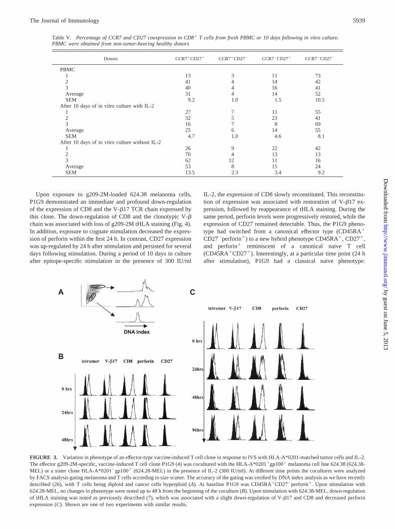
Upon exposure to g209-2M-loaded 624.38 melanoma cells,P1G9 demonstrated an immediate and profound down-regulationof the expression of CD8 and the V-�17 TCR chain expressed bythis clone. The down-regulation of CD8 and the clonotypic V-�chain was associated with loss of g209-2M tHLA staining (Fig. 4).In addition, exposure to cognate stimulation decreased the expres-sion of perforin within the first 24 h. In contrast, CD27 expressionwas up-regulated by 24 h after stimulation and persisted for severaldays following stimulation. During a period of 10 days in cultureafter epitope-specific stimulation in the presence of 300 IU/ml
IL-2, the expression of CD8 slowly reconstituted. This reconstitu-tion of expression was associated with restoration of V-�17 ex-pression, followed by reappearance of tHLA staining. During thesame period, perforin levels were progressively restored, while theexpression of CD27 remained detectable. Thus, the P1G9 pheno-type had switched from a canonical effector type (CD45RA�
CD27�perforin�) to a new hybrid phenotype CD45RA�, CD27�,and perforin� reminiscent of a canonical naive T cell(CD45RA�CD27�). Interestingly, at a particular time point (24 hafter stimulation), P1G9 had a classical naive phenotype:
FIGURE 3. Variation in phenotype of an effector-type vaccine-induced T cell clone in response to IVS with HLA-A*0201-matched tumor cells and IL-2.The effector g209-2M-specific, vaccine-induced T cell clone P1G9 (4) was cocultured with the HLA-A*0201�gp100� melanoma cell line 624.38 (624.38-MEL) or a sister clone HLA-A*0201�gp100� (624.28-MEL) in the presence of IL-2 (300 IU/ml). At different time points the cocultures were analyzedby FACS analysis gating melanoma and T cells according to size scatter. The accuracy of the gating was verified by DNA index analysis as we have recentlydescribed (26), with T cells being diploid and cancer cells hyperploid (A). At baseline P1G9 was CD45RA�CD27�perforin�. Upon stimulation with624.28-MEL, no changes in phenotype were noted up to 48 h from the beginning of the coculture (B). Upon stimulation with 624.38-MEL, down-regulationof tHLA staining was noted as previously described (7), which was associated with a slight down-regulation of V-�17 and CD8 and decreased perforinexpression (C). Shown are one of two experiments with similar results.
Table V. Percentage of CCR7 and CD27 coexpression in CD8� T cells from fresh PBMC or 10 days following in vitro culture.PBMC were obtained from non-tumor-bearing healthy donors
Donors CCR7�CD27� CCR7�CD27� CCR7�CD27� CCR7�CD27�
PBMC1 13 3 11 732 41 4 14 423 40 4 16 41Average 31 4 14 52SEM 9.2 1.0 1.5 10.5
After 10 days of in vitro culture with IL-21 27 7 11 552 32 5 23 413 16 7 8 69Average 25 6 14 55SEM 4.7 1.0 4.6 8.1
After 10 days of in vitro culture without IL-21 26 9 22 422 70 4 13 133 62 12 11 16Average 53 8 15 24SEM 13.5 2.3 3.4 9.2
5939The Journal of Immunology
by guest on June 5, 2013http://w
ww
.jimm
unol.org/D
ownloaded from

CD45RA�, CD27�, and perforin�. Because it is unlikely that thisCTL clone had regressed to an Ag-inexperienced status of differ-entiation, it is most likely that switches in CD27 expression andperforin detectability reflect different levels of activation of circu-lating and in vitro cultured T cells. In this study we did not addressin depth the differential role that cognate stimulation vs IL-2 ex-posure may play in affecting changes in T cell phenotype. How-ever, we believe that the intensity of cognate stimulation may havea predominant role. In one experiment in which the same stimu-latory conditions were applied to P1G9 (stimulation with 624.28MEL or 624.38 MEL with or without exogenous loading withg209-2M) in the presence or the absence of IL-2, no demonstrabledifferences in expression of the previously discussed markers werenoted up to 4 days following stimulation (data not shown). Itshould be emphasized that these data derived from a clonal pop-ulation may have limited relevance to the in vivo situation, asvarious CTL clones may be quite heterogeneous depending onvarious intrinsic and extrinsic factors. However, the switch in phe-notype observed in this clone in response to cognate stimulationsuggests that markers such as CD27 may vary independently of thestatus of differentiation of individual T cells.
DiscussionInsights in the status of activation and/or differentiation of vaccine-induced T cells may have relevance to the interpretation of clinicalresults following immunization protocols. In particular, epitope-specific vaccination allows a unique opportunity to simplify theanalysis of its effect onto a single HLA allele/epitope combination.This allows the study of human disease independently of HLApolymorphism and/or heterogeneity of individual tumor Ag pro-cessing and presentation efficiency. By following peptide-basedimmunization, we and others have repeatedly observed an en-hancement of vaccine-induced Tc-pf (3, 29, 33–41). In general,these studies have shown that vaccination with minimal T cell-directed epitopic sequences can be quite effective in inducing tu-mor-specific T cell responses, which can be easily observed amongcirculating lymphocytes of immunized patients. However, withfew exceptions (39–41), the identification of such immune re-sponses could not be consistently associated with significant im-provement in clinical outcome.
Several factors may be responsible for the lack of correlationbetween the identification of an immune response to the vaccineand the lack of cancer regression. Obviously, circulating T cellsinduced by vaccination may not reach the tumor site to exert theireffector function. However, attempts to expand vaccine-specific Tcells from biopsies through fine needle aspirates of the same lesionobtained before and after treatment were more frequently success-ful in postvaccination samples (42). In addition, we noted accu-mulation of gp100-specific lymphocytes in metastatic deposits fol-lowing vaccination in 8 of 11 patients. This accumulation wasassociated with increased expression of IFN-� in lesions that ex-pressed gp100. Yet this localization was not sufficient for tumorregression despite the expression by melanoma cells of the gp100Ag targeted by the vaccine (43). These findings suggest that vac-cine-elicited T cells can localize within the target tissue and thatthey are engaged by tumor cells to produce IFN-�, but this is notsufficient, and additional factors may be responsible for their lackof effect on tumor growth.
It could also be postulated that vaccine-induced Tc-pf, althoughclearly observable, may not be sufficient to induce tumor regres-sion, because the number of immunization-induced T cells ob-served in this study may not be above the threshold that may berequired to eliminate all tumor cells. In a mouse model we iden-tified a correlation between the intensity of immune response in-duced by the immunogen and tumor rejection. This correlationcould be easily identified in this experimental model, because theuse of a clonal population of tumor cells and of inbred animalsreduced tumor heterogeneity and/or variability of individual ani-mal immune responses providing a more homogeneous target forimmunotherapy (44). However, in humans a clear quantitative cor-relation between the extent of immune response and clinical out-come has remained elusive in most reported clinical trials, perhapsin relation to the heterogeneity of tumors in various individualsaffecting their immune responsiveness (45).
T cell responses documented by IVS or by direct ex vivo enu-meration of Tc-pf with vaccine-specific tHLA may not fully ad-dress their status of activation in vivo. In fact, Lee et al. (5) haveshown that tumor-specific T cells identified in vaccine-naive pa-tients by tHLA may not be able to exert effector function eitherthrough target killing or expression of cytokines such as IFN-�.
FIGURE 4. Variation in phenotype of aneffector-type vaccine-induced T cell clone inresponse to IVS with HLA-A*0201-matchedtumor cells loaded with 1 �M g209-2M pep-tide. The effector g209-2M-specific, vaccine-induced T cell clone P1G9 (4) was cocul-tured with the HLA-A*0201�gp100melanoma cell line 624.38 previously incu-bated with 1 �M g209-2M. Tumor and lym-phocytes were gated according to side scatteras shown in Fig. 3A. A profound down-reg-ulation of tHLA, V-�17, CD8, and perforinstaining was noted. In contrast, de novo ex-pression of CD27 was noted 24 h after stim-ulation and persisted up to 10 days. Duringthe same time course, all the other markersrecovered so that the originally CD45RA�
CD27� T cell clone became CD45RA�
CD27�perforin�. Shown are data from oneof two experiments with similar results.
5940 FUNCTIONAL STATUS OF VACCINE-INDUCED T CELLS
by guest on June 5, 2013http://w
ww
.jimm
unol.org/D
ownloaded from

We have previously argued that this concept may not pertain tovaccine-induced CD8� T cell responses, because in at least a per-centage of them, it is possible to document the expression of IFN-�upon cognate stimulation with vaccine-specific epitope or HLA-matched tumor cells (4–7). However, recent reports have empha-sized a dichotomy between IFN-� secretion by T cells and theirkiller function (8). This suggests that IFN-� expression in itselfmay not be the ultimate arbiter of T cell effector function, but mayrepresent a sensitive marker of T cell reactivity to cognatestimulation.
Hamann et al. (9) suggested a spectrum of circulating T cellphenotypes from naive to effector types based on the expression ofseveral cellular markers of which CD45RA and CD27 appeared tobest predict their status of differentiation and consequently theirfunction. Others suggested that homing receptors might be respon-sible for the effector function of T cells by regulating their local-ization within target organs (14). The observation in this and otherstudies (6) that vaccine-elicited T cells express low or undetectableCCR7 suggests that homing may not be, at least based on thismarker, a significant problem in the context of vaccines. This find-ing is consistent with an effector phenotype reported for flu- andCMV-specific T cells (6, 15) and correlates with our observationthat vaccine-induced T cell responses can be detected in lesionsresistant to therapy following vaccination (42, 43).
Segregation of T cell subsets into naive, memory, and effectortypes suggests a static and irrevocable status of differentiation thatmay not necessarily apply to the dynamic behavior of surfacemarkers in response to T cell stimulation. For instance, expressionof CD27 may play a role in the survival of activated T cells (46).Stimulation of the TCR complex with PHA or CD3 mAbs causesa dramatic increase in CD27, and addition of CD27 Abs to T cellcultures leads to enhanced proliferation (47). Interestingly, CD27could be immunoprecipitated from the membrane of activated, butnot resting, T cells (47). Recently, CD27 was described as a co-stimulatory receptor responsible for the generation and mainte-nance of T cell memory responses (48). Our findings suggest thatCD27 expression in circulating vaccine-induced T cells may rep-resent a marker of their status of activation rather than differenti-ation. Indeed, CD27 could be induced through IVS of vaccine-induced CD27� (effector) CD8� T cells. This increase in CD27-expressing T cells after 10 days of IVS is probably the result of itsde novo expression by CD27� PBMC, rather than the preferentialexpansion of few CD27� T cells. In particular, the substantialnumber of CD45RA�CD27� (naive) CD8� T cells (23%) ob-served after IVS is unlikely to be the result of expansion of rare(below the threshold of detection of tHLA) Ag-inexperienced pre-cursors in PBMC obtained after vaccination. We believe that allthe vaccine-specific T cells identifiable through IVS after immu-nization must have been exposed to the vaccine epitope, becausewe could never expand g209-2M-specific T cells before vaccina-tion using the exact in vitro conditions used here (2, 17). Thus,postvaccination IVS-induced T cells are most likely Ag-experi-enced derived from the CD45RA�CD27� PBMC pool. TheCD45RA expression in Ag-experienced T cells is reminiscent ofthe stable CD45RA�LFAhigh memory state recently described byFaint et al. (49). Unfortunately, because of the limited number ofcells available, LFA-1 as well as other potentially interestingmarkers such as CCR5, CXCR1, and CD62 could not be evaluatedin this study. The demonstration that a vaccine-induced, effector-type T cell clone could reverse its phenotype from CD27� toCD27� upon IVS suggests that the phenotype switch observed inIVS-expanded CTL is due to a change in functional status ratherthan preferential expansion of T cells in a particular status of dif-ferentiation. Thus, as suggested by others (6), CD27 may not be
the most accurate surface marker to segregate naive from Ag-ex-perienced CD8� T lymphocytes as also reported for CD45RA(49). Indeed, expression of the CD45RA isoform has been noted tooccur during the quiescent stage of memory T cell reversion to aresting state after in vivo priming by acute EBV infection (49).Interestingly, the phenotypic changes observed on P1G9 couldonly be induced by intense stimulation with saturating conditionsof epitope (pulsed tumor cells), while exposure to tumor cells ex-pressing natural amounts of HLA/epitope on their surface was notsufficient to induce such changes even in the presence of exoge-nous IL-2. This finding suggests that in vivo contact between vac-cine-induced T cells and tumor cells may not be sufficient to in-duce productive T cell stimulation capable of sustaining a briskimmune response as suggested by others (50, 51).
Of particular significance in this study was the lack of expres-sion of perforin in circulating vaccine-induced T cells. This inassociation with their small size and their lack of expression ofCD27 suggests that vaccine-induced circulating T cells are in aresting status, which can be reversed by IVS. It was beyond thepurpose of this study to address in depth whether this in vitroreactivation is due to re-exposure to cognate stimulation or to the10-day culture with IL-2. Whatever the cause of the striking dif-ference noted after 10 days of IVS, this change in phenotype maysuggest a reason for the discrepancy between satisfactory functionof T cells observed in vitro after IVS and their limited function invivo. This study also did not address when and where vaccine-induced T cells turn into this postulated resting phase. All samplesin this study were obtained 3 wk after immunization. Thus, it ispossible that this period marks the gradual regression of vaccine-induced T cells toward a resting/memory phenotype. Analysis ofvaccine-induced Tc-pf at an earlier time point might have depicteda totally different portrait. Perhaps a more intense and consistentexposure in vivo to vaccine may induce and sustain a more activeand functionally rewarding immune response as suggested by oth-ers (50, 52, 53).
In summary, we hypothesize that vaccine-induced T cells be-long to an effector subtype (predominantly CD27� and CCR7�)that is in a resting phase characterized by lack of perforin, smallcellular size, and lack of expression of activation markers such asCD27. These cells, however, are not totally anergic and, on thecontrary, are responsive to cognate stimulation, as they can pro-duce IFN-� upon exposure to relevant Ag (4, 7). It is possible thatif the stimulus is delivered in optimal conditions their full effectorfunction can be restored as modeled by the 10-day IVS conditions.These findings assign a limited level of functionality to circulatingvaccine-induced T cells and may partially explain why increases inTc-pf following immunization may not directly correlate with clin-ical cancer regression.
References1. Marchand, M., P. Weynants, E. Rankin, F. Arienti, F. Belli, G. Parmiani,
N. Cascinelli, A. Bourlond, R. Vanwijck, and Y. Humblet. 1995. Tumor regres-sion responses in melanoma patients treated with a peptide encoded by geneMAGE-3. Int. J. Cancer 63:883.
2. Lee, K.-H., E. Wang, M.-B. Nielsen, J. Wunderlich, S. Migueles, M. Connors,S. M. Steinberg, S. A. Rosenberg, and F. M. Marincola. 1999. Increased vaccine-specific T cell frequency after peptide-based vaccination correlates with increasedsusceptibility to in vitro stimulation but does not lead to tumor regression. J. Im-munol. 163:6292.
3. Rosenberg, S. A., J. C. Yang, D. Schwartzentruber, P. Hwu, F. M. Marincola,S. L. Topalian, N. P. Restifo, E. Dufour, L. Schwartzberg, P. Spiess, et al. 1998.Immunologic and therapeutic evaluation of a synthetic tumor associated peptidevaccine for the treatment of patients with metastatic melanoma. Nat. Med. 4:321.
4. Monsurro, V., M.-B. Nielsen, A. Perez-Diez, M. E. Dudley, E. Wang,S. A. Rosenberg, and F. M. Marincola. 2001. Kinetics of TCR use in response torepeated epitope-specific immunization. J. Immunol. 166:5817.
5941The Journal of Immunology
by guest on June 5, 2013http://w
ww
.jimm
unol.org/D
ownloaded from

5. Lee, P. P., C. Yee, P. A. Savage, L. Fong, D. Brockstedt, J. S. Weber, D. Johnson,S. Swetter, J. Thompson, P. D. Greenberg, et al. 1999. Characterization of cir-culating T cells specific for tumor-associated antigens in melanoma patients. Nat.Med. 5:677.
6. Pittet, M. J., A. Zippelius, D. E. Speiser, M. Assenmacher, P. Guillaume,D. Valmori, D. Lienard, F. Lejune, J.-C. Cerottini, and P. Romero. 2001. Ex vivoIFN-� secretion by circulating CD8 T lymphocytes: implications of a novel ap-proach for T cell monitoring in infectious and malignant diseases. J. Immunol.166:7634.
7. Nielsen, M.-B., V. Monsurro, S. Miguelse, E. Wang, A. Perez-Diez, K.-H. Lee,U. S. Kammula, S. A. Rosenberg, and F. M. Marincola. 2000. Status of activationof circulating vaccine-elicited CD8� T cells. J. Immunol. 165:2287.
8. Appay, V., D. F. Nixon, S. M. Donahoe, G. M. Gillespie, T. Dong, A. King,G. S. Ogg, H. M. Spiegel, C. Conlon, C. Spina, et al. 2000. HIV-specific CD8�
T cells produce antiviral cytokines but are impaired in cytolytic function. J. Exp.Med. 192:63.
9. Hamann, D., P. A. Baars, M. H. G. Rep, B. Hoolbrink, S. R. Kerkhof-Garde,M. R. Klein, and R. A. W. van Lier. 1997. Phenotype and functional separationof memory and effector human CD8� T cells. J. Exp. Med. 186:1407.
10. Baars, P. A., L. M. Ribeiro do Couto, J. H. W. Leusen, B. Hoolbrink,T. W. Kuijpers, S. M. A. Lens, and R. A. W. van Lier. 2000. Cytolytic mecha-nisms and expression of activation-regulating receptors on effector-typeCD8�CD45RA�CD27� human T cells. J. Immunol. 165:1910.
11. Hamann, D., S. Kostense, K. C. Wolthers, S. A. Otto, P. A. Baars, F. Miedema,and R. A. W. van Lier. 1999. Evidence that human CD8�CD45RA�CD27� cellsare induced by antigen and evolve through extensive rounds of division. Int.Immunol. 11:1027.
12. Gamadia, L. E., R. J. Rentenaar, P. A. Baars, E. B. M. Remmerswaal,S. Surachno, J. F. L. Weel, M. Toebes, T. N. Schumacher, J. M. ten Berge, andR. A. W. van Lier. 2001. Differentiation of cytomegalovirus-specific CD8� Tcells in healthy and immunosuppressed virus carriers. Blood 98:754.
13. Nagai, M., R. Kubota, T. F. Greten, J. P. Schneck, T. P. Leist, and S. Jacobson.2001. Increased activated human T cell lymphotropic virus type I (HTLV-I)Tax11–19-specific memory and effector CD8� cells in patients with HTLV-I-associated myelopathy/tropical spastic paraparesis: correlation with HTLV-I pro-virus load. J. Infect. Dis. 183:197.
14. Sallusto, F., D. Lenig, R. Forster, M. Lipp, and A. Lanzavecchia. 1999. Twosubsets of memory T lymphocytes with distinct homing potentials and effectorfunctions. Nature 401:659.
15. Champagne, P., G. S. Ogg, A. King, C. Knabenhans, K. Ellefsen, M. Nobile,V. Appay, G. P. Rizzardi, S. Fleury, M. Lipp, et al. 2001. Skewed maturation ofmemory HIV-specific CD8 T lymphocytes. Nature 410:106.
16. Parkhurst, M. R., M. L. Salgaller, S. Southwood, P. F. Robbins, A. Sette,S. A. Rosenberg, and Y. Kawakami. 1996. Improved induction of melanomareactive CTL with peptides from the melanoma antigen gp100 modified at HLA-A*0201 binding residues. J. Immunol. 157:2539.
17. Salgaller, M. L., F. M. Marincola, J. N. Cormier, and S. A. Rosenberg. 1996.Immunization against epitopes in the human melanoma antigen gp100 followingpatient immunization with synthetic peptides. Cancer Res. 56:4749.
18. Bunce, M., C. M. O’Neill, M. C. Barnardo, P. Krausa, M. J. Browning,P. J. Morris, and K. I. Welsh. 1995. Phototyping: comprehensive DNA typing forHLA-A, B, C, DRB1, DRB3, DRB4, DRB5 and DQB1 by PCR with 144 primermixes utilizing sequence-specific primers (PCR-SSP). Tissue Antigens 46:355.
19. Dudley, M. E., L. T. Ngo, J. Westwood, J. R. Wunderlich, and S. A. Rosenberg.2000. T-cell clones from melanoma patients immunized against an anchor modifiedgp100 peptide display discordant effector phenotypes. Cancer J. Sci. Am. 6:69.
20. Riddell, S. R., and P. D. Greenberg. 1994. Therapeutic reconstitution of humanviral immunity by adoptive transfer of cytotoxic T lymphocyte clones. Curr. Top.Microbiol. Immunol. 189:9.
21. Legerski, R., and C. Peterson. 1992. Expression cloning of a human DNA repairgene involved in xeroderma pigmentosum group C. Nature 359:70.
22. Rivoltini, L., K. C. Baracchini, V. Viggiano, Y. Kawakami, A. Smith, A. Mixon,N. P. Restifo, S. L. Topalian, T. B. Simonis, S. A. Rosenberg, et al. 1995. Quanti-tative correlation between HLA class I allele expression and recognition of melanomacells by antigen specific cytotoxic T lymphocytes. Cancer Res. 55:3149.
23. Wang, Z., F. M. Marincola, L. Rivoltini, G. Parmiani, and S. Ferrone. 1999.Selective human leukocyte antigen (HLA)-A2 loss caused by aberrant pre-mRNAsplicing in 624MEL28 melanoma cells. J. Exp. Med. 190:205.
24. Altman, J. D., P. H. Moss, P. R. Goulder, D. H. Barouch,M. G. McHeyzer-Williams, J. I. Bell, A. J. McMichael, and M. M. Davis. 1996.Phenotypic analysis of antigen-specific T lymphocytes. [Published erratum ap-pears in 1998 Science 280:1821.] Science 274:94.
25. Cormier, J. N., Y. M. Hijazi, A. Abati, P. Fetsch, M. Bettinotti, S. M. Steinberg,S. A. Rosenberg, and F. M. Marincola. 1998. Heterogeneous expression of mel-anoma-associated antigens (MAA) and HLA-A2 in metastatic melanoma in vivo.Int. J. Cancer 75:517.
26. Mocellin, S., P. A. Fetsch, A. Abati, G. Phan, E. Wang, M. Provenzano,D. Stroncek, S. A. Rosenberg, and F. M. Marincola. 2001. Laser scanner cytom-eter evaluation of MART-1, gp100 and HLA-A2 expression in melanoma me-tastases. J. Immunother. 24:447.
27. Rivoltini, L., Y. Kawakami, K. Sakaguchi, S. Southwood, A. Sette, P. F. Robbins,F. M. Marincola, M. Salgaller, J. R. Yannelli, E. Appella, et al. 1995. Inductionof tumor reactive CTL from peripheral blood and tumor infiltrating lymphocytesof melanoma patients by in vitro stimulation with an immunodominant peptide ofthe human melanoma antigen MART-1. J. Immunol. 154:2257.
28. Marincola, F. M., L. Rivoltini, M. L. Salgaller, M. Player, and S. A. Rosenberg.1996. Differential anti-MART-1/MelanA CTL activity in peripheral blood of
HLA-A2 melanoma patients in comparison to healthy donors: evidence for invivo priming by tumor cells. J. Immunother. 19:266.
29. Cormier, J. N., M. L. Salgaller, T. Prevette, K. C. Barracchini, L. Rivoltini,N. P. Restifo, S. A. Rosenberg, and F. M. Marincola. 1997. Enhancement ofcellular immunity in melanoma patients immunized with a peptide from MART-1/Melan A. Cancer J. Sci. Am. 3:37.
30. Cormier, J. N., M. C. Panelli, J. A. Hackett, M. Bettinotti, A. Mixon,J. Wunderlich, L. Parker, N. P. Restifo, S. Ferrone, and F. M. Marincola. 1999.Natural variation of the expression of HLA and endogenous antigen modulatesCTL recognition in an in vitro melanoma model. Int. J. Cancer 80:781.
31. Dudley, M. E., M. I. Nishimura, A. K. C. Holt, and S. A. Rosenberg. 1999.Anti-tumor immunization with a minimal peptide epitope (G9-209-2M) leads toa functionally heterogeneous CTL response. J. Immunother. 22:288.
32. Zaks, T. Z., D. B. Chappell, S. A. Rosenberg, and N. P. Restifo. 1999. Fas-Mediated suicide of tumor-reactive T cells following activation by specific tumor:selective rescue by caspase inhibition. J. Immunol. 162:3273.
33. Marchand, M., N. van Baren, P. Weynants, V. Brichard, B. Dreno, M. H. Tessier,E. Rankin, G. Parmiani, F. Arienti, Y. Humblet, et al. 1999. Tumor regressionsobserved in patients with metastatic melanoma treated with an antigenic peptideencoded by gene MAGE-3 and presented by HLA-A1. Int. J. Cancer 80:219.
34. Pittet, M. J., D. E. Speiser, D. Lienard, D. Valmori, P. Guillaume, V. Dutoit,D. Rimoldi, F. Lejeune, J. C. Cerottini, and P. Romero. 2001. Expansion andfunctional maturation of human tumor antigen-specific CD8� T cells after vac-cination with antigenic peptide. Clin. Cancer Res. 7:796s.
35. Nestle, F. O., S. Alijagic, M. Gilliet, Y. Sun, S. Grabbe, R. Dummer, G. Burg, andD. Schadendorf. 1998. Vaccination of melanoma patients with peptide- or tumorlysate-pulsed dendritic cells. Nat. Med. 4:328.
36. Thurner, B., I. Haendle, C. Roder, D. Dieckmann, P. Keikavoussi, H. Jonuleit,A. Bender, C. Maczek, D. Schreiner, P. von den Driesch, et al. 1999. Vaccinationwith MAGE-3A1 peptide-pulsed mature, monocyte-derived dendritic cells ex-pands specific cytotoxic T cells and induces regression of some metastases inadvanced stage IV melanoma. J. Exp. Med. 190:1669.
37. Scheibenbogen, C., A. Schmittel, U. Keilholz, T. Allgauer, U. Hofmann, R. Max,E. Thiel, and D. Schadendorf. 2000. Phase 2 trial of vaccination with tyrosinasepeptides and granulocyte-macrophage colony-stimulating factor in patients withmetastatic melanoma. J. Immunother. 23:275.
38. Laforet, M., N. Froelich, A. Parissiadis, H. Bausinger, B. Pfeiffer, andM. M. Tongio. 1997. An intronic mutation responsible for a low level of expres-sion of an HLA-A*24 allele. Tissue Antigens 50:340.
39. Jager, E., S. Gnjatic, Y. Nagata, E. Stockert, D. Jager, J. Karbach, A. Neumann,J. Rieckenberg, Y.-T. Chen, G. Ritter, et al. 2000. Induction of primary NY-ESO-1 immunity: CD8� T lymphocyte and antibody responses in peptide-vac-cinated patients with NY-ESO-1� cancers. Proc. Natl. Acad. Sci. USA 97:12198.
40. Jager, E., M. J. Maeurer, H. Hohn, J. Karbach, D. Jager, Z. Zidianakis,A. Bakhshandeh-Bath, J. Orth, C. Neukirch, A. Necker, et al. 2000. Clonal ex-pansion of melan A-specific cytotoxic T lymphocytes in a melanoma patientresponding to continued immunization with melanoma-associated peptides. Int. J.Cancer 86:538.
41. Wang, F., E. Bade, C. Kuniyoshi, L. Spears, G. Jeffery, V. Marthy, S. Groshen,and J. Weber. 1999. Phase I trial of a MART-1 peptide vaccine with incompleteFreund’s adjuvant for resected high-risk melanoma. Clin. Cancer Res. 5:2756.
42. Panelli, M. C., A. Riker, U. S. Kammula, K.-H. Lee, E. Wang, S. A. Rosenberg,and F. M. Marincola. 2000. Expansion of tumor/T cell pairs from fine needleaspirates (FNA) of melanoma metastases. J. Immunol. 164:495.
43. Kammula, U. S., K.-H. Lee, A. Riker, E. Wang, G. A. Ohnmacht,S. A. Rosenberg, and F. M. Marincola. 1999. Functional analysis of antigen-specific T lymphocytes by serial measurement of gene expression in peripheralblood mononuclear cells and tumor specimens. J. Immunol. 163:6867.
44. Perez-Diez, A., P. J. Spiess, N. P. Restifo, P. Matzinger, and F. M. Marincola.2002. Intensity of the vaccine-elicited immune response determines tumor clear-ance. J. Immunol. 168:338.
45. Marincola, F. M., E. M. Jaffe, D. J. Hicklin, and S. Ferrone. 2000. Escape ofhuman solid tumors from T cell recognition: molecular mechanisms and func-tional significance. Adv. Immunol. 74:181.
46. Camerini, D., G. Walz, W. A. M. Loenen, J. Borst, and B. Seed. 1991. The T cellactivation antigen CD27 is a member of the nerve growth factor/tumor necrosisfactor receptor gene family. J. Immunol. 147:3165.
47. van Lier, R. A. W., J. Borst, T. M. Vroom, H. Klein, P. van Mourik,W. P. Zeijlemaker, and C. J. Melief. 1987. Tissue distribution and biochemicaland functional properties of Tp55 (CD27), a novel T cell differentiation antigen.J. Immunol. 139:1589.
48. Hendriks, J., L. A. Gravestein, K. Tesselaar, R. A. W. van Lier, M. Schumacher,and J. Borst. 2000. CD27 is required for generation and long-term maintenanceof T cell immunity. Nat. Immunol. 1:433.
49. Faint, J. M., N. E. Annels, S. J. Curnow, P. Shields, D. Pilling, A. D. Hislop,L. Wu, A. N. Akbar, C. D. Buckley, P. A. Moss, et al. 2001. Memory T cellsconstitute a subset of the human CD8�CD45RA� pool with distinct phenotypicand migratory characteristics. J. Immunol. 167:212.
50. Fuchs, E. J., and P. Matzinger. 1996. Is cancer dangerous to the immune system?Semin. Immunol. 8:271.
51. Matzinger, P. 1998. An innate sense of danger. Semin. Immunol. 10:399.52. Speiser, D. E., R. Miranda, A. Zakarian, M. F. Bachmann, K. McKall-Faienza,
B. Odermatt, D. Hanahan, R. M. Zinkernagel, and P. S. Ohashi. 1997. Self an-tigens expressed by solid tumors do not efficiently stimulate naive or activated Tcells: implications for immunotherapy. J. Exp. Med. 186:645.
53. Zinkernagel, R. M. 2000. Localization dose and time of antigens determine im-mune reactivity. Semin. Immunol. 12:163.
5942 FUNCTIONAL STATUS OF VACCINE-INDUCED T CELLS
by guest on June 5, 2013http://w
ww
.jimm
unol.org/D
ownloaded from