Human fronto-tectal and fronto-striatal-tectal pathways activate differently during anti-saccades
Upload
independentCategory
view
1download
0
ORIGINAL RESEARCH ARTICLEpublished: 18 February 2013
doi: 10.3389/fimmu.2013.00033
Neutrophils activate tumoral CORTACTIN to enhanceprogression of orohypopharynx carcinomaClaudia A. Dumitru1, Agnes Bankfalvi 2, Xiang Gu1,Wilfried E. Eberhardt 3, Reinhard Zeidler 4, Stephan Lang1
and Sven Brandau1*1 Department of Otorhinolaryngology, University Hospital Essen, University of Duisburg-Essen, Essen, Germany2 Department of Pathology/Neuropathology, University Hospital Essen, University of Duisburg-Essen, Essen, Germany3 Department of Medical Oncology, West German Cancer Centre, University Hospital Essen, University of Duisburg-Essen, Essen, Germany4 Department of Otorhinolaryngology, Ludwig Maximilians University, Munich, Germany
Edited by:Jean-Pierre Abastado, Agency forScience, Technology and Research,Singapore
Reviewed by:Lai Guan Ng, Agency for Science,Technology and Research, SingaporeKar Wai Tan, Singapore ImmunologyNetwork, Singapore
*Correspondence:Sven Brandau, Department ofOtorhinolaryngology, University ofDuisburg-Essen, Hufelandstraße 55,45122 Essen, Germany.e-mail: [email protected]
CORTACTIN is an actin-binding protein critically involved in cellular migration and invasion.Here, we investigated the role of CORTACTIN in the pathophysiology of orohypopharynxcarcinoma – one of the major subtypes of head and neck cancer. To this end, we analyzedCORTACTIN expression in tumor tissues from 89 orohypopharynx carcinoma patients inrelation to clinical parameters. We found that high tumoral CORTACTIN expression asso-ciated with poor survival, higher T-stage, and higher lymph node metastasis (N-stage) inthese patients. Next, we combined the prognostic values of tumoral and stromal cell bio-logical parameters in our patient cohort.We determined the potential interaction of tumoralCORTACTIN with tumor-infiltrating neutrophils, which have been previously linked to poorclinical outcome in orohypopharynx carcinoma patients with advanced disease. Interest-ingly, we found that patients with both high tumoral CORTACTIN expression and highneutrophilic infiltration had significantly worse clinical outcome than all other patients in ourcohort. These findings suggest that tumoral CORTACTIN and tumor-infiltrating neutrophilsmight be functionally linked during progression of orohypopharynx carcinoma. In vitro, weshowed that neutrophils released soluble factors which phosphorylated CORTACTIN inthe tumor cells and promoted their migration. Furthermore, we demonstrated that strongCORTACTIN phosphorylation significantly correlated with strong neutrophilic infiltrationin tumor tissues from orohypopharynx carcinoma patients. Taken together, our findingsunravel a novel mechanism of tumor-stroma interaction, which might be relevant for amore accurate prognosis and improved therapeutic strategies in this tumor entity.
Keywords: cancer-related inflammation, tumor microenvironment, head and neck cancer, clinical outcome, tumormigration
INTRODUCTIONHead and neck cancer (HNC) is the eighth most common typeof cancer worldwide. Despite multiple and aggressive therapeuticinterventions, there has been no fundamental improvement in the5-year survival rates of the patients over the past decades (Jemalet al., 2007; Choong and Vokes, 2008). Therefore, there is an urgentneed to: (i) identify clinicopathological parameters (biomarkers)for accurate diagnosis, prognosis, and therapeutic prediction inthese patients and to (ii) understand the biology and molecularmechanisms behind the respective biomarkers.
A number of studies investigated the role of cortical actin-binding protein (CORTACTIN) as prognostic marker in differenttypes of cancer. CORTACTIN is a cytoplasmic protein whichpromotes the rearrangement of the actin cytoskeleton. Conse-quently, CORTACTIN is critically involved in regulation of cellularmotility/migration, invasion, or endocytosis/vesicular trafficking(reviewed in Ammer and Weed, 2008). To exert its functions, COR-TACTIN needs to be activated by tyrosine or serine/threoninephosphorylation. In particular, tyrosine phosphorylation of COR-TACTIN at residues Tyr 421, Tyr 466, and Tyr 482 has often beenshown to occur in response to various stimuli (Ammer and Weed,
2008). Consistent with its role in cellular migration and invasion,high CORTACTIN expression and/or activation has been found toassociate with increased metastasis and poor clinical outcome inseveral types of cancer, such as renal carcinoma (Wang et al., 2009),gastric cancer (Li et al., 2008), colorectal adenocarcinoma (Caiet al., 2010), or melanoma (Xu et al., 2010). In HNC, amplifica-tion of CORTACTIN gene has been linked to poor clinical outcomemainly in patients with laryngeal carcinoma (Gibcus et al., 2008;Rodrigo et al., 2009).
Solid tumors display an inflammatory microenvironment,characterized by large numbers of tumor-associated immune cells,which represent a biologically important component of the tumor-stroma (Coussens and Werb, 2002; Lin and Karin, 2007). Withinthe tumor microenvironment, cancer cells can “reprogram” thestromal immune cells of the host to acquire tumor-promotingactivities. Although less characterized than tumor-associatedmacrophages (TAMs) or tumor-infiltrating lymphocytes (TILs),tumor-infiltrating neutrophils are emerging as important play-ers in the pathophysiology of cancer. Within the tumor tissue,neutrophils can potentially modulate multiple cellular processeswhich may ultimately lead to tumor progression. In particular,
www.frontiersin.org February 2013 | Volume 4 | Article 33 | 1

Dumitru et al. Neutrophils and CORTACTIN in orohypopharyngeal cancer
neutrophils were shown to promote angiogenesis (Nozawa et al.,2006; Jablonska et al., 2010; Bekes et al., 2011; Kuang et al., 2011).However, neutrophils can directly modulate the biology and func-tions of tumor cells by promoting their migration, invasion, orproliferation (reviewed in Gregory and Houghton, 2011; Dumitruet al., 2012b). Thus, it is not surprising that very recent studiesreported an association of high numbers of tumor-infiltratingneutrophils with advanced disease and poor clinical outcomein patients with different types of cancer, such as renal cancer(Donskov and von der Maase, 2006; Jensen et al., 2009), hepato-cellular cancer (Kuang et al., 2011), non-small-cell lung carcinoma(NSCLC; Ilie et al., 2012), or melanoma (Jensen et al., 2012).Recently, we demonstrated that high neutrophilic infiltration ofthe tumor tissue associated with high tumor (T) stage and poorsurvival in head and neck (orohypopharynx) cancer patients withadvanced disease (Trellakis et al., 2011). Furthermore, our in vitrostudies indicated a direct interaction between neutrophils andHNC cells by showing that neutrophils were “primed” by thetumor cells to release pro-inflammatory factors which promotedtumoral migration in a feed-back manner (Dumitru et al., 2011,2012a).
In the present study we characterized the tumor-neutrophilinteractions in orohypopharyngeal cancer with particular focuson the role of tumoral CORTACTIN in this process. We demon-strate that high tumoral CORTACTIN levels together with highneutrophilic infiltration predict poor clinical outcome in univari-ate and multivariate analysis. These findings suggest that tumoralCORTACTIN and tumor-infiltrating neutrophils “cooperate” inthe progression of orohypopharynx carcinoma. Further in vitroand in situ studies confirm the clinical findings and demonstratethat neutrophils activate (phosphorylate) CORTACTIN in thetumor cells to promote their migration.
MATERIALS AND METHODSSTUDY SUBJECTSNeutrophils were isolated from the peripheral blood of healthyvolunteers. For immunohistochemical studies on frozen sections,malignant tissues were collected from 37 patients with orohy-popharyngeal cancer, treated at the Department of Otorhinolaryn-gology (University of Duisburg-Essen) between 2010 and 2012.Healthy epithelial tissues (n= 12) were collected from contralat-eral buccal mucosa of the same patients. For immunohistochem-ical studies on tissue microarrays (TMAs) and correlation withclinical parameters, tissue samples were collected from 89 patientswith head and neck squamous cell carcinoma of the oropharynxand hypopharynx. The patients were treated at the Departmentof Otorhinolaryngology (University of Duisburg-Essen) between1995 and 2000 and clinical follow-up was retrieved. The charac-teristics of these patients are shown in Table 1. All studies wereapproved by the ethics committee of the University Hospital Essen(nr. 09-4074, nr. 07-3500, and nr. 12-5192-BO).
CELL LINES, CONDITIONED SUPERNATANTS, AND siRNATRANSFECTIONSThe human hypopharyngeal carcinoma cell line FaDu wasobtained from the American Type Culture Collection (ATCC,
Table 1 | Characteristics of the patients used forTMA analysis and
association with clinical parameters (survival,T-stage, and N-stage).
Number % of total
All patients 89 100
GENDER
Male 71 79.8
Female 18 20.2
TUMOR LOCALIZATION
Oropharynx 65 73.0
Hypopharynx 24 27.0
T-STAGE
T1 13 14.6
T2 19 21.3
T3 12 13.5
T4 45 50.6
N-STAGE
NO 14 15.7
N1 13 14.6
N2a 6 6.7
N2b 20 22.5
N2c 32 36.0
N3 4 4.5
DISTANT METASTASIS
MO 86 96.6
M1 3 3.4
HISTOLOGIC GRADE
Grade 1 6 6.7
Grade 2 55 61.8
Grade 3 28 31.5
Manassas, VA, USA). The cells were cultured and main-tained in RPMI-1640 (Invitrogen, Karlsruhe, Germany) sup-plemented with 10% fetal calf serum (Biochrom, Berlin,Germany), 100 units/mL penicillin, and 100 (g/mL strepto-mycin (PAA-Laboratories GmbH, Coelbe, Germany). To obtaintumor-conditioned supernatants (FaDu SN), we incubated2× 106 cells/mL for 24 h at 37˚C in RPMI-1640 supplementedas above. The resulting supernatant was centrifuged to removecellular debris and stored at−20˚C.
To knock-down CORTACTIN, we transfected FaDu cells with25 nM validated CORTACTIN siRNA or 25 nM AllStars Nega-tive Control (mock) siRNA (both from Qiagen, Hilden, Germany)using the NEON™ transfection system (Invitrogen, Karlsruhe,Germany).
MIGRATION ASSAYMigration of FaDu cells was assessed with the ORIS™ cell migra-tion system (Platypus Technologies, Madison, WI, USA) accord-ing to the manufacturer’s instructions. The cells were allowed tomigrate for 48 h and were then fixed/stained with a solution con-taining 2% formaldehyde, 30% ethanol, 60 mM NaCl, and 0.7%crystal violet. Micrographs were taken at 25× magnification andquantification of “gap”-closure was performed with the ImageJsoftware.
Frontiers in Immunology | Tumor Immunity February 2013 | Volume 4 | Article 33 | 2

Dumitru et al. Neutrophils and CORTACTIN in orohypopharyngeal cancer
ISOLATION AND CULTURE OF NEUTROPHILSNeutrophils (purity >98%) were isolated from the blood ofhealthy donors as previously described (Dumitru et al., 2011) andwere cultured in RPMI-1640 supplemented as above.
AMIDA SCREENINGTumors elicit an immune response, leading to the generationof antibodies specific for tumoral antigens. AMIDA technology(autoantibody-mediated identification of antigens) identifies pro-teins exclusively recognized by antibodies from cancer patientsbut not from healthy controls. Here, we used AMIDA to screenfor tumor-associated antigens in four HNC patients versus fourhealthy donors. The screening was performed exactly as describedpreviously (Rauch et al., 2004).
ANTIBODIES AND INHIBITORSPolyclonal rabbit anti-human CORTACTIN and rabbit IgG anti-bodies were obtained from Santa Cruz Biotechnology (Santa Cruz,CA, USA). Rabbit anti-human phospho-CORTACTIN (Tyr421)antibodies were from Cell Signaling Technology (Danvers, MA,USA) or from Millipore (Temecula, CA, USA). Rabbit anti-humanGAPDH was from Cell Signaling Technology. Mouse anti-humanCD66b antibodies were obtained from Immunotech (MarseilleCedex 9, France). The secondary antibodies (DyLight 488-donkeyanti-rabbit, HRP-goat anti-rabbit, HRP-donkey anti-goat,AP-goatanti-rabbit) were from Dianova (Hamburg, Germany) while Alexa546-goat anti-mouse IgG was from Molecular Probes (Leiden, TheNetherlands). Protease inhibitor cocktail sets I and III were fromMerck (Darmstadt, Germany). The phosphatase inhibitor cocktailPhosStop was from Roche Diagnostics (Mannheim, Germany).
IMMUNOHISTOCHEMISTRY AND IMMUNOFLUORESCENCEFrozen sections of tumor or healthy epithelial tissue were fixedwith BD Cytofix/Cytoperm and incubated with primary anti-bodies overnight at 4˚C. Secondary (and tertiary reactions, forcolorimetric immunohistochemistry) were performed for 30 mineach at room temperature. Nuclei were counterstained usingHematoxylin – for immunohistochemistry (Thermo Scientific,Karlsruhe, Germany) or DRAQ5 – for immunofluorescence (eBio-science, San Diego, CA, USA). Samples were finally mounted inKaisers glycerol gelatine (Merck, Darmstadt, Germany) or in Flu-oprep (bioMerieux, Marcy l’Etoile, France) and analyzed with aZeiss Axioskop 2 microscope (Zeiss, Jena, Germany).
For TMAs, immunohistochemical staining was performed withan automated staining device (Dako Autostainer; DakoCytoma-tion, Hamburg, Germany) using rabbit anti-human CORTACTINantibodies. Secondary and tertiary immunoreactions were per-formed with commercially available anti-rabbit IgG detection kit(En-Vision; DakoCytomation, Hamburg, Germany). Analysis wasperformed with a Zeiss Axioscope 2 microscope. Staining andanalysis of TMAs with CD66b antibodies has been describedpreviously (Trellakis et al., 2011).
FLOW CYTOMETRYTo assess the levels of CORTACTIN after siRNA transfection, FaDucells were fixed with BD Cytofix/Cytoperm and incubated withrabbit anti-CORTACTIN or isotype control antibodies for 1 h
at room temperature. Secondary reactions were performed for20 min at room temperature using DyLight488-conjugated anti-rabbit antibodies. Samples were analyzed by flow cytometry witha BD FACSCanto II flow cytometer (BD, Heidelberg, Germany).
SDS-PAGE AND WESTERN BLOT ANALYSISFaDu cells (106 cells/sample) were lysed with a buffer contain-ing 25 mM HEPES (pH 7.3), 0.1% sodium dodecylsulfate (SDS),1% Triton X-100, 10 mM EDTA, 10 mM sodiumpyrophosphate,10 mM NaF, 125 mM NaCl, 1% protease inhibitor cocktail I, 1%protease inhibitor cocktail III, and 10% PhosStop. Cell debriswas removed by centrifugation and the lysates were incubatedwith SDS-sample buffer (final concentrations 50 mM Tris pH6.8, 4% glycerin, 0.8% SDS, 1.6% β-mercaptoethanol, and 0.04%bromophenol blue). Samples were further processed by SDS-PAGE and immunoblotting, respectively, as previously described(Dumitru et al., 2012a).
STATISTICAL ANALYSISFor the in vitro studies, data are presented as means and SD and sta-tistical analysis was performed with the two-tailed paired Student’st -test. Clinical data was analyzed with the SPSS statistical softwareversion 18.0 (SPSS Inc, Chicago, IL, USA). Survival curves (5-yearscutoff) were plotted according to the Kaplan–Meier method andsignificance was tested using univariate analysis (log-rank test) ormultivariate Cox regression analysis. Correlation between COR-TACTIN phosphorylation and neutrophilic infiltration was testedwith Spearmans’s rank correlation coefficient (Spearman ρ). Com-parison of: (i) CORTACTIN expression with clinicopathologicalvariables [T-stage and lymph node metastasis (N-stage)] or (ii)CORTACTIN phosphorylation with high versus low neutrophilicinfiltration, respectively, was performed with the χ2 test. In allstudies, the level of significance was set at p≤ 0.05.
RESULTSCORTACTIN IN OROHYPOPHARYNX CARCINOMA: EXPRESSION ANDSCORING SYSTEMUsing AMIDA technology (Rauch et al., 2004) we initially screenedfor potential tumor-associated antigens in HNC patients (seeMaterials and Methods). Subsequently, we performed an exten-sive literature search to select the antigens which might enhancethe aggressiveness and progression of tumors. Thus, we identifiedCORTACTIN as a promising candidate. We then determined theexpression of CORTACTIN by immunohistochemical analysis ofTMAs from 89 orohypopharynx carcinoma patients. Additionally,we determined CORTACTIN expression in frozen orohypophar-ynx carcinoma tissues (n= 16) versus healthy epithelial tissuesfrom buccal mucosa (n= 12). CORTACTIN expression was scoredas intensity of staining multiplied by the percentage of positivecells [immunoreactive score (IRS); Figure 1A]. An IRS of 1–4 wasconsidered as weak CORTACTIN, IRS 6–8 as medium, and IRS9–12 represented strong CORTACTIN levels (Figure 1A). Interest-ingly, in healthy epithelial tissues we observed weak and mediumlevels of CORTACTIN, but no strong CORTACTIN expression(Figure 1B). Consequently, we considered that weak and mediumCORTACTIN represent basal levels of the protein (from here
www.frontiersin.org February 2013 | Volume 4 | Article 33 | 3

Dumitru et al. Neutrophils and CORTACTIN in orohypopharyngeal cancer
FIGURE 1 | CORTACTIN expression and scoring in tissue microarrays.Malignant orohypopharyngeal and healthy epithelial tissues were stainedagainst CORTACTIN. (A) The immunoreactive score (IRS) was calculatedas intensity of the staining reaction multiplied by the percentage ofpositive cells. Based on the IRS values, CORTACTIN was scored as weak,medium, and strong. (B) Representative micrographs indicating that
strong levels of CORTACTIN are found only in malignant tissues, whilehealthy epithelial tissues displayed either weak or medium levels ofCORTACTIN. Subsequently, the weak and medium CORTACTIN-expressing specimens were considered as “CORTACTINlow” while strongCORTACTIN-expressing specimens were termed “CORTACTINhigh.”Magnification=200×.
on termed “CORTACTINlow”), while strong CORTACTIN rep-resents overexpressed levels of the protein (from here on termed“CORTACTINhigh”).
CORTACTIN AND CLINICAL OUTCOME IN OROHYPOPHARYNXCARCINOMA PATIENTSNext, we determined whether CORTACTIN levels associatedwith 5-years survival, T-stage, and lymph node metastasis (N-stage) in orohypopharynx carcinoma patients. Here and through-out the whole study, T-stage was expressed as T1, T2, T3,T4, while N-stage was expressed as either N0, N1, N2a, N2b,N2c/N3 (advanced regional lymphadenopathy), or as unilat-eral (N1, N2a, N2b) versus bilateral (N2c) lymph node metas-tasis. The results demonstrated that patients with high levelsof tumoral CORTACTIN had significantly shorter overall sur-vival (p= 0.028, log-rank) than patients with CORTACTINlow
(Figure 2A). Furthermore, χ2 analysis indicated that patients with
advanced disease were more likely to express high levels of COR-TACTIN than patients with earlier stages of disease (p= 0.027for T2 versus T4; p= 0.008 for T1–2 versus T3–4; p= 0.005 forN2a versus N2c-3; p= 0.016 for N unilateral versus bilateral).The distribution of patients with high and low expression ofCORTACTIN in relation to T-stage and N-stage is depicted inFigures 2B–D.
CORTACTIN AND NEUTROPHILIC INFILTRATION IN OROHYPOPHARYNXCARCINOMAAccumulating evidence indicates a critical role of tumor-infiltrating immune cells in the progression of different typesof solid cancers. Recently, we demonstrated that high num-bers of tumor-infiltrating neutrophils (CD66bhigh) associatedwith higher T-stage and poor survival in orohypopharynx car-cinoma patients with advanced disease (Trellakis et al., 2011).Evaluation of the entire cohort of or hypopharynx carcinoma
Frontiers in Immunology | Tumor Immunity February 2013 | Volume 4 | Article 33 | 4

Dumitru et al. Neutrophils and CORTACTIN in orohypopharyngeal cancer
FIGURE 2 | CORTACTIN overexpression associates withadvanced disease and poor clinical outcome in orohypopharynxcarcinoma patients. (A) Kaplan–Meier 5-years survival curves wereplotted for patients with low versus high tumoral CORTACTINexpression and statistical analysis was performed with the log-ranktest. Graphical depiction of (B) T-stage, (C) N-stage, and (D) laterality
of lymph node metastasis in patients with low CORTACTIN versushigh CORTACTIN expression. Statistical analysis was performed withthe χ2 test (p=0.027 for T2 versus T4; p= 0.008 for T1–2 versusT3–4; p=0.005 for N2a versus N2c-3; p=0.016 for N unilateralversus bilateral). The absolute patient numbers are indicated aboveeach bar of the graphs.
patients showed that high neutrophilic infiltration tended to asso-ciate with poor survival in those patients (p= 0.054; Figure 3A).Additionally, we found that patients with advanced N-stagewere more likely to exhibit high neutrophilic infiltration thanpatients with earlier N-stages (p= 0.005 for N1 versus N2c/N3;p= 0.015 for N2b versus N2c/N3; p= 0.001 for N unilateral ver-sus bilateral; χ2). The distribution of patients with high andlow neutrophilic infiltration in relation to N-stage is depicted inFigure 3B.
Next, we investigated the combined effect of CORTACTINand neutrophilic infiltration on the clinical outcome of oro-hypopharynx carcinoma patients. To this end, we dividedthe patients into four groups (CORTACTINlow/CD66blow,CORTACTINlow/CD66bhigh, CORTACTINhigh/CD66blow, andCORTACTINhigh/CD66bhigh) and determined the survival, T-stage, and the rate of lymph node metastasis. The results demon-strated that CORTACTINhigh/CD66bhigh patients had the shortestoverall survival (p= 0.010, log-rank; Figure 4A). In further analy-sis we performed χ2 tests for CORTACTINhigh/CD66bhigh patientsversus the other three groups of patients taken together. The resultsindicated that patients with advanced T-stage were more likely toexpress synchronous high levels of CORTACTIN and neutrophilic
infiltration than patients with earlier stages of disease (p= 0.036for T1 versus T4; p= 0.041 for T2 versus T3; p < 0.001 for T2versus T4; p < 0.001 for T1–2 versus T3–4). Similar results wereobtained for patients with advanced N-stage (p= 0.042 for N0versus N2c/N3; p= 0.007 for N1 versus N2c/N3; p= 0.023 forN2a versus N2c/N3; p= 0.003 for N2b versus N2c/N3; p < 0.001for N unilateral versus bilateral). The distribution of patients withvarious combinations of CORTACTIN/CD66b levels in relation toT-stage and N-stage is depicted in Figures 4B–D. To confirm theprognostic value of CORTACTIN and neutrophilic infiltration forthe survival of orohypopharynx carcinoma patients, we performedmultivariate Cox regression analysis in a model adjusted for thetype of therapy received, smoking/alcohol consumption and gen-der. The results showed that the CORTACTINhigh/CD66bhigh phe-notype was the only significant predictor for poor survival in thesepatients (HR= 2.34, 95% CI= 1.01–5.38, p= 0.045; Table 2).Taken together these data indicate that only the simultaneouspresence of high tumoral CORTACTIN levels and high numbersof neutrophils associates with poor clinical outcome in orohy-popharynx carcinoma patients. Furthermore, our findings suggestthat CORTACTIN and neutrophils might functionally“cooperate”to enhance the progression of orohypopharynx carcinoma in vivo.
www.frontiersin.org February 2013 | Volume 4 | Article 33 | 5

Dumitru et al. Neutrophils and CORTACTIN in orohypopharyngeal cancer
FIGURE 3 | High neutrophilic infiltration associates with poor survivaland metastasis to the lymph nodes in orohypopharynx carcinomapatients. (A) Kaplan–Meier 5-years survival curves were plotted for patientswith low versus high neutrophilic infiltration and statistical analysis wasperformed with the log-rank test. (B) Graphical depiction of N-stage inpatients with high versus low neutrophilic infiltration. Statistical testing was
performed with the χ2 test (p=0.005 for N1 versus N2c/N3; p=0.015 forN2b versus N2c/N3; p=0.001 for N unilateral versus bilateral). Patients withhigh numbers of infiltrating neutrophils (CD66bhigh) are depicted in black,while patients with low numbers of infiltrating neutrophils (CD66blow) aredepicted in gray. The absolute patient numbers are indicated above each barof the graphs.
FIGURE 4 | High CORTACTIN and high neutrophilic infiltration(CD66b) associate with the most unfavorable clinical outcome.Analysis of (A) 5-years survival, (B) T-stage, (C) N-stage, and (D) lateralityof lymph node metastasis in patients with various combinations ofCORTACTIN levels and neutrophilic infiltration, respectively. Statisticaltesting was performed with the log-rank test (for survival) and with the χ2
test for the remaining clinical parameters (p=0.036 for T1 versus T4;p=0.041 for T2 versus T3; p < 0.001 for T2 versus T4; p < 0.001 for T1–2versus T3–4; p=0.042 for N0 versus N2c/N3; p=0.007 for N1 versusN2c/N3; p=0.023 for N2a versus N2c/N3; p=0.003 for N2b versusN2c/N3; p < 0.001 for N unilateral versus bilateral). The absolute patientnumbers are indicated above each bar of the graphs.
Frontiers in Immunology | Tumor Immunity February 2013 | Volume 4 | Article 33 | 6

Dumitru et al. Neutrophils and CORTACTIN in orohypopharyngeal cancer
Table 2 | Multivariate Cox regression analysis of CORTACTIN and
neutrophilic infiltration adjusted for type of therapy, smoking/alcohol
consumption, and gender.
Cox regression multivariate Hazard ratio for survival
HR 95% CI p-Value
CORTACTIN/CD66b
CORTACTINlow/CD66blow 1
CORTACTINlow/CD66bhigh 1.26 0.50–3.19 0.617
CORTACTINhigh/CD66blow 1.37 0.54–3.50 0.502
CORTACTINhigh/CD66bhigh 2.34 1.01–5.38 0.045*
THERAPY
Surgery 1
Radio(chemo)therapy 1.61 0.41–6.36 0.493
Surgery+ radio(chemo)therapy 1.34 0.36–4.98 0.662
ALCOHOL/TOBACCO
None 1
Tobacco 0.97 0.28–3.37 0.970
Alcohol 0.61 0.06–6.12 0.611
Both 1.39 0.57–3.79 0.518
GENDER
Female 1
Male 2.04 0.88–4.73 0.093
HR, hazard ratio; 95% CI, 95% confidence interval. The asterisk (*) indicates the
parameters showing statistical significance (p≤0.05).
TUMORAL CORTACTIN AND NEUTROPHILS: MECHANISMS OFINTERACTIONIn a further set of studies we sought to clarify the relation-ship between tumoral CORTACTIN and neutrophils by usingan in vitro system well established in our group and previouslydescribed (Dumitru et al., 2011). This system mimics the recip-rocal (bi-directional) interaction of tumor cells and neutrophilsin the tumor microenvironment. As illustrated in Figure 5A,neutrophils are “primed” by tumoral factors and, subsequently,exert feed-back effects on the tumor cells (Figure 5A). We ini-tially tested whether neutrophils might upregulate the expres-sion of CORTACTIN in the tumor cells. To this end, FaDu cells(hypopharynx carcinoma) were stimulated with factors releasedby the “primed” neutrophils (FaDu/neutrophil SN). As controlwe used supernatants containing only tumoral factors (FaDuSN) or supernatants from “un-primed” neutrophils (neutrophilSN). We initially assessed the total levels of CORTACTIN in atime-course by western blot. We did not observe a significantchange in CORTACTIN expression upon stimulation by neu-trophils (Figure 5B – upper panel). However, when the sampleswere tested for phosphorylated CORTACTIN, we found increasedlevels of phospho-CORTACTIN in tumor cells exposed to factorsreleased by the “primed” neutrophils (Figure 5B – middle panel).
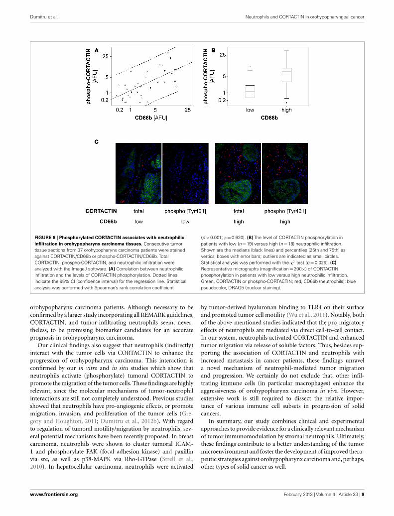
Next, we tested whether neutrophil-induced phosphorylationof CORTACTIN might occur also in vivo. To this end, we stainedconsecutive sections from frozen orohypopharynx carcinoma tis-sues (37 patients) against total and phosphorylated (Tyr421)forms of CORTACTIN, respectively. All samples were co-stained
against CD66b (neutrophil marker). Micrographs were taken at100-fold magnification from the same region of the tumor tissuefor CORTACTIN or phospho-CORTACTIN (each with CD66bco-staining). The micrographs were analyzed with the ImageJsoftware and the values obtained were considered as Arbitrary Flu-orescent Units (AFU). We then plotted the levels of CORTACTINphosphorylation [phospho-CORTACTIN (AFU)] against the lev-els of neutrophilic infiltration [CD66b (AFU); Figure 6A]. Statisti-cal analysis using Spearman’s rank test indicated that CORTACTINphosphorylation significantly correlated with neutrophilic infil-tration (p < 0.001, ρ = 0.620). Additionally, we divided thepatients into CD66blow and CD66bhigh according to the medianvalue of CD66b (AFU) and found that CD66bhigh patients had sig-nificantly higher CORTACTIN phosphorylation than CD66blow
patients (p= 0.029, χ2; Figure 6B). A representative example ofCORTACTIN phosphorylation in CD66blow versus CD66bhigh
patients is shown in Figure 6C. Taken together, our in vitroand in situ studies demonstrate that neutrophils interact withthe tumor cells by inducing CORTACTIN phosphorylation andactivation.
TUMORAL CORTACTIN – NEUTROPHIL INTERACTION: FUNCTIONALCONSEQUENCESFinally, we investigated which functional changes might neu-trophils induce in the tumor cells via CORTACTIN. Based on thefindings that: (i) CORTACTIN is an important regulator of cellularmigration (Ammer and Weed, 2008) and (ii) neutrophils enhancethe migration of HNC cells (Dumitru et al., 2011), we hypoth-esized that neutrophils “exploit” high tumoral CORTACTIN lev-els to promote the migration of the tumor cells. Conversely, inthe absence of tumoral CORTACTIN, neutrophils would not beable to stimulate the migration of the tumor cells. We, therefore,knocked-down CORTACTIN in FaDu cells by siRNA. As control,FaDu cells were transfected with mock siRNA (Figures 7A,B).We then determined the migratory potential of the tumor cellsin the presence or absence of factors released by “primed” neu-trophils. The results showed that mock-transfected FaDu cellsdisplayed accelerated migration in response to neutrophil stimula-tion, as indicated by enhanced closure of the detection zone (“gap”;Figure 7C). In contrast, this effect of neutrophil stimulation wasabolished in CORTACTIN knock-down FaDu cells (Figure 7C).These findings indicate that, consistent with the results presentedin Figure 4, high levels of tumoral CORTACTIN are required forneutrophil-induced tumoral migration and progression.
DISCUSSIONIncreased effort has been made to identify cellular/molecularfactors that could provide accurate information regarding can-cer diagnosis, prognosis, and response to therapy. In this study,we identify tumoral CORTACTIN as a prognostic factor for theclinical outcome in orohypopharynx carcinoma patients. Moreimportantly, we demonstrate that combined analysis of tumoralCORTACTIN and of neutrophilic infiltration in the tumor tis-sue has higher prognostic impact for the clinical outcome inthese patients. In addition to the clinical findings, our study pro-vides a biological and mechanistical link between neutrophils and
www.frontiersin.org February 2013 | Volume 4 | Article 33 | 7

Dumitru et al. Neutrophils and CORTACTIN in orohypopharyngeal cancer
FIGURE 5 | Neutrophils phosphorylate CORTACTIN in FaDu(hypopharynx carcinoma) cells. (A) Neutrophils were “primed” byco-incubation with FaDu supernatants (FaDu SN) for 24 h. The resultingsupernatants (FaDu/neutrophil SN) were used to stimulate FaDu cells for 24,48, or 72 h. FaDu SN or neutrophil SN only were used as controls. (B)
CORTACTIN expression and activation were assessed by western blot usingantibodies against total CORTACTIN (upper panel) or phosphorylatedCORTACTIN (Tyr 421; middle panel). GAPDH (lower panel) was used ascontrol for the quantity and quality of protein lysates. The results arerepresentative for five independent experiments each.
tumoral CORTACTIN by showing that neutrophils phosphorylateCORTACTIN in the tumor cells to promote their migration.
The potential role of CORTACTIN as prognostic biomarker hasbeen previously investigated in different types of cancer (Li et al.,2008; Wang et al., 2009; Cai et al., 2010; Xu et al., 2010). Severalstudies observed that CORTACTIN associated with poor clini-cal outcome also in HNC patients (Gibcus et al., 2008; Hofmanet al., 2008; Rodrigo et al., 2009). Among the HNC subtypes, COR-TACTIN has been mainly proposed as a potential biomarker forlaryngeal carcinoma (Gibcus et al., 2008; Rodrigo et al., 2009). Sur-prisingly, the study of Rodrigo et al. (2009) suggested that, whilehaving prognostic value in laryngeal carcinoma, CORTACTINdoes not associate with the clinical outcome in pharyngeal car-cinoma, which is in contrast to our findings. This discrepancymight result from the different read-outs used to assess COR-TACTIN (over)expression. While we analyzed directly the proteinlevels of CORTACTIN in relation to clinical parameters, the above-mentioned studies based their statistical analysis mainly on thegene amplification of CORTACTIN. However, it has been oftenshown that protein and gene expression do not perfectly correlate.In fact, even the study of Rodrigo et al. (2009) found that geneamplification of CORTACTIN did not correlate to the proteinlevels observed by immunohistochemistry in 35% of specimens.
Yet proteins are the ultimate effectors in biological processes andanalysis of protein levels is likely to provide more reliable biologi-cal insights than analysis of gene expression. Thus, by quantifyingthe protein expression of CORTACTIN, our study sheds a newlight on the role of this molecule in the prognosis of HNC.
Of novelty and significance are our findings regarding the inter-action between tumoral CORTACTIN and neutrophils (CD66b-positive cells) in orohypopharynx carcinoma. As outlined above,an association of either CORTACTIN or tumor-infiltrating neu-trophils with poor clinical outcome in cancer patients has alreadybeen proposed by previous studies (Donskov and von der Maase,2006; Jensen et al., 2009, 2012; Kuang et al., 2011; Trellakis et al.,2011; Ilie et al., 2012). However, a link between CORTACTINand neutrophils has not been shown in any type of cancer thusfar. Here, we demonstrate that patients exhibiting high levels oftumoral CORTACTIN together with high neutrophilic infiltra-tion (CORTACTINhigh/CD66bhigh) have worse clinical outcomethan all other groups of patients in our cohort. These findingsare further substantiated by multivariate analysis adjusted forfactors potentially relevant for progression of orohypopharynxcarcinoma, such as smoking/alcohol consumption, type of ther-apy, and gender. Within this model, CORTACTINhigh/CD66bhigh
phenotype was the only significant predictor of poor survival for
Frontiers in Immunology | Tumor Immunity February 2013 | Volume 4 | Article 33 | 8
Dumitru et al. Neutrophils and CORTACTIN in orohypopharyngeal cancer
FIGURE 6 | Phosphorylated CORTACTIN associates with neutrophilicinfiltration in orohypopharynx carcinoma tissues. Consecutive tumortissue sections from 37 orohypopharynx carcinoma patients were stainedagainst CORTACTIN/CD66b or phospho-CORTACTIN/CD66b. TotalCORTACTIN, phospho-CORTACTIN, and neutrophilic infiltration wereanalyzed with the ImageJ software. (A) Correlation between neutrophilicinfiltration and the levels of CORTACTIN phosphorylation. Dotted linesindicate the 95% CI (confidence interval) for the regression line. Statisticalanalysis was performed with Spearman’s rank correlation coefficient
(p < 0.001; ρ=0.620). (B) The level of CORTACTIN phosphorylation inpatients with low (n=19) versus high (n=18) neutrophilic infiltration.Shown are the medians (black lines) and percentiles (25th and 75th) asvertical boxes with error bars; outliers are indicated as small circles.Statistical analysis was performed with the χ2 test (p=0.029). (C)Representative micrographs (magnification=200×) of CORTACTINphosphorylation in patients with low versus high neutrophilic infiltration.Green, CORTACTIN or phospho-CORTACTIN; red, CD66b (neutrophils); bluepseudocolor, DRAQ5 (nuclear staining).
orohypopharynx carcinoma patients. Although necessary to beconfirmed by a larger study incorporating all REMARK guidelines,CORTACTIN, and tumor-infiltrating neutrophils seem, never-theless, to be promising biomarker candidates for an accurateprognosis in orohypopharynx carcinoma.
Our clinical findings also suggest that neutrophils (indirectly)interact with the tumor cells via CORTACTIN to enhance theprogression of orohypopharynx carcinoma. This interaction isconfirmed by our in vitro and in situ studies which show thatneutrophils activate (phosphorylate) tumoral CORTACTIN topromote the migration of the tumor cells. These findings are highlyrelevant, since the molecular mechanisms of tumor-neutrophilinteractions are still not completely understood. Previous studiesshowed that neutrophils have pro-angiogenic effects, or promotemigration, invasion, and proliferation of the tumor cells (Gre-gory and Houghton, 2011; Dumitru et al., 2012b). With regardto regulation of tumoral motility/migration by neutrophils, sev-eral potential mechanisms have been recently proposed. In breastcarcinoma, neutrophils were shown to cluster tumoral ICAM-1 and phosphorylate FAK (focal adhesion kinase) and paxillinvia src, as well as p38-MAPK via Rho-GTPase (Strell et al.,2010). In hepatocellular carcinoma, neutrophils were activated
by tumor-derived hyaluronan binding to TLR4 on their surfaceand promoted tumor cell motility (Wu et al., 2011). Notably, bothof the above-mentioned studies indicated that the pro-migratoryeffects of neutrophils are mediated via direct cell-to-cell contact.In our system, neutrophils activated CORTACTIN and enhancedtumor migration via release of soluble factors. Thus, besides sup-porting the association of CORTACTIN and neutrophils withincreased metastasis in cancer patients, these findings unravela novel mechanism of neutrophil-mediated tumor migrationand progression. We certainly do not exclude that, other infil-trating immune cells (in particular macrophages) enhance theaggressiveness of orohypopharynx carcinoma in vivo. However,extensive work is still required to dissect the relative impor-tance of various immune cell subsets in progression of solidcancers.
In summary, our study combines clinical and experimentalapproaches to provide evidence for a clinically relevant mechanismof tumor immunomodulation by stromal neutrophils. Ultimately,these findings contribute to a better understanding of the tumormicroenvironment and foster the development of improved thera-peutic strategies against orohypopharynx carcinoma and, perhaps,other types of solid cancer as well.
www.frontiersin.org February 2013 | Volume 4 | Article 33 | 9

Dumitru et al. Neutrophils and CORTACTIN in orohypopharyngeal cancer
FIGURE 7 | Neutrophils promote the migration of FaDu cells viaCORTACTIN. (A) FaDu cells in which CORTACTIN had been knocked-down bysiRNA were stimulated with FaDu/neutrophil SN and migration was assessed48 h later. (B) Efficiency of CORTACTIN knock-down was determined by flowcytometric analysis of total CORTACTIN levels at 72 h post-transfection. (C)FaDu cells (transfected with CORTACTIN or mock siRNA) were grown toconfluency in the presence of “gap”-forming culture inserts. The “gap” refers
to the cell-free round area visible in the figure. Stimulation was performed asindicated in A. Cells were fixed/stained with a solution containing crystalviolet and micrographs were taken at 25-fold magnification. “Gap”-closurewas quantified with the ImageJ software. Data are means±SD of threeindependent experiments (upper panel). A representative result is shown inthe lower panel of the figure. Statistical testing was performed with thetwo-tailed paired Student’s t -test.
ACKNOWLEDGMENTSWe thank Petra Altenhoff and Sebastian Vollmer (Depart-ment of Otorhynolaryngology, University of Duisburg-Essen)and Anja Peglow (Department of Pathology/Neuropathology,
University of Duisburg-Essen) for excellent technical support.This study was supported in part by the German Ministryfor Education and Research (BMBF) and by KrebsgesellschaftNRW.
REFERENCESAmmer, A. G., and Weed, S. A. (2008).
Cortactin branches out: roles inregulating protrusive actin dynam-ics. Cell Motil. Cytoskeleton 65,687–707.
Bekes, E. M., Schweighofer, B.,Kupriyanova, T. A., Zajac, E., Ardi,V. C., Quigley, J. P., et al. (2011).Tumor-recruited neutrophils andneutrophil TIMP-free MMP-9regulate coordinately the levels oftumor angiogenesis and efficiencyof malignant cell intravasation. Am.J. Pathol. 179, 1455–1470.
Cai, J. H., Zhao, R., Zhu, J. W., Jin, X.L., Wan, F. J., Liu, K., et al. (2010).Expression of cortactin correlateswith a poor prognosis in patients
with stages II-III colorectal adeno-carcinoma. J. Gastrointest. Surg. 14,1248–1257.
Choong, N., and Vokes, E. (2008).Expanding role of the medicaloncologist in the management ofhead and neck cancer. CA Cancer J.Clin. 58, 32–53.
Coussens, L. M., and Werb, Z. (2002).Inflammation and cancer. Nature420, 860–867.
Donskov, F., and von der Maase, H.(2006). Impact of immune para-meters on long-term survival inmetastatic renal cell carcinoma. J.Clin. Oncol. 24, 1997–2005.
Dumitru, C. A., Fechner, M. K., Hoff-mann, T. K., Lang, S., and Bran-dau, S. (2012a). A novel p38-MAPK
signaling axis modulates neutrophilbiology in head and neck cancer. J.Leukoc. Biol. 91, 591–598.
Dumitru, C. A., Moses, K., Trel-lakis, S., Lang, S., and Brandau,S. (2012b). Neutrophils and gran-ulocytic myeloid-derived suppres-sor cells: immunophenotyping, cellbiology and clinical relevance inhuman oncology. Cancer Immunol.Immunother. 61, 1155–1167.
Dumitru, C. A., Gholaman, H., Trel-lakis, S., Bruderek, K., Dominas, N.,Gu, X., et al. (2011). Tumor-derivedmacrophage migration inhibitoryfactor modulates the biology ofhead and neck cancer cells via neu-trophil activation. Int. J. Cancer 129,859–869.
Gibcus, J. H., Mastik, M. F., Menkema,L., de Bock, G. H., Kluin, P. M., Schu-uring, E., et al. (2008). Cortactinexpression predicts poor survival inlaryngeal carcinoma. Br. J. Cancer98, 950–955.
Gregory, A. D., and Houghton, A.M. (2011). Tumor-associatedneutrophils: new targets forcancer therapy. Cancer Res. 71,2411–2416.
Hofman, P., Butori, C., Havet, K., Hof-man, V., Selva, E., Guevara, N., etal. (2008). Prognostic significance ofcortactin levels in head and necksquamous cell carcinoma: compar-ison with epidermal growth factorreceptor status. Br. J. Cancer 98,956–964.
Frontiers in Immunology | Tumor Immunity February 2013 | Volume 4 | Article 33 | 10

Dumitru et al. Neutrophils and CORTACTIN in orohypopharyngeal cancer
Ilie, M., Hofman, V., Ortholan, C.,Bonnetaud, C., Coelle, C., Mouroux,J., et al. (2012). Predictive clini-cal outcome of the intratumoralCD66b-positive neutrophil-to-CD8-positive T-cell ratio inpatients with resectable nonsmallcell lung cancer. Cancer 118,1726–1737.
Jablonska, J., Leschner, S., Westphal,K., Lienenklaus, S., and Weiss,S. (2010). Neutrophils responsiveto endogenous IFN-beta regulatetumor angiogenesis and growth in amouse tumor model. J. Clin. Invest.120, 1151–1164.
Jemal,A., Siegel, R.,Ward, E., Murray, T.,Xu, J., and Thun, M. J. (2007). Can-cer statistics, 2007. CA Cancer J. Clin.57, 43–66.
Jensen, H. K., Donskov, F., Marcussen,N., Nordsmark, M., Lundbeck, F.,and von der Maase, H. (2009). Pres-ence of intratumoral neutrophils isan independent prognostic factorin localized renal cell carcinoma. J.Clin. Oncol. 27, 4709–4717.
Jensen, T. O., Schmidt, H., Moller, H.J., Donskov, F., Hoyer, M., Sjoegren,P., et al. (2012). Intratumoral neu-trophils and plasmacytoid dendriticcells indicate poor prognosis and areassociated with pSTAT3 expressionin AJCC stage I/II melanoma. Cancer118, 2476–2485.
Kuang, D. M., Zhao, Q., Wu, Y., Peng,C., Wang, J., Xu, Z., et al. (2011).Peritumoral neutrophils link inflam-matory response to disease progres-sion by fostering angiogenesis inhepatocellular carcinoma. J. Hepa-tol. 54, 948–955.
Li, X., Zheng, H., Hara, T., Taka-hashi, H., Masuda, S., Wang, Z.,et al. (2008). Aberrant expressionof cortactin and fascin are effectivemarkers for pathogenesis, invasion,metastasis and prognosis of gastriccarcinomas. Int. J. Oncol. 33, 69–79.
Lin, W. W., and Karin, M. (2007).A cytokine-mediated link betweeninnate immunity, inflammation,and cancer. J. Clin. Invest. 117,1175–1183.
Nozawa, H., Chiu, C., and Hanahan,D. (2006). Infiltrating neutrophilsmediate the initial angiogenic switchin a mouse model of multistage car-cinogenesis. Proc. Natl. Acad. Sci.U.S.A. 103, 12493–12498.
Rauch, J., Ahlemann, M., Schaffrik, M.,Mack, B., Ertongur, S., Andratschke,M., et al. (2004). Allogenic antibody-mediated identification of head andneck cancer antigens. Biochem. Bio-phys. Res. Commun. 323, 156–162.
Rodrigo, J. P., Garcia-Carracedo, D.,Garcia, L. A., Menendez, S., Allonca,E., Gonzalez, M. V., et al. (2009).Distinctive clinicopathological
associations of amplification of thecortactin gene at 11q13 in head andneck squamous cell carcinomas. J.Pathol. 217, 516–523.
Strell, C., Lang, K., Niggemann, B.,Zaenker, K. S., and Entschladen,F. (2010). Neutrophil granulocytespromote the migratory activity ofMDA-MB-468 human breast carci-noma cells via ICAM-1. Exp. CellRes. 316, 138–148.
Trellakis, S., Bruderek, K., Dumitru, C.A., Gholaman, H., Gu, X., Bankfalvi,A., et al. (2011). Polymorphonucleargranulocytes in human head andneck cancer: Enhanced inflamma-tory activity, modulation by cancercells and expansion in advanced dis-ease. Int. J. Cancer 129, 2183–2193.
Wang, G. C., Hsieh, P. S., Hsu, H. H.,Sun, G. H., Nieh, S., Yu, C. P., et al.(2009). Expression of cortactin andsurvivin in renal cell carcinoma asso-ciated with tumor aggressiveness.World J. Urol. 27, 557–563.
Wu, Y., Zhao, Q., Peng, C., Sun, L., Li, X.F., and Kuang, D. M. (2011). Neu-trophils promote motility of cancercells via a hyaluronan-mediatedTLR4/PI3K activation loop. J.Pathol. 225, 438–447.
Xu, X. Z., Garcia, M. V., Li, T. Y., Khor,L. Y., Gajapathy, R. S., Spittle, C., etal. (2010). Cytoskeleton alterationsin melanoma: aberrant expression of
cortactin, an actin-binding adapterprotein, correlates with melanocytictumor progression. Mod. Pathol. 23,187–196.
Conflict of Interest Statement: Theauthors declare that the research wasconducted in the absence of any com-mercial or financial relationships thatcould be construed as a potential con-flict of interest.
Received: 23 November 2012; accepted:30 January 2013; published online: 18February 2013.Citation: Dumitru CA, Bankfalvi A,Gu X, Eberhardt WE, Zeidler R,Lang S and Brandau S (2013) Neu-trophils activate tumoral CORTACTINto enhance progression of orohypophar-ynx carcinoma. Front. Immun. 4:33. doi:10.3389/fimmu.2013.00033This article was submitted to Frontiers inTumor Immunity, a specialty of Frontiersin Immunology.Copyright © 2013 Dumitru, Bankfalvi,Gu, Eberhardt , Zeidler , Lang and Bran-dau. This is an open-access article distrib-uted under the terms of the Creative Com-mons Attribution License, which per-mits use, distribution and reproductionin other forums, provided the originalauthors and source are credited and sub-ject to any copyright notices concerningany third-party graphics etc.
www.frontiersin.org February 2013 | Volume 4 | Article 33 | 11
Copyright © 2022 FDOKUMEN