
Static images of novel, moveable objects learned through touch activate visual area hMT+
10
This article appeared in a journal published by Elsevier. The attached copy is furnished to the author for internal non-commercial research and education use, including for instruction at the authors institution and sharing with colleagues. Other uses, including reproduction and distribution, or selling or licensing copies, or posting to personal, institutional or third party websites are prohibited. In most cases authors are permitted to post their version of the article (e.g. in Word or Tex form) to their personal website or institutional repository. Authors requiring further information regarding Elsevier’s archiving and manuscript policies are encouraged to visit: http://www.elsevier.com/copyright
Transcript of Static images of novel, moveable objects learned through touch activate visual area hMT+

This article appeared in a journal published by Elsevier. The attachedcopy is furnished to the author for internal non-commercial researchand education use, including for instruction at the authors institution
and sharing with colleagues.
Other uses, including reproduction and distribution, or selling orlicensing copies, or posting to personal, institutional or third party
websites are prohibited.
In most cases authors are permitted to post their version of thearticle (e.g. in Word or Tex form) to their personal website orinstitutional repository. Authors requiring further information
regarding Elsevier’s archiving and manuscript policies areencouraged to visit:
http://www.elsevier.com/copyright

Author's personal copy
Static images of novel, moveable objects learned through touch activate visualarea hMT+
Jason S. Chan ⁎, Cristina Simões-Franklin, Hugh Garavan, Fiona N. NewellSchool of Psychology and Institute of Neuroscience, Trinity College Dublin, Dublin, Republic of Ireland
a b s t r a c ta r t i c l e i n f o
Article history:Received 12 August 2009Revised 28 September 2009Accepted 29 September 2009Available online 6 October 2009
Keywords:Dynamic object recognitionHapticshMT+Multisensory perceptionObject recognition
Although many studies have found similar cortical areas activated during the recognition of objects encodedthrough vision or touch, little is known about cortical areas involved in the crossmodal recognition ofdynamic objects. Here, we investigated which cortical areas are involved in the recognition of moving objectsand were specifically interested in whether motion areas are involved in the recognition of dynamic objectswithin and across sensory modalities. Prior to scanning, participants first learned to recognise a set of 12novel objects, each presented either visually or haptically, and either moving or stationary. We thenconducted fMRI whilst participants performed an old-new task with static images of learned or not-learnedobjects. We found the fusiform and right inferior frontal gyri more activated to within-modal visual thancrossmodal object recognition. Our results also revealed increased activation in area hMT+, LOC and themiddle occipital gyrus, in the right hemisphere only, for the objects learned as moving compared to thelearned static objects, regardless of modality. We propose that the network of cortical areas involved in therecognition of dynamic objects is largely independent of modality and have important implications forunderstanding the neural substrates of multisensory dynamic object recognition.
© 2009 Elsevier Inc. All rights reserved.
Introduction
Although there is extensive research on visual, haptic, and visuo-haptic object recognition, most of these studies have focused on therecognition of static objects (e.g., Amedi et al., 2002, 2005; Ernst et al.,2007; Grill-Spector et al., 1998, 1999; Newell et al., 2001). However,real world object recognition not only involves static objects butmanyobjects can either move independently or have moveable parts forparticular functions (e.g., door handles and hand whisks). Despite thisfact, our knowledge of how information about moving objects isprocessed for the purpose of recognition is mainly limited to visualobject recognition and relatively little is known about haptic or cross-modal recognition of moving objects.
Many behavioural studies have suggested efficient sharing ofinformation across vision and touch for the purpose of the recogni-tion of static objects (e.g., Easton et al., 1997; Reales and Ballesteros,1999). Later neuroimaging investigations revealed an overlapbetween the cortical areas involved in visual object recognition andthose involved in the recognition of objects encoded through touch(Amedi et al., 2001, 2005; Lacey et al., 2009; Malach et al., 1995;Sathian and Zangaladze, 2002). In particular, haptic object recogni-
tion activates the lateral occipital complex (LOC; Amedi et al., 2001),an area which is consistently activated during the visual recognitionof objects. Together, these studies demonstrate that efficient beha-vioural performance in crossmodal object recognition is underpinnedby shared neural systems often identified as being within so-calledvisual areas.
Recent research into the visual recognition of moving objects hassuggested that objects are stored in memory as spatio-temporalrepresentations such that both the characteristic motion and shape ofan object are relevant cues for recognition (e.g., Newell et al., 2004;Setti and Newell, 2009; Stone, 1998). Other evidence, particularlyfrom studies on the perception of faces, has supported the findingsthat motion information can affect recognition in that changes in thecharacteristic dynamic patterns of faces can disrupt the recognition offamiliar faces (Hill and Johnston, 2001). Thus, evidence from be-havioural studies suggests that motion and shape information areintegrated in the representation of objects and faces.
Other evidence from neuroimaging studies has suggested that theprocessing of shape and motion information in object perception isunderpinned by the same neural systems. For example, although ithas been established that real visual-motion information activatesarea hMT+ (i.e., V5; Tootell et al., 1995b; Zeki, 1991, 1993), it has alsobeen shown that area hMT+ is activated to apparent motion, that is,visual-motion aftereffects caused by outwardly moving concentriccircles (Tootell et al., 1995a). In other words, activity in hMT+ can beelicited by the perception of motion and not just by motioninformation directly available in the visual stimulus itself. This
NeuroImage 49 (2010) 1708–1716
⁎ Corresponding author. School of Psychology/Institute of Neuroscience, TrinityCollege Dublin, 2 College Green, Lloyd Building, Dublin 2, Republic of Ireland.Fax: +353 1 896 3183.
E-mail address: [email protected] (J.S. Chan).
1053-8119/$ – see front matter © 2009 Elsevier Inc. All rights reserved.doi:10.1016/j.neuroimage.2009.09.068
Contents lists available at ScienceDirect
NeuroImage
j ourna l homepage: www.e lsev ie r.com/ locate /yn img

Author's personal copy
suggests that neural activation (as measured by blood oxygen-leveldependent or BOLD responses) in area hMT+ may be both top-downdependent and stimulus dependent (see also Alford et al., 2007;Beauchamp and DeYoe, 1996). Adding to the evidence that activity inhMT+ can be modulated by top-down processes, further brainimaging studies have also shown that the mental imagery of motion(Cohen et al., 1996; Goebel et al., 1998; O'Craven and Kanwisher,2000) and illusory motion (Tootell et al., 1995a) activate the samebrain regions as the direct perception of motion. Moreover, Kourtziand Kanwisher (2000) compared BOLD activation to participantsviewing still images in which motion was implied (e.g., an athleteperforming an action) or no motion was implied (e.g., an athlete atrest). They found greater activation in area hMT+ in the righthemisphere when presented with images containing implied motioncompared to the no-motion images. Interestingly, this effect was notrestricted to images of humans and animals, but to motion implied inscenes as well (e.g., a waterfall) suggesting that activation to eitherdirect or implied motion is not category specific.
Area hMT+ is not just related to the processing of motion in thevisual modality, but has also been shown to be related to motionstimuli presented via the auditory (Poirer et al., 2005; Smith et al.,2007) and tactile modalities (Hagen et al., 2002; Ricciardi et al.,2007). For example, Hagen and colleagues examined the effect oftactile stimulation on a participant's passive hand using paintbrushes which were moved across the hand. They found increasedactivation in area MT/V5 compared to static stimulation (i.e., brushesheld in place; see also Blake et al., 2004). Specifically, activation inarea MT was lower in the tactile motion condition than in the visual-motion condition (outwardly moving concentric circles). Theseresults are contrary to other studies that found no activation in thisarea to vibration or light touch stimulation on the skin (Coghill et al.,1994; Fox et al., 1986, 1987; Pardo et al., 1991; Seitz and Roland,1992). However, Hagen et al. suggest that this is because theseprevious studies used stimuli that did not provide a true perceptionof tactile motion. In other words, they did not use a stimulus that wasperceived as a single entity moving in space.
Others have suggested that area hMT+may not be a multisensorymotion processing area but that the neural activation to motionencoded from other modalities may be mediated by visual imagery. Inan attempt to dissociate tactile motion from visual imagery in sightedparticipants, Blake et al. (2004) used fMRI to measure activationwhenparticipants determined the direction of a rotating globe by eithertouching, viewing, or imagining the globe. They found increasedactivation in area hMT+ when touching a rotating globe relative tovisual imagery of the rotating globe or even imagining their fingertapping. Similar to the Hagen et al. study, activation to the tactilestimulus was lower than when viewing rotating dots but wasnevertheless significantly greater than baseline. Moreover, studieson participants who are visually impaired have also found that tactilemotion was sufficient to activate area hMT+ in the absence of visualstimulation or even visual familiarity with the stimuli (Goyal et al.,2006; Ricciardi et al., 2007). Taken together, these studies suggest thatvisual imagery does not fully account for activations in area hMT+induced by stimulation from modalities other than vision. Instead,these findings suggest that area hMT+ may be a general motionprocessing area.
There is evidence that changes in activation in brain areas asso-ciated with both the appearance and function of objects occurs as aconsequence of learning. For example, Weisberg et al. (2007)explicitly investigated the role of learning in visual object recogni-tion using novel objects which were attributed with a particularshape and function. They scanned participants in two fMRI sessions,before and after learning the novel objects. Before the trainingsession, activation in areas in the fusiform gyrus commonly asso-ciated with object shape properties was found to the static visualpresentation of the objects. During training, participants manipu-
lated each object and learned its particular shape and function.Following training, Weisberg et al. reported activation to the staticimages of the objects which was more focused in fusiform areas butalso activation was found in other areas of the brain normallyassociated with object motion (the left middle temporal gyrus) andobject manipulation (left intraparietal sulcus and premotor cortex).They attribute this activation to learned tool use and objectfunctionality. Interestingly, in their study, still images of the objectswere sufficient to activate motion areas in the cortex. As such, theactivation was due to motion information implied in the image ofthe object and not related to motion available in the stimulus or todirect tool-use itself. However, as the function of each object waslearned through both viewing and haptic manipulation it is unclear ifthis change in BOLD activation in area hMT+ is due to recalled visualobject motion acquired during the training session or imaginedaction motion of the hands manipulating the object. Valyear et al.(2007) found that action-related activation for tools and otherobjects is localized in the posterior intraparietal sulcus (IPS) and nothMT+, suggesting that the motion of body parts during tool use isunlikely to activate area hMT+. On the other hand, others haveargued that knowledge of tool-use and tool-motion is sufficient toactivate ventral areas in the cortex, particularly those in lateraltemporal areas which are activated by either static images or movingimages of familiar tools (Beauchamp et al., 2002).
In the current fMRI study, we examined the neural substratesunderlying the recognition of dynamic objects learned througheither vision or touch. In particular, we investigated whether thecortical areas underpinning visual object recognition and visualobject motion are also active to images of objects previously learnedthrough touch alone. To that end, we trained participants outsidethe scanner to recognise a series of novel object shapes which weremoving or stationary using either vision or haptics. In the scanner,we presented static, greyscale images of the learned objects to theparticipants who performed an old/new recognition task. Wehypothesised that there would be significantly greater BOLDactivation in areas hMT+ to static images of objects visuallylearned as moving compared to those learned as stationary objects.However, we were unclear as to whether moving objects learnedthrough haptics would also selectively activate this motion area.These results could have important implications in understandingthe role of area hMT+ in crossmodal processing. While previousstudies have shown that area hMT+ is involved in processingmotion information within vision and touch, it is unknown if areahMT+ processes implied motion in other modalities. Also, theseresults will further determine whether this information is sharedacross modalities. We were also interested in whether the lateraloccipital complex (LOC) was activated by images of the learnedobjects irrespective of learning modality and learned motion. Wepropose that if motion is integrated into a single representation ofthe object then we would expect to find increased activation in LOCas well as hMT+ to images of objects learned as moving. If motionis processed as a separate feature from shape information then wewould not expect to see any change in activation in area LOC acrossthe image types.
Methods
Participants
Seventeen participants (10 female and 7 male) between the agesof 20 and 48 years (mean age 30.38 years) took part in thisexperiment. All participants were right hand dominant, reported tohave normal or corrected to normal vision and were native Englishspeakers. All procedures were approved by the School of Psychology,Trinity College Dublin and, accordingly, informed consent wasobtained prior to the experiment.
1709J.S. Chan et al. / NeuroImage 49 (2010) 1708–1716

Author's personal copy
Apparatus and stimuli
A set of 12 novel objects was created using Lego™ blocks andeach was attached to a platform which prevented the participantfrom picking up the object. All objects were small enough to beeasily palpated by the hands (8 cm×8 cm). Each object shape wascomposed of between three and five blocks. For half of theseobjects, shape parts were built on a movable base structure whichallowed the objects to be moved in one of six different ways, i.e.,rotate, slide, pivot, slide and pivot, rotate and slide, and pivot androtate (see Fig. 1 for an illustration of the shapes and motions). Theremaining objects were similarly constructed but none of the partswere moveable.
In the learning phase, participants were presented with the 12three-dimensional objects. High resolution photographs of thesetarget objects were then taken from the viewpoint of the participantand converted to greyscale images using standard photo editingsoftware. These images were used as stimuli during the testingsession which took place in the fMRI scanner. We also constructed anextra set of eight objects, and images of these objects which were notpresented to subjects prior to imaging were used as non-targetstimuli in the fMRI task.
The test session of the experiment was programmed using“Presentation” software (Neurobehavioral Systems, California).Responses were recorded via an MR-compatible two-button responsepad (Cambridge Research Systems).
Fig. 1. A subset of six objects and motions used in the learning session. Each object has a unique shape and motion pairing. (A) Still images of a participant's haptic exploration in thelearning session. In the haptic-motion condition, participants moved the objects themselves. In the visual-motion condition, participants viewed the objects while the experimentermanipulated the object's motion. (B) Images used in the fMRI (test session).
1710 J.S. Chan et al. / NeuroImage 49 (2010) 1708–1716

Author's personal copy
A motion localizer stimulus was used to identify functional areahMT+ in each participant. The stimuli consisted of movingconcentric circles (frequency 50 Hz) alternating with a staticimage (Hampson et al., 2004; Kourtzi and Kanwisher, 2000; Tootellet al., 1995b). The static image appeared as a single frame of themoving counterpart. These stimuli were created using the Visgentoolkit associated with Presentation.
Design and procedure
There were two sessions to the experiment: a learning sessionimmediately followed by a test session. The learning session wasconducted prior to neuroimaging and the test session occurred whilethe participant was in the MRI scanner. During the learning session,participants learned to associate a nonsense name to each targetobject. The task inside the scanner was based on an old-newrecognition paradigm, where participants determined whether eachvisually presented object was previously learned (i.e., ‘old’) or not(i.e., ‘new’) regardless of the modality through which they werelearned or whether they were moving or not during learning.
Learning session (behavioural)
The learning phase consisted of four separate learning blocks: twohaptic-only blocks and two vision-only blocks. Each block consisted ofthree objects. Within each sensory block all objects were presented aseither fixed in position (i.e., static) or were movable. The presentationorder of these four blocks was counterbalanced across participants.During this session all target objects were placed behind a curtainwhich obscured the object from the participant's view. To ensure thatparticipants adequately learned each target object and all of itsproperties, they were instructed to explore all aspects of each targetobject. To aid explanation, participants were provided with examplesof manipulable objects in the real world which comprise severalfeatures and functions such as a door handle, a stapler or a handwhisk. These example objects have a characteristic shape and motionand were provided as an indication of the range of object types thatcould be tested but there was no emphasis given to a particularfeature (i.e., neither motion nor shape was singled out for attention).
During haptic learning, participants reached underneath thecurtain with both hands to feel the object for 30 s. All object partsof each target could be freely explored during haptic learning. Forthe moving target objects, the objects did not move independentlybut could be moved by the participant during active exploration.During visual learning, participants lifted the curtain for 10 s to seethe object and lowered it when instructed by the experimenter. Forthe moving visual target objects, participants viewed the targetobject while the experimenter moved the object. In the visual-staticblock, participants viewed the stationary object for 10 s andparticipants were encouraged to remain still. However, their headswere not fixed in place.
To ensure that participants were exploring all aspects of theobjects for recognition, participants were required to associate eachobject with a given nonsense name (e.g., “nof,” “zam,” “kuk”). Withineach block of trials, an object was presented four times in successionand the experimenter provided the name of each object to theparticipant. At the end of each block, the target objects were randomlypresented to the participant one at a time, and he or she was requiredto recall the correct name of the object.
After conducting all four blocks of learning, participants werethen tested on their object-name recall. Participants were randomlypresented with objects across all conditions. They were informedbeforehand in which modality the object would be presented butnot whether the object would be moving or static. This was anunspeeded task, thus participants could take as long as they neededto answer correctly. All responses were recorded by the experi-
menter. The learning session was repeated until participants wereable to recall the names of the objects with an accuracy of over 90%correct (i.e., 11 out of 12 correct responses). On average, traininglasted approximately 60 min. Participants were brought to the fMRIscanner immediately after training to take part in the test session.
Test session (neuroimaging)
Prior to functional acquisition during the test session, we acquiredstructural scans, which lasted approximately 12 min. During the testsession, participants were presented with static visual images only ofthe target objects which they learned during the learning session. Themain test session was based on a 2×2 repeated measures design withlearning modality (haptic or visual) and learned motion (static ormotion) as factors. For the purposes of this test, participants were toldto ignore the names of each object but instead indicate whether theobject was previously learned or not.
The test session was separated into six runs, with each run lastingapproximately 6 min. The protocol was based on an event-relateddesign which was implemented with a jittered inter-trial interval(ITI) of 6, 8, 10 or 12 s. A trial began with a red fixation cross whichwas presented for the duration of the ITI. This was immediatelyreplaced by a blue fixation cross which was presented for 2 s.Participants were instructed that the blue fixation cross signalled theimminent appearance of a stimulus object. A static image of an objectwas then presented for 4 s. Following the trial, participants weregiven 2 s to indicate whether the object image they saw was one theyhad learned before or not. Images of objects were presented in arandom order across all runs. In 60% of trials the old object waspresented with a new object presented in the remaining trials. Therewere 120 trials in total with 8 repetitions of each old object and 3repetitions of each new object. This was done to ensure thatparticipants did not become familiar with the new stimuli.
Following the test session, participants were presented with visualimages designed to localise functional area hMT+. There were 12interleaved blocks of 6 moving and 6 static visual images in total, witheach block lasting 16 s. The participants were required to look at thefixation point at the centre of each visual image but they were notrequired to perform a task. The entire scanning session lastedapproximately 50 min.
fMRI data acquisition and data analysis
Scanning was performed with a Philips Intera Achieva 3.0 Teslawhole-body MR system (Philips Healthcare, The Netherlands)equipped with a mirror that reflected a 1024×768 display, projectedona panel placed behind the participant's headoutside themagnet. Themirror wasmounted on the head coil in the participant's line of vision.
Imaging started with 31.5 s of standard scout images to adjust forhead positioning, followed by a reference scan to resolve sensitivityvariations. Imaging used a parallel SENSitivity Encoding (SENSE)approach (Pruessmann et al., 1999) with reduction factor 2. Onehundred and eighty high-resolution T1-weighted anatomic MPRAGEaxial images (FOV 230 mm, thickness 0.9 mm, voxel size0.9×0.9×0.9) were then acquired (for a total duration of 5.43 min),to allow subsequent activation localization and spatial normalization.
The functional scans consisted of 32 non-contiguous (10% gap)3.5 mm axial slices covering the entire brain were collected using aT2⁎ weighted echo-planar imaging sequence (TE=35 ms,TR=2000 ms, FOV 224 mm, 64×64 mm matrix size in Fourierspace). The spontaneous changes in frequency were automaticallycorrected by the Intera Achieva by means of dynamic stabilization(real-time frequency adjustment) after each TR.
The analyses of the functional activations were restricted to trialsto which the participant responded correctly. This was done to ensurethat we were analyzing brain activation related to an accurate
1711J.S. Chan et al. / NeuroImage 49 (2010) 1708–1716
Author's personal copy
perception of the learned object. The six functional task runs wereconcatenated (yielding 771 volumes) and motion corrected. Fourseparate time-series regressors were created by convolving the timingof each object stimulus (i.e., haptic motion, haptic static, visual motionand visual static) with a gamma-variate function to account for theBOLD hemodynamic response function and the regression analysiswas conducted using the ‘3dDeconvolve’ program in AFNI (Cox,1996). The magnitude of the signal change from baseline associatedwith each regressor was carried forward to group analyses. Spatialsmoothing was then applied for all time points of activation using a 3-mm full width half-maximum Gaussian kernel. For group analysis,individual brains were converted into standard anatomical space(Talairach and Tournoux, 1988).
For the single localiser run, a separate square-wave regressor wasconvolved with a gamma-variate function, contrasting the movingand static concentric circles and then motion corrected.
We conducted a repeated measures, voxel-wise, 2×2 randomeffects ANOVA on the activation, with learning modality (vision ortouch) and learned motion (motion or static) of the target object asfactors. The results of this group analysis were F-maps indicating theextent to which each factor or the interaction between factorsmodulated brain activity for each voxel. Voxels were then thresholdedat p≤0.005 which, when combined with a cluster size criterion of285 mm3, resulted in a false positive level of p≤0.05 (corrected) ascalculated by Monte Carlo simulation.
We were also specifically interested in determining if object areaLOC and motion area hMT+ were activated by images of objectspreviously learned either visually or haptically or as static or movableobjects. To that end, we created 5mm radius ROI spheres based on thecoordinates of hMT+ as revealed by the localiser (Talairachcoordinates: 40, −71, 4) and on the coordinates for LOC (Talairachcoordinates: 43,−73,−18) provided byMalach et al. (1995). For bothROIs, 2×2 repeated measures ANOVAs were conducted with learningmodality and learned motion as factors.
Results
Behavioural results
Accuracy averaged across participants and conditions during thetest session was 65%. A one-way ANOVA found no significant differ-ence in accuracy across conditions [F(4,65)b1, n.s.]. Performance ineach target condition was greater than chance level (15%) with sig-nificantdifferences between chanceperformance and the haptic-motioncondition (mean=64.58% [χ2=206.30, pb0.0001]), haptic-static con-dition (mean=68.15%) [χ2=247.22, pb0.0001], visual-motion condi-tion (mean=58.93%) [χ2=142.04, pb0.0001], visual-static condition(mean=72.62%) [χ2=292.96, pb0.0001], and the correct recognitionof new objects (chance=40%; mean=66.37%) [χ2=46.25, pb0.0005].
fMRI results
As previously stated, fMRI analyses were restricted to the correctresponses alone to ensure that activations were related to theveridical percept of the object. Furthermore, we excluded activationsto the presentation of the new objects from any further analyses asthese responses were not of relevance to the investigation (but werenevertheless important for the task).
Whole-brain analysis
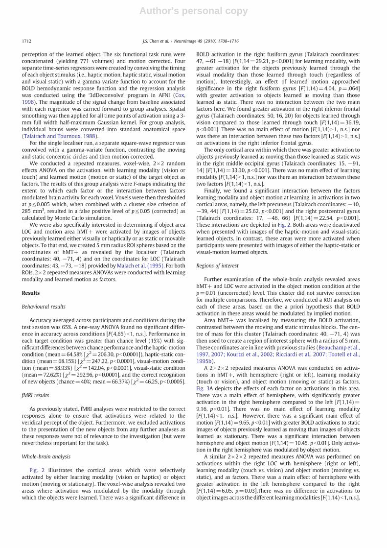
Fig. 2 illustrates the cortical areas which were selectivelyactivated by either learning modality (vision or haptics) or objectmotion (moving or stationary). The voxel-wise analysis revealed twoareas where activation was modulated by the modality throughwhich the objects were learned. There was a significant difference in
BOLD activation in the right fusiform gyrus (Talairach coordinates:47, −61 −18) [F(1,14=29.21, pb0.001] for learning modality, withgreater activation for the objects previously learned through thevisual modality than those learned through touch (regardless ofmotion). Interestingly, an effect of learned motion approachedsignificance in the right fusiform gyrus [F(1,14)=4.04, p=.064]with greater activation to objects learned as moving than thoselearned as static. There was no interaction between the two mainfactors here. We found greater activation in the right inferior frontalgyrus (Talairach coordinates: 50, 16, 20) for objects learned throughvision compared to those learned through touch [F(1,14)=36.19,pb0.001]. There was no main effect of motion [F(1,14)N1, n.s.] norwas there an interaction between these two factors [F(1,14)N1, n.s.]on activations in the right inferior frontal gyrus.
The only cortical area within which there was greater activation toobjects previously learned as moving than those learned as static wasin the right middle occipital gyrus (Talairach coordinates: 15, −91,14) [F(1,14)=33.30, pb0.001]. There was no main effect of learningmodality [F(1,14)b1, n.s.] nor was there an interaction between thesetwo factors [F(1,14)b1, n.s.].
Finally, we found a significant interaction between the factorslearning modality and object motion at learning, in activations in twocortical areas, namely, the left precuneus (Talairach coordinates:−10,−39, 44) [F(1,14)=25.62, pb0.001] and the right postcentral gyrus(Talairach coordinates: 17, −46, 66) [F(1,14)=22.54, pb0.001].These interactions are depicted in Fig. 2. Both areas were deactivatedwhen presented with images of the haptic-motion and visual-staticlearned objects. In contrast, these areas were more activated whenparticipants were presented with images of either the haptic-static orvisual-motion learned objects.
Regions of interest
Further examination of the whole-brain analysis revealed areashMT+ and LOC were activated in the object motion condition at thep=0.01 (uncorrected) level. This cluster did not survive correctionfor multiple comparisons. Therefore, we conducted a ROI analysis oneach of these areas, based on the a priori hypothesis that BOLDactivation in these areas would be modulated by implied motion.
Area hMT+ was localised by measuring the BOLD activation,contrasted between the moving and static stimulus blocks. The cen-tre of mass for this cluster (Talairach coordinates: 40, −71, 4) wasthen used to create a region of interest sphere with a radius of 5 mm.These coordinates are in linewith previous studies (Beauchamp et al.,1997, 2007; Kourtzi et al., 2002; Ricciardi et al., 2007; Tootell et al.,1995b).
A 2×2×2 repeated measures ANOVA was conducted on activa-tions in hMT+, with hemisphere (right or left), learning modality(touch or vision), and object motion (moving or static) as factors.Fig. 3A depicts the effects of each factor on activations in this area.There was a main effect of hemisphere, with significantly greateractivation in the right hemisphere compared to the left [F(1,14)=9.16, pb0.01]. There was no main effect of learning modality[F(1,14)b1, n.s.]. However, there was a significant main effect ofmotion [F(1,14)=9.65, pb0.01] with greater BOLD activations to staticimages of objects previously learned as moving than images of objectslearned as stationary. There was a significant interaction betweenhemisphere and object motion [F(1,14)=10.45, pb0.01]. Only activa-tion in the right hemisphere was modulated by object motion.
A similar 2×2×2 repeated measures ANOVA was performed onactivations within the right LOC with hemisphere (right or left),learning modality (touch vs. vision) and object motion (moving vs.static), and as factors. There was a main effect of hemisphere withgreater activation in the left hemisphere compared to the right[F(1,14)=6.05, p=0.03].There was no difference in activations toobject images across the different learningmodalities [F(1,14)b1, n.s.].
1712 J.S. Chan et al. / NeuroImage 49 (2010) 1708–1716
Author's personal copy
However, we found a main effect of motion [F(1,14)=7.87, p=0.01],with significantly greater BOLD response in right LOC when seeing astatic object image that was previously learned as moving objectcompared to seeing an image of an object previously learned asstationary. There were no other significant interactions (Table 1).
To assess the specificity of the ROI results, we conducted anadditional 5 mm spherical ROI analysis on Brodmann area 17(V1; Talairach coordinates: right: 10, −66, 17; left: −9, −67, 17)(Dougherty et al., 2003), a region in which we would nothypothesise activity to be influenced by learning modality or objectmotion. The same 2×2×2 repeated measures analysis performed forthe previous ROI areas was also conducted here. There were nosignificant effects or interactions.
Discussion
In summary, prior to scanning, participants were trained torecognise a set of novel objects which either had moving or static
parts as experienced through either touch or vision. In the fMRIscanner, participants were presented with static images of thesetarget objects and other non-target objects and were required toperform an old/new task. We found increased activation in severalareas which was specifically related to the learning modality andwhether the objects were learned as moving or not. In particular,activations in areas hMT+ and LOC within the right hemispherediffered for images of objects that implied motion and not images ofobjects that were stationary, regardless of modality.
This study investigated whether static images of moving objectspreviously learned through touch also activates hMT+, an areapreviously shown to be active for implied visual motion (Kourtzi andKanwisher, 2000). By presenting static visual images, we were able todemonstrate that area hMT+ processes the implied motion fromobjects previously learned through touch as well as vision. Activationfrom the hMT+ localiser was found in only the right hemispherewhich is similar to the results reported by Kourtzi and Kanwisher(2000). This lateralization of hMT+ activity could be due to a greater
Fig. 2. Activations represent the results from the voxel-wise whole-brain, 2×2 random effects ANOVA, with modality (vision vs. touch) and motion (motion vs. static) as factors.Graphs represent the clusters of significant activation for each main effect and interaction between the factors. Error bars represent the SEM. Images are using the neurologicalconvention.
1713J.S. Chan et al. / NeuroImage 49 (2010) 1708–1716

Author's personal copy
involvement of the right hemisphere in spatial processing, whereasthe left hemisphere is thought to be more involved in linguisticalprocessing (Grèzes et al., 2001; Large et al., 2007; Van Boven et al.,2005; Vandenbulcke et al., 2006).
In a related study, James and Gauthier (2003) investigatedcortical activations associated with learned associations betweenvisually presented unfamiliar objects and either auditory, action orencyclopedic semantic labels presented verbally. They found thateach concept activated sensory-specific networks. In particular,auditory concepts activated the superior temporal gyrus, whereasaction concepts activated more posterior regions in the superiortemporal sulcus. The authors conducted ROI analyses within thesespecific regions, however, and did not report activation specifically inarea hMT+ for the presentation of novel visual objects associatedwith action concepts. It may be the case, therefore, that previous
direct sensory experience with moving objects may be necessary toactivate this area (Beauchamp and Martin, 2007).
Interestingly, there was also a significant difference in BOLDactivation in area LOC when participants were shown objects thatwere learned moving compared to objects which did not moveduring learning. However, the increased activation in LOC for thelearned moving objects could be due to the additional informationprovided about the object while the object was moved (Gauthieret al., 2002; Grill-Spector et al., 1999) and not due to pure motioninformation. Gauthier et al. suggest that additional informationenhances mental imagery in the superior and inferior parietallobules, but they did not find that changes in viewpoint increasedactivation during an object recognition task in those areas of LOC.Thus, it is possible that the increase in BOLD response we see in LOCwas not due to additional viewpoint information of the motion
Fig. 3. (A) Results from the hMT+ ROI analysis. The 5 mm sphere represents the Talairach coordinates from the hMT+ localizer task. (B) The results from the LOC ROI analysis. The5mm sphere represents the Talairach coordinates taken fromMalach et al. (1995). For both analyses, results indicate the BOLD activation, thresholded to 0.005 and p correct to 0.05.Images are using the neurological convention.
Table 1A summary of BOLD activation when viewing static greyscale images of the target objects.
Brain area Hemifield Talairach coordinates Learning conditions Statistics
% Change score
x y z Haptic motion Haptic static Visual motion Visual static F -stat p -value
ROIMT+ Right/left ±40 −71 4 0.411 0.302 0.395 0.294 9.65 b0.01LOC Right/left ±43 −73 −18 0.541 0.425 0.572 0.495 7.87 0.01
Voxel-wise analysisFusiform gyrus Right 47 −61 −18 0.487 0.471 0.708 0.579 29.21 b0.0001Inferior frontal gyrus Right 50 16 20 0.241 0.19 0.406 0.41 36.188 b0.0001Middle occipital gyrus Right 15 −91 14 0.237 0.134 0.26 0.128 33.3 b0.0001Precuneus Left −10 −39 44 0.02 0.139 0.075 −0.101 25.62 0.0001Post-CG Right 17 −46 66 −0.04 0.062 0.063 −0.081 22.54 0.0003
ROI statistics represent the main effects of object motion analyses. Voxel-wise statistics are the results of the 2×2 whole-brain analysis.
1714 J.S. Chan et al. / NeuroImage 49 (2010) 1708–1716
Author's personal copy
component but due to an integration of motion and shape infor-mation (see e.g., Kriegeskorte et al., 2003).
The whole-brain analysis revealed activation in the precuneus andright postcentral gyrus under certain conditions. The precuneus hasbeen shown to be active during tasks involving visuo-spatial attention(see Cavanna and Trimble, 2006 for a review) as well as mentalimagery of self-generated movement (Hanakawa et al., 2003). Forexample, Hanakawa et al. asked participants either to finger-tap intime to a visual cue or to imagine themselves finger tapping. Theyfound greater activation in the precuneus for imagined finger tappingcompared to real finger tapping. We found increased activation in thisarea for the haptic-static and visual-motion conditions compared tothe haptic-motion and visual-static conditions. If the precuneus isinvolved in the mental imagery of self-generated motion we wouldhave expected activation in the haptic-motion condition instead ofthe haptic-static condition. Increased BOLD activation in the rightpostcentral gyrus, the primary somatosensory processing area, wassurprising given that participants did not feel the objects in thescanner. Activation in this brain region followed the same pattern asactivation in the precuneus in that there was increased activation forthe haptic-static and visual-motion conditions compared to thehaptic-motion and visual-static conditions. This may suggest in-creased spatial attention or mental imagery (Corbetta, 1998;Hopfinger et al., 2000). Clearly further research is needed to interpretthese interactions between learning modality and object position andthe role of mental imagery.
The right fusiform gyrus was found to be more active whenparticipants were presented with images of objects previouslylearned through vision than through touch. There was also a nearsignificant difference of motion in this brain area with images ofobjects previously learned as moving producing greater activationthan images of objects learned as stationary. This suggests that themotion of an object is a feature which is integrated into a repres-entation of an object and is not processed independently of objectshape (Wiggett et al., 2006).
Images of objects which were previously learned as moving alsoactivated the right middle occipital gyrus. This area has beenimplicated in tasks in which object shape is derived from motionand not to coherent motion without shape (Braddick et al., 2000).Taken together, the results from these studies suggest that the middleoccipital gyrus may be involved in the integration of shape andmotion, and not just deriving shape frommotion, and that activationsin the rMOG are independent of the modality through which themoving object was initially learned.
In the learning phase, participants either manipulated the objectsby touch or saw the objects beingmanipulated by the experimenter inthe object motion conditions. Clearly, there was less interaction withthe object features when the object was learned as stationary or whenthe object was learned through vision. Consequently, it may be arguedthat the activations in area hMT+ and the middle occipital gyrus areaction related instead of object related. However, we do not believethis to be the case for several reasons. First, the difference inactivations in area hMT+ to the images of objects learned as movingand those learned as stationary was modality independent. Ifactivations were related to interaction with the objects then itmight be expected that there would be greater activations to objectslearned through the haptic modality than those learned visually.Second, it has been shown that object–hand interactions activate theprecentral gyrus, pars triangularis and part opercularis in humans(BA44: Johnson-Frey et al., 2003). Johnson-Frey et al. presented stillimages of a hand grasping an object in a functional or non-functionalway, or simply touching the object (the palm was facing away fromthe object) and found that prehensile action was sufficient to elicitactivation in these areas. In other words, implied goal-directed actionwas enough to activate the inferior frontal gyrus. We did not find anysignificant changes in activation in these brain areas across the images
of objects learned through touch or vision. Instead, areas activated byimages of objects learned as moving or not and learned through visionor touch tended to activate a network within ventral areas of thecortex commonly associated with object recognition.
In this study, we present new findings suggesting that the onceso-called visual middle occipital gyrus, hMT+, and LOC are areasinvolved in the processing of implied motion of objects throughvision and touch. More importantly, this processing of impliedmotion is shared across modalities. These results further elucidatethe multisensory nature of information processing in the humanbrain for the purpose of object recognition and add to the growingevidence that object motion and shape is integrated for the purposeof recognition.
Acknowledgments
This research was funded by the Irish Research Council for theHumanities and Social Sciences.
This work was supported by access to the IITAC advancedcomputing and visualisation facilities, funded by the HEA PRTLIprogram and the NDP, provided by the Trinity Centre for HighPerformance Computing.
References
Alford, J.L., van Donkelaar, P., Dassonville, P., Marrocco, R.T., 2007. Transcranialmagnetic stimulation over MT/MST fails to impair judgments of implied motion.Cogn. Affect. Behav. Neurosci. 7, 225–232.
Amedi, A., Jacobson, G., Hendler, T., Malach, R., Zohary, E., 2002. Convergence of visualand tactile shape processing in the human lateral occipital complex. Cereb. Cortex12, 1202–1212.
Amedi, A., Malach, R., Hendler, T., Peled, S., Zohary, E., 2001. Visuo-haptic object-relatedactivation in the ventral visual pathway. Nat. Neurosci. 4, 324–330.
Amedi, A., von Kriegstein, K., van Atteveldt, N.M., Beauchamp, M.S., Naumer, M.J., 2005.Functional imaging of human crossmodal identification and object recognition.Exp. Brain 116, 559–571.
Beauchamp, M.S., DeYoe, E.A., 1996. Brain areas for processing motion and theirmodulation by selective attention. Neuroimage 3, S245.
Beauchamp, M.S., Martin, A., 2007. Grounding object concepts in perception and action:evidence from fMRI studies of tools. Cortex 43 (3), 461–468.
Beauchamp, M.S., Cox, R.W., Deyoe, E.A., 1997. Graded effects of spatial and featuralattention on human area MT and associated motion processing areas.J. Neurophysiol. 78, 516–520.
Beauchamp, M.S., Lee, K.E., Haxby, J.V., Martin, A., 2002. Parallel visual motionprocessing streams for manipulable objects and human movements. Neuron 34,149–159.
Beauchamp, M.S., Yasar, N.E., Kishan, N., Ro, T., 2007. Human MST but not MT respondsto tactile stimulation. J. Neurosci. 27, 8261–8267.
Blake, R., Sobel, K.V., James, T.W., 2004. Neural synergy between kinetic vision andtouch. Psychol. Sci. 15, 397–402.
Braddick, O.J., O'Brien, J.M.D., Wattam-Bell, J., Atkinson, J., Turner, R., 2000. Form andmotion coherence activate independent, but not dorsal/ventral segregated,networks in the human brain. Curr. Biol. 10, 731–734.
Cavanna, A.E., Trimble, M.R., 2006. The precuneus: a review of its functional anatomyand behavioural correlates. Brain 129, 564–583.
Coghill, R.C., Talbot, J.D., Evans, A.C., Meyer, E., Gjedde, A., Bushnell, M.C., Duncan, G.H.,1994. Distributed processing of pain and vibration by the human brain. J. Neurosci.14, 4095–4108.
Cohen, M.S., Kosslyn, S.M., Breiter, H.C., DiGirolamo, G.J., Thompson, W.L., Anderson,A.K., Bookheimer, S.Y., Rosen, B.R., Belliveau, J.W., 1996. Changes in cortical activityduring mental rotation A mapping study using functional MRI. Brain 119, 89–100.
Corbetta, M., 1998. Frontoparietal cortical networks for directing attention and theeye to visual locations: identical, independent, or overlapping neural systems?Proc. Natl. Acad. Sci. U. S. A. 95, 831–838.
Cox, R.W., 1996. AFNI: software for analysis and visualization of functional magneticresonance neuroimages. Comput. Biomed. Res. 29, 162–173.
Dougherty, R.F., Koch, V.M., Brewer, A.A., Fischer, B., Modersitzki, J., Wandell, B.A., 2003.Visual field representations and locations of visual areas V1/2/3 in human visualcortex. J. Vis. 3, 586–598.
Easton, R.D., Srinivas, K., Greene, A.J., 1997. Do vision and haptics share commonrepresentations? Implicit and explicit memory within and between modalities.J. Exper. Psychol., Learn., Mem., Cogn. 23, 153–163.
Ernst, M.O., Lang, C., Newell, F.N., 2007. Multisensory recognition of actively exploredobjects. Can. J. Exp. Psychol. 61, 242–253.
Fox, P.T., Mintun, M.A., Raichle, M.E., Miezin, F.M., Allman, J.M., Van Essen, D.C., 1986.Mapping human visual cortex with positron emission tomography. Nature 323,806–809.
Fox, P.T., Burton, H., Raichle, M.E., 1987. Mapping human somatosensory cortex withpositron emission tomography. J. Neurosurg. 67, 34–43.
1715J.S. Chan et al. / NeuroImage 49 (2010) 1708–1716

Author's personal copy
Gauthier, I., Hayward, W.G., Tarr, M.J., Anderson, A.W., Skudlarski, P., Gore, J.C., 2002.BOLD activity during mental rotation and viewpoint-dependent object recognition.Neuron 34, 161–171.
Goebel, R., Khorram-Sefat, D., Muckli, L., Hacker, H., Singer, W., 1998. The constructivenature of vision: direct evidence from functional magnetic resonance imagingstudies of apparent motion and motion imagery. Eur. J. NeuroSci. 10, 1563–1573.
Goyal, M.S., Hansen, P.J., Blakemore, C.B., 2006. Tactile perception recruits functionallyrelated visual areas in the late-blind. NeuroReport 17, 1381–1384.
Grèzes, J., Fonlupt, P., Bertenthal, B., Delon-Martin, C., Segebarth, C., Decety, J., 2001.Does perception of biological motion rely on specific brain regions? Neuroimage13, 775–785.
Grill-Spector, K., Kushnir, T., Edelman, S., Itzchak, Y., Malach, R., 1998. Cue-invariantactivation in object-related areas of the human occipital lobe. Neuron 1998,191–202.
Grill-Spector, K., Kushnir, T., Edelman, S., Avidan, G., Itzchak, Y., Malach, R., 1999.Differential processing of objects under various viewing conditions in the humanlateral occipital complex. Neuron 24, 187–203.
Hagen, M., Franzén, O., McGlone, F., Essick, G., Dancer, C., Pardo, J.V., 2002. Tactilemotion activates the human middle temporal/V5 (MT/V5) complex. Eur. J.NeuroSci. 16, 957–964.
Hampson, M., Olson, I.R., Leung, H.-C., Skudlarski, P., Gore, J.C., 2004. Changes infunctional connectivity of human MT/V5 with visual motion input. NeuroReport15, 1315–1319.
Hanakawa, T., Immisch, I., Toma, K., Dimyan, M.A., Van Gelderen, P., Hallett, M., 2003.Functional properties of brain areas associated with motor execution and imagery.J. Neurophysiol. 89, 989–1002.
Hill, H., Johnston, A., 2001. Categorizing sex and identity from the biological motion offaces. Curr. Biol. 11, 880–885.
Hopfinger, J.B., Buonocore, M.H., Mangun, G.R., 2000. The neural mechanisms of top-down attentional control. Nat. Neurosci. 3, 284–291.
James, T.W., Gauthier, I., 2003. Auditory and action semantic features activate sensory-specific perceptual brain regions. Curr. Biol. 13, 1792–1796.
Johnson-Frey, S.H., Maloof, F.R., Newman-Norlund, R., Farrer, C., Inati, S., Grafton, S.T.,2003. Actions or hand-object interactions? Human inferior frontal cortex andaction observation. Neuron 39, 1053–1058.
Kourtzi, Z., Kanwisher, N., 2000. Activation in human MT/MST by static images withimplied motion. J. Cogn. Neurosci. 12, 48–55.
Kourtzi, Z., Bülthoff, H.H., Erb, M., Grodd, W., 2002. Object-selective responses in thehuman motion area MT/MST. Nat. Neurosci. 5, 17–18.
Kriegeskorte, N., Sorger, B., Naumer, M., Schwarzbach, J., van den Boogert, E., Hussy, W.,Goebel, R., 2003. Human cortical object recognition from a visual motion flowfield.J. Neurosci. 23, 1451–1463.
Lacey, S., Tal, N., Amedi, A., Sathian, K., 2009. A putative model of multisensory objectrepresentation. Brain Topogr. 21, 269–274.
Large, M.-E., Aldcroft, A., Vilis, T., 2007. Task-related laterality effects in the lateraloccipital complex. Brain Res. 1128, 130–138.
Malach, R., Reppas, J.B., Benson, R.R., Kwong, K.K., Jiang, H., Kennedy, W.A., Ledden, P.J.,Brady, T.J., Rosen, B.R., Tootell, R.B., 1995. Object-related activity revealed byfunctional magnetic resonance imaging in human occipital cortex. Proc. Natl. Acad.Sci. U. S. A. 92, 8135–8139.
Newell, F.N., Ernst, M.O., Tjan, B.S., Bülthoff, H.H., 2001. Viewpoint dependence in visualand haptic object recognition. Psychol. Sci. 12, 37–42.
Newell, F.N., Wallraven, C., Huber, S., 2004. The role of characteristic motion in objectcategorization. J. Vis. 4, 118–129.
O'Craven, K.M., Kanwisher, N., 2000. Mental imagery of faces and places activatescorresponding stimulus-specific brain regions. J. Cogn. Neurosci. 12, 1013–1023.
Pardo, J.V., Fox, P.T., Raichle, M.E., 1991. Localization of a human system for sustainedattention by positron emission tomography. Nature 349, 61–64.
Poirer, C., Collignon, O., DeVolder, A.G., Renier, L., Venlierde, A., Tranduy, D., Scheiber, C.,2005. Specific activation of the V5 brain area by auditory motion processing: anfMRI study. Cogn. Brain Res. 25, 650–658.
Pruessmann, K.P., Weiger, M., Scheidegger, M.B., Boesiger, P., 1999. SENSE: sensitivityencoding for fast MRI. Magn. Reson. Med. 42, 952–962.
Reales, J.M., Ballesteros, S., 1999. Implicit and explicit memory for visual and hapticobjects: cross-modal priming depends on structural descriptions. J. Exper. Psychol.Learn. Mem. Cogn. 25, 644–663.
Ricciardi, E., Vanello, N., Sani, L., Gentili, C., Scilingo, E.P., Landini, L., Guazzelli, M., Bicchi,A., Haxby, J.V., Pietrini, P., 2007. The effect of visual experience on the developmentof functional architecture in hMT+. Cereberal. Cortex.
Sathian, K., Zangaladze, A., 2002. Feeling with the mind's eye: contribution of visualcortex to tactile perception. Behav. Brain Res. 135, 127–132.
Seitz, R.J., Roland, P.E., 1992. Vibratory stimulation increases and decreases the regionalcerebral blood flow and oxidative metabolism : a positron emission tomography(PET) study. Acta. Neurol. Scand. 86, 60–67.
Setti, A., Newell, F.N., 2009. The effect of body and part-basedmotion on the recognition ofunfamiliar objects. Visual Cognition 99999 (1), 1–25. Retrieved from http://www.informaworld.com/10.1080/13506280902830561. doi:10.1080/13506280902830561.
Smith, K.R., Saberi, K., Hickok, G., 2007. An event-related fMRI study of auditory motionperception: no evidence for a specialized cortical system. Brain Res. 1150, 94–99.
Stone, J.V., 1998.Object recognitionusing spatiotemporal signatures. Vis. Res. 38, 947–951.Talairach, J., Tournoux, P., 1988. Co-planar stereotaxic atlas of the human brain. Thieme,
New York.Tootell, R.B.H., Reppas, J.B., Dale, A.M., Look, R.B., Sereno, M.I., Malach, R., Brady, T.J.,
Rosen, B.R., 1995a. Visual motion aftereffect in human cortical area MT revealed byfunctional magnetic resonance imaging. Nature 375, 139–141.
Tootell, R.B.H., Reppas, J.B., Kwong, K.K., Malach, R., Born, R.T., Brady, T.J., Rosen, B.R.,Belliveau, J.W., 1995b. Functional analysis of human MT and related visual corticalareas using magnetic resonance imaging. J. Neurosci. 15, 3215–3230.
Valyear, K.F., Cavina-Pratesi, C., Stiglick, A.J., Culham, J.C., 2007. Does tool-related fMRIactivity within the intraparietal sulcus reflect the plan to grasp? Neuroimage 36,T94–T108.
Van Boven, R.W., Ingeholm, J.E., Beauchamp, M.S., Bikle, P.C., Ungerleider, L.G., 2005.Tactile form and location processing in the human brain. Proc. Natl. Acad. Sci. U. S. A.102, 12601–12605.
Vandenbulcke, M., Peeters, R., Fannes, K., Vandenberghe, R., 2006. Knowledge of visualattributes in the right hemisphere. Nat. Neurosci. 9, 964–970.
Weisberg, J., van Turennout, M., Martin, A., 2007. A neural system for learning aboutobject function. Cereberal Cortex 17, 513–521.
Wiggett, A.J., Peelen, M.V., Downing, P.E., 2006. Pattern analysis of biological motionselectivity. J. Vis. 6, -1031.
Zeki, S., 1993. A vision of the brain. Blackwell Scientific Publication, Cambridge, MA.Zeki, S., Watson, J.D., Lueck, C.J., Friston, K.J., Kennard, C., Frackowiak, R.S., 1991. A direct
demonstration of functional specialization in human visual cortex. J. Neurosci. 11,641–649.
1716 J.S. Chan et al. / NeuroImage 49 (2010) 1708–1716