Secreted aspartic proteases of Candida albicans activate the NLRP3 inflammasome
14
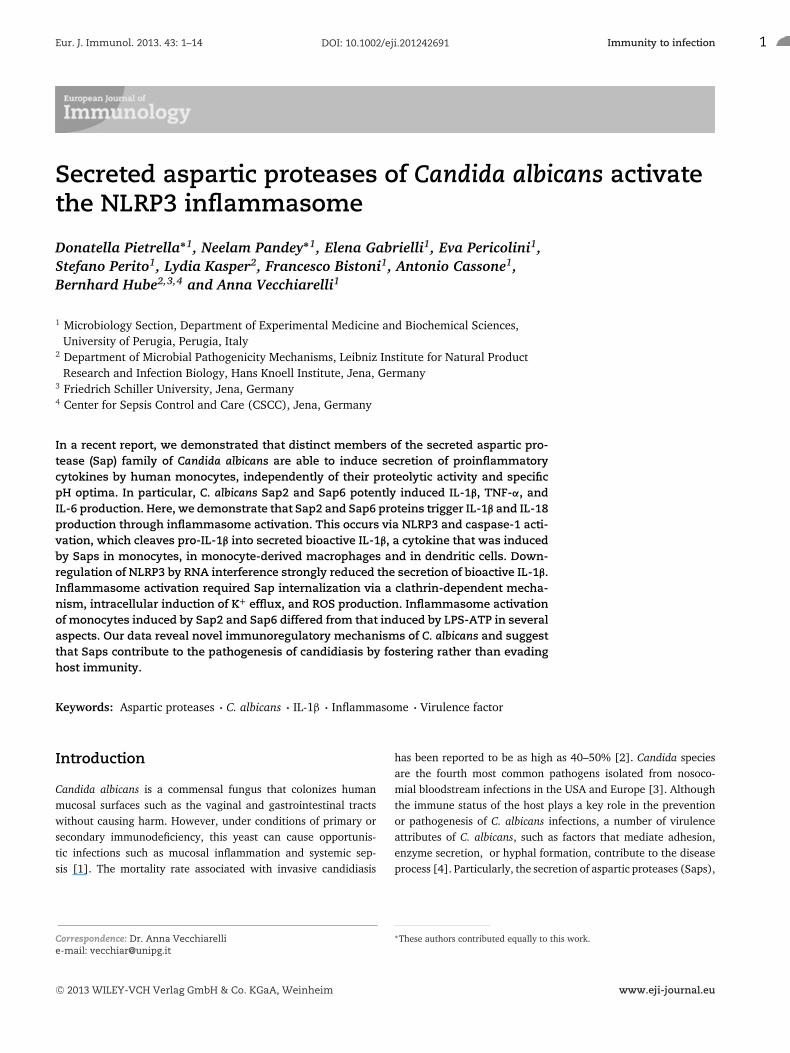
Eur. J. Immunol. 2013. 43: 1–14 Immunity to infection DOI: 10.1002/eji.201242691 1 Secreted aspartic proteases of Candida albicans activate the NLRP3 inflammasome Donatella Pietrella ∗1 , Neelam Pandey ∗1 , Elena Gabrielli 1 , Eva Pericolini 1 , Stefano Perito 1 , Lydia Kasper 2 , Francesco Bistoni 1 , Antonio Cassone 1 , Bernhard Hube 2,3,4 and Anna Vecchiarelli 1 1 Microbiology Section, Department of Experimental Medicine and Biochemical Sciences, University of Perugia, Perugia, Italy 2 Department of Microbial Pathogenicity Mechanisms, Leibniz Institute for Natural Product Research and Infection Biology, Hans Knoell Institute, Jena, Germany 3 Friedrich Schiller University, Jena, Germany 4 Center for Sepsis Control and Care (CSCC), Jena, Germany In a recent report, we demonstrated that distinct members of the secreted aspartic pro- tease (Sap) family of Candida albicans are able to induce secretion of proinflammatory cytokines by human monocytes, independently of their proteolytic activity and specific pH optima. In particular, C. albicans Sap2 and Sap6 potently induced IL-1β, TNF-α, and IL-6 production. Here, we demonstrate that Sap2 and Sap6 proteins trigger IL-1β and IL-18 production through inflammasome activation. This occurs via NLRP3 and caspase-1 acti- vation, which cleaves pro-IL-1β into secreted bioactive IL-1β, a cytokine that was induced by Saps in monocytes, in monocyte-derived macrophages and in dendritic cells. Down- regulation of NLRP3 by RNA interference strongly reduced the secretion of bioactive IL-1β. Inflammasome activation required Sap internalization via a clathrin-dependent mecha- nism, intracellular induction of K + efflux, and ROS production. Inflammasome activation of monocytes induced by Sap2 and Sap6 differed from that induced by LPS-ATP in several aspects. Our data reveal novel immunoregulatory mechanisms of C. albicans and suggest that Saps contribute to the pathogenesis of candidiasis by fostering rather than evading host immunity. Keywords: Aspartic proteases C. albicans IL-1β Inflammasome Virulence factor Introduction Candida albicans is a commensal fungus that colonizes human mucosal surfaces such as the vaginal and gastrointestinal tracts without causing harm. However, under conditions of primary or secondary immunodeficiency, this yeast can cause opportunis- tic infections such as mucosal inflammation and systemic sep- sis [1]. The mortality rate associated with invasive candidiasis Correspondence: Dr. Anna Vecchiarelli e-mail: [email protected] has been reported to be as high as 40–50% [2]. Candida species are the fourth most common pathogens isolated from nosoco- mial bloodstream infections in the USA and Europe [3]. Although the immune status of the host plays a key role in the prevention or pathogenesis of C. albicans infections, a number of virulence attributes of C. albicans, such as factors that mediate adhesion, enzyme secretion, or hyphal formation, contribute to the disease process [4]. Particularly, the secretion of aspartic proteases (Saps), ∗ These authors contributed equally to this work. C 2013 WILEY-VCH Verlag GmbH & Co. KGaA, Weinheim www.eji-journal.eu
-
Upload
independent -
Category
Documents
-
view
2 -
download
0
Transcript of Secreted aspartic proteases of Candida albicans activate the NLRP3 inflammasome
Eur. J. Immunol. 2013. 43: 1–14 Immunity to infectionDOI: 10.1002/eji.201242691 1
Secreted aspartic proteases of Candida albicans activatethe NLRP3 inflammasome
Donatella Pietrella∗1, Neelam Pandey∗1, Elena Gabrielli1, Eva Pericolini1,Stefano Perito1, Lydia Kasper2, Francesco Bistoni1, Antonio Cassone1,Bernhard Hube2,3,4 and Anna Vecchiarelli1
1 Microbiology Section, Department of Experimental Medicine and Biochemical Sciences,University of Perugia, Perugia, Italy
2 Department of Microbial Pathogenicity Mechanisms, Leibniz Institute for Natural ProductResearch and Infection Biology, Hans Knoell Institute, Jena, Germany
3 Friedrich Schiller University, Jena, Germany4 Center for Sepsis Control and Care (CSCC), Jena, Germany
In a recent report, we demonstrated that distinct members of the secreted aspartic pro-tease (Sap) family of Candida albicans are able to induce secretion of proinflammatorycytokines by human monocytes, independently of their proteolytic activity and specificpH optima. In particular, C. albicans Sap2 and Sap6 potently induced IL-1β, TNF-α, andIL-6 production. Here, we demonstrate that Sap2 and Sap6 proteins trigger IL-1β and IL-18production through inflammasome activation. This occurs via NLRP3 and caspase-1 acti-vation, which cleaves pro-IL-1β into secreted bioactive IL-1β, a cytokine that was inducedby Saps in monocytes, in monocyte-derived macrophages and in dendritic cells. Down-regulation of NLRP3 by RNA interference strongly reduced the secretion of bioactive IL-1β.Inflammasome activation required Sap internalization via a clathrin-dependent mecha-nism, intracellular induction of K+ efflux, and ROS production. Inflammasome activationof monocytes induced by Sap2 and Sap6 differed from that induced by LPS-ATP in severalaspects. Our data reveal novel immunoregulatory mechanisms of C. albicans and suggestthat Saps contribute to the pathogenesis of candidiasis by fostering rather than evadinghost immunity.
Keywords: Aspartic proteases � C. albicans � IL-1β � Inflammasome � Virulence factor
Introduction
Candida albicans is a commensal fungus that colonizes humanmucosal surfaces such as the vaginal and gastrointestinal tractswithout causing harm. However, under conditions of primary orsecondary immunodeficiency, this yeast can cause opportunis-tic infections such as mucosal inflammation and systemic sep-sis [1]. The mortality rate associated with invasive candidiasis
Correspondence: Dr. Anna Vecchiarellie-mail: [email protected]
has been reported to be as high as 40–50% [2]. Candida speciesare the fourth most common pathogens isolated from nosoco-mial bloodstream infections in the USA and Europe [3]. Althoughthe immune status of the host plays a key role in the preventionor pathogenesis of C. albicans infections, a number of virulenceattributes of C. albicans, such as factors that mediate adhesion,enzyme secretion, or hyphal formation, contribute to the diseaseprocess [4]. Particularly, the secretion of aspartic proteases (Saps),
∗These authors contributed equally to this work.
C© 2013 WILEY-VCH Verlag GmbH & Co. KGaA, Weinheim www.eji-journal.eu

2 Donatella Pietrella et al. Eur. J. Immunol. 2013. 43: 1–14
which are encoded by a gene family with ten members, has longbeen recognized as a virulence-associated trait of this pathogenicyeast [5].
We recently reported that various members of the Sap family,including Sap1, Sap2, Sap3, and Sap6, have different abilities toinduce secretion of pro-inflammatory cytokines by human mono-cytes via Akt/NF-κB activation. Sap1, Sap2, and Sap6 potentlyinduced IL-1β, TNF-α, and IL-6 production. Importantly, Sap-induced cytokine production was independent of the proteolyticactivity and of the optimal pH for the individual Sap activities [6].These data suggest that Saps contribute to the pathogenesis ofcandidiasis by fostering rather than evading host immunity.
Immune cells of the innate system recognize pathogensthrough PRR that include membrane-bound TLRs and intracellularproteins such as NOD-like receptors (NLRs) [7]. NLRP3 respondsto multiple stimuli and forms an intracellular multiprotein com-plex, called NLRP3 inflammasome, consisting of the apoptosis-associated speck-like protein containing a caspase recruitmentdomain, and caspase-1, which triggers the secretion of IL-1β [8]. Arecent report by Kayagaki et al. [9] demonstrated that caspase-11is also involved in a noncanonical inflammasome activation.
NLRP3 inflammasome is activated in response to a rangeof bacterial, viral, and fungal pathogens, including Aspergillusfumigatus and C. albicans [10–12]. IL-1β production via the inflam-masome generally requires two signals: an NF-κB-dependent sig-nal that induces the synthesis of pro-IL-1β, and a second signal thattriggers proteolytic pro-IL-1β processing to produce mature IL-1β.Spleen tyrosine kinase (Syk) coupled to fungal pattern recogni-tion receptors, such as Dectin-1, controls both pro-IL-1β synthesisand the activation of the inflammasome after cell stimulation withC. albicans [13]. IL-1β and IL-18, which are both induced by acti-vation of the inflammasome, are involved in the initiation of theadaptive Th1 and Th17 cellular responses to C. albicans [14].
To investigate whether the inflammasome activation is trig-gered by Saps, we selected Sap2 and Sap6 as members of twoSap subfamilies, Sap1–3 and Sap4–6 respectively, which differ intheir biological properties and potential roles in different types ofC. albicans infections [5,15].
Results
IL-1β and IL-18 production by monocytes in responseto Saps
We first analyzed whether Sap2 and Sap6 from C. albicans wereable to induce the production of IL-18 and IL-1β by humanmonocytes [16]. Figure 1 shows that Sap2 and Sap6 induce IL-1β
and IL-18 to a similar extent. The cytokine production levelswere generally comparable with those observed after stimulationwith a positive control stimulant (LPS-ATP). In contrast, Sap3did not induce significant upregulation of IL-18, even though amodest increase of this cytokine was observed. To rule out anyLPS involvement, we added polymyxin B to select basic assays.As shown in Figure 1, addition of polymyxin B did not cause any
Figure 1. IL-1β and IL-18 production by monocytes in response to Saps.Monocytes were treated for 4 h with Sap2, Sap3, and Sap6 (20 μg/mL)or LPS (1 μg/mL) plus 5 mM ATP. Selected experiments were carried outin the presence of 10 μg/mL of polymyxin B. After incubation, super-natants were recovered and tested for the presence of IL-1β or IL-18.Data are expressed as means ± SEM for six samples pooled from threeindependent experiments. *p < 0.05, **p < 0.01, (Sap or LPS-ATP-treatedcells versus untreated cells), ##p < 0.01, (polymyxin B plus Sap or LPS-ATP-treated cells versus Sap or LPS-ATP-treated cells). Differences wereanalyzed by ANOVA test.
significant decrease in the level of Sap-induced cytokine produc-tion, but it strongly reduced IL-1β and IL-18 production afterLPS-ATP stimulation of monocytes (see below for additionalevidence of the polymyxin B effect). To investigate whether theproduction of Sap-induced IL-1β requires proteolytic activity ofSaps, monocytes were treated with Sap2 and Sap6 in combina-tion with the aspartic protease inhibitor pepstatin A (15 μM).Pepstatin A did not affect the Sap2- or Sap6-induced productionof IL-1β (data not shown), confirming that the activation of theinflammasome by Saps is independent from their enzymatic activ-ity. To further confirm that the recombinant Sap proteins werenot contaminated with other molecules that may be responsiblefor the observed activation, experiments were repeated withheat-inactivated Saps and with Proteinase K. Heat inactivationand addition of Proteinase K abrogated the IL-1β production by
C© 2013 WILEY-VCH Verlag GmbH & Co. KGaA, Weinheim www.eji-journal.eu

Eur. J. Immunol. 2013. 43: 1–14 Immunity to infection 3
monocytes stimulated with Sap2 or Sap6 indicating that nativeSap proteins were required for inflammasome activation.
Inhibition of Sap internalization and regulation ofIL-1β and IL-18 production
To test whether Saps must be internalized for cytokine induction,monocytes were treated with known inhibitors of endocytic activ-ity such as nystatin, chlorpromazine (CPZ), and cytochalasin D.CPZ is known to inhibit clathrin-mediated endocytosis [17], whilenystatin is an inhibitor of the lipid raft-caveolae endocytosispathway [18]. As shown in Figure 2A, the treatment with nys-tatin or cytochalasin D did not cause any decrease of Sap2- orSap6-induced IL-1β production. Conversely, treatment with CPZmarkedly inhibited IL-1β secretion in response to both Sap2 andSap6. Since it is known that LPS-ATP recognition by monocytesalso depends on clathrin-mediated endocytosis [19], CPZ musthave caused the decrease in cytokine production following LPS-ATP stimulation (Fig. 2B). A CPZ concentration of 45 μM, which iscommonly used to inhibit phagocytosis [20], was effective in pro-ducing inhibitory effects. In parallel experiments, we also demon-strated that CPZ treatment also downregulated IL-18 productionafter induction by Sap2 and Sap6 (Fig. 2C).
Because TNF-α production is induced through regulatorypathways that differ from those leading to IL-1β and IL-18production, we tested whether CPZ was able to affect TNF-αproduction induced by Saps. As shown in Figure 2D, treatmentwith CPZ did not modify Sap induced TNF-α production. Next,we tested the uptake of FITC-conjugated Sap2 and Sap6 bymonocytes with and without CPZ treatment. Figure 2E andF shows a significant decrease in Sap2 and Sap6 uptake byCPZ-treated monocytes. When FITC-conjugated LPS was used,differences in LPS uptake after CPZ treatment were also observed(Fig. 2G). This is consistent with previous data showing thatinternalization of LPS occurs via a clathrin-dependent mechanism[21, 22]. FITC-conjugated CTLA-4 F(ab)2 was used as a negativecontrol for protein uptake (Figure 2H). Since recent data indicatethat autophagosome formation modulates IL-1β secretion [23]and many reports point to the involvement of the endolysosomalcompartment in unconventional secretion [24], it is possible thatCPZ affects not only endocytosis but also intracellular traffickingof endolysosomes or autophagosomes. To settle this point, wecarried out experiments with two TLR agonists that do notrequire clathrin-mediated endocytosis, FSL-1 (TLR2 agonist), andflagellin (TLR5 agonist). Both agonists were able to induce IL-1β
secretion by monocytes (Table 1). Pretreatment of monocyteswith 90, 45, 15 μM of CPZ did not significantly affect the IL-1β
production induced by either TLR agonist.
Intracellular signaling induced by Saps
Caspase-1 processes the inactive IL-1β and IL-18 precursors intotheir mature active forms, which are subsequently secreted by the
cell [25]. We next examined the intracellular production of theinactive precursor of IL-1β (pro-IL-1β) after addition of Saps bycytofluorimetric analysis. The results documented in Figure 3Ashow that both Sap2 and Sap6 induce abundant production of theIL-1β precursor. The addition of the specific caspase-1 inhibitor(Ac-YVAD 25 μM, referred to as IC-1) resulted in an increase of theinactive precursor of IL-1β (Fig. 3A), as expected when cytokinematuration is inhibited (see also Fig. 4C). Immunoblot analysiswas also performed to unravel the dynamics of IL-1β secretion inresponse to Saps. As shown in Figure 3B, a pro-IL-1β band wasobserved 2 h after stimulation with Sap2 and Sap6 only in thepresence of the inhibitor, while pro-IL-1β and IL-1β active bandswere manifested after 2 h of stimulation with LPS-ATP indepen-dently of the inhibitor presence. However, pro-IL-1β and activeIL-1β bands were detected in response to 4 h of stimulation withSap2, Sap6, and LPS-ATP. Two different doses of IC-1 (25 and100 μM) were used to inhibit the activation of caspase-1. IC-1treatment clearly downregulated the formation of the activeIL-1β at 4 h at the concentration of 25 μM, while the higher dosecompletely abolished the IL-1β maturation (Fig. 3C). The kinet-ics of caspase-1 activation was analyzed by western blot (Fig. 4Aand quantification in B). The results showed that stimulation withSap2 or Sap6 led to different kinetics of caspase-1 expression.Sap2 was able to induce the activation of caspase-1 after 1 hof stimulation in a pattern similar to that manifested after LPS-ATP stimulation. In contrast, Sap6 caused a weak activation ofcaspase-1 after 1 h of incubation, while a strong expression wasobserved after 2 h (Fig. 4A and B). The inhibition of caspase-1by IC-1 resulted in downregulation of caspase-1 activation in allexperiments, independently of whether Sap2, Sap6, or LPS-ATPwere used as stimulants (Fig. 4A and B). The secretion of bothIL-1β (Fig. 4C) and IL-18 (Fig. 4F) significantly decreased in thepresence of the IC-1. However, the inhibitor did not affect TNF-αproduction (Fig. 4G). Since monocytes, macrophages and den-dritic cells (DCs) have different requirements for inflammasomeactivation and the processing and release of IL-1β [26], we testedSap ability to stimulate human monocyte-derived macrophages(MDMs) and human DCs to produce IL-1β. As shown in Figure 4Dand E, IL-1β was also produced by both MDMs and DCs, followingstimulation with Sap2 and Sap6. Similarly, IC-1 caused a signifi-cant decrease in IL-1β production.
The activation of the NLRP3 inflammasome requires, in mostcell types, two different stimuli. One, often called priming, is nec-essary for the upregulation of NLRP3 and pro-IL-1β and is usuallycaused by microbial ligands. The second stimulus is the real acti-vator of the NLRP3 inflammasome (e.g. ATP). It has been demon-strated that human monocytes need only one stimulus, becausethey constitutively express active caspase-1 or because they cansecrete ATP in response to stimulation with microbial ligands.To clarify whether Saps act exclusively like other microbial lig-ands or whether they are direct activators of the NLRP3 inflam-masome, macrophages primed with LPS (signal 1) were thenstimulated with Saps. The results shown in Figure 4H demon-strate that Saps are able to directly activate the production ofIL-1β.
C© 2013 WILEY-VCH Verlag GmbH & Co. KGaA, Weinheim www.eji-journal.eu

4 Donatella Pietrella et al. Eur. J. Immunol. 2013. 43: 1–14
Figure 2. Inhibition of Sap internalization and reg-ulation of IL-1β and IL-18 production by mono-cytes. (A–D) Monocytes were pretreated with nys-tatin (5 μg/mL), chlorpromazine (45 μM), cytocha-lasin D (2 μM) for 30 min and then stimu-lated with Sap2, Sap6, or LPS-ATP. After incu-bation, supernatants were tested for the pres-ence of (A, B) IL-1β, (C) IL-18, and (D) TNF-α.(E–H) To inhibit Sap internalization, monocytes weretreated with chlorpromazine (45 μM) and then incu-bated for 10, 30, and 120 min with 20 μg/mL ofFITC-labeled (E) Sap2, (F) Sap6, (G) LPS, or (H) CTLA4(Fab)2. Fluorescence was analyzed by flow cytometryand it is expressed as mean intensity fluorescence.Data are expressed as means ± SEM of six samplespooled from three independent experiments carriedout with monocytes from three different donors.*p < 0.05, **p < 0.01 (chlorpromazine plus Saps or LPS-ATP-treated cells versus Saps or LPS-ATP-treatedcells). Differences were analyzed by ANOVA test.
Because the tyrosine kinase Syk is involved in the control ofboth pro-IL-1β synthesis and the activation of the inflammasomeinduced by Candida [10], we carried out experiments to assesswhether Syk is also involved in the activation of the NLRP3 inflam-masome induced by Saps. The phosphorylation of Tyr525/526 isessential for Syk function [27]. No activation of Syk was observedafter 4 h of stimulation with Sap2, Sap6, or LPS-ATP (data notshown).
The direct involvement of NLRP3 in the induction of IL-1β bySaps was investigated. By using Western blot analysis, a significant
increase in NLRP3 expression upon stimulation with Sap2, Sap6,or LPS-ATP was observed (Fig. 5A and B). Then, the involve-ment of NLRP3 was also analyzed by using small interfering RNA(siRNA). An RT-PCR control experiment (Fig. 5C) showed thatelectroporation of NLRP3 siRNA had a strong silencing efficiency(about 80%). As shown in Figure 5D, IL-1β production in silencedcells stimulated with Sap2, Sap6, or LPS-ATP was markedly atten-uated compared with that of control cells. Conversely, TNF-αsecretion was barely affected, indicating that Sap-induced IL-1β
production is regulated via NLRP3.
C© 2013 WILEY-VCH Verlag GmbH & Co. KGaA, Weinheim www.eji-journal.eu

Eur. J. Immunol. 2013. 43: 1–14 Immunity to infection 5
Table 1. Effect of chlorpromazine on IL-1β production induced by TLRagonists.
Chlorpromazinea) Untreated Flagellin FSL-1cells (Bacillus 12.5 μg/mL
subtilis)100 ng/mL
— 9.4 ± 2.2 31.0 ± 8.7b),c) 21.4 ± 2.4c)
15 μM 5.0 ± 2.1 42.8 ± 22.9 21.2 ± 4.045 μM 5.6 ± 2.7 33.1 ± 8.5 14.3 ± 0.890 μM 6.2 ± 1.4 22.4 ± 2.0 17.5 ± 1.3
a)Monocytes were pretreated with chlorpromazine for 30 min and thenstimulated with flagellin (TLR5 agonist) and FSL-1 (TLR2 agonist).
b)IL-1β is expressed as pg/mL.c) p < 0.05, Flagellin or FSL-1-treated cells versus untreated cells. Differ-
ences were analyzed by ANOVA test.
Inhibition of potassium efflux, superoxide production,and induction of lysosomal damage by Saps
Activation of the NLRP3 inflammasome can be mediated by dif-ferent, often concurrent, mechanisms, including stimulation of K+
efflux, induction of lysosomal damage, and production of ROS, asreviewed by Cassel et al. [28]. To investigate whether these mech-anisms are involved during Sap-induced responses, we used rec-ognized inhibitors such as glibenclamide, which inhibits K+ efflux
[29] and butylated hydroxyanisole (BHA), a potent inhibitor ofROS production [29]. Preliminary tests ruled out that the con-centrations of these inhibitors used in our assays were toxic formonocytes, both in the presence and in the absence of Sap stimu-lation (data not shown).
Glibenclamide was able to inhibit production of IL-1β uponstimulation with Saps, particularly with Sap6, in a dose-dependentfashion (data not shown). Additional experiments using a bufferrich in K+ (150 mM) were carried out. A reduction of IL-1β produc-tion by monocytes stimulated with Sap2 and Sap6 was observed(Fig. 6A), confirming that K+ efflux is involved in Sap-inducedinflammasome activation.
In a second series of experiments, BHA was used to inhibit ROSor cytokine production. Figure 6B shows that an increase of super-oxide anions was observed upon addition of Saps or LPS-ATP. Inthe presence of BHA the production of ROS was reversed to the lev-els of unstimulated monocytes. As shown in Figure 6C and D, BHAalso caused a statistically significant reduction of IL-1β and IL-18upon stimulation, although cytokine production was not reversedto the level of unstimulated cells, suggesting that additional path-ways that are not inhibited by BHA are involved in cytokine pro-duction after Sap induction. In further experiments, we evalu-ated the kinetics of ROS production. Stimulation of monocyteswith Saps induced generation of intracellular ROS that peakedat 30 min and decreased thereafter (Fig. 6E). Because ROS
Figure 3. Expression of immature pro-IL-1β and active IL-1β induced by Saps. Monocytes were stimulated with Sap2 or Sap6 (20 μg/mL) or LPS-ATPfor 4 h. (A) After incubation, cells were first fixed, permeabilized, and incubated with anti-human pro-IL-1β. To inhibit the activation of caspase-1monocytes were pretreated with IC-1 (25 and 100 μM), for 30 min. Fluorescence was analyzed by flow cytometer and expressed as mean intensityfluorescence. Data are expressed as means ± SEM of three samples pooled from three independent experiments. *p < 0.05 (Sap-treated cells versusuntreated cells), §p < 0.05 (Sap plus IC-1-treated cells versus Sap-treated cells). Differences were analyzed by ANOVA test. (B) For western blottinganalysis, after incubation, cell lysates were separated by SDS-PAGE, transferred to nitrocellulose membranes, and incubated with antibodiesagainst pro and active IL-1β. (C) An antibody to β-actin was used for normalization. The Western blots and normalization data are from oneexperiment representative of four independent experiments with similar results.
C© 2013 WILEY-VCH Verlag GmbH & Co. KGaA, Weinheim www.eji-journal.eu

6 Donatella Pietrella et al. Eur. J. Immunol. 2013. 43: 1–14
Figure 4. Caspase-1 activation induced by Saps. Monocytes, MDMs, or DCs were stimulated with Sap2 or Sap6 (20 μg/mL). (A) After incubation cellswere lysed and analyzed by western blotting to examine caspase-1 activation. (A–H) To inhibit the activation of caspase-1, cells were pretreatedwith IC-1 (25 μM). (B) Normalization of caspase-1 expression against β-actin was carried out. (C–G) Supernatants were recovered and testedfor the presence of (C–E) IL-1β, (F) IL-18, and (G) TNF-α. (H) MDMs were pretreated with LPS (1 μg/mL) and then stimulated with Saps for 2 hbefore lysis and western blotting analysis. Data are expressed as means + SEM of three samples pooled from three independent experiments. *p< 0.05, **p < 0.01 ((A) Sap-treated cells versus untreated cells (C–H) Sap plus IC-1-treated cells versus Sap-treated cells). Differences were analyzedby ANOVA test. The western blots and normalization data are from one out experiment representative of two performed with similar results.
production is generally counteracted by upregulation of antioxi-dant defenses to avoid oxidative stress [30], we analyzed the mod-ulation of intracellular glutathione (GSH). GSH was absent after30 min of stimulation and reached a maximum at 1 h (Fig. 6F).These data suggest that ROS have a priming role in the inductionof IL-1β production, which is found only after 4 h of stimulation.
In addition, we examined whether Sap stimulation could pro-voke lysosome rupture thereby inducing inflammasome activationby cathepsin B release [31]. Monocytes were treated for 2 h with
Saps and then analyzed for lysosome damage, assessed by thedecrement of acridine orange fluorescence intensity. The resultsshown in Figure 7A show that there is a consistent decrease offluorescence intensity in cells treated with Sap2 or Sap6, but notwith LPS-ATP, suggesting that Saps are able to induce cathepsin Brelease by lysosomes. It has been reported that the inhibitionof the lysosomal cysteine protease (cathepsin B) by its specificinhibitor CA-074 (IC-B) abrogated IL-1β production [32]. In ourexperimental system, IC-B inhibited the production of IL-1β by
C© 2013 WILEY-VCH Verlag GmbH & Co. KGaA, Weinheim www.eji-journal.eu

Eur. J. Immunol. 2013. 43: 1–14 Immunity to infection 7
Figure 5. The role of NLRP3 in the production and release of IL-1β. (A, B) Expression of NLRP3 in monocytes was evaluated by western blottingafter stimulation for 4 h with Sap2 or Sap6. (C) Monocytes were transfected with siRNA NLRP3 or nonsilencing control (siC). RT-PCR of lysates afterovernight incubation was performed for NLRP3 expression normalized against that of GAPDH. (D–E) After 24 h cells were primed for 4 h with Sap2,Sap6, or LPS plus ATP. (D) IL-1β or (E) TNF-α were assessed in the supernatants. The western blots and normalization data are from one experimentrepresentative of three independent experiments with similar results. Cytokine production data are expressed as means + SEM of six samplespooled from three independent experiments. *p < 0.05, **p < 0.01 (siRNA NLRP3-treated cells versus siC-treated cells). Differences were analyzedby ANOVA test.
monocytes stimulated with Sap2 or Sap6 (Fig. 7B). However, wecannot completely exclude that this effect is due to nonspecificeffects of IC-B as previously observed [10]. Moreover, to confirmthat lysosomal damage is not secondary to the induction of pyrop-tosis, experiments were carried out in the presence of the caspase-1inhibitor IC-1. IC-1 did not significantly affect the lysosome dam-age, assessed by the decrement of acridine orange fluorescenceintensity in cells treated with Sap2, Sap6, or LPS-ATP (data notshown).
Discussion
In a recent paper, we reported the capacity of secreted asparticproteases of C. albicans to induce secretion of proinflammatorycytokines by human monocytes. In particular, Sap2 and Sap6,which are members of two Sap subfamilies (Sap1–3 and Sap4–6)with distinct roles in C. albicans pathogenicity and disease spec-trum [5], strongly induced upregulation of IL-1β, TNF-α, and IL-6
[6] independently from their proteolytic activity, from the opti-mal pH of each individual Sap activity, and from activation ofprotease-activated receptors.
In this study, we propose that IL-1β and IL-18 productioncaused by Sap2 or Sap6 is the result of NLRP3 inflammasomeand caspase-1 activation. We demonstrate that Sap2 and Sap6induction of proinflammatory cytokines was due to a cascadeof events causing upstream inflammasome activation and down-stream caspase-1 mediated cytokine synthesis by procytokinesas proposed in Figure 8. To our knowledge, there has beenno prior demonstration that any single member of the Sap iso-enzyme family, which is one of the best known virulence deter-minants of C. albicans [33], nor any protein of C. albicans orany other fungus, can induce inflammasome activation. How-ever, although the biological relevance remains unclear, it hasbeen reported that selected proteinases of C. albicans can acti-vate pro-IL-1β proteolytically to IL-1β [34]. Furthermore, ithas been shown that mutants lacking functional SAP geneshave a reduced potential to induce cytokine production in oral
C© 2013 WILEY-VCH Verlag GmbH & Co. KGaA, Weinheim www.eji-journal.eu

8 Donatella Pietrella et al. Eur. J. Immunol. 2013. 43: 1–14
Figure 6. Inhibition of potassium efflux andsuperoxide production induced by Saps. Mono-cytes were stimulated with Saps for 4 h in aserum-free buffer with 150 mM KCl. In paral-lel, a buffer with 150 mM NaCl was used. (A)After incubation, supernatants were tested forthe presence of IL-1β. To analyze superoxideproduction, cells were pretreated with 10 μM ofH2DCFDA. (B) To inhibit superoxide production,monocytes were pretreated with BHA (50 μM)for 30 min. After incubation, cells were fixedand underwent cytofluorimetric analysis. (C, D)Supernatant fluids were recovered to test forthe presence of (C) IL-1β and (D) IL-18. (E, F) Tostudy the kinetics of ROS production, mono-cytes were stimulated with 20 μg/mL of Sap2 orSap6 for 30 min, 1, 2, and 4 h. (E) A 10 μM ofH2DCFDA to test intracellular ROS or (F) 100 μMof MCB to assess intracellular GSH were addedto cultures 30 min before the end of the incuba-tion. Results are expressed as relative fluores-cence units (RFU). Data are expressed as means± SEM of six samples pooled from three inde-pendent experiments. *p < 0.05, **p <0.01 ((A)Sap plus K+ buffer-treated cells versus Sap-treated cells, (B–D) Sap plus BHA-treated cellsversus Sap-treated cells). Differences were ana-lyzed by ANOVA test.
epithelial tissue models [35]. While this process is possibly asso-ciated with reduced damage of SAP mutants, our study presentedhere suggests that single Sap proteins can directly activate theinflammasome.
This assertion is supported by evidence of direct activation ofNLRP3 and caspase-1 by Sap2 and Sap6. In our experimental set-ting Sap2, Sap6, and the positive control LPS-ATP induce IL-1β andIL-18 to a similar extent. In previous studies, the amount of IL-1β
secreted by human monocytes upon LPS plus ATP stimulation wasmuch higher than the amount of IL-18 produced [36, 37]. How-ever, the experimental conditions in these studies were differentfrom ours. For example, Perregaux et al. [36] reported the pro-duction of IL-1β and IL-18 in plasma from LPS plus ATP-activatedblood cells, while Piccini and collaborators [37] used a differentconcentration of cells and a lower concentration of ATP.
Silencing of NLRP3 mRNA provided direct evidence that Sap2and Sap6 can activate the inflammasome. The kinetics of caspase-1activation showed that Sap6-induced activation of caspase-1occurred over longer periods (still evident 4 h poststimulation)than Sap2-induced activation. This may be due to the differentepitopes of the Saps that could stimulate caspase-1 differently.Although Sap2 and Sap6 are both aspartic proteases and membersof the Sap family, they belong to different subgroups. Sap2 belongsto the subfamily Sap1–3, while Sap6 belongs to the Sap4–6subfamily [15]. However, the kinetics of bioactive IL-1β expressionwas similar for both stimulating proteases. The expression wasat a maximum 4 h after stimulation with Saps and the amountof secreted bioactive IL-1β was similar. This suggests that theextended activation of caspase-1 does not result in a more consis-tent cleavage of pro-IL-1β.
C© 2013 WILEY-VCH Verlag GmbH & Co. KGaA, Weinheim www.eji-journal.eu

Eur. J. Immunol. 2013. 43: 1–14 Immunity to infection 9
Figure 7. Lysosomal damage induced by Saps.Monocytes were stimulated with Sap2, Sap6(20 μg/mL) or LPS-ATP for 2 h. (A, B) After incu-bation the (A) lysosomal damage and (B) secre-tion of IL-1β were evaluated. (A) After incuba-tion cells were treated with 10 μM of acridineorange for 10 min, fluorescence was analyzedby using a cytofluorimeter and expressed as thepercentage of mean intensity fluorescence decre-ment. (B) Cathepsin B activity was inhibited byIC-B 10 μM and IL-1β production was evalu-ated. Data are expressed as means + SEM of sixsamples pooled from three independent experi-ments. *p < 0.05, (Sap-treated cells versus untreatedcells). Differences were analyzed by ANOVAtest.
Using specific inhibitors of K+ efflux and ROS generation, weobserved that Sap-induced inflammasome activation seems to beassociated with physiological changes such as transient opening ofion channels and the production of ROS, as observed for NLRP3-induced inflammasome [38]. Interestingly, when ROS generationwas inhibited by BHA, which led to ROS levels similar to thoseof nonstimulated monocytes, cytokine production was only par-tially inhibited, but the reduction was significant. This suggeststhat inflammasome activation and cytokine production induced bySaps does not rely on just one signaling pattern but is likely drivenby concurrent intracellular mechanisms. In agreement with thishypothesis, TNF-α is also strongly induced by Saps without anyinvolvement of the NLRP3 inflammasome cascade. Coherently,cytokine production was also strongly affected, but not completely
blocked, by an inhibitor of caspase-1, which, as expected, causedan accumulation of biologically inactive pro-IL-1β. All these obser-vations suggest that Sap-mediated cytokine production is medi-ated via multiple pathways and confirm that Saps, well-knownvirulence factors of C. albicans, have a role as modulators of anti-candidal immunity.
The pathway that leads to Sap induced NLRP3 inflamma-some activation is not fully understood. However, our data maycontribute to unraveling important aspects of the induction pro-cess. First, it appears that inflammasome activation and cytokineproduction occur through Sap internalization via a clathrin-dependent endocytic process. This process is strongly affected bychlorpromazine, and left unaffected by nystatin and cytochalasinD, which are inhibitors of other mechanisms of intracellular entry
Figure 8. Model of NLRP3 inflammasomeactivation by secreted aspartic proteasesof Candida albicans. Sap2 and Sap6 induc-tion of proinflammatory cytokines is dueto a cascade of events causing upstreaminflammasome activation and down-stream caspase-1-mediated cytokinesynthesis.
C© 2013 WILEY-VCH Verlag GmbH & Co. KGaA, Weinheim www.eji-journal.eu

10 Donatella Pietrella et al. Eur. J. Immunol. 2013. 43: 1–14
[39]. Second, one of the sensing events could be an alternative cel-lular system involved in the recognition of microbial molecules,that is, the NLR pathway [40, 41]. Indeed, unlike several PRR,the NLR protein family senses microbial molecules intracellularlyin the cytosol [42–46]. This mechanism has been described forsome viral and bacterial proteins [47–49] and for some adju-vants such as aluminum salts [39]. Since the inhibition of Sapinternalization via the clathrin-dependent pathway results in aconsistent inhibition of IL-1β and IL-18, it can be concluded thatSaps must reach the cytoplasmic compartment to induce inflam-masome activation. Proteases could be internalized in endosomesand then released in the cytosol as suggested by the lysosomaldamage observed. Our results show that the inflammasome acti-vation and cytokine production induced by Saps in monocytes aredifferent from those induced by LPS-ATP treatment. In fact, unlikeLPS-ATP, Sap2 and Sap6 inflammasome induction results in lyso-some damage.
Since our experiments were generally performed in primaryhuman monocytes, which have been reported to display constitu-tive inflammasome activity [50], we performed some additionalexperiments with other cell types (MDMs and DCs), which donot display constitutive inflammasome activation. In these cells,activation of the inflammasome usually occurs in two stages. Inthe first stage, often called priming, upregulation of NLRP3 andpro-IL-1β, caused by exposure to microbial ligands, is necessary.In the second stage, NLRP3 needs to be directly activated. IndeedSap2 and Sap6 provide the second signal causing direct NLRP3activation, as demonstrated by their capacity of inducingcaspase-1-dependent IL-1β production. These data suggest thatSaps can genuinely activate the inflammasome, rather than spuri-ously affecting inflammasome auto-activation.
There is overwhelming experimental, and a certain amount ofclinical evidence that Saps are important factors of C. albicans vir-ulence [5, 15, 51–54]. The demonstration that proinflammatorycytokine production is induced by Saps through inflammasomeactivation implies a new role for these virulence factors relatedto their structural features. In addition, the observation thatselected Saps are capable of inducing the inflammasome directly,regardless of C. albicans, suggests that inflammasome activationtriggered during the course of infection may be independent ofdectin-1 recognition and Syk activation pathway, previouslydescribed for C. albicans [13].
Inflammasome activation may contribute to severe inflamma-tion and recruitment of neutrophils, a scenario characteristic in thepathology of vaginal candidiasis [55]. Expression of Sap proteasesmay contribute to the pathology of these superficial infections viaactivation of the inflammasome. This hypothesis is supported bythe fact that expression of SAP genes and Sap antigens, includingSap2 and Sap6, is frequently detected during vaginal infection[15,52], and that antibodies against Sap2 have been shown to beprotective against C. albicans challenges in a rat vaginal infectionmodel [56,57]. Differences in the magnitude and mechanisms ofinduction are also likely, as in part suggested by some differencesshown here between Sap2 and Sap6 in the kinetics of caspase-1activation. In conclusion, while these aspects remain to be inves-
tigated, our data suggest that Sap production may contribute tothe excessive inflammatory response observed during C. albicansinfections, which may be the hallmark of at least some Candidapathologies.
Materials and methods
Aspartic protease production
As previously described by Borg-von Zepelin et al. [50] recombi-nant C. albicans aspartic proteases Sap1, Sap2, Sap3, and Sap6were expressed as recombinant proteins using Pichia pastorisclones. After purification, Saps tested negative for endotoxin con-tamination in a Limulus assay (E-toxate, Sigma) with a sensitivityof 10 pg/mL of Escherichia coli LPS. Nevertheless, selected experi-ments were carried out at least once in the presence of 10 μg/mLof polymyxin B (Sigma) to neutralize any undetected contamina-tion with bacterial LPS.
Monocyte isolation
PBMCs were separated by density gradient centrifugation overFicoll-Hypaque Plus (Pharmacia Biotech), recovered, washedtwice, and suspended in RPMI 1640 supplemented with 10% FCS,100 U penicillin/mL, and 100 μg streptomycin/mL, plated in cellculture flasks (Corning), and incubated for 1 h at a density of2–3 × 10⁶/mL. Adherent cells recovered were >95% CD14+, eval-uated by flow cytometry (FACScan, Becton Dickinson).
In vitro generation of DCs and MDMs
The generation of DCs and MDMs from monocytes was performedas previously described, with minor modifications [58]. Imma-ture DCs were obtained by treating monocytes with 50 ng/mL ofhuman recombinant GM-CSF (Biosource) and 30 ng/mL of humanrecombinant IL-4 (Biosource) for 5–6 days. MDMs were obtainedby treatment with human recombinant M-CSF (50 ng/mL) for5 days.
Determination of proinflammatory cytokineproduction
Monocytes, MDMs, or DCs (2.5 × 10⁵) were incubated at 37◦Cfor 4 h with Sap2, Sap6 (20 μg/mL). As a positive control LPS(1 μg/mL) plus ATP (5 mM) was used for a further 15 min. Inselected experiments monocytes were stimulated with 12.5 μg/mLof diacylated lipoprotein FSL-1 (Pam2CGDPKHPKSF, InvivoGen)or with flagellin from Bacillus subtilis (100 ng/mL, InvivoGen).In selected experiments cells were pretreated with nys-tatin (5 μg/mL, Sigma), chlorpromazine (90, 45, 15 μM),
C© 2013 WILEY-VCH Verlag GmbH & Co. KGaA, Weinheim www.eji-journal.eu

Eur. J. Immunol. 2013. 43: 1–14 Immunity to infection 11
cytochalasin D (2 μM, Sigma), caspase-1 inhibitor Ac-YVAD-cmk(25 μM) (IC-1), glibenclamide (10, 25, 50, 75, and 100 μM), BHA(50 μM) for 30 min at 37◦C, or treated with CA074me (10 μM,Peptanova), an inhibitor of cathepsin B (IC-B). To inhibit the K+
efflux monocytes were stimulated with Saps for 4 h in a serum-free buffer with 150 mM KCl (10 mM Hepes, 5 mM NaH2PO4,150 mM KCl, 1 mM MgCl2, 1 mM CaCl2, 1% BSA, pH 7.4). In par-allel, a buffer with 150 mM NaCl was used (10 mM Hepes, 150 mMNaCl, 5 mM KH2PO4, 1 mM MgCl2, 1 mM CaCl2, 1% BSA, pH 7.4)[59]. After incubation, supernatant fluids were recovered, thenthe presence of human IL-1β, IL-18, and TNF-α was measured byELISA kit (Biosciences).
Cytotoxicity assay
Monocytes (2.5 × 10⁵) were treated for 4 and 18 h at 37◦C with7.5, 15, 30, 45, and 90 μM of chlorpromazine (Sigma); 10, 25,50, 75, and 100 μM of glibenclamide (Sigma); 10, 25, and 50 μMof Ac-YVAD-cmk (Bachem AG). After incubation cell viability wasevaluated by the use of an ATP bioluminescence kit (Via Light kit;Cambrex).
Protease FITC labeling
Proteases and CTLA-4 F(ab)2 (Ancell) were labeled with fluores-cein using the Fluoro Tag FITC Conjugation kit (Sigma). Briefly,fresh FITC solution in carbonate-bicarbonate buffer was addedto the Sap solutions and samples were incubated for 2 h atroom temperature with gentle stirring. The labeled proteins werepurified from unconjugated fluorescein by use of a SephadexG-25M column.
Sap2 and Sap6 uptake
The uptake of Sap2 and Sap6 by human monocytes was ana-lyzed by flow cytometry as previously described [58]. Monocytes(2.5 × 10⁵) were treated with or without 45 μM of chlorpro-mazine for 30 min at 37◦C, incubated with 20 μg/mL of FITC-labeled Sap2, Sap6, LPS, or CTLA-4 F(ab)2 for 30, 60, and 120min at 37◦C in the presence of 5% CO2. After incubation cell flu-orescence was evaluated by using FACScan and expressed as themean intensity fluorescence.
Determination of pro-IL-1β by flow cytometry
Monocytes were stimulated with 20 μg/mL of Sap2 or Sap6 at37◦C for 4 h, fixed with 4% paraformaldehyde, permeabilized with0.1% saponin (Sigma) in PBS, and labeled with goat anti humanpro-IL-1β (Santa Cruz Biotechnology). As a secondary antibodya FITC-conjugated anti-goat antibody was used. Cells expressingpro-IL-1β were analyzed using FACScan.
Immunoblotting
For immunoblotting, 106 cells were stimulated with 20 μg/mLof Sap2, Sap6, or LPS+ATP for the indicated times. In selectedexperiments, MDMs were pretreated with LPS for 1 h and thenstimulated with Sap2 and Sap6 (20 μg/mL). After stimulationcells were lysed and 30 μg of protein of each sample were sep-arated by SDS-PAGE and transferred to nitrocellulose membrane(Pierce). The membrane was blocked with 5% w/v milk powderin PBS and incubated overnight at 4◦C with goat anti human pro-IL-1β or goat anti-human-activated caspase-1, rabbit anti-humanSyk (Santa Cruz Biotechnology), rabbit anti-human phospho-Syk(Abcam) or rabbit anti-human NLRP3 (Imgenex) in TBST. Afterovernight incubation, the blots were washed with TBST and incu-bated with HRP-conjugated goat anti-rabbit antibody or mouseanti goat for 1 h at room temperature and incubated with thesubstrate ChemiLucent Trial Kit (Chemicon Int.). Immunoreactivebands were visualized and quantified by Chemidoc Instrument(Bio-Rad).
siRNA experiments
For siRNA experiments, the primary monocyte protocol for elec-troporation in the Amaxa chamber (AmaxaBiosystems) was used,according to the instructions of the manufacturer. Specific setsof siRNA for NLRP3 as well as nonsilencing siRNA for the con-trol were obtained from Ambion. Monocytes were resuspended in100 μL of Human Monocyte Nucleofactor solution. Two micro-gram siRNA per 106 cells was added, electroporation was per-formed and monocytes were allowed to recover overnight andstimulated with Sap2 or Sap6 or LPS.
RT-PCR
Control experiments to check the inhibition of NLRP3 expressionwere performed by RT-PCR. A total of 106 monocytes were treatedwith nonsilencing siRNA or with NLRP3 siRNA. After 18 h of incu-bation at 37◦C, total RNA was extracted, reverse-transcribed intocomplementary DNA. PCR was performed using a Bio-Rad Ther-mal Cycler-200. PCR conditions were as follows: 2 min at 50◦Cand 10 min at 95◦C, followed by 30 cycles of PCR reaction at 94◦Cfor 45 s, 70◦C for 2 min, and 59◦C for 1 min. GAPDH was used asa reference gene. The PCR products were run on 1% agarose gelsstained with ethidium bromide.
Superoxide assay
The production of superoxide was analyzed using a mem-brane permeable probe: 2′,7′-dichlorodihydrofluorescein diac-etate (H2DCF-DA, Sigma), which is oxidized by H2O2 to dichlo-rofluorescein. Monocytes (2.5 × 10⁵) were pretreated with 10 μM
C© 2013 WILEY-VCH Verlag GmbH & Co. KGaA, Weinheim www.eji-journal.eu

12 Donatella Pietrella et al. Eur. J. Immunol. 2013. 43: 1–14
of H2DCF-DA for 1 h at 37◦C, then incubated for 2 h with 20 μg/mLof Sap2 or Sap6. Cells were then fixed in 4% paraformaldehydeand analyzed using a flow cytometer. In selected experiments,to inhibit the superoxide production, monocytes were pretreatedwith a broad superoxide scavenger, BHA (50 μM).
To study the kinetics of ROS production, monocytes werestimulated with 20 μg/mL of Sap2 or Sap6 for 30 min, 1, 2,4 h. A 10 μM of H2DCF-DA to test intracellular ROS or 100 μMof monochlorobimane (MCB, Sigma) to assess intracellular GSHwere added to cultures 30 min before the end of the incubation.Fluorescence was measured in cell lysates with a microplate fluo-rometer (480 nm excitation, 530 nm emission for H2DCFDA; 355nm excitation and 485 nm emission for MCB).
Determination of lysosomal damage
To measure lysosomal membrane integrity, healthy, or apop-totic cells were stained with acridine orange (Sigma) as previ-ously described [60]. Briefly, monocytes after stimulation with20 μg/mL Sap2 or Sap6 or LPS+ATP for 2 h, were collected,washed, and incubated with 10 μM of acridine orange (SigmaAldrich) for 10 min. Monocytes were then analyzed using a FAC-Scan flow cytometer. In selected experiments cells were pretreatedwith caspase-1 inhibitor.
Statistical analysis
Statistical significance was determined using ANOVA test. Resultsare presented as means ± SEM from replicate samples of 2-6experiments. Post hoc comparison were done with Bonferroni’sTest. A value of p < 0.05 was considered significant.
Acknowledgments: The study was supported by the EU Marie-Curie project N◦ FP7-214004-2, FINSysB. We thank CatherineMacpherson for editorial assistance.
Conflict of interests: The authors declare no financial or com-mercial conflict of interest.
References
1 Dimopoulos, G., Karabinis, A., Samonis, G. and Falagas, M. E., Can-
didemia in immunocompromised and immunocompetent critically ill
patients: a prospective comparative study. Eur. J. Clin. Microbiol. Infect Dis.
2007. 26: 377–384.
2 Gudlaugsson, O., Gillespie, S., Lee, K., Vande Berg, J., Hu, J., Messer,
S., Herwaldt L. et al., Attributable mortality of nosocomial candidemia,
revisited. Clin. Infect Dis. 2003. 37: 1172–1177.
3 Chen, L. Y., Kuo, S. C., Wu, H. S., Yang, S. P., Chan, Y. J., Chen, L. K.
and Wang, F. D., Associated clinical characteristics of patients with can-
didemia among different Candida species. J. Microbiol. Immunol. Infect 2012.
In press. doi: 10.1016/j.jmii.2012.08.001.
4 Calderone, R. A. and Fonzi, W. A., Virulence factors of Candida albicans.
Trends Microbiol. 2001. 9: 327–335.
5 Naglik, J., Albrecht, A., Bader, O. and Hube, B., Candida albicans pro-
teinases and host/pathogen interactions. Cell Microbiol. 2004. 6: 915–926.
6 Pietrella, D., Rachini, A., Pandey, N., Schild, L., Netea, M., Bistoni, F.,
Hube, B. et al., The Inflammatory response induced by aspartic proteases
of Candida albicans is independent of proteolytic activity. Infect Immun.
2010. 78: 4754–4762.
7 Akira, S., Uematsu, S. and Takeuchi, O., Pathogen recognition and innate
immunity. Cell 2006. 124: 783–801.
8 Franchi, L., Eigenbrod, T., Munoz-Planillo, R. and Nunez, G.,
The inflammasome: a caspase-1-activation platform that regulates
immune responses and disease pathogenesis. Nat. Immunol. 2009. 10:
241–247.
9 Kayagaki, N., Warming, S., Lamkanfi, M., Vande Walle, L., Louie, S.,
Dong, J., Newton, K. et al., Non-canonical inflammasome activation tar-
gets caspase-11. Nature 2011. 479: 117–121.
10 Gross, O., Poeck, H., Bscheider, M., Dostert, C., Hannesschlager, N.,
Endres, S., Hartmann, G. et al., Syk kinase signalling couples to the Nlrp3
inflammasome for anti-fungal host defence. Nature 2009. 459: 433–436.
11 Joly, S. and Sutterwala, F. S., Fungal pathogen recognition by the NLRP3
inflammasome. Virulence 2010. 1: 276–280.
12 Said-Sadier, N., Padilla, E., Langsley, G. and Ojcius, D. M., Aspergillus
fumigatus stimulates the NLRP3 inflammasome through a pathway
requiring ROS production and the Syk tyrosine kinase. PLoS One 2010.
5: e10008.
13 Cheng, S. C., van de Veerdonk, F. L., Lenardon, M., Stoffels, M., Plantinga,
T., Smeekens, S., Rizzetto, L. et al., The dectin-1/inflammasome pathway
is responsible for the induction of protective T-helper 17 responses that
discriminate between yeasts and hyphae of Candida albicans. J. Leukoc.
Biol. 2011. 90: 357–366.
14 van de Veerdonk, F. L., Joosten, L. A., Shaw, P. J., Smeekens, S. P.,
Malireddi, R. K., van der Meer, J. W., Kullberg, B. J. et al., The inflamma-
some drives protective Th1 and Th17 cellular responses in disseminated
candidiasis. Eur. J. Immunol. 2011. 41: 2260–2268.
15 Naglik, J. R., Challacombe, S. J. and Hube, B., Candida albicans secreted
aspartyl proteinases in virulence and pathogenesis. Microbiol. Mol. Biol.
Rev. 2003. 67: 400–428.
16 van de Veerdonk, F. L., Netea, M. G., Dinarello, C. A. and Joosten, L.
A., Inflammasome activation and IL-1beta and IL-18 processing during
infection. Trends Immunol. 2011. 32: 110–116.
17 Wang, L. H., Rothberg, K. G. and Anderson, R. G., Mis-assembly of
clathrin lattices on endosomes reveals a regulatory switch for coated
pit formation. J. Cell Biol. 1993. 123: 1107–1117.
18 Schnitzer, J. E., Oh, P., Pinney, E. and Allard, J., Filipin-sensitive caveolae-
mediated transport in endothelium: reduced transcytosis, scavenger
endocytosis, and capillary permeability of select macromolecules. J. Cell
Biol. 1994. 127: 1217–1232.
19 Kitchens, R. L., Wang, P. and Munford, R. S., Bacterial lipopolysaccha-
ride can enter monocytes via two CD14-dependent pathways. J. Immunol.
1998. 161: 5534–5545.
20 Ghigo, E., Kartenbeck, J., Lien, P., Pelkmans, L., Capo, C., Mege, J. L. and
Raoult, D., Ameobal pathogen mimivirus infects macrophages through
phagocytosis. PLoS Pathog 2008. 4: e1000087.
C© 2013 WILEY-VCH Verlag GmbH & Co. KGaA, Weinheim www.eji-journal.eu

Eur. J. Immunol. 2013. 43: 1–14 Immunity to infection 13
21 Harnett, W. and Harnett, M. M., Helminth-derived immunomodulators:
can understanding the worm produce the pill? Nat. Rev. Immunol. 2010.
10: 278–284.
22 Husebye, H., Halaas, O., Stenmark, H., Tunheim, G., Sandanger, O.,
Bogen, B., Brech, A. et al., Endocytic pathways regulate Toll-like recep-
tor 4 signaling and link innate and adaptive immunity. EMBO J. 2006. 25:
683–692.
23 Dupont, N., Jiang, S., Pilli, M., Ornatowski, W., Bhattacharya, D. and
Deretic, V., Autophagy-based unconventional secretory pathway for
extracellular delivery of IL-1beta. EMBO J. 2011. 30: 4701–4711.
24 Manjithaya, R. and Subramani, S., Autophagy: a broad role in unconven-
tional protein secretion? Trends Cell Biol. 2011. 21: 67–73.
25 Eder, C., Mechanisms of interleukin-1beta release. Immunobiology 2009.
214: 543–553.
26 Netea, M. G., Nold-Petry, C. A., Nold, M. F., Joosten, L. A., Opitz, B., van
der Meer, J. H., van de Veerdonk, F. L. et al., Differential requirement
for the activation of the inflammasome for processing and release of
IL-1beta in monocytes and macrophages. Blood 2009. 113: 2324–2335.
27 Zhang, J., Billingsley, M. L., Kincaid, R. L. and Siraganian, R. P., Phospho-
rylation of Syk activation loop tyrosines is essential for Syk function. An
in vivo study using a specific anti-Syk activation loop phosphotyrosine
antibody. J. Biol. Chem. 2000. 275: 35442–35447.
28 Cassel, S. L., Joly, S. and Sutterwala, F. S., The NLRP3 inflammasome: a
sensor of immune danger signals. Semin. Immunol. 2009. 21: 194–198.
29 Abdul-Sater, A. A., Koo, E., Hacker, G. and Ojcius, D. M., Inflammasome-
dependent caspase-1 activation in cervical epithelial cells stimulates
growth of the intracellular pathogen Chlamydia trachomatis. J. Biol. Chem.
2009. 284: 26789–26796.
30 Tassi, S., Carta, S., Vene, R., Delfino, L., Ciriolo, M. R. and Rubartelli,
A., Pathogen-induced interleukin-1beta processing and secretion is reg-
ulated by a biphasic redox response. J. Immunol. 2009. 183: 1456–1462.
31 Jin, C. and Flavell, R. A., Molecular mechanism of NLRP3 inflammasome
activation. J. Clin. Immunol. 2010. 30: 628–631.
32 Shio, M. T., Eisenbarth, S. C., Savaria, M., Vinet, A. F., Bellemare, M. J.,
Harder, K. W., Sutterwala, F. S. et al., Malarial hemozoin activates the
NLRP3 inflammasome through Lyn and Syk kinases. PLoS Pathog 2009. 5:
e1000559.
33 Monod, M. and Borg-von, Z. M., Secreted aspartic proteases as virulence
factors of Candida species. Biol. Chem. 2002. 383: 1087–1093.
34 Beausejour, A., Grenier, D., Goulet, J. P. and Deslauriers, N., Proteolytic
activation of the interleukin-1beta precursor by Candida albicans. Infect
Immun. 1998. 66: 676–681.
35 Schaller, M., Bein, M., Korting, H. C., Baur, S., Hamm, G., Monod, M.,
Beinhauer, S. et al., The secreted aspartyl proteinases Sap1 and Sap2
cause tissue damage in an in vitro model of vaginal candidiasis based on
reconstituted human vaginal epithelium. Infect Immun. 2003. 71: 3227–
3234.
36 Perregaux, D. G., McNiff, P., Laliberte, R., Conklyn, M. and Gabel,
C. A., ATP acts as an agonist to promote stimulus-induced secre-
tion of IL-1 beta and IL-18 in human blood. J. Immunol. 2000. 165:
4615–4623.
37 Piccini, A., Carta, S., Tassi, S., Lasiglie, D., Fossati, G. and Rubartelli, A.,
ATP is released by monocytes stimulated with pathogen-sensing recep-
tor ligands and induces IL-1beta and IL-18 secretion in an autocrine way.
Proc. Natl. Acad. Sci. USA 2008. 105: 8067–8072.
38 Tschopp, J. and Schroder, K., NLRP3 inflammasome activation: the con-
vergence of multiple signalling pathways on ROS production? Nat. Rev.
Immunol. 2010. 10: 210–215.
39 Harris, J., Sharp, F. A. and Lavelle, E. C., The role of inflammasomes in
the immunostimulatory effects of particulate vaccine adjuvants. Eur. J.
Immunol. 2010. 40: 634–638.
40 Franchi, L., McDonald, C., Kanneganti, T. D., Amer, A. and Nunez, G.,
Nucleotide-binding oligomerization domain-like receptors: intracellular
pattern recognition molecules for pathogen detection and host defense.
J. Immunol. 2006. 177: 3507–3513.
41 Franchi, L., Park, J. H., Shaw, M. H., Marina-Garcia, N., Chen, G., Kim, Y.
G. and Nunez, G., Intracellular NOD-like receptors in innate immunity,
infection and disease. Cell Microbiol. 2008. 10: 1–8.
42 Kanneganti, T. D., Lamkanfi, M., Kim, Y. G., Chen, G., Park, J. H., Franchi,
L., Vandenabeele, P. et al., Pannexin-1-mediated recognition of bacterial
molecules activates the cryopyrin inflammasome independent of Toll-
like receptor signaling. Immunity 2007. 26: 433–443.
43 Kim, Y. G., Park, J. H., Shaw, M. H., Franchi, L., Inohara, N. and Nunez, G.,
The cytosolic sensors Nod1 and Nod2 are critical for bacterial recognition
and host defense after exposure to Toll-like receptor ligands. Immunity
2008. 28: 246–257.
44 Mariathasan, S., Weiss, D. S., Newton, K., McBride, J., O)Rourke, K.,
Roose-Girma, M., Lee, W. P. et al., Cryopyrin activates the inflamma-
some in response to toxins and ATP. Nature 2006. 440: 228–232.
45 Marina-Garcia, N., Franchi, L., Kim, Y. G., Miller, D., McDonald, C.,
Boons, G. J. and Nunez, G., Pannexin-1-mediated intracellular delivery
of muramyl dipeptide induces caspase-1 activation via cryopyrin/NLRP3
independently of Nod2. J. Immunol. 2008. 180: 4050–4057.
46 Martinon, F., Petrilli, V., Mayor, A., Tardivel, A. and Tschopp, J., Gout-
associated uric acid crystals activate the NALP3 inflammasome. Nature
2006. 440: 237–241.
47 Brereton, C. F., Sutton, C. E., Ross, P. J., Iwakura, Y., Pizza, M., Rappuoli,
R., Lavelle, E. C. et al., Escherichia coli heat-labile enterotoxin promotes
protective Th17 responses against infection by driving innate IL-1 and
IL-23 production. J. Immunol. 2011. 186: 5896–5906.
48 Dunne, A., Ross, P. J., Pospisilova, E., Masin, J., Meaney, A., Sutton, C.
E., Iwakura, Y. et al., Inflammasome activation by adenylate cyclase
toxin directs Th17 responses and protection against Bordetella pertussis.
J. Immunol. 2010. 185: 1711–1719.
49 McNeela, E. A., Burke, A., Neill, D. R., Baxter, C., Fernandes, V. E., Ferreira,
D., Smeaton, S. et al., Pneumolysin activates the NLRP3 inflammasome
and promotes proinflammatory cytokines independently of TLR4. PLoS
Pathog 2010. 6: e1001191.
50 Borg-von Zepelin, M., Beggah, S., Boggian, K., Sanglard, D. and Monod,
M., The expression of the secreted aspartyl proteinases Sap4 to Sap6
from Candida albicans in murine macrophages. Mol. Microbiol. 1998. 28:
543–554.
51 Cassone, A., De Bernardis, F. and Santoni, G., Anticandidal immunity
and vaginitis: novel opportunities for immune intervention. Infect Immun.
2007. 75: 4675–4686.
52 De Bernardis, F., Sullivan, P. A. and Cassone, A., Aspartyl proteinases
of Candida albicans and their role in pathogenicity. Med. Mycol. 2001. 39:
303–313.
53 Hube, B., Candida albicans secreted aspartyl proteinases. Curr. Top Med.
Mycol. 1996. 7: 55–69.
54 Schaller, M., Borelli, C., Korting, H. C. and Hube, B., Hydrolytic
enzymes as virulence factors of Candida albicans. Mycoses 2005. 48:
365–377.
55 Fidel, P. L. Jr., Luo, W., Steele, C., Chabain, J., Baker, M. and Wormley,
F. Jr., Analysis of vaginal cell populations during experimental vaginal
candidiasis. Infect Immun. 1999. 67: 3135–3140.
C© 2013 WILEY-VCH Verlag GmbH & Co. KGaA, Weinheim www.eji-journal.eu

14 Donatella Pietrella et al. Eur. J. Immunol. 2013. 43: 1–14
56 De Bernardis, F., Liu, H., O)Mahony, R., La Valle, R., Bartollino, S., Sandini,
S., Grant, S. et al., Human domain antibodies against virulence traits
of Candida albicans inhibit fungus adherence to vaginal epithelium and
protect against experimental vaginal candidiasis. J. Infect Dis. 2007. 195:
149–157.
57 Sandini, S., La Valle, R., Deaglio, S., Malavasi, F., Cassone, A. and De
Bernardis, F., A highly immunogenic recombinant and truncated protein
of the secreted aspartic proteases family (rSap2t) of Candida albicans as
a mucosal anticandidal vaccine. FEMS Immunol. Med. Microbiol. 2011. 62:
215–224.
58 Pietrella, D., Lupo, P., Rachini, A., Sandini, S., Ciervo, A., Per-
ito, S., Bistoni, F. et al., A Candida albicans mannoprotein deprived
of its mannan moiety is efficiently taken up and processed by
human dendritic cells and induces T-cell activation without stimu-
lating proinflammatory cytokine production. Infect Immun. 2008. 76:
4359–4367.
59 Cassel, S. L., Eisenbarth, S. C., Iyer, S. S., Sadler, J. J., Colegio, O. R.,
Tephly, L. A., Carter, A. B. et al., The Nalp3 inflammasome is essen-
tial for the development of silicosis. Proc. Natl. Acad. Sci. USA 2008. 105:
9035–9040.
60 Oberle, C., Huai, J., Reinheckel, T., Tacke, M., Rassner, M., Ekert, P. G.,
Buellesbach, J. et al., Lysosomal membrane permeabilization and cathep-
sin release is a Bax/Bak-dependent, amplifying event of apoptosis in
fibroblasts and monocytes. Cell Death Differ. 2010. 17: 1167–1178.
Abbreviations: BHA: butylated hydroxyanisole · CPZ: chlorpromazine ·IC-1: caspase-1 inhibitor · IC-B: cathepsin B inhibitor · MDM: monocyte-
derived macrophage · NLR: NOD-like receptor · Sap: aspartic protease ·Syk: spleen tyrosine kinase
Full correspondence: Dr. Anna Vecchiarelli, Microbiology Section,Department of Experimental Medicine and Biochemical Sciences,University of Perugia, Via del Giochetto, 06126 Perugia, ItalyFax: +39-075-585-7407e-mail: [email protected]
Received: 22/5/2012Revised: 6/12/2012Accepted: 20/12/2012Accepted article online: 26/12/2012
C© 2013 WILEY-VCH Verlag GmbH & Co. KGaA, Weinheim www.eji-journal.eu