BYC, an atypical aspartic endopeptidase from Rhipicephalus (Boophilus) microplus eggs
9
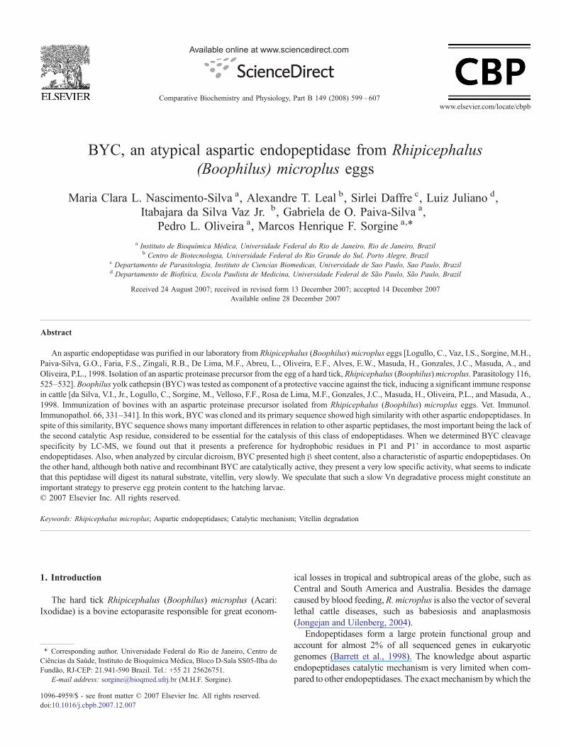
BYC, an atypical aspartic endopeptidase from Rhipicephalus (Boophilus) microplus eggs Maria Clara L. Nascimento-Silva a , Alexandre T. Leal b , Sirlei Daffre c , Luiz Juliano d , Itabajara da Silva Vaz Jr. b , Gabriela de O. Paiva-Silva a , Pedro L. Oliveira a , Marcos Henrique F. Sorgine a, ⁎ a Instituto de Bioquímica Médica, Universidade Federal do Rio de Janeiro, Rio de Janeiro, Brazil b Centro de Biotecnologia, Universidade Federal do Rio Grande do Sul, Porto Alegre, Brazil c Departamento de Parasitologia, Instituto de Ciencias Biomedicas, Universidade de Sao Paulo, Sao Paulo, Brazil d Departamento de Biofisica, Escola Paulista de Medicina, Universidade Federal de São Paulo, São Paulo, Brazil Received 24 August 2007; received in revised form 13 December 2007; accepted 14 December 2007 Available online 28 December 2007 Abstract An aspartic endopeptidase was purified in our laboratory from Rhipicephalus (Boophilus) microplus eggs [Logullo, C., Vaz, I.S., Sorgine, M.H., Paiva-Silva, G.O., Faria, F.S., Zingali, R.B., De Lima, M.F., Abreu, L., Oliveira, E.F., Alves, E.W., Masuda, H., Gonzales, J.C., Masuda, A., and Oliveira, P.L., 1998. Isolation of an aspartic proteinase precursor from the egg of a hard tick, Rhipicephalus (Boophilus) microplus. Parasitology 116, 525–532]. Boophilus yolk cathepsin (BYC) was tested as component of a protective vaccine against the tick, inducing a significant immune response in cattle [da Silva, V.I., Jr., Logullo, C., Sorgine, M., Velloso, F.F., Rosa de Lima, M.F., Gonzales, J.C., Masuda, H., Oliveira, P.L., and Masuda, A., 1998. Immunization of bovines with an aspartic proteinase precursor isolated from Rhipicephalus (Boophilus) microplus eggs. Vet. Immunol. Immunopathol. 66, 331–341]. In this work, BYC was cloned and its primary sequence showed high similarity with other aspartic endopeptidases. In spite of this similarity, BYC sequence shows many important differences in relation to other aspartic peptidases, the most important being the lack of the second catalytic Asp residue, considered to be essential for the catalysis of this class of endopeptidases. When we determined BYC cleavage specificity by LC-MS, we found out that it presents a preference for hydrophobic residues in P1 and P1' in accordance to most aspartic endopeptidases. Also, when analyzed by circular dicroism, BYC presented high β sheet content, also a characteristic of aspartic endopeptidases. On the other hand, although both native and recombinant BYC are catalytically active, they present a very low specific activity, what seems to indicate that this peptidase will digest its natural substrate, vitellin, very slowly. We speculate that such a slow Vn degradative process might constitute an important strategy to preserve egg protein content to the hatching larvae. © 2007 Elsevier Inc. All rights reserved. Keywords: Rhipicephalus microplus; Aspartic endopeptidases; Catalytic mechanism; Vitellin degradation 1. Introduction The hard tick Rhipicephalus (Boophilus) microplus (Acari: Ixodidae) is a bovine ectoparasite responsible for great econom- ical losses in tropical and subtropical areas of the globe, such as Central and South America and Australia. Besides the damage caused by blood feeding, R. microplus is also the vector of several lethal cattle diseases, such as babesiosis and anaplasmosis (Jongejan and Uilenberg, 2004). Endopeptidases form a large protein functional group and account for almost 2% of all sequenced genes in eukaryotic genomes (Barrett et al., 1998). The knowledge about aspartic endopeptidases catalytic mechanism is very limited when com- pared to other endopeptidases. The exact mechanism by which the Available online at www.sciencedirect.com Comparative Biochemistry and Physiology, Part B 149 (2008) 599 – 607 www.elsevier.com/locate/cbpb ⁎ Corresponding author. Universidade Federal do Rio de Janeiro, Centro de Ciências da Saúde, Instituto de Bioquímica Médica, Bloco D-Sala SS05-Ilha do Fundão, RJ-CEP: 21.941-590 Brazil. Tel.: +55 21 25626751. E-mail address: [email protected] (M.H.F. Sorgine). 1096-4959/$ - see front matter © 2007 Elsevier Inc. All rights reserved. doi:10.1016/j.cbpb.2007.12.007
-
Upload
independent -
Category
Documents
-
view
0 -
download
0
Transcript of BYC, an atypical aspartic endopeptidase from Rhipicephalus (Boophilus) microplus eggs
Available online at www.sciencedirect.com
gy, Part B 149 (2008) 599–607www.elsevier.com/locate/cbpb
Comparative Biochemistry and Physiolo
BYC, an atypical aspartic endopeptidase from Rhipicephalus(Boophilus) microplus eggs
Maria Clara L. Nascimento-Silva a, Alexandre T. Leal b, Sirlei Daffre c, Luiz Juliano d,Itabajara da Silva Vaz Jr. b, Gabriela de O. Paiva-Silva a,
Pedro L. Oliveira a, Marcos Henrique F. Sorgine a,⁎
a Instituto de Bioquímica Médica, Universidade Federal do Rio de Janeiro, Rio de Janeiro, Brazilb Centro de Biotecnologia, Universidade Federal do Rio Grande do Sul, Porto Alegre, Brazil
c Departamento de Parasitologia, Instituto de Ciencias Biomedicas, Universidade de Sao Paulo, Sao Paulo, Brazild Departamento de Biofisica, Escola Paulista de Medicina, Universidade Federal de São Paulo, São Paulo, Brazil
Received 24 August 2007; received in revised form 13 December 2007; accepted 14 December 2007Available online 28 December 2007
Abstract
An aspartic endopeptidase was purified in our laboratory from Rhipicephalus (Boophilus) microplus eggs [Logullo, C., Vaz, I.S., Sorgine, M.H.,Paiva-Silva, G.O., Faria, F.S., Zingali, R.B., De Lima, M.F., Abreu, L., Oliveira, E.F., Alves, E.W., Masuda, H., Gonzales, J.C., Masuda, A., andOliveira, P.L., 1998. Isolation of an aspartic proteinase precursor from the egg of a hard tick, Rhipicephalus (Boophilus)microplus. Parasitology 116,525–532]. Boophilus yolk cathepsin (BYC) was tested as component of a protective vaccine against the tick, inducing a significant immune responsein cattle [da Silva, V.I., Jr., Logullo, C., Sorgine, M., Velloso, F.F., Rosa de Lima, M.F., Gonzales, J.C., Masuda, H., Oliveira, P.L., and Masuda, A.,1998. Immunization of bovines with an aspartic proteinase precursor isolated from Rhipicephalus (Boophilus) microplus eggs. Vet. Immunol.Immunopathol. 66, 331–341]. In this work, BYCwas cloned and its primary sequence showed high similarity with other aspartic endopeptidases. Inspite of this similarity, BYC sequence shows many important differences in relation to other aspartic peptidases, the most important being the lack ofthe second catalytic Asp residue, considered to be essential for the catalysis of this class of endopeptidases. When we determined BYC cleavagespecificity by LC-MS, we found out that it presents a preference for hydrophobic residues in P1 and P1' in accordance to most asparticendopeptidases. Also, when analyzed by circular dicroism, BYC presented high β sheet content, also a characteristic of aspartic endopeptidases. Onthe other hand, although both native and recombinant BYC are catalytically active, they present a very low specific activity, what seems to indicatethat this peptidase will digest its natural substrate, vitellin, very slowly. We speculate that such a slow Vn degradative process might constitute animportant strategy to preserve egg protein content to the hatching larvae.© 2007 Elsevier Inc. All rights reserved.
Keywords: Rhipicephalus microplus; Aspartic endopeptidases; Catalytic mechanism; Vitellin degradation
1. Introduction
The hard tick Rhipicephalus (Boophilus) microplus (Acari:Ixodidae) is a bovine ectoparasite responsible for great econom-
⁎ Corresponding author. Universidade Federal do Rio de Janeiro, Centro deCiências da Saúde, Instituto de Bioquímica Médica, Bloco D-Sala SS05-Ilha doFundão, RJ-CEP: 21.941-590 Brazil. Tel.: +55 21 25626751.
E-mail address: [email protected] (M.H.F. Sorgine).
1096-4959/$ - see front matter © 2007 Elsevier Inc. All rights reserved.doi:10.1016/j.cbpb.2007.12.007
ical losses in tropical and subtropical areas of the globe, such asCentral and South America and Australia. Besides the damagecaused by blood feeding,R. microplus is also the vector of severallethal cattle diseases, such as babesiosis and anaplasmosis(Jongejan and Uilenberg, 2004).
Endopeptidases form a large protein functional group andaccount for almost 2% of all sequenced genes in eukaryoticgenomes (Barrett et al., 1998). The knowledge about asparticendopeptidases catalytic mechanism is very limited when com-pared to other endopeptidases. The exactmechanism bywhich the

600 M.C.L. Nascimento-Silva et al. / Comparative Biochemistry and Physiology, Part B 149 (2008) 599–607
hydrolysis reaction takes place is not completely understood, butit is known that, in the catalytic pocket of aspartic endopeptidases,two aspartic acid residues are responsible for binding watermolecules, essential for the cleavage of the peptide-bond.
Structural characterization of retropepsins, the aspartic endo-peptidases of retroviruses, provided support to the concept thatthe two aspartic acid residues are essential for peptide-bondhydrolysis by aspartic endopeptidases. Since retropepsins haveonly one Asp residue in each polypeptide chain, this enzymeonly works as a homodimer (Lapatto et al., 1989).
In this work, the cDNA coding for BYC was cloned by RT-PCR and the analysis of its deduced amino acid sequenceconfirmed great similarity with other aspartic endopeptidases.However, BYC sequence showed a unique feature, which wasthe lack of the highly conserved second Asp residue found inthe active site of this class of endopeptidases. We have tried todefine molecular aspects of the protein catalysis for this uniqueenzyme and speculate about the consequence of the lack of oneAsp catalytic residue to the enzyme role in yolk degradation andtick development.
2. Materials and methods
2.1. Animals
R. (B.) microplus of Porto Alegre strain, were reared oncalves obtained from a tick-free area and maintained at theFaculdade de Veterinaria of the Universidade Federal do RioGrande do Sul, Brazil. Engorged adult females were kept in Petridishes at 28 C and 85% relative humidity. Eggs were collected atthe first day after oviposition and were frozen at−70 C until use.
2.2. Egg homogenates
Eggs were homogenized in a Potter-Elvehjem tissue grinder in20 mM Tris–HCl buffer, pH 7.4, with soybean trypsin inhibitor(20 μM), leupeptin (10 μM), and benzamidine (20 μM). Egghomogenates were centrifuged at 45,000 g for 60min at 4 °C. Thefloating lipids and the pellet were discarded, and the crude eggextract supernatant was used for protein isolation.
2.3. Electrophoresis
Polyacrylamide gels (10%) were run in the presence of SDS ata constant current of 20 mA. Gels were stained with CoomassieBlue G according to the method of Neuhoff et al., 1988. anddestained with deionized water.
2.4. Native BYC purification
BYC was purified as previously described (Logullo et al.,1998). Briefly, first-day egg homogenate was applied to an anionexchange DEAE-Toyopearl (20×3 cm) column equilibratedwith 10 mM Tris–HCl, pH 8.4 and eluted with a NaCl gradient(0 to 0.3MNaCl) at a 1 mL/min flow. Fractions containing BYC(identified by SDS-PAGE) were concentrated in a Speed-Vacsystem (Savant) and applied to a Sephacryl S-200 column
(120×2.4 cm) equilibrated with 0.15 MNaCl, 10 mM Tris–HClbuffer, pH 7.2. Protein purity was monitored by SDS-PAGE.
2.5. cDNA cloning and analysis
Total RNA from fat bodies of engorged female ticks waspurified using TRIzol reagent (Invitrogen Inc.). Five microgramsof total RNAwere reverse transcribed using the ‘Superscript pre-amplification system’ (Invitrogen Inc). A degenerated primer(CGNAARGAYCGNATHATHATG) based on BYC amino-terminal sequence (Logullo et al., 1998) was used together witha NotI-(dT)18 primer (Amersham Pharmacia Biotech) to amplifyBYC cDNA in a 35 cycles PCR reaction (94 °C for denaturation,55 °C for annealing and 70 °C for extension) using Pfx DNApolymerase. The PCR product was analyzed in 1% agarose geland extracted using “Concert Rapid Gel Extraction System” (LifeTechnologies Inc.). This product was cloned into a pT7Blue-3vector using the Perfectly Blunt TM cloning kit (Novagen Inc.),according to the manufacturer's instructions. DNA sequencingwas performed using the dideoxy method at the MolecularGenetics Instrumentation Facility of the University of Georgia.Several clones produced in different PCR and cloning reactionswere sequenced.
The obtained sequences were subjected to similaritysearches in non-redundant sequence databases such as NCBI(www.ncbi.nlm.nih.gov/BLAST). Multiple alignments weremade using ClustalW (www.expasy.org) (Thompson et al.,1994) and T-coffee, version 1.4(http://www.ch.embnet.org/software/TCoffee.htmL) (Notredame et al., 2000). Physico-chemical and structural features were analyzed using the fol-lowing programs: Prot-Param (www.expasy.org), for generalparameters (Guruprasad et al., 1990); SignalP 3.0 (http://www.cbs.dtu.dk/services/SignalP/) for signal peptide cleavage siteidentification; PROSITE search (www.expasy.org) for second-ary structure prediction (Bairoch et al., 1997); SwissModelFirst Approach Mode (http://www.expasy.org/spdbv/) forsecondary structure comparison and alignment and SwissMo-del First Approach Mode (http://www.expasy.org/spdbv/) fortertiary structure modeling (Peitsch and Tschopp, 1995; Guexand Peitsch, 1997; Schwede et al., 2003).
2.6. Recombinant BYC
The amino acid sequence of the BYC clone obtained in thiswork was incomplete when compared to the sequence de-termined by Logullo et al., 1998, since the six N-terminal aminoacids were absent. In order to express recombinant BYC, the fullcoding sequence was restored by PCR and sub-cloned into apET32b (Novagen) vector, producing a recombinant proteinfused with histidine tagged thioredoxin protein (rBYC-trx) (Lealet al., 2006).
Recombinant BYC (rBYC-trx) was expressed in inclusionbodies. The inclusion bodies were purified as described byMarks et al., 2001. Briefly, the induction was made using1 mM IPTG in 250 mL of LB medium for 6 h at 37 °C. Cultureswere centrifuged at 3000 ×g for 10 min at 4 °C, the cell pelletwas incubated in KTE (50 mM Tris pH 8, 100 mM KCl, 1 mM

601M.C.L. Nascimento-Silva et al. / Comparative Biochemistry and Physiology, Part B 149 (2008) 599–607
EDTA) with 5 μg/mL DNAase for 10 min and sonicated on ice.After that, the preparation was centrifuged at 15,000 ×g for15 min at 4 °C and washed with KTE. This procedure wasrepeated four times. The obtained inclusion bodies werewashed twice in 20 mM Tris pH 8.5, 2.5 mM EDTA, 5 mMimidazole, 1% Triton X-100, centrifuged at 27,000 ×g for10 min at 4 °C and solubilized in 15 mL 0.3% N-laurosylsarcosine with 50 mM CAPS, pH 11, and 1 mM DTT for30 min at room temperature. After that, the preparation wascentrifuged at 11,000 ×g for 15 min at 4 °C and the supernatantwas collected and dialyzed at 4 °C against 0.15 M NaCl,20 mM Tris–HCl, pH 8.0, using three refolding steps, 4 h perstep: 1) 0.1 mM DTT 2) buffer only 3) with 0.2 mM oxidizedglutathione and 1 mM reduced glutathione. Protein purity wasmonitored by SDS-PAGE.
2.7. Proteolytic activity
To determine peptidase activity, both native and recombinantBYC were incubated in 0.1 M sodium acetate buffer, pH 3.5, at37 °C with its substrates. Abz-peptidyl-EDDnp fluorogenicsubstrates (6 μM) were incubated with 0.5 μM of the enzymeand the reactions were monitored after 12 h by measuring thefluorescence (excitation at 320 nm wavelength, emission at420 nm) with a Cary Eclipse spectrofluorometer.
Fig. 1. BYC cloning. BYC complete nucleotide and amino acid sequence. The boxcorresponds to the pre-pro-region. The asterisk corresponds to the cleavage site ofdetermined by Edmann degradation (Logullo et al., 1998).
2.8. Hemoglobin hydrolysis and BYCs cleavage specificityanalysis by LC-MS
To follow hydrolysis of hemoglobin, 20 μg of native BYCand 20 μg of bovine hemoglobin were incubated in 0.1 Msodium acetate buffer pH 3.5, at 37 °C for different times.Reactions were stopped by the addition of 3 μl of SDS samplebuffer to the 15 μl of the reaction mixture and the samples wereimmediately boiled for 3 min. Hemoglobin hydrolysis wasobserved by SDS-PAGE.
For analysis of cleavage specificity, 50 μg of native BYC and65 μg of bovine hemoglobin were incubated in 0.1 M sodiumacetate buffer pH 3.5, at 37 °C for 12 h in a 50 μl reaction. Thisreaction was diluted 10 times in 0.05% TFA and fractioned in aC18 capillaryHPLCcolumn equilibrated in 5% acetonitrile, 0.05%TFA and eluted by a 5–80% acetonitrile gradient. This columnwascoupled to a Finnigan LCQDuo ion Trap mass spectrometer. Datawas obtained with 3 s intervals in a 150–2000 m/z range. Thesequenced fragments were analyzed by Turbo Sequest (ThermoFinnigan) software, using bovine proteins database.
2.9. Circular dichroism
CD spectra were made at room temperature in a Jasco J-715spectropolarimeter, between 190–260 nm, using a 1 mm cuvette
marks the region corresponding to the primer used for PCR. The shaded regionthe pre-pro-BYC, predicted using SignalP, which matches the amino terminal

602 M.C.L. Nascimento-Silva et al. / Comparative Biochemistry and Physiology, Part B 149 (2008) 599–607
and 0.05 mg/mL of BYC, in 0.10 M sodium acetate buffer, pH3.5.
2.10. Fluorogenic synthetic peptides
All fluorogenic peptides used in this work were synthesizedas previously described (Hirata et al., 1991).
3. Results
3.1. BYC cloning
The amino-terminal sequence, obtained by Edman degrada-tion of native BYC (Logullo et al., 1998), was used to construct adegenerated primer (CGNAARGAYCGNATHATHATG) that,together with a NotI-(dT) 18 primer, was used to clone BYCfrom tick fat body RNA (GenBank accession no. AY966003).The obtained cDNAwas Blasted against B. microplus sequencesavailable at The Institute for Genomic Research (TIGR) GeneIndex Project (http://compbio.dfci.harvard.edu/tgi/cgi-bin/tgi/gimain.pl?gudb=b_microplus) and a sequence namedBEACL02 was identified as identical to BYC, containing notonly its coding sequence but also its 5'UTR. When translated,the obtained sequences encodes a protein of 375 amino acids(41,844.6 Da) with a pI of 5.38 (Fig. 1). When this cDNAsequence was used to perform a search in NCBI Blastp database,some of the closest obtained identities were with aspartic
Fig. 2. Alignment of BYC amino acid sequence with Aedes aegypti (GenBank aSMU60995) aspartic endopeptidases. The structural and functional domains areboxes marked with an arrow; S2 substrate subsite is assigned with a dark stararrowheads.
endopeptidases from Schistossoma mansoni (GenBank acces-sion no. SMU60995) and Aedes aegypti (GenBank accessionno. H477377) (34% and 35%, respectively), which were alignedwith BYC in Fig. 2.
3.2. BYC sequence analysis
When BYC sequence was compared with other asparticendopeptidases, we noticed some important differences in theamino-acids of its active site (Fig. 2). The substrate binding S2sub-site, in the carboxy-end of BYC amino acid sequence wasnot preserved, as well as the nearby conserved cysteine pair.But the most important change was the absence of the secondaspartic acid residue, highly conserved in this enzyme class. Aspreviously described, aspartic endopeptidases have twoaspartic residues, responsible for binding the water moleculesin its active site, leading to substrate hydrolysis (Barrett et al.,1998).
3.3. Secondary and tertiary structure prediction
BYC amino acid sequence was analyzed using Predictpro-tein (Columbia University) and SWISS-MODEL software forsecondary structure prediction. The higher identity matches inSWISS-MODEL template database were with rennin, fromMus musculus (code 1smrA, 32% identity) and Homo sapiens(code 1hrnA, 33% identity). The predicted secondary structure
ccession no. H477377) and Schistossoma mansoni (GenBank accession no.assigned: Conserved loops are shaded, catalytic aspartic residues are in gray; S3 substrate subsite is assigned with a white star and Cys residues with

Fig. 3. Circular Dichroism of BYC. BYC presented a β-sheet typical profile,with a negative peak at 216 nm.
Fig. 5. Bovine hemoglobin hydrolysis by BYC. SDS PAGE (20%) of BYCincubated with hemoglobin for 12 h. 1-BYC and hemoglobin without incu-bation, 2-acidic medium pH 3.5, 3-acidic medium in the presence of pepstatinA and 4-neutral medium.
603M.C.L. Nascimento-Silva et al. / Comparative Biochemistry and Physiology, Part B 149 (2008) 599–607
showed a predominance of β-sheet, which was in accordancewith the circular dichroism spectra of native BYC, whichdisplayed a typical β-sheet profile, with a negative peak at216 nm (Fig. 3).
Using the alignments with M. musculus and H. sapiensrennins we obtained a tertiary structure model for BYC, usingthe “first approach” mode of Swiss-Model. Besides, anotheralignment was done, using T-Coffee software, having humanrecombinant renin (PDB- 2Ren) as template. From this secondalignment, another BYCmodel was made, using the “AlignmentInterface”mode of Swiss-Model. Both models were analyzed onRasTop and SPDBV software and the main residues werelocated in nearly identical positions. The comparison of BYCmodel with renin structure showed a serine residue in BYCsequence that would be in the same position occupied by one ofthe rennin catalytic asp residues, suggesting a role for this aminoacid in BYC catalytic mechanism (Fig. 4).
Fig. 4. BYC Structure prediction. (A) The prediction was made using first approachAsp-residues are shown in (B) and BYC residues that align in the active region are
3.4. Determination of cleavage specificity by LC-MS
Native BYC activity was tested against hemoglobin, a classicsubstrate for aspartic endopeptidases (Fig. 5). When incubatedin acidic medium for 12 h, purified native BYC was able tohydrolyze hemoglobin. However, when incubated in neutralmedium or in presence of pepstatin A, an aspartic endopeptidaseinhibitor, proteolysis was totally abolished.
Considering that BYC is an active aspartic endopeptidasethat lacks an important residue in its catalytic site, we wanted todetermine its cleavage specificity. BYC (20 μg) was incubated
mode, SWISS-MODEL. Human recombinant renin PDB — 2ren_ two catalyticshown in (C).

Fig. 6. Clevage specificity analysis. (A) Cleavage sites identificed by “⁎” in α and β chains of bovine hemoglobin by overnight 15 h incubations with BYC weredetermined by mass spectrometry LC-MS (B) Cleavage profile of the fluorogenic peptides by BYC after a 20 h incubation, pH 3,5 and 37 °C. Results are indicated asmeans±S.E.M. of three independent experiments.
604 M.C.L. Nascimento-Silva et al. / Comparative Biochemistry and Physiology, Part B 149 (2008) 599–607
with hemoglobin (20 μg) for 15 h. The reaction mixture wasanalyzed in a HPLC capillary C18 column coupled to a massspectrometer (LC-MS). The proteolysis products were partiallysequenced and analyzed using a bovine protein database. Itwas possible to identify 17 different BYC cleavage sites inhemoglobin sequence (Fig. 6A).
3.5. Fluorogenic synthetic peptides
Based on the cleavage profile obtained by the LC-MS an-alysis, 14 synthetic peptides with 8 amino acids were made(from P4 to P4'). In these peptides, P1 and P1' were theprobable cleavage site determined by bovine hemoglobinproteolysis (Fig. 6A). One more peptide was synthesized,using a consensus hydrolysis sequence for aspartic endopepti-dases (peptide A, AIAFFSRQ). All the synthetic peptides wereincubated with native BYC for 48 h and the reaction mediumfluorescence was registered at different times. When the fluo-rescence values obtained were analyzed, three peptides weremore efficiently cleaved than the others, reaching the maximumof fluorescence after 20 h of incubation (HSLLVTLA, VVV-
LARNF and AIAFFSRQ); eight peptides presented a lowercleavage rate (LERMFLSP, AEALERMF, SHSLLVTL, LV-TLASHL, TAFWGKV, LGRLLVVY, TQRFFESF and GR-LLVVYP) and the other four were not susceptible at all toproteolysis by BYC (LDKFLANV, STVLTSKY, FLANVSTVand LQADFQKV) (Fig. 6B).
3.6. Recombinant BYC activity
Bacteria containing pET-32b/BYC (Leal et al., 2006) weregrown in LB medium with IPTG. The recombinant BYC, fusedwith histidine tagged thioredoxin (Trx) was expressed ininclusion bodies, which were purified to obtain rBYC-Trx.After refolding, the extraction product presented the expectedmolecular mass for rBYC-Trx (50 kDa ) when observed by SDSPAGE (Fig. 7A).
Recombinant BYC activity was measured using the syn-thetic peptide A, consensus for aspartic endopeptidases, in thesame conditions as the native protein. Recombinant BYC wasable to hydrolyze peptide A and its activity was abolished inthe presence of pepstatin A, a specific aspartic endopeptidase

Fig. 7. Recombinant BYC is proteolytic active (A) SDS PAGE (10%) ofrecombinant BYC 1-Molecular mass marker and 2-purified inclusion bodies.The arrow indicates BYC. (B) Synthetic peptide A fluorescence when incubatedwith recombinant BYC in the absence (1) or presence (2) of pepstatin A. Resultsare representative of three independent experiments.
605M.C.L. Nascimento-Silva et al. / Comparative Biochemistry and Physiology, Part B 149 (2008) 599–607
inhibitor (Fig. 7B). Bacterial extracts containing only PET32bplasmid showed no activity under these conditions (data notshown).
4. Discussion
4.1. An atypical aspartic endopeptidase
BYC amino terminal sequence (Logullo et al., 1998) was usedto design a degenerated primer to clone its cDNA by RT-PCR(Fig. 1). The first amino-acids and the 5'UTR of the gene werefurther obtained by blasting the obtained sequence against theBoophilus microplus EST database at the TIGR Gene IndexProject. BYC cDNA sequence (Fig. 1) was compared to otheraspartic endopeptidases. BLASTp analysis showed greatsequence identities (about 35%) with arthropods endopeptidases,and with those of Schistossoma mansoni (34%). Despite overallsequence similarity, BYC showed important differences towardsthese typical aspartic endopeptidases (Fig. 2). Themost importantis the absence of the second Asp residue from the catalytic site.Since present knowledge about the reaction mechanism ofaspartic endopeptidases establishes that two conserved Aspresiduesmust bind the water molecules responsible for hydrolysisof substrate peptide bond, we wonder how BYC could preserveproteolytic activity even in the lack of this residue of aspartic acid.
A few reports on aspartic endopeptidases that also lack im-portant residues in their active sites can be found in the literatureand include mammal PSPs, cockroach proteins and an asparticpeptidase from Plasmodium falciparum.
The Pregnancy Specific Proteins (PSPs) are found in placentaof ungulate mammals and are described as inactive members ofthe aspartic endopeptidase family (Xie et al., 1991). PSPspresent several modifications in its catalytic site, among whichthe most relevant is the absence of the second Asp residue, afeature that is also observed in BYC sequence. These proteins
are proposed to have no catalytic activity, in spite of beingcapable of binding peptides (up to 7 amino acids) to its active site(Butler et al., 1982).
Lma-p54 is a surface protein present in adult Leucophaeamaderae and proposed to be closely related to aspartic endo-peptidases. However, some amino-acid substitutions at the activesite seem to result in the absence of enzymatic activity. Proteolysisassays performed in conditions close to those observed in thenatural secretions suggested that Lma-p54 is not active on theinsect body surface (Cornette et al., 1991). The most similarprotein to Lma-p54 is the cockroach allergen Bla g 2, of B.germanica (Arruda et al., 1995) (48.5% amino acid Identity). Thestructure of Bla g 2, obtained using recombinant enzymeexpressed in Pichia pastoris revealed that this protein has abilobal fold with a large cleft located between the two domains, asexpected for aspartic endopeptidases. However, four criticalamino-acid substitutions in the active site and in the flap regioncause important modifications in the orientation of the aspartateresidues important to its catalytic mechanism (Gustchina et al.,2005). No enzymatic activity was measured using milk-clottingand hemoglobin assays but a residual proteolytic activity wasfound at high protein concentrations (approximately 4 mM),suggesting that proteolysis may not be the primary function of thisallergen. (Wünschmann et al., 2005).
Despite a very low activity, both Bla g 2 and PSPs arecapable of biding ligands to its active site (Butler et al., 1982,Wünschmann et al., 2005) and their role as a ligand-binding orcarrier molecules are under investigation.
Another aspartic endopeptidase presenting a modification inits catalytic site has been characterized. HAP (Histo-asparticendopeptidase) is an aspartic endopeptidase located in thedigestive vacuole of Plasmodium falciparum that presents a Hisresidue in the place of the first Asp catalytic residue (Berry et al.,1999). This substitution allows the enzyme to be active,although it results in a change of the enzyme optimum pHtowards more neutral instead of acidic ones. This data shows thatother amino-acids can also bind the water molecule, performingrole of at least one of the aspartic acid residues in hydrolysis ofsubstrate peptide bonds. Nevertheless, it is important to noticethat, in the case of BYC, a change in the enzyme optimumactivity pH is not observed, being its activity maximum at acidicpHs, similar to all the other “classical” aspartic endopeptidases(Fig. 5).
An alternative explanation would be that BYC, as well asHIV retropepidase, works as a dimmer, using only one asparticacid residue from each subunit to break the peptide bond.Nevertheless, all molecular weight determinations, by nativePAGE or gel exclusion chromatography, showed no evidence ofsuch fact and, indeed, indicate that BYC works as a monomer(Logullo et al., 1998).
Comparison of BYC amino acid sequence with other asparticendopeptidases shows that the region around the first Aspresidue is highly conserved, including the Gln residue of the S3sub-site, important in substrate binding. In contrast, the regionnear the position of the missing Asp residue of BYC is alsodifferent from other aspartic endopeptidases since a sequencesimilar to the whole S2 sub-site is not found in the tick peptidase.
606 M.C.L. Nascimento-Silva et al. / Comparative Biochemistry and Physiology, Part B 149 (2008) 599–607
Several studies with vertebrate and fungi aspartic endopepti-dases suggested that S2 and S3 sub-sites are both essential forpeptidase-substrate interaction (Dunn et al., 1986; Dunn et al.,1987 and Wong et al., 1997).
Vertebrate cathepsins D present eight conserved loops, someof them involved in maturation and processing of precursorpolypeptides. Loop 3 is responsible for a cleavage that leads todimer formation, stabilizing the tertiary structure of the enzyme(Yonezawa et al., 1988). Some enzymes do not have this loopbut, instead, present other cleavage site or never form a dimer.BYC and the peptidases that alignment better with it did notpresent loop 3 (Fig. 2).
Secondary structure prediction using BYC amino acidsequence indicates a high β sheet content, a known characteristicof aspartic endopeptidases (Davies, 1990). This prediction was inagreement with circular dichroism spectra of native BYC (Fig. 3).When BYC was compared to other aspartic endopeptidases thatalready have its tertiary structure determined (SWISS-PROTdatabase), it presented higher similarity with rennins. Based on thestructure of human rennin, a model for BYC tertiary structure wascreated, making it possible to compare the different amino acidpositions in the active site region (Fig. 4). The residue that betterfits the second Asp position, in this model, was a serine. The lackof a cysteine pair in this region could lead to a modified con-formation in comparison to other aspartic endopeptidases, sug-gesting that only a complete resolution of BYC structure, togetherwith a site-directedmutagenesis study, could answer this question.
4.2. BYC activity, cleavage specificity and importance to thetick larvae
Brindley et al., 1997, studied carefully the cleavagespecificity of the cathepsin D — like aspartyl endopeptidasesof S. mansoni and S. japonicum. Using human hemoglobin assubstrate they found 15 and 13 cleavages sites, respectively.Both enzymes showed preference for hydrophobic residues inP1 and P1', specially leucine and phenylalanine. BYC cleavagespecificity analysis by LC-MS identified seventeen cleavagesites in bovine hemoglobin (Fig. 6A). Ten of these sites were inhemoglobin α chain and seven in β chain. Among them, thirteenhad leucine or phenylalanine in P1 and six in P1'. This profile isvery similar to the one exhibited by S. mansoni cathepsin Dwhen assayed against human hemoglobin (Brindley et al., 1997).Furthermore, BYC shares six of the thirteen human cathepsin Dcleavage sites in bovine hemoglobin (Fruitier et al., 1998).
The cleavage profile of synthetic peptides by BYC confirmedthe enzyme preference for hydrophobic and aromatic residues inP1 and P1' (Fig. 6B). These results showed that BYC displays atypical cathepsin D-like cleavage primary specificity, althoughlacking an important residue in its active site.
To exclude the possibility that BYC previously demonstratedactivity (Logullo et al., 1998) was due to a minor contaminationof the preparation by another egg aspartic endopeptidase, suchas THAP (Sorgine et al., 2000), we expressed the protein in E.coli system (Leal et al., 2006). After purification, activity wastested using synthetic peptide A, showing that the recombinantenzyme is proteolytically active (Fig. 7) and shows the same
cleavage specificity of the native enzyme. For these reasons, webelieve that BYC, as described here, might constitute a novelaspartic endopeptidase with a unique catalytic mechanism.Further studies, particularly using single point mutations, arestill necessary to better elucidate its action mechanism.
Vitellins (Vns) are the major storage proteins of arthropodeggs and are degraded during embryogenesis to provide rawmaterials/substrates for embryo development (Postlethwait andGiorgi, 1985). BYC is associated with yolk degradation in R.microplus (Abreu et al., 2004). Logullo et al., 2002, showed thatonly one third of total Vn is degraded during the embryogenesisof R. microplus – which takes about three weeks fromoviposition to hatching – suggesting that this is a very slowand controlled process. Besides BYC, two other yolkendopeptidases from R. (B.) microplus eggs have already beendescribed: a cysteine endopeptidase VTDCE (vitellin degradingcysteine endopeptidase) (Seixas et al., 2003) and anotheraspartic endopeptidases, THAP (Tick Heme-binding Asparticendopeptidase) (Sorgine et al., 2000). If the activity of thesepeptidases were not tightly controlled, Vn would probably bedegraded much faster than it actually occurs in vivo. Therefore,the mechanisms by which each one of these enzymes isregulated are not only extremely important for a betterunderstanding of embryogenesis in the tick, but also representsa very interesting model to increase our understanding onsubstrate-enzyme interaction and cleavage specificity.
One important issue about BYC activity is that significantlevels of activity can only be detected after long incubations inhigh enzyme/substrate molar ratios, sometimes close to 1:1,indicating that the enzyme possesses a very low specificactivity. It seems much likely that this low specific activity bedue to the lack of the second Asp catalytic residue. BYC is themost abundant endopeptidase in R. microplus eggs. Whendetermined by densitometry of SDS gels, this proteinrepresents approximately 6% of egg total protein content(data not shown). Considering that Vn may constitute up to90% of the egg protein content (Logullo et al., 2002), one mayconclude that the physiological molar ratio of BYC to Vn inthe egg is of approximately 0.6 to 1, which is probably one ofthe highest enzyme to substrate ratios ever described. In thisway, if BYC had a high specific activity, Vn would bedegraded too soon, a fact that cannot happen since by the timeof hatching only 40% of the original Vn content has beenconsumed and the rest of this protein is necessary to sustain thelarvae before it finds a new host (a process that can take a fewmonths) (Logullo et al., 2002). In this way, we believe that thepresence of an endopeptidase with high specificity and lowactivity might constitute an important strategy developed bythe tick to spare egg energetic resources for the first periods oflarvae existence.
Acknowledgements
We are grateful to Dr. Misao Onuma for cooperation incloning experiments and S R Cassia for technical assistance.This work was supported by grants from HHMI, PRONEXand CNPq.
607M.C.L. Nascimento-Silva et al. / Comparative Biochemistry and Physiology, Part B 149 (2008) 599–607
References
Abreu, L.A., Valle, D., Manso, P.P., Facanha, A.R., Pelajo-Machado, M.,Masuda, H., Masuda, A., Vaz Jr., I., Lenzi, H., Oliveira, P.L., Logullo, C.,2004. Proteolytic activity of Rhipicephalus (Boophilus) microplus yolk pro-c athepsin D (BYC) is coincident with cortical acidification duringembryogenesis. Insect Biochem. Mol. Biol. 34, 443–449.
Arruda, L.K., Vailes, L.D., Hayden, M.L., Benjamin, D.C., Chapman, M.D.,1995. Cloning of cockroach allergen, Bla g 4, identifies ligand bindingproteins (or calycins) as a cause of IgE antibody responses. J. Biol. Chem.270, 31196–31201.
Bairoch, A., Bucher, P., Hofmann, K., 1997. The PROSITE database, its statusin 1997. Nucleic Acids Res. 25, 217–221.
Barrett, A.J., Rawlings, N.D., Woessner, J.F. (Eds.), 1998. Handbook ofproteolytic enzymes. Academic Press, London.
Berry, C., Humphreys, M.J., Matharu, P., Granger, R., Horrocks, P., Moon, R.P.,Certa, U., Ridley, R.G., Bur, D., Kay, J., 1999. A distinct member of theaspartic proteinase gene family from the human malaria parasite Plasmo-dium falciparum. FEBS Lett. 447, 149–154.
Brindley, P.J., Kalinna, B.H., Dalton, J.P., Day, S.R., Wong, J.Y., Smythe, M.L.,McManus, D.P., 1997. Proteolytic degradation of host hemoglobin byschistosomes. Mol. Biochem. Parasitol. 89, 1–9.
Butler, J.E., Hamilton, W.C., Sasser, R.G., Ruder, C.A., Hass, G.M., Williams,R.J., 1982. Detection and partial characterization of two bovine pregnancy-specific proteins. Biol. Reprod. 26, 925–933.
Cornette, R., Farine, J.P., Quennedey, B., Riviere, S., Brossut, R., 1991.Molecular characterization of Lma-p54, a new epicuticular surface protein inthe cockroach Leucophaea maderae (Dictyoptera, oxyhaloinae). InsectBiochem. Mol. Biol. 32, 1635–1642.
Davies, D.R., 1990. The structure and function of the aspartic proteinases. Annu.Rev. Biophys. Biophys. Chem. 19, 189–215.
Dunn, B.M., Jimenez, M., Parten, B.F., Valler, M.J., Rolph, C.E., Kay, J., 1986.A systematic series of synthetic chromophoric substrates for asparticproteinases. Biochem. J. 237, 899–906.
Dunn, B.M., Valler, M.J., Rolph, C.E., Foundling, S.I., Jimenez, M., Kay, J.,1987. The pH dependence of the hydrolysis of chromogenic substrates of thetype, Lys-Pro-Xaa-Yaa-Phe-(NO2)Phe-Arg-Leu, by selected aspartic pro-teinases: evidence for specific interactions in subsites S3 and S2. Biochim.Biophys. Acta 913, 122–130.
Fruitier, I., Garreau, I., Piot, J.M., 1998. Cathepsin D is a good candidate for thespecific release of a stable hemorphin from hemoglobin in vivo: VV-hemorphin-7. Biochem. Biophys. Res. Commun. 246, 719–724.
Guex, N., Peitsch, M.C., 1997. SWISS-MODEL and the Swiss-PdbViewer: anenvironment for comparative protein modeling. Electrophoresis 18,2714–2723.
Guruprasad, K., Reddy, B.V., Pandit, M.W., 1990. Correlation between stabilityof a protein and its dipeptide composition: a novel approach for predicting invivo stability of a protein from its primary sequence. Protein Eng 4, 155–161.
Gustchina, A., Li, M., Wünschmann, S., Chapman,M.D., Pomés, A., Wlodawer,A., 2005. Crystal structure of cockroach allergen Bla g 2, an unusual zincbinding aspartic protease with a novel mode of self-inhibition. J. Mol. Biol.348, 433–444.
Hirata, I.Y., Boschcov, P., Oliveira,M.C., Juliano,M.A.,Miranda, A., Chagas, J.R.,Tsuboi, S., Okada, Y., Juliano, L., 1991. Synthesis of human angiotensinogen(1–17) containing one of the putative glycosylation binding sites and itshydrolysis by human renin and porcine pepsin. Int. J. Pept. Protein Res. 38,298–307.
Jongejan, F., Uilenberg, G., 2004. The global importance of ticks. Parasitology129, S3–S14.
Lapatto, R., Blundell, T., Hemmings, A., Overington, J., Wilderspin, A., Wood,S., Merson, J.R., Whittle, P.J., Danley, D.E., Geoghegan, K.F., 1989. X-rayanalysis of HIV-1 proteinase at 2.7 A resolution confirms structuralhomology among retroviral enzymes. Nature 342, 299–302.
Leal, A.T., Pohl, P.C., Ferreira, C.A., Nascimento-Silva, M.C., Sorgine, M.H.,Logullo, C., Oliveira, P.L., Farias, S.E., da Silva Jr., V.I., Masuda, A., 2006.Purification and antigenicity of two recombinant forms of Rhipicephalus(Boophilus) microplus yolk pro-cathepsin expressed in inclusion bodies.Protein Expr. Purif. 45, 107–114.
Logullo, C., Vaz, I.S., Sorgine, M.H., Paiva-Silva, G.O., Faria, F.S., Zingali, R.B.,De Lima, M.F., Abreu, L., Oliveira, E.F., Alves, E.W., Masuda, H.,Gonzales, J.C., Masuda, A., Oliveira, P.L., 1998. Isolation of an asparticproteinase precursor from the egg of a hard tick, Rhipicephalus(Boophilus) microplus. Parasitology 116, 525–532.
Logullo, C., Moraes, J., Dansa-Petretski, M., Vaz, I.S., Masuda, A., Sorgine,M.H.,Braz, G.R., Masuda, H., Oliveira, P.L., 2002. Binding and storage of heme byvitellin from the cattle tick, Rhipicephalus (Boophilus) microplus. InsectBiochem. Mol. Biol. 32, 1805–1811.
Marks, R.M., Lu, H., Sundaresan, R., Toida, T., Suzuki, A., Imanari, T., Hernaiz,M.J., Linhardt, R.J., 2001. Probing the interaction of dengue virus envelopeprotein with heparin: assessment of glycosaminoglycan-derived inhibitors.J. Med. Chem. 44, 2178–2187.
Neuhoff, V., Arold, N., Taube, D., Ehrhardt, W., 1988. Improved staining ofproteins in polyacrylamide gels including isoelectric focusing gels with clearbackground at nanogram sensitivity using Coomassie Brilliant Blue G-250and R-250. Electrophoresis 9, 255–262.
Notredame, C., Higgins, D.G., Heringa, J., 2000. T-Coffee: A novel method forfast and accurate multiple sequence alignment. J. Mol. Biol. 302, 205–217.
Peitsch, M.C., Tschopp, J., 1995. Comparative molecular modelling of the Fas-ligand and other members of the TNF family. Mol. Immunol. 32, 761–772.
Postlethwait, J.H., Giorgi, F., 1985. Vitellogenesis in insects. Dev. Biol. (N. Y.1985) 1, 85–126.
Schwede, T., Kopp, J., Guex, N., Peitsch, M.C., 2003. SWISS-MODEL:An automated protein homology-modeling server. Nucleic Acids Res. 31,3381–3385.
Seixas, A., Dos Santos, P.C., Velloso, F.F., da Silva Jr., V.I., Masuda, A., Horn,F., Termignoni, C., 2003. A Rhipicephalus (Boophilus) microplus vitellin-degrading cysteine endopeptidase. Parasitology 126, 155–163.
Sorgine,M.H.F., Logullo, C., Zingali, R.B., Paiva-Silva,G.O., Juliano, L., Oliveira,P.L., 2000. A heme-binding aspartic proteinase from the eggs of the hard tickRhipicephalus (Boophilus) microplus. J. Biol. Chem. 275, 28659–28665.
Thompson, J.D., Higgins, D.G., Gibson, T.J., 1994. CLUSTAL W: improvingthe sensitivity of progressive multiple sequence alignment through sequenceweighting, position-specific gap penalties and weight matrix choice. NucleicAcids Res. 22, 4673–4680.
Wong, J.Y., Harrop, S.A., Day, S.R., Brindley, P.J., 1997. Schistosomes expresstwo forms of cathepsin D. Biochim. Biophys. Acta 1338, 156–160.
Wünschmann, S., Gustchina, A., Chapman, M.D., Pomés, A., 2005. Cockroachallergen Bla g 2: an unusual aspartic proteinase. J. Allergy Clin. Immunol.116, 140–145.
Xie, S.C., Low, B.G., Nagel, R.J., Kramer, K.K., Anthony, R.V., Zoli, A.P.,Beckers, J.F., Roberts, R.M., 1991. Identification of the major pregnancy-specific antigens of cattle and sheep as inactive members of the asparticproteinase family. Proc. Natl. Acad. Sci. U. S. A. 88, 10247–10251.
Yonezawa, S., Takahashi, T., Wang, X.J., Wong, R.N., Hartsuck, J.A., Tang, J.,1988. Structures at the proteolytic processing region of cathepsin D. J. Biol.Chem. 263, 16504–16511.