Epidemiology and molecular phylogeny of Babesia sp. in Little Penguins Eudyptula minor in Australia
BioMed CentralParasites & Vectors
ss
Open AcceResearchSilencing of a putative immunophilin gene in the cattle tick Rhipicephalus (Boophilus) microplus increases the infection rate of Babesia bovis in larval progenyReginaldo G Bastos*1, Massaro W Ueti1, Felix D Guerrero2, Donald P Knowles1,3 and Glen A Scoles3Address: 1Program in Vector-Borne Diseases, Department of Veterinary Microbiology and Pathology, Washington State University, Pullman, WA 99164-7040, USA, 2USDA-ARS, Knipling Bushland US Livestock Insect Research laboratory, 2700 Fredericksburg Road, Kerrville, TX 78028, USA and 3Animal Disease Research Unit, USDA-ARS, Washington State University, 3003 ADBF, P.O. Box 646630, Pullman, WA 99164-6630, USA
Email: Reginaldo G Bastos* - [email protected]; Massaro W Ueti - [email protected]; Felix D Guerrero - [email protected]; Donald P Knowles - [email protected]; Glen A Scoles - [email protected]
* Corresponding author
AbstractBackground: The cattle tick Rhipicephalus (Boophilus) microplus is involved in the transmission ofthe protozoan Babesia bovis, the etiological agent of bovine babesiosis. Interactions between ticksand protozoa are poorly understood and the investigation of tick genes that affect tick fitness andprotozoan infection can set the stage for dissecting the molecular interactions between the twospecies.
Results: In this study, RNA interference was used to silence R. microplus genes that had beenpreviously shown to be up-regulated in response to B. bovis infection. The silencing of a putativeimmunophilin gene (Imnp) in female ticks fed on a calf acutely infected with B. bovis decreased thehatching rate and survival of larval progeny. Interestingly, Imnp was up-regulated significantly inovaries of R. microplus in response to B. bovis infection and its silencing in female ticks significantlyincreased the infection rate of the protozoan in larval progeny. The results also showed that thesilencing of a putative Kunitz-type serine protease inhibitor (Spi) gene and a putative lipocalin (Lpc)gene decreased the fitness of R. microplus females, but had no significant effect on the infection rateof B. bovis in larval progeny.
Conclusion: The silencing of the Imnp, Spi or Lpc genes decreased the fitness of R. microplusfemales fed on a calf during acute B. bovis infection. The Imnp gene data suggest that this putativeimmunophilin gene is involved in the defense system of R. microplus against B. bovis and may play arole in controlling the protozoan infection in tick ovaries and larval progeny.
BackgroundTicks are obligate hematophagous ectoparasites that canaffect human and animal health both directly by bloodfeeding and indirectly by transmitting pathogens. The cat-
tle tick Rhipicephalus (Boophilus) microplus is an economi-cally important ectoparasite of bovines implicated in thetransmission of the apicomplexan protozoan Babesiabovis, the etiological agent of bovine babesiosis (also
Published: 20 November 2009
Parasites & Vectors 2009, 2:57 doi:10.1186/1756-3305-2-57
Received: 13 October 2009Accepted: 20 November 2009
This article is available from: http://www.parasitesandvectors.com/content/2/1/57
© 2009 Bastos et al; licensee BioMed Central Ltd. This is an Open Access article distributed under the terms of the Creative Commons Attribution License (http://creativecommons.org/licenses/by/2.0), which permits unrestricted use, distribution, and reproduction in any medium, provided the original work is properly cited.
Page 1 of 11(page number not for citation purposes)
Parasites & Vectors 2009, 2:57 http://www.parasitesandvectors.com/content/2/1/57
known as tick fever) [1]. Adult females R. microplusacquire B. bovis merozoites by ingesting blood from aninfected bovine and pass the protozoan transovarially totheir larvae progeny that can transmit B. bovis sporozoitesto cattle during subsequent feeding [1-3]. The control ofbovine babesiosis relies on the control of tick populationsand the use of live attenuated vaccines in some endemicareas [1,3]. The control of R. microplus is based on the useof acaricides and to a lesser extent by vaccination [4,5];however the efficacy of commercial anti-tick vaccines isinconsistent in different regions of the world and therecent development of tick populations resistant to acari-cides represents a serious threat to the cattle industry[6,7]. Additionally, the reemergence of R. microplus inareas that had been considered to be free of this tick, suchas the area outside the permanent quarantine zone insouth Texas, US, is causing concerns about the reintroduc-tion of B. bovis into regions that are currently free ofbovine babesiosis [8]. Exposure of naïve cattle to B. boviswould lead to significant mortality since no protectiveimmunity is present in the US population. A better under-standing of the interactions among ticks, protozoan andcattle is required for the development of epidemiologicalmodels and strategies to prevent the reinvasion of R.microplus into zones free of this tick and the introductionof B. bovis into non-endemic areas.
Interactions between R. microplus and B. bovis wererecently investigated using a proteomic approach and dif-ferential protein expression was demonstrated in femaleticks in response to B. bovis infection [9,10]. Up-regula-tion of proteins encoded by a putative immunophilin(Imnp) gene and a putative Kunitz-type serine proteaseinhibitor (Spi) gene was demonstrated in ovaries of ticksfed on cattle infected with B. bovis. Immunophilin pro-teins, also known as cyclophilin, are linked to multiplecellular processes, such as protein folding, trafficking anddefense mechanisms [11], whereas serine protease inhib-itors are involved in blocking blood coagulation in R.microplus [12]. Preliminary analyses showed that anotherR. microplus gene, which encodes for a putative lipocalin(Lpc), was up-regulated in the guts of female ticks fed onB. bovis-infected cattle (F.D. Guerrero, unpublished data).Lipocalin proteins, also known as histamine-binding pro-teins, play a role in the synthesis and transport of smallmolecules implicated in regulatory functions [13].
Cellular and molecular responses of R. microplus to B. bovisinfection are poorly understood, but it is reasonable toconsider that adaptation between the two species mayhave led the ticks to develop defenses against the proto-zoan infection. Therefore, it is rationale to assume that R.microplus genes up-regulated during B. bovis infection mayplay a role in tick fitness and defense against the proto-zoan. In the present study we tested the hypothesis that
the silencing of the Imnp, Spi or Lpc genes through RNAinterference (RNAi) affects the fitness of R. microplusfemales and the rate of infection with B. bovis in larvalprogeny. The data showed that the silencing of the Imnp,Spi or Lpc genes decreased the fitness of female ticks; how-ever only the silencing of Imnp had a significant effect onincreasing B. bovis infection in larval progeny.
ResultsTranscription level and gene silencingThe level of expression and silencing of the Imnp, Spi andLpc genes were investigated by quantitative real-time RT-PCR (qRT-PCR) in tick gut and ovary samples collected atday 5 of feeding (Figure 1). As a first step for standardiza-tion of the qRT-PCR, actin, tubulin, glucose 6-phosphatedehydrogenase (G6PDH), and phospholipid-hydroperox-ide glutathione peroxidase (PHGPx) were evaluated as tickreference gene candidates for data normalization. ThegeNorm [14] analyses showed that none of reference genecandidates were stably expressed and therefore these can-didates were considered inadequate for qRT-PCR normal-ization (data not shown). Consequently, the qRT-PCRwas normalized to the total amount of RNA used to gen-erate the cDNA and the transcription level was calculatedas a relative expression using the formula: Relative expres-sion(sample) = 2[Ct(control)-Ct(sample)], where the control is thelowest Ct value for a given gene of interest (GOI). Effi-ciency of amplification of the qRT-PCR for the GOIranged from 102 to 109% and melt curve analysesshowed the absence of primer dimers and nonspecificamplification.
To assess gene silencing, female ticks were injected withdsRNA identical to the Imnp, Spi or Lpc genes and fed on acalf acutely infected with B. bovis, and at day 5 of feeding,6 biological replicates of individual ovaries and guts wereexamined by qRT-PCR (Figure 1 and 2). The Imnp genewas silenced 92.9% (± 3.1%) and 93.6% (± 2.8%) in ova-ries and guts of dsRNA-injected ticks, respectively, and itsrelative expression decreased more than 14 times (P =0.0150) in ovaries and more than 32 times (P = 0.0406)in guts. The Spi gene was silenced 93.4% (± 7.0%) and85.3% (± 26.4%) in ovaries and guts of dsRNA-injectedticks, respectively, and the relative expression of this genedecreased more than 15 times (P = 0.0234) in ovaries andmore than 6 times (P = 0.0213) in guts. The Lpc gene wassilenced 81.5% (± 9.6%) and 86.8% (± 12.5%) in ovariesand guts of dsRNA-injected ticks, respectively, and its rel-ative expression decreased more than 8 times in ovaries (P= 0.0021) and guts (P = 0.0284).
Biological effect of gene silencingTo investigate the biological effect of gene silencing,dsRNA-injected female ticks were fed on a calf duringacute B. bovis infection and tick fitness was evaluated
Page 2 of 11(page number not for citation purposes)
Parasites & Vectors 2009, 2:57 http://www.parasitesandvectors.com/content/2/1/57
(Table 1). Fifty two out of 180 female ticks (28.8%) fed torepletion in the Spi silenced group whereas 83 out of 180females (46%) fed to repletion in the control group dem-onstrating that the silencing of the Spi gene significantlydecreased (P = 0.0011) the number of engorged females.In contrast, the silencing of the imnp and Lpc genes had noeffect on the number of engorged females. The averageweight of engorged females in the Lpc silenced group was364.3 mg and significantly higher (P = 0.0167) than theaverage weight of engorged females in the control group(342.4 mg). However, the silencing of imnp and Spi didnot affect the weight of engorged females. Overall, the rateof oviposition was not affected by gene silencing. Theaverage weight of egg masses in the Spi silenced group was117.8 mg and significantly lower (P = 0.0329) than theaverage of weight of egg masses in the control group
(141.1 mg). The silencing of Imnp and Lpc had no effecton the weight of egg masses. Fifty seven out of 82 eggmasses (69.5%) hatched in the Imnp silenced groupwhereas 62 out of 93 egg masses (66.6%) hatched in theLpc silenced group. In comparison, 74 out of 82 eggmasses (90.2%) hatched in the control group showingthat the silencing of Imnp or Lpc decreased significantlythe percentage of hatching (P = 0.0016 and P = 0.0002,respectively). The silencing of the Spi gene did not affectthe percentage of hatching. The silencing of Imnpdecreased the percentage of larvae survival significantly (P< 0.0001) and 11 out of 57 larval progeny (19.3%) died45 days after hatching whereas 100% (n = 74) of the larvalprogeny of the control group survived in the same period.In contrast, the silencing of Spi or Lpc showed no effect onthe percentage larval survival. Additionally, the cumula-
Parasitemia of B. bovis during acute infection and timeline indicating the time-point for double stranded RNA injection, collec-tion of ticks to assess gene silencing, and feeding periodFigure 1Parasitemia of B. bovis during acute infection and timeline indicating the time-point for double stranded RNA injection, collection of ticks to assess gene silencing, and feeding period. The B. bovis parasitemia was determined by quantitative real-time PCR and presented as log10 parasites per ml of peripheral blood.
2
3
4
5
1 2 3 4 5 6 7 8 9 10 11 12 13 14
Log
10pa
rasi
tes
/ ml b
lood
Days post Babesia bovis infection
End of tick feeding period
6 female ticks from each group were collected to
evaluate gene silencing
Unfed female ticks were injected with
either dsRNA or buffer and put to feed on the B. bovis infected calf
Page 3 of 11(page number not for citation purposes)
Parasites & Vectors 2009, 2:57 http://www.parasitesandvectors.com/content/2/1/57
Page 4 of 11(page number not for citation purposes)
Transcript level of the imunophilin (Imnp) (A), serine protease inhibitor (Spi) (B) and lipocalin (Lpc) (C) genes in ovaries and guts of six partially engorged R. microplus females injected with dsRNA (grey bar) or buffer (black bar) and fed on a calf acutely infected with B. bovisFigure 2Transcript level of the imunophilin (Imnp) (A), serine protease inhibitor (Spi) (B) and lipocalin (Lpc) (C) genes in ovaries and guts of six partially engorged R. microplus females injected with dsRNA (grey bar) or buffer (black bar) and fed on a calf acutely infected with B. bovis. Imnp was silenced 92.9% (± 3.1%) and 93.6% (± 2.8%) in ova-ries and guts, respectively. Spi was silenced 93.4% (± 7.0%) and 85.3% (± 26.4%) in ovaries and guts, respectively. Lpc was silenced 81.5% (± 9.6%) and 86.8% (± 12.5%) in ovaries and guts, respectively. The data represent mean values of relative expression of the genes of interest. Asterisk (*) indicates difference between control and dsRNA-injected groups as deter-mined by t-test (P < 0.05).
Parasites & Vectors 2009, 2:57 http://www.parasitesandvectors.com/content/2/1/57
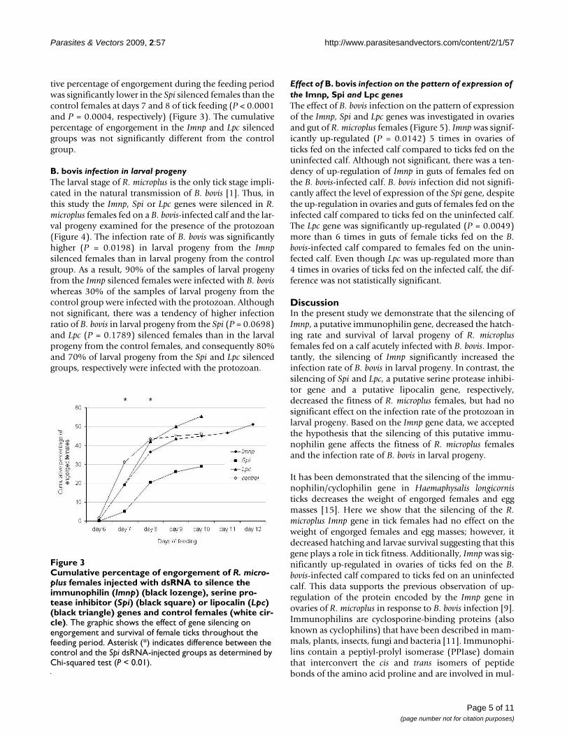
tive percentage of engorgement during the feeding periodwas significantly lower in the Spi silenced females than thecontrol females at days 7 and 8 of tick feeding (P < 0.0001and P = 0.0004, respectively) (Figure 3). The cumulativepercentage of engorgement in the Imnp and Lpc silencedgroups was not significantly different from the controlgroup.
B. bovis infection in larval progenyThe larval stage of R. microplus is the only tick stage impli-cated in the natural transmission of B. bovis [1]. Thus, inthis study the Imnp, Spi or Lpc genes were silenced in R.microplus females fed on a B. bovis-infected calf and the lar-val progeny examined for the presence of the protozoan(Figure 4). The infection rate of B. bovis was significantlyhigher (P = 0.0198) in larval progeny from the Imnpsilenced females than in larval progeny from the controlgroup. As a result, 90% of the samples of larval progenyfrom the Imnp silenced females were infected with B. boviswhereas 30% of the samples of larval progeny from thecontrol group were infected with the protozoan. Althoughnot significant, there was a tendency of higher infectionratio of B. bovis in larval progeny from the Spi (P = 0.0698)and Lpc (P = 0.1789) silenced females than in the larvalprogeny from the control females, and consequently 80%and 70% of larval progeny from the Spi and Lpc silencedgroups, respectively were infected with the protozoan.
Effect of B. bovis infection on the pattern of expression of the Imnp, Spi and Lpc genesThe effect of B. bovis infection on the pattern of expressionof the Imnp, Spi and Lpc genes was investigated in ovariesand gut of R. microplus females (Figure 5). Imnp was signif-icantly up-regulated (P = 0.0142) 5 times in ovaries ofticks fed on the infected calf compared to ticks fed on theuninfected calf. Although not significant, there was a ten-dency of up-regulation of Imnp in guts of females fed onthe B. bovis-infected calf. B. bovis infection did not signifi-cantly affect the level of expression of the Spi gene, despitethe up-regulation in ovaries and guts of females fed on theinfected calf compared to ticks fed on the uninfected calf.The Lpc gene was significantly up-regulated (P = 0.0049)more than 6 times in guts of female ticks fed on the B.bovis-infected calf compared to females fed on the unin-fected calf. Even though Lpc was up-regulated more than4 times in ovaries of ticks fed on the infected calf, the dif-ference was not statistically significant.
DiscussionIn the present study we demonstrate that the silencing ofImnp, a putative immunophilin gene, decreased the hatch-ing rate and survival of larval progeny of R. microplusfemales fed on a calf acutely infected with B. bovis. Impor-tantly, the silencing of Imnp significantly increased theinfection rate of B. bovis in larval progeny. In contrast, thesilencing of Spi and Lpc, a putative serine protease inhibi-tor gene and a putative lipocalin gene, respectively,decreased the fitness of R. microplus females, but had nosignificant effect on the infection rate of the protozoan inlarval progeny. Based on the Imnp gene data, we acceptedthe hypothesis that the silencing of this putative immu-nophilin gene affects the fitness of R. microplus femalesand the infection rate of B. bovis in larval progeny.
It has been demonstrated that the silencing of the immu-nophilin/cyclophilin gene in Haemaphysalis longicornisticks decreases the weight of engorged females and eggmasses [15]. Here we show that the silencing of the R.microplus Imnp gene in tick females had no effect on theweight of engorged females and egg masses; however, itdecreased hatching and larvae survival suggesting that thisgene plays a role in tick fitness. Additionally, Imnp was sig-nificantly up-regulated in ovaries of ticks fed on the B.bovis-infected calf compared to ticks fed on an uninfectedcalf. This data supports the previous observation of up-regulation of the protein encoded by the Imnp gene inovaries of R. microplus in response to B. bovis infection [9].Immunophilins are cyclosporine-binding proteins (alsoknown as cyclophilins) that have been described in mam-mals, plants, insects, fungi and bacteria [11]. Immunophi-lins contain a peptiyl-prolyl isomerase (PPIase) domainthat interconvert the cis and trans isomers of peptidebonds of the amino acid proline and are involved in mul-
Cumulative percentage of engorgement of R. microplus females injected with dsRNA to silence the immunophilin (Imnp) (black lozenge), serine protease inhibitor (Spi) (black square) or lipocalin (Lpc) (black triangle) genes and control females (white circle)Figure 3Cumulative percentage of engorgement of R. micro-plus females injected with dsRNA to silence the immunophilin (Imnp) (black lozenge), serine pro-tease inhibitor (Spi) (black square) or lipocalin (Lpc) (black triangle) genes and control females (white cir-cle). The graphic shows the effect of gene silencing on engorgement and survival of female ticks throughout the feeding period. Asterisk (*) indicates difference between the control and the Spi dsRNA-injected groups as determined by Chi-squared test (P < 0.01).
Page 5 of 11(page number not for citation purposes)
Parasites & Vectors 2009, 2:57 http://www.parasitesandvectors.com/content/2/1/57
tiple cellular processes, such as protein folding, traffickingand defense mechanisms [11,16]. Numerous putativeimmunophilin/cyclophilin genes have been identified inthe Ixodes scapularis tick genome http://iscapularis.vectorbase.org/; however, no functional study has been reportedyet. The deduced amino acid sequence of the Imnp geneused in the present study contains a PPIase domain(amino acid 49 to 208, Pfam prediction, E value = 1.80e-117), and has 90% and 82% identity with the immu-nophilyn/cyclophilin proteins of H. longicornis and I.scapularis, respectively (data not shown), suggesting thatImp encodes for a putative R. microplus immunophilingene.
The silencing of the Spi gene reduced the number ofengorged females causing a delay in the engorgement time
suggesting that this gene is involved in the processes offeeding and digestion in R. microplus as previouslydescribed for other members of serine protease inhibitors[12,17]. Although not statistically significant, the up-reg-ulation of Spi in ovaries of ticks fed on a calf acutelyinfected with B. bovis may have biological relevance andexplain the up-regulation of the Spi protein in ovaries ofR. microplus fed on cattle infected with the protozoandescribed elsewhere [9]. It has been shown that a Kunitz-type serine protease inhibitor is involved in the defensemechanisms of adult Dermacentor variabilis ticks againstRickettsia montanensis [18]. Here we showed that silencingof the Spi gene in female ticks did not result in a signifi-cant increase of B. bovis infection in R. microplus larvae.Despite the fact that the serine protease inhibitor is a largeprotein family that includes several members, these datasuggest that different tick species or tick stages may usedifferent strategies to control Babesial or Rickettsial infec-tion. Serine proteases are part of the clotting mechanismsof mammals and serine protease inhibitors play a role inblocking blood coagulation. Considering hematopha-gous ectoparasites, the blood coagulation cascade of themammalian host is essentially inhibited by proteaseinhibitors [19]. Several serine protease inhibitors havebeen identified in R. microplus and shown to inhibit majorprocoagulant enzymes, such as thrombin, trypsin andplasmin [12,17]. The N-terminal amino-acid sequence ofthe protein encoded by the R. microplus Spi gene has 85%identity with the R. microplus Kunitz-type serine proteaseinhibitor (GeneBank accession no. P83609) and conser-vation among cysteine position, suggesting that Spiencodes for a putative Kunitz-type serine protease inhibi-tor [9]. The Imnp and Spi genes were up-regulated in R.microplus ovaries and guts, respectively in response to B.bovis infection and their silencing affected the fitness ofthe ticks and rate of B. bovis infection in larval progeny.Despite the fact that there is no direct association betweenthe putative function of the two genes, it is reasonable tospeculate that concurrent silencing of these genes may
Table 1: Biological effect of gene silencing in R. microplus females fed on a calf during acute Babesia bovis infection.
Experimental groups
Percentage of engorged females
Weight (mg)of engorged
females (SD)
Oviposition rate Egg mass (mg) (SD)
Percentage of hatching
Percentage of larvae survival
Control 46.1%(83/180)
342.4 (± 0.567) 98.7% (82/83) 141.1 (± 0.423) 90.2% (74/82) 100% (74/74)
Imnp dsRNA 51.1%(92/180)
349.3 (± 0.907) 89.1% (82/92) 132.3 (± 0.662) 69.5%1 (57/82) 80.7%1 (46/57)
Spi dsRNA 28.8%1
(52/180)354.1 (± 0.615) 98.0% (51/52) 117.82 (± 0.552) 94.1% (48/51) 97.9% (47/48)
Lpc dsRNA 55.5%(100/180)
364.32 (± 0.662) 93.0% (93/100) 128.3 (± 0.558) 66.6%1 (62/93) 96.7% (60/62)
Imnp - a putative immunophilin gene; Spi - a putative Kunitz-type serine protease inhibitor; Lpc - a putative lipocalin gene1 Chi-squared test (P < 0.01)2 t test (P < 0.05)
Infection rate of B. bovis in larval progeny from gene silenced R. microplus femalesFigure 4Infection rate of B. bovis in larval progeny from gene silenced R. microplus females. Imnp - a putative immu-nophilin gene; Spi - a putative Kunitz-type serine protease inhibitor; Lpc - a putative lipocalin gene. The percentages of larval progeny infected with B. bovis were compared by Chi-squared test and P values for each experimental group are shown.
0
10
20
30
40
50
60
70
80
90
100
Per
cen
tag
e o
f la
rval
pro
gen
y in
fect
ed w
ith
B. b
ovi
s
P=0.0198
Larval progeny from the Imnp
silenced females
Larval progeny from the Spi
silenced females
Larval progeny from the Lpc
silenced females
Larval progeny from the control
females
P=0.0698
P=0.1789
Page 6 of 11(page number not for citation purposes)
Parasites & Vectors 2009, 2:57 http://www.parasitesandvectors.com/content/2/1/57
Page 7 of 11(page number not for citation purposes)
Transcript level of the imunophilin (Imnp) (A), serine protease inhibitor (Spi) (B) and lipocalin (Lpc) (C) genes in ovaries and guts of six partially engorged R. microplus females fed on either an uninfected calf (black bar) or a B. bovis-infected calf (grey bar)Figure 5Transcript level of the imunophilin (Imnp) (A), serine protease inhibitor (Spi) (B) and lipocalin (Lpc) (C) genes in ovaries and guts of six partially engorged R. microplus females fed on either an uninfected calf (black bar) or a B. bovis-infected calf (grey bar). The data represent mean values of relative expression of the genes of interest. Asterisk (*) indicates difference between dsRNA-injected and control groups as determined by t-test (P < 0.05).
0.0
0.2
0.4
0.6
0.8
uninfected infected0.0
0.2
0.4
0.6
0.8
uninfected infected
B Spi - ovaries Spi - guts
Rel
ativ
e ex
pres
sion
0.0
0.2
0.4
0.6
0.8
uninfected infected0.0
0.2
0.4
0.6
0.8
1.0
uninfected infected
Imnp - ovaries
Rel
ativ
e ex
pres
sion
Imnp - gutsA
*
C
0.0
0.2
0.4
0.6
0.8
1.0
uninfected infected0.0
0.2
0.4
0.6
0.8
uninfected infected
Lpc - ovaries Lpc - guts
Rel
ativ
e ex
pres
sion *
Parasites & Vectors 2009, 2:57 http://www.parasitesandvectors.com/content/2/1/57
have a synergetic effect in controlling the protozoan infec-tion in ticks and further experiments are needed to inves-tigate this hypothesis.
The R. microplus Lpc gene encodes for a putative lipocalinprotein which contains a histamine-binding domain fromresidue 35 to 180 (Pfam E-value: 1.30e-29) (data notshown). The lipocalin protein family is a large group ofextracellular proteins which exhibit a variety of functionsthat include transport of small molecules, prostaglandinsynthesis, smell reception and regulation of immuneresponse [13,20]. It has been shown that the earlierdetachment of R. microplus larvae from resistant cattle isrelated to the release of histamine at the attachment site[21]. The silencing of tick genes that encode for hista-mine-binding proteins has shown to have a marked effecton tick fitness [22,23]. Here we show that the silencing ofthe R. microplus Lpc gene had no affect on the duration ofthe tick feeding period, but it decreased the weight ofengorged females and the percentage of hatching demon-strating that this putative lipocalin gene affects tick fitness.Lpc was significantly up-regulated in gut of ticks fed on acalf during acute B. bovis infection compared to ticks fedon an uninfected calf, confirming previous observation(F.D. Guerrero, unpublished data). High-affinity hista-mine-binding proteins involved in the suppression ofhost inflammatory response during tick feeding have beenidentified in saliva of Rhipicephalus appendiculatus ticks[24]. It has been shown that the bovine immune systemproduces inflammatory mediators, such as nitric oxideand cytokines, in response to acute B. bovis infection [25].Considering that lipocalin proteins suppress inflamma-tion, it is tempting to speculate that the Lpc gene may playa role in R. microplus gut by suppressing the inflammatorymediators present in the blood meal from acutely B. bovis-infected cattle.
R. microplus females acquire B. bovis infection and pass theprotozoan transovarially to their larval progeny whichwill transmit it to naïve cattle [1]. Therefore, it was ofinterest to silence genes that had been previously demon-strated to be up-regulated in female ticks in response to B.bovis infection and investigate the presence of the proto-zoan in larval progeny. It has been shown in Anophelesgambiae mosquito that targeting up-regulated genes, suchas C-type lectin and leucine-rich genes, for silencingaffected the infection rate of Plasmodium berghei in mos-quitoes [26]. We show that the infection rate of B. bovis inlarval progeny from the Imnp silenced females was signif-icantly higher than the control group suggesting that thisgene plays a role in defending R. microplus against the pro-tozoan infection. Thirty percent of the larvae samples ofthe control group were infected with B. bovis, showing thatonly a minor proportion of larval progeny is transovari-ally infected when R. microplus females fed on an acutely
infected calf as described elsewhere [27]. In contrast, 90%of the larval progeny samples from the Imnp silencedgroup were infected with B. bovis demonstrating the bio-logical relevance of this gene as an antagonist of the pro-tozoan infection. It has been shown that infection withBabesia spp can affect tick fitness and the severity of theseeffects is related to the degree of parasitemia [1]. In thisstudy, clinical signs and parasitemia may have affected thetick fitness; however the overall biological impact cannotbe solely attributed to B. bovis infection, considering thedifferences in tick fitness observed between the silencedand control groups.
RNAi has been widely used to knock-down gene expres-sion in a sequence-specific manner, making it a powerfultool for investigating gene function. However, off-targeteffects have been described in numerous species [28-30]and cannot be entirely ruled out as the cause of the resultsobserved in this experiment. The R. microplus genomesequence is not available [31] and the alignment analysesare restricted to sequences listed in databases. Blast analy-sis of the dsRNA sequences used in this study did notreveal significant homology to any known tick sequenceother than the GOI. Additionally, the GOI were specifi-cally silenced by the dsRNA injection. Taken together,these two aspects support the data and represent the bestpossible strategy to make solid scientific observationsregarding biological effect of gene silencing in R. micro-plus.
In summary, we show that the silencing of Imnp, Spi orLpc, three novel genes of R. microplus, decreased the fitnessof females ticks fed on a calf during acute B. bovis infec-tion. Notably, the Imnp gene was significantly up-regu-lated in ovaries of R. microplus in response to infection andits silencing in female ticks increased significantly theinfection rate of B. bovis in larval progeny. Collectively,these data demonstrate that Imnp acts as an antagonistagainst B. bovis infection suggesting that this gene mayplay a role in controlling the transovarial transmission ofthe protozoan in R. microplus. These observations mayalso account for the presence of a tick defense system tocontrol protozoan infection and further studies areneeded to characterize the mechanism used by the Imnpgene to affect B. bovis infection in R. microplus.
Materials and methodsCattle, ticks and protozoanTwo Holstein calves 3-4 months of age that tested negativefor B. bovis by nested PCR [27] were used in this study. Theanimals were maintained according to protocolsapproved by the University of Idaho Institutional AnimalCare and Use Committee. One calf remained uninfectedthroughout the experiment and the other calf was inocu-lated intravenously with approximately 1.4 × 10⁸ B. bovis-
Page 8 of 11(page number not for citation purposes)
Parasites & Vectors 2009, 2:57 http://www.parasitesandvectors.com/content/2/1/57
infected erythrocytes (T2Bo strain) [32]. Parasitemia inperipheral blood was examined by quantitative real timePCR to amplify the single copy B. bovis msa-1 gene. For themsa-1 quantitative real time PCR, specific primers (5' gat-gcgtttgcacatgctaag 3' and 5' cgggtacttcggtgctctca 3') andprobe (FAM 5'-cacgctcaagtaggaaattttgttaaacctgga-3'TAMRA) were used following the assay conditionsdescribed elsewhere [27]. At day 6 after the B. bovis inocu-lation, the infected calf started showing clinical signs ofacute babesioses marked by a decrease of 20% in packedcell volume and temperature above 39°C. The La Minitastrain of R. microplus used in this study has been main-tained at the University of Idaho Holm Research Centersince 1999 as previously described [33]. To obtain unin-fected and unfed adult ticks, approximately 40,000 larvae(from 2 g of eggs) were placed under a cloth patch on theuninfected calf. After 13-14 days, engorged nymphs weremanually removed with forceps and incubated in vitro at26°C to induce molting to adult stage. Unfed adult tickswere sexed and the females used for gene expression andsilencing experiments. The biological effect of gene silenc-ing was investigated in female ticks fed until repletion onthe B. bovis-infected calf whereas the pattern of geneexpression was examined in females fed until repletion oneither the B. bovis-infected calf or the uninfected calf.
Target genes and synthesis of double stranded RNASpecific primers were designed based on the cDNAsequences of the Imnp (TC12157), Spi (TC9311) and Lpc(TC8123) genes described in the R. microplus Gene IndexProject http://compbio.dfci.harvard.edu/tgi/tgipage.html(Table 2). PCR products from each one of the GOI wereindividually cloned into pCR® 4-TOPO® (Invitrogen,Carlsbad, CA, USA), sequenced and analyzed in silico forthe presence of putative small interfering RNA (siRNA) by
the algorithm siRNA Target Finder (Ambion, Austin, TX,USA). A fragment with approximately 400 bp from eachGOI containing the highest number of putative siRNA wasamplified by PCR, cloned into pCR®II-TOPO® (Invitro-gen) and used as template for the dsRNA synthesis (Table2). The MEGAscript® Transcription Kit (Ambion) was usedfor the dsRNA synthesis following the manufacturer's pro-tocol. The dsRNA molecules were checked by electro-phoresis on agarose gel, quantified by spectrophotometryand kept at -20°C until used for tick injection.
Injection of ticks with double stranded RNAFreshly molted unfed females were used for the dsRNAinjection. Three groups with 200 female ticks eachreceived a single injection with dsRNA identical to the tar-get region of the Imnp, Spi or Lpc genes. Another groupalso with 200 female ticks was injected with buffer (0.1mM EDTA) and used as control. For injection, individualfemales were placed on double-sided tape with the ventralside upwards and injected with either 1 μl of dsRNA(approximately 1 × 10¹¹ molecules dissolved in buffer 0.1mM EDTA) or 1 μl of EDTA buffer through the coxalmembrane at the base of the 4th leg on the right ventralside. Injections were accomplished using a 10 μl syringewith a 33 gauge needle (Hamilton, Bonaduz, Switzerland)and the microprocessor controlled UMP3 injection pumpapparatus (World Precision Instruments, Berlin, Ger-many). After the injection, the female ticks of each groupplus an equal number of males were placed under individ-ual stockinet sleeves glued to the side of the B. bovis-infected calf (2 days after the experimental B. bovis infec-tion). At day 5 post-dsRNA injection, 6 partially engorgedfemales from each group were dissected and individualovaries and guts collected in RNAlater (Ambion) to evalu-ate gene silencing.
Table 2: Gene identification, primers, gene region amplified, amplicon size and PCR annealing temperature.
Gene identification (purpose)
TC number* or GenBank identification
Forward primer(5'-3')
Reverse primer(5'-3')
Size (bp) Annealing temperature (°C)
Imnp (full-length) TC12157 gcagaattaactatcgcaaa tttgtgtggtggaaatgaa 928 58Imnp (dsRNA synthesis) TC12157 agacttcacgaatcacaa taagaagaaaggaaaagttg 380 58Imnp (real-time PCR) TC12157 ggttacttgtgtggttgt gtgtggtggaaatgaat 64 60Spi (full-length) TC9311 caattcatcctgtgtattctcg aacccattcgcctgttacg 1652 58Spi (dsRNA synthesis) TC9311 caattcatcctgtgtattctcg caacaacttcgtgactct 396 58Spi (real-time PCR) TC9311 taatagcgagggaagtag caccaaggagaccatttc 135 60Lpc (full-length) TC8123 accccgaatccctgtgtt cagtgccgaccaaagtaagg 731 58Lpc (dsRNA synthesis) TC8123 gagaagataggaaatgtgta tctctgttgtagacgaagt 379 58Lpc (real-time PCR) TC8123 aaggagggaagaatacac ctcaagtttcaaggacag 134 60Actin (real-time PCR) AY255624 tcctctccttccttctac aaagttctgttcgtcgt 103 60tubulin (real-time PCR) AA257910 tgtgccccgtgccgtattt ggcaccagagcgaacct 64 60G6PDH (real-time PCR) EU595881 ttctctctctcctcagtg gaagatgtgttgttgtcc 88 60PHGPx (real-time PCR) DQ180067 accaacaagaactacacg cttctttatgtctgcctca 131 60
Imnp - a putative immunophilin gene; Spi - a putative Kunitz-type serine protease inhibitor; Lpc - a putative lipocalin gene; TC - tentative consensus; * the R. microplus Gene Index Project http://compbio.dfci.harvard.edu/tgi/tgipage.html.
Page 9 of 11(page number not for citation purposes)
Parasites & Vectors 2009, 2:57 http://www.parasitesandvectors.com/content/2/1/57
Evaluation of tick fitnessAfter the dsRNA injection, individual stockinet sleevescontaining each experimental group were monitoreddaily for the presence of engorged females. The effect ofgene silencing on engorgement and survival of femaleticks was investigated by assessing the cumulative percent-age of engorged females during the feeding period. Indi-vidual engorged females were weighed and put in 24-wellplates at 26°C to assess oviposition. At day 14 after thebeginning of oviposition, eggs laid by individual femaleswere weighed, put in individual vials and incubated at26°C to evaluate hatching. Hatching was evaluated at 30days after the egg masses were collected and was definedas the presence of any larvae in eggs from each individualfemale. The larval progeny was maintained in individualvials at 26°C for 45 days and the larvae survival was deter-mined as the presence of any live larvae in larval progenyfrom individual females.
Gene expression and silencingA qRT-PCR was standardized for each GOI to assess geneexpression and silencing. Total RNA was extracted fromindividual ovary or gut samples using the RNAqueous® Kit(Ambion) according to the manufacturer's protocol andquantified by Qubit® Fluorometer (Invitrogen). The RNAsamples were treated with DNase I (Invitrogen) followingthe manufacturer's protocol and 200 ng of total RNA ofeach sample were used for cDNA synthesis using theSuperscript® Vilo™ cDNA Synthesis Kit (Invitrogen) fol-lowing the manufacturer's protocol. For the qRT-PCR,specific primers for each GOI were designed to avoid thegene region used to synthesize the dsRNA (Table 2). Forthe standardization of the qRT-PCR, the R. microplus actin,tubulin, G6PDH and PHGPx genes were evaluated as tickreference gene candidates (Table 2). The geNorm applet[14] was used to examine the stability of expression of thereference gene candidates. The qRT-PCR for the GOI andreference gene candidates were performed in a CFX96™Real-Time PCR Detection System using the Express SYBR®
GreenER™ Supermix Kit (Invitrogen). The cycling condi-tions consisted of a Uracil-DNA Glycosylase inactivationstep of 50°C for 30 sec, initial denaturation of 95°C for 2min followed by 40 cycles of 95°C denaturation for 15 secand annealing/extension of 60°C for 45 sec. Reactionswere performed in duplicate in 20 μl using 200 nM ofeach primer and 2 μl of a 1/20 dilution of cDNA as tem-plate. An inter-run calibrator was included to assess inter-run variations. The CFX Manager™ Software (Bio-Rad,Hercules, CA, USA) was used to analyze the qRT-PCRdata. Efficiency of amplification and melt curve analyseswere performed to evaluate analytical sensitivity and spe-cificity of the qRT-PCR for each GOI.
Infection rate of B. bovis in larvae of R. microplusThe presence of B. bovis in larval progeny of the silencedand control R. microplus females was examined by nestedPCR with primers to the B. bovis msa1 gene as previouslydescribed [27]. Briefly, a total of 10 larval progeny sam-ples (containing approximately 100 larvae per sample) ofindividual females from each experimental group weretested. Genomic DNA (gDNA) was extracted from eachlarval sample using the Puregene® Genomic DNA Purifica-tion Kit (Gentra, Minneapolis, MN, USA) following themanufacturer's protocol and quantified by spectropho-tometry. The nested PCR were performed using the Hot-StarTaq® Plus Master Mix Kit (QIAGEN, Valencia, CA,USA) in 20 μl containing 0.2 μM of each primer. For thefirst run of the nested PCR, 100 ng of larvae gDNA wereused as template with 55°C of annealing temperature. Forthe second run, 2 μl of a 1/100 dilution of the first PCRrun was used as template with 60°C of annealing temper-ature. The presence of amplifiable DNA was examined byPCR in all negative samples using primers specific for theR. microplus actin gene described in Table 2.
Statistical analysesThe Instat® software (GraphPad Software, Inc., USA), ver-sion 3.06, was used to perform the statistical analyses. Therelative gene expression and weights of engorged femalesand egg masses were compared by t-test. The percentagesof engorged females, oviposition, hatching, larvae sur-vival and infection rate of B. bovis were compared by Chi-squared test.
Competing interestsThe authors declare that they have no competing interests.
Authors' contributionsRGB designed and performed the experiment, and wrotethe first draft of the manuscript. MWU designed and per-formed the experimental procedures involving ticks. FDGsuggested the tick genes used in this study and contributedto the experimental design. DPK designed, supervised andacquired funding for the experiment. GAS designed andsupervised the experiment and made intellectual contri-bution regarding data interpretation. All authors reviewedand approved the final version of the manuscript.
AcknowledgementsWe thank Kathy Mason, Ralph Horn, James Allison and Sara Davis for excellent technical assistance. This work was supported by USDA-ARS CRIS project number 5348-32000-028-00D.
References1. Friedhoff KT: Transmission of babesia. In Babesiosis of Domestic
Animals and Man Edited by: Ristic M. Boca Raton, FL, USA: CRC Press;1988:23-52.
Page 10 of 11(page number not for citation purposes)
Parasites & Vectors 2009, 2:57 http://www.parasitesandvectors.com/content/2/1/57
Publish with BioMed Central and every scientist can read your work free of charge
"BioMed Central will be the most significant development for disseminating the results of biomedical research in our lifetime."
Sir Paul Nurse, Cancer Research UK
Your research papers will be:
available free of charge to the entire biomedical community
peer reviewed and published immediately upon acceptance
cited in PubMed and archived on PubMed Central
yours — you keep the copyright
Submit your manuscript here:http://www.biomedcentral.com/info/publishing_adv.asp
BioMedcentral
2. Mahoney DF, Mirre GB: A note on the transmission of Babesiabovis (syn B argentina) by the one-host tick, Boophilus micro-plus. Res Vet Sci 1979, 26:253-254.
3. Bock R, Jackson L, de Vos A, Jorgensen W: Babesiosis of cattle.Parasitology 2004, 129(Suppl):S247-S269.
4. Fragoso H, Rad PH, Ortiz M, Rodriguez M, Redondo M, Herrera L, dela Fuente J: Protection against Boophilus annulatus infestationsin cattle vaccinated with the B. microplus Bm86-containingvaccine Gavac. Vaccine 1998, 16:1990-1992.
5. Jonsson NN, Matschoss AL, Pepper P, Green PE, Albrecht MS, Hun-gerford J, Ansell : Evaluation of TickGARDPLUS, a novel vac-cine against Boophilus microplus, in lactating Holstein-Friesian cows. Veterinary Parasitology 2000, 88:275-285.
6. Miller RJ, Davey RB, George JE: First report of organophosphate-resistant Boophilus microplus (Acari: Ixodidae) within theUnited States. J Med Entomol 2005, 42:912-917.
7. de la Fuente J, Almazán C, Canales M, de la Lastra JMP, Kocan KM,Willadsen P: A ten-year review of commercial vaccine per-formance for control of tick infestations on cattle. AnimalHealth Research Reviews 2007, 8:23-28.
8. Cattle fever tick surveillance in Texas NAHSS outlook 2005[http://www.aphis.usda.gov/vs/ceah/ncahs/nsu/outlook/issue7/cattle_fever_tick_surveillance.pdf].
9. Rachinsky A, Guerrero FD, Scoles GA: Differential proteinexpression in ovaries of uninfected and Babesia-infectedsouthern cattle ticks, Rhipicephalus (Boophilus) microplus.Insect Biochemistry and Molecular Biology 2007, 37:1291-1308.
10. Rachinsky A, Guerrero FD, Scoles GA: Proteomic profiling of Rhi-picephalus (Boophilus) microplus midgut responses to infec-tion with Babesia bovis. Veterinary Parasitology 2008, 152:294-313.
11. Wang P, Heitman J: The cyclophilins. Genome Biology 2005, 6:226.12. Macedo-Ribeiro S, Almeida C, Calisto BM, Friedrich T, Mentele R,
Sturzebecher J, Fuentes-Prior P, Pereira PJ: Isolation, Cloning andStructural Characterisation of Boophilin, a MultifunctionalKunitz-Type Proteinase Inhibitor from the Cattle Tick. PLoSONE 2008, 3:e1624.
13. Flower DR, North ACT, Sansom CE: The lipocalin protein family:structural and sequence overview. Biochimica et Biophysica Acta(BBA) - Protein Structure and Molecular Enzymology 2000, 1482:9-24.
14. Vandesompele J, De Preter K, Pattyn F, Poppe B, Van Roy N, DePaepe A, Speleman F: Accurate normalization of real-timequantitative RT-PCR data by geometric averaging of multi-ple internal control genes. Genome Biology 2002, 3:research0034.
15. Boldbaatar D, Kilonzo RM, Battur B, Umemiya R, Liao M, Tanaka T,Xuan X, Fujisaki K: Identification of two forms of cyclophilinfrom the hard tick Haemaphysalis longicornis. Process Biochem-istry 2008, 43:615-625.
16. Galat A: Peptidylproline cis-trans-isomerases: immunophi-lins. Eur J Biochem 1993, 216:689-707.
17. Andreotti R, Gomes A, Malavazi-Piza KC, Sasaki SD, Sampaio CAM,Tanaka AS: BmTI antigens induce a bovine protectiveimmune response against Boophilus microplus tick. Interna-tional Immunopharmacology 2002, 2:557-563.
18. Ceraul SM, Dreher-Lesnick SM, Mulenga A, Rahman MS, Azad AF:Functional characterization and novel rickettsiostatic effectsof a kunitz-type Serine protease inhibitor from the tick Der-macentor variabilis. Infect Immun 2008, 76:5429-5435.
19. Mans BJ, Neitz AWH: Adaptation of ticks to a blood-feedingenvironment: evolution from a functional perspective. InsectBiochemistry and Molecular Biology 2004, 34:1-17.
20. Flower DR: The lipocalin protein family: structure and func-tion. Biochem J 1996, 318:1-14.
21. Kemp DH, Bourne A: Boophilus microplus: the effect of hista-mine on the attachment of cattle-tick larvae-studies in vivoand in vitro. Parasitology 1980, 80:487-496.
22. Aljamali MN, Bior AD, Sauer JR, Essenberg RC: RNA interferencein ticks: a study using histamine binding protein dsRNA inthe female tick Amblyomma americanum. Insect Molecular Biology2003, 12:299-305.
23. Ramakrishnan VG, Aljamali MN, Sauer JR, Essenberg RC: Applica-tion of RNA interference in tick salivary gland research. Jour-nal of Biomolecular Techniques 2005, 16:297-305.
24. Paesen GC, Adams PL, Harlos K, Nuttall PA, Stuart DI: Tick Hista-mine-Binding Proteins: Isolation, Cloning, and Three-Dimensional Structure. Molecular Cell 1999, 3:661-671.
25. Goff WL, Johnson WC, Parish SM, Barrington GM, Elsasser TH, DavisWC, Valdez RA: IL-4 and IL-10 inhibition of IFN-gamma- andTNF-alpha-dependent nitric oxide production from bovinemononuclear phagocytes exposed to Babesia bovis mero-zoites. Vet Immunol Immunopathol 2002, 84:237-251.
26. Osta MA, Christophides GK, Kafatos FC: Effects of mosquitogenes on plasmodium development. Science 2004,303:2030-2032.
27. Howell JM, Ueti MW, Palmer GH, Scoles GA, Knowles DP: Transo-varial transmission efficiency of Babesia bovis tick stagesacquired by Rhipicephalus (Boophilus) microplus duringacute infection. J Clin Microbiol 2007, 45:426-431.
28. Ma Y, Creanga A, Lum L, Beachy PA: Prevalence of off-targeteffects in Drosophila RNA interference screens. Nature 2006,443:359-363.
29. Cullen BR: Enhancing and confirming the specificity of RNAiexperiments. Nat Meth 2006, 3:677-681.
30. Rual JF, Klitgord N, Achaz G: Novel insights into RNAi off-targeteffects using C. elegans paralogs. BMC Genomics 2007, 8:106.
31. Guerrero FD, Nene VM, George JE, Barker SC, Willadsen P:Sequencing a new target genome: the Boophilus microplus(Acari: Ixodidae) genome project. J Med Entomol 2006, 43:9-16.
32. Goff WL, Johnson WC, Cluff CW: Babesiabovis immunity. Invitro and in vivo evidence for IL-10 regulation of IFN-gammaand iNOS. Ann N Y Acad Sci 1998, 849:161-180.
33. Stiller D, Goff WL, Johnson LW, Knowles DP: Dermacentor varia-bilis and Boophilus microplus (Acari: Ixodidae): experimentalvectors of Babesia equi to equids. J Med Entomol 2002,39:667-670.
Page 11 of 11(page number not for citation purposes)
Copyright © 2022 FDOKUMEN