First survey for Babesia bovis and Babesia bigemina infection in cattle from Central and Southern...
7
First survey for Babesia bovis and Babesia bigemina infection in cattle from Central and Southern regions of Portugal using serological and DNA detection methods Marta G. Silva a, *, Gisela Henriques a , Claudia Sa ´ nchez a , Patrı ´cia X. Marques a , Carlos E. Suarez b , Abel Oliva a a Biomolecular Diagnostic Laboratory, ITQB/IBET, Universidade Nova de Lisboa, Av. da Repu ´blica, Apt 12, 2781-901 Oeiras, Portugal b Animal Disease Research Unit, USDA-ARS, 3003 ADBF, WSU, Pullman, WA 99163-6630, USA 1. Introduction Bovine babesiosis is a tick-borne disease caused by infection with intraerythrocytic Apicomplexa protozoan parasites from the genus Babesia that are distributed worldwide (McCrosker, 1981). Babesia bovis and Babesia bigemina are the most common species infecting cattle (Figueroa et al., 1998). In Portugal there are at least three classes of ticks identified that are competent for transmission of B. bovis and B. bigemina: Ixodes ricinus, Rhipicephalus bursa and Rhipicephalus (Boophilus) annulatus (Caeiro, 1999; Estrada- Pena et al., 2004). The species R. bursa and R. annulatus are mostly restricted to the Southern part of the country under hot and dry climate conditions of mediterranean type. I. ricinus is present in Southern zones of the country, and also in Western parts where the climate is habitually humid (Estrada-Pena and Santos-Silva, 2005). Previous studies suggest the occurrence of B. bovis infections in Portugal (Caeiro, 1999; Criado-Fornelio et al., 2003), however the incidence and distribution of Babesia infections among the estimated 1.1 millions cattle population currently in Portugal remains unknown and systematic studies for determining the magnitude of these infections are needed for the development of a control strategy for babesiosis in this country. Such studies required the use of state-of-the-art diagnostic tools that are both sensitive and specific. Accurate serological detection of Babesia infections was achieved using ELISA tests based on the C-terminus portion of the B. bovis and B. bigemina rhoptry-associated protein-1 (RAP-1) proteins (Boonchit et al., 2004, 2006; Goff et al., 2006, 2008). In addition, although these RAP-1 protein- based serological tests provided a good level for detection of early and late infections in cattle (Goff et al., 2003), and due to the time required for the emergence of detectable Veterinary Parasitology 166 (2009) 66–72 ARTICLE INFO Article history: Received 29 April 2009 Received in revised form 10 July 2009 Accepted 15 July 2009 Keywords: Babesia bovis Babesia bigemina Portugal ELISA PCR ABSTRACT Incidence of bovine babesiosis in Portugal is currently unknown. In this study, a first survey of Babesia bovis and Babesia bigemina infection in cattle was carried out using blood samples from 406 clinically healthy individuals from different districts from Central and Southern regions of Portugal and analyzed by indirect enzyme linked immunosorbent assay (iELISA) and nested polymerase chain reaction (nPCR). Overall, serological testing revealed that 79% and 52% of cattle were positive for B. bovis and B. bigemina antibodies, respectively, whereas nPCR testing detected 71% and 34% cattle infected with B. bovis and B. bigemina protozoan, respectively. This is the first report of the prevalence of B. bovis and B. bigemina in cattle obtained by serological and DNA analysis studies in Central and Southern regions of Portugal. These data suggests high incidence of Babesia sp. infection in Portugal and can be used for designing adequate control programs. ß 2009 Elsevier B.V. All rights reserved. * Corresponding author. Tel.: +351 214 469 428; fax: +351 214 411 277. E-mail address: [email protected] (M.G. Silva). Contents lists available at ScienceDirect Veterinary Parasitology journal homepage: www.elsevier.com/locate/vetpar 0304-4017/$ – see front matter ß 2009 Elsevier B.V. All rights reserved. doi:10.1016/j.vetpar.2009.07.031
-
Upload
independent -
Category
Documents
-
view
0 -
download
0
Transcript of First survey for Babesia bovis and Babesia bigemina infection in cattle from Central and Southern...
Veterinary Parasitology 166 (2009) 66–72
First survey for Babesia bovis and Babesia bigemina infection in cattlefrom Central and Southern regions of Portugal using serological andDNA detection methods
Marta G. Silva a,*, Gisela Henriques a, Claudia Sanchez a, Patrıcia X. Marques a,Carlos E. Suarez b, Abel Oliva a
a Biomolecular Diagnostic Laboratory, ITQB/IBET, Universidade Nova de Lisboa, Av. da Republica, Apt 12, 2781-901 Oeiras, Portugalb Animal Disease Research Unit, USDA-ARS, 3003 ADBF, WSU, Pullman, WA 99163-6630, USA
A R T I C L E I N F O
Article history:
Received 29 April 2009
Received in revised form 10 July 2009
Accepted 15 July 2009
Keywords:
Babesia bovis
Babesia bigemina
Portugal
ELISA
PCR
A B S T R A C T
Incidence of bovine babesiosis in Portugal is currently unknown. In this study, a first
survey of Babesia bovis and Babesia bigemina infection in cattle was carried out using blood
samples from 406 clinically healthy individuals from different districts from Central and
Southern regions of Portugal and analyzed by indirect enzyme linked immunosorbent
assay (iELISA) and nested polymerase chain reaction (nPCR). Overall, serological testing
revealed that 79% and 52% of cattle were positive for B. bovis and B. bigemina antibodies,
respectively, whereas nPCR testing detected 71% and 34% cattle infected with B. bovis and
B. bigemina protozoan, respectively. This is the first report of the prevalence of B. bovis and
B. bigemina in cattle obtained by serological and DNA analysis studies in Central and
Southern regions of Portugal. These data suggests high incidence of Babesia sp. infection in
Portugal and can be used for designing adequate control programs.
� 2009 Elsevier B.V. All rights reserved.
Contents lists available at ScienceDirect
Veterinary Parasitology
journa l homepage: www.e lsevier .com/ locate /vetpar
1. Introduction
Bovine babesiosis is a tick-borne disease caused byinfection with intraerythrocytic Apicomplexa protozoanparasites from the genus Babesia that are distributedworldwide (McCrosker, 1981). Babesia bovis and Babesia
bigemina are the most common species infecting cattle(Figueroa et al., 1998).
In Portugal there are at least three classes of ticksidentified that are competent for transmission of B. bovis
and B. bigemina: Ixodes ricinus, Rhipicephalus bursa andRhipicephalus (Boophilus) annulatus (Caeiro, 1999; Estrada-Pena et al., 2004). The species R. bursa and R. annulatus aremostly restricted to the Southern part of the country underhot and dry climate conditions of mediterranean type. I.
ricinus is present in Southern zones of the country, and also
* Corresponding author. Tel.: +351 214 469 428; fax: +351 214 411 277.
E-mail address: [email protected] (M.G. Silva).
0304-4017/$ – see front matter � 2009 Elsevier B.V. All rights reserved.
doi:10.1016/j.vetpar.2009.07.031
in Western parts where the climate is habitually humid(Estrada-Pena and Santos-Silva, 2005). Previous studiessuggest the occurrence of B. bovis infections in Portugal(Caeiro, 1999; Criado-Fornelio et al., 2003), however theincidence and distribution of Babesia infections among theestimated 1.1 millions cattle population currently inPortugal remains unknown and systematic studies fordetermining the magnitude of these infections are neededfor the development of a control strategy for babesiosis inthis country.
Such studies required the use of state-of-the-artdiagnostic tools that are both sensitive and specific.Accurate serological detection of Babesia infections wasachieved using ELISA tests based on the C-terminus portionof the B. bovis and B. bigemina rhoptry-associated protein-1(RAP-1) proteins (Boonchit et al., 2004, 2006; Goff et al.,2006, 2008). In addition, although these RAP-1 protein-based serological tests provided a good level for detectionof early and late infections in cattle (Goff et al., 2003), anddue to the time required for the emergence of detectable
M.G. Silva et al. / Veterinary Parasitology 166 (2009) 66–72 67
humoral immune responses, there is also the possibility ofvery early infections being undetected using only serolo-gical methods. In contrast, parasites in blood are usuallymore easily detectable during this early acute stage of thedisease; therefore these cases can also be identified usinghighly sensitive nested PCR techniques (Goff et al., 1988;Brown et al., 1996; Calder et al., 1996; Dorigo-Zetsma et al.,1999; Birkenheuer et al., 2003; Miyama et al., 2005;Oyamada et al., 2005; Boonchit et al., 2006; Heim et al.,2007). This study was planned in order to survey the statusof B. bovis and B. bigemina infections in selected Central andSouthern regions of Portugal that were presumed endemicfor Babesia infection. The survey was conducted byserological assessment for B. bovis and B. bigemina
infections with an indirect ELISA (iELISA) based on RAP-1 antigens derived from B. bovis and B. bigemina Portuguesestrains, and nested-PCR (nPCR) reactions on total DNAextracted from cattle blood samples.
2. Material and methods
2.1. Sampling of cattle blood
A total of 406 cattle blood samples were randomlycollected under sterile conditions and with or withoutEDTA as anticoagulant between March 2006 and January2007. The blood samples were collected from apparentlyclinically healthy cattle located in four different districtsfrom Central and Southern regions of Portugal: 29 animalsfrom 2 farms in Santarem (Central region), and 377samples from 45 farms in Southern region [163 animalsfrom 24 farms from Setubal (Southern West of Portugal),194 animals from 20 farms in Beja and 20 animals from 1farm in Evora (Southern East of Portugal)] (map with thelocalization of the four districts is shown in Supp. data, Fig.1). Blood samples from approximately 10% of the totalnumber of cattle existent in the each farm were used in thisstudy.
2.2. Processing of DNA and cattle blood sera
DNA was extracted from whole blood collected withEDTA as an anticoagulant using the Puregene DNAPurification System Blood KitTM (Gentra/Qiagen), accord-ing to manufacturer’s instructions, and stored at �20 8Cuntil used.
Serum was obtained from the blood samples collectedwithout anticoagulants. After centrifugation, the super-natant was incubated for 1 h at 37 8C with 50 mg/ml ofEscherichia coli lysate in order to minimize non-specificreactivity of the sera with E. coli cells present in the coatedrecombinant antigen (Silva et al., 2008).
2.3. Production of recombinant (r)RAP-1/CT-STR protein from
Portuguese B. bovis and B. bigemina strains
The rap-1 carboxyl-terminal of B. bovis (B. bovis-rap-1/CT-STR) and rap-1 carboxyl-terminal of B. bigemina (B.
bigemina-rap-1/CT-STR) genes were amplified from geno-mic DNA from the B. bovis and B. bigemina isolates ofinfected Portuguese cattle by PCR, cloned into the pBAD/
TOPO ThioFusion vector (Invitrogen), and transformed intoTOP-10 E. coli cells. Purified recombinant proteins wereused to develop the ELISA method for the detection ofantibodies against B. bovis and B. bigemina in cattle seraand for immunoblots as described below. The cloning,expression and purification procedure was performed asdescribed in detail by Silva et al. (2008).
2.4. Immunoblot
Monoclonal anti-B. bovis-rRAP-1, BABB75A4 (Palmeret al., 1991), and anti-B. bigemina-rRAP-1, 64/04.10.3(Vidotto et al., 1995), antibodies were used in immuno-blots against B. bovis-recombinant (r)RAP-1/CT-STR pro-tein and B. bigemina-rRAP-1/CT-STR protein, respectively.In each case, 1 mg of recombinant protein was electro-phoresed in a 4–20% Tris–HCl gradient SDS-PAGE gel (Bio-Rad) and transferred to 0.45 mm nitrocellulose membranefor 1 h at 100 V. Membranes were blocked with 200 ml ofELISA blocking buffer – 5% skim milk in PBST (phosphatebuffer saline – 0.150 M NaCl, 0.027 M KCl, 0.570 MNa2HPO4, 0.003 M KH2PO4, pH 7.2 – with 0.1% of Tween20) – for 1 h at room temperature in an orbital shaker.Membranes were then incubated for 40 min with mono-clonal antibodies diluted 1:3200 in ELISA blocking buffer,washed three times for 10 min each with PBST, incubatedwith horseradish peroxidase (HRP)-conjugated goat anti-mouse bovine immunoglobulin (Ig) G (KPL) diluted1:10,000 with PBST, for 30 min. After three washings for10 min each with PBST, the blots were developed using achemiluminescence ECL Plus kit (Amersham) and an X-rayfilm.
2.5. iELISA assays
Indirect ELISA was carried out as previously describedby Silva et al. (2008) for the detection of anti-B. bovis-rRAP-1/CT-STR and anti-B. bigemina-rRAP-1/CT-STR antibodiesin bovine sera samples.
Briefly, 96-well microtiter plates (Immuno Plate Max-iSorpTM Nunc International) were coated overnight at 4 8Cwith 100 ml of the B. bovis-rRAP-1/CT-STR protein at aconcentration of 2 ng/ml per well in a coating buffer(15 mM Na2CO3; 35 mM Na2HCO3, pH 9.6). The plateswere then incubated with 200 ml of ELISA blocking bufferfor 2 h at 37 8C. Serum samples were diluted to 1:40 and1:80 in PBST, added to a well, separately, and thenincubated for 1 h at room temperature. After three washeswith PBST, wells were incubated with 100 ml of HRP-conjugated mouse anti-bovine IgG (Sigma, diluted 1:1000in PBST) for 1 h at room temperature. The plates werewashed six times with PBST, and then 100 ml of thesubstrate solution, TMB (Pierce) was added to each well.After 30 min of incubation at room temperature, thereaction was stopped by the addition of 75 ml of stop buffer(2 M H2SO4). The mean optical density (OD) was measuredat wavelength 450 nm using a Spectra max 340 (MolecularDevices). The OD450 cut-off was set as the mean value ofthe 12 negative bovine sera plus three standard deviations.
An identical iELISA protocol was performed using B.
bigemina-rRAP-1/CT-STR purified recombinant protein.
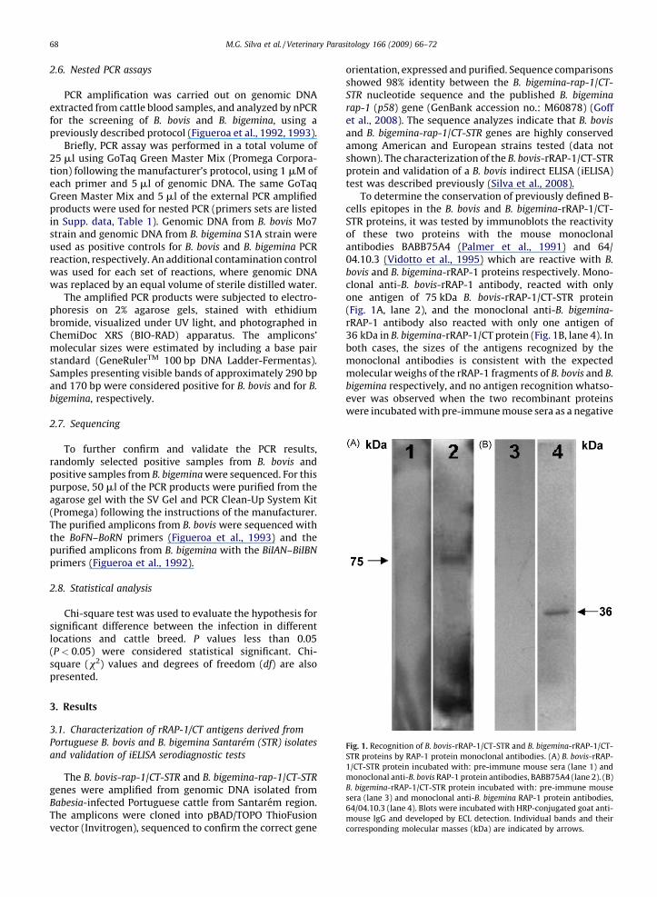
Fig. 1. Recognition of B. bovis-rRAP-1/CT-STR and B. bigemina-rRAP-1/CT-
STR proteins by RAP-1 protein monoclonal antibodies. (A) B. bovis-rRAP-
1/CT-STR protein incubated with: pre-immune mouse sera (lane 1) and
monoclonal anti-B. bovis RAP-1 protein antibodies, BABB75A4 (lane 2). (B)
B. bigemina-rRAP-1/CT-STR protein incubated with: pre-immune mouse
sera (lane 3) and monoclonal anti-B. bigemina RAP-1 protein antibodies,
64/04.10.3 (lane 4). Blots were incubated with HRP-conjugated goat anti-
mouse IgG and developed by ECL detection. Individual bands and their
corresponding molecular masses (kDa) are indicated by arrows.
M.G. Silva et al. / Veterinary Parasitology 166 (2009) 66–7268
2.6. Nested PCR assays
PCR amplification was carried out on genomic DNAextracted from cattle blood samples, and analyzed by nPCRfor the screening of B. bovis and B. bigemina, using apreviously described protocol (Figueroa et al., 1992, 1993).
Briefly, PCR assay was performed in a total volume of25 ml using GoTaq Green Master Mix (Promega Corpora-tion) following the manufacturer’s protocol, using 1 mM ofeach primer and 5 ml of genomic DNA. The same GoTaqGreen Master Mix and 5 ml of the external PCR amplifiedproducts were used for nested PCR (primers sets are listedin Supp. data, Table 1). Genomic DNA from B. bovis Mo7strain and genomic DNA from B. bigemina S1A strain wereused as positive controls for B. bovis and B. bigemina PCRreaction, respectively. An additional contamination controlwas used for each set of reactions, where genomic DNAwas replaced by an equal volume of sterile distilled water.
The amplified PCR products were subjected to electro-phoresis on 2% agarose gels, stained with ethidiumbromide, visualized under UV light, and photographed inChemiDoc XRS (BIO-RAD) apparatus. The amplicons’molecular sizes were estimated by including a base pairstandard (GeneRulerTM 100 bp DNA Ladder-Fermentas).Samples presenting visible bands of approximately 290 bpand 170 bp were considered positive for B. bovis and for B.
bigemina, respectively.
2.7. Sequencing
To further confirm and validate the PCR results,randomly selected positive samples from B. bovis andpositive samples from B. bigemina were sequenced. For thispurpose, 50 ml of the PCR products were purified from theagarose gel with the SV Gel and PCR Clean-Up System Kit(Promega) following the instructions of the manufacturer.The purified amplicons from B. bovis were sequenced withthe BoFN–BoRN primers (Figueroa et al., 1993) and thepurified amplicons from B. bigemina with the BiIAN–BiIBN
primers (Figueroa et al., 1992).
2.8. Statistical analysis
Chi-square test was used to evaluate the hypothesis forsignificant difference between the infection in differentlocations and cattle breed. P values less than 0.05(P < 0.05) were considered statistical significant. Chi-square (x2) values and degrees of freedom (df) are alsopresented.
3. Results
3.1. Characterization of rRAP-1/CT antigens derived from
Portuguese B. bovis and B. bigemina Santarem (STR) isolates
and validation of iELISA serodiagnostic tests
The B. bovis-rap-1/CT-STR and B. bigemina-rap-1/CT-STR
genes were amplified from genomic DNA isolated fromBabesia-infected Portuguese cattle from Santarem region.The amplicons were cloned into pBAD/TOPO ThioFusionvector (Invitrogen), sequenced to confirm the correct gene
orientation, expressed and purified. Sequence comparisonsshowed 98% identity between the B. bigemina-rap-1/CT-STR nucleotide sequence and the published B. bigemina
rap-1 (p58) gene (GenBank accession no.: M60878) (Goffet al., 2008). The sequence analyzes indicate that B. bovis
and B. bigemina-rap-1/CT-STR genes are highly conservedamong American and European strains tested (data notshown). The characterization of the B. bovis-rRAP-1/CT-STRprotein and validation of a B. bovis indirect ELISA (iELISA)test was described previously (Silva et al., 2008).
To determine the conservation of previously defined B-cells epitopes in the B. bovis and B. bigemina-rRAP-1/CT-STR proteins, it was tested by immunoblots the reactivityof these two proteins with the mouse monoclonalantibodies BABB75A4 (Palmer et al., 1991) and 64/04.10.3 (Vidotto et al., 1995) which are reactive with B.
bovis and B. bigemina-rRAP-1 proteins respectively. Mono-clonal anti-B. bovis-rRAP-1 antibody, reacted with onlyone antigen of 75 kDa B. bovis-rRAP-1/CT-STR protein(Fig. 1A, lane 2), and the monoclonal anti-B. bigemina-rRAP-1 antibody also reacted with only one antigen of36 kDa in B. bigemina-rRAP-1/CT protein (Fig. 1B, lane 4). Inboth cases, the sizes of the antigens recognized by themonoclonal antibodies is consistent with the expectedmolecular weighs of the rRAP-1 fragments of B. bovis and B.
bigemina respectively, and no antigen recognition whatso-ever was observed when the two recombinant proteinswere incubated with pre-immune mouse sera as a negative
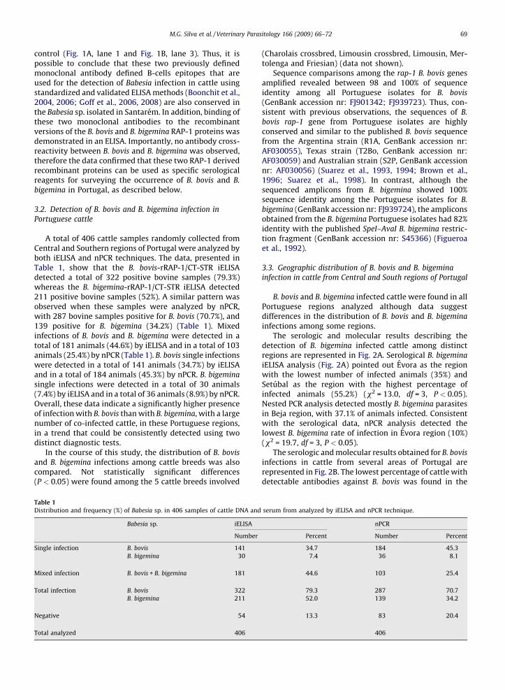
M.G. Silva et al. / Veterinary Parasitology 166 (2009) 66–72 69
control (Fig. 1A, lane 1 and Fig. 1B, lane 3). Thus, it ispossible to conclude that these two previously definedmonoclonal antibody defined B-cells epitopes that areused for the detection of Babesia infection in cattle usingstandardized and validated ELISA methods (Boonchit et al.,2004, 2006; Goff et al., 2006, 2008) are also conserved inthe Babesia sp. isolated in Santarem. In addition, binding ofthese two monoclonal antibodies to the recombinantversions of the B. bovis and B. bigemina RAP-1 proteins wasdemonstrated in an ELISA. Importantly, no antibody cross-reactivity between B. bovis and B. bigemina was observed,therefore the data confirmed that these two RAP-1 derivedrecombinant proteins can be used as specific serologicalreagents for surveying the occurrence of B. bovis and B.
bigemina in Portugal, as described below.
3.2. Detection of B. bovis and B. bigemina infection in
Portuguese cattle
A total of 406 cattle samples randomly collected fromCentral and Southern regions of Portugal were analyzed byboth iELISA and nPCR techniques. The data, presented inTable 1, show that the B. bovis-rRAP-1/CT-STR iELISAdetected a total of 322 positive bovine samples (79.3%)whereas the B. bigemina-rRAP-1/CT-STR iELISA detected211 positive bovine samples (52%). A similar pattern wasobserved when these samples were analyzed by nPCR,with 287 bovine samples positive for B. bovis (70.7%), and139 positive for B. bigemina (34.2%) (Table 1). Mixedinfections of B. bovis and B. bigemina were detected in atotal of 181 animals (44.6%) by iELISA and in a total of 103animals (25.4%) by nPCR (Table 1). B. bovis single infectionswere detected in a total of 141 animals (34.7%) by iELISAand in a total of 184 animals (45.3%) by nPCR. B. bigemina
single infections were detected in a total of 30 animals(7.4%) by iELISA and in a total of 36 animals (8.9%) by nPCR.Overall, these data indicate a significantly higher presenceof infection with B. bovis than with B. bigemina, with a largenumber of co-infected cattle, in these Portuguese regions,in a trend that could be consistently detected using twodistinct diagnostic tests.
In the course of this study, the distribution of B. bovis
and B. bigemina infections among cattle breeds was alsocompared. Not statistically significant differences(P < 0.05) were found among the 5 cattle breeds involved
Table 1
Distribution and frequency (%) of Babesia sp. in 406 samples of cattle DNA and
Babesia sp. iELISA
Number
Single infection B. bovis 141
B. bigemina 30
Mixed infection B. bovis + B. bigemina 181
Total infection B. bovis 322
B. bigemina 211
Negative 54
Total analyzed 406
(Charolais crossbred, Limousin crossbred, Limousin, Mer-tolenga and Friesian) (data not shown).
Sequence comparisons among the rap-1 B. bovis genesamplified revealed between 98 and 100% of sequenceidentity among all Portuguese isolates for B. bovis
(GenBank accession nr: FJ901342; FJ939723). Thus, con-sistent with previous observations, the sequences of B.
bovis rap-1 gene from Portuguese isolates are highlyconserved and similar to the published B. bovis sequencefrom the Argentina strain (R1A, GenBank accession nr:AF030055), Texas strain (T2Bo, GenBank accession nr:AF030059) and Australian strain (S2P, GenBank accessionnr: AF030056) (Suarez et al., 1993, 1994; Brown et al.,1996; Suarez et al., 1998). In contrast, although thesequenced amplicons from B. bigemina showed 100%sequence identity among the Portuguese isolates for B.
bigemina (GenBank accession nr: FJ939724), the ampliconsobtained from the B. bigemina Portuguese isolates had 82%identity with the published SpeI–AvaI B. bigemina restric-tion fragment (GenBank accession nr: S45366) (Figueroaet al., 1992).
3.3. Geographic distribution of B. bovis and B. bigemina
infection in cattle from Central and South regions of Portugal
B. bovis and B. bigemina infected cattle were found in allPortuguese regions analyzed although data suggestdifferences in the distribution of B. bovis and B. bigemina
infections among some regions.The serologic and molecular results describing the
detection of B. bigemina infected cattle among distinctregions are represented in Fig. 2A. Serological B. bigemina
iELISA analysis (Fig. 2A) pointed out Evora as the regionwith the lowest number of infected animals (35%) andSetubal as the region with the highest percentage ofinfected animals (55.2%) (x2 = 13.0, df = 3, P < 0.05).Nested PCR analysis detected mostly B. bigemina parasitesin Beja region, with 37.1% of animals infected. Consistentwith the serological data, nPCR analysis detected thelowest B. bigemina rate of infection in Evora region (10%)(x2 = 19.7, df = 3, P < 0.05).
The serologic and molecular results obtained for B. bovis
infections in cattle from several areas of Portugal arerepresented in Fig. 2B. The lowest percentage of cattle withdetectable antibodies against B. bovis was found in the
serum from analyzed by iELISA and nPCR technique.
nPCR
Percent Number Percent
34.7 184 45.3
7.4 36 8.1
44.6 103 25.4
79.3 287 70.7
52.0 139 34.2
13.3 83 20.4
406
Fig. 2. Analysis of Babesia sp. infection by iELISA and nPCR techniques
among cattle samples from the four district-based studied: Santarem
(n = 29), Setubal (n = 163), Evora (n = 20) and Beja (n = 194). (A)
Distribution and frequency (%) of infected B. bigemina cattle. (B)
Distribution and frequency (%) of infected B. bovis cattle.
M.G. Silva et al. / Veterinary Parasitology 166 (2009) 66–7270
Setubal region (77.3%), whereas the highest percentagewas detected in the Evora region (85%) (Fig. 2B). However,the serological differences found for the geographicaldistribution of B. bovis infection in Portugal were notsignificant (x2 = 1.05, df = 3, P > 0.05). Nested PCR analysisrevealed that B. bovis parasites are present at the highestpercentage in cattle from the Santarem region (93.1%) andwith the lowest percentage of infection in Setubal region(66.3%), with evidence of statistical difference (x2 = 9.45,df = 3, P < 0.05) (Fig. 2B).
4. Discussion
In this study, serological and molecular tools were usedfor the first time to detect B. bovis and B. bigemina in cattleblood from the previously uncharacterized Central andSouthern regions of Portugal. Recombinant carboxyl-terminal-RAP-1 proteins (B. bovis-rRAP-1/CT-STR proteinand B. bigemina-rRAP-1/CT-STR protein) were used todetect antibodies against B. bovis and B. bigemina in cattlesera samples by iELISA, and nPCR was used to amplify theconserved rap-1 B. bovis gene and SpeI–AvaI B. bigemina
fragment in genomic DNA (Figueroa et al., 1992, 1993,1994). These two methods have been previously reportedto have high specificity and sensitivity for their use inepidemiology surveys (Figueroa et al., 1993; Goff et al.,
2006, 2008). The results of this study indicate a highpresence of infection of cattle by B. bovis and B. bigemina
parasites in the surveyed areas and suggest that properpreventive measures will be required for the control of thiscostly disease.
Serologic assays detected a higher number of infectedBabesia sp. cattle than nPCR. These differences areconsistent with previous findings that B. bovis and B.
bigemina parasites are difficult to detect using PCRtechniques due to the small number of parasites thatoccur in peripheral blood during chronic infections (Pipanoet al., 2002); another possible reason for the high numberof cattle with detectable Babesia sp. antibodies could bethat these antibodies remain in circulation for a longperiod of time after acute infection. In such cases, it ispossible, either that the parasites are cleared fromcirculation, or that the parasite concentration in the blooddrops below the limit of detection of the nPCR (Tjornehojet al., 1996; Pipano et al., 2002). Before this research wasconducted, no reliable diagnostic studies were pursued bythe incumbent farmers despite bovines showing signsconsistent with Babesia clinical disease. Therefore, therewere no previous reports on B. bovis and B. bigemina
clinical manifestations in the regions of Portugal analyzedin this study, and the history of occurrence of clinical casesin these farms remains unknown.
Nucleotide sequence comparisons of several rap-1 fromPortuguese B. bovis field isolates showed a higher level ofsequence conservation when compared to the Argentina,Texas and Australia B. bovis strains. The SpeI–AvaI B.
bigemina nucleotide sequences obtained from the Portu-guese isolates revealed some level of polymorphism whencompared to the previously published strain. In contrast,the nucleotide sequence of this fragment showed highconservation within B. bigemina isolates from North andCentral America and the Caribbean Region (Figueroa et al.,1992). No previous information was available on theconservation of the SpeI–AvaI fragment in B. bigemina
isolates from Europe, suggesting that more Europeanisolates need to be collected and sequenced to further testthe level of nucleotide conservation of this SpeI–AvaI B.
bigemina DNA fragment.The antibody prevalence has been used as an indicator
of endemic stability or instability situations (Mahoney andGoodger, 1972; Carrique et al., 2000). Overall, anti-B. bovis
antibodies were detected in over 75% of the cattleanalyzed, suggesting that all the regions analyzed in thisstudy might be in an endemically stable situation. Thus, itis possible that cattle may have developed immunity to theparasite, decreasing the occurrence of detectable clinicalbabesiosis cases (Homer et al., 2000). Conversely, it is alsopossible that an endemically unstable situation occurs forB. bigemina where less than 75% of the cattle areseropositive, in a scenario in which cattle may be at asignificantly higher risk of clinical disease (L’Hostis andSeegers, 2002).
5. Conclusions
The present study provides the first survey of B. bovis
and B. bigemina in cattle from Central and Southern regions
M.G. Silva et al. / Veterinary Parasitology 166 (2009) 66–72 71
of Portugal. Evidence of B. bovis and B. bigemina infectioncould have an important impact in weight loss, milkproduction and possible abortion (Wagner et al., 2002).
The outcomes of this preliminary study suggests theneed for the implementation of a more complete studyassessing the epidemiological situation of bovine babe-siosis in Portugal, including testing animals from allPortuguese regions and an evaluation of the relationshipsbetween the disease and animal ages, breeds, environ-mental conditions, and tick vector prevalence. These datawill be of importance in order to carry out a comprehensiveand rationally designed plan to control and prevent thesecurrently neglected diseases, at a regional level.
Conflict of interest statement
None of the authors of this paper has a financial orpersonal relationship with other people or organisationsthat could inappropriately influence or bias the content ofthe paper.
Acknowledgements
This work was financed by the European Project(MEDLABAB) INCO-DEV - 2005-2007 Proj. No: 003691.Marta Silva is grateful for a scholarship from Fundacao paraa Ciencia e Tecnologia (Portugal). Thanks to Paul Lacy forall his help, Debbie Alperin and Carl Johnson for their helpin protein expression, purification and ELISA. To SilvinaWilkowsky who kindly provided us a purified sample of B.
bigemina DNA. An acknowledgement to Prof. Isabel Netofor her help in statistic analyzes, and also the authors wantto acknowledge Isabel Fazendeiro and all veterinarysupport: ADS Beja, ADS Litoral Alentejano and Dr. Carsten.
Appendix A. Supplementary data
Supplementary data associated with this article can be
found, in the online version, at doi:10.1016/j.vetpar.2009.
07.031.
References
Birkenheuer, A.J., Levy, M.G., Breitschwerdt, E.B., 2003. Development andevaluation of a seminested PCR for detection and differentiation ofBabesia gibsoni and B. canis DNA in canine blood samples. J. Clin.Microbiol. 41, 4172–4177.
Boonchit, S., Alhassan, A., Chan, B., Xuan, X., Yokoyama, N., Ooshiro, M.,Goff, W.L., Waghela, S.D., Wagner, G., Igarashi, I., 2006. Expression ofC-terminal truncated and full-length Babesia bigemina rhoptry-asso-ciated protein 1 and their potential use in enzyme-linked immuno-sorbent assay. Vet. Parasitol. 137, 28–35.
Boonchit, S., Xuan, X., Yokoyama, N., Goff, W.L., Waghela, S.D., Wagner, G.,Igarashi, I., 2004. Improved enzyme-linked immunosorbent assayusing C-terminal truncated recombinant antigens of Babesia bovisrhoptry-associated protein-1 for detection of specific antibodies. J.Clin. Microbiol. 42, 1601–1604.
Brown, W.C., McElwain, T.F., Ruef, B.J., Suarez, C.E., Shkap, V., Chitko-McKown, C.G., Tuo, W., Rice-Ficht, A.C., Palmer, G.H., 1996. Babesiabovis rhoptry-associated protein 1 is immunodominant for T helpercells of immune cattle and contains T-cell epitopes conserved amonggeographically distant B. bovis strains. Infect. Immun. 64, 3341–3350.
Caeiro, V., 1999. General review of tick species present in Portugal.Parassitologia 41, 11–15.
Calder, J.A., Reddy, G.R., Chieves, L., Courtney, C.H., Littell, R., Livengood,J.R., Norval, R.A., Smith, C., Dame, J.B., 1996. Monitoring Babesia bovis
infections in cattle by using PCR-based tests. J. Clin. Microbiol. 34,2748–2755.
Carrique, J.J., Morales, G.J., Edelsten, M., 2000. Endemic instability forbabesiosis and anaplasmosis in cattle in the Bolivian Chaco. Vet. J.160, 162–164.
Criado-Fornelio, A., Martinez-Marcos, A., Buling-Sarana, A., Barba-Carre-tero, J.C., 2003. Molecular studies on Babesia, Theileria and Hepatozoonin southern Europe. Part II. Phylogenetic analysis and evolutionaryhistory. Vet. Parasitol. 114, 173–194.
Dorigo-Zetsma, J.W., Zaat, S.A., Wertheim-van Dillen, P.M., Spanjaard, L.,Rijntjes, J., van Waveren, G., Jensen, J.S., Angulo, A.F., Dankert, J., 1999.Comparison of PCR, culture, and serological tests for diagnosis ofMycoplasma pneumoniae respiratory tract infection in children. J. Clin.Microbiol. 37, 14–17.
Estrada-Pena, A., Bouattour, A., Camicas, J.-L., Walker, A.R., 2004. Ticks ofDomestic Animals in the Mediterranean Region: A Guide to Identi-fication of Species. University of Zaragoza, Zaragoza.
Estrada-Pena, A., Santos-Silva, M.M., 2005. The distribution of ticks ofdomestic livestock in Portugal. Exp. Appl. Acarol. 36, 233–246.
Figueroa, J.V., Alvarez, J.A., Ramos, J.A., Rojas, E.E., Santiago, C., Mosqueda,J.J., Vega, C.A., Buening, G.M., 1998. Bovine babesiosis and anaplas-mosis follow-up on cattle relocated in an endemic area for hemopar-asitic diseases. Ann. N.Y. Acad. Sci. 849, 1–10.
Figueroa, J.V., Alvarez, J.A., Ramos, J.A., Vega, C.A., Buening, G.M., 1993. Useof multiplex polymerase chain reaction-based assay to conduct epi-demiological studies on bovine hemoparasites in Mexico. Revued’elevage et de Medecine Veterinaire des Pays Tropicaux 46, 71–75.
Figueroa, J.V., Chieves, L.P., Johnson, G.S., Buening, G.M., 1992. Detectionof Babesia bigemina-infected carriers by polymerase chain reactionamplification. J. Clin. Microbiol. 30, 2576–2582.
Figueroa, J.V., Chieves, L.P., Johnson, G.S., Goff, W.L., Buening, G.M., 1994.Polymerase chain reaction-based diagnostic assay to detect cattlechronically infected with Babesia bovis. Revista Latinoamericana deMicrobiologia 36, 47–55.
Goff, W.L., Davis, W.C., Palmer, G.H., McElwain, T.F., Johnson, W.C., Bailey,J.F., McGuire, T.C., 1988. Identification of Babesia bovis merozoitesurface antigens by using immune bovine sera and monoclonalantibodies. Infect. Immun. 56, 2363–2368.
Goff, W.L., Johnson, W.C., Molloy, J.B., Jorgensen, W.K., Waldron, S.J.,Figueroa, J.V., Matthee, O., Adams, D.S., McGuire, T.C., Pino, I., Mos-queda, J., Palmer, G.H., Suarez, C.E., Knowles, D.P., McElwain, T.F.,2008. Validation of a competitive enzyme-linked immunosorbentassay for detection of Babesia bigemina antibodies in cattle. Clin.Vaccine Immunol. 15, 1316–1321.
Goff, W.L., McElwain, T.F., Suarez, C.E., Johnson, W.C., Brown, W.C.,Norimine, J., Knowles, D.P., 2003. Competitive enzyme-linked immu-nosorbent assay based on a rhoptry-associated protein 1 epitopespecifically identifies Babesia bovis-infected cattle. Clin. Diagn. Lab.Immunol. 10, 38–43.
Goff, W.L., Molloy, J.B., Johnson, W.C., Suarez, C.E., Pino, I., Rhalem, A.,Sahibi, H., Ceci, L., Carelli, G., Adams, D.S., McGuire, T.C., Knowles, D.P.,McElwain, T.F., 2006. Validation of a competitive enzyme-linkedimmunosorbent assay for detection of antibodies against Babesiabovis. Clin. Vaccine Immunol. 13, 1212–1216.
Heim, A., Passos, L.M., Ribeiro, M.F., Costa-Junior, L.M., Bastos, C.V., Cabral,D.D., Hirzmann, J., Pfister, K., 2007. Detection and molecular char-acterization of Babesia caballi and Theileria equi isolates from endemicareas of Brazil. Parasitol. Res. 102, 63–68.
Homer, M.J., Aguilar-Delfin, I., Telford 3rd, S.R., Krause, P.J., Persing, D.H.,2000. Babesiosis. Clin. Microbiol. Rev. 13, 451–469.
L’Hostis, M., Seegers, H., 2002. Tick-borne parasitic diseases in cattle:current knowledge and prospective risk analysis related to the ongoingevolution in French cattle farming systems. Vet. Res. 33, 599–611.
Mahoney, D.F., Goodger, B.V., 1972. Babesia argentina: immunogenicity ofplasma from infected animals. Exp. Parasitol. 32, 71–85.
McCrosker, 1981. The Global Importance of Babesiosis. Academic Press,Inc., New York, pp. 1–24.
Miyama, T., Sakata, Y., Shimada, Y., Ogino, S., Watanabe, M., Itamoto, K.,Okuda, M., Verdida, R.A., Xuan, X., Nagasawa, H., Inokuma, H., 2005.Epidemiological survey of Babesia gibsoni infection in dogs in easternJapan. J. Vet. Med. Sci. 67, 467–471.
Oyamada, M., Davoust, B., Boni, M., Dereure, J., Bucheton, B., Hammad, A.,Itamoto, K., Okuda, M., Inokuma, H., 2005. Detection of Babesia canisrossi, B. canis vogeli, and Hepatozoon canis in dogs in a village of easternSudan by using a screening PCR and sequencing methodologies. Clin.Diagn. Lab. Immunol. 12, 1343–1346.
Palmer, G.H., McElwain, T.F., Perryman, L.E., Davis, W.C., Reduker, D.R.,Jasmer, D.P., Shkap, V., Pipano, E., Goff, W.L., McGuire, T.C., 1991.Strain variation of Babesia bovis merozoite surface-exposed epitopes.Infect. Immun. 59, 3340–3342.

M.G. Silva et al. / Veterinary Parasitology 166 (2009) 66–7272
Pipano, E., Shkap, V., Kriegel, Y., Leibovitz, B., Savitsky, I., Fish, L., 2002.Babesia bovis and B. bigemina: persistence of infection in friesian cowsfollowing vaccination with live antibabesial vaccines. Vet. J. 164, 64–68.
Silva, M.G., Helali, S., Esseghaier, C., Suarez, C.E., Oliva, A., Abdelghanib, A.,2008. An impedance spectroscopy method for the detection andevaluation of Babesia bovis antibodies in cattle. Sensors Actuat. B:Chem. 135, 206–213.
Suarez, C.E., McElwain, T.F., Echaide, I., Torioni de Echaide, S., Palmer, G.H.,1994. Interstrain conservation of babesial RAP-1 surface-exposed B-cell epitopes despite rap-1 genomic polymorphism. Infect. Immun.62, 3576–3579.
Suarez, C.E., Palmer, G.H., Hines, S.A., McElwain, T.F., 1993. ImmunogenicB-cell epitopes of Babesia bovis rhoptry-associated protein 1 are
distinct from sequences conserved between species. Infect. Immun.61, 3511–3517.
Suarez, C.E., Palmer, G.H., Hotzel, I., McElwain, T.F., 1998. Structure,sequence, and transcriptional analysis of the Babesia bovis rap-1multigene locus. Mol. Biochem. Parasitol. 93, 215–224.
Tjornehoj, K., Lawrence, J.A., Whiteland, A.P., Kafuwa, P.T., 1996. Fieldobservations on the duration of immunity in cattle after vaccinationagainst Anaplasma and Babesia species. Onderstepoort J. Vet. Res. 63,1–5.
Vidotto, O., McElwain, T.F., Machado, R.Z., Perryman, L.E., Suarez, C.E.,Palmer, G.H., 1995. Babesia bigemina: identification of B cell epitopesassociated with parasitized erythrocytes. Exp. Parasitol. 81, 491–500.
Wagner, G.G., Holman, P., Waghela, S., 2002. Babesiosis and heartwater:threats without boundaries. Vet. Clin. North Am. 18, 417–430.