Babesia lengau associated with cerebral and haemolytic babesiosis in two domestic cats
6
SHORT REPORT Open Access Babesia lengau associated with cerebral and haemolytic babesiosis in two domestic cats Anna-Mari Bosman 1 , Marinda C Oosthuizen 1 , Estelle H Venter 1 , Johan CA Steyl 2 , Tertius A Gous 3 and Barend L Penzhorn 1* Abstract Background: Although reported sporadically from various countries, feline babesiosis appears to be a significant clinical entity only in South Africa, where Babesia felis is usually incriminated as the causative agent. Babesia lengau, recently described from asymptomatic cheetahs, has now possibly been incriminated as the causative agent in two severe clinical cases in domestic cats. Findings: Both cats were euthanised in extremis. While typical feline babesiosis in South Africa is an afebrile disease with a chronic manifestation, there was acute onset of severe clinical signs in both cats and their body temperatures were above the normal range when they were presented for treatment. Haemolytic anaemia was confirmed in one case. To our knowledge, this is the first report of cerebral babesiosis in cats. On reverse line blot 18S rDNA PCR products obtained from both cats showed positive hybridization profiles with the B. lengau species-specific probe. The two partial parasite 18S rRNA gene sequences obtained, showed high sequence similarity (99.9%) to B. lengau. In a representative tree constructed by the neighbor-joining method using the two-parameter model of Kimura the two obtained partial 18S rDNA sequences and that of B. lengau formed a monophyletic group with B. conradae and sequences previously isolated from humans and wildlife in the western USA. Conclusion: All clinical cases of feline babesiosis in South Africa are not necessarily caused by B. felis. Other piroplasms, e.g. B. lengau, may be incriminated in clinical cases, especially those occurring outside the known endemic area. Keywords: Babesia lengau, Cerebral babesiosis, Feline babesiosis, Haemolytic anaemia Findings Babesiosis of domestic cats has been reported sporadically from various countries, including France [1], Germany [2], India [3], Israel [4], Poland [5], Portugal [6], Thailand [7] and Zimbabwe [8], but appears to be a significant clini- cal entity only in South Africa where it occurs primarily along the eastern and southern seaboard (KwaZulu-Natal, Eastern and Western Cape Provinces), as well as in iso- lated foci along the eastern escarpment of Mpumalanga and Limpopo Provinces [9-11] (Figure 1). This would sug- gest that the vector(s) only occur(s) in these areas, but the vector(s) remain(s) unknown. Clinical cases occurring in- land, e.g. in Gauteng Province, usually concern pets taken along when the owners visited the coast [9,12]. Feline babesiosis has been well documented in South Africa. Apart from case reports [13], there have been detailed studies on signalment, clinical manifestation and patho- logy [9,14,15] as well as treatment [12,16]. Lethargy, ano- rexia and anaemia generally occur, while icterus is only occasionally seen [14]. Elevated body temperature is not a feature of this disease [14]. In clinical cases, parasitaemia is usually high and may exceed 50% [14]. Babesia felis, the piroplasm usually incriminated as causing feline babesiosis in South Africa, was initially de- scribed from a Sudanese wild cat (Felis ocreata, syn. F. sylvestris) [17]. Fourteen domestic cats inoculated with blood from the wild cat became persistently infected, with parasitaemias < 1%, but did not develop any clinical signs [17]. When feline babesiosis was first described in South Africa a few years later, the parasite was considered to be * Correspondence: [email protected] 1 Department of Veterinary Tropical Diseases, Faculty of Veterinary Science, University of Pretoria, Private Bag X04, Onderstepoort 0110, South Africa Full list of author information is available at the end of the article © 2013 Bosman et al.; licensee BioMed Central Ltd. This is an Open Access article distributed under the terms of the Creative Commons Attribution License (http://creativecommons.org/licenses/by/2.0), which permits unrestricted use, distribution, and reproduction in any medium, provided the original work is properly cited. Bosman et al. Parasites & Vectors 2013, 6:128 http://www.parasitesandvectors.com/content/6/1/128
Transcript of Babesia lengau associated with cerebral and haemolytic babesiosis in two domestic cats
Bosman et al. Parasites & Vectors 2013, 6:128http://www.parasitesandvectors.com/content/6/1/128
SHORT REPORT Open Access
Babesia lengau associated with cerebral andhaemolytic babesiosis in two domestic catsAnna-Mari Bosman1, Marinda C Oosthuizen1, Estelle H Venter1, Johan CA Steyl2, Tertius A Gous3
and Barend L Penzhorn1*
Abstract
Background: Although reported sporadically from various countries, feline babesiosis appears to be a significantclinical entity only in South Africa, where Babesia felis is usually incriminated as the causative agent. Babesia lengau,recently described from asymptomatic cheetahs, has now possibly been incriminated as the causative agent in twosevere clinical cases in domestic cats.
Findings: Both cats were euthanised in extremis. While typical feline babesiosis in South Africa is an afebriledisease with a chronic manifestation, there was acute onset of severe clinical signs in both cats and their bodytemperatures were above the normal range when they were presented for treatment. Haemolytic anaemia wasconfirmed in one case. To our knowledge, this is the first report of cerebral babesiosis in cats.On reverse line blot 18S rDNA PCR products obtained from both cats showed positive hybridization profiles withthe B. lengau species-specific probe. The two partial parasite 18S rRNA gene sequences obtained, showed highsequence similarity (99.9%) to B. lengau. In a representative tree constructed by the neighbor-joining methodusing the two-parameter model of Kimura the two obtained partial 18S rDNA sequences and that of B. lengauformed a monophyletic group with B. conradae and sequences previously isolated from humans and wildlife inthe western USA.
Conclusion: All clinical cases of feline babesiosis in South Africa are not necessarily caused by B. felis. Otherpiroplasms, e.g. B. lengau, may be incriminated in clinical cases, especially those occurring outside the knownendemic area.
Keywords: Babesia lengau, Cerebral babesiosis, Feline babesiosis, Haemolytic anaemia
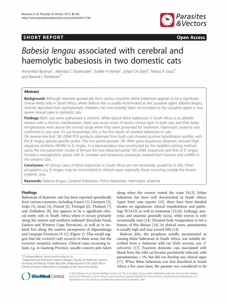
FindingsBabesiosis of domestic cats has been reported sporadicallyfrom various countries, including France [1], Germany [2],India [3], Israel [4], Poland [5], Portugal [6], Thailand [7]and Zimbabwe [8], but appears to be a significant clini-cal entity only in South Africa where it occurs primarilyalong the eastern and southern seaboard (KwaZulu-Natal,Eastern and Western Cape Provinces), as well as in iso-lated foci along the eastern escarpment of Mpumalangaand Limpopo Provinces [9-11] (Figure 1). This would sug-gest that the vector(s) only occur(s) in these areas, but thevector(s) remain(s) unknown. Clinical cases occurring in-land, e.g. in Gauteng Province, usually concern pets taken
* Correspondence: [email protected] of Veterinary Tropical Diseases, Faculty of Veterinary Science,University of Pretoria, Private Bag X04, Onderstepoort 0110, South AfricaFull list of author information is available at the end of the article
© 2013 Bosman et al.; licensee BioMed CentraCommons Attribution License (http://creativecreproduction in any medium, provided the or
along when the owners visited the coast [9,12]. Felinebabesiosis has been well documented in South Africa.Apart from case reports [13], there have been detailedstudies on signalment, clinical manifestation and patho-logy [9,14,15] as well as treatment [12,16]. Lethargy, ano-rexia and anaemia generally occur, while icterus is onlyoccasionally seen [14]. Elevated body temperature is not afeature of this disease [14]. In clinical cases, parasitaemiais usually high and may exceed 50% [14].Babesia felis, the piroplasm usually incriminated as
causing feline babesiosis in South Africa, was initially de-scribed from a Sudanese wild cat (Felis ocreata, syn. F.sylvestris) [17]. Fourteen domestic cats inoculated withblood from the wild cat became persistently infected, withparasitaemias < 1%, but did not develop any clinical signs[17]. When feline babesiosis was first described in SouthAfrica a few years later, the parasite was considered to be
l Ltd. This is an Open Access article distributed under the terms of the Creativeommons.org/licenses/by/2.0), which permits unrestricted use, distribution, andiginal work is properly cited.
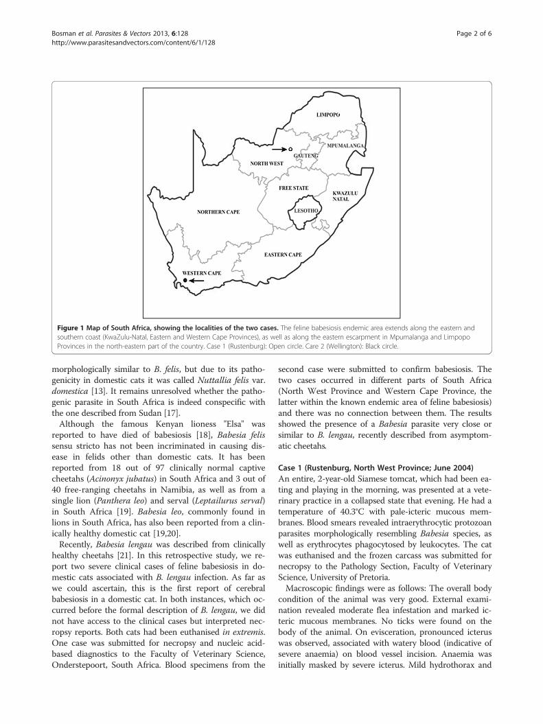
Figure 1 Map of South Africa, showing the localities of the two cases. The feline babesiosis endemic area extends along the eastern andsouthern coast (KwaZulu-Natal, Eastern and Western Cape Provinces), as well as along the eastern escarpment in Mpumalanga and LimpopoProvinces in the north-eastern part of the country. Case 1 (Rustenburg): Open circle. Care 2 (Wellington): Black circle.
Bosman et al. Parasites & Vectors 2013, 6:128 Page 2 of 6http://www.parasitesandvectors.com/content/6/1/128
morphologically similar to B. felis, but due to its patho-genicity in domestic cats it was called Nuttallia felis var.domestica [13]. It remains unresolved whether the patho-genic parasite in South Africa is indeed conspecific withthe one described from Sudan [17].Although the famous Kenyan lioness "Elsa" was
reported to have died of babesiosis [18], Babesia felissensu stricto has not been incriminated in causing dis-ease in felids other than domestic cats. It has beenreported from 18 out of 97 clinically normal captivecheetahs (Acinonyx jubatus) in South Africa and 3 out of40 free-ranging cheetahs in Namibia, as well as from asingle lion (Panthera leo) and serval (Leptailurus serval)in South Africa [19]. Babesia leo, commonly found inlions in South Africa, has also been reported from a clin-ically healthy domestic cat [19,20].Recently, Babesia lengau was described from clinically
healthy cheetahs [21]. In this retrospective study, we re-port two severe clinical cases of feline babesiosis in do-mestic cats associated with B. lengau infection. As far aswe could ascertain, this is the first report of cerebralbabesiosis in a domestic cat. In both instances, which oc-curred before the formal description of B. lengau, we didnot have access to the clinical cases but interpreted nec-ropsy reports. Both cats had been euthanised in extremis.One case was submitted for necropsy and nucleic acid-based diagnostics to the Faculty of Veterinary Science,Onderstepoort, South Africa. Blood specimens from the
second case were submitted to confirm babesiosis. Thetwo cases occurred in different parts of South Africa(North West Province and Western Cape Province, thelatter within the known endemic area of feline babesiosis)and there was no connection between them. The resultsshowed the presence of a Babesia parasite very close orsimilar to B. lengau, recently described from asymptom-atic cheetahs.
Case 1 (Rustenburg, North West Province; June 2004)An entire, 2-year-old Siamese tomcat, which had been ea-ting and playing in the morning, was presented at a vete-rinary practice in a collapsed state that evening. He had atemperature of 40.3°C with pale-icteric mucous mem-branes. Blood smears revealed intraerythrocytic protozoanparasites morphologically resembling Babesia species, aswell as erythrocytes phagocytosed by leukocytes. The catwas euthanised and the frozen carcass was submitted fornecropsy to the Pathology Section, Faculty of VeterinaryScience, University of Pretoria.Macroscopic findings were as follows: The overall body
condition of the animal was very good. External exami-nation revealed moderate flea infestation and marked ic-teric mucous membranes. No ticks were found on thebody of the animal. On evisceration, pronounced icteruswas observed, associated with watery blood (indicative ofsevere anaemia) on blood vessel incision. Anaemia wasinitially masked by severe icterus. Mild hydrothorax and
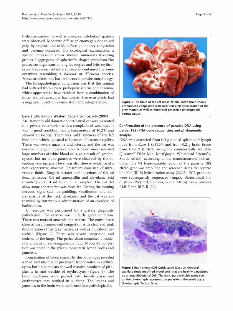
Figure 2 The brain of the cat (Case 2): The entire brain shows
Bosman et al. Parasites & Vectors 2013, 6:128 Page 3 of 6http://www.parasitesandvectors.com/content/6/1/128
hydropericardium as well as acute, centrilobular hepatosiswere observed. Moderate diffuse splenomegaly due to redpulp hyperplasia and mild, diffuse pulmonary congestionand oedema occurred. On cytological examination, asplenic impression smear showed numerous free-lyinggroups / aggregates of spherically shaped piroplasm-likeprotozoan organisms among leukocytes and lytic erythro-cytes. Occasional intact erythrocytes contained the sameorganism resembling a Babesia or Theileria species.Freeze artefacts may have influenced parasite morphology.The histopathological conclusion was that this animal
had suffered from severe prehepatic icterus and anaemia,which appeared to have resulted from a combination ofintra- and extravascular haemolysis. Freeze artefacts hada negative impact on examination and interpretation.
pronounced congestion with clear red-pink discoloration of thegrey matter, as well as multifocal petechiae (Photograph:Tertius Gous).
Figure 3 Brain smear (Diff Quick stain) (Case 2): Cerebralcapillary sludging of red blood cells that are heavily parasitizedby a large Babesia (x1000) The dark, purple-bluish spots seenon the photograph represent the parasite in the erythrocyte(Photograph: Tertius Gous).
Case 2 (Wellington, Western Cape Province; July 2007)An 18-month-old domestic short-haired cat was presentedto a private veterinarian with a complaint of weakness. Itwas in good condition, had a temperature of 40.2°C andshowed anisocoria. There was mild lameness of the lefthind limb, which appeared to be more of extensor rigidity.There was severe anaemia and icterus, and the cat wascovered in large numbers of ticks. A blood smear revealedlarge numbers of white blood cells, as a result of lympho-cytosis, but no blood parasites were observed by the at-tending veterinarian. The smear also showed evidence of anon-regenerative anaemia. Treatment consisted of intra-venous fluids (Ringer’s lactate) and injections of 0.3 mldexamethasone, 0.4 ml amoxycillin and clavulanic acid(Synulox) and 0.4 ml Vitamin B Complex. The cat didshow some appetite but was force-fed. During the eveningnervous signs such as paddling, vocalisation and clo-nic spasms of the neck developed and the cat was eu-thanised by intravenous administration of an overdose ofbarbiturates.A necropsy was performed by a private diagnostic
pathologist. The carcass was in fairly good condition.There was marked anaemia and icterus. The entire brainshowed very pronounced congestion with clear red-pinkdiscoloration of the grey matter, as well as multifocal pe-techiae (Figure 2). There was severe congestion andoedema of the lungs. The pericardium contained a mode-rate amount of serosanguineous fluid. Moderate conges-tion was noted in the spleen, mesenteric lymph nodes andpancreas.Examination of blood smears by the pathologist revealed
a mild parasitaemia of piroplasm trophozoites in erythro-cytes, but brain smears showed massive numbers of piro-plasms in and outside of erythrocytes (Figure 3). Thebrain capillaries were packed with heavily parasitisederythrocytes that resulted in sludging. The lesions andparasites in the brain were confirmed histopathologically.
Confirmation of the presence of parasite DNA usingpartial 18S rRNA gene sequencing and phylogeneticanalysisDNA was extracted from 0.2 g pooled spleen and lymphnode from Case 1 (BF226) and from 0.2 g brain tissuefrom Case 2 (BF463), using the commercially availableQIAampW DNA Mini Kit (Qiagen, Whitehead Scientific,South Africa), according to the manufacturer’s instruc-tions. The V4 hypervariable region of the parasite 18SrRNA gene was amplified and screened using the reverseline blot (RLB) hybridization assay [22,23]. PCR productswere subsequently sequenced (Inqaba Biotechnical In-dustries (Pty) Ltd, Pretoria, South Africa) using primersRLB-F and RLB-R [23].
Bosman et al. Parasites & Vectors 2013, 6:128 Page 4 of 6http://www.parasitesandvectors.com/content/6/1/128
The obtained sequence data were assembled and editedusing GAP 4 of the Staden package (Version 1.6.0 forWindows) [24]. A search for homologous sequences wasperformed using BLASTn (www.ncbi.nlm.nih.gov) andsequences of closely related Babesia species were alignedwith the obtained parasite sequences using ClustalX(Version 1.81 for Windows). The alignment was manuallytruncated to the size of the smallest sequence (405 bp). Aphylogenetic tree was constructed using the neighbor-joining method [25] in combination with the boot-strapmethod (1000 replicates/tree) [26]. The consensus treewas edited using MEGA v4.0.2 [27].Reverse line blot results from Case 1 (BF226) and Case
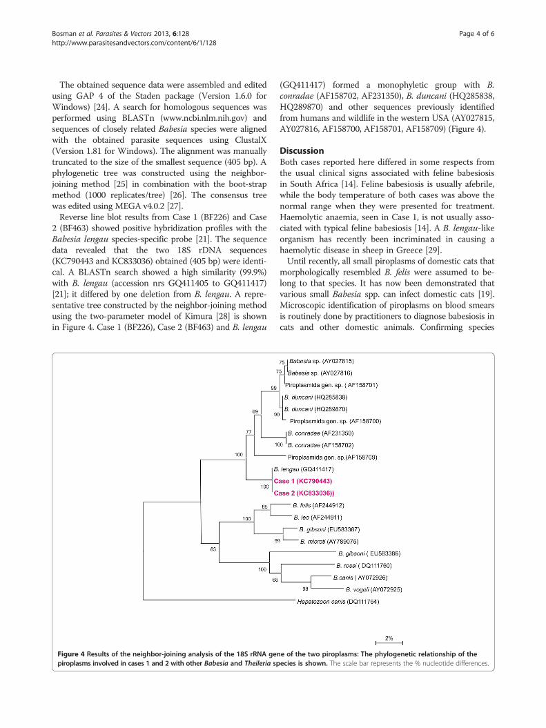
2 (BF463) showed positive hybridization profiles with theBabesia lengau species-specific probe [21]. The sequencedata revealed that the two 18S rDNA sequences(KC790443 and KC833036) obtained (405 bp) were identi-cal. A BLASTn search showed a high similarity (99.9%)with B. lengau (accession nrs GQ411405 to GQ411417)[21]; it differed by one deletion from B. lengau. A repre-sentative tree constructed by the neighbor-joining methodusing the two-parameter model of Kimura [28] is shownin Figure 4. Case 1 (BF226), Case 2 (BF463) and B. lengau
Figure 4 Results of the neighbor-joining analysis of the 18S rRNA genpiroplasms involved in cases 1 and 2 with other Babesia and Theileria sp
(GQ411417) formed a monophyletic group with B.conradae (AF158702, AF231350), B. duncani (HQ285838,HQ289870) and other sequences previously identifiedfrom humans and wildlife in the western USA (AY027815,AY027816, AF158700, AF158701, AF158709) (Figure 4).
DiscussionBoth cases reported here differed in some respects fromthe usual clinical signs associated with feline babesiosisin South Africa [14]. Feline babesiosis is usually afebrile,while the body temperature of both cases was above thenormal range when they were presented for treatment.Haemolytic anaemia, seen in Case 1, is not usually asso-ciated with typical feline babesiosis [14]. A B. lengau-likeorganism has recently been incriminated in causing ahaemolytic disease in sheep in Greece [29].Until recently, all small piroplasms of domestic cats that
morphologically resembled B. felis were assumed to be-long to that species. It has now been demonstrated thatvarious small Babesia spp. can infect domestic cats [19].Microscopic identification of piroplasms on blood smearsis routinely done by practitioners to diagnose babesiosis incats and other domestic animals. Confirming species
e of the two piroplasms: The phylogenetic relationship of theecies is shown. The scale bar represents the % nucleotide differences.
Bosman et al. Parasites & Vectors 2013, 6:128 Page 5 of 6http://www.parasitesandvectors.com/content/6/1/128
identity, however, often requires molecular diagnostictools. The reverse line blot hybridization assay and phylo-genetic analysis are also more sensitive than blood smearexamination in demonstrating the presence of Babesiaspp. in subclinically infected animals.The macroscopic and histopathological findings of both
cases reported here suggested severe illness due to infec-tion by a Babesia parasite, as described for dogs [30]. Mo-lecular diagnostic methods confirmed the presence ofBabesia parasites in blood and brain smears. Phylogeneticanalysis revealed a high similarity to B. lengau.Babesia lengau was described in asymptomatic chee-
tahs [21] and to date no evidence existed to suggest thatB. lengau causes clinical disease in that host. The onlyother piroplasm B. lengau genotypically relates to closelyis a canine parasite, B. conradae, which was describedfrom dogs in California, USA, and is associated withhaemolytic anaemia in the host [31,32]. Babesia lengauand B. conradae form part of the previously described“western clade” of piroplasms, which also includes B.duncani and piroplasms isolated from both humans andwildlife from the western USA. It clustered separatelyfrom the Babesia sensu stricto, the B. microti clade andTheileria and Cytauxzoon species.Cerebral babesiosis has frequently been reported in cat-
tle (e.g., caused by B. bovis) [33] and dogs (e.g., caused byB. rossi) [34,35]. To our knowledge, this serves as a firstreport of cerebral babesiosis in a domestic cat.
ConclusionAll clinical cases of feline babesiosis in South Africa arenot necessarily caused by B. felis. Other Babesia species,e.g. B. lengau, may be incriminated in clinical cases, espe-cially those occurring outside the known endemic area. Asa routine, the identity of the Babesia species involvedshould ideally be confirmed by molecular techniqueswhen specimens from suspected feline babesiosis casesare submitted to diagnostic laboratories. Examining brainsmears or sections is also recommended. Since the vectorof B. felis has not yet been confirmed, our findings empha-sise the urgency of further investigations to enhanceunderstanding the epidemiology of this enigmatic disease.
Competing interestsThe authors declare that they have no competing interests.
Authors’ contributionsAMB carried out the molecular genetic studies, participated in the sequencealignment and wrote the first draft of the manuscript; MCO supervised thelaboratory work and participated in the sequence alignment andconstruction of the phylogenetic trees; EHV co-supervised the laboratorywork; JCAS and TAG performed the necropsies and histological diagnosticinvestigation on Cases 1 and 2, respectively; BLP coordinated theinvestigation, conducted literature searches and wrote the final version ofthe report. All authors read and approved the final manuscript.
AcknowledgementsWe thank Dr T.A. Schouwstra (Rustenburg) and Dr Marisa Naudé (Cape Town)for referring the cases. This research was funded by a South African NationalResearch Foundation grant (NRF grant GUN 2069496) to BL Penzhorn.Publication of the CVBD8 thematic series has been sponsored by Bayer AnimalHealth GmbH.
Author details1Department of Veterinary Tropical Diseases, Faculty of Veterinary Science,University of Pretoria, Private Bag X04, Onderstepoort 0110, South Africa.2Department of Paraclinical Sciences, Faculty of Veterinary Science, Universityof Pretoria, Private Bag X04, Onderstepoort 0110, South Africa. 3SpecialistVeterinary Pathologist, P.O. Box 5371, Helderberg 7135, South Africa.
Received: 30 January 2013 Accepted: 23 April 2013Published: 1 May 2013
References1. Bourdeau P: Feline babesiosis. Point Vet 1996, 27:947–953.2. Moik K, Gothe R: Babesia infections of felids and a report on a case in a
cat in Germany. Tierärztl Praxis 1997, 25:532–535.3. Mangrulkar MY: On a piroplasm of the Indian cat (Felis domesticus). Ind J
Vet Sci Anim Husb 1937, 7:243–246.4. Baneth G, Kenny MJ, Tasker S, Anug Y, Shkap V, Levy A, Shaw SE: Infection
with a proposed new subspecies of Babesia canis, Babesia canis subsppresentii, in domestic cats. J Clin Microbiol 2004, 42:99–105.
5. Adaszek L, Winiarczyk S, Lukaszewska J, Heile C: Babesiose bei einer Katze(Feline babesiosis). Kleintierpraxis 2010, 55:624–628.
6. Vilhena H, Martinez-Días VL, Cardoso L, Vieira L, Altet L, Francino O, Pastor J,Silvestre-Ferreira AC: Feline vector-borne pathogens in the north and centreof Portugal. Parasites & Vectors 2013, 6:99. doi:10.1186/1756-3305-6-99.
7. Jittapalapong S, Jansawan W: Preliminary survey on blood parasites ofcats in Bangkhen District Area. Kasetsart J Nat Sci 1993, 27:330–335.
8. Stewart CG, Hackett KJW, Collett MC: An unidentified Babesia of thedomestic cat (Felis domesticus). J S Afr Vet Assoc 1980, 51:219–221.
9. Jacobson LS, Schoeman T, Lobetti RG: A survey of feline babesiosis inSouth Africa. J S Afr Vet Ass 2000, 71:222–228.
10. Penzhorn BL, Schoeman T, Jacobson LS: Feline babesiosis in South Africa:a review. Ann NY Acad Sci 2004, 1026:183–186.
11. Penzhorn BL, Stylianides E, Coetzee MA, Viljoen JM, Lewis BD: A focus offeline babesiosis at Kaapschehoop on the Mpumalanga escarpment. J SAfr Vet Assoc 1999, 70:60.
12. Potgieter FT: Chemotherapy of Babesia felis infection: efficacy of certaindrugs. J S Afr Vet Assoc 1981, 52:289–293.
13. Jackson C, Dunning FJ: Biliary fever (nuttaliosis) of the cat: a case in theStellenbosch district. J S Afr Vet Med Assoc 1937, 8:83–87.
14. Futter GJ, Belonje PC: Studies on feline babesiosis. 2. Clinicalobservations. J S Afr Vet Assoc 1980, 51:143–146.
15. Schoeman T, Lobetti RG, Jacobson LS, Penzhorn BL: Feline babesiosis:signalment, clinical pathology and co-infections. J S Afr Vet Assoc 2001,72:4–11.
16. Penzhorn BL, Lewis B, López-Rebollar LM, Swan GE: Screening of five drugsfor efficacy against Babesia felis in experimentally infected cats. J S AfrVet Assoc 2000, 71:53–57.
17. Davis LJ: On a piroplasm of the Sudanese wild cat (Felis ocreata). Trans RSoc Trop Med Hyg 1929, 22:523–534.
18. Barnett SF, Brocklesby DW: Some piroplasms of wild mammals. Symp ZoolSoc Lond 1968, 24:159–176.
19. Bosman AM, Venter EH, Penzhorn BL: Occurrence of Babesia felis andBabesia leo in various wild felids and domestic cats in South Africa,based on reverse line blot analysis. Vet Parasitol 2007, 144:33–38.
20. Penzhorn BL, Kjemtrup AM, López-Rebollar LM, Conrad PA: Babesia leo n.sp. from lions in the Kruger National Park, South Africa, and its relationto other small piroplasms. J Parasitol 2001, 87:681–685.
21. Bosman AM, Oosthuizen EC, Peirce MA, Venter EH, Penzhorn BL: Babesialengau sp. nov., a novel Babesia species in cheetah (Acinonyx jubatus,Schreber, 1775) populations in South Africa. J Clin Microbiol 2010,48:2703–2708.
22. Gubbels JM, de Vos AP, van der Weide M, Viseras J, Schouls LM, de Vries E,Jongejan F: Simultaneous detection of bovine Theileria and Babesia speciesby reverse line blot hybridization. J Clin Microbiol 1999, 37:1782–1789.

Bosman et al. Parasites & Vectors 2013, 6:128 Page 6 of 6http://www.parasitesandvectors.com/content/6/1/128
23. Nijhof A, Pillay V, Steyl J, Prozesky L, Stoltsz WH, Lawrence JA, Penzhorn BL,Jongejan F: Molecular characterization of Theileria species associatedwith mortality in four species of African antelopes. J Clin Microbiol 2005,43:5907–5911.
24. Staden R, Beal KF, Bonfield JK: The Staden package, 1998. Methods Mol Biol2000, 132:115–130.
25. Saitou N, Nei M: The neighbor-joining method: a new method forreconstructing phylogenetic trees. Mol Biol Evol 1987, 4:406–425.
26. Felsenstein J: Confidence limits on phylogenies: an approach using thebootstrap. Evolution 1985, 39:783–791.
27. Tamura K, Dudley J, Nei M, Kumar S: MEGA4: Molecular EvolutionaryGenetics Analysis (MEGA) software version 4.0. Mol Biol Evol 2007,24:1596–1599.
28. Kimura M: A simple method for estimating evolutionary rates of basesubstitutions though comparative studies of nucleotide sequences. J MolEvol 1980, 16:111–120.
29. Giadinis ND, Chochlakis D, Kritsepi-Konstantinou M, Makridaki E, Tselentis Y,Kostopoulou D, Karatzias H, Psaroulaki A: Haemolytic disease in sheepattributed to a Babesia lengau-like organism. Vet Rec 2012, 170:155b.doi:10.1136/vr.100394.
30. Boozer AL, MacIntire DK: Canine babesiosis. Vet Clin N Am Small Anim Pract2003, 33:885–904.
31. Conrad PA, Thomford JW, Marsh A, Telford SR III, Anderson JF, Spielman A,Sabin EA, Yamane L, Persing DH: Hemolytic anemia caused by Babesiagibsoni infection in dogs. J Am Vet Med Assoc 1991, 199:601–605.
32. Kjemtrup AM, Wainwright K, Miller M, Penzhorn BL, Carreno RA: Babesiaconradae, sp. nov., a small canine Babesia identified in California. Vet Parasitol2006, 138:103–111.
33. Nevils MA, Figueroa JV, Turk JR, Canto GJ, Le V, Ellersieck MR, Carson CA:Cloned lines of Babesia bovis differ in their ability to induce cerebralbabesiosis in cattle. Parasitol Res 2000, 86:437–443.
34. Jacobson LS: The South African form of severe and complicated caninebabesiosis: Clinical advances 1994-2004. Vet Parasitol 2006, 138:126–139.
35. Malherbe WD, Parkin BS: Atypical symptomatology in Babesia canisinfection. J S Afr Vet Med Assoc 1951, 22:25–36.
doi:10.1186/1756-3305-6-128Cite this article as: Bosman et al.: Babesia lengau associated withcerebral and haemolytic babesiosis in two domestic cats. Parasites &Vectors 2013 6:128.
Submit your next manuscript to BioMed Centraland take full advantage of:
• Convenient online submission
• Thorough peer review
• No space constraints or color figure charges
• Immediate publication on acceptance
• Inclusion in PubMed, CAS, Scopus and Google Scholar
• Research which is freely available for redistribution
Submit your manuscript at www.biomedcentral.com/submit