Structure-Activity Relationships of Haemolytic Saponins
10
PB137 Pharmaceutical Biology 1388-0209/02/4001-001$16.00 2002, Vol. 40, No. ••, pp. ••–•• © Swets & Zeitlinger Abstract The haemolytic activity of 59 samples, natural or partially synthetic pure saponins and mixtures thereof, was deter- mined. The tested saponins possessed triterpenic genins and six structural skeleton types were studied. Structure-activity relationships were established by comparison of the func- tional groups of each saponin and of the branched sugar chains attached to aglycone. The haemolytic activity is shown to depend on the number of sugar units in the chains, as well as on the presence of an osidic chain in position 3 or of func- tionnal groups on the genin such as carboxylic or 16a-OH. Comparison of activities of monodesmosidic and bidesmo- sidic saponins showed that monodesmosidic saponins were generally more active and suggested a polar balance between the two sugar chains at positions 3 and 28. Twelve saponins showed strong haemolytic activity and five possessed a Haemolytic Index (HI) greater than 100,000, such as the a and b aescines (HI: 200,000). Keywords: Haemolytic activity, Haemolytic Index, struc- ture-activity relationships, triterpene saponins. Introduction The rupture of erythrocyte membranes (haemolysis) by saponins is one of the most spectacular properties of these molecules, having found application as reagents in blood cell counters (Hostettmann & Marston, 1995). The origin of the phenomenon is generally ascribed to their amphiphilic prop- erties, but there is a lack of knowledge of the intimate mol- ecular requirements for this activity. This fact is probably linked to the difficulties in obtaining sizeable quantities of pure material and in chemically modifying the structures. Recently, Oda et al. (2000) studied 47 triterpenic or steroidic saponins purified from medicinal and food plants to estab- lish the relationship between adjuvant and haemolytic activ- ities and concluded that there is no correlation. The purpose of this article is to report the investigation of the haemolytic activities of 59 compounds isolated from plants or produced by partial synthesis and try to deduce minimum structure activity relationships. The 59 samples of this study are classified according to the structure of their genins (Fig. 1) and are glycosides of glycyrrhetinic acid (g), oleanolic acid (o), echinocystic acid (e), hederagenin (h), medicagenic acid (m), zanhic acid (z), protobassic acid (pb), 16a-hydroxy-protobassic acid (hp), protoprimulagenin A (pp), cyclamiretin A (c), protoaesci- genin (pa), barringtogenol C (b), R1-barrigenol (Rb), A1- barrigenol (Ab), camelliagenin A (cA), asiatic acid (a), and soyasapogenols B and E (sB and sE). Materials and methods Saponin samples Tables 1 to 4 list the samples subjected to the study, arranged according to the nature of their triterpene. There are 53 pure compounds and 4 purified extracts of known composition characterized by a common genin. The panel was completed by the reference saponin compounds from Merck ® and Sigma ® , as well as commercially available chrysantellin A, a- and b-aescines, glycyrrhizin, asiaticoside, madecasso- side and by partially synthetic compounds. The saponins were extracted from: leaves and roots of Medicago sativa (Fabaceae) (Massiot et al., 1988a,b, 1991), root bark of Tridesmostemon claessenssi (Sapotaceae) (Massiot et al., 1990), leaves of Steganotaenia araliacea (Apiaceae) (Lavaud et al., 1992), stem bark of Petersianthus macrocarpus (Lecythidaceae) (Massiot et al., 1992), leaves of Aphloia madagascariensis (Flacourtiaceae) (Dijoux et al., 1993), leaves and roots of Beta vulgaris (Chenopodiaceae) (Ridout et al., 1994; Massiot et al., 1994), stem bark of Myrsine pel- lucida (Myrsinaceae) (Lavaud et al., 1994a), root bark of Accepted: March 13, 2002 Address correspondence to: Laurence Voutquenne, UMR CNRS 6013, Bât 18, BP 1039, 51097 Reims Cedex 2, France. Fax: 33 (0)3 26 91 35 96; E-mail: [email protected] Structure-Activity Relationships of Hemolytic Saponins Laurence Voutquenne 1 , Catherine Lavaud 1 , Georges Massiot 1,2 and Louisette Le Men-Olivier 1 1 Laboratoire de Pharmacognosie, Reims, France; 2 Institut de Recherche Pierre Fabre, Ramonville, France
-
Upload
independent -
Category
Documents
-
view
0 -
download
0
Transcript of Structure-Activity Relationships of Haemolytic Saponins

PB137
Pharmaceutical Biology 1388-0209/02/4001-001$16.002002, Vol. 40, No. ••, pp. ••–•• © Swets & Zeitlinger
Abstract
The haemolytic activity of 59 samples, natural or partiallysynthetic pure saponins and mixtures thereof, was deter-mined. The tested saponins possessed triterpenic genins andsix structural skeleton types were studied. Structure-activityrelationships were established by comparison of the func-tional groups of each saponin and of the branched sugarchains attached to aglycone. The haemolytic activity is shownto depend on the number of sugar units in the chains, as wellas on the presence of an osidic chain in position 3 or of func-tionnal groups on the genin such as carboxylic or 16a-OH.Comparison of activities of monodesmosidic and bidesmo-sidic saponins showed that monodesmosidic saponins weregenerally more active and suggested a polar balance betweenthe two sugar chains at positions 3 and 28. Twelve saponinsshowed strong haemolytic activity and five possessed aHaemolytic Index (HI) greater than 100,000, such as the aand b aescines (HI: 200,000).
Keywords: Haemolytic activity, Haemolytic Index, struc-ture-activity relationships, triterpene saponins.
Introduction
The rupture of erythrocyte membranes (haemolysis) bysaponins is one of the most spectacular properties of thesemolecules, having found application as reagents in blood cellcounters (Hostettmann & Marston, 1995). The origin of thephenomenon is generally ascribed to their amphiphilic prop-erties, but there is a lack of knowledge of the intimate mol-ecular requirements for this activity. This fact is probablylinked to the difficulties in obtaining sizeable quantities ofpure material and in chemically modifying the structures.Recently, Oda et al. (2000) studied 47 triterpenic or steroidicsaponins purified from medicinal and food plants to estab-lish the relationship between adjuvant and haemolytic activ-ities and concluded that there is no correlation. The purpose
of this article is to report the investigation of the haemolyticactivities of 59 compounds isolated from plants or producedby partial synthesis and try to deduce minimum structureactivity relationships.
The 59 samples of this study are classified according tothe structure of their genins (Fig. 1) and are glycosides ofglycyrrhetinic acid (g), oleanolic acid (o), echinocystic acid(e), hederagenin (h), medicagenic acid (m), zanhic acid (z),protobassic acid (pb), 16a-hydroxy-protobassic acid (hp),protoprimulagenin A (pp), cyclamiretin A (c), protoaesci-genin (pa), barringtogenol C (b), R1-barrigenol (Rb), A1-barrigenol (Ab), camelliagenin A (cA), asiatic acid (a), andsoyasapogenols B and E (sB and sE).
Materials and methods
Saponin samples
Tables 1 to 4 list the samples subjected to the study, arrangedaccording to the nature of their triterpene. There are 53 purecompounds and 4 purified extracts of known compositioncharacterized by a common genin. The panel was completedby the reference saponin compounds from Merck® andSigma®, as well as commercially available chrysantellin A, a- and b-aescines, glycyrrhizin, asiaticoside, madecasso-side and by partially synthetic compounds. The saponinswere extracted from: leaves and roots of Medicago sativa(Fabaceae) (Massiot et al., 1988a,b, 1991), root bark ofTridesmostemon claessenssi (Sapotaceae) (Massiot et al.,1990), leaves of Steganotaenia araliacea (Apiaceae) (Lavaudet al., 1992), stem bark of Petersianthus macrocarpus(Lecythidaceae) (Massiot et al., 1992), leaves of Aphloiamadagascariensis (Flacourtiaceae) (Dijoux et al., 1993),leaves and roots of Beta vulgaris (Chenopodiaceae) (Ridoutet al., 1994; Massiot et al., 1994), stem bark of Myrsine pel-lucida (Myrsinaceae) (Lavaud et al., 1994a), root bark of
Accepted: March 13, 2002
Address correspondence to: Laurence Voutquenne, UMR CNRS 6013, Bât 18, BP 1039, 51097 Reims Cedex 2, France. Fax: 33 (0)3 26 9135 96; E-mail: [email protected]
Structure-Activity Relationships of Hemolytic Saponins
Laurence Voutquenne1, Catherine Lavaud1, Georges Massiot1,2 and Louisette Le Men-Olivier1
1Laboratoire de Pharmacognosie, Reims, France; 2Institut de Recherche Pierre Fabre, Ramonville, France

2 L. Voutquenne et al.
Smelophyllum capense (Sapindaceae) (Lavaud et al., 1994b),leaves of Pisonia umbellifera (Nyctaginaceae) (Lavaud et al.,1996a), seed kernel of Mimusops sp. (Sapotaceae) (Lavaudet al., 1996b), stem bark of Tapeinosperma clethroides(Myrsinaceae) (Kitagawa et al., 1980; Lavaud et al., 1999), stem bark of Filicium decipiens (Sapindaceae)(Lavaud et al., 1998), stem bark of Harpullia cupanioides(Sapindaceae) (Dimbi et al., 1983; Voutquenne et al., 1998),and leaves of Desmodium adscendens (Fabaceae) (McManuset al., 1993).
The oleanolic derivatives o-1 to o-4 were prepared fromoleanolic acid (o) and the corresponding peracetobromosug-ars under phase transfer conditions (Bliard et al., 1994). Thesame conditions were used for the preparation of m-5 and m-6 from m-3, of a-1 from asiatic acid (a) and of g-2 fromglycyrrhetinic acid (g). Saponin o-5 was prepared from o-1by Koenigs-Knorr condensation with acetyl lactosyl bromide(Nazabadioko, 1996). Partial acid hydrolysis of a-hederin (h-2) in 0.04 N H2SO4 in 50% EtOH solution provided h-1.Partial alkaline hydrolysis in 1% KOH in 30% EtOH
PB137
COOH
O
HO
g glycyrrhetinic acid
COOH
R2
R3
R1
HO
R42
3
16
28
6
R1 R2 R3 R4 o oleanolic acid H CH3 H H
e echinocystic acid H CH3 H OH
h hederagenin H CH2OH H H
m medicagenic acid OH COOH H H
z zanhic acid OH COOH H OH
pb protobassic acid OH CH2OH OH H
hp 16a-hydroxy-protobassic acid OH CH2OH OH OH
OH
O
HO
R
3
13
28
16
R
OHOH
R
2CH R1
OH
HO3
1516
21
22
28
2
3
R R1 R2 R3 pp Protoprimulagenin A CH3 pa protoaescigenin OH H OH
c cyclamiretin A CHO b barringtogenol C H H OH
Rb R1-barrigenol H OH OH
Ab A1-barrigenol H OH H
cA camelliagenin A H H H
HO
HO
COOH
asiatic acidaCH R
R
R
1
2
3
2
R =OH1 R = R = H2 3,
HO
CH OH
OH2
soyasapogenol BsB
R
sE =O
R
soyasapogenol E
Figure 1. Structures of genins of tested saponins.

Structure-activity relationships of hemolytic saponins 3PB137
Table 1. Haemolytic activity of saponins with carboxylic function in sapogenin part.
OR-3 COOR-23, 28 or 30 Trivial name HD100 mM (mg/mL) reference
o H Hˆ28 oleanolic acid >2200 –o-1 H Glcˆ28 – >1600 [15%] Bliard, 1994o-2 H Glcˆ
4Glcˆ28 – nha Bliard, 1994o-3 H Rhaˆ
6Glcˆ6Glcˆ28 – nha Bliard, 1994
o-4 H – >900 [15%] Bliard, 1994
o-5 Galˆ4Glcˆ Glcˆ28 calenduloside B 1061 [96%] (1) –o-6 Galˆ2GlcAˆ Glcˆ28 – >523 [0%] Lavaud, 1992e-1 Glcˆ Hˆ28 – 118 (75) –e-2 Glcˆ Rhaˆ
3Xylˆ4Rhaˆ2Xylˆ28 chrysantellin A 16.8 (20) –
Pisonia 1, 2 or 4 sugars Glcˆ28 ~75mg/ml Lavaud, 1996aextractBeta l. 3 sugars H or Glcˆ28 ~500mg/ml Ridout, 1994extract Massiot, 1994h H Hˆ28 hederagenin >2000 [68%] –h-1 Araˆ Hˆ28 fatsiaside B1 1656 (1000) –h-2 Rhaˆ
2Araˆ Hˆ28 a-hederin 26.7 (20) Lavaud, 1994bh-3 Xylˆ3Rhaˆ
2Araˆ Hˆ28 sapindoside B 22.7 (20) Lavaud, 1994bh-4 Araˆ
3Rhaˆ2Araˆ Hˆ28 – 11.3 (10) Lavaud, 1994b
h-5 Araˆ2Rhaˆ Glcˆ28 – 1096 (1000) Lavaud, 1994b
h-6 Araˆ2Glcˆ
2Araˆ Glcˆ28 medicoside I 94.3 (100) Massiot, 1988,1991
h-7 Araˆ2Glcˆ
2Araˆ Hˆ28 medicoside C 55.7 (50) –pb-1 Glcˆ Rhaˆ
3Xylˆ4Rhaˆ2Araˆ28 Mi-saponin A 61.4 (75) Lavaud, 1996b
pb-2 GlcAˆ Rhaˆ3Xylˆ4Rhaˆ
2Araˆ28 butyroside C 202 (250) Lavaud, 1996bpb-3 Glcˆ
3Glcˆ ˆRhaˆ3Xylˆ4Rhaˆ
2Araˆ28 18.1 (25) Lavaud, 1996bhp-1 Glcˆ Rhaˆ
3Xylˆ4Rhaˆ2Araˆ28 arganine C 20.2 (25) Lavaud, 1996b
hp-2 GlcAˆ Rhaˆ3Xylˆ4Rhaˆ
2Araˆ28 59.9 (75) Lavaud, 1996b
hp-3 Glcˆ3Glcˆ tridesmosaponin B 162 (250) Massiot, 1990
hp-4 Rhaˆ tridesmosaponin A 54.8 (75) Massiot, 1990
m-1 Glcˆ Hˆ28, Hˆ23 medicoside A 90.4 (60) Massiot, 1988m-2 Glcˆ Glcˆ28, Hˆ23 medicoside G >605 [0%] 1991m-3 Glcˆ Xylˆ4Rhaˆ
2Araˆ28, Hˆ23 medicoside J 233 (250) Massiot, 1988m-4 Glcˆ
2Glcˆ Xylˆ4Rhaˆ2Araˆ28, Hˆ23 809 (1000) 1991
m-5 Glcˆ Xylˆ4Rhaˆ2Araˆ28, Glcˆ23 405 (500) –
m-6 Glcˆ Xylˆ4Rhaˆ2Araˆ28, 715 [82%] (1000) –
Galˆ4Glcˆ23
m-7 Glcˆ2Glcˆ 15.8 (25) Lavaud, 1998
z-1 Glcˆ 34.8 (50) Lavaud, 1998
sB-1 Rhaˆ2Galˆ2GlcAˆ – soyasaponin I nha Mc Manus,
sB-2 Galˆ2GlcAˆ – soyasaponin III nha 1993sE-1 Rhaˆ
2Galˆ2GlcAˆ – dehydrosoyasaponin I nha ≤g-1 (GlcAO-NH4
+)2ˆ NH4+ˆ30 glycyrrhizin nha –
g-2 H Glcˆ4Glcˆ30 nha –
nha: no haemolytic activity. Each monosaccharide unit is abbreviated as follows: arabinose (Ara), xylose (Xyl), glucose (Glc), glucuronicacid (GlcA), galactose (Gal), fucose (Fuc), rhamnose (Rha). Acyls groups are described as follows: acetyl (Ac), niloyl (Nil), angeloyl (Ang)and tigloyl (Tig).
Xyl 4Rha 2Fuc 28,H 23
Ara2 4
Nil Nil
Xyl 4Rha 2Fuc 28,H 23
Ara2 4
Nil Nil
Rha 3Xyl 4Rha 2Xyl 28
Rha4
Rha 3Xyl 4Rha 2Xyl 28
Rha3
Glc6 Gal4 Glc 28
Glc6

4 L. Voutquenne et al.
solution of bidesmosidic saponins e-2, h-6 or ester saponinsRb1, pa-1 and pa-2 gave the corresponding monodesmosidicor saponified saponins e-1, h-7, Rb-4, and pa-3.
The commercially saponins Merck®, mixture from Gyps-ophyla paniculata (Caryophyllaceae), and Sigma®, mixturefrom Quillaja saponaria (Rosaceae), were dialysed in
Spectra/Por® molecular porous membrane tubing withMCWO: 6- 8000. Saponins (1g) were dissolved in 15ml ofpure water and the solution was introduced into theSpectra/Por® membrane tubing. The saponin mixture was dialysed against pure water for 72h and then the contentof the tube was lyophilised. The powdered residues consti-
PB137
Table 3. Haemolytic activity of saponins with a polyhydroxylated ring D or E in sapogenin part.
Trivial HD100 mMOR-3 OR-21 OR-22 OR-28 name (mg/mL) reference
pa-1 Angˆ CH3COˆ H a-aescine 6.6 (7.5) –
pa-2 Tigˆ CH3COˆ H b-aescine 6.6 (7.5) –
pa-3 H H H – >497 [0%] –
b-1 TigˆNilˆAraˆ
+ Tig H H – 76.2 (100) Massiot, 1992
b-2 PhˆCOˆ H Rhaˆ –
Rb-1 Tigˆ H H >460 [0%] Lavaud, 1992
Rb-2 Angˆ H H >460 [0%] Lavaud, 1992
Rb-3 Angˆ H H >473 [0%] Lavaud, 1992
Rb-4 H H H – >497 [0%] –
Harpullia – H or Ang Ac or Ang – 9.6 Voutquenne, 1998extract
Rha 3GlcA
hexose2
Gal 3GlcA
Gal2
Xyl 4GlcA
Glc2
Gal 3GlcA
Gal2
Gal 3GlcA
Gal2
Gal 3GlcA
Gal2
Gal 3GlcA
Gal2
Glc 4GlcA
Glc2
Glc 4GlcA
Glc2
Glc 4GlcA
Glc2
Table 2. Haemolytic activity of cyclamiretin A (c) and protoprimulagenin A (pp).
OR-3 C-28 Trivial name HD100 mM (mg/mL) reference
c-1 13,28-epoxy saxifragoline B 7.1 (7.5) Lavaud, 1994a
c-2 Xylˆ2Glcˆ4Araˆ 13,28-epoxy primulanin 55.7 (50) Lavaud, 1994a
c-3 Rhaˆ2Glcˆ
4Araˆ 13,28-epoxy – 54.8 (50) Lavaud, 1994a
c-4 CH2OH – >472 [0%] Lavaud, 1994a
pp-1 13,28-epoxy sakuraso-saponin 12 (15) Lavaud, 1999Rha 2Rha 2Gal 3GlcA
Glc2
Xyl 2Glc 4Ara
Glc2
Xyl 2Glc 4Ara
Glc2

Structure-activity relationships of hemolytic saponins 5
tuted the reference saponin mixtures Merck®D and Sigma®D(175mg, yield 17.5%), respectively.
Haemolysis test
Material
Sheep erythrocytes were purchased from Biomerieux-Lyonas a 50% suspension and diluted with phosphate buffer saline(PBS) to give a 10% suspension. Saponins were dissolved inPBS and pH was adjusted to 7.4; 2ml samples were preparedwith concentrations ranging from 10-3 mg/ml to 1mg/ml.Each sample was tested at least twice. In the case ofsapogenins, the mother solution was a mixture of DMSO:water (5 :1); further dilutions were made with PBS (Segal et al., 1974).
Test
The erythrocyte suspension (50 ml) was added to 2ml of thesample to be tested and the mixture was rapidly stirred, incu-bated for 60min at room temperature, then centrifuged at3000 rpm for 5min. Absorbance of the supernatant was mea-sured at 540nm. The 100% haemolytic dose (HD100) wasdetermined as the saponin concentration (mg/ml or mM)inducing the same haemolysis that was caused by Sigma®
dialyzed mixture (Sigma® D). For this sample, 100% haemol-ysis (HD100) was obtained at a concentration of 75 mg/ml. The haemolytic index (HI) was calculated according to theEuropean Pharmacopoeia as HI = 30,000 ¥ (a) / (b) where(a) and (b) are the concentrations in (g/l) of the saponin ref-erence “Merck® D” and of the saponin under study whichgives 100% haemolysis.
Haemolytic time course measurements
The erythrocyte suspension (50 ml) was added to 2ml of commercial saponins whose final concentrations caused100% haemolysis within 60min, and the mixture was rapidlystirred, incubated at room temperature. After 0.25, 0.5, 0.75,1, 2, 3, 4, 5, 15, 30 or 60min of incubation, the whole mixturewas centrifuged at 3000 rpm for 5min. The absorbance ofeach supernatant was measured at 540nm. The value %haemolysis was the ratio of this absorbance to that obtainedat 100% haemolysis within 60min.
Results
The commercial saponins from Merck® and Sigma® pos-sessed a 100% haemolytic dose (HD100) at a concentration of250mg/ml. A similar activity is obtained at respective con-centrations of 50 and 75 mg/ml after a step of purification ofthese mixtures by dialysis against pure water. Figure 2 showsrepresentative examples of haemolysis curves in whichabsorbance is plotted against concentration. The series ofdata are obtained with saponins c-1 to c-3, pp-1 and h-4 containing cyclamiretin A (c), protoprimulagenin A (pp) andhederagenin (h) as aglycone and the dialysed referencesaponin from Sigma®. All the curves are of the sigmoidaltype which means that the hemolysis needs a minimum concentration to start, after which the reaction rapidly takesplace.
Figure 3 compares the haemolytic time courses of the two dialysed reference saponin from Merck® and Sigma®
with three commercially available saponins a-hederin (h-2),chrysantellin A (e-2) and a-aescine (pa-1). The time coursesof the reference compounds are similar and 90% haemolysisis obtained in one minute. The haemolytic rates of the purecommercial saponins h-2 and e-2 are fast (15sec) while thatof the mixture pa-1 is slow (10min) compared to the refer-ence saponins. The difference in the time course is not easilyexplained but it seems that the haemolytic time course ofpure saponins is faster than the mixture. Takechi and Tanaka(1995) have previously mentioned that differences inhaemolytic time courses could be due to structural differ-ences of the sapogenins and not of the sugars moieties as proposed by Segal et al. (1974).
The results presented in Table 1 show that monodesmo-sidic saponins with an ester chain at C-28 (o-1 to o-4) or C-30 (g-2) are not haemolytic. The same result is observed withthe monodesmosidic ursane derivatives: a-1 to a-3 and withsaponins from Aphloia madagascariensis (Table 4). Theintroduction of an osidic residue in position 3 to the saponino-1 gives the bidesmosidic saponin o-5 which displays a lowhaemolytic activity.
The comparison of haemolytic potency of three saponinsmonoglycosylated in position 3 with sapogenins h-1, e-1 andm-1, shows that the presence of one additional hydroxylgroup at position 2 (m-1) or 16a (e-1) multiplies the activ-ity by a factor of fifteen (Fig. 4). A similar effect is observedbetween the bidesmosidic saponins hp-1 and hp-2 which
PB137
Table 4. Haemolytic activity of saponins derivatives of ursane skeleton.
R1-23 R2-6 R3-19 COOR-28 Trivial name HD100 mM reference
a-1 OH H H Glcˆ4Glcˆ – nha –
a-2 OH H H Rhaˆ4Glcˆ
6Glcˆ asiaticoside nha –a-3 OH OH H Rhaˆ
4Glcˆ6Glcˆ madecassoside nha –
Aphloia H or OH OH OH Glcˆ – nha Dijoux,extract 1993
nha: no haemolytic activity.

6 L. Voutquenne et al.
contain an hydroxyl group at position 16a and saponins pb-1 and pb-2 which are about three times less active (Table 1).
The haemolytic activity of monodesmosidic saponins con-taining a 13b,28-epoxygenin as cyclamiretin A (c) (Table 2)and of similar monodesmosidic saponins with hederagenin(h) (Table 1) were compared. The bi- or triglycosylatedsaponins c-2 and c-3 exhibit the same range of haemolyticactivity as h-2, h-3, h-4 and h-7, suggesting that the geninsinduce similar effects. When the ether ring is cleaved into acarbinol at position C-28 (c-4), the haemolytic activity isstrongly reduced. Comparison of activity of c-4 with that ofe-1 shows that the carboxylic function at position C-28enhances the activity.
Table 3 reports the haemolytic activity of saponins with apolyhydroxylated sapogenin in rings D or E. The a and baescines pa-1 and pa-2 which contain two free hydroxyls andtwo esters in ring D and/or E are strongly haemolytic withHD100 6.6mM as observed by Oda et al. (2000). The saponinsextracted from Harpullia cupanioides show a strong activityand their structures possess two esters and two free hydrox-yls. A decrease in activity is observed with saponins con-taining less than two ester functions like saponins b-1 andb-2 from Petersianthus macrocarpus. No activity appearswith saponins which contain four or more free hydroxylssuch as the Steganotaenia saponins (Rb-1 to Rb-3) and theproducts pa-3 and Rb-4 resulting from alkaline hydrolysis.
PB137
0
20
40
60
80
100
0 2 4 6 8 10 12 14 16 18 20
Time (min)
Sigma® DMerck ® Dh-2=e-2pa-1
Hae
mol
ysis
%
Figure 3. Haemolytic time course measurements.
Hae
mol
ysis
%
0
20
40
60
80
100
0 5 10 15 20 25 30 35 40 45 50
Conc mg/mL
Sigma® Dc-1c-2=c-3pp-1h-4
Figure 2. Haemolytic activity of saponins with cyclamiretin A (c), protoprimulagenin A (pp) and hederagenin (h).

Structure-activity relationships of hemolytic saponins 7
Monodesmosidic diesterified saponins with protoaescigenin(pa) and barringtogenol (b) (Table 3) possess the same activ-ity or are as much as ten-fold more active than the corre-sponding monodesmosidic saponins with cyclamiretin A (c)(Table 2), hederagenin (h) or medicagenic acid (m) (Table 1)as aglycones.
The saponin extract from Desmodium adscendens con-taining soyasaponin I (sB1), III (sB-2) and dehydrosoyas-aponin I (sE-1) whith one hydroxyl in ring E is inactive(Table 1). This seems to be due to the free hydroxyl in posi-tion 22 and to the absence of a carboxylic group in position28. This absence of haemolytic activity of soyasaponins hasbeen previously related by Gestetner et al. (1971) and morerecently by Oda et al. (2000). Thus, the inactivity of the gly-cyrrhizin (g-1) can be explained by the absence of carboxylicgroup at C-28 (Table 1).
From the results presented in Table 1, it appears that thehaemolytic activity of di-and triglycosides saponins h-2, h-3, h-4 and h-7 is higher than that of the monoglycoside h-1,so the number of sugars residues also affects the haemolyticactivity of monodesmosidic saponins. The saponins c-1 andpp-1 with four or five osidic units are more active than trigly-cosidic saponins c-2 and c-3 (Table 2, Fig. 3). The activityof monodesmosidic saponin in C-3 increases with thenumber of sugar units and is maximum with four sugar units.The decrease of activity between c-1 and pp-1 can be due tothe increased number of osidic units. Abe et al. (1978) haspreviously reported that haemolytic activity of saikosaponinsdepends on the number of sugars.
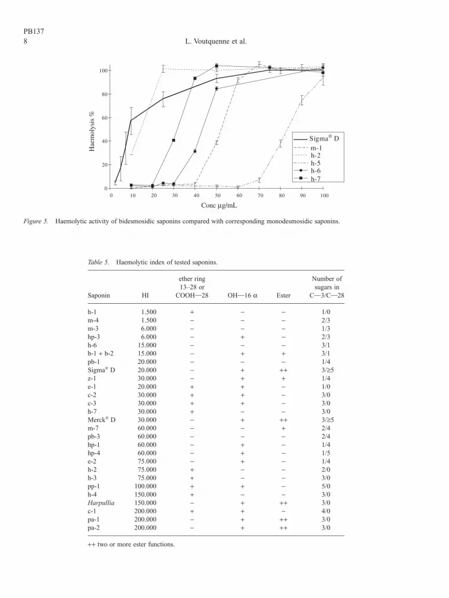
In Fig. 5, haemolytic potency of monodesmosidicsaponins m-1, h-2 and h-7 is compared with one of the cor-responding bidesmosidic saponins m-2, h-5 and h-6. It showsthat glucosylation in C-28 decreased the activity. The mon-odesmosidic medicoside A (m-1) shows the greatest activitycompared to the bidesmosidic medicosides G and J (m-2 and
m-3) and medicoside m-4 as previously reported by Oleszek(1990) with alfalfa saponins (Table 1).
Comparison of the haemolytic activity of bidesmosidicsaponins with only one glucose unit in position 28 like o-5,o-6, h-5, h-6, m-2, and saponin extracts of Pisonia umbell-ifera and of Beta vulgaris, shows activity increasing with the number of sugars branched in position 3 as previouslyobserved for monodesmosidic saponins and with a maximumfor three sugars units as in saponin h-6 (Table 1). In bidesmo-sidic saponins with the same trisaccharide in position 28 ofmedicagenic acid, haemolytic activity is related to thenumber of sugars attached to the saponin, m-3 being the mostactive. Increasing the number of sugar branches on ring A atpositions 3 or 23 (saponins m-4, m-5 and m-6) has a nega-tive effect on activity (Table 1).
In the case of saponins with one tetrasaccharidic esterchain as saponins pb-1, pb-2 and pb-3, the highest activity isobserved for pb-3 with two glucoses in the chain at C-3(Table 1). A similar effect is observed for the couple ofsaponins m-7 and m-4. A strong activity is observed insaponin z-1 with the same ester chain but only with oneglucose at C-3, this may result from the beneficial effect ofthe 16a-hydroxyl to the haemolytic activity (Table 1). Onesupplementary sugar in the ester chain at position 28 as insaponins hp-3 and hp-4 decreases activity. The activity is bestwhen only one sugar constitutes the chain at C-3 (Table 1).
Discussion
Table 5 gives a classification of the saponins according totheir haemolytic index (HI) with comparison with a com-mercial saponin Merck®. We distinguish saponins with low(HI < 10.000), moderate (10.000 £ HI ≥ 30.000) and stronghaemolytic activity (HI > 50.000). The table also includes
PB137
0
20
40
60
80
100
0 50 100 150 200 250 300 350 400 450 500
Conc mg/mL
Sigma® De-1h-1m-1
Hae
mol
ysis
%
Figure 4. Influence of one polar group in rings A or D on haemolytic activity.
8 L. Voutquenne et al.PB137
0
20
40
60
80
100
0 10 20 30 40 50 60 70 80 90 100
Conc mg/mL
Sigma® Dm-1h-2h-5h-6h-7
Hae
mol
ysis
%
Figure 5. Haemolytic activity of bidesmosidic saponins compared with corresponding monodesmosidic saponins.
Table 5. Haemolytic index of tested saponins.
ether ring Number of13–28 or sugars in
Saponin HI COOHˆ28 OHˆ16 a Ester Cˆ3/Cˆ28
h-1 1.500 + - - 1/0m-4 1.500 - - - 2/3m-3 6.000 - - - 1/3hp-3 6.000 - + - 2/3h-6 15.000 - - - 3/1b-1 + b-2 15.000 - + + 3/1pb-1 20.000 - - - 1/4Sigma® D 20.000 - + ++ 3/≥5z-1 30.000 - + + 1/4e-1 20.000 + + - 1/0c-2 30.000 + + - 3/0c-3 30.000 + + - 3/0h-7 30.000 + - - 3/0Merck® D 30.000 - + ++ 3/≥5m-7 60.000 - - + 2/4pb-3 60.000 - - - 2/4hp-1 60.000 - + - 1/4hp-4 60.000 - + - 1/5e-2 75.000 - + - 1/4h-2 75.000 + - - 2/0h-3 75.000 + - - 3/0pp-1 100.000 + + - 5/0h-4 150.000 + - - 3/0Harpullia 150.000 - + ++ 3/0c-1 200.000 + + - 4/0pa-1 200.000 - + ++ 3/0pa-2 200.000 - + ++ 3/0
++ two or more ester functions.

Structure-activity relationships of hemolytic saponins 9
some important structural features of the molecules. Resultsof this investigation lead to the conclusion that monodesmo-sidic and bidesmosidic saponins must be separated, for estab-lishment of structure-activity relationships. Comparison ofthe haemolytic index shows that monodesmosidic saponinsare generally more active than bidesmosidic saponins.
Monodesmosidic saponins with a single ester chain atposition 28 are not haemolytic. The introduction of an osidicresidue in position 3 gives haemolytic potency thus confirm-ing the work of Hase et al. (1981). The haemolytic potencyof monoglycosidic saponins in position 3 increases with thepresence of one a-hydroxyl at position 16 or at position 2 aspreviously described by Schlösser and Wulff (1969) whichnoticed that for activity to be observed, the aglycone requiresa polar group in ring A (OH and/or COOH) and a moder-ately polar group in ring D or E. Saponins with a 13b,28-epoxy bridge are highly haemolytic and this activity isreduced when the ether ring is cleaved (Nose et al., 1989;Sindambiwe et al., 1998). Saponins without a carboxylicgroup in C-28 are inactive as with the soyasaponins (Gestetner et al., 1971; Oda et al., 2000). These results allowto conclude that haemolytic activity of monodesmosidicsaponins in position 3 was connected with a carboxylic func-tion at position 28 or a 13b,28-epoxy bridge, and with onea-hydroxyl at position 16. This activity increases with analcohol function in ring A at positions 2 or 23 or in ring Bat position 6.
The presence of many free hydroxyls in the ring E as inthe saponins Rb-1 to Rb-4 and pa-3, induces loss ofhaemolytic activity as previously announced by Hase et al.(1981) and Schlösser and Wulff (1969). The esterification ofthese hydroxyls gives haemolytic potency as in saponinsfrom Harpullia cupanioides and the aescines (Table 3).These results confirm the previous reported findings of Segalet al. (1966, 1970) on activity enhancement by esterification,independant of the type of ester, as well as of Schlösser andWulff (1969) on the requirement for the genin to possess alimited number of OH groups in rings D and/or E to beactive. In our case, the presence of one ester at position 22seems to be necessary to display haemolytic activity as in theaescines pa-1 and pa-2 (Table 5). We confirm that polarity inring A and relative hydrophobicity in rings D/E are essentialfor haemolytic activity (Hostettmann & Marston, 1995).
Moreover we observe with the two types of genins (hed-eragenin and cyclamiretin A) that the haemolytic activity ofmonodesmosidic saponins increases with the number ofsugar units as Romussi et al. (1980) and Takechi and Tanaka(1992) have observed with oleanolic acid saponins, and Abeet al. (1978) with saikosaponins. Haemolytic activity of mon-odesmosidic saponins in position 3 is thus higher with threeor four sugars units in the sugar chain.
Comparison of monodesmosidic saponins and corre-sponding bidesmosidic saponins shows that glucosylation inC-28 decreases the haemolytic activity as previously reportedby Hase et al. (1981). In the case of bidesmosidic saponins,with one ester sugar unit at the position 28, the presence of
three sugars at the position 3 is necessary to keep a moder-ate activity. Whereas when the number of sugars in the sugarchain at position 28 is higher than four or five units, thenumber of sugars in the sugar chain at position 3 must bedecreased to one sugar. These results suggest the necessityof a polar balance between the two sugar chains at positions3 and 28. The activity of bidesmosidic saponins is the highestwhen the osidic ester chain in position 28 is ramified withfour sugars and one glycoside in position 3 (Table 5). As in monodesmosidic saponins, the presence of a 16a-OHincreases the haemolytic activity. One hydroxyl in position 2and of a polar function like COOH or CH2OH in C-23 seemto be necessary for haemolytic activity.
All these results do not include the nature of the sugar unitin structure-activity relationships. The presence of one ter-minal xylose in saponin h-3 slightly decreases the activity ascompared to the arabinose in saponin h-4. The glucose unitin the middle of the chain of saponin h-7 decreases the activ-ity in comparing to the presence of rhamnose as in saponinh-4 (Table 1). The presence of glucuronic acid seems todecrease activity as suggested by the comparison of saponinspb-1, hp-1 and o-5 which contain one glucose linked tooxygen-3 with the saponins pb-2, hp-2 and o-6 where oneglucuronic acid takes the place of glucose.
Twelve saponins are found more active than the saponinmixture Merck® D which is known to possess a HI of 30,000(Table 5). They are the bidesmosidic saponins hp-1, pb-3 andtridesmosaponin A (hp-4) isolated from Mimusops sp., thesaponin m-7 isolated from Filicium decipiens and thechrysantellin A (e-2). The monodesmosidic active saponinsare a-hederin (h-2), sapindoside B (h-3) and sapindoside h-4, the saxifragoline B (c-1), the sakuraso-saponin (pp-1),the a-and b-aescines (pa-1, pa-2).
In this study, we have attempted to increase the knowl-edge of haemolytic activity of triterpene saponins. The greatvariability in the structure of saponins makes this type of study delicate and the results may be difficult to rational-ize. New structure-activity relating to the chemical structureof the aglycon and of the sugar chains have been proposedin this work. In order further to confirm the above assump-tions as well as investigate how the nature and the sequenc-ing osidic units affect haemolytic activity, it is necessary totest many other saponins from various sources. This studyshould be completed with haemolytic tests on human bloodcells.
References
Abe H, Sakaguchi M, Konishi H, Tani T, Arichi S (1978): Theeffects of saikosaponins on biological membranes. 1. Therelationship between the structures of saikosaponins andHaemolytic activity. Planta Med 34: 160–166.
Bliard C, Massiot G, Nazabadioko S (1994): Glycosylation ofacids under phase transfer conditions. Partial synthesis ofsaponins. Tetrahedron Lett 35: 6107–6108.
PB137

10 L. Voutquenne et al.
Dijoux MG, Lavaud C, Massiot G, Le Men-Olivier L, SheeleyDM (1993): A saponin from leaves of Aphloia madagas-cariensis. Phytochemistry 34: 497–499.
Dimbi MZ, Warin R, Delaude C, Huls R, Kapundu M (1983):Triterpenoïdes of Harpullia cupanioides. Bull Soc ChimBelg 92: 473–484.
Gestetner B, Assa Y, Henis Y, Birk Y, Bondi A (1971): LucerneSaponins. IV. Relationship between their chemical consti-tution, and haemolytic and antifungal activities. J Sci FoodAgr 22: 168–172.
Hase J, Kobashi K, Mitsui K, Namba T et al. (1981): The struc-ture-hemolysis relationship of oleanolic acid derivativesand inhibition of the saponin-induced hemolysis withsapogenins. J Pharm Dyn 4: 833–837.
Hostettmann K, Marston A (1995): Saponins. Chemistry andPharmacology of Natural Products. Cambridge, UniversityPress, pp. 232–286.
Kitagawa I, Yoshikawa M, Kobayashi K, Imakura Y et al. (1980):Saponin and sapogenol. XXVIII. Reinvestigation of thebranching positions in the glucuronide moities of three glucuronide saponins: Desacyl-jegosaponin, desacyl-boninsaponin A, and sakuraso-saponin. Chem Pharm Bull 28: 296–300.
Lavaud C, Massiot G, Le Men-Olivier L, Viari A et al. (1992):Saponins from Steganotaenia araliacea. Phytochemistry31: 3177–3181.
Lavaud C, Massiot G, Bravo Barrera J, Moretti C, Le Men-Olivier L (1994a): Triterpene saponins from Myrsinepellucida. Phytochemistry 37: 1671–1677.
Lavaud C, Voutquenne L, Massiot G, Le Men-Olivier L,Delaude C (1994b): Saponines triterpéniques de Smelo-phyllum capense (Sapindaceae). Bull Soc Roy Sci Liège 63:455–463.
Lavaud C, Beauviere S, Massiot G, Le Men-Olivier L, BourdyG (1996a): Saponins from Pisonia umbellifera. Phyto-chemistry 43: 189–194.
Lavaud C, Massiot G, Becchi M, Misra G, Nigam SK (1996b):Saponins from three species of Mimusops. Phytochemistry41: 887–893.
Lavaud C, Voutquenne L, Massiot G, Le Men-Olivier L et al.(1998): Saponins from stem bark of Filicum decipiens. Phytochemistry 47: 441–449.
Lavaud C, Pichelin O, Massiot G, Le Men-Olivier L et al.(1999): Sakuraso-saponin from Tapeinosperma clethroides.Fitoterapia, 70: 116–118.
Massiot G, Lavaud C, Guillaume D, Le Men-Olivier L (1988a):Reinvestigation of the sapogenins and prosapogenins fromalfalfa (Medicago sativa). J Agric Food Chem 36: 902–909.
Massiot G, Lavaud C, Le Men-Olivier L, Van Binst G et al.(1988b): Structural elucidation of alfalfa root saponins bymass spectrometry and nuclear magnetic resonance analy-sis. J Chem Soc Perkin I, 3071–3079.
Massiot G, Lavaud C, Delaude C, Van Binst GF et al. (1990):Saponins from Tridesmostemon claessenssi. Phytochem-istry 29: 3291–3298.
Massiot G, Lavaud C, Besson V, Le Men-Olivier L, Van Binst
G (1991): Saponins from aerial parts of alfalfa (Medicagosativa). J Agric Food Chem 39: 78–82.
Massiot G, Chen XF, Lavaud C, Le Men-Olivier L et al. (1992):Saponins from stem bark of Petersianthus macrocarpus.Phytochemistry 31: 3571–3576.
Massiot G, Dijoux MG, Lavaud C, Le Men-Olivier L et al.(1994): Seco-glycosides of oleanolic acid from Beta vul-garis. Phytochemistry 37: 1667–1670.
McManus OB, Harris GH, Giangiacomo KM, Feigenbaum P et al. (1993): An activator of calcium-dependant potassiumchannels isolated from a medicinal herb. Biochemistry 32:6128–6133.
Nazabadioko S (1996): Synthèse d’oligosaccharides pourl’hémisynthèse de saponines. Th Pharm Reims, P202.
Nose M, Amagaya S, Ogihara Y (1989): Effects of saikosaponinmetabolites on the hemolysis of red blood cells and theiradsorbability on the cell membrane. Chem Pharm Bull 37:3306–3310.
Oda K, Matsuda H, Murakami T, Katayama S et al. (2000):Adjuvant and haemolytic activities of 47 saponins derivedfrom medicinal and food plants. Biol Chem 381: 67–74.
Oleszek W (1990): Structural specificity of alfalfa (Medicagosativa). Saponin haemolysis and its impact on two haemol-ysis-based quantification methods. J Sci Food Agric 53:477–485.
Ridout CL, Price KR, Parkin G, Dijoux MG, Lavaud C (1994):Saponins from sugar beet and the floc probem. J Agric FoodChem 42: 279–282.
Romussi G, Cafaggi S, Bignardi G (1980): Hemolytic action and surface activity of triterpene saponins from Anchusaofficinalis L., part 2: On the constituents of Boraginaceae.Pharmazie 35: 498–499.
Schlösser E, Wulff G (1969): Uber die strukturspezifität desSaponinhämolyse. I. Triterpensaponine und aglykone. ZNaturforsch 24b: 1284–1290.
Segal R, Mansour M, Zaitschek DV (1966): Effect of estergroups on the haemolytic action of some saponins andsapogenins. Biochem Pharmacol 15: 1411–1416.
Segal R, Milo-Goldzweig I, Schupper H, Zaitschek DV (1970):Effect of ester groups on the haemolytic action ofsapogenins. II. Esterification with bifunctional acids.Biochem Pharmacol 19: 2501–2507.
Segal R, Shatkovsky P, Milo-Goldzweig I (1974): On the mech-anism of saponin hemolysis. I. Hydrolysis of the glycosidicbond. Biochem Pharmacol 23: 973–981.
Sindambiwe JB, Calomme M, Geerts S, Pieters L et al. (1998):Evaluation of Biological activities of triterpenoid saponinsfrom Maesa lanceolata. J Nat Prod 61: 585–590.
Takechi M, Tanaka Y (1992): Structure-activity relationships ofsynthetic methyl oleanolate glycosides. Phytochemistry 31:3789–3791.
Takechi M, Tanaka Y (1995): Hemolytic time course differencesbetween steroid and triterpenoid saponins. Planta Med 61:76–77.
Voutquenne L, Lavaud C, Massiot G, Delaude C (1998):Saponins from Harpullia cupanioides. Phytochemistry 49:2081–2085.
PB137