Saponins-containing medicinal plants and herbal drugs
12
Saponins-containing medicinal plants and herbal drugs 187 MORPHOLOGICAL, ANATOMICAL AND CHEMICAL ANALYSIS OF SAPONINS- CONTAINING MEDICINAL PLANTS AND HERBAL DRUGS Saponins are a group of natural organic compounds, which due to their chemical structure appear to be glycosides, possessing high surface activity. They exert hemolytic and toxic action, especially on cold-blooded animals. Many saponins are used as fish poisons. Saponin glycosides are widely distributed in higher plants and possess the following properties: a. They form colloidal solutions in water which foam on shaking. b. There are usually sternutatory and irritating to mucous membranes and possess a bitter taste. c. They destroy erythrocytes via hemolysis and are especially toxic to cold-blooded animals (fish poison). d. Hydrolysis affords aglycone (genin) known as a sapogenin. e. The more poisonous saponins are called sapotoxins. f. The saponins themselves are frequently amorphous and difficult to separate and purify but do form nicely crystalline acetylation products. Questions on topic “Saponins“ 1. Which compounds are called saponins? 2. What physical and chemical properties saponins do have? 3. What do underlie the classifications of the saponins? 4. What does underlie the chemical classification of the saponins? 5. Which methods of qualitative analysis of saponins are used? 6. Which methods of quantitative analysis of saponins are used? What is advantage of these methods? 7. What kinds of pharmacological activities do saponins have? (What purposes are saponins employed for?) 8. How should herbal drugs contained saponins be picked up? 9. How should herbal drugs contained saponins be stored? 10. List the Latin name of plant sources of steroidal hormones. Quantitative analysis of saponins in herbal drugs Definition of haemolytic index by Koffler method. Haemolytic index is minimal concentration of the substance (mkg/ml) that exerts complete haemolysis of 2 °/o suspension of defibrinated blood. Place 2 g of a powered plant drug (weigh with precision ±0,01 g), into a flask and add 100 ml of hot physiological solution with phosphate buffer pH 7,4. Weigh the flask with the solution (with precision ±0,01 g) and make an incision in a boiling water bath for 15 min. Add water into ^e flask to maintain the previous weight of the flask, and filtrate. Carry out the experiment using 10 test tubes. Add 1,0, 0,9, 0,8; 0,7; 0,6; 0,5; 0,4; 0,3; 0,2; 0,1 ml of the obtained extract in each of 10 test tubes. Add the necessary quantity of physiological solution to maintain I ml, then add 1 ml erythrocyte suspension and shake. Find the last test-tube, where haemolysis occurred after 15 min. If haemolysis occurs in all test tubes, prepare a new portion of solutions. The results write in Calculate haemolytic index from the expression: b a X 100 2 a - primary concentration of extraction in %, b - quantitative of primary solution in final test-tube, where haemolysis occurred, ml. Determination of foaming index Many medicinal plant materials contain saponins that can cause persistent foam when an aqueous decoction is shaken. The foaming ability of an aqueous decoction of plant materials and their extracts is measured in terms of a foaming index.
-
Upload
khangminh22 -
Category
Documents
-
view
3 -
download
0
Transcript of Saponins-containing medicinal plants and herbal drugs
Saponins-containing medicinal plants and herbal drugs
187
MORPHOLOGICAL, ANATOMICAL AND CHEMICAL ANALYSIS OF SAPONINS-
CONTAINING MEDICINAL PLANTS AND HERBAL DRUGS
Saponins are a group of natural organic compounds, which due to their chemical structure
appear to be glycosides, possessing high surface activity. They exert hemolytic and toxic action,
especially on cold-blooded animals. Many saponins are used as fish poisons.
Saponin glycosides are widely distributed in higher plants and possess the following properties:
a. They form colloidal solutions in water which foam on shaking.
b. There are usually sternutatory and irritating to mucous membranes and possess a bitter taste.
c. They destroy erythrocytes via hemolysis and are especially toxic to cold-blooded animals (fish
poison).
d. Hydrolysis affords aglycone (genin) known as a sapogenin.
e. The more poisonous saponins are called sapotoxins.
f. The saponins themselves are frequently amorphous and difficult to separate and purify but do
form nicely crystalline acetylation products.
Questions on topic “Saponins“
1. Which compounds are called saponins?
2. What physical and chemical properties saponins do have?
3. What do underlie the classifications of the saponins?
4. What does underlie the chemical classification of the saponins?
5. Which methods of qualitative analysis of saponins are used?
6. Which methods of quantitative analysis of saponins are used? What is advantage of these
methods?
7. What kinds of pharmacological activities do saponins have? (What purposes are saponins
employed for?)
8. How should herbal drugs contained saponins be picked up?
9. How should herbal drugs contained saponins be stored?
10. List the Latin name of plant sources of steroidal hormones.
Quantitative analysis of saponins in herbal drugs
Definition of haemolytic index by Koffler method. Haemolytic index is minimal
concentration of the substance (mkg/ml) that exerts complete haemolysis of 2 °/o suspension of
defibrinated blood.
Place 2 g of a powered plant drug (weigh with precision ±0,01 g), into a flask and add 100
ml of hot physiological solution with phosphate buffer pH 7,4. Weigh the flask with the solution
(with precision ±0,01 g) and make an incision in a boiling water bath for 15 min. Add water into ^e
flask to maintain the previous weight of the flask, and filtrate. Carry out the experiment using 10
test tubes. Add 1,0, 0,9, 0,8; 0,7; 0,6; 0,5; 0,4; 0,3; 0,2; 0,1 ml of the obtained extract in each of 10
test tubes. Add the necessary quantity of physiological solution to maintain I ml, then add 1 ml
erythrocyte suspension and shake. Find the last test-tube, where haemolysis occurred after 15 min.
If haemolysis occurs in all test tubes, prepare a new portion of solutions. The results write in
Calculate haemolytic index from the expression:
baX
1002
a - primary concentration of extraction in %,
b - quantitative of primary solution in final test-tube, where haemolysis occurred, ml.
Determination of foaming index
Many medicinal plant materials contain saponins that can cause persistent foam when an
aqueous decoction is shaken. The foaming ability of an aqueous decoction of plant materials and
their extracts is measured in terms of a foaming index.
188
Recommended procedure
Reduce about I g of the plant material to a coarse powder (sieve size no. 1250), weigh
accurately and transfer to a 500-ml conical flask containing 100 ml of boiling water. Maintain at
moderate boiling for 30 minutes. Cool and filter into a 100 ml volumetric flask and add sufficient
water through the filter to dilute to volume.
Pour the decoction into 10 stoppered test-tubes (height 16 cm, diameter 16 mm) in
successive portions of I ml, 2 ml, 3 ml, etc. up to 10 ml, and adjust the volume of the liquid in each
tube with water to 10 ml. Stopper the tubes and shake them in a lengthwise motion for 15 seconds,
two shakes per second. Allow to stand for 15 minutes and measure the height of the foam. The
results are assessed as follows.
If the height of the foam in every tube is less than I cm, the foaming index is less than 100.
• If a height of foam of I cm is measured in any tube, the volume of the plant material
decoction in this tube (a) is used to determine the index. If this tube is the first or second tube in a
series, prepare an intermediate dilution in a similar manner to obtain a more precise result.
If the height of the foam is more than 1 cm in every tube the foaming index is over 1000 In
this case repeat the determination using a new series of the decoction in the order to obtain a result.
Calculate the foaming index using the following formula
X = a
1000
a - the volume in ml of the decoction used for preparing the dilution in the tube foaming to a
height of 1cm is observed
Tests for saponins
I. Identification of saponin according to the physical properties.
a) Shake vigorously 3 ml of the extract for several seconds. Foam appears if saponin are
present.
b) Carry out of Fontan-Kandell reaction for determination of a chemical group of saponins.
Take two test-tubes of the same colour and diameter, containing 2 ml of the extract; add 2
ml of 0,5 N solution of HCI solution (pH=1,0) to the first test-tube; and 2 ml 0,5 N NaOH solution
(pH= 13) to the second one and shake both test-tubes. Determine the height of the foam and its
stability. Indicate conditions of letter forming of foam (pH of medium) and make the conclusion
about the chemical group of saponins.
If foam is stable in an alkaline medium, the sample contains steroid saponins. The foam of
triterpenoid saponins is stable both in alkaline and acid mediums
2. Identification of saponins by their chemical properties.
a) Precipitation reactions.
add several drops of Pb (CH3COOH)2 to 2 ml of the extract; white precipitation is foamed
add several drops of Nessicr reagent to 2 ml of the extract
add several drops of saturated solution of Ba(OH)2 to 2 ml of the extract, white precipitation is
foamed
b) reactions of forming of colour substances:
Libermann-Burchard reaction. Evaporate 2 ml of the extract in the porcelanous cup, dissolve the
residue in the 0,5 ml of acetate anhydrate and place it into the test tube. Carefully (!) add the equal
volume of concentrate sulphuric acid.
3. Identification of saponins by biological properties.
Add 2 ml of erythrocyte suspension in a physiological solution to 2 ml of the extract. Note the result
of the reaction after 15 min. Describe changes you watch and explain them.
Saponins-containing medicinal plants and herbal drugs
189
MORPHOLOGICAL AND ANATOMICAL ANALYSIS OF SAPONINS -CONTAINING
MEDICINAL PLANTS AND HERBAL DRUGS
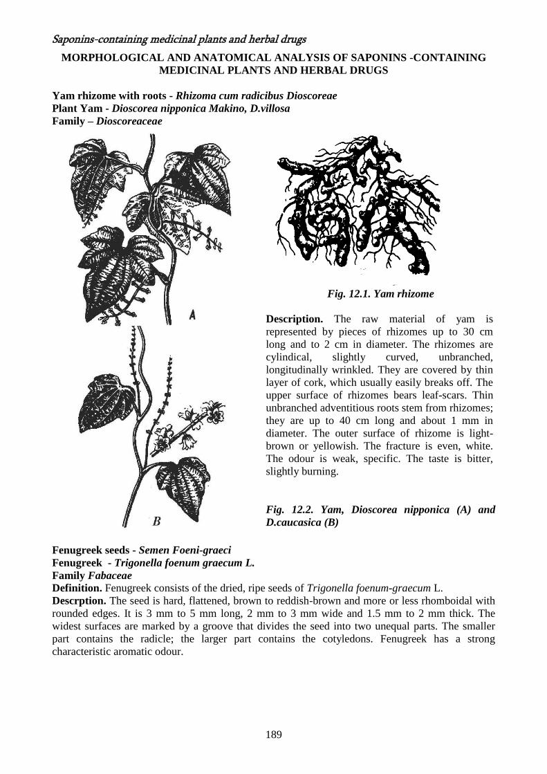
Yam rhizome with roots - Rhizoma cum radicibus Dioscoreae
Plant Yam - Dioscorea nipponica Makino, D.villosa
Family – Dioscoreaceae
Fig. 12.1. Yam rhizome
Description. The raw material of yam is
represented by pieces of rhizomes up to 30 cm
long and to 2 cm in diameter. The rhizomes are
cylindical, slightly curved, unbranched,
longitudinally wrinkled. They are covered by thin
layer of cork, which usually easily breaks off. The
upper surface of rhizomes bears leaf-scars. Thin
unbranched adventitious roots stem from rhizomes;
they are up to 40 cm long and about 1 mm in
diameter. The outer surface of rhizome is light-
brown or yellowish. The fracture is even, white.
The odour is weak, specific. The taste is bitter,
slightly burning.
Fig. 12.2. Yam, Dioscorea nipponica (A) and
D.caucasica (B)
Fenugreek seeds - Semen Foeni-graeci
Fenugreek - Trigonella foenum graecum L.
Family Fabaceae Definition. Fenugreek consists of the dried, ripe seeds of Trigonella foenum-graecum L.
Descrption. The seed is hard, flattened, brown to reddish-brown and more or less rhomboidal with
rounded edges. It is 3 mm to 5 mm long, 2 mm to 3 mm wide and 1.5 mm to 2 mm thick. The
widest surfaces are marked by a groove that divides the seed into two unequal parts. The smaller
part contains the radicle; the larger part contains the cotyledons. Fenugreek has a strong
characteristic aromatic odour.
190
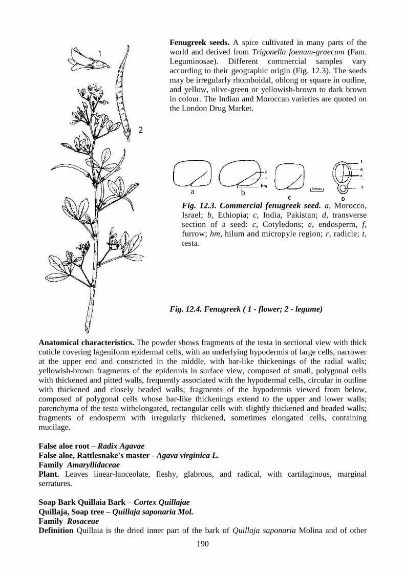
Fenugreek seeds. A spice cultivated in many parts of the
world and derived from Trigonella foenum-graecum (Fam.
Leguminosae). Different commercial samples vary
according to their geographic origin (Fig. 12.3). The seeds
may be irregularly rhomboidal, oblong or square in outline,
and yellow, olive-green or yellowish-brown to dark brown
in colour. The Indian and Moroccan varieties are quoted on
the London Drug Market.
a
b
Fig. 12.3. Commercial fenugreek seed. a, Morocco,
Israel; b, Ethiopia; c, India, Pakistan; d, transverse
section of a seed: c, Cotyledons; e, endosperm, f,
furrow; hm, hilum and micropyle region; r, radicle; t,
testa.
Fig. 12.4. Fenugreek ( 1 - flower; 2 - legume)
Anatomical characteristics. The powder shows fragments of the testa in sectional view with thick
cuticle covering lageniform epidermal cells, with an underlying hypodermis of large cells, narrower
at the upper end and constricted in the middle, with bar-like thickenings of the radial walls;
yellowish-brown fragments of the epidermis in surface view, composed of small, polygonal cells
with thickened and pitted walls, frequently associated with the hypodermal cells, circular in outline
with thickened and closely beaded walls; fragments of the hypodermis viewed from below,
composed of polygonal cells whose bar-like thickenings extend to the upper and lower walls;
parenchyma of the testa withelongated, rectangular cells with slightly thickened and beaded walls;
fragments of endosperm with irregularly thickened, sometimes elongated cells, containing
mucilage.
False aloe root – Radix Agavae
False aloe, Rattlesnake's master - Agava virginica L.
Family Amaryllidaceae
Plant. Leaves linear-lanceolate, fleshy, glabrous, and radical, with cartilaginous, marginal
serratures.
Soap Bark Quillaia Bark – Cortex Quillajae
Quillaja, Soap tree – Quillaja saponaria Mol.
Family Rosaceae
Definition Quillaia is the dried inner part of the bark of Quillaja saponaria Molina and of other

Saponins-containing medicinal plants and herbal drugs
191
species of Quillaja. Extractive soluble in ethanol (45%). Not less than 22.0%.
Description Pieces flat, up to about 1 metre
long, 10 to 20 cm broad and 3 to 10 mm, usually
6 mm, thick. Outer surface brownish white or
pale reddish brown, longitudinally striated or
coarsely reticulated, with occasional blackish
brown patches of adherent outer bark; inner
surface yellowish white, smooth and very hard;
fracture splintery and laminated, the broken
surface showing numerous large prisms of
calcium oxalate as glistening points. Smoothed
transversely cut surface appearing chequered,
with delicate radial lines representing medullary
rays and tangential lines formed by alternating
tangential bands of fibrous and non-fibrous
phloem. Odourless or almost odourless; dust
strongly sternutatory.
Fig. 12.5. Flowering Soap tree
Anatomical characteristics Outer bark, when present, consisting of reddish brown cork cells
alternating with bands of brown parenchyma containing numerous groups of phloem fibres and
large prisms of calcium oxalate. Inner bark consisting of alternating bands of tortuous fibres,
irregularly enlarged at intervals, about 500 to 1000 µm long and 20 to 50 µm wide and of sieve
tissue mixed with parenchyma. Medullary rays mostly three to four, but sometimes up to six cells
wide, with occasional pitted, subrectangular sclereids adjacent to the bundles of phloem fibres.
Starch granules 5 to 20 µm, usually about 10 µm, in diameter and prisms of calcium oxalate usually
50 to 170 µm long and up to 30 µm wide present in the parenchymatous cells.
Sarsaparilla root - Radix Sarsaparillae
Smilax - Smilax china L.
Smilax officinale - Smilax officinalis H.B.K.
Family Liliaceae
A
B
Fig. 12.6. A - Sassaparilla or smilax, B -
Sassaparilla roots
Description. Mexican Sarsaparilla. In long roots up to 6 mm. in diameter, frequently attached to a
tough, woody crown possessing one or more stem bases; externally light grayish-brown or weak
192
reddish-brown to yellowish-brown, longitudinally ridged and broadly furrowed, the furrows
sometimes containing blackish earth; with relatively few fibrous rootlets; fracture of cortex brittle,
central cylinder tough and fibrous; the cut or fractured surface exhibiting It mealy and pale orange,
or light yellowish brown and horny cortex, a yellow band porous woody zone and a lighter colored
central pith.
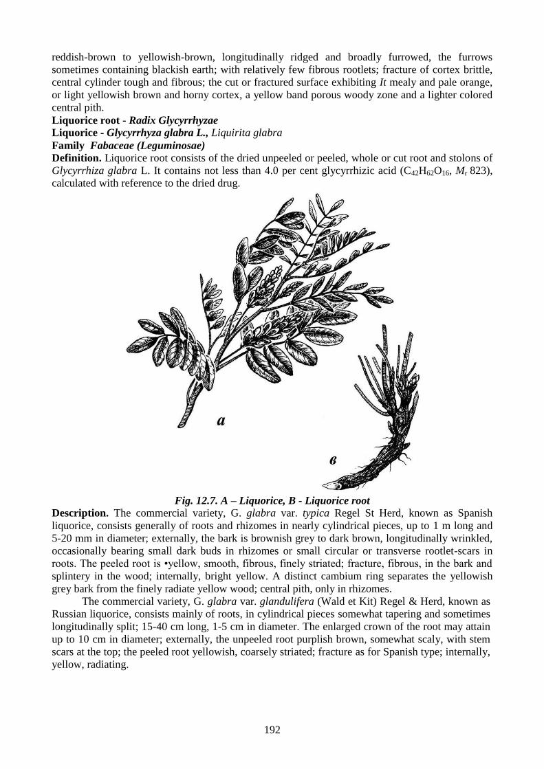
Liquorice root - Radix Glycyrrhyzae
Liquorice - Glycyrrhyza glabra L., Liquirita glabra
Family Fabaceae (Leguminosae)
Definition. Liquorice root consists of the dried unpeeled or peeled, whole or cut root and stolons of
Glycyrrhiza glabra L. It contains not less than 4.0 per cent glycyrrhizic acid (C42H62O16, Mr 823),
calculated with reference to the dried drug.
Fig. 12.7. A – Liquorice, B - Liquorice root
Description. The commercial variety, G. glabra var. typica Regel St Herd, known as Spanish
liquorice, consists generally of roots and rhizomes in nearly cylindrical pieces, up to 1 m long and
5-20 mm in diameter; externally, the bark is brownish grey to dark brown, longitudinally wrinkled,
occasionally bearing small dark buds in rhizomes or small circular or transverse rootlet-scars in
roots. The peeled root is •yellow, smooth, fibrous, finely striated; fracture, fibrous, in the bark and
splintery in the wood; internally, bright yellow. A distinct cambium ring separates the yellowish
grey bark from the finely radiate yellow wood; central pith, only in rhizomes.
The commercial variety, G. glabra var. glandulifera (Wald et Kit) Regel & Herd, known as
Russian liquorice, consists mainly of roots, in cylindrical pieces somewhat tapering and sometimes
longitudinally split; 15-40 cm long, 1-5 cm in diameter. The enlarged crown of the root may attain
up to 10 cm in diameter; externally, the unpeeled root purplish brown, somewhat scaly, with stem
scars at the top; the peeled root yellowish, coarsely striated; fracture as for Spanish type; internally,
yellow, radiating.
Saponins-containing medicinal plants and herbal drugs
193
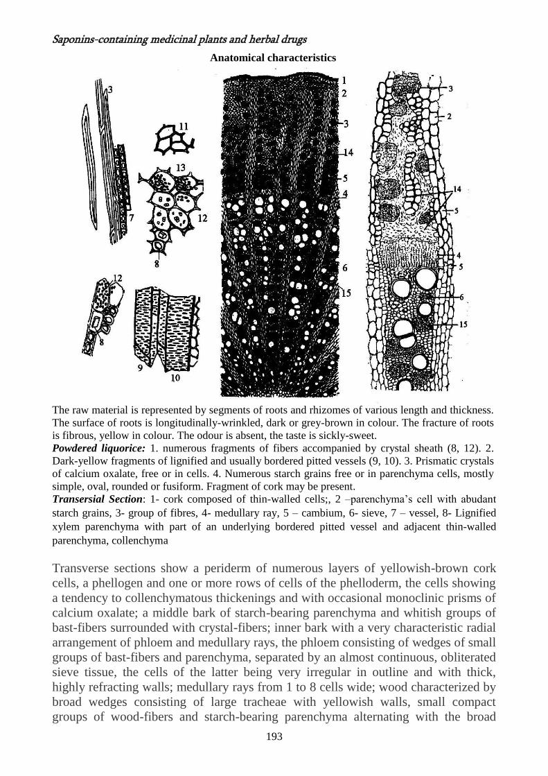
Anatomical characteristics
The raw material is represented by segments of roots and rhizomes of various length and thickness.
The surface of roots is longitudinally-wrinkled, dark or grey-brown in colour. The fracture of roots
is fibrous, yellow in colour. The odour is absent, the taste is sickly-sweet.
Powdered liquorice: 1. numerous fragments of fibers accompanied by crystal sheath (8, 12). 2.
Dark-yellow fragments of lignified and usually bordered pitted vessels (9, 10). 3. Prismatic crystals
of calcium oxalate, free or in cells. 4. Numerous starch grains free or in parenchyma cells, mostly
simple, oval, rounded or fusiform. Fragment of cork may be present.
Transersial Section: 1- cork composed of thin-walled cells;, 2 –parenchyma’s cell with abudant
starch grains, 3- group of fibres, 4- medullary ray, 5 – cambium, 6- sieve, 7 – vessel, 8- Lignified
xylem parenchyma with part of an underlying bordered pitted vessel and adjacent thin-walled
parenchyma, collenchyma
Transverse sections show a periderm of numerous layers of yellowish-brown cork
cells, a phellogen and one or more rows of cells of the phelloderm, the cells showing
a tendency to collenchymatous thickenings and with occasional monoclinic prisms of
calcium oxalate; a middle bark of starch-bearing parenchyma and whitish groups of
bast-fibers surrounded with crystal-fibers; inner bark with a very characteristic radial
arrangement of phloem and medullary rays, the phloem consisting of wedges of small
groups of bast-fibers and parenchyma, separated by an almost continuous, obliterated
sieve tissue, the cells of the latter being very irregular in outline and with thick,
highly refracting walls; medullary rays from 1 to 8 cells wide; wood characterized by
broad wedges consisting of large tracheae with yellowish walls, small compact
groups of wood-fibers and starch-bearing parenchyma alternating with the broad
194
medullary rays; pith composed of parenchyma, the cells being large, more or less
polygonal in outline and containing numerous starch grains, or prisms of calcium
oxalate. In sections of roots the pith is absent.
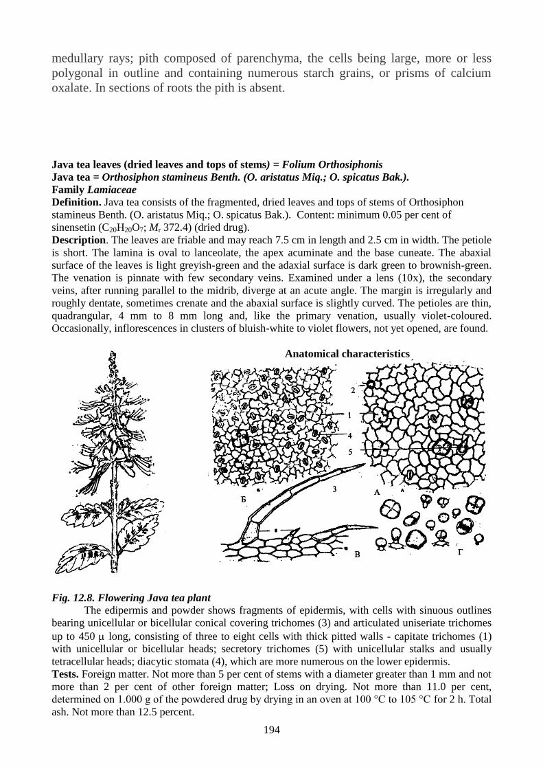
Java tea leaves (dried leaves and tops of stems) = Folium Orthosiphonis
Java tea = Orthosiphon stamineus Benth. (O. aristatus Miq.; O. spicatus Bak.).
Family Lamiaceae
Definition. Java tea consists of the fragmented, dried leaves and tops of stems of Orthosiphon
stamineus Benth. (O. aristatus Miq.; O. spicatus Bak.). Content: minimum 0.05 per cent of
sinensetin (C20H20O7; Mr 372.4) (dried drug).
Description. The leaves are friable and may reach 7.5 cm in length and 2.5 cm in width. The petiole
is short. The lamina is oval to lanceolate, the apex acuminate and the base cuneate. The abaxial
surface of the leaves is light greyish-green and the adaxial surface is dark green to brownish-green.
The venation is pinnate with few secondary veins. Examined under a lens (10x), the secondary
veins, after running parallel to the midrib, diverge at an acute angle. The margin is irregularly and
roughly dentate, sometimes crenate and the abaxial surface is slightly curved. The petioles are thin,
quadrangular, 4 mm to 8 mm long and, like the primary venation, usually violet-coloured.
Occasionally, inflorescences in clusters of bluish-white to violet flowers, not yet opened, are found.
Anatomical characteristics
Fig. 12.8. Flowering Java tea plant The edipermis and powder shows fragments of epidermis, with cells with sinuous outlines
bearing unicellular or bicellular conical covering trichomes (3) and articulated uniseriate trichomes
up to 450 long, consisting of three to eight cells with thick pitted walls - capitate trichomes (1)
with unicellular or bicellular heads; secretory trichomes (5) with unicellular stalks and usually
tetracellular heads; diacytic stomata (4), which are more numerous on the lower epidermis.
Tests. Foreign matter. Not more than 5 per cent of stems with a diameter greater than 1 mm and not
more than 2 per cent of other foreign matter; Loss on drying. Not more than 11.0 per cent,
determined on 1.000 g of the powdered drug by drying in an oven at 100 °C to 105 °C for 2 h. Total
ash. Not more than 12.5 percent.
Saponins-containing medicinal plants and herbal drugs
195
Horse chestnut seeds = Semen Hippocastani
Horse chestnut = Aesculum hippocastanum L.
Family Hippocastanaceae
Definition. Semen Hippocastani consists of the dried ripe seeds of
Aesculum hippocastanum L. It contains not less than 3, 0% triterpen
saponins calculated as aescin (escin) determined by spectrophotometry at 540
nm.
Description. Globulous or ovoid, 2-4 cm in diameter. The 2 large cotyledons, oily and starchy,
often connate with a line of suture more or less visible; covered by a shiny dark-brown tegument
with a large whitish spot corresponding to the hilum; tegument creamy white in the immature seed,
takes on a brown tinge during maturation, becoming dark brown when mature. Curved radicle
occupies a depression either on the commissure of the cotyledons or the dorsal side of the
cotyledons. Odour slight; taste bitter, acrid.
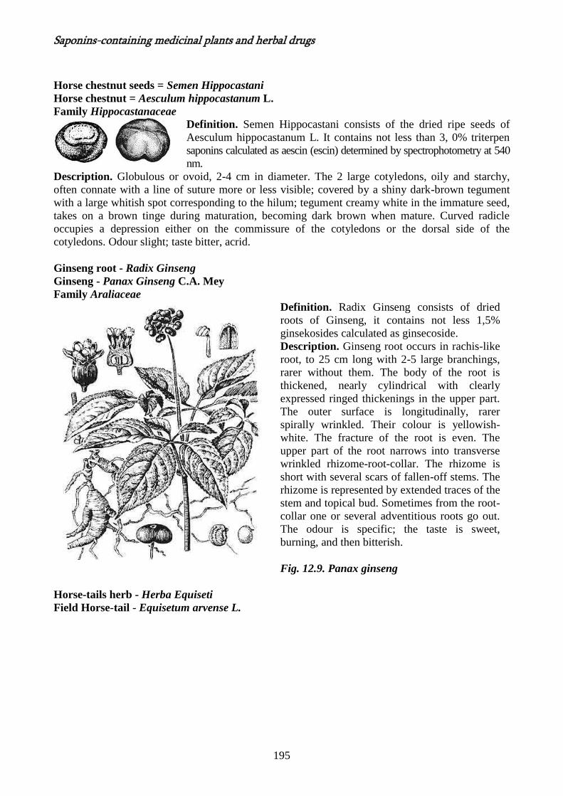
Ginseng root - Radix Ginseng
Ginseng - Panax Ginseng C.A. Mey
Family Araliaceae
Definition. Radix Ginseng consists of dried
roots of Ginseng, it contains not less 1,5%
ginsekosides calculated as ginsecoside.
Description. Ginseng root occurs in rachis-like
root, to 25 cm long with 2-5 large branchings,
rarer without them. The body of the root is
thickened, nearly cylindrical with clearly
expressed ringed thickenings in the upper part.
The outer surface is longitudinally, rarer
spirally wrinkled. Their colour is yellowish-
white. The fracture of the root is even. The
upper part of the root narrows into transverse
wrinkled rhizome-root-collar. The rhizome is
short with several scars of fallen-off stems. The
rhizome is represented by extended traces of the
stem and topical bud. Sometimes from the root-
collar one or several adventitious roots go out.
The odour is specific; the taste is sweet,
burning, and then bitterish.
Fig. 12.9. Panax ginseng
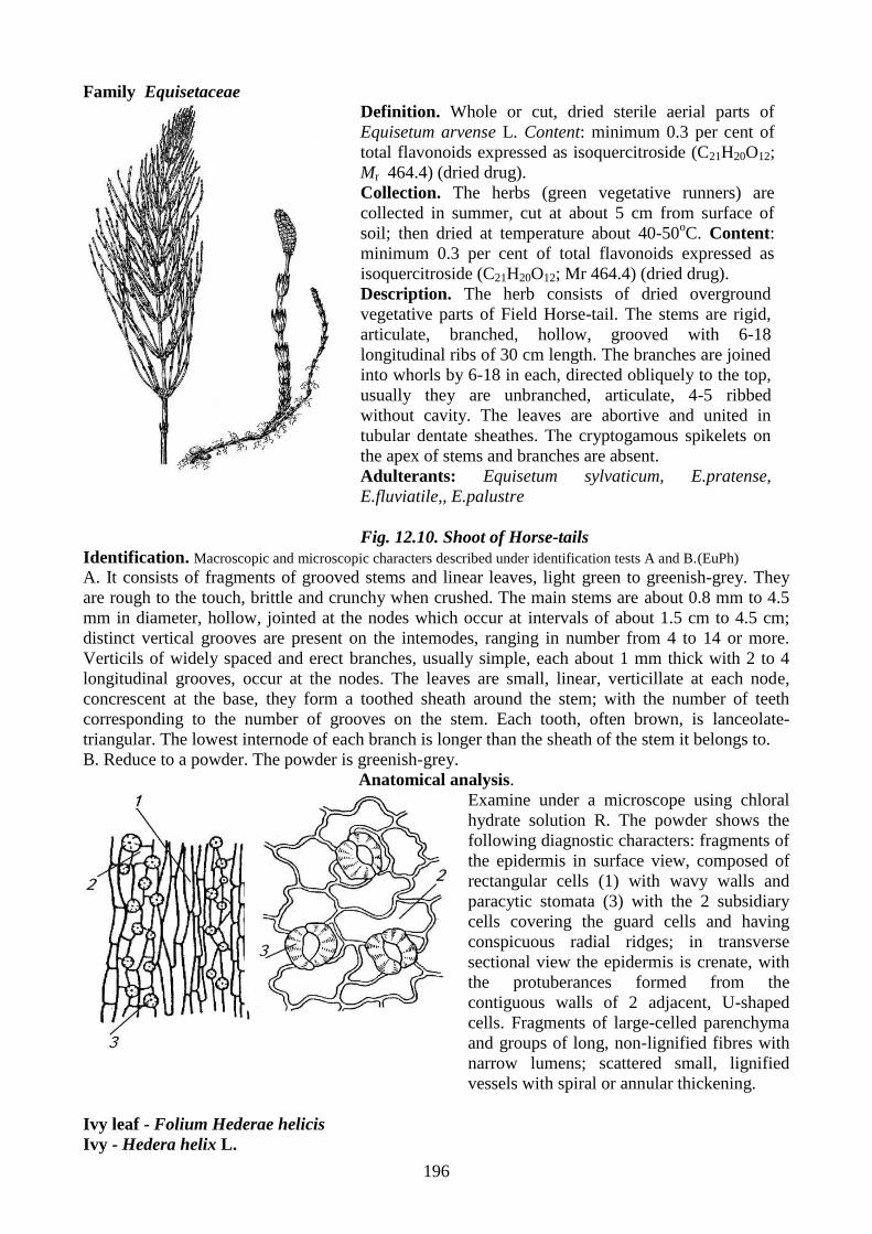
Horse-tails herb - Herba Equiseti
Field Horse-tail - Equisetum arvense L.
196
Family Equisetaceae
Definition. Whole or cut, dried sterile aerial parts of
Equisetum arvense L. Content: minimum 0.3 per cent of
total flavonoids expressed as isoquercitroside (C21H20O12;
Mr 464.4) (dried drug).
Collection. The herbs (green vegetative runners) are
collected in summer, cut at about 5 cm from surface of
soil; then dried at temperature about 40-50oC. Content:
minimum 0.3 per cent of total flavonoids expressed as
isoquercitroside (C21H20O12; Mr 464.4) (dried drug).
Description. The herb consists of dried overground
vegetative parts of Field Horse-tail. The stems are rigid,
articulate, branched, hollow, grooved with 6-18
longitudinal ribs of 30 cm length. The branches are joined
into whorls by 6-18 in each, directed obliquely to the top,
usually they are unbranched, articulate, 4-5 ribbed
without cavity. The leaves are abortive and united in
tubular dentate sheathes. The cryptogamous spikelets on
the apex of stems and branches are absent.
Adulterants: Equisetum sylvaticum, E.pratense,
E.fluviatile,, E.palustre
Fig. 12.10. Shoot of Horse-tails
Identification. Macroscopic and microscopic characters described under identification tests A and B.(EuPh)
A. It consists of fragments of grooved stems and linear leaves, light green to greenish-grey. They
are rough to the touch, brittle and crunchy when crushed. The main stems are about 0.8 mm to 4.5
mm in diameter, hollow, jointed at the nodes which occur at intervals of about 1.5 cm to 4.5 cm;
distinct vertical grooves are present on the intemodes, ranging in number from 4 to 14 or more.
Verticils of widely spaced and erect branches, usually simple, each about 1 mm thick with 2 to 4
longitudinal grooves, occur at the nodes. The leaves are small, linear, verticillate at each node,
concrescent at the base, they form a toothed sheath around the stem; with the number of teeth
corresponding to the number of grooves on the stem. Each tooth, often brown, is lanceolate-
triangular. The lowest internode of each branch is longer than the sheath of the stem it belongs to.
B. Reduce to a powder. The powder is greenish-grey.
Anatomical analysis.
Examine under a microscope using chloral
hydrate solution R. The powder shows the
following diagnostic characters: fragments of
the epidermis in surface view, composed of
rectangular cells (1) with wavy walls and
paracytic stomata (3) with the 2 subsidiary
cells covering the guard cells and having
conspicuous radial ridges; in transverse
sectional view the epidermis is crenate, with
the protuberances formed from the
contiguous walls of 2 adjacent, U-shaped
cells. Fragments of large-celled parenchyma
and groups of long, non-lignified fibres with
narrow lumens; scattered small, lignified
vessels with spiral or annular thickening.
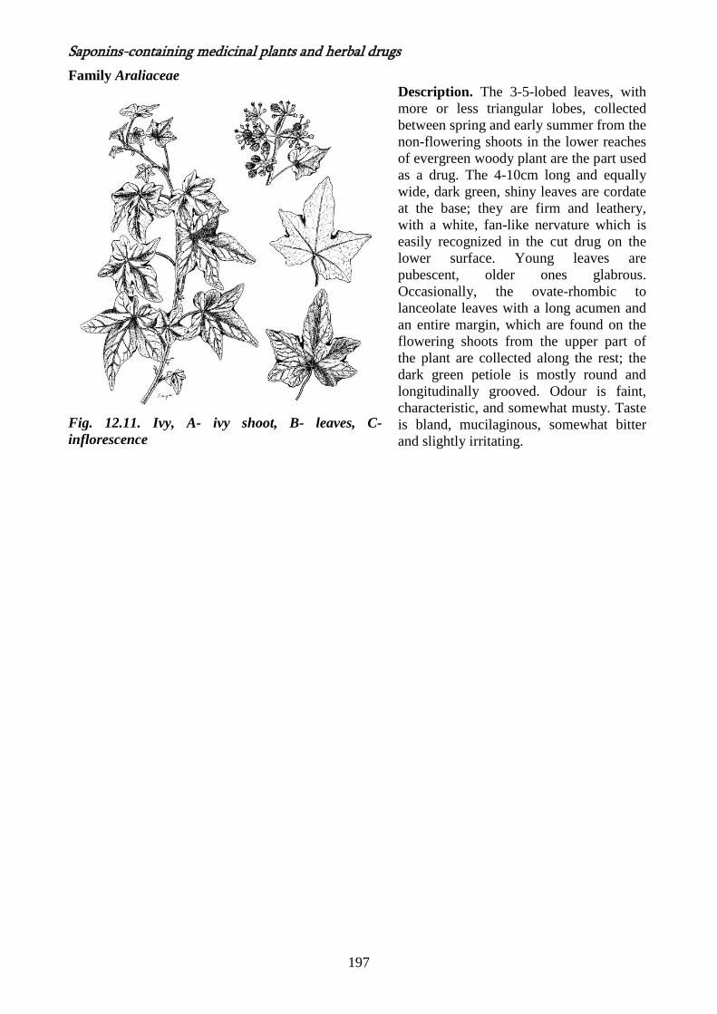
Ivy leaf - Folium Hederae helicis
Ivy - Hedera helix L.
Saponins-containing medicinal plants and herbal drugs
197
Family Araliaceae
Fig. 12.11. Ivy, A- ivy shoot, B- leaves, C-
inflorescence
Description. The 3-5-lobed leaves, with
more or less triangular lobes, collected
between spring and early summer from the
non-flowering shoots in the lower reaches
of evergreen woody plant are the part used
as a drug. The 4-10cm long and equally
wide, dark green, shiny leaves are cordate
at the base; they are firm and leathery,
with a white, fan-like nervature which is
easily recognized in the cut drug on the
lower surface. Young leaves are
pubescent, older ones glabrous.
Occasionally, the ovate-rhombic to
lanceolate leaves with a long acumen and
an entire margin, which are found on the
flowering shoots from the upper part of
the plant are collected along the rest; the
dark green petiole is mostly round and
longitudinally grooved. Odour is faint,
characteristic, and somewhat musty. Taste
is bland, mucilaginous, somewhat bitter
and slightly irritating.
198
The root of Soapwort - Radix Saponaria
Soapwort - Saponaria officinalis
Family Caryophyllaceae
Description. It occurs in cylindrical, long, hard, thin pieces. The
outer surface is red-brown in colour, the fracture is yellowish. The
odour is absent, the taste is slightly burning, irritating remaining
for a long time.
Fig. 12.12. Flowering shoot of soapwort
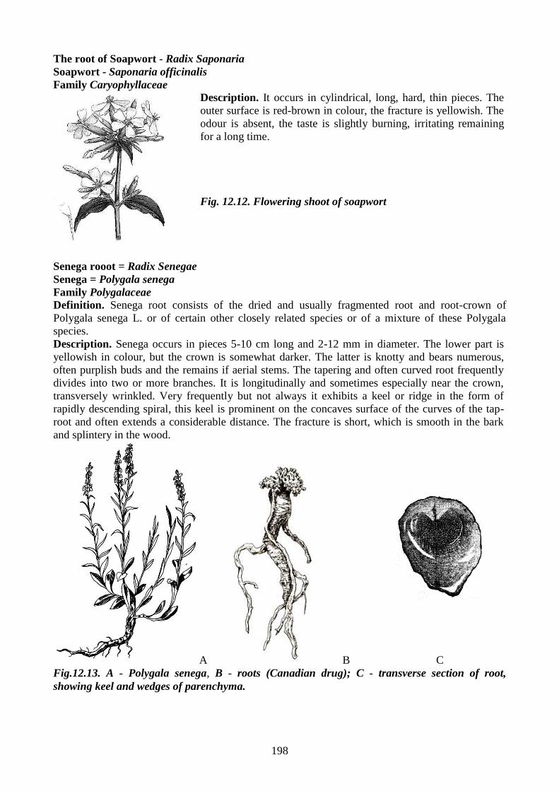
Senega rooot = Radix Senegae
Senega = Polygala senega
Family Polygalaceae
Definition. Senega root consists of the dried and usually fragmented root and root-crown of
Polygala senega L. or of certain other closely related species or of a mixture of these Polygala
species.
Description. Senega occurs in pieces 5-10 cm long and 2-12 mm in diameter. The lower part is
yellowish in colour, but the crown is somewhat darker. The latter is knotty and bears numerous,
often purplish buds and the remains if aerial stems. The tapering and often curved root frequently
divides into two or more branches. It is longitudinally and sometimes especially near the crown,
transversely wrinkled. Very frequently but not always it exhibits a keel or ridge in the form of
rapidly descending spiral, this keel is prominent on the concaves surface of the curves of the tap-
root and often extends a considerable distance. The fracture is short, which is smooth in the bark
and splintery in the wood.
A B
C
Fig.12.13. A - Polygala senega, B - roots (Canadian drug); C - transverse section of root,
showing keel and wedges of parenchyma.