Carbohydrates analysis in herbal glycomics
15
Review Carbohydrates analysis in herbal glycomics Shao-ping Li ⇑ , Ding-tao Wu 1 , Guang-ping Lv 1 , Jing Zhao 1 State Key Laboratory of Quality Research in Chinese Medicine, Institute of Chinese Medical Sciences, University of Macau, Macao SAR, China article info Keywords: Atomic force microscopy (AFM) Carbohydrate Chromatography Functional food Herbal glycomics Herbal ingredient Lectin array Mass spectrometry (MS) Multiple-angle laser-light scattering (MALLS) Quality control (QC) abstract Herbs serve primary healthcare needs and some herbs are widely used as functional foods. Analysis of carbohydrates is essential to understand their structure–function relationships in food science and herbal medicine. However, accurately defining chemical structures of glycans is a challenge due to their chemical heterogeneity and diversity. We review and discuss carbohydrates as new functional ingredi- ents in herbs and functional foods, and their analysis, a novel aspect in quality control and herbal glycomics. Ó 2013 Elsevier Ltd. All rights reserved. Contents 1. Introduction ......................................................................................................... 156 2. Functions and structures of herbal carbohydrates ........................................................................... 156 3. Analysis of carbohydrates in herbal glycomics.............................................................................. 157 3.1. Analysis of monosaccharides ...................................................................................... 158 3.1.1. HPTLC ................................................................................................. 158 3.1.2. HPLC .................................................................................................. 160 3.1.3. GC .................................................................................................... 161 3.1.4. CE..................................................................................................... 161 3.2. Analysis of oligosaccharides ....................................................................................... 161 3.2.1. HPTLC ................................................................................................. 161 3.2.2. HPLC .................................................................................................. 161 3.2.3. CE..................................................................................................... 163 3.3. Analysis of polysaccharides and glycoconjugates ...................................................................... 163 3.3.1. SEC-MALLS-(VIS) ......................................................................................... 163 3.3.2. AFM ................................................................................................... 165 3.4. Lectin array .................................................................................................... 166 4. Conclusion .......................................................................................................... 166 Acknowledgements ................................................................................................... 166 References .......................................................................................................... 167 0165-9936/$ - see front matter Ó 2013 Elsevier Ltd. All rights reserved. http://dx.doi.org/10.1016/j.trac.2013.05.020 Abbreviations: AFM, Atomic force microscopy; ANTS, 8-aminonaphthalene-1,3,6-trisulfonic acid; CAD, Charged aerosol detector; CZE, Capillary zone electrophoresis; C 1 , Limited characteristic ratio; d, Diameter of the chain; DAD, Diode-array detector; DLS, Dynamic light scattering; DPA, Diphenylamine-aniline-phosphoric acid; ELSD, Evaporative light-scattering detector; ESI-MS, Electrospray-ionization mass spectrometry; FLD, Fluorescence detector; FTIR, Fourier-transform infrared; HILIC, Hydrophilic interaction liquid chromatography; HPAEC, High-performance anion-exchange chromatography; HPTLC, High-performance thin-layer chromatography; LIF, Laser-induced fluorescence; LLS, Laser-light scattering; MALDI, Matrix-assisted laser desorption/ionization; MEKC, Micellar eletrokinetic chromatography; mf-MELDI–MS, Material enhanced laser desorption/ionization time of flight mass spectrometry; M L , Molar mass per unit contour length; M w , Molecular weight; OPLC, Overpressured layer chromatography; P, Persistence length; PAD, Pulsed amperometry detection; PDA, Photodiode array; PDI, Polydispersity index; PMP, 1-phenyl-3-methyl-5-pyrazolone; Q- TOF, Quadrupole time-of-flight; Rho110, Rhodamine 110; RID, Refractive index detector; SEC, Size-exclusion chromatography; THC, Triple helical conformation; t-ITP, Transient isotachophoretic preconcentration; TMS, Trimethylsilyl; VIS, Viscometer. ⇑ Corresponding author. Fax: +853 2884 1358. E-mail address: [email protected] (S.-p. Li). 1 These authors contributed equally to this work. Trends in Analytical Chemistry 52 (2013) 155–169 Contents lists available at ScienceDirect Trends in Analytical Chemistry journal homepage: www.elsevier.com/locate/trac
-
Upload
independent -
Category
Documents
-
view
1 -
download
0
Transcript of Carbohydrates analysis in herbal glycomics

Trends in Analytical Chemistry 52 (2013) 155–169
Contents lists available at ScienceDirect
Trends in Analytical Chemistry
journal homepage: www.elsevier .com/locate / t rac
Review
Carbohydrates analysis in herbal glycomics
Shao-ping Li ⇑, Ding-tao Wu 1, Guang-ping Lv 1, Jing Zhao 1
State Key Laboratory of Quality Research in Chinese Medicine, Institute of Chinese Medical Sciences, University of Macau, Macao SAR, China
a r t i c l e i n f o a b s t r a c t
Keywords:Atomic force microscopy (AFM)
CarbohydrateChromatographyFunctional foodHerbal glycomicsHerbal ingredientLectin arrayMass spectrometry (MS)Multiple-angle laser-light scattering(MALLS)Quality control (QC)0165-9936/$ - see front matter � 2013 Elsevier Ltd. Ahttp://dx.doi.org/10.1016/j.trac.2013.05.020
Abbreviations: AFM, Atomic force microscopy; ANTLimited characteristic ratio; d, Diameter of the chaiEvaporative light-scattering detector; ESI-MS, Electrointeraction liquid chromatography; HPAEC, High-perffluorescence; LLS, Laser-light scattering; MALDI, Menhanced laser desorption/ionization time of flightchromatography; P, Persistence length; PAD, Pulsed aTOF, Quadrupole time-of-flight; Rho110, RhodamineTransient isotachophoretic preconcentration; TMS, Tr⇑ Corresponding author. Fax: +853 2884 1358.
E-mail address: [email protected] (S.-p. Li).1 These authors contributed equally to this work.
Herbs serve primary healthcare needs and some herbs are widely used as functional foods. Analysis ofcarbohydrates is essential to understand their structure–function relationships in food science andherbal medicine. However, accurately defining chemical structures of glycans is a challenge due to theirchemical heterogeneity and diversity. We review and discuss carbohydrates as new functional ingredi-ents in herbs and functional foods, and their analysis, a novel aspect in quality control and herbalglycomics.
� 2013 Elsevier Ltd. All rights reserved.
Contents
1. Introduction . . . . . . . . . . . . . . . . . . . . . . . . . . . . . . . . . . . . . . . . . . . . . . . . . . . . . . . . . . . . . . . . . . . . . . . . . . . . . . . . . . . . . . . . . . . . . . . . . . . . . . . . . 1562. Functions and structures of herbal carbohydrates . . . . . . . . . . . . . . . . . . . . . . . . . . . . . . . . . . . . . . . . . . . . . . . . . . . . . . . . . . . . . . . . . . . . . . . . . . . 1563. Analysis of carbohydrates in herbal glycomics. . . . . . . . . . . . . . . . . . . . . . . . . . . . . . . . . . . . . . . . . . . . . . . . . . . . . . . . . . . . . . . . . . . . . . . . . . . . . . 157
3.1. Analysis of monosaccharides . . . . . . . . . . . . . . . . . . . . . . . . . . . . . . . . . . . . . . . . . . . . . . . . . . . . . . . . . . . . . . . . . . . . . . . . . . . . . . . . . . . . . . 158
3.1.1. HPTLC . . . . . . . . . . . . . . . . . . . . . . . . . . . . . . . . . . . . . . . . . . . . . . . . . . . . . . . . . . . . . . . . . . . . . . . . . . . . . . . . . . . . . . . . . . . . . . . . . 1583.1.2. HPLC . . . . . . . . . . . . . . . . . . . . . . . . . . . . . . . . . . . . . . . . . . . . . . . . . . . . . . . . . . . . . . . . . . . . . . . . . . . . . . . . . . . . . . . . . . . . . . . . . . 1603.1.3. GC . . . . . . . . . . . . . . . . . . . . . . . . . . . . . . . . . . . . . . . . . . . . . . . . . . . . . . . . . . . . . . . . . . . . . . . . . . . . . . . . . . . . . . . . . . . . . . . . . . . . 1613.1.4. CE. . . . . . . . . . . . . . . . . . . . . . . . . . . . . . . . . . . . . . . . . . . . . . . . . . . . . . . . . . . . . . . . . . . . . . . . . . . . . . . . . . . . . . . . . . . . . . . . . . . . . 1613.2. Analysis of oligosaccharides . . . . . . . . . . . . . . . . . . . . . . . . . . . . . . . . . . . . . . . . . . . . . . . . . . . . . . . . . . . . . . . . . . . . . . . . . . . . . . . . . . . . . . . 161
3.2.1. HPTLC . . . . . . . . . . . . . . . . . . . . . . . . . . . . . . . . . . . . . . . . . . . . . . . . . . . . . . . . . . . . . . . . . . . . . . . . . . . . . . . . . . . . . . . . . . . . . . . . . 1613.2.2. HPLC . . . . . . . . . . . . . . . . . . . . . . . . . . . . . . . . . . . . . . . . . . . . . . . . . . . . . . . . . . . . . . . . . . . . . . . . . . . . . . . . . . . . . . . . . . . . . . . . . . 1613.2.3. CE. . . . . . . . . . . . . . . . . . . . . . . . . . . . . . . . . . . . . . . . . . . . . . . . . . . . . . . . . . . . . . . . . . . . . . . . . . . . . . . . . . . . . . . . . . . . . . . . . . . . . 1633.3. Analysis of polysaccharides and glycoconjugates . . . . . . . . . . . . . . . . . . . . . . . . . . . . . . . . . . . . . . . . . . . . . . . . . . . . . . . . . . . . . . . . . . . . . . 163
3.3.1. SEC-MALLS-(VIS). . . . . . . . . . . . . . . . . . . . . . . . . . . . . . . . . . . . . . . . . . . . . . . . . . . . . . . . . . . . . . . . . . . . . . . . . . . . . . . . . . . . . . . . . 1633.3.2. AFM . . . . . . . . . . . . . . . . . . . . . . . . . . . . . . . . . . . . . . . . . . . . . . . . . . . . . . . . . . . . . . . . . . . . . . . . . . . . . . . . . . . . . . . . . . . . . . . . . . . 1653.4. Lectin array . . . . . . . . . . . . . . . . . . . . . . . . . . . . . . . . . . . . . . . . . . . . . . . . . . . . . . . . . . . . . . . . . . . . . . . . . . . . . . . . . . . . . . . . . . . . . . . . . . . . 166
4. Conclusion . . . . . . . . . . . . . . . . . . . . . . . . . . . . . . . . . . . . . . . . . . . . . . . . . . . . . . . . . . . . . . . . . . . . . . . . . . . . . . . . . . . . . . . . . . . . . . . . . . . . . . . . . . 166Acknowledgements . . . . . . . . . . . . . . . . . . . . . . . . . . . . . . . . . . . . . . . . . . . . . . . . . . . . . . . . . . . . . . . . . . . . . . . . . . . . . . . . . . . . . . . . . . . . . . . . . . . 166References . . . . . . . . . . . . . . . . . . . . . . . . . . . . . . . . . . . . . . . . . . . . . . . . . . . . . . . . . . . . . . . . . . . . . . . . . . . . . . . . . . . . . . . . . . . . . . . . . . . . . . . . . . 167
ll rights reserved.
S, 8-aminonaphthalene-1,3,6-trisun; DAD, Diode-array detector; DLspray-ionization mass spectrometrormance anion-exchange chromatatrix-assisted laser desorption/ionmass spectrometry; ML, Molar mmperometry detection; PDA, Phot110; RID, Refractive index detec
imethylsilyl; VIS, Viscometer.
lfonic acid; CAD, Charged aerosol detector; CZE, Capillary zone electrophoresis; C1,S, Dynamic light scattering; DPA, Diphenylamine-aniline-phosphoric acid; ELSD,y; FLD, Fluorescence detector; FTIR, Fourier-transform infrared; HILIC, Hydrophilicography; HPTLC, High-performance thin-layer chromatography; LIF, Laser-inducedization; MEKC, Micellar eletrokinetic chromatography; mf-MELDI–MS, Materialass per unit contour length; Mw, Molecular weight; OPLC, Overpressured layer
odiode array; PDI, Polydispersity index; PMP, 1-phenyl-3-methyl-5-pyrazolone; Q-tor; SEC, Size-exclusion chromatography; THC, Triple helical conformation; t-ITP,

156 S.-p. Li et al. / Trends in Analytical Chemistry 52 (2013) 155–169
1. Introduction
Carbohydrates, the most abundant natural products, may beclassified according to their degree of polymerization (DP) and di-vided initially into three principal groups, namely sugars (DP 1–2),oligosaccharides (DP 3–9) and polysaccharides (DP > 9). Generally,carbohydrates are often thought of as food sources and structuralbuilding blocks. Their biological roles have in many ways beenoverlooked in the past, compared with the roles of proteins and nu-cleic acids [1]. However, unlike nucleic acids and proteins, thecomplexity of carbohydrates is unique. The number of possiblestructures calculated for reducing hexasaccharides comprised ofD-hexoses is >1012 alone [2], though it is recognized that many the-oretical combinations may not be synthesized in nature. Neverthe-less, over the past decade, researchers increasingly turned theirattention toward understanding the role of carbohydrates in nor-mal cellular function and disease, and opened up new researchfronts in terms of probing glycans as targets in the design andthe development of novel drugs or new therapies for infectiousand neurological diseases, cancer, and metabolic disorders [3,4].
Herbs, originating from almost every part of the globe, serve theprimary healthcare needs of up to 80% of people in Africa, and ac-count for 30–50% of the total medicinal consumption in China.These herbs are complex mixtures, usually containing hundredsof chemical constituents, but only a few compounds are responsiblefor the beneficial effects. Carbohydrates, especially oligosaccha-rides, polysaccharides, and glycoconjugates are a relevant part ofthe bioactive components of the natural products exploited intherapeutics, diagnostics, food additives, and biomaterials [5–9].Carbohydrates analysis is therefore very important for qualitycontrol (QC) of herbal medicine. But accurately defining chemicalstructures of glycans is a challenge due to their chemicalheterogeneity and diversity [10]. Glycomics, an integrated systemsapproach to structure-function relationships of glycans, rapidlydeveloped in the past decade. Herbal glycomics, glycomics inherbal medicine, involves all methods and approaches, includingstructural analysis of glycans, study of interactions betweenglycans and counter molecules and glyco-informatics, used toprofile the glycome (all components of glycans) in herbs.
As we know, the biological activities of carbohydrates areclosely correlated to their physico-chemical properties, such asmolecular size, types and ratios of constituent monosaccharides,and features of glycosidic linkages (e.g., configuration and positionof glycosidic linkages, and sequence of monosaccharides) [11].Characterization of carbohydrates is therefore necessary to ensuretheir efficacy and safety. Herbal glycomics is necessary and crucialfor good understanding of the herbal glycome and the holistic ef-fects of herbs, and is a powerful tool for rational selection of qualitymarkers and improving approaches to the QC of herbal medicines.However, carbohydrates analysis is a bottleneck due to their com-plexity. In this review, we summarize and discuss advances in car-bohydrates analysis in herbal glycomics, based on function-relatedcharacteristics of herbal carbohydrates.
2. Functions and structures of herbal carbohydrates
The targets for QC of herbal medicine should be bioactivecomponents and/or functional characteristics. A great deal hasbeen learnt in recent years about the biological roles of carbohy-drates, with a rapid increase of publications in this field [12]. In-deed, carbohydrates may directly influence human diseases byaffecting physiological and metabolic processes (Table 1).The simplest carbohydrates are the monosaccharides, which canbe joined together to make disaccharides, oligosaccharides andpolysaccharides.
D-allose, the C-3 epimer of D-glucose, is one of the rare mono-saccharides existing in nature, and it has been shown to have mul-tiple effects, including immunosuppressive [13,14], anti-inflammatory [15], anti-oxidant [13,14,16,17], and anti-prolifera-tion [13,14] activities. D-allose may be a useful tool for controllingthe growth of certain types of cancers, and it also has potential todevelop a new therapeutic strategy with high expression of thiore-doxin-interacting protein for leukemia patients [13]. D-allose canalso reinforce the action of metronidazole and trichomonad on Tri-trichomonas fetus [18].
Similar results were also investigated for another rare monosac-charide, D-psicose, C-3 epimer of D-fructose, which originates fromwheat, Itea plants, processed cane and beet molasses [18,19]. D-psi-cose has shown antidiabetic activity and may have antidyslipidem-ic effects in type II diabetes [19]. It also protects pancreas b-isletsand thus improves insulin resistance in OLETF rats [20].
Besides the significant pharmacological activities of rare mono-saccharides, some abundant monosaccharides in nature, such asmannose [21] and arabinose [22], are effective in reducing lipo-polysaccharide (LPS)-induced acute lung injury [21] and protectingagainst hyperglycemia [22], respectively (Table 1).
Trehalose, a glucose disaccharide (a, a-1,1-glucoside bond), canbe abundant in some mushrooms, such as Cordyceps sinensis, C.liangshanensis and C. gunnii [23]. The sugar possesses unique phys-ical and chemical properties, which have been used for preserva-tion of biological molecules, including lipids and proteins, andmore recently, stem cells, tissues, and organs [24]. Besides, treha-lose also inhibits proinflammatory phenotype activation in macro-phages, prevents endotoxin-induced mortality [25], blocks theinflammatory cascade triggered by endotoxin shock, stabilizesthe bio-membranes and switches off water-diffusive dynamics[26]. Trehalose, which has suppressive effects on several patholog-ical events after subarachnoid hemorrhage (SAH), including vaso-spasm, inflammatory responses, and lipid peroxidation, mayprovide a new therapeutic approach for treatment of complicationsafter SAH [27]. In addition, trehalose could promote the clearanceof A53T mutant a-synuclein (a-Syn) in PC12 cells mediatedthrough the macroautophagy pathway. Trehalose may thereforebe effective in treating Parkinson’s disease, though the macroauto-phagy pathway is not a dominant way for wild type WTa-Synclearance [28].
Consumers are increasingly interested in their personal health,and expect their foods to be – beyond tasty and attractive – alsosafe and healthy. Non-digestible oligosaccharides (NDOs), whichcan be obtained by direct extraction from natural sources, or pro-duced by chemical processes hydrolyzing polysaccharides, or byenzymatic and chemical synthesis from disaccharides, possessimportant physicochemical and physiological properties, and areclaimed as dietary fibers and prebiotics [29]. Xu et al. [30] dis-cussed the potential of the functional oligosaccharides to modulatethe gut flora, to affect different gastrointestinal activities and lipidmetabolism, to enhance immunity, and to reduce diabetes, obesityand cardiovascular risk. Common commercial NDOs, includingfructo-oligosaccharides (FOSs) and galacto-oligosaccharides, havebeen reviewed [29]. Besides the important beneficial physiologicaleffects [31], FOSs from Morinda officinalis [32], Smallanthus son-chifolius [33] and Asparagus racemosus [34] also possess antidepres-sant, anti-inflammatory and potentiating NK-cell activities,respectively. The biological effects of oligosaccharides are summa-rized in Table 1.
Polysaccharides, especially water-soluble, from medicinal plantsand fungi are most important for their significant pharmaceuticalapplications. The pharmaceutical effects of more and more kinds ofnatural polysaccharide with different curative effects have beentested and even applied in therapies. Major pharmaceutical activities,such as antitumor [35,36], antiviral [37], immunoregulatory [9,38],

Table 1Summary of biological activities of carbohydrates from medicinal plants and fungi (Data from 178 journal articles collected in Web of Science mainly dated 2008–12)
Carbohydrates (or constituentmonosaccharides)
Origin Types of linkage Activitya
1 2 3 4 5 6
Monosaccharides
D-Allose Protea rubropilosa N/Ab + + + + + +
1,2,6-tri-O-galloyl-b-D-allose Euphorbia species N/A +
L-Arabinose Sugar beet N/A + +
1,5-Anhydro-D-fructose Fungi; Red algae N/A + + +
L-Fucose Plants N/A + +
L-Rhamnose Buckthorn; Poison sumac; Uncaria N/A + +
1,2,3,4,6-penta-O-galloyl-b-D-glucose Rhus chinensis Mill; Paeonia suffruticosa N/A + + + + +
D-Mannose Plants N/A + + +
D-Psicose Wheat; Itea plants; Beet molasses N/A + + +
7-O-Galloyl-D-sedoheptulose Cornus officinalis N/A +
OligosaccharidesFructooligosaccharide Morinda officinalis; Smallanthus sonchifolius;
Asparagus racemosus Willdb-(2,1); a-(1,2) + + + +
b-Galacto-oligosaccharide Lupinus polyphyllus Lindl; Nerium indicum Mill b-(1,4) +Malto-oligosaccharide Panax ginseng a-(1,4)(1,6) + +Pectic-oligosaccharide Valencia orange albedo; Crataegus pinnatifida
Bge.a-(1,4) + + +
Stachyose, manninotriose, verbascose Lycopus lucidus Turcz; Rehmannia glutinosaLibosch;Lupinus albus var. Multolupa; Soybean
a-(1,6)(1,2) + + + + +
Xylo-oligosaccharide Cicer arietinum L.; Triticum aestivum; Almondshells;Corncob; Ragi bran; Wheat bran
b-(1,4); a-(1,3) + + + +
Oligosaccharides with Glc and GlcA Ganoderma lucidum b-(1,4); a-(1,4) +Galactose type oligosaccharides Longan fruit pericarp 1,3-Gal-;1,6-Gal-; 1,3-Glc- +
PolysaccharidesLentinan Lentinus edodes b-(1,3)(1,6)(1,4); a-(1,6) + + + + + +Inulin Cichorium intybus b-(2,1); a-(1,2) + + +⁄Gal, ⁄Fuc, Man, Glc, GalN Antrodia camphorata a-(1,6) + *+ + + + +Glc, Gal, Ara, GalA, GlcA, Rha, Xyl Astragalus membranaceus ⁄b-(1,3)(1,6)(1,4); a-(1,4)(1,2) *+ + + + +Glc, Man, Gal, GlcA Cordyceps sinensis b-(1,3)(1,6); a-(1,4)(1,3)(1,6) + + + + + +Glc, Man, Gal, GalA, Ara, Xyl Ganoderma lucidum b-(1,3)(1,6); a-(1,2)(1,4) + + + +Glc, Gal, Man, Ara, Rib, Xyl, Fuc Poria cocos b-(1,3); a-(1,3)(1,6) + + + + +Glc, Man Agaricus blazei Murrill. b-(1,6)(1,3); a-(1,4) + +Glc, Man, Gal, Rha, Ara Grifola frondosa b-(1,3)(1,4)(1,6); a-(1,2)(1,3)(1,4) + + +Xyl, Glc, Rha, Man, Gal, Ara, GalA Lycium barbarum b-(1,3)(1,5); a-(1,4)(1,5)(1,6) + + + +Glc, Gal, Ara, Rha, Man, GalA Angelica sinensis (Oliv.) Diels b-(1,3)(1,4)(1,6); a-
(1,2)(1,3)(1,4)(1,5)(1,6)+ + + + + +
Glc, Gal, Man, Ara, Rha, GalA, GlcA Panax ginseng b-(1,3)(1,6); a-(1,2)(1,4)(1,6) + + + +Glc, Man, Gal, Ara, GalA, Fuc, Xyl Aloe vera b-(1,4); a-(1,4)(1,6) + + + + +Glc, Man, Gal, Rha, Xyl, Ara, GalA Dendrobium b-(1,6)(1,4)(1,3); a-(1,2)(1,4)(1,6) + + + +Gal, Man, Glc, Ara, Rha, GalA Ginkgo biloba b-(1,3)(1,4); a-(1,3)(1,4) + + + +Glc, Rha, Ara, Gal, GalA Cistanche deserticola a-(1,4)(1,6) + + +
Ara, Arabinose; Fuc, Fucose; Gal, Galactose; GalA, Galacturonic acid; GalN, Galactosamine; Glc, Glucose; GlcA, Glucuronic acid; Man, Mannose; Rha, Rhamnose; Rib, Ribose;Xyl, Xylose.* Major elements related to the effects.
a The number of 1, 2, 3, 4, 5 and 6 represent the immunomodulatory, antitumor, antioxidant, hypoglycemic, anti-inflammatory and other activities, respectively.b Not available.
S.-p. Li et al. / Trends in Analytical Chemistry 52 (2013) 155–169 157
hypolipidemic effects [39] and biomineralization [40], of the polysac-charides in herbs have been extensively reviewed [8,41]. The poly-saccharides from Angelica sinensis [42], Panax ginseng [43] andCordyceps species [44,45] were also summarized and discussed.
The biological activities of polysaccharides are closely corre-lated to their physico-chemical properties. The effect of a rangeof commercially-available oligosaccharides, including laminari-oli-gosaccharides, malto-oligosaccharides, pullulan-heptaose, cyclo-dextrins, arabino-oligosaccharides, mannuro-oligosaccharides andgulurono-oligosaccharides, on human immune cells were mea-sured [46]. It was found that laminariheptaose increased produc-tion of reactive oxidizing species (ROS), whilst mannan-oligosaccharides with a DP of 6–7 decrease their production. Oligo-saccharides with a DP greater than 6 and with a tendency to formhelical structures are most effective at influencing the productionof ROS in the immune system. Fucose residues featuring a numberof 1,2-linkages are essential for immuno-modulation activity of
Ganoderma lucidum polysaccharides [47]. Though it was reportedthat (1 ? 3)-b-glucans from fungi commonly have a tumor-inhibi-tion percentage of 99–l00%, while other polysaccharides exhibitl0–40% inhibition, the immune response of polysaccharides wasconsidered as in part non-specific, determined by size rather thanchemical structure [48]. Anyway, effects of carbohydrates, espe-cially polysaccharides and oligosaccharides, are structural andsize-dependent [42,49–51], and related to their three-dimensionalstructure, such as triple-helical types [51]. Carbohydrates analysistargeted on the activity-related characteristics is therefore crucialfor QC of herbal medicine. Table 1 also summarizes the structuralcharacteristics of herbal polysaccharides with their activities.
3. Analysis of carbohydrates in herbal glycomics
Carbohydrates analysis is necessary to know the herbalglycome, the entirety of carbohydrates in a herb. The strategy for
158 S.-p. Li et al. / Trends in Analytical Chemistry 52 (2013) 155–169
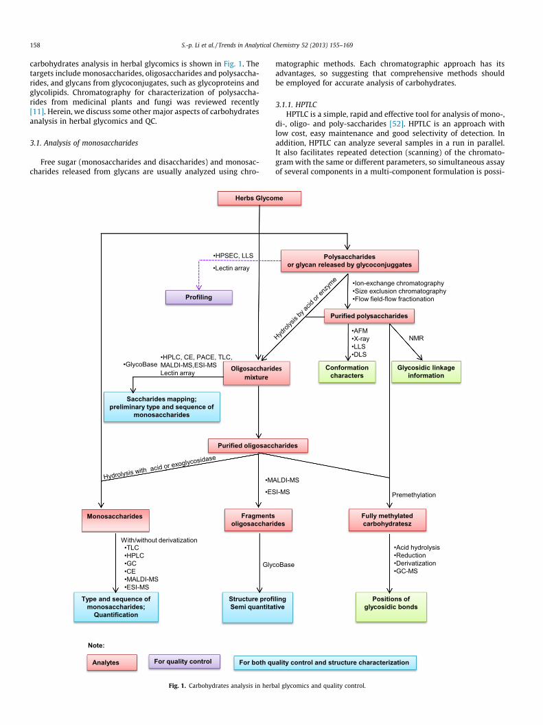
carbohydrates analysis in herbal glycomics is shown in Fig. 1. Thetargets include monosaccharides, oligosaccharides and polysaccha-rides, and glycans from glycoconjugates, such as glycoproteins andglycolipids. Chromatography for characterization of polysaccha-rides from medicinal plants and fungi was reviewed recently[11]. Herein, we discuss some other major aspects of carbohydratesanalysis in herbal glycomics and QC.
3.1. Analysis of monosaccharides
Free sugar (monosaccharides and disaccharides) and monosac-charides released from glycans are usually analyzed using chro-
Herbs Glyco
With/without derivatization•TLC•HPLC•GC•CE•MALDI-MS•ESI-MS
Monosaccharides
Type and sequence of monosaccharides;
Quantification
Oligosaccharidmixture
•GlycoBase
Saccharides mapping; preliminary type and sequence of
monosaccharides
Purified oligosacc
•HPSEC, LLS
Profiling
Structure profiSemi quantita
•MA
•ES
Glyc
Fragmentsoligosacchari
•HPLC, CE, PACE, TLC, MALDI-MS,ESI-MS Lectin array
Analytes For quality control For both qu
Note:
•Lectin array
Fig. 1. Carbohydrates analysis in herb
matographic methods. Each chromatographic approach has itsadvantages, so suggesting that comprehensive methods shouldbe employed for accurate analysis of carbohydrates.
3.1.1. HPTLCHPTLC is a simple, rapid and effective tool for analysis of mono-,
di-, oligo- and poly-saccharides [52]. HPTLC is an approach withlow cost, easy maintenance and good selectivity of detection. Inaddition, HPTLC can analyze several samples in a run in parallel.It also facilitates repeated detection (scanning) of the chromato-gram with the same or different parameters, so simultaneous assayof several components in a multi-component formulation is possi-
me
Polysaccharidesor glycan released by glycoconjuggates
•Ion-exchange chromatography•Size exclusion chromatography•Flow field-flow fractionation
Premethylation
Fully methylatedcarbohydratesz
•Acid hydrolysis•Reduction•Derivatization•GC-MS
Positions of glycosidic bonds
Conformation characters
•AFM •X-ray •LLS•DLS
es
Purified polysaccharides
harides
lingtive
LDI-MS
I-MS
oBase
des
Glycosidic linkage information
NMR
ality control and structure characterization
al glycomics and quality control.
S.-p. Li et al. / Trends in Analytical Chemistry 52 (2013) 155–169 159
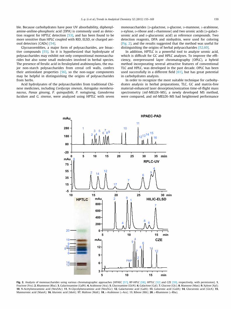
ble. Because carbohydrates have poor UV absorbability, diphenyl-amine-aniline-phosphoric acid (DPA) is commonly used as detec-tion reagent for HPTLC detection [53], and has been found to bemore sensitive than HPLC coupled with RID, ELSD, or charged aer-osol detectors (CADs) [54].
Glycoassemblies, a major form of polysaccharides, are bioac-tive compounds [55]. So it is hypothesized that hydrolysate ofpolysaccharides may exhibit not only compositional monosaccha-rides but also some small molecules involved in herbal species.The presence of ferulic acid in feruloylated arabinoxylans, the ma-jor non-starch polysaccharides from cereal cell walls, conferstheir antioxidant properties [56], so the non-sugar componentsmay be helpful in distinguishing the origins of polysaccharidesfrom herbs.
Acid hydrolysates of the polysaccharides from traditional Chi-nese medicines, including Cordyceps sinensis, Astragalus membera-naceus, Panax ginseng, P. quinquefolii, P. notogiseng, Ganodermalucidum and G. sinense, were analyzed using HPTLC with seven
mAu
1
2 3 5 4
7 6 8 9
0 5 10 15 20-20
80
180
280
mAu 8
2 14
17
12
10 15 20 -5
15
35
55
75
mAu 2
5 0
200
400
600
800
mAu
5 3
6
9
12
HPTLC
14
6 7
18
19 9
20
12
8
Fig. 2. Analysis of monosaccharides using various chromatographic approaches {HPAEFructose (Fru); 2, Rhamnose (Rha); 3, Galactosamine (GalN); 4, Arabinose (Ara); 5, Glucosa10, N-Acetylneuraminic acid (Neu5Ac); 11, N-Glycolylneuraminic acid (Neu5Gc); 12, GMannuronic acid (ManA); 16, Iduronic acid (IdoA); 17, Maltose (Malt); 18, L-Arabinose (
monosaccharides (D-galactose, D-glucose, D-mannose, L-arabinose,D-xylose, D-ribose and L-rhamnose) and two uronic acids (D-galact-uronic acid and D-glucuronic acid) as reference compounds. Twodetection reagents, DPA and ninhydrin, were used for coloring(Fig. 2), and the results suggested that the method was useful fordistinguishing the origins of herbal polysaccharides [52,60].
In addition, HPTLC is a powerful tool to analyze uronic acid,which is difficult for GC and HPLC analyses. To improve the effi-ciency, overpressured layer chromatography (OPLC), a hybridmethod incorporating several attractive features of conventionalTLC and HPLC, was developed in the past decade. OPLC has beenused successfully in a different field [61], but has great potentialin carbohydrates analysis.
In order to recognize the most suitable technique for carbohy-drates analysis in herbal preparations, TLC, GC and matrix-freematerial-enhanced laser desorption/ionization time-of-flight massspectrometry (mf-MELDI–MS), a newly developed MS method,were compared, and mf-MELDI–MS had heightened performance
HPAEC-PAD
min
11 10
12 15
16 14 13
25 30 35 RPLC-UV
min
7
6
4 9
25 30 HILIC-ELSD
min
4
1 9
6 8
7
10 15
min
9 4
2
7 8
6
14 12
PMP
CZE
10 15
C [57], RP-HPLC [58], HPTLC [52] and CZE [59], respectively, with permission}. 1,mine (GlcN); 6, Galactose (Gal); 7, Glucose (Glc); 8, Mannose (Man); 9, Xylose (Xyl);
alacturonic acid (GalA); 13, Guluronic acid (GulA); 14, Glucuronic acid (GlcA); 15,L-Ara); 19, Ribose (Rib); 20, L-Rhamnose (L-Rha).

160 S.-p. Li et al. / Trends in Analytical Chemistry 52 (2013) 155–169
for the qualitative analysis of complex mixtures, as targets do notneed modification and the analysis requires only a few minutes[62].
HPTLC has many coupled detection techniques, such as UV, FLD,FTIR and MS [63]. MS is very helpful for unambiguous structuralelucidation of the spots obtained in TLC. TLC–MS was applied toseparation and detection of large and small biomolecules [64,65],but carbohydrates are generally less frequently investigated [64].
Recently, a new approach in glycobiology by TLC-Blot/MALDI-TOF–MS system was proposed [66,67].
3.1.2. HPLCVarious modes of HPLC separation have been developed for the
separation of carbohydrates. Normal phase (or hydrophilic interac-tion), ion exchange, reversed phase and adsorption (on porousgraphitized carbon columns) are typical.
High-performance anion-exchange chromatography (HPAEC)was, and might still be, the most effective HPLC mode for the anal-ysis of carbohydrates [68]. HPAEC is a combination of a polymer-based pellicular resin (the active component of which is a quater-nary ammonium ion), equilibrated by an alkaline mobile phasewith pulsed amperometry detection (PAD). Carbohydrates, in theform of polyhydric molecules, behave as weak acids, having pKavalues of 12–14. Thus, in basic solutions, all carbohydrates are neg-atively-charged oxyanions. The slight differences in relative pKavalues aid the chromatographic separation of individual monosac-charides. A major advantage of the technique is that it does not re-quire prior derivatization or clean-up of carbohydrate samples inorder to allow quantification. However, anomeric forms of mono-saccharides, without pre-derivatization, yield a single peak as thevery rapid mutarotation of sugars at high pH. But the high chro-matographic efficiency is satisfactory in the separation of aldose,ketose, uronic acid, amino sugar, disaccharide as well as oligosac-charides. Commonly used HPAEC columns for carbohydrates anal-ysis in plants were summarized [69].
Recently, a method to profile complete monosaccharides,including most neutral, amino, and acidic sugars, was developed[57]. The separation was carried out on Carbopac PA1 columnwith a multiple-step gradient elution using isocratic 15mMNaOH for 10min followed by a linear NaOAc gradient with fixed15mM NaOH for the next 30min (Fig. 2). Unfortunately, co-elu-tion of sugars, notably for closely-eluting pairs of sugars (e.g.,rhamnose/arabinose and xylose/mannose), was still not resolved[70].
Hydrophilic interaction liquid chromatography (HILIC) is analternative HPLC mode for separating polar compounds. The op-tions for the characterization of HILIC stationary phases [71] andtheir applications [71–73] were reviewed for separation of polarcompounds in complex matrices, though carbohydrates wererarely the focus.
HILIC with its unique advantages is a significant tool in analysisof carbohydrates from natural sources. HILIC easily separates thesugars (ribose, glucose, sucrose, lactose, and raffinose), whichfailed to separate using RPLC [74]. Seven neutral monosaccharides(L-rhamnose, D-arabinose, D-xylose, D-fructose, D-mannose, D-gal-actose and D-glucose), including the pairs of sugars not separatedon HPAEC [70], were successfully separated in our laboratory inless than 20min using HILIC (Fig. 2).
A simple and sensitive HILIC–MS/MS method was developed fordetermination of monosaccharides liberated from marine polysac-charides by acidic hydrolysis. Optimal separation of diastereomericmonosaccharides, including D-arabinose, D-xylose, D-galactose, D-glucose, L-fucose and L-rhamnose, was achieved using an amino-propyl-bonded column with mobile phase containing ternary sol-vents (acetonitrile/methanol/water) in selected reactionmonitoring mode. Mechanisms for fragmentation of deprotonated
monosaccharides with regard to cross-ring cleavage were pro-posed [75].
HILIC-ESI–MS was also used for the analysis of highly polar car-bohydrate-related metabolites commonly found in plants, andseparation of a mixture of eight authentic standard compoundscontaining glucose, sucrose, raffinose, verbascose, mannitol, malt-itol, glucose-6-phosphate and trehalose-6-phosphate wasachieved in less than 15min. The method was successfully usedto quantify glucose, sucrose, raffinose, and Glc6P in Arabidopsisthaliana extracts [76]. In addition, the HILIC-APCI–MS methodwas developed for identification and quantification of monosac-charides (rhamnose, xylose, fructose, glucose, fucose, arabinose,mannose, and galactose) and disaccharides (cellobiose and su-crose) in orange juice or enzymatic hydrolysis of sunflower seeds[77].
Although HILIC was very convenient for the analysis of carbohy-drates, some inherent problems must be paid more attention dur-ing analysis, such as the temperature and the proportion of theorganic phase, which significantly influence the response of theanalytes [78]. Direct separation of acid sugars without derivatiza-tion also especially challenges some HILIC materials.
RP-HPLC is seldom directly used for analysis of carbohydratesbecause of their high polarity and poor UV absorbability, soderivatization is usually employed to improve separation abilityand detection sensitivity. RP-HPLC columns for separation of car-bohydrates were summarized in a previous review [69]. Asmooth, quantitative derivatization by reductive amination wasusually used. The first reagent, 2-aminopyridine, was soonaccompanied by 2-aminobenzamide, 2-aminobenzoic acid andmany others [79]. The methods, including labeling reagentsand sites for derivatization of carbohydrate, have been reviewed[80].
RP-HPLC was used for analysis of sugars in enzymatic hydroly-sates of polysaccharides, after derivatization with 1-phenyl-3-methyl-5-pyrazolone (PMP), from Panax ginseng, P. notoginseng, P.quinquefolium, Cordyceps sinensis, C. militaris, Ganoderma lucidum,G. sinense, and detected by UV (Fig. 2) and MS [58,81,82]. UPLC-UV/MS was also developed for simultaneous determination of theabsolute configuration of 16 monosaccharides including six pairsof aldose enantiomers (D/L-glucose, D/L-galactose, D/L-allose, D/L-arabinose, D/L-xylose, and D/L-fucose) and four other monosaccha-rides (L-rhamnose, 2-deoxy-D-glucose, 6-deoxy-D-glucose, and 2-deoxy-D-glalactose) in less than 25min. One-pot reaction was usedfor derivatization of monosaccharide with L-cysteine methyl esterand phenyl isothiocyanate [83].
Although many applications have been achieved, RP station-ary-phase materials have problems in separating carbohydrateswith charged groups, such as negatively-charged groups, includ-ing sialic acid, phosphate and sulfate, which are often critical tothe function of the carbohydrates. A different strategy for RP-HPLC analysis of carbohydrates is ion-pairing, which was appliedto glycosaminoglycan fragments [84] and heparin-sulfate disac-charides [85].
Though UV detection is widely used, HPLC analysis of carbohy-drates is usually based on PAD, RID, ELSD, CADs or MS to avoidderivatization (pre-column or post-column) [69,86,87]. CAD, a uni-versal detector that provides a uniform response for all com-pounds, regardless of their physicochemical properties, passes acounter current flow of gas over a high-voltage corona wire, whichtransfers a charge to particles and provides a response when thecharged particles come into contact with a highly sensitive elec-trometer [88]. CAD has been employed for analysis of carbohy-drates [89,90]. It might show the best LOD compared with UVdetection and ELSD [91]. The application of CAD may be helpfulto improve the quantification of carbohydrates without an appro-priate reference compound.

S.-p. Li et al. / Trends in Analytical Chemistry 52 (2013) 155–169 161
3.1.3. GCGC presents several characteristics that make it suitable for the
analysis of complex mixtures, such as high resolution, high sensi-tivity and easy coupling to different detectors, including massspectrometers. In turn, MS is a technique that provides rather com-plete structural information, so GC and GC–MS are excellent tech-niques for the analysis of carbohydrates, but the preparation ofadequate derivatives is necessary. Among the various derivativesof acetates, trifluoroacetates, trimethylsilyl (TMS) derivatives, oxi-mes and aldononitrile acetates, only aldononitrile acetates of car-bohydrates (aldoses, sugar acids, amino and imino sugars)produce one peak, but derivatization is not available for ketoses.All derivatizations of alditols produce a single compound, onechromatographic peak, which is easy for quantification andquantitation.
Derivatization of carbohydrates for GC and GC–MS analyses wasreviewed recently [92]. These derivatization processes typically re-quire reaction times from 30min up to several hours at elevatedtemperature. In contrast, microwave protocols can reduce the timerequired for derivatization to a few minutes, so they can very effec-tively shorten the overall analysis time, in particular when carriedout in a high-throughput format. The application of microwaves inthis field and the techniques for performing parallel GC–MS deriv-atization protocols have been summarized and discussed [93].
A new method for droplet sampling and manipulation routinesin capillaries for extraction, derivation, and partitioning weredeveloped for picogram-scale quantitative determination of solu-ble saccharides from fresh tissues with GC–MS [94]. In brief, 5–10 lL of sap was sampled with a glass capillary containing ribitol(internal standard). Subsequently, the analytes were acetylatedwith acetic anhydride and catalyzed by 1-methylimidazole. Finally,the soluble saccharides were qualitatively and quantitativelydetermined with GC–MS selected-ion monitoring mode. The meth-od is suitable and applicable to analysis of soluble monosaccha-rides in fresh tissues and other aqueous samples.
3.1.4. CEBesides HPLC and GC, CE has attracted substantial interest in
the analysis of both derivatized and underivatized carbohydrates.Advances in CE analysis of carbohydrates were covered in recentreviews [95,96]. Among different CE modes, capillary-zone electro-phoresis (CZE) and micellar eletrokinetic chromatography (MEKC)have been employed most commonly in the analysis ofcarbohydrates.
A CZE method based on pre-column derivatization with PMPhas been applied for the simultaneous determination of neutraland acidic sugars of purified polysaccharides from Ephedra sinica[59]. The standard curves coupled with correction factors wereused to calculate the molar ratios of different monosaccharides.
A promising CE method with direct UV detection was proposedfor analysis of neutral carbohydrates under extremely high alkalineconditions, and the neutral sugar composition of acid hydrolyzedextracts of cellulose-fiber samples was determined [97]. Followingthis unusual detection phenomena, more attention was paid tounderstanding the detection mechanism, and photochemical reac-tions were found in the detection window. The influence of manyparameters was investigated (e.g., carbohydrate nature, electrolytepH, residence time in the detection window, and capillarydiameter).
It is interesting that much faster separations could be obtainedif the work was performed under cathodic (reversed) electro-os-motic flow conditions (using polybrene-modified capillaries) [98].This study opened up new avenues for the detection in the mid-UV range of non-UV-absorbing compounds with reducing moie-ties, such as carbohydrates. The validation of this method for the
determination of fructose, glucose, lactose and sucrose in forensic,pharmaceutical and beverage samples was also performed [99].
CE–MS has attracted attention due to its efficient, selective sep-aration in combination with powerful detection, which allowsidentification and detailed characterization. Recent approachesand applications of CE–MS relevant to (bio) pharmaceuticals arereviewed and discussed to show developments and future pros-pects [100].
3.2. Analysis of oligosaccharides
Generally, apart from GC, the methods used for analysis ofmonosaccharides are available for analysis of intact oligosaccha-rides with or without derivatization.
3.2.1. HPTLCHPTLC-UV was compared with HPLC-ELSD for analysis of seven
sugars in various food samples. The benchmarking of both meth-ods showed HPTLC is highly suitable or even better than HPLCfor quantitation of sugars in food samples with regard to capabilityof detection, intermediate precision, accuracy, and efficiency [54].HPTLC is also a good method for separation of fructo-oligosaccha-rides (Fig. 3).
Fuchs et al. [64] reviewed MALDI-TOF–MS directly combinedwith TLC before 2009. Recently, on-line coupling of TLC with mf-MELDI–MS was described for analysis of carbohydrate referencestandards and a plant extract of Quercus robur [105]. Different com-plex oligosaccharides could easily identified by MALDI-TOF–MS di-rectly from a standard TLC plate [106].
3.2.2. HPLCHPAEC-PAD is a powerful technique to analyze carbohydrates,
such as oligosaccharides (Fig. 3), and is well suited for mappingand characterization of oligosaccharides [102,107]. Oligosaccha-ride profiles of 19 pure agave syrups representing the three majorproduction regions and four processing facilities in Mexico wereanalyzed by HPAEC-PAD, and the oligosaccharide profiling was ex-tremely successful for detecting adulteration [108].
HILIC-ELSD–MSn is a valuable tool for identifying a wide rangeof neutral and acidic cell-wall-derived oligosaccharides. The sepa-ration potential for acidic oligosaccharides observed with HILIC ismuch better than other techniques, such as CE, and RP and porousgraphitic carbon (PGC) chromatography. Important structuralinformation, such as the presence of methyl esters and acetylgroups, is retained during analysis. Separation of acidic oligosac-charides with equal charge yet different degrees of polymerizationcan be obtained. The method enables characterization and quanti-fication of many different oligosaccharide structures present incomplex mixtures [109].
PGC has unique properties as a stationary phase in HPLC, whichretains carbohydrates better (even underivatized) than other RPresins. PGC has been widely used for oligosaccharide separations[110]. The stationary phase and separation mechanism, compari-son with other stationary phases and applications have been wellreviewed [110,111]. The factors influencing detection and elutionof charged oligosaccharides during PGC-ESI–MS analysis wereinvestigated. As a consequence, detection in the positive-ion modeappears preferable when neutral and charged glycans are quanti-tated in the same sample. While retention of neutral glycans isunaffected by pH, sialylated species are retained more at acidicpH. Remarkably, retention of glycans on PGC increased at highertemperatures [112]. PGC-ESI–MS for analysis of oligosaccharideshas been reviewed. Though PGC–MS for glycan analysis is still onlyapplied by a limited number of groups, more users are expected toapply this method when databases become available to supportstructural assignment [113].

0 10 20 40 50 60 70 min 30 0
5
10
15
20
25 30
DP5
DP10
DP15 DP20 DP25 DP30 DP35
HILIC-ELSD 10mV
DP10
DP20
DP30
HPAEC-PAD
0 50 min
DP1
DP5
DP10 DP20
CE-LIF RFU
min 15 25 35 0
1
2
3
PGC-HPLC
min
DP7 DP8
DP5 DP4 DP6 DP3 DP1 DP2
0 5 10 15
DP5
DP10
DP15
HPTLC
Fig. 3. Analysis of oligosaccharides with different degree of polymerization (DP) using various chromatographic approaches {HILIC [101], HPAEC [102], CZE [103], and PGC[104], respectively, with permission}.
162 S.-p. Li et al. / Trends in Analytical Chemistry 52 (2013) 155–169
PGC-ELSD–MS, superior to HPAEC, was used for separation andunambiguous identification of linear and branched arabino-oligo-saccharides. The elution behavior of all arabino-oligosaccharideswas correlated with their chemical structures and conclusionswere drawn about the retention mechanisms of the arabino-oligo-saccharides on the chromatographic system. The combination ofthe elution behavior on PGC and the MS-fragmentation patternsof the arabino-oligosaccharides led to predictions of structuresfor new DP6 arabino-oligosaccharides in complex enzyme digests[104].
Generally, positive-ion-mode tandem MS is used for the struc-tural analysis of neutral saccharides. However, their behavior inthe positive-ion mode is not always adequate for identifying thestructures of oligosaccharides, so fragmentation of [M–H]� or an-ion adducts produced in the negative-ion mode MS offers an alter-native, as their fragmentation patterns provide more structure-related information. The main drawback of the negative-ion modeMS of neutral saccharides is low sensitivity. Several salts weretested as additives to saccharide samples for sensitivity improve-ment in the negative-ion mode. The addition of NH4H2PO4 forming[M + H2PO4]� seemed to be the best choice due to a relatively large
increase in sensitivity in both MS and MSn spectra. The addition ofNH4H2PO4 assisted detection and identification of fructans and oli-gosaccharides of the raffinose family. This approach is a promisingalternative to the more common MS analysis of neutral oligosac-charides in the positive-ion mode or chloride or nitrate additionsin the negative-ion mode [114].
Oligosaccharide structures could be accurately and specificallyidentified by employing tandem MS. Identification was aided bysoftware that compared the sample spectra from tandem MS withspectra in the library. The method incorporated Q-TOF–MS alongwith an annotated oligosaccharide-structure library and theMassHunter Personal Compound Database and Library software.With automated spectra search, oligosaccharide structures indifferent samples were readily identified [115]. Similarly, identifi-cation and accurate quantitation of biological oligosaccharide mix-tures was also performed [116]. The methods were useful in thestudy of milk oligosaccharides, but can be readily applied to oligo-saccharide pools in other biological tissues with the developmentof related MS databases.
As functional components, some oligosaccharides, includinginulin-type oligosaccharides in Morinda officinalis [117], a-galac-
S.-p. Li et al. / Trends in Analytical Chemistry 52 (2013) 155–169 163
to-oligosaccharides in Lycopus lucidus [118], fructo- and malto-oli-gosaccharides in Nicotiana tabacum [119], were separated, charac-terized and/or quantitatively determined (Table 2).
3.2.3. CECE with laser-induced fluorescence (CE-LIF) detection is a pow-
erful tool that provides rapid, high-resolution, highly-sensitivequantitative and/or structural analysis of oligosaccharides. A vari-ety of reagents, including APTS, 3-aminobenzoic acid, and 8-aminonaphthalene-1,3,6-trisulfonic acid (ANTS), have been usedfor CE-LIF detection. But the absorption maximum wavelengthsof the above-mentioned reagents are not perfectly suited to Arion and He/Cd laser emissions at 488nm and 325nm, respectively.Moreover, their molar extinction coefficients and fluorescencequantum yields, which are important factors for highly-sensitivedetection, are not uniformly sufficient. Rhodamine 110 (Rho110)is a highly-sensitive derivatization reagent, which possesses a largefluorescence quantum yield, a high molar extinction coefficient,and an absorption maximum wavelength very close to the wave-length of the Ar ion laser line at 488 nm, so a CE-LIF method wasdeveloped for analyzing N-linked oligosaccharides in glycopro-teins, using Rho110 as fluorescence derivatization reagent. In themethod, CE separation of sialo-oligosaccharides and asialo-oligo-saccharides was according to the number of sialic acids and theirsize using a fused-silica capillary and neutral buffer conditionsand the same capillary with acidic buffer conditions. The analysesof N-linked sialo- and asialo-oligosaccharides in glycoproteinswere also performed [103]. Generally, in order to overcome thelimits of the maximum sample-injection volume and the concen-tration-based limit of detection, numerous on-line preconcentra-tion techniques, such as field-amplified sample stacking,sweeping, dynamic pH junction and isotachophoresis, have beendeveloped. Cationic derivatives of oligosaccharides by quaternaryammonium label were analyzed by CE. Transient isotachophoreticpreconcentration (t-ITP) was performed by injecting the samplesolution complemented by ammonium acetate into the acetic-acidbackground electrolyte. In this case, the ammonium ions served asleading electrolyte followed by the sample zone and the back-ground electrolyte of acetic acid acted as terminating electrolyte.Finally, the oligosaccharides were focused into narrow zones byITP [120].
A CE-ESI–MS method, using a semi-permanent phospholipidcoating for the first time, was also developed for analysis of oligo-saccharides labeled with 1-aminopyrene-3,6,8-trisulfonic acid[121].
3.3. Analysis of polysaccharides and glycoconjugates
Glycosylation produces a diverse, abundant range of glycans,which are collectively known as the glycome. Glycans are one ofthe four fundamental macromolecular components of all cells,and are highly regulated in the immune system. Glycan diversityarises from not only differences in monosaccharide composition,anomeric state, linkage, and branching of a monosaccharide, butalso substituted components and linkage to their aglycones[122]. Various glycosylated natural products exhibit high activity,and the glycosidic residue can be crucial [123].
Advances in chromatography for characterization of polysac-charides were reviewed and discussed recently [11]. Glycan shape(chain conformation) obviously influences activity [124,125]. Gen-erally, polysaccharides mainly exist as random coil, double helix,triple helix, worm-like and rod-like chain conformations, and asaggregates in solution, depending on the type of monosaccharideand glycosidic link, the branch-chain structure and intermolecularinteractions. The chain conformations could be characterized using
light scattering, viscometry, and microscopy such as atomic forcemicroscopy (AFM) [126].
3.3.1. SEC-MALLS-(VIS)Size-exclusion chromatography (SEC) is the most popular ap-
proach for determination of molecular weight and distribution.The serial combination of multiple SEC columns allows broadmolecular weight distribution, and provides higher resolution.Multi-angle laser-light scattering (MALLS) is a technique for deter-mining, independently, the absolute molar mass and the averagesize of particles in solution, by detecting how they scatterlight. SEC coupled with MALLS detection is recognized as one ofthe most powerful techniques for the investigation of macromole-cules. The principles and the theory for absolute characterization ofmacromolecules using SEC-MALLS have been intensively reviewed[127].
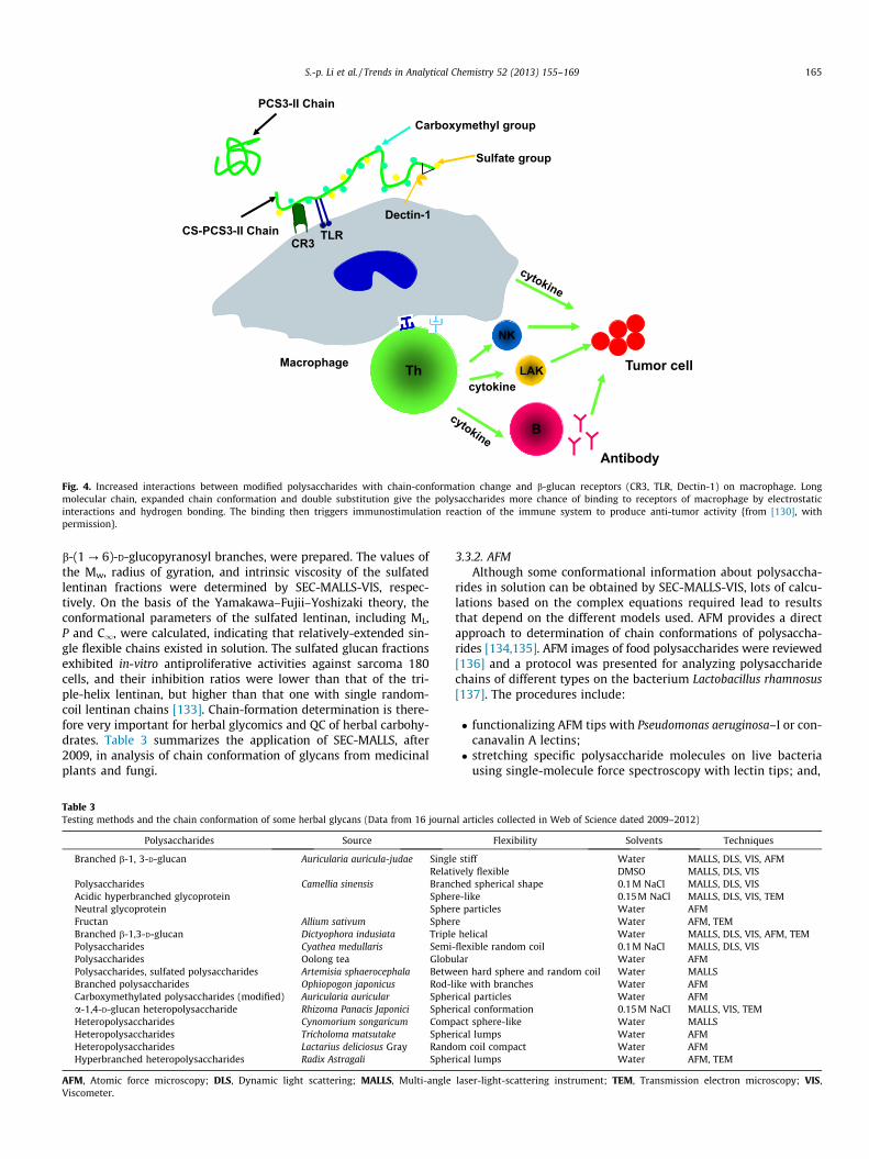
Lentinan, a b-(1 ? 3)-D-glucan isolated from a common ediblemushroom, Lentinus edodes, is a biologically-active macromole-cule with very strong host-mediated anti-cancer activity, via acti-vation of the human immune system. It was also widelyemployed as a therapeutic agent for treatment of malignant tu-mors since 1995, and there are six lentinan injections or powdersfor injection produced by five manufacturers in China. It is con-sidered that molecular weight (Mw) and triple helical conforma-tion (THC) are strongly related to the immuno-potentiationactivity of b-D-glucans besides their primary structures. Mw andTHC should therefore be considered seriously for quality assess-ment of lentinan.
Recently, an SEC-MALLS method was developed for qualityevaluation of lentinan injection (or powder for injection) producedin China based on Mw, Mw distribution, molecular size and struc-ture properties [127]. The results showed that variations on Mw,polydispersity index (PDI) and molecular size of different lentinansamples were significant. The ratio of Mw in water to Mw in DMSOof some samples close to 3, indicating that lentinan THC existed inaqueous solution. However, the Mw (in water)/Mw (in DMSO) ratioof other samples was close to 1, which proved the lack of helicalconformation. The difference between samples may be attributedto the variation in Mw or molecular size.
SEC-MALLS is usually coupled with a viscometer (VIS) for deter-mination of intrinsic viscosity, generating Mark-Houwink plotsthat provide parameters, such as molar mass per unit contourlength (ML), persistence length (P), diameter of the chain (d), andthe limited characteristic ratio (C1), of the chain conformation ofa polysaccharides at a given temperature in a given solvent. Usingthis methodology, ML and P of b-D-glucan [125] and acidic hetero-polysaccharide [128] from Auricularia auricula-judae, and b-D-glu-can from Poria cocos [129], were determined. The resultssuggested that they existed as semi-stiff, duplex or flexible chainsin the given solvents, respectively.
A carboxymethylated-sulfated derivative of (1 ? 3)-b-D-glucan(PCS3-II) extracted from Poria cocos was synthesized and codedas CS-PCS3-II. By using SEC-MALLS, the dependence of radius ofgyration on the molecular weight for CS-PCS3-II was establishedin 0.15 M NaCl solution at 25�C, suggesting that CS-PCS3-II existedas an extended flexible chain [130]. In addition, CS-PCS3-II exhib-ited significantly higher inhibition ratio to Sarcoma 180 tumor inBALB/c mice than PCS3-II, so it was postulated that introductionof the carboxymethyl and sulfate groups to PCS3-II increased itspossible contact with the receptors of immune cells throughhydrogen binding and electrostatic attraction, leading to the stron-ger immunological responses that resulted in inhibition of tumor-cell proliferation (Fig. 4).
Similar studies also showed that single-helix chain might playan important role in activating lymphocytes and NK cells for thedegraded derivatives (LPI1 and LPI2) of longan pulp polysaccharide

Table 2HPLC and CE analysis of mono- and oligosaccharides in food and herbs (Data from 36 journal articles collected in Web of Science dated 2008–2012)
Carbohydrates Origin Column Mobile phase/Running buffer Detection
HPLCFru Grapevine berry Aminex HPX-87H ion
exchange5mM H2SO4 UV 278nm, RI
Glc, Xyl, Ara Meranti wood sawdust Rezex RHMmonosaccharide H+
Water RI
Glc, Xyl, Ara Corncob hydrolysate Aminex HPX-87H 0.35mM H2SO4, 0.6ml/min ELSDGal, Man Natural galactomannans Zorbax Eclipse XDB-C18 Gradient elution of 25 mM phosphate
buffer and acetonitrileUV 271nm
Sugars in oligosaccharides Panax ginseng DIKMA Inertsil ODS-3 Phosphate buffer-acetonitrile (82:18)
UV 245nm
Oligosaccharides Sugar beet Carbopac PA1 Gradient elution of 150mM NaOHand 500mM sodium acetate in150mM NaOH
PAD
Oligosaccharides Barley and malt Carbohydrate ES Water-acetonitrile (2:3), Water-acetonitrile (7:13)
RI, MSn
Oligosaccharides Plant cell wall Hypercarb Gradient elution of water, acetonitrileand 0.2% trifluoroacetic acid
ELSD, MSn
Inulin-type oligosaccharides Morinda officinalis Cyclobond I 2000 Acetonitrile-water (73:27) ELSDInulin-type oligosaccharides Morinda officinalis Cyclobond I 2000 Water-acetonitrile RI, MS/MSInulin-type oligosaccharides Chicory, artichoke Carbohydrate Gradient elution of 0.04% NH3 and
acetonitrileELSD
Fructo-oligosaccharides Musa acuminata L. Carbopac PA1 18mM NaOH PADSuc, 1-kestose, fructo-
oligosaccharidesMature green bananas Carbopac PA1 Gradient elution of 1M NaOH and1 M
sodium acetatePAD
Glc, Fru, Gal, raffitrinose,saccharose
Salvia miltiorrhiza Bunge BP 100 carbohydrateCa++
Water ELSD
Raffinose, stachyose, verbascose Rehmannia glutinosa ZIC-HILIC 0.03% formic acid-acetonitrile (3:7) ELSD, MSRaffinose, stachyose, verbascose Cicer arietinum L. Sugar-D Water-acetonitrile (1:3) RIManno-oligosaccharides Gleditsia sinensis SUGAR PAK I Water RIGalacto-oligosaccharides Lycopus lucidus Turcz. TSK gel amide-80 Water-acetonitrile (3:1) RIXylo-oligosaccharides, pectic-
oligosaccharidesOlive Dionex Carbopac PA-10 Gradient elution of 150mM NaOH,
150mM NaOH in 600mM sodiumacetate and 18mM NaOH
PAD
Feruloylated oligosaccharides Wheat bran Diamonsil C18 Water-acetonitrile (90: 10) with 0.5%formic acid
UV 325nm, MSn
Oligosaccharide esters Polygala tenuifolia Willd Kromasil SB-C18 Gradient elution of water andacetonitrile
UV 318nm
CEGlc, Fru Punica granatum juice Fused silica capillary 75mM glycylglycine, pH 12.85 UV 207nmGlc, Fru, Suc Carrot, cabbage and beet juices;
potato, onion, tomato and cucumberjuices
Fused silica capillary 10–50mM ionic liquids-0–50mMNaOH, pH 12.4
UV 207nm
Glc, Fru, Suc Honey Fused silica capillary 20mM sorbic acid-0.2mM cetyltrimethylammonium bromide-40mM NaOH, pH 12.2
UV 254nm
Glc, Fru, Suc Tomato, pepper, muskmelon, wintersquash, orange
Fused silica capillary 20mM 2,6-pyridine dicarboxylicacid-0.1% hexadimethrine bromide,pH 12.1
UV 200nm UV 214nm
Glc, Fru, Suc Chinese oolong tea Fused silica capillary 30mM borate-40mM phosphate, pH8.5
AD
Glc, Fru, Suc, Man Tobacco Fused silica capillary 100mM NaOH ADGlc, Fru, Suc, Ma-ol Lysium chinensis Mill Fused silica capillary 50mM NaOH ADGlc, Gal, Man, Xyl, Ara, Rib,
GlcNAc, MelMaple syrup, Maple sugar Fused silica capillary 200mM borate buffer, pH 10.5 UV 254nm
Glc, Gal, Fru, Lac, Suc Apple and orange juice, red and whitewin, milk, honey, yoghurt white andjogobella
Fused silica capillary 75mM NaOH, pH 12.8 Contactlessconductivity
Monosaccharides in fibers Oat spelt, wheat straw, sprucethermomechanical pulp, aspenstemwood, bleached birch kraft pulp
Fused silica capillary 130mM NaOH-36mM Na2H2PO4, pH12.6
UV 270nm
Monosaccharides inpolysaccharides
Ganoderma lucidum Dendrobiumhuoshanense
Fused silica capillary 100mM borate buffer, pH 9.0 300 mMphosphate buffer, pH 3.0
UV 254nm
Codonopsis pilosula Fused silica capillary 200mM borate buffer, pH 11 UV 250nmAsparagus officinalis Linn. Fused silica capillary 120mM NaOH AD
Xylo-oligosaccharides Birch kraft pulp Fused silica capillary 130mM NaOH-36mM Na2HPO4, pH12.6
UV 280nmUV278nmUV 240nm
Glucomannan-derivedoligosaccharides
Amorphophallus konjac Polyvinyl alcohol-coated capillary
25mM acetate buffer containing 0.4%polyethylene oxide, pH 4.75
LIF
AD, Amperometric detection; Ara, Arabinose; ED, Electrochemical detection; ELSD, Evaporative light-scattering detection; Fru, Fructose; Gal, Galactose; Glc, Glucose; Lac,Lactose; LIF, Laser-induced fluorescence; Man, Mannose; Ma-ol, Mannitol; Mel, Melibiose; PAD, Pulsed amperometric detection; RI, Refractive index; Rib, Ribose; Suc,Sucrose; Xyl, Xylose.
164 S.-p. Li et al. / Trends in Analytical Chemistry 52 (2013) 155–169
(LPI) [131], and that extended chain conformation was beneficial inenhancing the anti-tumor activity of phosphorylated derivatives of
(1 ? 3)-b-D-Glucan isolated from Poria cocos [132]. Five water-sol-uble sulfated derivatives of lentinan, a b-(1 ? 3)-D-glucan bearing
PCS3-II Chain
CS-PCS3-II Chain
Carboxymethyl group
Sulfate group
TLR CR3
Dectin-1
Macrophage Th
NK
LAK cytokine
B
Antibody
Tumor cell
Fig. 4. Increased interactions between modified polysaccharides with chain-conformation change and b-glucan receptors (CR3, TLR, Dectin-1) on macrophage. Longmolecular chain, expanded chain conformation and double substitution give the polysaccharides more chance of binding to receptors of macrophage by electrostaticinteractions and hydrogen bonding. The binding then triggers immunostimulation reaction of the immune system to produce anti-tumor activity {from [130], withpermission}.
S.-p. Li et al. / Trends in Analytical Chemistry 52 (2013) 155–169 165
b-(1 ? 6)-D-glucopyranosyl branches, were prepared. The values ofthe Mw, radius of gyration, and intrinsic viscosity of the sulfatedlentinan fractions were determined by SEC-MALLS-VIS, respec-tively. On the basis of the Yamakawa–Fujii–Yoshizaki theory, theconformational parameters of the sulfated lentinan, including ML,P and C1, were calculated, indicating that relatively-extended sin-gle flexible chains existed in solution. The sulfated glucan fractionsexhibited in-vitro antiproliferative activities against sarcoma 180cells, and their inhibition ratios were lower than that of the tri-ple-helix lentinan, but higher than that one with single random-coil lentinan chains [133]. Chain-formation determination is there-fore very important for herbal glycomics and QC of herbal carbohy-drates. Table 3 summarizes the application of SEC-MALLS, after2009, in analysis of chain conformation of glycans from medicinalplants and fungi.
Table 3Testing methods and the chain conformation of some herbal glycans (Data from 16 journ
Polysaccharides Source
Branched b-1, 3-D-glucan Auricularia auricula-judae SinglRelat
Polysaccharides Camellia sinensis BrancAcidic hyperbranched glycoprotein SpherNeutral glycoprotein SpherFructan Allium sativum SpherBranched b-1,3-D-glucan Dictyophora indusiata TriplePolysaccharides Cyathea medullaris SemiPolysaccharides Oolong tea GlobuPolysaccharides, sulfated polysaccharides Artemisia sphaerocephala BetwBranched polysaccharides Ophiopogon japonicus Rod-lCarboxymethylated polysaccharides (modified) Auricularia auricular Sphera-1,4-D-glucan heteropolysaccharide Rhizoma Panacis Japonici SpherHeteropolysaccharides Cynomorium songaricum CompHeteropolysaccharides Tricholoma matsutake SpherHeteropolysaccharides Lactarius deliciosus Gray RandHyperbranched heteropolysaccharides Radix Astragali Spher
AFM, Atomic force microscopy; DLS, Dynamic light scattering; MALLS, Multi-angleViscometer.
3.3.2. AFMAlthough some conformational information about polysaccha-
rides in solution can be obtained by SEC-MALLS-VIS, lots of calcu-lations based on the complex equations required lead to resultsthat depend on the different models used. AFM provides a directapproach to determination of chain conformations of polysaccha-rides [134,135]. AFM images of food polysaccharides were reviewed[136] and a protocol was presented for analyzing polysaccharidechains of different types on the bacterium Lactobacillus rhamnosus[137]. The procedures include:
� functionalizing AFM tips with Pseudomonas aeruginosa–I or con-canavalin A lectins;� stretching specific polysaccharide molecules on live bacteria
using single-molecule force spectroscopy with lectin tips; and,
al articles collected in Web of Science dated 2009–2012)
Flexibility Solvents Techniques
e stiff Water MALLS, DLS, VIS, AFMively flexible DMSO MALLS, DLS, VIShed spherical shape 0.1M NaCl MALLS, DLS, VISe-like 0.15M NaCl MALLS, DLS, VIS, TEMe particles Water AFMe Water AFM, TEMhelical Water MALLS, DLS, VIS, AFM, TEM
-flexible random coil 0.1M NaCl MALLS, DLS, VISlar Water AFM
een hard sphere and random coil Water MALLSike with branches Water AFMical particles Water AFMical conformation 0.15M NaCl MALLS, VIS, TEMact sphere-like Water MALLSical lumps Water AFM
om coil compact Water AFMical lumps Water AFM, TEM
laser-light-scattering instrument; TEM, Transmission electron microscopy; VIS,

Fig. 5. Lectin array interacts with herbal glycans to provide a potentially powerful approach in herbal glycomics and quality control of herbal carbohydrates.
166 S.-p. Li et al. / Trends in Analytical Chemistry 52 (2013) 155–169
� mapping localization, adhesion and extension of individualpolysaccharide chains.
The molecular mass and size of five water-soluble polysaccha-rides isolated from Rhizoma Panacis Japonici were determined withlaser-light scattering (LLS), SEC-LLS, and dynamic light scattering(DLS). The results revealed that all of the polysaccharides existedas spheres in 0.15 M NaCl aqueous solution. AFM further confirmedthe spherical morphologies of these molecules [138]. Similarly, thespherical shape of three water-soluble heteropolysaccharides (GL-Ito GL-III), isolated from the cultured fruiting body of Ganodermalucidum, was also confirmed by AFM [139].
Table 3 summarizes the application of AFM to chain conforma-tion of polysaccharides from medicinal plants and fungi.
3.4. Lectin array
Lectins are a group of highly diverse proteins of non-immuneorigin that contain at least one non-catalytic domain, whichenables them to recognize and bind reversibly to specific free sug-ars or glycans selectively without altering the structure of the car-bohydrate [140]. Lectins are therefore especially used inmicroarrays for glycome profiling, as well as in cancer diagnosisand/or therapeutics, and as anti-microbial, anti-viral, and anti-in-sect molecules [141]. Glycans profiling is very important for char-acterization of herbal carbohydrates, which is beneficial forunderstanding herbal glycomics and QC.
Lectin array is a newly developed technique that enables ultra-sensitive glycan profiling in a high-throughput format. Recent ad-vances in the analysis of carbohydrates for biomedical use werereviewed [86,142]. In order to overcome the limitations of label-based technologies, several label-free approaches, such as sur-face-plasmon resonance, carbon nanotubes and nanowires, andmicrocantilevers, have advanced in recent years [143]. In general,the glycan–lectin interaction is very weak and most conventionalmicroarray scanners require a washing process. However, oncebound to a lectin on an array, some glycans may dissociate duringthe washing process, and this often results in a significant reduc-tion in the signal intensity. With the development of a unique lec-tin microarray based on the principle of evanescent-field
fluorescence detection, array analysis greatly improved [144]. Alectin array-based method was developed for rapid analysis of gly-cosylation profiles of glycoproteins. The method is based on bind-ing an intact glycoprotein to the arrayed lectins, resulting in acharacteristic fingerprint that is highly sensitive to changes of gly-can composition. The large number of lectins, each with its specificrecognition pattern, ensures high sensitivity to changes in the gly-can pattern. A set of proprietary algorithms automatically inter-prets the fingerprint signals to provide a comprehensive glycan-profile output [145]. Fig. 5 shows herbal glycan profiling usingthe lectin microarray. Unfortunately, few works have been for her-bal glycans.
We consider that rapid development will take place in theimmediate future with the interesting research focused on herbalglycomics.
4. Conclusion
Carbohydrates are one of major functional ingredients in herbs,and their biological activities depend on their chemical character-istics. Elucidation of active components or fractions is necessary forQC of herbal medicine, which provides the basis for rational mar-ker selection and ensuring the safety and the efficacy of herbs.Therefore, herbal glycomics is crucial for good understanding ofthe herbal glycome, active fractions and characteristics related toactivity. Chromatographic approaches and MS, commonly used inglycomics, are also very helpful in improving carbohydrate analy-sis. MALLS, viscometry and AFM are powerful tools for determina-tion of carbohydrate-chain conformation. Lectin array has a greatpotential as a valuable new method for characterization and QCof herbal glycans. Herbal glycomics is a trend in research on herbalmedicine.
Acknowledgements
We are grateful to Chen Yiwen from our institute for his assis-tance in preparing the figures. The research was partially sup-ported by grants from the University of Macau (MYRG140 andMYRG085), the Science and Technology Development Fund of Ma-
S.-p. Li et al. / Trends in Analytical Chemistry 52 (2013) 155–169 167
cau (059/2011/A3) and the National Natural Science Foundation ofChina (No. 30928033).
References
[1] B. Ernst, J.L. Magnani, From carbohydrate leads to glycomimetic drugs, Nat.Rev. Drug Discov. 8 (2009) 661–677.
[2] R.D. Cummings, The repertoire of glycan determinants in the human glycome,Mol. Biosyst. 5 (2009) 1087–1104.
[3] P.H. Seeberger, D.B. Werz, Synthesis and medical applications ofoligosaccharides, Nature 446 (2007) 1046–1051.
[4] N. Blow, Glycobiology: A spoonful of sugar, Nature 457 (2009) 617–622.[5] I. Chlubnova, B. Sylla, C. Nugier-Chauvin, R. Daniellou, L. Legentil, B. Kralova,
V. Ferrieres, Natural glycans and glycoconjugates as immunomodulatingagents, Nat. Prod. Rep. 28 (2011) 937–952.
[6] J. Courtois, Oligosaccharides from land plants and algae: production andapplications in therapeutics and biotechnology, Curr. Opin. Microbiol. 12(2009) 261–273.
[7] C. Morris, G.A. Morris, The effect of inulin and fructo-oligosaccharidesupplementation on the textural, rheological and sensory properties ofbread and their role in weight management: A review, Food Chem. 133 (2012)237–248.
[8] M. Thakur, A. Weng, H. Fuchs, V. Sharma, C.S. Bhargava, N.S. Chauhan, V.K.Dixit, S. Bhargava, Rasayana properties of Ayurvedic herbs: Arepolysaccharides a major contributor, Carbohydr. Polym. 87 (2012) 3–15.
[9] J.E. Ramberg, E.D. Nelson, R.A. Sinnott, Immunomodulatory dietarypolysaccharides: A systematic review of the literature, Nutr. J. 9 (2010).
[10] R. Raman, S. Raguram, G. Venkataraman, J.C. Paulson, R. Sasisekharan,Glycomics: an integrated systems approach to structure-functionrelationships of glycans, Nat. Methods 2 (2005) 817–824.
[11] D.J. Hu, K.L. Cheong, J. Zhao, S.P. Li, Chromatography in characterization ofpolysaccharides from medicinal plants and fungi, J. Sep. Sci. 36 (2013) 1–19.
[12] N.F. Reuel, B. Mu, J.Q. Zhang, A. Hinckley, M.S. Strano, Nanoengineered glycansensors enabling native glycoprofiling for medicinal applications: towardsprofiling glycoproteins without labeling or liberation steps, Chem. Soc. Rev.41 (2012) 5744–5779.
[13] Y. Hirata, M. Saito, I. Tsukamoto, F. Yamaguchi, L. Sui, K. Kamitori, Y.Y. Dong,E. Uehara, R. Konishi, N. Janjua, M. Tokuda, Analysis of the inhibitorymechanism of D-allose on MOLT-4F leukemia cell proliferation, J. Biosci.Bioeng. 107 (2009) 562–568.
[14] S. Tanaka, H. Sakamoto, Effects of D-allose on the endocytic activity ofdendritic cells and the subsequent stimulation of T cells, Cell. Immunol. 271(2011) 141–146.
[15] D.K. Gao, N. Kawai, T. Tamiya, The anti-inflammatory effects of D-allosecontribute to attenuation of cerebral ischemia-reperfusion injury, Med.Hypotheses 76 (2011) 911–913.
[16] Y. Ishihara, K. Katayama, M. Sakabe, M. Kitamura, M. Aizawa, M. Takara, K.Itoh, Antioxidant properties of rare sugar D-allose: Effects on mitochondrialreactive oxygen species production in Neuro2A cells, J. Biosci. Bioeng. 112(2011) 638–642.
[17] T. Nakamura, S. Tanaka, K. Hirooka, T. Toyoshima, N. Kawai, T. Tamiya, F.Shiraga, M. Tokuda, R.F. Keep, T. Itano, O. Miyamoto, Anti-oxidative effects ofD-allose, a rare sugar, on ischemia-reperfusion damage following focalcerebral ischemia in rat, Neurosci. Lett. 487 (2011) 103–106.
[18] M. Harada, E. Kondo, H. Hayashi, C. Suezawa, S. Suguri, M. Arai, D-Allose andD-psicose reinforce the action of metronidazole on trichomonad, Parasitol.Res. 110 (2012) 1565–1567.
[19] S.H. Baek, S.J. Park, H.G. Lee, D-Psicose, a sweet monosaccharide, amelioratehyperglycemia, and dyslipidemia in C57BL/6J db/db mice, J. Food Sci. 75(2010) H49–H53.
[20] A. Hossain, F. Yamaguchi, T. Matsunaga, Y. Hirata, K. Kamitori, Y. Dong, L. Sui,I. Tsukamoto, M. Ueno, M. Tokuda, Rare sugar D-psicose protects pancreas b-islets and thus improves insulin resistance in OLETF rats, Biochem. Biophys.Res. Commun. 425 (2012) 717–723.
[21] X.L. Xu, Q.M. Xie, Y.H. Shen, J.J. Jiang, Y.Y. Chen, H.Y. Yao, J.Y. Zhou, Mannoseprevents lipopolysaccharide-induced acute lung injury in rats, Inflamm. Res.57 (2008) 104–110.
[22] Y.B. Song, B. Kim, M.J. Choi, Y.O. Song, E.J. Cho, Protective effect of arabinoseand sugar beet pulp against high glucose-induced oxidative stress in LLC-PK1
cells, Food Chem. 134 (2012) 189–194.[23] S. Wang, F.Q. Yang, K. Feng, D.Q. Li, J. Zhao, S.P. Li, Simultaneous
determination of nucleosides, myriocin, and carbohydrates in Cordyceps byHPLC coupled with diode array detection and evaporative light scatteringdetection, J. Sep. Sci. 32 (2009) 4069–4076.
[24] S. Ohtake, Y.J. Wang, Trehalose: Current use and future applications, J. Pharm.Sci. 100 (2011) 2020–2053.
[25] L. Minutoli, D. Altavilla, A. Bitto, F. Polito, E. Bellocco, G. Laganà, D. Giuliani, T.Fiumara, S. Magazù, P. Ruggeri, S. Guarini, F. Squadrito, The disaccharidetrehalose inhibits proinflammatory phenotype activation in macrophagesand prevents mortality in experimental septic shock, Shock 27 (2007) 91–96.
[26] L. Minutoli, D. Altavilla, A. Bitto, F. Polito, E. Bellocco, G. Laganà, T. Fiumara, S.Magazù, F. Migliardo, F.S. Venuti, F. Squadrito, Trehalose: A biophysicsapproach to modulate the inflammatory response during endotoxic shock,Eur. J. Pharmacol. 589 (2008) 272–280.
[27] R. Echigo, N. Shimohata, K. Karatsu, F. Yano, Y. Kayasuga-Kariya, A. Fujisawa,T. Ohto, Y. Kita, M. Nakamura, S. Suzuki, M. Mochizuki, T. Shimizu, U.I. Chung,N. Sasaki, Trehalose treatment suppresses inflammation oxidative stress andvasospasm induced by experimental subarachnoid hemorrhage, J. Transl.Med. 10 (2012).
[28] D.M. Lan, F.T. Liu, J. Zhao, Y. Chen, J.J. Wu, Z.T. Ding, Z.Y. Yue, H.M. Ren, Y.P.Jiang, J. Wang, Effect of trehalose on PC12 cells overexpressing wild-type or A53T mutant alpha-synuclein, Neurochem. Res. 37 (2012) 2025–2032.
[29] S.I. Mussatto, I.M. Mancilha, Non-digestible oligosaccharides: A review,Carbohydr. Polym. 68 (2007) 587–597.
[30] Q. Xu, Y.L. Chao, Q.B. Wan, Health benefit application of functionaloligosaccharides, Carbohydr. Polym. 77 (2009) 435–441.
[31] M. Sabater-Molina, E. Larque, F. Torrella, S. Zamora, Dietaryfructooligosaccharides and potential benefits on health, J. Physiol. Biochem.65 (2009) 315–328.
[32] Y.F. Li, Z.H. Gong, M. Yang, Y.M. Zhao, Z.P. Luo, Inhibition of theoligosaccharides extracted from Morinda officinalis, a Chinese traditionalherbal medicine, on the corticosterone induced apoptosis in PC12 cells, LifeSci. 72 (2003) 933–942.
[33] G.T.C. Delgado, R. Thome, D.L. Gabriel, W.M.S.C. Tamashiro, G.M. Pastore,Yacon (Smallanthus sonchifolius)-derived fructooligosaccharides improves theimmune parameters in the mouse, Nutr. Res. 32 (2012) 884–892.
[34] M. Thakur, P. Connellan, M.A. Deseo, C. Morris, W. Praznik, R. Loeppert, V.K.Dixit, Characterization and in vitro immunomodulatory screening of fructo-oligosaccharides of Asparagus racemosus Willd, Int. J. Biol. Macromol. 50(2012) 77–81.
[35] A.Z. Zong, H.Z. Cao, F.S. Wang, Anticancer polysaccharides from naturalresources: A review of recent research, Carbohydr. Polym. 90 (2012) 1395–1410.
[36] L. Ren, C. Perera, Y. Hemar, Antitumor activity of mushroom polysaccharides:a review, Food Funct. 3 (2012) 1118–1130.
[37] W. Wang, S.X. Wang, H.S. Guan, The antiviral activities and mechanisms ofmarine polysaccharides: An overview, Mar. Drugs 10 (2012) 2795–2816.
[38] M.H. Jiang, L.A. Zhu, J.G. Jiang, Immunoregulatory actions of polysaccharidesfrom Chinese herbal medicine, Expert Opin. Ther. Targets 14 (2010) 1367–1402.
[39] P. Gunness, M.J. Gidley, Mechanisms underlying the cholesterol-loweringproperties of soluble dietary fibre polysaccharides, Food Funct. 1 (2010) 149–155.
[40] J.L. Arias, M.S. Fernandez, Polysaccharides and proteoglycans in calciumcarbonate-based biomineralization, Chem. Rev. 108 (2008) 4475–4482.
[41] H. Kiyohara, T. Matsuzaki, T. Matsumoto, T. Nagai, H. Yamada, Elucidation ofstructures and functions through Peyer’s patches of responsible carbohydratechains in intestinal immune system modulating polysaccharides fromJapanese medicinal herbs, Yakugaku Zasshi-J. Pharm. Soc. Jpn. 128 (2008)709–716.
[42] M.L. Jin, K. Zhao, Q.S. Huang, C.L. Xu, P. Shang, Isolation, structure andbioactivities of the polysaccharides from Angelica sinensis (Oliv.) Diels: Areview, Carbohydr. Polym. 89 (2012) 713–722.
[43] Y.X. Sun, Structure and biological activities of the polysaccharides from theleaves, roots and fruits of Panax ginseng CA Meyer: An overview, Carbohydr.Polym. 85 (2011) 490–499.
[44] S. Zhong, H.J. Pan, L.F. Fan, G.Y. Lv, Y.Z. Wu, B. Parmeswaran, A. Pandey, C.R.Soccol, Advances in research of polysaccharides in Cordyceps species, FoodTechnol. Biotechnol. 47 (2009) 304–312.
[45] X. Zhou, Z. Gong, Y. Su, J. Lin, K. Tang, Cordyceps fungi: Natural products,pharmacological functions and developmental products, J. Pharm. Pharmacol.61 (2009) 279–291.
[46] E.J. Bland, T. Keshavarz, C. Bucke, The influence of small oligosaccharides onthe immune system, Carbohydr. Res. 339 (2004) 1673–1678.
[47] K.F. Hua, H.Y. Hsu, L.K. Chao, S.T. Chen, W.B. Yang, J. Hsu, C.H. Wong,Ganoderma lucidum polysaccharides enhance CD14 endocytosis of LPS andpromote TLR4 signal transduction of cytokine expression, J. Cell. Physiol. 212(2007) 537–550.
[48] F. Firenzuoli, L. Gori, G. Lombardo, The medicinal mushroom Agaricus blazeimurrill: Review of literature and pharmaco-toxicological problems, Evid.Based Complement. Alternat. Med. 5 (2008) 3–15.
[49] V.K. Morya, J. Kim, E.K. Kim, Algal fucoidan: Structural and size-dependentbioactivities and their perspectives, Appl. Microbiol. Biotechnol. 93 (2012)71–82.
[50] V.H. Pomin, An overview about the structure-function relationship of marinesulfated homopolysaccharides with regular chemical structures, Biopolymers91 (2009) 601–609.
[51] Y. Zhang, S. Li, X. Wang, L. Zhang, P.C.K. Cheung, Advances in lentinan:Isolation, structure, chain conformation and bioactivities, Food Hydrocolloids25 (2011) 196–206.
[52] C. Yang, J. Guan, J.S. Zhang, S.P. Li, Use of HPTLC to differentiate among thecrude polysaccharides in six traditional Chinese medicines, JPC-J. PlanarChromatogr.-Mod. TLC 23 (2010) 46–49.
[53] Z.Q. Zhang, Z.P. Xiao, R.J. Linhardt, Thin layer chromatography for theseparation and analysis of acidic carbohydrates, J. Liq. Chromatogr. Relat.Technol. 32 (2009) 1711–1732.
[54] G.E. Morlock, G. Sabir, Comparison of two orthogonal liquid chromatographicmethods for quantitation of sugars in food, J. Liq. Chromatogr. Relat. Technol.34 (2011) 902–919.

168 S.-p. Li et al. / Trends in Analytical Chemistry 52 (2013) 155–169
[55] C.R. Bertozzi, L.L. Kiessling, Chemical glycobiology, Science 291 (2001) 2357–2364.
[56] G. Nino-Medina, E. Carvajal-Millan, A. Rascon-Chu, J.A. Marquez-Escalante, V.Guerrero, E. Salas-Munoz, Feruloylated arabinoxylans and arabinoxylan gels:structure, sources and applications, Phytochem. Rev. 9 (2010) 111–120.
[57] Z.Q. Zhang, N.M. Khan, K.M. Nunez, E.K. Chess, C.M. Szabo, Completemonosaccharide analysis by high-Performance anion-Exchangechromatography with pulsed amperometric detection, Anal. Chem. 84(2012) 4104–4110.
[58] J. Xu, J. Guan, X.J. Chen, J. Zhao, S.P. Li, Comparison of polysaccharides fromdifferent Dendrobium using saccharide mapping, J. Pharm. Biomed. Anal. 55(2011) 977–983.
[59] Y.G. Xia, Q.H. Wang, J. Liang, B.Y. Yang, G.Y. Li, H.X. Kuang, Developmentand application of a rapid and efficient CZE method coupled withcorrection factors for determination of monosaccharide composition ofacidic hetero-polysaccharides from Ephedra sinica, Phytochem. Anal 22(2011) 103–111.
[60] J. Xie, J. Zhao, D.J. Hu, J.A. Duan, Y.P. Tang, S.P. Li, Comparison of polysaccharidesfrom two species of Ganoderma, Molecules 17 (2012) 740–752.
[61] E. Tyihak, E. Mincsovics, A.M. Moricz, Overpressured layer chromatography:From the pressurized ultramicro chamber to BioArena system, J. Chromatogr.A 1232 (2012) 3–18.
[62] M.N. Qureshi, G. Stecher, T. Sultana, G. Abel, M. Popp, G.K. Bonn,Determination of carbohydrates in medicinal plants-comparison betweenTLC, mf-MELDI–MS and GC–MS, Phytochem. Anal 22 (2011) 296–302.
[63] G. Morlock, W. Schwack, Hyphenations in planar chromatography, J.Chromatogr. A 1217 (2010) 6600–6609.
[64] B. Fuchs, R. Süß, A. Nimptsch, J. Schiller, MALDI-TOF–MS directly combinedwith TLC: A Review of the current state, Chromatographia 69 (2009) S95–S105.
[65] T. Tuzimski, Application of different modes of thin-layer chromatography andmass spectrometry for the separation and detection of large and smallbiomolecules, J. Chromatogr. A 1218 (2011) 8799–8812.
[66] T. Taki, An approach to glycobiology from glycolipidomics: Gangliosidemolecular scanning in the brains of patients with Alzheimer’s disease by TLC-Blot/matrix assisted laser desorption/ionization-time of flight MS, Biol.Pharm. Bull. 35 (2012) 1642–1647.
[67] T. Valdes-Gonzalez, N. Goto-Inoue, W. Hirano, H. Ishiyama, T. Hayasaka, M.Setou, T. Taki, New approach for glyco- and lipidomics- molecular scanning ofhuman brain gangliosides by TLC-Blot and MALDI-QIT-TOF MS, J. Neurochem.116 (2011) 678–683.
[68] J.L. Behan, K.D. Smith, The analysis of glycosylation: a continued need for highpH anion exchange chromatography, Biomed. Chromatogr. 25 (2011) 39–46.
[69] M. Raessler, Sample preparation and current applications of liquidchromatography for the determination of non-structural carbohydrates inplants, TrAC-Trends Anal. Chem. 30 (2011) 1833–1843.
[70] C. Panagiotopoulos, R. Sempere, The molecular distribution of combinedaldoses in sinking particles in various oceanic conditions, Mar. Chem. 95(2005) 31–49.
[71] B. Buszewski, S. Noga, Hydrophilic interaction liquid chromatography (HILIC)-apowerful separation technique, Anal. Bioanal. Chem. 402 (2012) 231–247.
[72] B. Dejaegher, S. Pieters, Y.V. Heyden, Emerging analytical separationtechniques with high throughput potential for pharmaceutical analysis,part II: Novel chromatographic modes, Comb. Chem. High ThroughputScreening 13 (2010) 530–547.
[73] J. Bernal, A.M. Ares, J. Pól, S.K. Wiedmer, Hydrophilic interaction liquidchromatography in food analysis, J. Chromatogr. A 1218 (2011) 7438–7452.
[74] S. Louw, A.S. Pereira, F. Lynen, M. Hanna-Brown, P. Sandra, Serial coupling ofreversed-phase and hydrophilic interaction liquid chromatography tobroaden the elution window for the analysis of pharmaceutical compounds,J. Chromatogr. A 1208 (2008) 90–94.
[75] Q.J. Gao, U. Nilsson, L.L. Ilag, C. Leck, Monosaccharide compositional analysisof marine polysaccharides by hydrophilic interaction liquid chromatography-tandem mass spectrometry, Anal. Bioanal. Chem. 399 (2011) 2517–2529.
[76] C. Antonio, T. Larson, A. Gilday, I. Graham, E. Bergstrom, J. Thomas-Oates,Hydrophilic interaction chromatography/electrospray mass spectrometryanalysis of carbohydrate-related metabolites from Arabidopsis thaliana leaftissue, Rapid Commun. Mass Spectrom. 22 (2008) 1399–1407.
[77] G. Ricochon, C. Paris, M. Girardin, L. Muniglia, Highly sensitive, quick andsimple quantification method for mono and disaccharides in aqueous media,J. Chromatogr. B 879 (2011) 1529–1536.
[78] Z.G. Hao, B.M. Xiao, N.D. Weng, Impact of column temperature and mobilephase components on selectivity of hydrophilic interaction chromatography(HILIC), J. Sep. Sci. 31 (2008) 1449–1464.
[79] M. Pabst, F. Altmann, Glycan analysis by modern instrumental methods,Proteomics 11 (2011) 631–643.
[80] D.J. Harvey, Derivatization of carbohydrates for analysis by chromatography;electrophoresis and mass spectrometry, J. Chromatogr. B 879 (2011) 1196–1225.
[81] J. Guan, S.P. Li, Discrimination of polysaccharides from traditional Chinesemedicines using saccharide mapping-enzymatic digestion followed bychromatographic analysis, J. Pharm. Biomed. Anal. 51 (2010) 590–598.
[82] J. Guan, J. Zhao, K. Feng, D.J. Hu, S.P. Li, Comparison and characterization ofpolysaccharides from natural and cultured Cordyceps using saccharidemapping, Anal. Bioanal. Chem. 399 (2011) 3465–3474.
[83] Y.H. Wang, B. Avula, X. Fu, M. Wang, I.A. Khan, Simultaneous determination ofthe absolute configuration of twelve monosaccharide enantiomers fromnatural products in a single injection by a UPLC-UV/MS Method, Planta Med.78 (2012) 834–837.
[84] J. Zaia, On-Line Separations combined with MS for analysis ofglycosaminoglycans, Mass Spectrom. Rev. 28 (2009) 254–272.
[85] Y. Wang, M.L. Tian, W.T. Bi, K.H. Row, Application of ionic liquids in highperformance reversed-phase chromatography, Int. J. Mol. Sci. 10 (2009)2591–2610.
[86] K. Yamada, K. Kakehi, Recent advances in the analysis of carbohydrates forbiomedical use, J. Pharm. Biomed. Anal. 55 (2011) 702–727.
[87] G. Zauner, A.M. Deelder, M. Wuhrer, Recent advances in hydrophilicinteraction liquid chromatography (HILIC) for structural glycomics,Electrophoresis 32 (2011) 3456–3466.
[88] J.P. Hutchinson, J.F. Li, W. Farrell, E. Groeber, R. Szucs, G. Dicinoski, P.R.Haddad, Universal response model for a corona charged aerosol detector, J.Chromatogr. A 1217 (2010) 7418–7427.
[89] B. Godin, R. Agneessens, P.A. Gerin, J. Delcarte, Composition of structuralcarbohydrates in biomass: Precision of a liquid chromatography methodusing a neutral detergent extraction and a charged aerosol detector, Talanta85 (2011) 2014–2026.
[90] J.P. Hutchinson, T. Remenyi, P. Nesterenko, W. Farrell, E. Groeber, R. Szucs, G.Dicinoski, P.R. Haddad, Investigation of polar organic solvents compatiblewith corona charged aerosol detection and their use for the determination ofsugars by hydrophilic interaction liquid chromatography, Anal. Chim. Acta750 (2012) 199–206.
[91] J. Shaodong, W.J. Lee, J.W. Ee, J.H. Park, S.W. Kwon, J. Lee, Comparison ofultraviolet detection, evaporative light scattering detection and chargedaerosol detection methods for liquid-chromatographic determination of anti-diabetic drugs, J. Pharm. Biomed. Anal. 51 (2010) 973–978.
[92] A.I. Ruiz-Matute, O. Hernández-Hernández, S. Rodríguez-Sánchez, M.L. Sanz,I. Martínez-Castro, Derivatization of carbohydrates for GC and GC–MSanalyses, J. Chromatogr. B 879 (2011) 1226–1240.
[93] S.L. Soderholm, M. Damm, C.O. Kappe, Microwave-assisted derivatizationprocedures for gas chromatography/mass spectrometry analysis, Mol. Divers.14 (2010) 869–888.
[94] Y.P. Tan, K. Li, L. Hu, S. Chen, Y. Gai, X.N. Jiang, Fast and simple dropletsampling of sap from plant tissues and capillary microextraction of solublesaccharides for picogram-scale quantitative determination with GC–MS, J.Agric. Food. Chem. 58 (2010) 9931–9935.
[95] H.M. Tseng, S. Gattollin, J. Pritchard, H.J. Newbury, D.A. Barrett, Analysis ofmono-, di- and oligosaccharides by CE using a two-stage derivatizationmethod and LIF detection, Electrophoresis 30 (2009) 1399–1405.
[96] R. Gotti, Capillary electrophoresis of phytochemical substances in herbaldrugs and medicinal plants, J. Pharm. Biomed. Anal. 55 (2011) 775–801.
[97] S. Rovio, H. Simolin, K. Koljonen, H. Sirén, Determination of monosaccharidecomposition in plant fiber materials by capillary zone electrophoresis, J.Chromatogr. A 1185 (2008) 139–144.
[98] C. Sarazin, N. Delaunay, C. Costanza, V. Eudes, J.M. Mallet, P. Gareil, Newavenue for mid-UV-range detection of underivatized carbohydratesand amino acids in capillary electrophoresis, Anal. Chem. 83 (2011) 7381–7387.
[99] C. Sarazin, N. Delaunay, C. Costanza, V. Eudes, P. Gareil, Application of a newcapillary electrophoretic method for the determination of carbohydrates inforensic, pharmaceutical, and beverage samples, Talanta 99 (2012) 202–206.
[100] M. Pioch, S.C. Bunz, C. Neususs, Capillary electrophoresis/mass spectrometryrelevant to pharmaceutical and biotechnological applications,Electrophoresis 33 (2012) 1517–1530.
[101] A. Kiss, P. Forgo, Investigations on inulin-type oligosaccharides with regard toHPLC analysis and prospective food applicability, Monatsh. Chem. 142 (2011)547–553.
[102] S.N. Ronkart, C.S. Blecker, H. Fourmanoir, C. Fougnies, C. Deroanne, J.C. VanHerck, M. Paquot, Isolation and identification of inulooligosaccharidesresulting from inulin hydrolysis, Anal. Chim. Acta 604 (2007) 81–87.
[103] S. Ijiri, K. Todoroki, H. Yoshida, T. Yoshitake, H. Nohta, M. Yamaguchi, Highlysensitive capillary electrophoresis analysis of N-linked oligosaccharides inglycoproteins following fluorescence derivatization with rhodamine 110 andlaser-induced fluorescence detection, Electrophoresis 32 (2011) 3499–3509.
[104] Y. Westphal, S. Kuhnel, H.A. Schols, A.G.J. Voragen, H. Gruppen, LC/CE–MStools for the analysis of complex arabino-oligosaccharides, Carbohydr. Res.345 (2010) 2239–2251.
[105] M.N. Qureshi, G. Stecher, C. Huck, G.K. Bonn, Online coupling of thin layerchromatography with matrix-assisted laser desorption/ionization time-of-flight mass spectrometry: synthesis and application of a new material for theidentification of carbohydrates by thin layer chromatography/matrix freematerial enhanced laser desorption/ionization mass spectrometry, RapidCommun. Mass Spectrom. 24 (2010) 2759–2764.
[106] K. Nimptsch, R. Süß, T. Riemer, A. Nimptsch, M. Schnabelrauch, J. Schiller,Differently complex oligosaccharides can be easily identified by matrix-assisted laser desorption and ionization time-of-flight mass spectrometrydirectly from a standard thin-layer chromatography plate, J. Chromatogr. A1217 (2010) 3711–3715.
[107] C. Grey, P. Edebrink, M. Krook, S.P. Jacobsson, Development of a highperformance anion exchange chromatography analysis for mapping ofoligosaccharides, J. Chromatogr. B 877 (2009) 1827–1832.

S.-p. Li et al. / Trends in Analytical Chemistry 52 (2013) 155–169 169
[108] J.L. Willems, N.H. Low, Major carbohydrate, polyol, and oligosaccharideprofiles of Agave Syrup. application of this data to authenticity analysis, J.Agric. Food. Chem. 60 (2012) 8745–8754.
[109] A.G.M. Leijdekkers, M.G. Sanders, H.A. Schols, H. Gruppen, Characterizingplant cell wall derived oligosaccharides using hydrophilic interactionchromatography with mass spectrometry detection, J. Chromatogr. A 1218(2011) 9227–9235.
[110] C. West, C. Elfakir, M. Lafosse, Porous graphitic carbon: A versatile stationaryphase for liquid chromatography, J. Chromatogr. A 1217 (2010) 3201–3216.
[111] L. Pereira, Porous graphitic carbon as a stationary phase in HPLC: Theory andapplications, J. Liq. Chromatogr. Relat. Technol. 31 (2008) 1687–1731.
[112] M. Pabst, F. Altmann, Influence of electrosorption, solvent, temperature, andion polarity on the performance of LC-ESI–MS using graphitic carbon foracidic oligosaccharides, Anal. Chem. 80 (2008) 7534–7542.
[113] L.R. Ruhaak, A.M. Deelder, M. Wuhrer, Oligosaccharide analysis bygraphitized carbon liquid chromatography-mass spectrometry, Anal.Bioanal. Chem. 394 (2009) 163–174.
[114] I. Fabrik, R. Cmelík, J. Bobálová, Analysis of free oligosaccharides by negative-ion electrospray ion trap tandem mass spectrometry in the presence ofH2PO4� anions, Int. J. Mass spectrom. 309 (2012) 88–96.
[115] S. Wu, J. Salcedo, N. Tang, K. Waddell, R. Grimm, J.B. German, C.B. Lebrilla,Employment of tandem mass spectrometry for the accurate and specificidentification of oligosaccharide structures, Anal. Chem. 84 (2012) 7456–7462.
[116] J.S. Strum, J. Kim, S. Wu, M.L.A. De Leoz, K. Peacock, R. Grimm, J.B. German,D.A. Mills, C.B. Lebrilla, Identification and accurate quantitation of biologicaloligosaccharide mixtures, Anal. Chem. 84 (2012) 7793–7801.
[117] Z. Yang, J. Hu, M. Zhao, Isolation and quantitative determination of inulin-type oligosaccharides in roots of Morinda officinalis, Carbohydr. Polym. 83(2011) 1997–2004.
[118] X. Yang, Y. Zhao, N. He, K.D. Croft, Isolation, characterization, andimmunological effects of a-Galacto-oligosaccharides from a new source,the Herb Lycopus lucidus Turcz, J. Agric. Food. Chem. 58 (2010) 8253–8258.
[119] A. Nagai, T. Yamamoto, H. Wariishi, Identification of Fructo- and Malto-oligosaccharides in Cured Tobacco Leaves (Nicotiana tabacum), J. Agric. Food.Chem. 60 (2012) 6606–6612.
[120] J. Partyka, F. Foret, Cationic labeling of oligosaccharides for electrophoreticpreconcentration and separation with contactless conductivity detection, J.Chromatogr. A 1267 (2012) 116–120.
[121] T.J. Langan, L.A. Holland, Capillary electrophoresis coupled to electrospraymass spectrometry through a coaxial sheath flow interface and semi-permanent phospholipid coating for the determination of oligosaccharideslabeled with 1-aminopyrene-3,6,8-trisulfonic acid, Electrophoresis 33 (2012)607–613.
[122] D.B. Werz, R. Ranzinger, S. Herget, A. Adibekian, C.W. von der Lieth, P.H.Seeberger, Exploring the structural diversity of mammalian carbohydrates(‘‘Glycospace’’) by statistical databank analysis, ACS Chem. Biol. 2 (2007)685–691.
[123] B. La Ferla, C. Airoldi, C. Zona, A. Orsato, F. Cardona, S. Merlo, E. Sironi, G.D’Orazio, F. Nicotra, Natural glycoconjugates with antitumor activity, Nat.Prod. Rep. 28 (2011) 630–648.
[124] S. Li, S.Q. Xu, L.N. Zhang, Advances in conformations and characterizations offungi polysaccharides, Acta Polym. Sin. (2010) 1359–1375.
[125] Z.C. Ma, J.G. Wang, L.N. Zhang, Structure and chain conformation of ss-glucanisolated from Auricularia auricula-judae, Biopolymers 89 (2008) 614–622.
[126] L.Q. Yang, L.M. Zhang, Chemical structural and chain conformationalcharacterization of some bioactive polysaccharides isolated from naturalsources, Carbohydr. Polym. 76 (2009) 349–361.
[127] Y.W. Chen, D.J. Hu, K.L. Cheong, J. Li, J. Xie, J. Zhao, S.P. Li, Quality evaluation oflentinan injection produced in China, J. Pharm. Biomed. Anal. 78–79 (2013)176–182.
[128] Z.C. Ma, L.N. Zhang, Y. Nishiyama, M.F. Marais, K. Mazeau, M. Vignon, Themolecular structure and solution conformation of an acidicheteropolysaccharide from Auricularia auricula-judae, Biopolymers 95(2011) 217–227.
[129] X.Y. Chen, X.J. Xu, L. Zhang, J.F. Kennedy, Flexible chain conformation of (1,3)-beta-D-glucan from Poria cocos sclerotium in NaOH/urea aqueous solution,Carbohydr. Polym. 75 (2009) 586–591.
[130] X.Y. Chen, L.N. Zhang, P.C.K. Cheung, Immunopotentiation and anti-tumoractivity of carboxymethylated-sulfated beta-(1,3)-D-glucan from Poria cocos,Int. Immunopharmacol. 10 (2010) 398–405.
[131] Y. Yi, M.W. Zhang, S.T. Liao, R.F. Zhang, Y.Y. Deng, Z.C. Wei, B. Yang, Effects ofalkali dissociation on the molecular conformation and immunomodulatoryactivity of longan pulp polysaccharide (LPI), Carbohydr. Polym. 87 (2012)1311–1317.
[132] X.Y. Chen, X.J. Xu, L.N. Zhang, F.B. Zeng, Chain conformation and anti-tumoractivities of phosphorylated (1,3)-beta-D-glucan from Poria cocos, Carbohydr.Polym. 78 (2009) 581–587.
[133] X.H. Wang, L.N. Zhang, Physicochemical properties and antitumor activitiesfor sulfated derivatives of lentinan, Carbohydr. Res. 344 (2009)2209–2216.
[134] P.E. Marszalek, Y.F. Dufrene, Stretching single polysaccharides and proteinsusing atomic force microscopy, Chem. Soc. Rev. 41 (2012) 3523–3534.
[135] J. Wu, X. Deng, Y. Zhang, L. Wang, B.Q. Tian, B.J. Xie, Application of atomicforce microscopy in the study of polysaccharide, Agric. Sci. China 8 (2009)1458–1465.
[136] T. Funami, Atomic force microscopy imaging of food polysaccharides, FoodSci. Technol. Res. 16 (2010) 1–12.
[137] G. Francius, D. Alsteens, V. Dupres, S. Lebeer, S. De Keersmaecker, J.Vanderleyden, H.J. Gruber, Y.F. Dufrene, Stretching polysaccharides on livecells using single molecule force spectroscopy, Nat. Protoc. 4 (2009) 939–946.
[138] Z.P. Huang, Y.A. Huang, X.B. Li, L.N. Zhang, Molecular mass and chainconformations of Rhizoma Panacis Japonici polysaccharides, Carbohydr.Polym. 78 (2009) 596–601.
[139] J.G. Wang, Z.C. Ma, L.N. Zhang, Y.P. Fang, F.T. Jiang, G.O. Phillips, Structure andchain conformation of water-soluble heteropolysaccharides from Ganodermalucidum, Carbohydr. Polym. 86 (2011) 844–851.
[140] W.J. Peumans, E.J.M. Vandamme, Lectins as plant defense proteins, PlantPhysiol. 109 (1995) 347–352.
[141] C. Oliveira, J.A. Teixeira, L. Domingues, Recombinant lectins: an array oftailor-made glycan-interaction biosynthetic tools, Crit. Rev. Biotechnol. 33(2013) 66–80.
[142] J. Hirabayashi, A. Kuno, H. Tateno, Lectin-based structural glycomics: Apractical approach to complex glycans, Electrophoresis 32 (2011) 1118–1128.
[143] H. Chandra, P.J. Reddy, S. Srivastava, Protein microarrays and novel detectionplatforms, Expert Rev. Proteomics 8 (2011) 61–79.
[144] H. Narimatsu, H. Sawaki, A. Kuno, H. Kaji, H. Ito, Y. Ikehara, A strategy fordiscovery of cancer glyco-biomarkers in serum using newly developedtechnologies for glycoproteomics, FEBS J. 277 (2010) 95–105.
[145] R. Rosenfeld, H. Bangio, G.J. Gerwig, R. Rosenberg, R. Aloni, Y. Cohen, Y. Amor,I. Plaschkes, J.P. Kamerling, R.B.-Y. Maya, A lectin array-based methodologyfor the analysis of protein glycosylation, J. Biochem. Bioph. Meth. 70 (2007)415–426.