Interactions between -haemolytic streptococci and the human ...
105
Interactions between -haemolytic streptococci and the human host. Heart, skin and beyond. Bläckberg, Anna 2022 Document Version: Publisher's PDF, also known as Version of record Link to publication Citation for published version (APA): Bläckberg, A. (2022). Interactions between β-haemolytic streptococci and the human host. Heart, skin and beyond. Lund University, Faculty of Medicine. Total number of authors: 1 General rights Unless other specific re-use rights are stated the following general rights apply: Copyright and moral rights for the publications made accessible in the public portal are retained by the authors and/or other copyright owners and it is a condition of accessing publications that users recognise and abide by the legal requirements associated with these rights. • Users may download and print one copy of any publication from the public portal for the purpose of private study or research. • You may not further distribute the material or use it for any profit-making activity or commercial gain • You may freely distribute the URL identifying the publication in the public portal Read more about Creative commons licenses: https://creativecommons.org/licenses/ Take down policy If you believe that this document breaches copyright please contact us providing details, and we will remove access to the work immediately and investigate your claim.
-
Upload
khangminh22 -
Category
Documents
-
view
3 -
download
0
Transcript of Interactions between -haemolytic streptococci and the human ...

LUND UNIVERSITY
PO Box 117221 00 Lund+46 46-222 00 00
Interactions between -haemolytic streptococci and the human host. Heart, skin andbeyond.
Bläckberg, Anna
2022
Document Version:Publisher's PDF, also known as Version of record
Link to publication
Citation for published version (APA):Bläckberg, A. (2022). Interactions between β-haemolytic streptococci and the human host. Heart, skin andbeyond. Lund University, Faculty of Medicine.
Total number of authors:1
General rightsUnless other specific re-use rights are stated the following general rights apply:Copyright and moral rights for the publications made accessible in the public portal are retained by the authorsand/or other copyright owners and it is a condition of accessing publications that users recognise and abide by thelegal requirements associated with these rights. • Users may download and print one copy of any publication from the public portal for the purpose of private studyor research. • You may not further distribute the material or use it for any profit-making activity or commercial gain • You may freely distribute the URL identifying the publication in the public portal
Read more about Creative commons licenses: https://creativecommons.org/licenses/Take down policyIf you believe that this document breaches copyright please contact us providing details, and we will removeaccess to the work immediately and investigate your claim.
Interactions between β-haemolytic streptococci and the human hostHeart, skin and beyondANNA BLÄCKBERG
DEPARTMENT OF CLINICAL SCIENCES, LUND | FACULTY OF MEDICINE | LUND UNIVERSITY


1
Interactions between -haemolytic streptococci and the human host

2

3
Interactions between -haemolytic streptococci and the human host
Heart, skin and beyond
Anna Bläckberg
DOCTORAL DISSERTATION
by due permission of the Faculty of Medicine, Lund University, Sweden. To be defended at Belfragesalen, BMC D15, on the 16th of September 2022 at 0900.
Faculty opponent
Prof. Steinar Skrede Haukeland University Hospital, University of Bergen, Norway

4
Organization LUND UNIVERSITY
Document name Doctoral dissertation
Department of Clinical Sciences Division of Infection Medicine
Date of issue September 16th 2022
Author(s) Anna Bläckberg Sponsoring organization
Title and subtitle Interactions between -haemolytic streptococci and the human host Heart, skin and beyond
Abstract Group C and G streptococci (GCS/GGS) and group A streptococci (GAS) belong to -haemolytic streptococci (BHS). GCS/GGS can further be species determined to S. dysgalactiae, whereas S. pyogenes are GAS. S. dysgalactiae and S. pyogenes cause similar diseases, (e.g., skin and soft tissue infections, erysipelas, bacteraemia, and infective endocarditis (IE)). Erysipelas often presents as a sharply demarcated oedematous erythema and often reoccurs in the same host. The diagnosis relies on the clinical presentation since cultures from blood and/or wounds often are negative. IE due to BHS is rare but is a challenging infection to treat. Combination therapy using a -lactam and an aminoglycoside has been employed to treat the condition, but the evidence for synergy between the two antibiotics is weak.
This thesis began investigating erysipelas and predominantly found GCS/GGS, but also GAS as important pathogens to erysipelas, (Paper I). To further investigate the disease panorama of GCS/GGS, the second study comprised cases of IE due to S. dysgalactiae from a nationwide registry. IE with S. dysgalactiae was found to have an acute onset of symptoms with substantial mortality and embolic event rate, (Paper II). Synergy between penicillin G and gentamicin was observed in some blood isolates of S. dysgalactiae. However, in most cases, penicillin G alone showed bactericidal action so strong, that any further killing action of gentamicin was difficult to detect.
Recurrent infections with S. dysgalactiae, involving erysipelas but also bacteraemia, are common. The cell surface attached M protein is an important virulence determinant for the bacteria. There are different M proteins which render an antigenic diversity and facilitate the bacteria’s evasion of the host defence system. In a prospectively based study, type-specific antibodies were developed in convalescent serum from patients with prior bacteraemia with S. dysgalactiae, (Paper III). However, further analysis with bactericidal and phagocytosis assays could not establish that these evolved antibodies opsonised the bacteria or enhanced the killing of the bacteria.
The quest for prognostic factors in bacteraemia with BHS is challenging. Time to positivity (TTP) from blood cultures may reflect bacterial concentration in blood and was identified as an independent prognostic factor for 30-day mortality in invasive infections due to both S. pyogenes and S. dysgalactiae respectively, (Paper IV and V).
All things considered, this thesis highlights the clinical and microbiological aspects of infections caused by BHS and their interactions with the human host. Recurrent infections due to the bacteria are common, and a lack of development of opsonising antibodies may partially explain the presence of recurrent bacteraemia with S. dysgalactiae. Key words -haemolytic streptococci; Streptococcus dysgalactiae; Streptococcus pyogenes; Erysipelas; Infective endocarditis; Antibiotic synergy; Bacteraemia; Time to positivity
Classification system and/or index terms (if any)
Supplementary bibliographical information Language English
ISSN 1652-8220 Lund University, Faculty of Medicine Doctoral Dissertation Series 2022:116
ISBN 978-91-8021-277-9
Recipient’s notes Number of pages 100 Price
Security classification
I, the undersigned, being the copyright owner of the abstract of the above-mentioned dissertation, hereby grant to all reference sources permission to publish and disseminate the abstract of the above-mentioned dissertation. Signature Date August 10th 2022

5
Interactions between -haemolytic streptococci and the human host
Heart, skin and beyond
Anna Bläckberg
Main supervisor: Magnus Rasmussen
Co-supervisor: Rolf Lood

6
Anna Bläckberg
Department of Clinical Sciences, Lund
Division of Infection Medicine
Faculty of Medicine
Lund University
Biomedical Centre, B14
221 84 Lund, Sweden
email: [email protected]
Cover illustration: Interaction between bacteria and the human host, in aquarelle by Charlotta Palmqvist
Copyright pp 1—100 Anna Bläckberg
Paper 1 © by the Authors (Open Access at Springer Nature)
Paper 2 © by the Authors (Open Access at Springer)
Paper 3 © by the Authors (Open Access at Frontiers)
Paper 4 © by the Authors (Open Access at Oxford Academic)
Paper 5 © by the Authors (Unpublished Manuscript)
Faculty of Medicine Department of Clinical Sciences, Lund ISBN 978-91-8021-277-9 ISSN 1652-8220 Lund University, Faculty of Medicine Doctoral Dissertation Series 2022:116 Printed in Sweden by Media-Tryck, Lund University, Lund 2022

7
To my family and friends for their love and support
Now is the time to understand more, so that we may fear less
Marie Curie

8
Table of contents
Original articles ............................................................................................. 10
Articles not included in the thesis .................................................................. 11
Abbreviations ................................................................................................ 12
Abstract ......................................................................................................... 13
Preface ........................................................................................................... 14
Introduction ......................................................................................................... 15
Brief history ................................................................................................... 15
Disease and aetiology ........................................................................................... 18
Blood culturing ............................................................................................. 18 Time to positivity from blood cultures .................................................. 19
The Gram-stain ............................................................................................. 19
Lancefield grouping ....................................................................................... 20
Rapid strep test for early detection of -haemolytic streptococci .................... 20
MALDI-TOF MS ......................................................................................... 21
Sequencing .................................................................................................... 21
emm typing .................................................................................................... 22
ELISA ........................................................................................................... 23
Antibiotics ........................................................................................................... 24 -lactam antibiotics .............................................................................. 24 Aminoglycosides ................................................................................... 25 Clindamycin ......................................................................................... 25 Antibiotic susceptibility testing ............................................................. 26 Antibiotic synergy ................................................................................. 29 Synergistic antibiotic activities against -haemolytic streptococci .......... 30
Streptococcus dysgalactiae and Streptococcus pyogenes ............................................. 32
Classification ................................................................................................. 32
Microbiological aspects .................................................................................. 34
Emergence of invasive diseases ....................................................................... 34
9
Streptococcal infections at a glance ................................................................ 35 Erysipelas .............................................................................................. 36 Sepsis .................................................................................................... 38 Necrotizing soft tissue infection (NSTI) ............................................... 39 Infective endocarditis (IE) ..................................................................... 40 Tonsillitis ............................................................................................. 43 Pneumonia ........................................................................................... 43 Postpartum endometritis ....................................................................... 43
Bacterial molecules to fight the human host defence ...................................... 43 M protein ............................................................................................. 44 M protein and interaction with host ligands ......................................... 46 Protein G .............................................................................................. 48 IdeS ...................................................................................................... 48 Toxins .................................................................................................. 48 Streptokinase ........................................................................................ 49 Superantigens ....................................................................................... 49 SIC and sicG ......................................................................................... 49 EndoS ................................................................................................... 49
Aspects of the immune defence ............................................................................ 50
The first line defence ..................................................................................... 50
The innate system .......................................................................................... 51
The complement system ................................................................................ 51
The adaptive immune system ........................................................................ 52
Immunoglobulins .......................................................................................... 53 Immunoglobulin G .............................................................................. 54
Antibody responses following infection with BHS ......................................... 55
Present investigations ........................................................................................... 57
Paper I ........................................................................................................... 57
Paper II ......................................................................................................... 59
Paper III ........................................................................................................ 61
Paper IV and Paper V .................................................................................... 63
Concluding remarks ............................................................................................. 65
Future perspectives ............................................................................................... 66
Sammanfattning ................................................................................................... 67
Stort tack! ............................................................................................................ 70
Bibliography ........................................................................................................ 72

10
Original papers
I. Bläckberg A, Trell K, Rasmussen M. Erysipelas, a large retrospective study of aetiology and clinical presentation. BMC Infect Dis. 2015;15(1):402.
II. Bläckberg A, Nilson B, Özenci V, Olaison L, Rasmussen M. Infective endocarditis due to Streptococcus dysgalactiae: clinical presentation and microbiological features. Eur J Clin Microbiol Infect Dis. 2018;37(12):2261—72.
III. Bläckberg A, de Neergard T, Frick IM, Nordenfelt P, Lood R, Rasmussen M. Lack of opsonic antibody responses to invasive infections with Streptococcus dysgalactiae. Front Microbiol. 2021;12:635591.
IV. Bläckberg A, Svedevall S, Lundberg K, Nilson B, Kahn F, Rasmussen M. Time to blood culture positivity: an independent predictor of mortality in Streptococcus pyogenes bacteremia. Open Forum Infect Dis. 2022;9(6):ofac163.
V. Bläckberg A, Lundberg K, Svedevall S, Nilson B, Rasmussen M. Time to positivity of blood cultures in bloodstream infections with Streptococcus dysgalactiae and correlation to outcome. Manuscript 2022.

11
Papers not included in the thesis
Kahn F, Tverring J, Mellhammar L, Wetterberg N, Bläckberg A, Studahl E, et al, Heparin-binding protein as a prognostic biomarker of sepsis and disease severity at the emergency department. Shock. 2019;52(6): e135—e45.
Andersson T, Bläckberg A, Lood R, Ertürk Bergdahl G. Development of a molecular imprinting-based surface plasmon resonance biosensor for rapid and sensitive detection of Staphylococcus aureus alpha hemolysin from human serum. Front Cell Infect Microbiol. 2020; 10:571578.
De Marinis Y, Sunnerhagen T, Bompada P, Bläckberg A, Yang R, Svensson J, et al. Serology assessment of antibody response to SARS-CoV-2 in patients with COVID-19 by rapid IgM/IgG antibody test. Infect Ecol Epidemiol. 2020;10(1):1821513.
Bläckberg A, Falk L, Oldberg K, Olaison L, Rasmussen M. Infective endocarditis due to Corynebacterium species: clinical features and antibiotic resistance. Open Forum Infect Dis. 2021;8(3): ofab055.
Bläckberg A, Morenius C, Olaison L, Berge A, Rasmussen M. Infective endocarditis caused by HACEK group bacteria–a registry-based comparative study. Eur J Clin Microbiol Infect Dis. 2021;40(9):1919—24.
Bläckberg A, Fernström N, Sarbrant E, Rasmussen M, Sunnerhagen T. Antibody kinetics and clinical course of COVID-19 a prospective observational study. PLoS One. 2021;16(3): e0248918.
Chao Y, Rebetz J, Bläckberg A, Hovold G, Sunnerhagen T, Rasmussen M, et al. Distinct phenotypes of platelet, monocyte, and neutrophil activation occur during the acute and convalescent phase of COVID-19. Platelets. 2021;32(8):1092—102.
Bahnan W, Wrighton S, Sundwall M, Bläckberg A, Larsson O, Höglund U et al. Spike-dependent opsonization indicates both dose-dependent inhibition of phagocytosis and that non-neutralizing antibodies can confer protection to SARS-CoV-2. Front Immunol. 2021; 12:808932.

12
Abbreviations ADN-B anti-deoxyribonuclease-B ASO anti-streptolysin O AST antibiotic susceptibility testing BHS -haemolytic streptococci C4BP C4-binding protein CDC Centers for Disease Control and Prevention CFU colony-forming unit CLSI Clinical and Laboratory Standards Institute DNA deoxyribonucleic acid ELISA enzyme-linked immunosorbent assay ESC European Society of Cardiology EUCAST European Committee on Antimicrobial Susceptibility Testing FHL1 Factor H-like protein 1 GAS group A streptococci GBS group B streptococci GCS group C streptococci GGS group G streptococci GRAB protein G-like 2-macroglobulin-binding HBP heparin-binding protein IE infective endocarditis IVIg intravenous immunoglobulin LTA lipoteichoic acid MAC membrane-attack-complex MALDI-TOF MS matrix-assisted laser desorption/ionization-time-of-flight mass
spectrometry MBL mannose-binding lectin MASP MBL-associated serine protease MIC minimum inhibitory concentration MLST multilocus sequence typing NBHS non--haemolytic streptococci NSTI necrotizing soft tissue infection PBPs penicillin-binding proteins PCR polymerase chain reaction rRNA ribosomal ribonucleic acid S. aureus Staphylococcus aureus S. agalactiae Streptococcus agalactiae S. dysgalactiae Streptococcus dysgalactiae S. pyogenes Streptococcus pyogenes SIRS systemic inflammatory response syndrome SLO streptolysin O SLS streptolysin S Spes streptococcal pyrogenic exotoxins SRIE Swedish Registry of Infective Endocarditis STSS streptococcal toxic shock syndrome TTP time to positivity WGS whole genome sequencing WHO World Health Organization

13
Abstract
Group C and G streptococci (GCS/GGS) and group A streptococci (GAS) belong to -haemolytic streptococci (BHS). GCS/GGS can further be species determined to S. dysgalactiae, whereas S. pyogenes are GAS. S. dysgalactiae and S. pyogenes cause similar diseases, (e.g., skin and soft tissue infections, erysipelas, bacteraemia, and infective endocarditis (IE)). Erysipelas often presents as a sharply demarcated oedematous erythema and often reoccurs in the same host. The diagnosis relies on the clinical presentation since cultures from blood and/or wounds often are negative. IE due to BHS is rare but is a challenging infection to treat. Combination therapy using a -lactam and an aminoglycoside has been employed to treat the condition, but the evidence for synergy between the two antibiotics is weak.
This thesis began investigating erysipelas and predominantly found GCS/GGS, but also GAS as important pathogens to erysipelas, (Paper I). To further investigate the disease panorama of GCS/GGS, the second study comprised cases of IE due to S. dysgalactiae from a nationwide registry. IE with S. dysgalactiae was found to have an acute onset of symptoms with substantial mortality and embolic event rate, (Paper II). Synergy between penicillin G and gentamicin was observed in some blood isolates of S. dysgalactiae. However, in most cases, penicillin G alone showed bactericidal action so strong, that any further killing action of gentamicin was difficult to detect.
Recurrent infections with S. dysgalactiae, involving erysipelas but also bacteraemia, are common. The cell surface attached M protein is an important virulence determinant for the bacteria. There are different M proteins which render an antigenic diversity and facilitate the bacteria’s evasion of the host defence system. In a prospectively based study, type-specific antibodies were developed in convalescent serum from patients with prior bacteraemia with S. dysgalactiae, (Paper III). However, further analysis with bactericidal and phagocytosis assays could not establish that these evolved antibodies opsonised the bacteria or enhanced the killing of the bacteria.
The quest for prognostic factors in bacteraemia with BHS is challenging. Time to positivity (TTP) from blood cultures may reflect bacterial concentration in blood and was identified as an independent prognostic factor for 30-day mortality in invasive infections due to both S. pyogenes and S. dysgalactiae respectively, (Paper IV and V).
All things considered, this thesis highlights the clinical and microbiological aspects of infections caused by BHS and their interactions with the human host. Recurrent infections due to the bacteria are common, and a lack of development of opsonising antibodies may partially explain the presence of recurrent bacteraemia with S. dysgalactiae.
14
Preface
I began my research career trying to identify the aetiology of erysipelas. I, therefore, became acquainted with -haemolytic streptococci, or more precisely Streptococcus dysgalactiae and Streptococcus pyogenes. The two pathogens share many virulence determinants and cause similar diseases. During my clinical practice, it became evident that despite infections with S. dysgalactiae and S. pyogenes being relatively common, knowledge of diagnostic procedures, prognostic factors, and treatment options is limited. My curiosity led me to further investigations of the clinical and microbiological aspects of the two bacteria. My thesis comprises studies of translational research where I observed a dilemma at the clinic and brought the bacteria back to the laboratory. Findings from the laboratory were correlated with clinical characteristics of diseases caused by S. dysgalactiae and S. pyogenes.
This thesis aimed to investigate clinical features, patterns of resistance, antibody responses, and prognostic factors in infections caused by S. dysgalactiae and S. pyogenes. The thesis is structured into seven different parts. The first section encompasses important historical aspects in the fields of bacteriology and immunology. The next section comprises aspects of microbiology, species determination and methods to find the aetiology of different diseases. The third section describes antibiotic susceptibility testing and antibiotic synergy. The fourth section covers a presentation of diseases and important molecules of S. dysgalactiae and S. pyogenes. The fifth section describes important aspects of immunology, as well as a description of different parts of the interactions between the human host and S. dysgalactiae and S. pyogenes. A summary of the present investigations is discussed in the sixth section. The final sections summarize the major findings of this thesis put into a broader perspective and elaborate on future perspectives related to the present research.
15
Introduction
The following section provides a historical background of some of the aspects of bacteriology and immunology that are important for this thesis. Methods from the laboratory, e.g., Lancefield grouping, are still relevant today and essential for the grouping of -haemolytic streptococci (BHS). Group A streptococci (GAS) and group C and G streptococci (GCS/GGS) belong to BHS. In recent years the molecular technique has improved making further species determination within these groups possible. Streptococcus pyogenes carries group A antigen, whereas Streptococcus dysgalactiae usually expresses group C or G antigen. This thesis investigates the clinical and microbiological aspects of GAS, S. pyogenes and GCS/GGS, S. dysgalactiae. Since species and group determination was not always performed or has been available, the bacteria are either referred to as their Lancefield group or species throughout this thesis.
Brief history
“Many were attacked by the erysipelas all over the body when the exciting cause was a trivial accident…flesh, sinews, and bones fell away in large quantities…there were many deaths”
Descamps V, Lancet, 19941
Observations of the progression of different infections have been documented throughout the years. Erysipelas, Greek for “red skin” was described during the early age of Hippocrates in the 5th century BC. “Erysipelas” is quite an adequate description since it generally presents an acute onset of erythema with enlargement and sometimes blisters. However, microorganisms causing this disease were first identified during the 19th century.
Developed in 1881, Robert Koch’s postulates assessed different criteria to determine whether an organism is causing a specific disease. Moreover, in those criteria, Koch described that the microorganism should be cultured from the putatively infected person. Injection of the cultured organism into a healthy animal or human should then generate the same disease. Thereafter, the same pathogen should be possible to identify

16
again2. Additionally, Koch developed a solid culture medium method for culturing bacteria resulting in the isolation of pure cultures of bacteria and sub-culturing them on different broth media. Furthermore, the molecular Koch’s postulates comprise genetic analysis of the bacterial pathogenesis3.
Theodor Billroth observed rounded microorganisms, organized in chains when investigating cultures obtained from wound and skin infections in the early 1870s4. To describe his findings, he applied the term Streptococcus. Streptococcus comes from the Greek streptos denoting “chain” and kokkos meaning “grain, berry”. In 1919, James Brown presented a method for grouping streptococci based on their haemolysis on blood agar5. -haemolysis, sometimes called green haemolysis, refers to partial haemolysis and is caused by a hydrogen peroxidase. -haemolysis is defined as the complete lysis of red blood cells and degradation of haemoglobin in the media adjacent to the colonies. The mechanism is suggested to be a result of the activity by streptolysin S. -haemolysis refers to non-haemolytic streptococci and the media around these colonies remain unchanged, (Fig. 1).
Figure 1. Different types of haemolysis are displayed on a blood agar plate. From left to right: -, and -haemolysis. Photo 1 by Anna Bläckberg.
“Hemolytic streptococci can be differentiated serologically by means of the precipitin reaction in distinct and sharply defined groups which are not disclosed by the agglutination reaction. The test is relatively simple and gives results which are strikingly uniform and consistent.”
Lancefield R, J Exp Med, 19336
Rebecca Lancefield was a great pioneer of her time and is sometimes referred to as the “PI in the Scotland Yard of Streptococcal Mysteries”7. In 1933, Lancefield established a further classification of the streptococci based on the presence of the carbohydrate

17
antigens on the bacterial surface6. Lancefield grouping comprises 20 groups, where BHS often carry either group A, B, C or G antigen. Moreover, in 1937, James Sherman classified the streptococci into either the pyogenic division, viridans division, lactic division, or enterococci8. BHS belong to the pyogenic division.
We are constantly exposed to different microorganisms and some of them can cause severe infections. Long-lasting immunity, against different microorganisms responsible for epidemics, was thought to be encountered in persons surviving the epidemics. In the late 18th century, Edward Jenner described that prior inoculation with cowpox virus seemed to confer protection against smallpox, a mechanism called “vaccination”9. The term vaccine is derived from the Latin word vacca for cow. Several years after, in the 1890s, Emil von Behring and Shibasaburo Kitaso described an “antitoxic activity” when they analysed serum from animals immune to diphtheria or tetanus resulting in protection against the toxins of diphtheria or tetanus10. Paul Ehrlich further developed this theory and introduced the development of antiserum as a treatment for diphtheria and tetanus. This mechanism of “antitoxic activity” is a way of protective immunity in which antibodies bind to and neutralizes toxins. The term antibody was first mentioned by Paul Ehrlich11, 12, who also described the “lock and key mechanism” of antibody-antigen interaction, which was later confirmed by Linus Pauling in 194013. In 1923, Michael Heidelberger and Oswald Avery demonstrated that these antibodies are proteins14. In 1948, the Swedish immunologist Astrid Fagraeus displayed the function of plasma cells and their generation of antibodies15.
“The specific serologic types of group A streptococci are based, not on carbohydrates, but on protein components, designated M antigens, which determine the production of protective antibodies specific for each type”
Lancefield R, J Immunol, 196216
Concurrently, Rebecca Lancefield observed the persistence of type-specific antibodies against GAS in human sera17. Since then, knowledge of the molecular structure of the antibodies has increased18-20. Focus on the development of protective antibodies and effective vaccines to prevent S. pyogenes infections have been in the spotlight for several decades, but still, there is no licensed vaccine. Clinical trials to develop a GAS vaccine dates back to 192321. Approaches to vaccine using heat-killed streptococci or cell walls of GAS were initially undertaken22. The M protein is an important virulence determinant for both S. pyogenes and S. dysgalactiae, and recent studies have tried to develop GAS vaccines containing type-specific M protein epitopes23, 24. In summary, the evidence of protective immunity following streptococcal bacteraemia is still inconclusive, and different ways to prevent infections due to BHS are investigated.

18
Disease and aetiology
Cultures from blood, throat, wounds, and so forth may be obtained to find the bacteria responsible for causing a disease in a patient. The procedure to find the causative pathogen is not always successful. There are several different diagnostic procedures to process cultures and determine species as described below.
Blood culturing
Bloodstream infections represent a major problem causing high mortality and morbidity throughout the world. Blood culture remains the gold standard to detect bacteraemia and makes it possible to discriminate bacterial species as well as antibiotic susceptibility of the isolated organism. The standard procedure of blood culturing includes four bottles (two sets), of which each set consists of paired aerobic and anaerobic culture bottles25, 26. Adequate volume sampling is important since the density of bacteria and fungi can be very low in patients with bacteraemia or fungemia27-29. Each culture bottle should contain at least 10 ml of blood, corresponding to a total of 40 ml of blood for bacterial culturing. However, filling the bottle > 10 ml may also result in falsely flagged positively by the blood culture system30. The blood culture bottles are incubated at 35–37C for a maximum duration of 5–7 days31, 32. Different laboratories utilize different automated blood culture system33. The automated blood culture system BACTEC FX from Becton Dickinson has been used continuously since 2014 in Region Skåne, Sweden. The system uses fluorescent CO2 sensors that detect growth by the metabolic activity of bacteria growing in blood cultures.

19
Time to positivity from blood cultures
“Making use of the hidden information”
Lamy B, CMI, 201834
Time to positivity from blood cultures (TTP) is defined as the time interval between the start of incubation of the blood culture bottle and the detection of growth, using an automated monitoring system (as previously mentioned, in Region Skåne the BACTEC FX blood culture system is utilized). A short TTP may correlate with a higher bacterial concentration which in turn may be associated with severe infection and elicit a stronger immune response by the host. Short TTP has been implied to be an important prognostic factor for outcomes, such as 30-day mortality as well as distinguishing infective endocarditis in bacteraemia with several major pathogens35-37. TTP is also an important tool and implemented in clinical practice when diagnosing catheter-related bloodstream infections38. However, many circumstances may affect TTP, such as inadequate blood volume, logistic transportation of blood culture bottles, polymicrobial growth, and so forth.
The Gram-stain
“The experiments resulted from the accidental observation that aniline-gentian violet preparations of tissues after treatment with iodine-potassium iodide, are completely and rapidly decolorized in alcohol”
Gram C, Fortschr Med, 188439
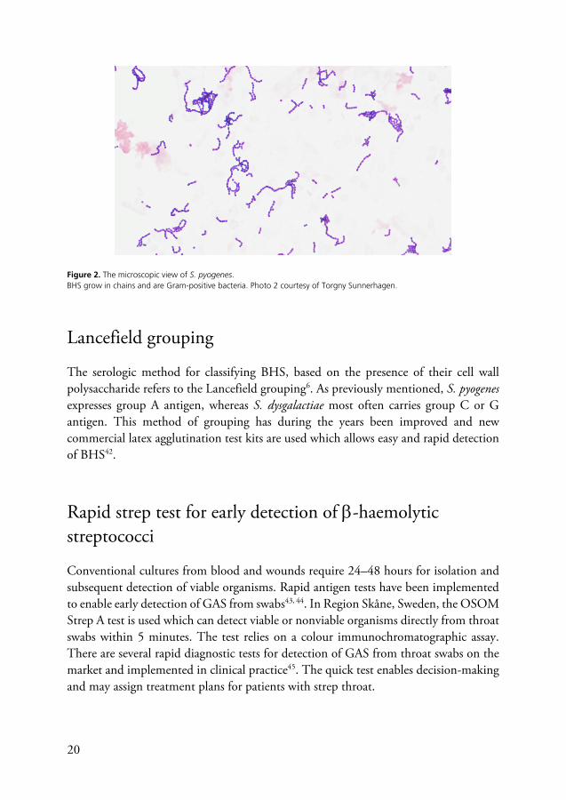
In 1884, Hans Christian Gram described a method to differentiate microorganisms based on their cell wall composition and reactions to different series of staining and decolourization steps39. The Gram stain is still utilized today, with some steps of modification during the years. The procedure includes a primary stain of crystal violet, followed by the addition of iodine, decolourization with ethanol or acetone and in the end counterstaining with e.g., safranin40. Microorganisms with cell walls containing thick layers of peptidoglycan retain the primary dye, stain purple, and are called Gram-positive bacteria. Gram-negative bacteria have thinner layers of peptidoglycan and retain only the counter stain resulting in a pink staining41. The microscopic view of BHS reveals a pattern of growth and shape as chains. The bacteria stain purple due to their thick cell wall and fall into the category of Gram-positive bacteria, (Fig. 2).
20
Figure 2. The microscopic view of S. pyogenes. BHS grow in chains and are Gram-positive bacteria. Photo 2 courtesy of Torgny Sunnerhagen.
Lancefield grouping
The serologic method for classifying BHS, based on the presence of their cell wall polysaccharide refers to the Lancefield grouping6. As previously mentioned, S. pyogenes expresses group A antigen, whereas S. dysgalactiae most often carries group C or G antigen. This method of grouping has during the years been improved and new commercial latex agglutination test kits are used which allows easy and rapid detection of BHS42.
Rapid strep test for early detection of -haemolytic streptococci
Conventional cultures from blood and wounds require 24–48 hours for isolation and subsequent detection of viable organisms. Rapid antigen tests have been implemented to enable early detection of GAS from swabs43, 44. In Region Skåne, Sweden, the OSOM Strep A test is used which can detect viable or nonviable organisms directly from throat swabs within 5 minutes. The test relies on a colour immunochromatographic assay. There are several rapid diagnostic tests for detection of GAS from throat swabs on the market and implemented in clinical practice45. The quick test enables decision-making and may assign treatment plans for patients with strep throat.

21
MALDI-TOF MS
In 1975, Catherine Fenselau and John Anhalt suggested a method based on mass spectrometry to improve species identification of bacteria46. A decade later, the technique matrix-assisted laser desorption/ionization time-of-flight mass spectrometry (MALDI-TOF MS) was introduced and developed for rapid microbiological species identification of bacterial isolates47-49. A given species has a unique set of protein content and the procedure involves colonies from agar plates that are smeared onto a metal plate to which a special solution of a matrix is later added, and the plate is left to dry. The plate is later irradiated by using a UV laser beam resulting in ionization and sublimation of the sample. The generated ions are separated based on their mass-to-charge ratio (m/z) during time-of-flight in the mass spectrometer. Ions with the smallest mass and highest charge will travel faster to the detector followed by ions with larger masses and lower charges. This reflects the protein profile in the sample. Altogether, this results in a generation of a “mass spectrum” which is compared and matched against certain protein profiles in a database to identify a certain species level50. It took 30 years after the MALDI-TOF MS was developed until it was utilized in routine clinical practice and commercially introduced51, 52. MALDI-TOF MS has a relatively low cost, it is fast, and has enabled species determination of bacteria that were previously not possible to identify53-55. MALDI-TOF MS has improved species determination within BHS, separating S. pyogenes from S. dysgalactiae56, but can formally not separate subspecies within S. dysgalactiae57. Moreover, MALDI-TOF MS may have difficulties in separating S. dysgalactiae from S. canis. However, by a refinement of certain reference spectra of MALDI-TOF MS, the identification of S. dysgalactiae and S. canis can be improved58.
Sequencing
Sequencing is used to determine the structure in a certain nucleotide sequence of a DNA or RNA molecule. Frederick Sanger developed the chain terminator sequencing “Sanger sequencing” in 1977, which was the first reliable method for DNA sequencing59. The method involves a double-stranded DNA that is denatured into two single-stranded DNA (ssDNA), and a primer that binds to the DNA corresponding to the beginning of the sequencing. It also involves polymerase solutions (four deoxynucleotide triphosphate (dNTP), one type of di-deoxynucleotide triphosphate (ddNTP), and a DNA polymerase59.

22
Sequencing 16S ribosomal ribonucleic acid (rRNA) is important to determine species within BHS. 16S rRNA is present in all bacteria and encodes the 30S which is a small subunit in the prokaryotic ribosome. The method of this sequencing is therefore very valuable to distinguish certain species of bacteria even after antibiotic treatment has been administered when the bacterial isolate can no longer be cultured60. 16S rRNA can be useful in distinguishing different species within BHS, but not to the level of subspecies. In those cases, multilocus sequence typing (MLST) may be applied. MLST is a DNA sequence-based method and involves sequencing of multiple loci, often house-keeping genes within the bacterial genome61. A sequence type or lineage is assigned by comparing the results to other already known isolate profiles in a database. This enables species identification in species and subspecies that are closely related. MLST is used for further characterization of S. pyogenes62 and is also available for delineation of S. dysgalactiae63. In addition, whole genome sequencing (WGS), can discriminate closely related species and subspecies. The method analyses the entire DNA sequence of an organism and provides a genomic fingerprint which can enable a better understanding of clonal groups in bacterial infections64, 65.
emm typing
“As a general rule, a single M antigen is specific for the type concerned”
Lancefield R, J Immunol, 196216
Rebecca Lancefield developed a serotyping system based on the antigenic variation of the M protein to distinguish different emm types of GAS16. Frederick Griffith developed another system, called the T protein agglutination system with the purpose of typing66. The M protein system is more utilized in the scientific world. Both S. pyogenes and S. dysgalactiae express the M protein, and this is an important virulence factor for the bacteria. emm typing involves sequencing of the emm gene that encodes the M protein. The method involves sequencing of the 5- end of the emm gene which encodes the hypervariable N-terminus of the M protein67. There are currently more than 90 different emm types among S. dysgalactiae68 and > 250 different emm types among S. pyogenes68, 69. Centers for Disease Control and Prevention (CDC) has set up a database for emm typing70.

23
ELISA
Sometimes there is a need to detect levels of antibodies following an infection. Enzyme immunoassay (EIA) and enzyme-linked immunosorbent assay (ELISA) incorporate an immunoassay technique which is routinely utilized in the laboratory and integrated into diagnostic methods globally71. Anton Schurs and Bauke van Weemen, from the Netherlands, developed the EIA technique in the 1970s72, and almost concurrently the ELISA method was developed and conceptualized in Sweden by Peter Perlman and Eva Engvall73. The ELISA methodology is used to detect any presence of antibodies, antigens and/or different proteins in biological samples. The basic principle for ELISA is the immobilization of an antigen to a solid surface. A multi-well 96-plate is often used for that purpose. The antigen is later complexed with an antibody, which in turn is conjugated with a molecule that enables detection by another enzyme or a fluorophore. There are four different assays of ELISA. The indirect ELISA carries a two-step process for detection, in which a primary antibody binds to the antigen, followed by a secondary antibody that binds to the primary antibody for detection. In experimental settings, the primary antibody can be substituted with serum. Altogether, the ELISA methodology is easy to perform and has high sensitivity and specificity to detect antigens in biological samples74-76. Serologic testing is sometimes used to diagnose different infections due to BHS. The anti-streptolysin O (ASO) test, and the anti-deoxyribonuclease-B (ADN-B) test are specific for both S. dysgalactiae and S. pyogenes. Increased titres of both ASO and ADN-B have been observed in patients with erysipelas77, but in most of the cases, no serologic evidence for a BHS aetiology of the infection can be found78.
Figure 3. Schematic view of indirect ELISA. The antigen (pink) is immobilized into a microtitre plate. A primary antibody (blue) is subsequently added and binds to the antigen. This is followed by a secondary antibody (purple) that binds to the primary antibody. The secondary antibody is typically conjugated to an enzyme, such as horseradish peroxidase (HRP) (green). A substrate is added which reacts with the enzyme conjugate, a colour is developed, and a measurable by-product is produced. The colour is proportional to the amount of bound antigen. Figure 3 was created with Biorender.

24
Antibiotics
-lactam antibiotics
-lactam antibiotics were early discovered and remain the most utilized antibiotic in clinical practice throughout the world. -lactam antibiotics have a -lactam ring and exert their bactericidal activity by inhibiting the cell wall of the bacteria79, 80. The targets for the -lactam antibiotics are the penicillin-binding proteins (PBPs). PBPs are enzymes that catalyse several reactions involved in the crosslinking of the peptidoglycan by the bacteria81. When -lactam antibiotics bind to the PBPs, the last step in the peptidoglycan synthesis is interrupted resulting in cell lysis and eventually cell death79,
82.
Penicillin
“Streptococcus pyogenes is also very sensitive. There were small differences in the titre with different strains, but it may be said generally that it is slightly more sensitive than Staphylococcus”
Fleming A, Br J Exp Pathol, 192983
Penicillin G, also known as benzylpenicillin, was the first discovered and marketed group of -lactam antibiotics84. In 1928, Alexander Fleming discovered by chance that the growth of Staphylococcus aureus on an agar plate could be inhibited by a mould, belonging to the Penicillium genus. He determined that this mould had an antibacterial effect on Gram-positive bacteria and named the agent penicillin83. Penicillin G is employed for the treatment of several streptococcal infections, e.g., tonsillitis, pneumonia, infective endocarditis, meningitis, and skin and soft tissue infections. Since its bactericidal activity correlates with time over the minimum inhibitory concentration (MIC), it is important to administer the antibiotic equally during the day. In contrast to other pathogens, (e.g. S. aureus, Escherichia coli) that have evolved resistance mechanisms towards penicillin, such as producing -lactamases that hydrolyse the -lactam ring and deactivate penicillin85, 86, S. dysgalactiae and S. pyogenes have remained uniformly sensitive to penicillin87, 88. Quite recently though, four isolates of S.

25
dysgalactiae causing human infections exhibited decreased susceptibility to penicillin, with the detection of identical mutations in the PBPs89. Reduced penicillin susceptibility, with certain amino acid substitutions in the PBPs, has been noted in isolates of S. pyogenes90, 91, but these mutations have not shown any trending increase.
Aminoglycosides
Aminoglycosides are members of the protein synthesis inhibitors. The first member of the class to be discovered was streptomycin in 1943. Since then, several members have been discovered, and gentamicin is commonly used in clinical practice in Region Skåne, Sweden. Gentamicin binds to the 30S ribosomal subunit. This binding to the 16S rRNA leads to a misreading between transfer RNA and messenger RNA of the ribosome. This results in a defect protein synthesis92-95. Accumulation of misreading during protein synthesis cause changes in the cytoplasmic membrane which permits further uptake of aminoglycosides and eventually cell lysis96, 97. Aminoglycosides are concentration-dependent killers, thereby requiring high concentration to eradicate the microorganism. The bactericidal activity of aminoglycosides is dependent on both the peak concentration and the time the concentration is over the MIC, of the bacterium. This results in the area under the curve of the antibiotic concentration over time being a good measure of the expected effect. Aminoglycosides have several side effects and can be highly ototoxic and nephrotoxic, for which the risk increases with several doses98-
100. The addition of an aminoglycoside to a -lactam antibiotic is often used for treating infective endocarditis due to Gram-positive bacteria, based on presumptive synergy101,
102.
Clindamycin
Clindamycin belongs to the group of lincosamides, which is a class of antibiotics that once inside the bacterial cell, binds to the 50S ribosomal subunit of the ribosome of the bacteria. The binding results in blocking of the peptide bond formation on the ribosome and thereby interfering with the synthesis of proteins103, 104. Clindamycin is often added together with penicillin in the treatment of necrotizing soft tissue infections, partially as it has been shown to inhibit the production of exotoxins in vitro105, 106. Increased resistance to clindamycin has been observed for S. pyogenes107, but in particular for S. dysgalactiae108. This is important information when a streptococcal infection is solitarily treated with clindamycin.

26
Antibiotic susceptibility testing
Antibiotic susceptibility testing (AST) of bacteria is performed ex vivo in clinical practice. Bacteria can be inherently resistant to antibiotics or evolve resistance mechanisms to certain antibiotics during and after treatment. Therefore, it is of great importance to know the patterns of resistance for the bacteria when treating the patients. Susceptibility testing can be performed in liquid media (broth) and/or on solid media (agar). By these two methods, one can determine the inhibition of growth and the bactericidal effect of the antibiotic on the bacteria. The European Committee on Antimicrobial Susceptibility Testing (EUCAST) harmonizes clinical breakpoints for existing antimicrobial agents and incorporates guidelines and protocols for performing AST109. Of note, in addition to EUCAST, the Clinical and Laboratory Standards Institute (CLSI) is the American representative organization for providing AST110. The two reference methods are similar with some differences.
Broth dilution Broth dilution test has been utilized throughout the years to provide AST for different agents and comprises two-fold dilutions of antibiotics (e.g., 16, 8, 4, 2 g/mL) in a liquid growth medium containing tubes. These tubes are later inoculated with a bacteria solution of a standardized concentration and incubated overnight under appropriate conditions111. The method of broth dilution is recommended by EUCAST and corresponds with the recommendations from the International Standards Organisations109. Bacterial growth is distinguished as evidenced by turbidity in the medium. The principal advantage of this method is the ability to generate concrete results, e.g., MIC which is the lowest antibiotic concentration that inhibits bacterial growth. Fig. 4 visualizes the method of broth dilution.

27
Figure 4. Broth dilution method. The broth dilution method comprises a liquid growth medium with different antibiotic concentrations. The first evident clarity of the wells indicates the MIC for the two different antibiotics respectively. Photo 4 by Anna Bläckberg.

28
The antimicrobial gradient method The antimicrobial gradient diffusion method utilizes a strip, with a concentration gradient of the antimicrobial compound in question to determine the antimicrobial susceptibility of the bacterial isolate. This antibiotic gradient may be, e.g., the Etest (bioMérieux), and the MIC test strip (Liofilchem). An agar plate is streaked with a standardized inoculum of bacteria, the gradient is applied, and the plate is later incubated overnight at appropriate conditions. The MIC can be imprinted on the MIC reading scale in g/mL and is determined by the intersection of the lower ellipse, which is the area of inhibition, (Fig. 5).
Figure 5. The Etest methodology of S. dysgalactiae streaked on agar plates. The agar plates are inoculated with the same strain of S. dysgalactiae. The MIC is measured as the inhibition zone that intersects the graded antibiotic strip respectively. The Etest strip carrying the penicillin gradient results in a large ellipse (A), whereas the Etest strip with gentamicin, presents a smaller ellipse (B). Photo 5 by Anna Bläckberg.
The disk diffusion method In contrast to the antimicrobial gradient method which gives an exact MIC value, the disc diffusion method gives a qualitative measurement of antibiotic susceptibility, stated as “susceptible”, “increased exposure” or “resistant”, in some species for some antibiotics, according to the EUCAST breakpoints109, 112. The disc diffusion method comprises a standard bacterial inoculum on an agar plate with an antibiotic disk in the centre. The plate is incubated overnight at appropriate conditions and later a zone may be visualized around the antibiotic disk, indicating inhibition of bacterial growth.
29
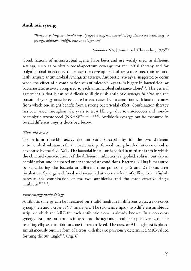
Antibiotic synergy
“When two drugs act simultaneously upon a uniform microbial population the result may be synergy, addition, indifference or antagonism”
Simmons NA, J Antimicrob Chemother, 1975113
Combinations of antimicrobial agents have been and are widely used in different settings, such as to obtain broad-spectrum coverage for the initial therapy and for polymicrobial infections, to reduce the development of resistance mechanisms, and lastly acquire antimicrobial synergistic activity. Antibiotic synergy is suggested to occur when the effect of a combination of antimicrobial agents is bigger in bactericidal or bacteriostatic activity compared to each antimicrobial substance alone113. The general agreement is that it can be difficult to distinguish antibiotic synergy in vitro and the pursuit of synergy must be evaluated in each case. IE is a condition with fatal outcomes from which one might benefit from a strong bactericidal effect. Combination therapy has been used throughout the years to treat IE, e.g., due to enterococci and non--haemolytic streptococci (NBHS)101, 102, 114-116. Antibiotic synergy can be measured in several different ways as described below.
Time-kill assays To perform time-kill assays the antibiotic susceptibility for the two different antimicrobial substances for the bacteria is performed, using broth dilution method as advocated by the EUCAST. The bacterial inoculum is added in nutrient broth in which the obtained concentrations of the different antibiotics are applied, solitary but also in combination, and incubated under appropriate conditions. Bacterial killing is measured by subculturing the bacteria at different time points, e.g., 6 and 24 hours after incubation. Synergy is defined and measured at a certain level of difference in cfu/mL between the combination of the two antibiotics and the most effective single antibiotic117, 118.
Etest synergy methodology Antibiotic synergy can be measured on a solid medium in different ways, a non-cross synergy test and a cross or 90 angle test. The two tests employ two different antibiotic strips of which the MIC for each antibiotic alone is already known. In a non-cross synergy test, one antibiotic is infused into the agar and another strip is overlayed. The resulting ellipse or inhibition zone is then analysed. The cross or 90 angle test is placed simultaneously but in a form of a cross with the two previously determined MIC-valued forming the 90 angle119, (Fig. 6).

30
Figure 6. Etest synergy methodology with cross or 90 angle test. The agar plate is inoculated with a bacterial strain of S. dysgalactiae. The Etest strips of penicillin and gentamicin are overlayed on the agar plate in a cross-approach. MIC is measured as the inhibition zone that intersects the graded antibiotic strip for each Etest strip respectively. Photo 6 by Anna Bläckberg.
The obtained MIC values in the combined approach are calculated against each antibiotic alone and synergy can be determined as described by the equation below119.
FICIndex FIC CA B
CA + FICB CB A
CB
The fractional inhibitory value (FIC) of each antibiotic can be calculated by distinguishing the MIC values of each inhibitory zone for the antimicrobial alone and in combination. The fractional inhibitory index (FICIndex) is calculated by the sums of FICA and FICB. A value < 0.5 suggests the presence of synergy.
Synergistic antibiotic activities against -haemolytic streptococci
Combination therapy may be imperative when strong bactericidal action is desired. As previously mentioned, combination therapy is recommended in IE due to some bacteria101, 102, 114-116. IE due to BHS is rare but has an acute onset of presentation120-122. Combination therapy with a -lactam and an aminoglycoside for treating IE due to BHS is suggested in the current European and American guidelines123, 124. This is partly based on previous publications implying synergy in vitro between penicillin G and an aminoglycoside on isolates of GGS125, 126. The Swedish guidelines recommend monotherapy with penicillin G for IE due to BHS127. This is partly based on a lack of any certain beneficial effect with combination therapy in IE due to group B streptococci (GBS)128. Aminoglycosides are also notorious for their adverse effects including nephrotoxicity and ototoxicity129, 130. As previously mentioned, clindamycin is often added in the treatment of necrotizing soft tissue infections due to BHS, partly due to

31
the observation of greater efficacy of clindamycin in mouse models with necrotizing soft tissue infections131. However, there are also reports that a combination of penicillin and clindamycin did not show any evident bactericidal advantage over each agent alone in the killing of S. pyogenes performed in time-kill assays132. In addition, older studies have reported antagonism of penicillin together with protein synthesis inhibitors both in vitro133 an in vivo134. Of note, results from studies in vitro cannot be directly implemented in vivo. Therefore, synergy may be observed in vitro, but may not be clinically effective in vivo. Combination therapy may be justified in patients with severe infections, but the risk of unwanted side effects should be considered and the need for synergistic bactericidal action can be evaluated in each case.
Antimicrobial resistance has emerged as one of the most critical public health problems worldwide, according to the World Health Organization (WHO)135. Bacteria have evolved versatile resistance mechanisms to antibiotics, such as enzymatic inactivation, modifications of drug targets, reduced permeability of the outer membrane, and active drug efflux136. S. pyogenes and S. dysgalactiae have remained sensitive to penicillin. On the other hand, they have evolved resistance mechanisms to other classes of antibiotics, in particular to erythromycin and clindamycin137-139.

32
Streptococcus dysgalactiae and Streptococcus pyogenes
S. dysgalactiae and S. pyogenes are the two main bacteria of this thesis. This chapter begins by describing the historical aspects of species determination, as well as microbiological characteristics, and diseases of S. dysgalactiae and S. pyogenes. This is followed by a brief description of the important molecules that these bacteria possess to interact with the host defence system.
Classification
S. dysgalactiae and S. pyogenes are members of the genus Streptococcus and belong to the phylum Firmicutes. Friedrich Rosenbach was the first to use the term S. pyogenes when isolating the organism from a wound and skin infection in 1884140. S. pyogenes expresses group A antigen (GAS) and almost all strains are -haemolytic. The “pyo” denoting “pus” and “genes” meaning “to produce” in Greek.
Another -haemolytic pathogen is S. dysgalactiae which usually carries group C or G antigen (GCS/GGS). S. dysgalactiae was initially recognized as causing bovine mastitis. The dys denoting “ill, hard” and galactia pertaining to “milk secretion”, and dysgalactiae meaning “loss or impairment of milk”. S. dysgalactiae was first mentioned by Diernhofer141. Furthermore, Frost and Engelbrecht described Streptococcus equisimilis for human BHS carrying group C antigen in 1936142. Alongside the recognition of the Lancefield grouping, this was the preferred nomenclature used. Years later, results from DNA-DNA hybridization data indicated similarities between S. dysgalactiae, S. equisimilis and large colony forming group C, G, or L streptococci. The species were transferred into a single species, S. dysgalactiae. This was revived and recognized in the approved lists of bacterial names in 1983143, 144. Thirteen years later, Vandamme et al proposed a new classification and proposed that S. dysgalactiae should be classified into distinct subspecies as they show different phenotypic characteristics145. Furthermore, S. dysgalactiae was divided into two subspecies; S. dysgalactiae subsp. equisimilis (refers to human origin, presents -haemolysis, or -haemolysis, and reacts with group A, C, G or L antigen) and S. dysgalactiae subsp. dysgalactiae (proposed for animal origin, can

33
present -haemolysis, -haemolysis, or -haemolysis and expresses group C antigen146). The division of S. dysgalactiae is complex and results from whole genome sequencing data have shown both host-restricted and lineage-specific restricted distribution of the subspecies of S. dysgalactiae147. Transmission of S. dysgalactiae is usually person to person, and sites of colonization or focal infections may serve as reservoirs for transmission148. S. pyogenes is transmitted directly by air droplets, and indirectly through contaminated surfaces61, 149. Similar to S. pyogenes, outbreaks of community- or hospital-acquired infection have been observed with S. dysgalactiae150. Human infections due to S. dysgalactiae subsp. dysgalactiae are rare and have predominantly been reported in patients after animal contact148, 151-155. In addition to S. dysgalactiae, Streptococcus equi and Streptococcus canis carry group C and G antigens respectively156,
157 . These species are often of animal origin but can cause diseases in humans158. Table 1 summarizes some of the different groups, species, subspecies, and types within BHS68,
145, 146, 159-161.
Table 1. -haemolytic streptococci
Group Species Subspecies Haemolysis Types
A
S. pyogenes > 250 emm types68, 69
S. dysgalactiae
S. dysgalactiae subsp. equisimilis *
B
S. agalactiae 10 serotypes162
C
S. dysgalactiae , ,
S. dysgalactiae subsp. equisimilis *
S. dysgalactiae subsp. dysgalactiae
S. equi
S. equi subsp. equi
S. equi subsp. zooepidemicus
S. equi subsp. ruminatorium
G
S. dysgalactiae
S. dysgalactiae subsp. equisimilis *
S. canis
L
S. dysgalactiae
S. dysgalactiae subsp. equisimilis *
The number of emm types is constantly shifting, and the exact number of emm types are therefore fluctuating. * S. dysgalactiae expresses > 90 emm types and they can be found in group A, C, G or L streptococci. The current emm types can be found in the StrepLab database, at CDC68, 70.

34
Microbiological aspects
S. pyogenes and S. dysgalactiae are facultative anaerobic cocci and display white-greyish colour with a diameter of 0.5 mm, surrounded by a -haemolysis on a blood agar plate, 24 hours after incubation in 5% CO2 at 35–37C, (Fig. 7).
Figure 7. S. dysgalactiae streaked on a blood agar plate. White-greyish colonies are displayed surrounded by -haemolysis after 24 hours of incubation in 5% CO2 at 35–37C. Photo 7 by Anna Bläckberg.
The PYR test refers to the presence of the enzyme pyrrolidonyl arylamidase. Since S. pyogenes is positive and S. dysgalactiae negative, the bacteria can be separated by this test161. Another way to differentiate the pathogens is that S. pyogenes is susceptible to bacitracin whereas S. dysgalactiae is resistant. However, bacitracin-resistant strains of S. pyogenes have been reported throughout Europe163, 164. Of note, streptococci are difficult to classify purely based on the presence of the carbohydrate antigen, as many of the streptococci within NBHS (e.g., Streptococcus anginosus) may carry A, C, G or F antigen, and may also display -, - or -haemolysis on a blood agar plate165. Other bacteria such as enterococci are also able to display -haemolysis and are also PYR positive. Enterococci can be relatively easily distinguished from S. pyogenes due to their differences in colony morphology and results from a combination of phenotypic testing.
Emergence of invasive diseases
The burden of invasive diseases due to both S. pyogenes and S. dysgalactiae varies in time and by geographical distribution. The estimated annual incidence of invasive diseases due to S. pyogenes ranges from 2.2–3.8 per 100 000 inhabitants166-168, with a case-fatality

35
rate of 14–16%167-170 in high-income countries. S. pyogenes still causes invasive diseases with millions of deaths yearly in less developed countries171, and since 1980 an increase in infections with S. pyogenes has also been reported in developed countries172. A plausible explanation for the increase of invasive diseases may be due to fluctuations of different emm types both temporally and geographically. emm1, expressed by S. pyogenes, is the most common emm type but has also been associated with causing invasive diseases with high mortality168, 173. emm3 has also been associated with invasive infections with high mortality rates168. These two emm types have also been found in patients with necrotizing soft tissue infections174.
S. dysgalactie has historically been perceived as non-pathogenic flora of the upper respiratory tract, the gastrointestinal tract and the female genital tract, and was not recognized as an important human pathogen until 1980s175. However, in recent years the burden of invasive S. dysgalactiae diseases approximates that of invasive S. pyogenes diseases. The annual incidence of invasive diseases due to S. dysgalactiae reaches from 2.2–6.3 per 100 000 inhabitants176-180, with a case-fatality rate ranging from 2–18%179,
181-183. Prolonged survival in adults and improved species determination with MALDI-TOF MS may be an explanation for the high detected rate of invasive S. dysgalactiae diseases. The distribution of emm types among isolates of S. dysgalactiae shows greater diversity than among S. pyogenes. In a study from the United States, the most predominant emm types were stG6 and stgG2078184, which was dissimilar from a study from Finland in which stG480 and stG485 were the most common183. A study from Japan linked certain emm types, (stG2078 and stG10), to invasive diseases185, whereas other studies have not established such a distinct correlation186, 187.
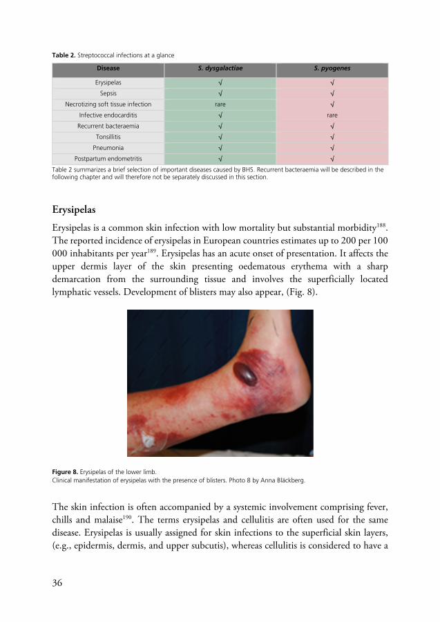
Streptococcal infections at a glance
Both S. pyogenes and S. dysgalactiae give rise to a wide range of infections, from mild tonsillitis to sepsis and severe soft tissue infection. The species cause similar infections with some exceptions. S. dysgalactiae often affects elderly people with comorbidities, while S. pyogenes affects a younger age group. Table 2 summarizes different diseases the pathogens cause, and some will be separately discussed below.
36
Table 2. Streptococcal infections at a glance
Disease S. dysgalactiae S. pyogenes
Erysipelas
Sepsis
Necrotizing soft tissue infection rare
Infective endocarditis rare
Recurrent bacteraemia
Tonsillitis
Pneumonia
Postpartum endometritis
Table 2 summarizes a brief selection of important diseases caused by BHS. Recurrent bacteraemia will be described in the following chapter and will therefore not be separately discussed in this section.
Erysipelas
Erysipelas is a common skin infection with low mortality but substantial morbidity188. The reported incidence of erysipelas in European countries estimates up to 200 per 100 000 inhabitants per year189. Erysipelas has an acute onset of presentation. It affects the upper dermis layer of the skin presenting oedematous erythema with a sharp demarcation from the surrounding tissue and involves the superficially located lymphatic vessels. Development of blisters may also appear, (Fig. 8).
Figure 8. Erysipelas of the lower limb. Clinical manifestation of erysipelas with the presence of blisters. Photo 8 by Anna Bläckberg.
The skin infection is often accompanied by a systemic involvement comprising fever, chills and malaise190. The terms erysipelas and cellulitis are often used for the same disease. Erysipelas is usually assigned for skin infections to the superficial skin layers, (e.g., epidermis, dermis, and upper subcutis), whereas cellulitis is considered to have a

37
deeper involvement, (extending from the dermis into the subcutaneous tissue), and seldom presents a demarcation line191. The definitions and clinical presentations are overlapping, and some clinicians use the term cellulitis also for more cutaneous infections and erysipelas only for facial cutaneous infections. To distinguish erysipelas from cellulitis or even necrotizing soft tissue infections may be challenging192. Cellulitis is more often associated with a more diverse variety of organisms, e.g., S. aureus is often recognized193. BHS have been more associated with erysipelas and cellulitis194-197, and predominantly GCS/GGS have been implicated as important pathogens195, 198. But again, the diagnosis of skin and soft tissue infection relies on the clinical presentation, and different terms are used for the same diseases making the comparison between studies difficult. Bacteraemia is rare in erysipelas, (81-87% of blood cultures are negative), and obtaining blood cultures may not be compulsory in uncomplicated erysipelas192. Erysipelas has thus remained a clinical diagnosis during the years. When and if skin lesions are present, the causative agent is rarely found from obtained cultures192. Other methods such as cultures from needle aspirates, and or punch biopsies have identified BHS in a minority of cases78, 194, 199. Other analyses may be serologic testing77, 78, 194 and or direct immunofluorescence200. These methods are often time-consuming, difficult to analyse and therefore hard to implement in clinical routine practice. As the condition is an infection of the skin, punch biopsies from infected sites may generate the bacteria by the PCR technique. However, in most of the cases, no bacterial agent to cellulitis could be detected using this movement and molecular technique201. Furthermore, if pathogenic bacteria are found, it may also be difficult to assess their clinical relevance.
Recurrence of erysipelas is common, often affects the same anatomic site and is present in up to 40% of cases202, 203. Two- and five-year recurrence rates have been estimated in up to 17%, and 57% of patients with a previous history of cellulitis204-206. The major risk factor for recurrent erysipelas is lymphedema202, 207. Other risk factors may be previous radiation, venous insufficiency, and the presence of disruption of cutaneous barriers resulting in entrance sites for the bacteria202, 208, 209. Colonization of BHS has been recognized in up to 44% of patients presenting with erysipelas, in which S. dysgalactiae has been the most predominant bacteria210. Some patients still carry the organism after concluded antibiotic treatment. Another plausible explanation for recurrence may be the ability of both GAS and GCS/GGS for intracellular uptake into and persistence in epithelial and endothelial cells creating a streptococcal reservoir211,
212. These intracellular reservoirs are likely not eliminated by penicillin since penicillin does not inevitably reach sufficient bactericidal intracellular concentration209. Treatment with antibiotics is often directed towards the streptococci with penicillin. However, when the depth of the infection in the tissue is unknown, antibiotic agents

38
may be administered to establish a broader coverage including S. aureus213. Preventing the recurrence of erysipelas is challenging. Prophylactic administration of low-dose penicillin has been proven to prevent recurrences at a low cost, but these results have been based on smaller studies214, 215. Other studies have not observed any significant reduction of recurrent erysipelas with the administration of prophylactic antibiotics216. Occasionally some patients experience relapses of erysipelas although they are on long-term prophylaxis with penicillin217, 218. In addition, erysipelas often reoccurs within a short time after prophylaxis has been discontinued203, 219. Other interventions to prevent recurrent erysipelas/cellulitis have been compression therapy. In a randomized controlled trial study, compression therapy has proven to reduce the recurrence of cellulitis in the lower limbs with chronic oedema220.
Figure 9. Erysipelas of the lower limb. Typical edematous erythema and swelling of the lower limb. The right leg is shown for comparison. Photo 9 by Anna Bläckberg.
Sepsis
Sepsis is a major health problem causing high morbidity and mortality with increasing incidence globally221-223. Gram-negative bacteria have traditionally been perceived as the most common causative pathogens in sepsis, but more recently, Gram-positive bacteria

39
have been associated with sepsis224, 225. As previously mentioned, S. dysgalactiae and S. pyogenes cause sepsis with high mortality and morbidity throughout the world171, 178.
The term sepsis has been used since ancient Greek and comes from the Greek word “”, denoted “I rot”226, 227. The definition of sepsis has been re-evaluated for several years based on pathophysiology, diagnosis, and degrees of severity. In 1992, Roger C Bone and the American College of Chest Physicians and the Society of Critical Care Medicine (ACCP/SSCM) Consensus Conference Committee, published their definition of sepsis (Sepsis-1)228. Sepsis was defined as the systemic inflammatory response syndrome (SIRS) in the presence of infection. The SIRS criteria comprised tachycardia, tachypnoea, hypothermia or hyperthermia, and leucocytosis. Furthermore, sepsis was divided based on the degree of severity. Severe sepsis was defined as organ dysfunction accompanied by hypoperfusion or hypotension and septic shock was defined as organ dysfunction with persisting hypotension despite adequate fluid resuscitation. Since then, the criteria for sepsis have been evaluated. Although the SIRS criteria may occur outside the presence of infection, the evidence of changing the pre-existing criteria was low and the definition of sepsis was only slightly altered in 2001, then comprising an expansion of signs and criteria for diagnosing sepsis, (Sepsis-2)229. The Sepsis-3 criteria were proposed and published in 2016 where Singer et al stated that “Sepsis is a life-threatening organ dysfunction caused by a dysregulated host response to infection”230. As it was considered that sepsis was always to be perceived as severe, the term severe sepsis was removed and the scoring system, Sequential Organ Failure Assessment (SOFA) was applied to assess the definition of organ dysfunction231,
232. Sepsis-3 is still in use today and the use of the SOFA score has been validated to be an important predictor for mortality as well as for staging of severity of sepsis233, 234.
Streptococcal toxic shock syndrome (STSS) was first described by Cone et al235. and in 1993 the case definition was established236. STSS involves parameters that largely mirror those that define septic shock but coupled with evidence of GAS pathology230,
236. STSS indicates multiorgan failure and is often present in severe soft tissue infection due to GAS. STSS has also been frequently encountered in severe infections due to GCS/GGS237-243. Bacteraemia, necrotizing soft tissue infection, and puerperal sepsis due to BHS are often associated with STSS173.
Necrotizing soft tissue infection (NSTI)
“The infection essentially produces a gangrene of the subcutaneous tissue, subsequently it causes death of a part of the overlying skin”
Meleney FL, Archives of Surgery, 1924244

40
Necrotizing soft tissue infection (NSTI) is a progressive deep infection of the subcutaneous tissue associated with serious and life-threatening conditions with a high mortality rate. The symptoms were initially described by Hippocrates in the 5th century BC and the condition has thereafter been described in a multiplicity of terminology. For example, in 1871, the surgeon Joseph Jones described it as “hospital gangrene”245, and in 1918, Pfanner described a patient with -haemolytic streptococcal infection to the skin as a necrotizing erysipelas246. In 1924, Meleney published a series of patients with rapidly developed gangrene and described the infection as “haemolytic streptococcus gangrene”, as he considered the causative organism to be what he called haemolytic streptococci244. The condition often involves extensive necrosis of subcutaneous tissue and local ischemia and can be caused by other bacteria that are not haemolytic. Wilson considered this and was the first to use the term “necrotizing fasciitis” in 1952247. This term has later been used in succeeding publications248-250, and the term NSTI is now used to encompass infections that also involve other soft tissues than fascia. S. pyogenes is associated with NSTI, but S. aureus and other Gram-negative bacteria are also linked to the condition248, 251. NSTI due to GCS/GGS has also been described174, 178, 252. NSTI is often accompanied by STSS and severe pain. Penicillin is the primary treatment for NSTI due to BHS. Further addition of clindamycin has proven to be valuable, and this combination therapy has shown lower mortality rates compared to penicillin monotherapy253, 254. It has been suggested that intravenous immunoglobulins (IVIg) may inhibit pro-inflammatory responses of superantigens from S. pyogenes and aid the opsonisation of the bacteria255, 256. Administration of IVIg has proven to be effective in the treatment of patients with NSTI and STSS254, 257-260. In a Cochrane review from 2013, Alejandria et al showed that administration of IVIg significantly reduced the mortality rate in patients with bacterial sepsis261. However, the studies included were small and the overall results were not robust enough to implement IVIg in the recommended treatment of NSTI in the current guidelines from the Infectious Diseases Society America192.
Infective endocarditis (IE)
Infective endocarditis (IE) is an infection of the heart valves, the endocardium or intracardiac devices. In 1885, Sir William Osler described the condition as a “mycosis” that could be spread throughout the body262. In developed countries, the incidence is 2.6–7.0 cases per 100 000 inhabitants per year with a mortality rate of 10–30%124, 263. Diagnosing IE may be difficult and is summarized by clinical findings, symptoms, microbiological findings, and results from imaging. In 1994, David Durack proposed criteria to adequately diagnose IE, the so-called “Duke Criteria”264. These criteria have during the past years been evaluated and resulted in the utilization of the modified

41
Duke criteria which are commonly used today265, with the addition of the European Society of Cardiology (ESC) 2015 modified diagnostic criteria123. Table 3 summarizes these criteria. S. aureus is an important pathogen in causing IE, followed by streptococci from the oral flora266, 267. IE due to BHS is rare, constituting up to 3% of all cases of IE in some countries268. Although endocardial infections with BHS is rare, these cases constitute challenges in clinical practice and have an acute presentation with a substantial embolization rate122, 269, 270. A study from Norway investigated sixteen cases of IE due to S. pyogenes and S. dysgalactiae that occurred in the period 1999–2013. The embolic event and 30-day mortality rate was 50% and 25% respectively120. Most of the patients were treated with a combination of a -lactam and an aminoglycoside. Recently, a study described cases of IE due to BHS based on results from data from the International Collaboration on Endocarditis Cohort Study268. This is a large multinational database comprising patients with IE recruited from 64 sites and 28 countries using a standardized case report form. BHS with GCS/GGS were more common than IE due to GAS. In contrast to the other bacteria within BHS, only patients with GCS/GGS aetiology had any form of relapse268. Altogether, BHS with IE is characterized by an acute presentation, often requiring surgery, and has a high rate of embolization. Findings of BHS in blood cultures do not, according to the modified Duke criteria, count as a major criterion. Current European and American guidelines consider some streptococcal species and their prevalence of IE, but not all streptococci are encountered in that calculation123, 124. Therefore, it may be difficult to know when further investigations of looking for a cardiac focus of infection should be performed in patients with BHS bacteraemia. In a study by Sunnerhagen et al, a score for directions on when to perform echocardiography was proposed in patients with bacteraemia with NBHS271. In addition, a retrospectively based study from Denmark proposed a flowchart for when to perform echocardiography in patients with IE due to S. dysgalactiae and S. pyogenes respectively, based on numbers of positive blood cultures, risk factors and the prevalence of IE272. Patients with S. pyogenes bacteraemia were regarded as having a low risk of having IE and echocardiography was not recommended unless they had 3 positive blood cultures and risk factor/-s associated with IE. Bacteraemia with S. dysgalactiae was considered as a moderate risk for having IE and echocardiography was suggested with the presence of 3 positive blood cultures and/or risk factor/-s associated with IE. This stratification of low risk (< 3%) and moderate risk (3-10%) for the diagnosis of IE was based on cases of streptococcal bloodstream infections from Danish nationwide registries272.

42
Table 3A. Definition of IE according to the modified Duke Criteria265
Definitive infective endocarditis
2 major criteria, or 1 major criterion and 3 minor criteria, or 5 minor criteria
Possible infective endocarditis
1 major criterion and 1 minor criterion, or 3 minor criteria
Table 3B. Terms used in the modified Duke criteria265, with addition of the ESC 2015 modified diagnostic criteria123
Major criteria
Blood culture positive for IE Typical organisms consistent with IE from 2 separate blood cultures or Microorganisms consistent with IE from persistently positive blood cultures or Single positive blood culture for Coxiella burnetti or IgG titre > 1: 800
Evidence of endocardial involvement
Echocardiogram positive for IE Oscillating intracardiac mass on valve or supporting structures, in the path of regurgitant jets, or on implanted material in the absence of an alternative anatomical explanation or Abscess or New partial dehiscence of prosthetic valve
New valvular regurgitation
Identification of paravalvular lesion by cardiac CT123
Abnormal activity around the site of prosthetic valve implantation detected by 18F-FDG PET/CT (only if the prosthesis was implanted for > 3 months), or radiolabelled leucocytes SPECT/CT123
Minor criteria
Predisposition Predisposing heart conditions or intravenous drug use
Fever, temperature > 38 C
Vascular phenomena Major arterial emboli, or septic pulmonary infarcts, or mycotic aneurysm, or intracranial hemorrhage, or conjunctival hemorrhages, or/and Janeway’s lesions
Immunological phenomena Glomerulonephritis, Osler’s nodes, Roth’s spots, and rheumatoid factor
Microbiological evidence Positive blood culture, but does not meet the criteria for a major criterion or Serological evidence of active infection with organism consistent with IE
Identification of recent embolic events or infectious aneurysms by imaging only (silent events)123
Table 3B presents the modified Duke Criteria265, with the addition of the proposed ESC 2015 modified diagnostic criteria123. Abbreviations used are; computed tomography (CT), fluorodeoxyglucose (FDG), positron emission tomography (PET), and single photon emission computerized tomography (SPECT).

43
Tonsillitis
Tonsillitis encounters an acute inflammation of the pharynx and tonsils. S. pyogenes has been known to cause this disease273, but other BHS have also been encountered274. In a study from Sweden, the asymptomatic carrier rate of GAS was 6.4% in persons < 10 years of age, 9.4% in persons 10–15 years of age, and 0.2% in persons 16 years of age275. In the present study, the carrier rate of GCS/GGS was 0.04%, 3.1%, and 2.7% respectively for the same age categories275. Additionally, the prevalence of GAS in symptomatic patients was 25% in patients 10–15 years of age and 14.1% in patients 16 years of age275. Concurrently, the prevalence of GCS/GGS was 6.3% and 4.5% respectively in the same age categories275.
Pneumonia
Community-acquired pneumonia (CAP) due to S. pyogenes was often reported during the prebiotic era in the 20th century276, 277 . Today it is a rare entity, sporadically detected following viral infection, e.g., influenza278-280. A study from Spain observed an annual incidence of S. pyogenes pneumonia of 0.29–2.29 per 100 000 inhabitants with a case-fatality rate of 20%. The most prevalent type was emm1281. Pneumonia due to S. dysgalactiae seems to be a rare presentation282, 283.
Postpartum endometritis
Postpartum endometritis is an infection of the uterine lining and is one of the major causes of maternal infection following delivery. Since the 17th century puerperal sepsis following abortion or vaginal delivery has been described with mortality rates of up to 40%284-286. S. pyogenes has been known to cause postpartum endometritis, but endometritis with S. dysgalactiae does also occur287 288, 289. Cultures from blood and cervix are usually obtained. Surveillance with emm typing may be of importance when nosocomial outbreaks occur290.
Bacterial molecules to fight the human host defence
S. dysgalactiae and S. pyogenes possess similar molecules to fight the human host defence. The pathogens express fibronectin-binding proteins which mediate adherence to the human epithelial cells and facilitate internalization into host cells291-294. The presence of hyaluronic acid capsule has been shown to confer phagocytosis by both masking

44
adhesins on the bacterial cell wall and resisting complement-mediated opsonophagocytic killing295, 296. Horizontal genetic transfer between S. dysgalactiae and S. pyogenes may occur to acquire genetic material297. Table 4 summarizes a brief selection of molecules that either S. dysgalactiae and/or S. pyogenes possess and that interact with the human host. Several of these molecules will be specified in separate sections below.
Table 4. Important molecules of S. dysgalactiae and S. pyogenes that interact with the host defence
Molecule S. dysgalactiae S. pyogenes Function
Fibronectin-binding proteins
Adhesion. Invasion291-294
Capsule Resist phagocytosis295, 296
M protein Evascape from the host defence system298-303
Protein G - Antibody binding304, 305
IdeS - Antibody degradation306, 307
Streptolysin S -haemolysis. Tissue damage308-
310
Streptolysin O Tissue damage311, 312
Streptokinase Bacteria spread in the tissue148, 313,
314
Superantigens speA, speC, speG, speK, speL, speM, ssa, smeZ
speA, speC, speG, speH, speI, speJ, speK, speL, speM,
ssa, smeZ
Proinflammatory response. Non-specific activation of T-cells147, 315-
317.
SIC sicG Inhibition of complement318-321
EndoS EndoSd Antibody deglycosylation, loss of function322, 323
C5a peptidase Inhibition of complement147, 324, 325
SpeB - Tissue necrosis326
The molecules may also have other functions in fighting the immune defence. Some of these molecules are present in S. dysgalactiae subsp. equisimilis but not in S. dysgalactiae subsp. dysgalactiae. Abbreviations used are; Immunoglobulin G-degrading enzyme of S. pyogenes (IdeS), streptococcal pyrogenix exotoxin (Spe), streptococcal inhibitor of complement (SIC), sic-like gene (sicG), streptococcal cysteine protease (SpeB).
M protein
The M protein is a cell surface attached protein and is a major virulence determinant for both S. dysgalactiae and S. pyogenes. The M protein is encoded by the emm gene and is the basis of the Lancefield serotyping. As previously mentioned, there are more than 90 different emm types among S. dysgalactiae68, 327 and > 250 different emm types among S. pyogenes68, 69. The M protein is an -helical coiled-coil protein that extends up to 0.6 m from the cell wall299, 328. It consists of various domains. The N-terminal part, on which molecular typing is based, consists of a hypervariable domain. This region is a target for type-specific protective antibodies, but intriguingly has also been shown in experimental studies sensitive to proteolysis and thereby weakly immunogenic329. This

45
weak immunogenicity of the hypervariable domain329, 330 and its ability of sequence variation enables the bacteria to escape the immune defence300. Proximal to the hypervariable domain is the A region, followed by the B repeats and later the C repeats of which the latter goes into the D domain. The D region contains an LPxTG motif for cell wall-anchoring331-333, (Fig. 10). Cross-immunity against emm types do not seem to be present due to the antigenic diversity of the N-terminal. Therefore, it may be a more strain-restricted protective immunity. The M protein is an important molecule and its interaction with the human host defence is further discussed below.
Figure 10. The M protein Schematic figure of the M protein, inspired by Frost et al298. The PTGS domain contains the LPXTG motif which is cleaved to form the mature protein, resulting in a helical portion (HP) enfolded in the cell membrane and a polar tail (PT) in the cell cytoplasm. Figure 10 by Anna Bläckberg.

46
Apart from these classical M proteins, there are M-like proteins, a term that was first presented by Bessen and Fischetti in 1992334. The fact that GAS were able to express more than one M protein was initially described in the 1960s by Wiley and Wilson335. The M-like proteins are encoded by the enn or mrp genes in contrast to the M protein, which is encoded by the emm gene, as previously mentioned. These emm-like genes as well as the emm gene are found within the chromosomal locus, regulated by mga (multiple gene activator) regulon298. This regulon coordinates the expression of the M protein, C5 peptidase and many other molecules336.
M protein and interaction with host ligands
The M protein has a variety of different immunomodulatory assets. Figure 11 summarizes different interactions between the M protein and the human host. Some of these interactions are described in separate sections below.
Figure 11. Interactions between the M protein and different host ligands. Schematic outlined how the M protein interplays with the immune defence, inspired by Frost et al298, and by Bisno et al337. Some of these proteins have also other immunomodulatory assets. Abbreviations used are; Factor H-like 1 protein (FHL1), C4-binding protein (C4BP), lipoteichoic acid (LTA), peptidoglycan (PG), and membrane (M). Figure 11 by Anna Bläckberg.

47
Inhibition of the activation of the complement system Many M proteins can bind to C4-binding protein (C4BP). This protein inhibits the classical pathway complement activation302. Moreover, different M proteins can bind to another complement inhibitory protein, factor H-like protein 1 (FHL1)301. Bound FHL1 and/or bound C4B to the hypervariable region of the M protein results in the protection of the bacteria against complement activation. This inhibition of complement activation results in partial resistance to phagocytosis300.
Interaction with fibrinogen and albumin The M protein may bind to fibrinogen. This binding may in turn block the deposition of complement and indirectly inhibit complement activation338, 339. The C-domain of the M protein may bind to human serum albumin340. This binding may have a role for the bacteria to avoid antibody opsonisation and resist phagocytosis341.
Biofilm formation The different M proteins possess the ability to form a complex with lipoteichoic acid (LTA) resulting in enhancement of the hydrophobic surface of the streptococci which may contribute to adhesion and biofilm formation342. The ability to form biofilm enables the bacteria to survive and proliferate in hostile environments.
Immunoglobulin binding The M protein can interact with the Fc region of the IgG and thereby block their function and partially resist phagocytosis343. The ability of the bacteria to bind and coat different host molecules may result in a “masking” of antigenic epitopes which enables the bacteria to avoid opsonisation and thereby evading the host immune system. On the other hand, it is believed that type-specific antibodies may be directed towards the hypervariable domain and confer protection after infection with S. pyogenes. This may result in protective immunity against the certain emm type341. Studies from mouse models have discriminated development of antibodies towards the C-terminal of the M protein following invasive S. pyogenes infection, but these antibodies did not seem to opsonise and promote phagocytosis of the bacteria330, but other studies indicate that antibodies towards the C-terminal of the M protein may be protective344. Moreover, the orientation of the IgGs at the bacterial surfaces may be dissimilar from different sites, e.g., in saliva, the IgGs have mostly been found to bind to S. pyogenes via Fc345. In blood, the IgGs were preferentially bound to the bacteria via Fab facilitating phagocytosis of the bacteria345.

48
Protein G
Protein G is a cell wall protein that is expressed by S. dysgalactiae and has unique protein-binding properties304. Protein G can bind to immunoglobulin G, as well as to serum albumin. The protein has an affinity for all human IgG subclasses, whereas there is no binding to IgM, IgA or IgD. The main function of protein G is binding to the Fc region of the IgG, but interaction with the Fab fragments of the IgG has also been established. The latter binding may therefore confer protection against opsonisation304,
305, 346. Protein G has also an affinity to 2-macroglobulin (proteinase inhibitor of human plasma)347, 348. Altogether, protein G is important for the bacteria in both evading the immune system by its binding to immunoglobulins and conferring protection against proteolytic degradation. S. pyogenes has a protein G-like 2-macroglobulin-binding (GRAB) protein that protects the bacteria from proteolysis but lacks the ability of binding to immunoglobulins349.
IdeS
IdeS is an IgG-degrading enzyme expressed by S. pyogenes306. IdeS cleaves the IgG molecule just beneath the hinge region which generates one F(ab')2 fragment and two monomeric Fc fragments306. This degradation of IgG results in an accumulation of Fc and F(ab')2 fragments that retain the ability to bind to surface antigens of the bacteria but have difficulties in activating the complement system and facilitating phagocytosis by immune cells307. Due to this highly unique enzymatic reaction, the use of IdeS as a therapeutic agent has been discussed as a future treatment for clinical conditions involving pathogenic immunoglobulins.
Toxins
Streptolysin S (SLS) is responsible for causing the ß-haemolysis308. This is an oxygen-stable oligopeptide, whereas the other cytotoxin, streptolysin O (SLO) is a cholesterol-binding bacterial exotoxin. SLO binds to the cholesterol in eukaryotic cell membranes eventually causing cell lysis and tissue damage311, 312. The exact mechanism of SLS is not fully understood, but it has been suggested that the peptide contributes to soft tissue damage, and tissue necrosis, and indirectly mediates cell death of macrophages309,
310.

49
Streptokinase
S. dysgalactiae and S. pyogenes express streptokinase. This enzyme can bind to and activate the circulatory plasminogen into plasmin350. Plasmin is a central proteinase in the fibrinolytic system and contributes to tissue damage and bacterial spread across barrier148, 313, 314.
Superantigens
Streptococcal pyrogenic exotoxins (Spes) are superantigens. They bind to the T-cell receptor and MHC class II which results in co-stimulation of a large number of T-cells351. This stimulation mediates the massive release of proinflammatory cytokines and contributes to the pathogenesis of invasive diseases. Production of superantigens has been associated with multi-organ failure and STSS352. S. dysgalactiae also expresses superantigens, such as SpeA353 and SpeG. However, the exact role superantigens play in the pathogenesis of severe infections with S. dysgalactiae remains unclear182, 354.
SIC and sicG
Streptococcal inhibitor of complement (SIC) is an extracellular protein that is expressed by some types of S. pyogenes, mostly emm1, and is known to inhibit the membrane attack complex of complement318, 319. Like S. pyogenes, some types of S. dysgalactiae possess a streptococcal inhibitor of complement-mediated cell lysis-like gene, denoted sicG. The presence of this gene has been associated with severe soft tissue infections321,
355.
EndoS
EndoS is expressed by S. pyogenes and some types of S. dysgalactiae express EndoSd322,
323. This secreted endoglycosidase is important in its ability to hydrolyse the N-linked glycan on IgG. This hydrolyzation results in the loss of IgG glycan and affects the binding between the Fc region of IgG and the Fc receptor on phagocytes356. This reduces complement activation through the classical pathway and enables escape from the mediated antibody response.

50
Aspects of the immune defence
We are constantly surrounded by organisms and some of them can cause invasive diseases. The human host possesses many defence mechanisms to fight these organisms. The innate immune system encompasses the first barrier of defence. It relies on epithelial surfaces, secreted proteins, and cell-associated receptors to detect infection and separate pathogens from host tissue. The adaptive system is on the other hand based upon antigen-binding properties of immunoglobulins and lymphocytes resulting in the identification and efficient eradication of the invading microbe. In addition, the adaptive system acquires an immunologic memory. It remembers the initial antigens that elicited a primary immune response, and this leads to an enhanced immune response when encountering the same pathogens again. As previously mentioned, infections with BHS tend to reoccur, this is true for erysipelas but also for bacteraemia with S. dysgalactiae. This chapter begins by describing different important sections of the immune defence to fight bacterial infections. This is followed by a detailed description of the humoral response and development of antibodies following invasive infections with BHS. Lastly, the interplay between these developed antibodies and BHS is further discussed.
The first line defence
Anatomical and chemical barriers are the first line of defence that a microorganism meets when invading the human defence. The anatomical barrier consists of epithelia lining the external and internal parts of the body. This could be the skin but also the respiratory and gastrointestinal tract. The chemical barrier consists of mucous, cilia, and antimicrobial peptides (AMPs). AMPs are expressed in different parts of the body and can disrupt bacterial cell walls and bacterial cell membranes. AMPs are also expressed by cells and are crucial constituents of the innate system, and involved in the chemotactic and immunomodulating actions during an infection357.

51
The innate system
Once the invading microbes succeed to overcome the anatomical and chemical barrier of the immune system, the host defence has indirect strategies to fight them. Some pathogens express pathogen-associated-molecular-patterns (PAMPs). These PAMPs can be recognized by pattern-recognition receptors (PRRs) that the host’s immune cells possess358, 359. PRRs include Toll-like receptors (TLRs)360, 361, NOD-like receptors (NLRs)362, 363, retinoic acid-inducible gene I (RIG-I)-like receptors (RLRs)364, 365, and C-type lectin receptors (CLRs)366, 367 . This recognition of PAMPs by PRRs induces an efficient immune response which ultimately may result in the eradication of the invading microbe. The host’s immune cells carrying such receptors are often called phagocytes and include monocytes, macrophages, neutrophils, and dendritic cells. The microbes are internalized into a membrane-enclosed endocytic vesicle, a phagosome and are eliminated and killed within the phagocyte. This is a complex process called phagocytosis and was first discovered and described by Élie Metchnikoff, in the 20th century368. This microbial recognition and killing activity constitute a central part of the innate immune system369, 370. Further enhancement of phagocytosis occurs when certain proteins bind to the pathogen surface which promotes early identification of the invading microbe by the host’s cells. Opsonisation refers to the coating of a pathogen with soluble host proteins called opsonins. Phagocytic cells carrying specific receptors recognize these opsonins and can thereby easier eradicate opsonised microbes371. Opsonins may be immunoglobulins and/or complement proteins368.
The complement system
The complement system is a major counterpart of the immune system but also acts as a bridge to the adaptive immune system. This collection of soluble proteins was first described by Buchner et al372. The complement system is composed of more than 30 plasma proteins that are mostly synthesized in the liver and circulate in the plasma or on cell surfaces as inactive precursors373. Complement activation comprises a cascade of proteolysis. Within complement activation, there are three different pathways. Each pathway results in the formation of C3 convertase which is the centre protein of the complement cascade. Further cleavage of C3 convertase results in C3a and C3b374. C3a acts as an anaphylatoxin and can recruit inflammatory cells. C3b acts as an opsonin and enables the eradication of the pathogen by phagocytes expressing receptors for C3b. C5 convertase can be cleaved into C5a and C5b. C5a acts as a chemoattractant and recruits

52
immune cells while C5b triggers the membrane-attack-complex (MAC) in which pores are inserted in the bacterial cell membranes leading to cell lysis375.
The classical pathway is an antibody-triggered pathway that was the first to be discovered and described in the 1940s by Louis Pillemer374. The classical pathway is initiated when C1q, (the first complement protein of the cascade), interacts with the surface of the pathogen or forms an antigen-antibody complex with either IgM or IgG. Further cleaving results in the formation of C3 convertase. In 1954, Louis Pillemer proposed the alternative pathway, which became highly controversial and was first rejected by the research field. A decade later, the alternative pathway was accepted and established as a part of the scientific body of knowledge376-378. The pathway can be spontaneously activated which results in the formation of a unique C3 convertase379. In the late 20th century, the third pathway was discovered. This is called the lectin-pathway which is initiated by mannose-binding lectin (MBL) that recognizes and binds to the surface of the pathogen380, 381. When MBL binds to carbohydrate ligands on the pathogen surface, MBL-associated serine protease 1 (MASP-1) is induced which in turn cleaves and activates MASP-2. MASP-2 can cleave C4 allowing a conformational change eventually resulting in a generation of C3 convertase382, 383.
The adaptive immune system
The keystones of the adaptive immune system are adaption, specificity, and memory. The term immunity refers to the immunomodulatory development and actions of the adaptive immune system which results in the protection against the recurrence of an infection you have already encountered. The adaptive system is made up of T-and B-lymphocytes, and antibodies. There are two different ways in which the adaptive immune system carries out the immune response, cell-mediated immune response (cellular immunity) and antibody response (humoral immunity). T-lymphocytes are classified into cytotoxic T-cells and helper T-cells. The helper T-cells mediate adaptive immunity to invading microbes and regulate both B-lymphocytes and cytotoxic cells384-
387. Humoral immunity is established by B-lymphocytes that produce antibodies. These antibodies may neutralize the pathogen and block the actions of the toxins the pathogens express. The antibodies may also opsonise the pathogen either directly or through activation of the complement system. Phagocytes carrying receptors for the Fc region of IgG molecules (FcRs) or for the C3b fragments recognize and ingest targets coated with IgG and/or C3b fragments. This facilitation of phagocytosis is as earlier mentioned called opsonisation. The B-cell receptor (BCR) can bind to the antigen directly but also deliver the antigen to intracellular sites in which it is degraded,

53
producing antigenic peptides bound to MHC class II that are returned to the surface of the B-lymphocyte388, 389. If this MHC II complex is recognized by antigen-specific helper-T-cells, these cells stimulate the B-lymphocyte leading to proliferation and differentiation into antibody-secreting plasma cells and memory B-cells390, 391.
Immunoglobulins
Immunoglobulins or antibodies are arranged according to different classes structured on and adapted to the function of the compartment of the body. The different classes, also referred to as isotypes, are IgA, IgD, IgE, IgG and IgM.
The Ig molecule is composed of two identical heavy chains and two identical light chains that are connected by disulphide bonds. The light chain is composed of a variable domain, VL, and a constant domain, CL. The heavy chain consists of one variable domain, VH and three constant domains, CH1-3, (Fig. 12). Each domain consists of approximately 110 amino acids, folded as a cylinder composed of two extended layers of -sheets. The light chains are of two types, lambda () or kappa (). The heavy chain is made up of five different types , , , and respectively which defines the classes of the immunoglobulins392.
The Ig molecule can be regarded in three different parts based on the function of the Ig molecule. There are two identical antigen-binding parts, (Fab) and a constant region (Fc) that are linked by a flexible hinge region. Within the domains of VH and VL, there are hypervariable regions, and this is the binding site for the antigen393. Since it is the combination of both the heavy chain and the light chain that makes up the antigen binding site, the immune system has a combinatorial diversity generating different antibodies based on the combination of the heavy-and light-V region. Digestion of the Ig molecule with the enzyme, papain, results in the separation of both Fab from Fc generating two Fab fragments and one Fc fragment. Another protease pepsin digests the Ig molecule leaving the hinge region intact generating a F(ab')2 fragment. Since the Fab region of the antibodies is important in protective immunity and antigen recognition, pathogens have evolved mechanisms to overcome this antigen detection. For example, Finegoldia magna (formerly known as Peptostreptococcus magnus) possesses a cell wall molecule called protein L that has an affinity for the light chain () of the IgG molecule, thereby binding to the Fab fragment of the antibody394-396, (Fig. 12).

54
Immunoglobulin G
IgG is the most abundant antibody in the blood, with a concentration of 7–16 mg/ml. The total molecular weight is 150 kDa, 50 kDa for each heavy chain and 25 kDa for each light chain. IgG is found in four different subclasses, (IgG1, IgG2, IgG3, and IgG4) dependent on the structure of the heavy chain, . IgG may bind to the antigen through the Fab region and coat the surface of an invading pathogen. As previously mentioned, the Fc region of certain isotypes can bind to FcRs that are expressed on the surface of phagocytic cells. This recognition facilitates phagocytosis of the pathogen. IgG is also able to activate the complement system. The Fc region binds to C1q and activates the classical pathway which eventually results in the formation of C3 convertase. The Fc region of the Ig molecule facilitates transportation, which is why IgG can be transported across the placenta providing an immune defence to the foetus and the new-born.
Figure 12. Immunoglobulin G. To the left molecules interacting with the immunoglobulin, the arrows indicate the location sites. Figure 12 was created with Biorender.

55
Antibody responses following infection with BHS
Both S. dysgalactiae and S. pyogenes possess many molecules that interact with the human host defence system. S. dysgalactiae expresses protein G which mainly has an affinity for the Fc region of the antibody, preventing the pathogen from being eliminated by the host’s cells314, 346. S. pyogenes possesses IdeS. This is an IgG-cleaving enzyme that cleaves the Ig molecule in a way like the previously mentioned enzyme pepsin. The cleaving is beneath the hinge region and generates two separate chains of the Fc region and a F(ab')2 fragment (Fig. 12). This splitting of the IgG molecule prevents the normal function of the IgG307, 397. Furthermore, the binding of the M protein to the IgG enables the bacteria to avoid phagocytosis398, 399, (Fig. 12).
“Persistence of type-specific antibodies in man following infection with group A streptococci”
Lancefield R, J Exp Med, 195917
Recurrent bacteraemia with S. pyogenes is rare and there have been many plausible explanations for this phenomenon In 1927, Todd performed an in vitro assay that involved inoculation of S. pyogenes into donor whole blood to investigate immunity400. This bactericidal assay was further established by Lancefield and is sometimes referred to as the “Lancefield whole blood killing assay”. Years later the bactericidal test is still used but has been improved with the addition of several modifications to assess opsonisation and killing of different types of GAS401, 402 Initially the method was used to identify serotypes of GAS with type-specific antisera prepared in rabbits403, and in 1959 Lancefield described the persistence of type-specific antibodies in some individuals 4–32 years after a GAS infection17. These antibodies were thought to be directed towards the hypervariable region of the emm type and confer immunity against that certain M type. The development of type-specific antibodies towards the M protein has been described in several other studies404, 405. In addition, a lack of opsonising antibodies was present in a patient with recurrent bacteraemia with S. pyogenes406. Since the Lancefield study of 195917, there is currently one study that has confirmed the persistence of opsonic antibodies following GAS infection407. Bencivenga et al reported the persistence of opsonic antibodies in one out of two patients with previously confirmed rheumatic fever407. Type-specific immunity following infection with GAS seems to rely on the development of antibodies towards the hypervariable terminal part of the M protein408, and cross-immunity between different emm types does not seem to be present409. However, the presence of protective antibodies towards the conserved epitopes of the M protein has been observed in repeated infections with GAS344. Similar findings from studies regarding immunity following GCS/GGS do not seem to be

56
present. In a murine model, in which cellulitis was induced by a strain of GGS, there was no evidence of acquired protective immunity by opsonic antibodies410. Recurrent bacteraemia with S. dysgalactiae occurs in up to 10% of cases183, 327, 411, 412. The second episode often occurs within 6 months from the first episode, and the same emm type from the first episode is often encountered412. In addition, recurrent bacteraemia with S. dysgalactiae seems to come from the same clonal origin as the first episode65. This may indicate a carriage between the episodes of bacteraemia. Perianal colonization with S. dysgalactaie may confer such carriage and could be a potential risk factor for recurrent infection210. The interplay between BHS and the human host seems to be essential when infections with BHS reoccur.

57
Present investigations
Understanding the pathophysiology of infection is important to find the organism responsible for the disease. This knowledge is also of importance when it comes to diagnostic modalities and administering adequate therapy for infections. In this section, papers I–V will be briefly and separately discussed. For complete papers, see the articles in appendices I–V.
The overall aims of this thesis were to further investigate:
o Microbiological findings and clinical features of erysipelas (Paper I)
o Clinical characteristics, and presence of antibiotic synergy in IE due to S. dysgalactiae (Paper II)
o Antibody responses following invasive infections with S. dysgalactiae (Paper III)
o Prognostic factors in invasive infections with S. pyogenes and S. dysgalactiae (Paper IV and V)
Paper I
Erysipelas, a large retrospective study of aetiology and clinical presentation Erysipelas is a common but also ambiguous infection of the skin. Ambiguous may refer to the difficulties in the terminology, as well as the lack of conclusive and consistent findings from microbiological cultures. Erysipelas has therefore remained a clinical diagnosis and typically presents as an acute progressively oedematous erythema that is sharply demarcated from adjacent unaffected skin. However, all that is red is not erysipelas/cellulitis. Deep vein thrombosis, stasis dermatitis, and other infections can also mimic the symptoms of erysipelas. Since it is an infection of the skin, one may hypothesize that streptococci can be found from punch biopsies from the affected skin. Punch biopsies were performed in eight clinically diagnosed patients with erysipelas. In seven punch biopsies, no organisms were discovered, but in one patient the organism Vibrio vulnificus was found by the PCR technique. This patient had acquired a V. vulnificus wound infection, vibriosis, or in Swedish called “badsårsfeber”. Identifying

58
streptococcal DNA from punch biopsies from affected skin by the PCR-approach did not seem to be successful. These findings were later in accordance with other studies. Crisp et al performed skin biopsy specimens from paired infected and non-infected sites of 49 subjects and were not able to identify S. pyogenes or S. dysgalactiae with the PCR technique used201.
Alongside undertaking punch biopsies, medical records of patients with erysipelas were studied trying to identify the aetiology of the disease. These episodes of erysipelas occurred in Region Skåne, Sweden during a seventh-year study period. Patients with a diagnosis of cellulitis were not included since this diagnosis was considered to refer to a deeper infection of the skin. Demographics and microbiological findings were extracted from medical records and analysed.
In accordance with other studies of erysipelas, the presence of an underlying disease or predisposing factor for erysipelas was common whereas only 128 patients (13%) had neither an underlying disease nor predisposing factor. The most common location for erysipelas was the leg (68%) followed by the arm (12%) and the face (9%). Wound cultures were obtained in 343 episodes (30%). GAS were identified in 53 cultures (23%), and in 64 episodes (26%) GCS/GGS were identified. Additionally, S. aureus was identified in 153 wound cultures (63%). In 555 episodes (49%) blood cultures were obtained, of which only 50 (9%) were positive. The low rate of positive blood cultures in erysipelas was not unexpected, and blood culturing is not mandatory in patients with uncomplicated erysipelas according to the guidelines192. GCS/GGS were the most common pathogens from blood cultures of which all were species determined to S dysgalactiae subsp. equisimilis, followed by S. pyogenes, S. agalactiae, and S. aureus. Other studies have suggested GGS as important pathogens in erysipelas195. This has also been demonstrated in more recent studies, suggesting a predominance of GCS/GGS causing erysipelas/cellulitis198. Paper I indicated BHS aetiology in only 13% of the cases which is lower compared to other studies. During the same period, a Norwegian study prospectively included patients with erysipelas. BHS were confirmed in 72%, (146/203), by combining results from serologic testing, blood cultures and/or cultures from tissue198. This rate increased to 85% when cases of “probable BHS caused erysipelas” were considered, (confirmed by penicillin response, or findings of BHS in superficial cultures)198. The exact role of S. aureus in erysipelas/cellulitis seems to be undetermined based on the results from Paper I. However, the patients with S. aureus bacteraemia had another infection than erysipelas. Moreover, the six patients who had co-cultures with BHS and S. aureus had bacteraemia with BHS. In addition, since all patients with a diagnosis of erysipelas were included, these patients might have been excluded if patients with a primary diagnosis of erysipelas would have been included.

59
In conclusion, the results from this thesis indicate that GCS/GGS are important pathogens to erysipelas. Erysipelas tends to reoccur, and this is more probable when the causative agents are GCS/GGS compared to GAS. The difficulties in finding the aetiology of erysipelas highlight the importance to improve diagnostic procedures to better assess antibiotic therapy.
Paper II
Infective endocarditis due to Streptococcus dysgalactiae: clinical presentation and microbiological features Infective endocarditis (IE) due to BHS is rare but has been described and recognized in earlier publications. In 2016, El Rafei et al described IE due to BHS as an acute onset of presentation with a high rate of embolization. The study encompassed 49 cases of IE, but only eight cases were due to GCS/GGS, and further species determination was not performed122. At the beginning of Paper II, only a few case reports of IE due to S. dysgalactiae had been published. However, during data collection, another research group from Norway published nine cases of IE due to S. dysgalactiae. The results were in accordance with previous findings and showed that IE with S. dysgalactiae has a high embolic event and mortality rate120. Most of the patients received a combination therapy of a -lactam and an aminoglycoside in the two described studies. This is in accordance with the European and American guidelines that suggest combination therapy of a -lactam and an aminoglycoside for the first 2 weeks of a 4–6 weeks course of antibiotic therapy in IE due to BHS123, 124. The rationale behind this has been based on in vitro studies showing better bactericidal action on isolates of GGS with the addition of gentamicin to penicillin G compared to penicillin G monotherapy125. In addition, a more favourable outcome with combination therapy compared to monotherapy has also been suggested in IE due to BHS413. However, this has not been confirmed in other more recent studies. El Rafei et al did not detect any favourable outcome in patients treated with adjunctive aminoglycosides. In contrast, this group seemed to have a higher rate of acute kidney injury (41%) compared to patients with monotherapy (22%). In addition, Ruppen et al did detect an initially faster killing in time-kill assays with the addition of gentamicin to penicillin G on blood isolates of GBS, but this was only observed within the first few hours414. Moreover, Sendi et al did not detect any better outcome in patients with IE due to GBS who received adjunctive aminoglycosides compared to monotherapy128. Swedish guidelines recommend monotherapy with penicillin G, due to a lack of evident benefits of combination therapy, thus avoiding potential toxic actions of aminoglycosides.

60
Clinical characteristics of cases of IE due to S. dysgalactiae were compared with cases of IE due to other major pathogens. Since IE due to S. dysgalactiae is rare, a nationwide study was conducted. Cases of IE occurring in Sweden are continuously reported to the Swedish Registry of Infective Endocarditis (SRIE). SRIE is managed by the Swedish Society of Infectious Diseases and since its inception, departments of infectious diseases throughout Sweden have participated in reporting cases treated at their respective clinics. In addition to the parameters from the SRIE, medical records of included patients with IE due to S. dysgalactiae, were collected from each clinic after obtaining written consent. In addition, blood isolates of S. dysgalactiae were collected from the different microbiological departments to establish species determination by MALDI-TOF MS. Antibiotic susceptibility testing and the presence of any possible synergistic effect between penicillin G and gentamicin were also performed on collected isolates of S. dysgalactiae.
Patients with IE due to S. dysgalactiae had a similar presentation as IE due to S. aureus. The condition has an acute onset of presentation, often requiring surgery and has a high rate of embolic events. Patients with IE due to S. dysgalactiae were older compared to patients with IE due to the other major pathogens. stC74a and stG62647 were the most common emm types in the study cohort, but the distribution was diverse and no correlation between hospital mortality and certain emm types was distinguished. However, the study was likely underpowered to detect such differences. The antibiotic synergy between penicillin G and gentamicin was not detected with the Etest methodology. In four out of nine isolates of S. dysgalactiae, antibiotic synergy was detected utilizing time-kill methods, but in most cases, further additive killing with gentamicin could not be discriminated, since the action of penicillin G alone was bactericidal enough to kill the bacteria within 24 hours. There was no statistical difference in outcome with patients receiving combination therapy compared to monotherapy, however, the study was likely underpowered to detect such difference. This study involves 50 cases of IE due to S. dysgalactiae which is considerably larger compared to other published studies, but of course limited in its retrospective design.
In conclusion, the results from Paper II indicate that IE due to S. dysgalactiae occurs in elderly people and has an acute onset of presentation. Synergy between penicillin G and gentamicin was demonstrated in vitro against some blood isolates of S. dysgalactiae. However, in most of the cases, penicillin G alone displayed bactericidal action so strong, that further additive killing with gentamicin could not be detected by the time-kill method used. The results from this study together with other published studies reporting a lack of favourable outcomes with combination therapy do not provide any robust evidence of adjunctive gentamicin in IE due to BHS. In severe infections, one might tend to either change or add an antibiotic. The need for potential strong

61
bactericidal action with combination therapy may be evaluated in everyone, nevertheless, administering a potential toxic additive antibiotic should be carefully considered and reassessed in clinical practice. The current Swedish guidelines already suggest monotherapy in IE due to BHS. It may now be the time to consider abandoning the recommendation of adjunctive aminoglycosides in IE due to BHS in the current international guidelines as well.
Paper III
Lack of opsonic antibody responses to invasive infections with Streptococcus dysgalactiae Infections with S. dysgalactiae tend to reoccur. Patients may have 3–4 episodes of erysipelas per year. The tendency to reoccur is not only true for erysipelas, but also for bacteraemia with S. dysgalactiae. Recurrent bacteraemia with S. pyogenes seems to be a very rare presentation. The persistence of protective opsonic type-specific antibodies following GAS infection17, as well as the lack of development of these antibodies in recurrent bacteraemia with S. pyogenes406 suggest that the activity of opsonic type-specific antibodies is important in preventing infections with S. pyogenes. However, knowledge of immunity following infections with GCS/GGS is limited. Most of the reoccurrence of bacteraemia with S. dysgalactiae strikes within 6 months from the first episode, and the same emm type is often encountered in the second episode412 .
The purpose of this study was to further investigate any presence and function of type-specific antibodies following bacteraemia with S. dysgalactiae. Furthermore, this study aimed to determine if these antibodies could confer type-specific protective immunity. To answer all these questions, patients with S. dysgalactiae bacteraemia in Region Skåne were prospectively enrolled. Acute sera were collected within 5 days from hospital admission and convalescent sera were obtained 4–6 weeks after onset of hospitalization. Medical records of included patients were reviewed, and sera were analysed for the development of type-specific antibodies. Blood isolates of S. dysgalactiae were emm typed. Fragments of the M protein, corresponding to the two predominantly emm types were synthesised to establish recombinant M proteins. ELISA was performed to detect levels of IgG in the paired acute and convalescent sera. Furthermore, bactericidal assay, as originally described by Lancefield17, 403, was applied to detect any enhancement of further opsonisation and killing of the bacteria with the presence of convalescent compared to paired acute sera. In addition, a phagocytosis assay, “the PAN-method”, developed by de Neergaard et al415, was undertaken to investigate any further phagocytosis of the bacteria with the presence of convalescent compared to paired acute sera.

62
Recurrent bacteraemia with S. dysgalactiae was recognized in one patient during the study period. There might have been more patients with recurrent bacteraemia, but the study period was limited. Overall, there were increased levels of IgG in the convalescent sera compared to the paired acute sera towards the bacteria. In addition, increased levels of IgG in convalescent sera compared to paired acute sera were observed towards fragments of the recombinant M protein. Additionally, in one patient with recurrent bacteraemia, type-specific antibodies had been developed in the convalescent sera. However, there was no robust evidence that these antibodies enhanced the killing of the bacteria in the bactericidal assay, nor did these evolved antibodies improve any significant phagocytosis of the bacteria in the phagocytosis assay.
The fact that there was a development of type-specific antibodies but that these failed to opsonise the bacteria may partially explain the tendency of reoccurrence. But as in the study of Lancefield of 195917, other immunological aspects that may affect the results were not considered. Some of the included patients had findings of S. dysgalactiae perianal or from a wound, and this turned out to be the same emm type as in blood, suggesting a carriage or colonisation. A Swedish study by Trell et al detected carriage of BHS in patients with erysipelas, (11/25 patients), in which some patients, (3/9 patients), still were colonized with BHS after concluded antibiotic treatment210. Skin and soft tissue infections caused by BHS, e.g., pyoderma reoccur, although, type-specific antibodies to M protein have been detected in patients with GAS pyoderma409,
416. In a study from Australia, the development of type-specific antibodies was observed in mice with induced skin infections with different types of GAS409. But the profound immunity was short-lived as there was no presence of signs of development of B-memory cells. Nevertheless, reinfection with the same type within three weeks generated B-cells and long-term immunity was accomplished.
In conclusion, this thesis detected the development of type-specific antibodies following invasive disease with S. dysgalactiae. These antibodies failed to opsonise and did not clearly improve the phagocytosis of the bacteria. This lack of opsonising antibodies may partially explain why bacteraemia with S. dysgalactiae tends to reoccur. In addition, the lack of antibodies towards the conserved region of the M protein provides a plausible explanation for why cross-immunity against different emm types following bacteraemia with S. dysgalactiae does not seem to be present. Overall, the results from this study underline the complexity of the antibody responses to BHS.

63
Paper IV and Paper V
Time to blood culture positivity: an independent predictor of mortality in Streptococcus pyogenes bacteremia
Time to positivity of blood cultures in bloodstream infections with Streptococcus dysgalactiae and correlation to outcome
Finding novel prognostic biomarkers in sepsis is challenging. Heparin-binding protein (HBP) was found to be a valuable tool for detecting infection-induced organ dysfunction in patients with sepsis, (with no regard to aetiology)417. Other prognostic biomarkers may be C-reactive protein, procalcitonin and so forth, but these biomarkers, including HBP, (although HBP was able to discriminate infection-related organ dysfunction), may also be elevated in non-infectious diseases. TTP is a valuable prognostic factor in bacteraemia with different bacteria35, 36. TTP is the time interval from the insertion of the blood culture bottle into the incubator and the detection of growth, utilizing an automated system. Short TTP may indirectly reflect a higher bacterial concentration and be associated with a severe infection that triggers a heavier immune response by the host. The use of the differential TTP as a part of diagnosing intravascular catheter-related caused bacteraemia is supported by the current guidelines from the Infectious Disease Society of America418. A time of 120 minutes of differential TTP has been suggested to have a high sensitivity and specificity for diagnosing catheter-related bacteraemia38.
Short TTP has also been suggested to be a tool for determining the risk of IE419, 420, as well as a prognostic factor for 30-day mortality in patients with bacteraemia with major pathogens35. Some researchers suggest that TTP may give a clue into which selected microorganisms are involved421, however, this remains to be determined. These studies are of the retrospective design, and knowledge of TTP and correlation with BHS is limited.
Paper IV and V investigated if there was any correlation between levels of TTP and outcome in patients with BHS bacteraemia. Episodes of bacteraemia with S. pyogenes and S. dysgalactiae occurring in Region Skåne, Sweden were identified and retrospectively studied. Medical records of included patients were reviewed and information on TTP for each episode was obtained from the Department of Clinical Microbiology, Skåne, Sweden. Since IE due to BHS is a rare condition, the study did not have enough power to correlate levels of TTP with the diagnosis. The primary

64
outcome was set as 30-day mortality, and secondary outcomes as the development of sepsis with or without septic shock within 48 hours from blood culturing.
Short TTP, in patients with S. pyogenes bacteraemia, was correlated with 30-day mortality, even after adjustments for covariables such as age, focus of infection, and comorbidities. Focus of infection to the lungs was associated with a fatal outcome. This is in line with other publications, suggesting that pneumonia due to S. pyogenes has a severe progression with high mortality rate281. Furthermore, any correlation between emm types and outcomes was not observed in this study. This may be a bit surprising as several other studies have identified an association of emm1 and emm3 to more severe infections168. The study did perhaps not have enough power to detect such difference, or the lack of such correlation may reflect that different emm types do not have to be associated with certain diseases and or outcomes.
As for S. pyogenes bacteraemia, short TTP was correlated with 30-day mortality in patients with S. dysgalactiae bacteraemia. The rate of unknown focus was substantially high, and this is in line with another study, that reported a high presence of bacteraemia with unknown origin in patients with S. dysgalactiae bacteraemia in Switzerland422.
A recent study from Hamilton et al, investigated TTP and its correlation to outcome in bloodstream infections with different organisms33. Data were from a multicentre randomized controlled trial. In contrast to other described studies, they did not discriminate any association between mortality and TTP in bacteraemia with different organisms. However, candida and BHS seemed to be an exemption from this conclusion, in which longer TTP was associated with a worse outcome for fungemia while shorter TTP was associated with a fatal outcome for BHS bacteraemia33.
In conclusion, both Paper IV and Paper V indicate that TTP may be a valuable additive prognostic tool and marker for point of care in patients with bacteraemia with BHS. Thus, there are several factors that may influence TTP and the actual value of TTP must be carefully interpreted.

65
Concluding remarks
o Erysipelas due to GCS/GGS affects older patients with a higher rate of reoccurrence, compared to erysipelas caused by GAS.
o IE due to S. dysgalactiae has an acute onset of symptoms with a high rate of embolization. Antibiotic synergy between penicillin G and gentamicin is observed in isolates of S. dysgalactiae. However, in most cases penicillin G alone has a bactericidal action so strong that any additional killing with gentamicin is difficult to detect. There is no robust evidence to recommend combination therapy in IE due to S. dysgalactiae.
o Type-specific antibodies are developed following invasive infections with S. dysgalactiae. These antibodies do not seem to be efficient at opsonising the bacteria. The lack of development of opsonic antibodies may partially explain why bacteraemia with S. dysgalactiae reoccurs in the same host.
o TTP is independently associated with 30-day mortality rates in patients with bacteraemia with S. pyogenes and S. dysgalactiae respectively. TTP may be a useful additional tool in determining the prognosis in patients with invasive diseases due to BHS.

66
Future perspectives
Each episode of erysipelas can damage the lymphatic vessels and may eventually result in irreversible lymphedema. This in turn is a major risk factor for recurrence of the disease. Optimal management of infections due to BHS is therefore needed to prevent reoccurrence. In addition to adequate antibiotic treatment, compression therapy is important to avoid this vicious circle of erysipelas. Corticosteroid therapy has been suggested for a more favourable outcome in patients with erysipelas, however, the studies have been small and to fully establish any effective treatment a double-blind study should be performed. Future aspects may therefore focus on developing strategies to prevent the reoccurrence of infections with BHS and to find novel strategies capable of treating but also eliminating the streptococci. In patients with recurrent erysipelas, it may be favourable to obtain perianal cultures and cultures from the throat after concluded antibiotic treatment, to look for any host-specific colonisation. If such persistence occurs, a combination of antibiotic treatments may be administered to eradicate the bacteria. Since the same emm type often is encountered in patients with recurrent S. dysgalactiae bacteraemia, this eradication therapy would potentially not only decrease the reoccurrence rate of erysipelas but also of bacteraemia.
Today invasive diseases due to S. dysgalactiae almost equal the burden of invasive diseases due to S. pyogenes. emm typing is mandatory for invasive infections with S. pyogenes in Region Skåne. This has in turn increased knowledge of outbreaks related to the emergence of clonal outbreaks of new or common emm types. This epidemiological surveillance system is not established for invasive diseases caused by S. dysgalactiae. Several studies have not shown any significant correlation between specific emm types and outcomes in bacteraemia with S. dysgalactiae, but since we do not routinely perform emm typing any possible changes in the epidemiology are difficult to determine. In addition to emm typing, the use of WGS may be useful to detect clonal bacterial infections connected to an epidemiological pattern. Genomic analysis of BHS can contribute to identifying important virulence factors or resistance genes and to further understanding of the phylogenetic relationship within BHS. All things considered, the quest to find novel strategies and approaches to tackle infections due to BHS is challenging, but also within reach. The results of this thesis highlight different clinical and microbiological aspects of infections due to BHS and may in turn provide better future diagnostic and prognostic modalities for these infections.

67
Sammanfattning
Bakterier är en grupp encelliga organismer där vissa har förmåga att attackera det mänskliga immunförsvaret och orsaka sjukdom. Erysipelas, eller i dagligt tal rosfeber är en vanligt förekommande infektion som drabbar människor i olika åldrar. Sjukdomsbilden innebär en rodnad, värmeökning och svullnad i huden. Rodnaden är kraftigt avgränsad och ibland kan det även uppstå blåsor i det drabbade hudområdet. Ofta föreligger det sår i huden som skulle kunna vara ingångsport för bakterier, men i andra fall kan detta saknas. Odlingar från blod och/eller eventuella sår tas innan administrering av antibiotika, men i majoriteten av fall påvisas inte några bakterier. Därför är det svårt att fastställa vilka bakterier som ligger bakom erysipelas. I delarbete I kunde vi konstatera grupp A streptokocker (GAS) och framför allt grupp C och G streptokocker (GCS/GGS) som viktiga bakterier som kan orsaka erysipelas. Dessa bakterier tillhör gruppen beta-hemolytiska streptokocker (BHS). Streptokocker ur gruppen BHS kan delas in i grupp A, C och G utifrån hur de reagerar med särskilda sockerarter. Denna indelning kallad Lancefield- gruppering etablerades på 1950-talet av Rebecca Lancefield. Därefter har man med hjälp av tekniken masspektrometri kunnat ytterligare förfina kartläggningen av olika bakterier och artidentifiera bakterier som tillhör grupperna GAS, GCS eller GGS. Tekniken kallas matrix-assisted time of flight mass spectrometry (MALDI-TOF MS). Bakteriearten Streptococcus pyogenes uttrycker grupp A antigen och Streptococcus dysgalactiae bär oftast grupp C eller G antigen.
Både S. dysgalactiae och S. pyogenes kan orsaka ett flertal olika infektioner, allt från halsfluss till svåra hud-och mjukdelsinfektioner, sepsis, och hjärtklaffsinfektion. Historiskt sett har S. pyogenes varit mer känd för allmänheten och kopplad till sjukdomsfall av barnsängsfeber och lunginflammation. Under senare år har dock svåra infektioner orsakat av S. dysgalactiae blivit allt vanligare och utgör nu en liknande sjukdomsbörda jämfört med infektioner orsakade av S. pyogenes. Infektiös endokardit eller hjärtklaffsinfektion är en allvarlig infektion som framför allt kan orsakas av S. dysgalactiae, men även av S. pyogenes. Infektionen är som namnet avslöjar, en infektion på hjärtats klaffar och innebär att små ”bakterieklumpar”, även kallade vegetationer, får fäste på antingen en klaff, inopererad pacemaker eller på hjärtats insida, endokardiet. Infektiös endokardit med BHS är en ovanlig sjukdom och har ett akut och aggressivt förlopp. Tidig antibiotikabehandling är mycket viktigt för överlevnad och

68
tillfrisknande. Kombinationsbehandling med två olika sorters antibiotika används ofta vid svåra infektioner för att åstadkomma en bättre avdödning av bakterierna jämfört med om ett preparat skulle användas ensamt. Detta kallas för antibiotikasynergi. Internationella terapiriktlinjer rekommenderar en kombination av två olika antibiotikapreparat, penicillin och aminoglyokosider, för behandling av infektiös endokardit orsakat av BHS. Bakomliggande evidens för detta är delvis baserat på äldre studier som har visat synergi in vitro, det vill säga i laboratoriet. Aminoglykosider kan dock ge svåra biverkningar. I Sverige rekommenderar man behandling med enbart penicillin vid infektiös endokardit orsakat av BHS, baserat på avsaknad av tillräcklig vetenskaplig evidens för kombinationsbehandling. I delarbete II undersökte vi infektiös endokardit orsakat av S. dysgalactiae och ställde våra resultat i jämförelse med infektiös endokardit orsakat av andra mer vanliga organismer. Vi kunde fastställa att infektiös endokardit med S. dysgalactiae framför allt drabbar äldre patienter, och har ett akut och aggressivt förlopp med en relativt hög dödlighet. Vidare undersökte vi om det föreligger en synergi mellan penicillin G och gentamicin (aminoglykosid) på bakterieisolat av S. dysgalactiae i provrör. I flera fall kunde vi urskilja en bättre avdödning när gentamicin var närvarande, men i majoriteten av fallen hade penicillin G ensamt en så potent effekt att ytterligare avdödning med gentamicin inte kunde detekteras med metoden. Sammantaget fann vi inga starka belägg för att det var bättre med kombinationsbehandling vid infektiös endokardit orsakat av BHS.
Kunskap om interaktionen mellan bakterier och det mänskliga immunförsvaret är viktig för att förstå hur vissa organismer kan orsaka sjukdom. S. dysgalactiae kan ofta upptäckas upprepande gånger i blodet. Återkommande fynd av bakterien i blodet, kallas återkommande bakteriemi. Under 1950-talet upptäckte Rebecca Lancefield antikroppar mot GAS i blod från människor som tidigare haft infektion med bakterien. Man trodde att dessa antikroppar var typspecifika och kunde skydda mot framtida infektioner med GAS. Typspecifika antikroppar skulle nämligen kunna fästa till ytstrukturer på bakterien, så kallad opsonisering, och därmed underlätta för immunförsvarets celler att avdöda bakterien. Detta skulle delvis kunna förklara varför man nästan aldrig får återkommande bakteriemi med S. pyogenes. Liknande data har inte studerats när det gäller bakteriemi med S. dysgalactiae. I delarbete III inkluderade vi därför patienter med S. dysgalactiae i blodet och erhöll initiala blodprov (akut serum) vid inläggning med förnyad blodprovstagning 4–6 veckor efter att patienten blivit utskriven (konvalescentserum). Blodproven analyserade med hjälp av olika mikrobiologiska samt immunologiska metoder för att utforska om det förelåg en utveckling av typspecifika antikroppar mot S. dysgalactiae. Vi konstaterade en utveckling av typspecifika antikroppar i konvalescentserum hos patienter med S. dysgalactiae bakteriemi. Dessa typspecifika antikroppar bidrog dock inte till någon

69
bättre avdödning av bakterien i avdödningsexperiment. Avsaknad av opsoniserande antikroppar skulle delvis kunna förklara hur S. dysgalactiae kan orsaka återkommande infektioner i samma värd.
När patienter kommer in till sjukhuset med misstänkt bakteriell infektion tas flera olika blodprov vilket ofta inkluderar blododlingar. Bakteriemi med S. dysgalactiae eller S. pyogenes är vanligt förekommande och kan vara mycket farligt. Det är dock svårt att förutsäga vilka patienter med fynd av BHS i blodet som kommer att utveckla en sjukdom med dödlig utgång. Tid från att blododlingsflaskan ställs i inkubatorn till detektion av bakterietillväxt, med hjälp av ett automatiserat blododlingssystem, så kallad tid till positivitet (TTP) har visat sig vara ett viktigt prognostiskt verktyg för att avgöra hur stor risk för allvarlig sjukdom bakterierna i blodet utgör. Kort TTP kan innebära en större bakteriekoncentration vilket kan avspegla en svårare infektion hos patienten. Det finns flertal faktorer som kan påverka TTP och informationen bör tolkas med försiktighet. I delarbete IV och V identifierade vi episoder av bakteriemi med S. pyogenes och S. dysgalactiae under en fyra-årsperiod. Medicinska journaler granskades och information om TTP inhämtades och ställdes i relation med det primära utfallet, som var död inom 30 dagar från det att blododlingen togs. Vi kunde konstatera att vid fynd av S. pyogenes eller S. dysgalactiae var TTP en oberoende prognostisk faktor för att dö inom 30 dagar. TTP skulle därför kunna vara ett användbart verktyg i den kliniska vardagen för att avgöra vilka patienter som kommer att utveckla en svår sjukdom.
Sammanfattningsvis belyser min doktorsavhandling mikrobiologiska, immunologiska och kliniska perspektiv kring sjukdomar orsakade av S. dysgalactiae och S. pyogenes. Min förhoppning är att resultaten som presenteras i avhandlingen leder till ökad kunskap och förståelse kring infektioner orsakade av BHS och att detta kan möjliggöra en mer effektiviserad handläggning och optimerad behandling av dessa sjukdomar i framtiden.

70
Stort tack!
Det finns flera som jag vill tacka och som jag har fått stöd av. Stort tack till alla ni som har hjälpt mig under vägen!
Tack Magnus Rasmussen, min huvudhandledare. Sedan dag ett har du inspirerat mig till olika forskningsprojekt och engagerat mig i olika samarbeten. Jag har under och med hjälp av din handledning vuxit i rollen som både forskare, och som infektionsläkare. När du ringer vet jag aldrig riktigt vilket eller snarare vilka projekt vi ska prata om. Du är alltid öppen för att diskutera mina idéer, tankar och ger mycket värdefull och konstruktiv feedback. Du sprider trevnadskänsla och skapar en härlig atmosfär runt omkring dig både på kliniken och på B14. Jag är så otroligt glad att jag har fått möjligheten att starta min forskarkarriär med handledning av dig. Jag har fått utvecklas och fick tidigt ta ansvar för och möjlighet att styra mina projekt. Stort tack för att du är och har varit en förebild för mig, mentor och framför allt min vän. Att du också var hyfsad i padel var ju bara en bonus. Med denna bas som du har hjälpt mig att skapa ser jag nu framemot att bygga vidare på min forskarkarriär och att leda projekt. Vem vet, tror vi möts på forskar-och/eller padelbanan inom kort. Tack Magnus!
Tack Rolf Lood, min bihandledare för alla diskussioner om olika metoder och laboratorietekniker. Du har varit ett stort stöd för mig och hjälpt mig med den molekylärbiologiska biten. På något sätt lyckas jag alltid få dig att skratta och prata om allt annat än just molekylärbiologi.
Tack Oonagh Shannon för att du sprider en fantastisk forskningsmiljö och för att du är en sådan ”power woman”. Tack för alla trevliga diskussioner och all input till olika forskningsprojekt. Även stort tack till all hjälp med bildhantering.
Tack Anita Berglund för all ovärderlig hjälp som du har gett mig under de här åren. Du är ”spindeln i nätet” som håller ihop allting (dock skulle jag inte säga spindel, utan mer en lady). Den värme och vänligheten du sprider på B14 är ovärderlig och jag vet inte hur många gånger du har hjälpt mig och flera andra med olika ärenden.
Tack Fredrik Kahn, för alla bra diskussioner, din briljanta statistikhjälp och allt stöd som jag känt när du varit bakjour. Tack även för all värdefull handledning under mitt examensarbete på läkarprogrammet.

71
Torgny Sunnerhagen, stort tack för all hjälp och input till alla mina projekt och korrekturläsning. Din hjälp är fantastisk och jag ser så mycket framemot alla nya projekt som vi i detta nu tillsammans genomdriver.
Stort tack Karl Oldberg för all hjälp avseende mikrobiologiska metoder och din input på alla arbeten. Tack Bo Nilson för all hjälp när du visade mig MALDI-TOF tekniken.
Tack Fredrik och Christofer för ert engagemang och trevliga diskussioner under min halvtidskontroll.
Tack Mattias, Inga-Maria och Lars för att ni skapar sådan trevlig atmosfär på B14.
Stort tack till alla medarbetare på B14 och ett speciellt tack till Gisela, Lisa, Frida, Gustav, Eleni, Yosh, Karin, Magnus P och Sara för alla diskussioner och trevliga luncher.
Jag vill även tacka alla mina medförfattare för all support, trevliga diskussioner och samarbeten.
Stort tack till alla mina läkarstudenter under era kandidat-samt examensarbeten och tack för all er hjälp i olika projekt.
Stort tack till alla mina kollegor på infektionskliniken och till vår boss Malin som värnar om en så bra arbetsmiljö i Lund. Ni får mig verkligen att trivas och jag uppskattar alla trevliga samtal och diskussioner vi har.
Tack till Gisela Otto, min kliniska handledare. Tack för allt stöd du har visat mig och för att vi alltid skrattar så mycket när vi ses.
Stort tack till mina kära vänner för all den fina support som ni alltid ger mig. Tack till alla mina padelvänner som har dragit ut mig på träningar och tävlingar före, mellan och efter arbetet. Tack Peter, Mathilda och Hanna för korrekturläsning av min avhandling. Tack till Woop för att ni alltid ger mig energi och finns där. Stort tack Sigrid, vi har alltid så roligt ihop och du har alltid trott på mig, även vi de tillfällen då mitt självförtroende har sviktat. Tack Charlotta för alla fina samtal som vi alltid har och för din magiska insats med att rita omslagsbilden till min avhandling.
Till min Mamma, Pappa, Martin, Mikaela och lille Malcolm. Tack för all kärlek ni ger mig. Tack för att ni alltid finns där för mig, förstår mig och tror på mig. Tack för att ni har låtit mig att utvecklas, uppmuntrat mig att gå min egen väg, och varit engagerade i alla mina olika projekt. Er fina support är helt fantastisk.
Stort tack! Vamos!

72
Bibliography
1. Descamps V, Aitken J, Lee MG. Hippocrates on necrotising fasciitis. Lancet. 1994;344(8921):556.
2. Koch R. Die Aetiologie der Tuberculose. Mittheilung aus dem Kaiserlichen Gesundheitsamte. 1884;2:1-88.
3. Falkow S. Molecular Koch's postulates applied to microbial pathogenicity. Rev Infect Dis. 1988;10 Suppl 2:S274-6.
4. Billroth T. Untersuchungen über die Vegetationsformen von “ Coccobacteria septica” und den Antheil welchen sie an der Entstehung und Verbreitung der accidentellen Wunderkrankheiten haben, Versuch einer Wissenschaftlichen Kritik der verschiedene Methode antiseptischer Wundbehandlung. G Reimers. 1874.
5. Brown JH. The use of blood agar for the study of streptococci: Rockefeller Institute for Medical Research; 1919.
6. Lancefield RC. A serological differentiation of human and other groups of hemolytic streptococci. J Exp Med. 1933;57(4):571-95.
7. Carroll KC. Biographical feature: Rebecca Lancefield, Ph.D. J Clin Microbiol. 2019;57(8).
8. Sherman JM. The streptococci. Bacteriol Rev. 1937;1(1):3-97. 9. Jenner E. An inquiry into the causes and effects of variolae vaccinae, a disease discovered
in some western counties of England. London: Sampson Low. 1798. 10. von Behring E, Kitaso S. Über das Zustandekommen der Diphtherie-Immunität und
der Tetanus-Immunität bei Tieren. Deutsche Medizinische Wochenschrift. 1890;16(49): 113-4.
11. Ehrlich P. Experimentelle untersuchungen über immunität. I. Ueber ricin. Deutsche Medizinische Wochenschrift. 1891;17(32):976-9.
12. Ehrlich P. Experimentelle untersuchungen über immunität. II. Ueber abrin. Deutsche Medizinische Wochenschrift. 1891;17(44):1218-9.
13. Pauling L. Molecular architecture and biological reactions. Chemical and engineering news. 1946;24(10):1375-7.
14. Heidelberger M, Avery OT. The soluble specific substance of pneumococcus. JEM. 1923;38(1):73.

73
15. Fagraeus A. The plasma cellular reaction and its relation to the formation of antibodies in vitro. J Immunol. 1948;58(1):1-13.
16. Lancefield RC. Current knowledge of type-specific M antigens of group A streptococci. J Immunol. 1962;89(3):307-13.
17. Lancefield RC. Persistence of type-specific antibodies in man following infection with group A streptococci. J Exp Med. 1959;110(2):271-92.
18. Porter RR. The hydrolysis of rabbit y-globulin and antibodies with crystalline papain. Biochem J. 1959;73(1):119-26.
19. Edelman GM, Poulik MD. Studies on structural units of the gamma-globulins. J Exp Med. 1961;113(5):861-84.
20. Tonegawa S. Somatic generation of antibody diversity. Nature. 1983;302(5909):575-81. 21. Bloomfield A. FAR. Prophylactic vaccination against acute tonsillitis. Bulletin of the
Johns Hopkins Hospital. 1923;34:251. 22. Potter EV, Stollerman GH, Siegel AC. Recall of type specific antibodies in man by
injections of streptococcal cell walls. J Clin Invest. 1962;41(2):301-10. 23. Dale JB, Batzloff MR, Cleary PP, Courtney HS, Good MF, Grandi G, et al. Current
approaches to group A streptococcal vaccine development. In Ferretti JJ, Stevens DL, Fischetti VA, editors, Streptococcus pyogenes: Basic Biology to Clinical Manifestations. Oklahoma City (OK): University of Oklahoma Health Sciences Center; 2016.
24. Dale JB, Penfound T, Chiang EY, Long V, Shulman ST, Beall B. Multivalent group A streptococcal vaccine elicits bactericidal antibodies against variant M subtypes. Clin Diagn Lab Immunol. 2005;12(7):833-6.
25. Patel R, Vetter EA, Harmsen WS, Schleck CD, Fadel HJ, Cockerill FR, 3rd. Optimized pathogen detection with 30- compared to 20-milliliter blood culture draws. J Clin Microbiol. 2011;49(12):4047-51.
26. Patel R. New developments in clinical bacteriology laboratories. Mayo Clin Proc. 2016;91(10):1448-59.
27. Li J, Plorde JJ, Carlson LG. Effects of volume and periodicity on blood cultures. J Clin Microbiol. 1994;32(11):2829-31.
28. Cockerill FR, 3rd, Wilson JW, Vetter EA, Goodman KM, Torgerson CA, Harmsen WS, et al. Optimal testing parameters for blood cultures. Clin Infect Dis. 2004;38(12):1724-30.
29. Bouza E, Sousa D, Rodríguez-Créixems M, Lechuz JG, Muñoz P. Is the volume of blood cultured still a significant factor in the diagnosis of bloodstream infections? J Clin Microbiol. 2007;45(9):2765-9.
30. Reimer LG, Wilson ML, Weinstein MP. Update on detection of bacteremia and fungemia. Clin Microbiol Rev. 1997;10(3):444-65.

74
31. Baron EJ, Miller JM, Weinstein MP, Richter SS, Gilligan PH, Thomson RB, Jr., et al. A guide to utilization of the microbiology laboratory for diagnosis of infectious diseases: 2013 recommendations by the Infectious Diseases Society of America (IDSA) and the American Society for Microbiology (ASM)(a). Clin Infect Dis. 2013;57(4):e22-e121.
32. Cornaglia G, Courcol R, Herrmann J-L, Kahlmeter G, Peigue-Lafeuille H, Jordi V. European manual of clinical microbiology: European Society for Clinical Microbiology and Infections Diseases; 2012.
33. Hamilton F, Evans R, Ghazal P, MacGowan A. Time to positivity in bloodstream infection is not a prognostic marker for mortality: analysis of a prospective multicentre randomized control trial. Clin Microbiol Infect. 2022;28(1):136.e7-.e13.
34. Lamy B. Blood culture time-to-positivity: making use of the hidden information. Clin Microbiol Infect. 2019;25(3):268-71.
35. Siméon S, Le Moing V, Tubiana S, Duval X, Fournier D, Lavigne JP, et al. Time to blood culture positivity: an independent predictor of infective endocarditis and mortality in patients with Staphylococcus aureus bacteraemia. Clin Microbiol Infect. 2019;25(4):481-8.
36. Peralta G, Rodríguez-Lera MJ, Garrido JC, Ansorena L, Roiz MP. Time to positivity in blood cultures of adults with Streptococcus pneumoniae bacteremia. BMC Infect Dis. 2006;6:79.
37. Simos PA, Holland DJ, Stewart A, Isler B, Hughes I, Price N, et al. Clinical prediction scores and the utility of time to blood culture positivity in stratifying the risk of infective endocarditis in Staphylococcus aureus bacteraemia. J Antimicrob Chemother. 2022.
38. Raad I, Hanna HA, Alakech B, Chatzinikolaou I, Johnson MM, Tarrand J. Differential time to positivity: a useful method for diagnosing catheter-related bloodstream infections. Ann Intern Med. 2004;140(1):18-25.
39. Gram C. Über die isolirte Färbung der Schizomyceten in Schnitt-und Trockenpräparaten. Fortschr Med. 1884:185-9.
40. Coico R. Gram staining. Curr Protoc Microbiol. 2005;Appendix 3:Appendix 3C. 41. Bartholomew JW, Mittwer T. The Gram stain. Bacteriol Rev. 1952;16(1):1-29. 42. Lue YA, Howit IP, Ellner PD. Rapid grouping of beta-hemolytic streptococci by latex
agglutination. J Clin Microbiol. 1978;8(3):326-8. 43. Lean WL, Arnup S, Danchin M, Steer AC. Rapid diagnostic tests for group A
streptococcal pharyngitis: a meta-analysis. Pediatrics. 2014;134(4):771-81. 44. Plainvert C, Duquesne I, Touak G, Dmytruk N, Poyart C. In vitro evaluation and
comparison of 5 rapid antigen detection tests for the diagnosis of beta-hemolytic group A streptococcal pharyngitis. Diagn Microbiol Infect Dis. 2015;83(2):105-11.

75
45. Sølvik U, Boija EE, Ekvall S, Jabbour A, Breivik AC, Nordin G, et al. Performance and user-friendliness of the rapid antigen detection tests QuickVue Dipstick Strep A test and DIAQUICK Strep A Blue Dipstick for pharyngotonsillitis caused by Streptococcus pyogenes in primary health care. Eur J Clin Microbiol Infect Dis. 2021;40(3):549-58.
46. Anhalt JP, Fenselau C. Identification of bacteria using mass spectrometry. Analytical Chemistry. 1975;47(2):219-25.
47. Karas M, Bachmann D, Hillenkamp F. Influence of the wavelength in high-irradiance ultraviolet laser desorption mass spectrometry of organic molecules. Analytical Chemistry. 1985;57(14):2935-9.
48. Tanaka K, Waki H, Ido Y, Akita S, Yoshida Y, Yoshida T, et al. Protein and polymer analyses up to m/z 100 000 by laser ionization time-of-flight mass spectrometry. Rapid Communications in Mass Spectrometry. 1988;2(8):151-3.
49. Tanaka K. The origin of macromolecule ionization by laser irradiation (Nobel lecture). Angew Chem Int Ed Engl. 2003;42(33):3860-70.
50. Clark AE, Kaleta EJ, Arora A, Wolk DM. Matrix-assisted laser desorption ionization-time of flight mass spectrometry: a fundamental shift in the routine practice of clinical microbiology. Clin Microbiol Rev. 2013;26(3):547-603.
51. Bizzini A, Durussel C, Bille J, Greub G, Prod'hom G. Performance of matrix-assisted laser desorption ionization-time of flight mass spectrometry for identification of bacterial strains routinely isolated in a clinical microbiology laboratory. J Clin Microbiol. 2010;48(5):1549-54.
52. Bizzini A, Greub G. Matrix-assisted laser desorption ionization time-of-flight mass spectrometry, a revolution in clinical microbial identification. Clin Microbiol Infect. 2010;16(11):1614-9.
53. Croxatto A, Prod'hom G, Greub G. Applications of MALDI-TOF mass spectrometry in clinical diagnostic microbiology. FEMS Microbiol Rev. 2012;36(2):380-407.
54. Sandalakis V, Goniotakis I, Vranakis I, Chochlakis D, Psaroulaki A. Use of MALDI-TOF mass spectrometry in the battle against bacterial infectious diseases: recent achievements and future perspectives. Expert Rev Proteomics. 2017;14(3):253-67.
55. Nomura F. Proteome-based bacterial identification using matrix-assisted laser desorption ionization-time of flight mass spectrometry (MALDI-TOF MS): A revolutionary shift in clinical diagnostic microbiology. Biochim Biophys Acta. 2015;1854(6):528-37.
56. Cherkaoui A, Emonet S, Fernandez J, Schorderet D, Schrenzel J. Evaluation of matrix-assisted laser desorption ionization-time of flight mass spectrometry for rapid identification of beta-hemolytic streptococci. J Clin Microbiol. 2011;49(8):3004-5.
57. Jensen CS, Dam-Nielsen C, Arpi M. Matrix-assisted laser desorption/ionization-time of flight mass spectrometry identification of large colony beta-hemolytic streptococci containing Lancefield groups A, C, and G. Infect Dis (Lond). 2015;47(8):575-9.

76
58. Nybakken EJ, Oppegaard O, Gilhuus M, Jensen CS, Mylvaganam H. Identification of Streptococcus dysgalactiae using matrix-assisted laser desorption/ionization-time of flight mass spectrometry; refining the database for improved identification. Diagn Microbiol Infect Dis. 2021;99(1):115207.
59. Sanger F, Nicklen S, Coulson AR. DNA sequencing with chain-terminating inhibitors. Proc Natl Acad Sci U S A. 1977;74(12):5463-7.
60. Kemp M, Jensen KH, Dargis R, Christensen JJ. Routine ribosomal PCR and DNA sequencing for detection and identification of bacteria. Future Microbiol. 2010;5(7):1101-7.
61. Hamburger M, Jr. Transfer of beta hemolytic streptococci by shaking hands. Am J Med. 1947;2(1):23-5.
62. McGregor KF, Spratt BG, Kalia A, Bennett A, Bilek N, Beall B, et al. Multilocus sequence typing of Streptococcus pyogenes representing most known emm types and distinctions among subpopulation genetic structures. J Bacteriol. 2004;186(13):4285-94.
63. McMillan DJ, Bessen DE, Pinho M, Ford C, Hall GS, Melo-Cristino J, et al. Population genetics of Streptococcus dysgalactiae subspecies equisimilis reveals widely dispersed clones and extensive recombination. PLoS One. 2010;5(7):e11741.
64. Chochua S, Metcalf BJ, Li Z, Rivers J, Mathis S, Jackson D, et al. Population and whole genome sequence based characterization of invasive group A streptococci recovered in the United States during 2015. mBio. 2017;8(5).
65. Senneby E, Hallström B, Rasmussen M. Genetic relatedness of Streptococcus dysgalactiae isolates causing recurrent bacteraemia. J Med Microbiol. 2021;70(3).
66. Griffith F. The serological classification of Streptococcus pyogenes. J Hyg (Lond). 1934;34(4):542-84.
67. Beall B, Facklam R, Thompson T. Sequencing emm-specific PCR products for routine and accurate typing of group A streptococci. J Clin Microbiol. 1996;34(4):953-8.
68. Centers for Disease Control and Prevention. [data accessed 2022 July 9th]. Available from: https://ftp.cdc.gov/pub/infectious_diseases/biotech/tsemm/.
69. McMillan DJ, Drèze PA, Vu T, Bessen DE, Guglielmini J, Steer AC, et al. Updated model of group A Streptococcus M proteins based on a comprehensive worldwide study. Clin Microbiol Infect. 2013;19(5):E222-9.
70. Blast-emm & emm databases [data accessed 2022 July 9th]. Available from: https://www.cdc.gov/streplab/groupa-strep/index.html.
71. Lequin RM. Enzyme immunoassay (EIA)/enzyme-linked immunosorbent assay (ELISA). Clin Chem. 2005;51(12):2415-8.
72. Van Weemen BK, Schuurs AH. Immunoassay using antigen-enzyme conjugates. FEBS Lett. 1971;15(3):232-6.
73. Engvall E, Perlmann P. Enzyme-linked immunosorbent assay (ELISA). Quantitative assay of immunoglobulin G. Immunochemistry. 1971;8(9):871-4.

77
74. Aydin S. A short history, principles, and types of ELISA, and our laboratory experience with peptide/protein analyses using ELISA. Peptides. 2015;72:4-15.
75. Levine M, Brumley RL, Jr. Fast ELISA for measuring serum antibody responses. J Immunol Methods. 1989;119(2):211-5.
76. Gan SD, Patel KR. Enzyme immunoassay and enzyme-linked immunosorbent assay. J Invest Dermatol. 2013;133(9):e12.
77. Karppelin M, Siljander T, Haapala AM, Aittoniemi J, Huttunen R, Kere J, et al. Evidence of streptococcal origin of acute non-necrotising cellulitis: a serological study. Eur J Clin Microbiol Infect Dis. 2015;34(4):669-72.
78. Leppard BJ, Seal DV, Colman G, Hallas G. The value of bacteriology and serology in the diagnosis of cellulitis and erysipelas. Br J Dermatol. 1985;112(5):559-67.
79. Bush K, Bradford PA. β-lactams and β-lactamase inhibitors: an overview. Cold Spring Harb Perspect Med. 2016;6(8).
80. Mora-Ochomogo M, Lohans CT. β-lactam antibiotic targets and resistance mechanisms: from covalent inhibitors to substrates. RSC Med Chem. 2021;12(10):1623-39.
81. Sauvage E, Kerff F, Terrak M, Ayala JA, Charlier P. The penicillin-binding proteins: structure and role in peptidoglycan biosynthesis. FEMS Microbiol Rev. 2008;32(2):234-58.
82. Dever LA, Dermody TS. Mechanisms of bacterial resistance to antibiotics. Arch Intern Med. 1991;151(5):886-95.
83. Fleming A. On the antibacterial action of cultures of a penicillium, with special reference to their use in the isolation of B. influenzæ. Br J Exp Pathol. 1929;10(3):226-36.
84. Lima LM, Silva B, Barbosa G, Barreiro EJ. β-lactam antibiotics: an overview from a medicinal chemistry perspective. Eur J Med Chem. 2020;208:112829.
85. Bush K. Past and present perspectives on β-lactamases. Antimicrob Agents Chemother. 2018;62(10).
86. Tooke CL, Hinchliffe P, Bragginton EC, Colenso CK, Hirvonen VHA, Takebayashi Y, et al. β-lactamases and β-lactamase inhibitors in the 21st Century. J Mol Biol. 2019;431(18):3472-500.
87. Horn DL, Zabriskie JB, Austrian R, Cleary PP, Ferretti JJ, Fischetti VA, et al. Why have group A streptococci remained susceptible to penicillin? Report on a symposium. Clin Infect Dis. 1998;26(6):1341-5.
88. Bonofiglio L, Gagetti P, García Gabarrot G, Kaufman S, Mollerach M, Toresani I, et al. Susceptibility to β-lactams in β-hemolytic streptococci. Rev Argent Microbiol. 2018;50(4):431-5.

78
89. Fuursted K, Stegger M, Hoffmann S, Lambertsen L, Andersen PS, Deleuran M, et al. Description and characterization of a penicillin-resistant Streptococcus dysgalactiae subsp. equisimilis clone isolated from blood in three epidemiologically linked patients. J Antimicrob Chemother. 2016;71(12):3376-80.
90. Vannice KS, Ricaldi J, Nanduri S, Fang FC, Lynch JB, Bryson-Cahn C, et al. Streptococcus pyogenes pbp2x mutation confers reduced susceptibility to β-lactam antibiotics. Clin Infect Dis. 2020;71(1):201-4.
91. Musser JM, Beres SB, Zhu L, Olsen RJ, Vuopio J, Hyyryläinen HL, et al. Reduced in vitro susceptibility of Streptococcus pyogenes to β-lactam antibiotics associated with mutations in the pbp2x gene is geographically widespread. J Clin Microbiol. 2020;58(4).
92. Carter AP, Clemons WM, Brodersen DE, Morgan-Warren RJ, Wimberly BT, Ramakrishnan V. Functional insights from the structure of the 30S ribosomal subunit and its interactions with antibiotics. Nature. 2000;407(6802):340-8.
93. Moazed D, Noller HF. Interaction of antibiotics with functional sites in 16S ribosomal RNA. Nature. 1987;327(6121):389-94.
94. Ogle JM, Murphy FV, Tarry MJ, Ramakrishnan V. Selection of tRNA by the ribosome requires a transition from an open to a closed form. Cell. 2002;111(5):721-32.
95. Demirci H, Murphy Ft, Murphy E, Gregory ST, Dahlberg AE, Jogl G. A structural basis for streptomycin-induced misreading of the genetic code. Nat Commun. 2013;4:1355.
96. Davis BD. Mechanism of bactericidal action of aminoglycosides. Microbiol Rev. 1987;51(3):341-50.
97. Montie T, Patamasucon P. Aminoglycosides: the complex problem of antibiotic mechanisms and clinical applications. Eur J Clin Microbiol Infect Dis. 1995;14(2):85-7.
98. Selimoglu E. Aminoglycoside-induced ototoxicity. Curr Pharm Des. 2007;13(1):119-26. 99. Bates DE. Aminoglycoside ototoxicity. Drugs Today (Barc). 2003;39(4):277-85. 100. Wargo KA, Edwards JD. Aminoglycoside-induced nephrotoxicity. J Pharm Pract.
2014;27(6):573-7. 101. Gavaldá J, Onrubia PL, Gómez MT, Gomis X, Ramírez JL, Len O, et al. Efficacy of
ampicillin combined with ceftriaxone and gentamicin in the treatment of experimental endocarditis due to Enterococcus faecalis with no high-level resistance to aminoglycosides. J Antimicrob Chemother. 2003;52(3):514-7.
102. Sande MA, Irvin RG. Penicillin-aminoglycoside synergy in experimental Streptococcus viridans endocarditis. J Infect Dis. 1974;129(5):572-6.
103. LeFrock JL, Molavi A, Prince RA. Clindamycin. Med Clin North Am. 1982;66(1):103-20.
104. Spízek J, Rezanka T. Lincomycin, clindamycin and their applications. Appl Microbiol Biotechnol. 2004;64(4):455-64.

79
105. Schlievert PM, Kelly JA. Clindamycin-induced suppression of toxic-shock syndrome--associated exotoxin production. J Infect Dis. 1984;149(3):471.
106. Mascini EM, Jansze M, Schouls LM, Verhoef J, Van Dijk H. Penicillin and clindamycin differentially inhibit the production of pyrogenic exotoxins A and B by group A streptococci. Int J Antimicrob Agents. 2001;18(4):395-8.
107. Pesola AK, Sihvonen R, Lindholm L, Pätäri-Sampo A. Clindamycin resistant emm33 Streptococcus pyogenes emerged among invasive infections in Helsinki metropolitan area, Finland, 2012 to 2013. Euro Surveill. 2015;20(18).
108. Loubinoux J, Plainvert C, Collobert G, Touak G, Bouvet A, Poyart C. Adult invasive and noninvasive infections due to Streptococcus dysgalactiae subsp. equisimilis in France from 2006 to 2010. J Clin Microbiol. 2013;51(8):2724-7.
109. EUCAST. Antimicrobial susceptibility testing. EUCAST. 2022. 110. Humphries R, Bobenchik AM, Hindler JA, Schuetz AN. Overview of changes to the
clinical and laboratory standards institute performance standards for antimicrobial susceptibility testing, M100, 31st Edition. J Clin Microbiol. 2021;59(12):e0021321.
111. Jorgensen JH, Ferraro MJ. Antimicrobial susceptibility testing: a review of general principles and contemporary practices. Clin Infect Dis. 2009;49(11):1749-55.
112. Matuschek E, Brown DF, Kahlmeter G. Development of the EUCAST disk diffusion antimicrobial susceptibility testing method and its implementation in routine microbiology laboratories. Clin Microbiol Infect. 2014;20(4):O255-66.
113. Simmons NA. Antibiotic synergy. J Antimicrob Chemother. 1975;1(3):257-61. 114. Jawetz E, Gunnison JB. Studies on antibiotic synergism and antagonism: a scheme of
combined antibiotic action. Antibiot Chemother (Northfield). 1952;2(5):243-8. 115. Jawetz E, Gunnison JB, Coleman VR. The combined action of penicillin with
streptomycin or chloromycetin on enterococci in vitro. Science. 1950;111(2880):254-6. 116. Sunnerhagen T, Nilson B, Rasmussen M. Antibiotic synergy against viridans
streptococci isolated in infective endocarditis. Int J Antimicrob Agents. 2015;45(5):550-1.
117. Weinstein AJ, Moellering RC, Jr. Studies of cephalothin: aminoglycoside synergism against enterococci. Antimicrob Agents Chemother. 1975;7(5):522-9.
118. Sunnerhagen T HP, Rasmussen M. A case of suspected infective endocarditis with Lactococcus garvieae: lack of in vitro synergy between ampicillin and gentamicin. JMM Case Rep. 2015;2(1):e000018.
119. Pankey GA, Ashcraft DS, Dornelles A. Comparison of 3 Etest(®) methods and time-kill assay for determination of antimicrobial synergy against carbapenemase-producing Klebsiella species. Diagn Microbiol Infect Dis. 2013;77(3):220-6.
120. Oppegaard O, Mylvaganam H, Skrede S, Jordal S, Glambek M, Kittang BR. Clinical and molecular characteristics of infective β-hemolytic streptococcal endocarditis. Diagn Microbiol Infect Dis. 2017;89(2):135-42.

80
121. Lother SA, Jassal DS, Lagacé-Wiens P, Keynan Y. Emerging group C and group G streptococcal endocarditis: A Canadian perspective. Int J Infect Dis. 2017;65:128-32.
122. El Rafei A, DeSimone DC, DeSimone CV, Lahr BD, Steckelberg JM, Sohail MR, et al. Beta-haemolytic streptococcal endocarditis: clinical presentation, management and outcomes. Infect Dis (Lond). 2016;48(5):373-8.
123. Habib G, Lancellotti P, Antunes MJ, Bongiorni MG, Casalta JP, Del Zotti F, et al. 2015 ESC guidelines for the management of infective endocarditis: the task force for the management of infective endocarditis of the European Society of Cardiology (ESC). endorsed by: European association for Cardio-Thoracic Surgery (EACTS), the European Association of Nuclear Medicine (EANM). Eur Heart J. 2015;36(44):3075-128.
124. Baddour LM, Wilson WR, Bayer AS, Fowler VG, Jr., Tleyjeh IM, Rybak MJ, et al. Infective endocarditis in adults: diagnosis, antimicrobial therapy, and management of complications: a scientific statement for healthcare professionals from the American heart association. Circulation. 2015;132(15):1435-86.
125. Lam K, Bayer AS. In vitro bactericidal synergy of gentamicin combined with penicillin G, vancomycin, or cefotaxime against group G streptococci. Antimicrob Agents Chemother. 1984;26(2):260-2.
126. Lam K, Bayer AS. Serious infections due to group G streptoccocci. Report of 15 cases with in vitro-in vivo correlations. Am J Med. 1983;75(4):561-70.
127. Infective endocarditis working group, Swedish society of infectious diseases, Sweden. 2021. Available from: https://infektion.net/wp-content/uploads/2021/11/vardprogram-infektios-endokardit-2021.pdf.
128. Sendi P, Ericsson M, Olaison L. Infective endocarditis caused by group B Streptococcus: the role of aminoglycoside-combination. J Infect. 2012;64(1):127-9.
129. Ahmed RM, Hannigan IP, MacDougall HG, Chan RC, Halmagyi GM. Gentamicin ototoxicity: a 23-year selected case series of 103 patients. Med J Aust. 2012;196(11):701-4.
130. Halmagyi GM, Fattore CM, Curthoys IS, Wade S. Gentamicin vestibulotoxicity. Otolaryngol Head Neck Surg. 1994;111(5):571-4.
131. Stevens DL, Gibbons AE, Bergstrom R, Winn V. The eagle effect revisited: efficacy of clindamycin, erythromycin, and penicillin in the treatment of streptococcal myositis. J Infect Dis. 1988;158(1):23-8.
132. Stevens DL, Madaras-Kelly KJ, Richards DM. In vitro antimicrobial effects of various combinations of penicillin and clindamycin against four strains of Streptococcus pyogenes. Antimicrob Agents Chemother. 1998;42(5):1266-8.
133. Jawetz E, Gunnison JB, Speck RS, Coleman VR. Studies on antibiotic synergism and antagonism; the interference of chloramphenicol with the action of penicillin. AMA Arch Intern Med. 1951;87(3):349-59.

81
134. Lepper MH, Dowling HF. Treatment of pneumococcic meningitis with penicillin compared with penicillin plus aureomycin; studies including observations on an apparent antagonism between penicillin and aureomycin. AMA Arch Intern Med. 1951;88(4):489-94.
135. Antimicrobial resistance: global report on surveillance. (2014). Available from: https://www.who.int/publications/i/item/9789241564748.
136. Sefton AM. Mechanisms of antimicrobial resistance: their clinical relevance in the new millennium. Drugs. 2002;62(4):557-66.
137. Bassetti M, Manno G, Collidà A, Ferrando A, Gatti G, Ugolotti E, et al. Erythromycin resistance in Streptococcus pyogenes in Italy. Emerg Infect Dis. 2000;6(2):180-3.
138. White BP, Siegrist EA. Increasing clindamycin resistance in group A Streptococcus. Lancet Infect Dis. 2021;21(9):1208-9.
139. Lu B, Fang Y, Huang L, Diao B, Du X, Kan B, et al. Molecular characterization and antibiotic resistance of clinical Streptococcus dysgalactiae subsp. equisimilis in Beijing, China. Infect Genet Evol. 2016;40:119-25.
140. Rosenbach AJF. Mikro-organismen bei den Wund-infections-krankheiten des Menschen: JF Bergmann; 1884.
141. Diernhofer K. Aesculinbouillon as Hilfsmittel für die Differenzierung von Euter-and Milchstreptokokken bei Masse Untersuchungen. Milchwirts Forsch. 1932;13:368-74.
142. Frost WD, Engelbrecht MA. The streptococci: their descriptions, classification, and distribution, with special reference to those in milk: Willdof book Company; 1940.
143. Garvie EI, Farrow JAE, Bramley AJ. Streptococcus dysgalactiae (Diernhofer) nom. rev. International Journal of Systematic and Evolutionary Microbiology. 1983;33(2):404-5.
144. Farrow JAE, Collins MD. Taxonomic studies on streptococci of serological groups C, G and L and possibly related taxa. Systematic and Applied Microbiology. 1984;5(4):483-93.
145. Vandamme P, Pot B, Falsen E, Kersters K, Devriese LA. Taxonomic study of Lancefield streptococcal groups C, G, and L (Streptococcus dysgalactiae) and proposal of S. dysgalactiae subsp. equisimilis subsp. nov. Int J Syst Bacteriol. 1996;46(3):774-81.
146. Vieira VV, Teixeira LM, Zahner V, Momen H, Facklam RR, Steigerwalt AG, et al. Genetic relationships among the different phenotypes of Streptococcus dysgalactiae strains. Int J Syst Bacteriol. 1998;48 Pt 4:1231-43.
147. Porcellato D, Smistad M, Skeie SB, Jørgensen HJ, Austbø L, Oppegaard O. Whole genome sequencing reveals possible host species adaptation of Streptococcus dysgalactiae. Sci Rep. 2021;11(1):17350.
148. Hughes JM, Wilson ME, Brandt CM, Spellerberg B. Human infections due to Streptococcus dysgalactiae subspecies equisimilis. Clinical Infectious Diseases. 2009;49(5):766-72.

82
149. Hamburger M, Jr., Green MJ. The problem of the dangerous carrier of hemolytic streptococci; observations upon the role of the hands, of blowing the nose, of sneezing, and of coughing in the dispersal of these microorganisms. J Infect Dis. 1946;79:33-44.
150. Efstratiou A. Outbreaks of human infection caused by pyogenic streptococci of Lancefield groups C and G. J Med Microbiol. 1989;29(3):207-19.
151. Jordal S, Glambek M, Oppegaard O, Kittang BR. New tricks from an old cow: infective endocarditis caused by Streptococcus dysgalactiae subsp. dysgalactiae. J Clin Microbiol. 2015;53(2):731-4.
152. Koh TH, Binte Abdul Rahman N, Sessions OM. Comparative genomic analysis of Streptococcus dysgalactiae subspecies dysgalactiae, an occasional cause of zoonotic infection. Pathology. 2020;52(2):262-6.
153. Schrieber L, Towers R, Muscatello G, Speare R. Transmission of Streptococcus dysgalactiae subsp. equisimilis between child and dog in an Aboriginal Australian community. Zoonoses Public Health. 2014;61(2):145-8.
154. Koh TH, Sng LH, Yuen SM, Thomas CK, Tan PL, Tan SH, et al. Streptococcal cellulitis following preparation of fresh raw seafood. Zoonoses Public Health. 2009;56(4):206-8.
155. Calvinho LF, Almeida RA, Oliver SP. Potential virulence factors of Streptococcus dysgalactiae associated with bovine mastitis. Vet Microbiol. 1998;61(1-2):93-110.
156. Edwards AT, Roulson M, Ironside MJ. A milk-borne outbreak of serious infection due to Streptococcus zooepidemicus (Lancefield group C). Epidemiol Infect. 1988;101(1):43-51.
157. Pinho MD, Matos SC, Pomba C, Lübke-Becker A, Wieler LH, Preziuso S, et al. Multilocus sequence analysis of Streptococcus canis confirms the zoonotic origin of human infections and reveals genetic exchange with Streptococcus dysgalactiae subsp. equisimilis. J Clin Microbiol. 2013;51(4):1099-109.
158. Fulde M, Valentin-Weigand P. Epidemiology and pathogenicity of zoonotic streptococci. Curr Top Microbiol Immunol. 2013;368:49-81.
159. Tanaka D, Isobe J, Watahiki M, Nagai Y, Katsukawa C, Kawahara R, et al. Genetic features of clinical isolates of Streptococcus dysgalactiae subsp. equisimilis possessing Lancefield's group A antigen. J Clin Microbiol. 2008;46(4):1526-9.
160. Brandt CM, Haase G, Schnitzler N, Zbinden R, Lütticken R. Characterization of blood culture isolates of Streptococcus dysgalactiae subsp. equisimilis possessing Lancefield's group A antigen. J Clin Microbiol. 1999;37(12):4194-7.
161. Jensen A, Kilian M. Delineation of Streptococcus dysgalactiae, its subspecies, and its clinical and phylogenetic relationship to Streptococcus pyogenes. J Clin Microbiol. 2012;50(1):113-26.
162. Breeding KM, Ragipani B, Lee KD, Malik M, Randis TM, Ratner AJ. Real-time PCR-based serotyping of Streptococcus agalactiae. Sci Rep. 2016;6:38523.

83
163. Malhotra-Kumar S, Wang S, Lammens C, Chapelle S, Goossens H. Bacitracin-resistant clone of Streptococcus pyogenes isolated from pharyngitis patients in Belgium. J Clin Microbiol. 2003;41(11):5282-4.
164. Mihaila-Amrouche L, Bouvet A, Loubinoux J. Clonal spread of emm type 28 isolates of Streptococcus pyogenes that are multiresistant to antibiotics. J Clin Microbiol. 2004;42(8):3844-6.
165. Facklam R. What happened to the streptococci: overview of taxonomic and nomenclature changes. Clin Microbiol Rev. 2002;15(4):613-30.
166. Lamagni TL, Darenberg J, Luca-Harari B, Siljander T, Efstratiou A, Henriques-Normark B, et al. Epidemiology of severe Streptococcus pyogenes disease in Europe. J Clin Microbiol. 2008;46(7):2359-67.
167. Lepoutre A, Doloy A, Bidet P, Leblond A, Perrocheau A, Bingen E, et al. Epidemiology of invasive Streptococcus pyogenes infections in France in 2007. J Clin Microbiol. 2011;49(12):4094-100.
168. O'Loughlin RE, Roberson A, Cieslak PR, Lynfield R, Gershman K, Craig A, et al. The epidemiology of invasive group A streptococcal infection and potential vaccine implications: United States, 2000-2004. Clin Infect Dis. 2007;45(7):853-62.
169. Darenberg J, Luca-Harari B, Jasir A, Sandgren A, Pettersson H, Schalén C, et al. Molecular and clinical characteristics of invasive group A streptococcal infection in Sweden. Clin Infect Dis. 2007;45(4):450-8.
170. Luca-Harari B, Ekelund K, van der Linden M, Staum-Kaltoft M, Hammerum AM, Jasir A. Clinical and epidemiological aspects of invasive Streptococcus pyogenes infections in Denmark during 2003 and 2004. J Clin Microbiol. 2008;46(1):79-86.
171. Carapetis JR, Steer AC, Mulholland EK, Weber M. The global burden of group A streptococcal diseases. Lancet Infect Dis. 2005;5(11):685-94.
172. Efstratiou A. Group A streptococci in the 1990s. J Antimicrob Chemother. 2000;45 Suppl:3-12.
173. Luca-Harari B, Darenberg J, Neal S, Siljander T, Strakova L, Tanna A, et al. Clinical and microbiological characteristics of severe Streptococcus pyogenes disease in Europe. J Clin Microbiol. 2009;47(4):1155-65.
174. Bruun T, Kittang BR, de Hoog BJ, Aardal S, Flaatten HK, Langeland N, et al. Necrotizing soft tissue infections caused by Streptococcus pyogenes and Streptococcus dysgalactiae subsp. equisimilis of groups C and G in western Norway. Clin Microbiol Infect. 2013;19(12):E545-50.
175. Gaunt PN, Seal DV. Group G streptococcal infections. J Infect. 1987;15(1):5-20. 176. Oppegaard O, Mylvaganam H, Kittang BR. Beta-haemolytic group A, C and G
streptococcal infections in Western Norway: a 15-year retrospective survey. Clin Microbiol Infect. 2015;21(2):171-8.

84
177. Rantala S, Vuopio-Varkila J, Vuento R, Huhtala H, Syrjänen J. Clinical presentations and epidemiology of beta-haemolytic streptococcal bacteraemia: a population-based study. Clin Microbiol Infect. 2009;15(3):286-8.
178. Ekelund K, Skinhøj P, Madsen J, Konradsen HB. Invasive group A, B, C and G streptococcal infections in Denmark 1999-2002: epidemiological and clinical aspects. Clin Microbiol Infect. 2005;11(7):569-76.
179. Kittang BR, Bruun T, Langeland N, Mylvaganam H, Glambek M, Skrede S. Invasive group A, C and G streptococcal disease in western Norway: virulence gene profiles, clinical features and outcomes. Clin Microbiol Infect. 2011;17(3):358-64.
180. Laupland KB, Pasquill K, Parfitt EC, Steele L. Bloodstream infection due to β-hemolytic streptococci: a population-based comparative analysis. Infection. 2019;47(6):1021-5.
181. Rantala S, Vuopio-Varkila J, Vuento R, Huhtala H, Syrjänen J. Predictors of mortality in beta-hemolytic streptococcal bacteremia: a population-based study. J Infect. 2009;58(4):266-72.
182. Tsai CT, Chi CY, Ho CM, Lin PC, Chou CH, Wang JH, et al. Correlation of virulence genes to clinical manifestations and outcome in patients with Streptococcus dysgalactiae subspecies equisimilis bacteremia. J Microbiol Immunol Infect. 2014;47(6):462-8.
183. Rantala S. Streptococcus dysgalactiae subsp. equisimilis bacteremia: an emerging infection. Eur J Clin Microbiol Infect Dis. 2014;33(8):1303-10.
184. Broyles LN, Van Beneden C, Beall B, Facklam R, Shewmaker PL, Malpiedi P, et al. Population-based study of invasive disease due to beta-hemolytic streptococci of groups other than A and B. Clin Infect Dis. 2009;48(6):706-12.
185. Pinho MD, Melo-Cristino J, Ramirez M. Clonal relationships between invasive and noninvasive Lancefield group C and G streptococci and emm-specific differences in invasiveness. J Clin Microbiol. 2006;44(3):841-6.
186. Trell K, Nilson B, Rasmussen M. Species and emm-type distribution of group C and G streptococci from different sites of isolation. Diagn Microbiol Infect Dis. 2016;86(4):467-9.
187. Wajima T, Morozumi M, Hanada S, Sunaoshi K, Chiba N, Iwata S, et al. Molecular characterization of invasive Streptococcus dysgalactiae subsp. equisimilis, Japan. Emerg Infect Dis. 2016;22(2):247-54.
188. Goettsch WG, Bouwes Bavinck JN, Herings RM. Burden of illness of bacterial cellulitis and erysipelas of the leg in the Netherlands. J Eur Acad Dermatol Venereol. 2006;20(7):834-9.
189. Bartholomeeusen S, Vandenbroucke J, Truyers C, Buntinx F. Epidemiology and comorbidity of erysipelas in primary care. Dermatology. 2007;215(2):118-22.
190. Bonnetblanc JM, Bédane C. Erysipelas: recognition and management. Am J Clin Dermatol. 2003;4(3):157-63.

85
191. Hirschmann JV, Raugi GJ. Lower limb cellulitis and its mimics: part I. Lower limb cellulitis. J Am Acad Dermatol. 2012;67(2):163.e1-12; quiz 75-6.
192. Stevens DL, Bisno AL, Chambers HF, Dellinger EP, Goldstein EJ, Gorbach SL, et al. Practice guidelines for the diagnosis and management of skin and soft tissue infections: 2014 update by the Infectious Diseases Society of America. Clin Infect Dis. 2014;59(2):e10-52.
193. Raff AB, Kroshinsky D. Cellulitis: A Review. Jama. 2016;316(3):325-37. 194. Eriksson B, Jorup-Rönström C, Karkkonen K, Sjöblom AC, Holm SE. Erysipelas:
clinical and bacteriologic spectrum and serological aspects. Clin Infect Dis. 1996;23(5):1091-8.
195. Hugo-Persson M, Norlin K. Erysipelas and group G streptococci. Infection. 1987;15(3):184-7.
196. Jorup-Rönström C. Epidemiological, bacteriological and complicating features of erysipelas. Scand J Infect Dis. 1986;18(6):519-24.
197. Gunderson CG, Martinello RA. A systematic review of bacteremias in cellulitis and erysipelas. J Infect. 2012;64(2):148-55.
198. Bruun T, Oppegaard O, Kittang BR, Mylvaganam H, Langeland N, Skrede S. Etiology of cellulitis and clinical prediction of streptococcal disease: a prospective study. Open Forum Infect Dis. 2016;3(1):ofv181.
199. Hook EW, 3rd, Hooton TM, Horton CA, Coyle MB, Ramsey PG, Turck M. Microbiologic evaluation of cutaneous cellulitis in adults. Arch Intern Med. 1986;146(2):295-7.
200. Bernard P, Bedane C, Mounier M, Denis F, Catanzano G, Bonnetblanc JM. Streptococcal cause of erysipelas and cellulitis in adults. A microbiologic study using a direct immunofluorescence technique. Arch Dermatol. 1989;125(6):779-82.
201. Crisp JG, Takhar SS, Moran GJ, Krishnadasan A, Dowd SE, Finegold SM, et al. Inability of polymerase chain reaction, pyrosequencing, and culture of infected and uninfected site skin biopsy specimens to identify the cause of cellulitis. Clin Infect Dis. 2015;61(11):1679-87.
202. Inghammar M, Rasmussen M, Linder A. Recurrent erysipelas--risk factors and clinical presentation. BMC Infect Dis. 2014;14:270.
203. Thomas KS, Crook AM, Nunn AJ, Foster KA, Mason JM, Chalmers JR, et al. Penicillin to prevent recurrent leg cellulitis. N Engl J Med. 2013;368(18):1695-703.
204. Jorup-Rönström C, Britton S. Recurrent erysipelas: predisposing factors and costs of prophylaxis. Infection. 1987;15(2):105-6.
205. McNamara DR, Tleyjeh IM, Berbari EF, Lahr BD, Martinez J, Mirzoyev SA, et al. A predictive model of recurrent lower extremity cellulitis in a population-based cohort. Arch Intern Med. 2007;167(7):709-15.

86
206. Karppelin M, Siljander T, Aittoniemi J, Hurme M, Huttunen R, Huhtala H, et al. Predictors of recurrent cellulitis in five years. Clinical risk factors and the role of PTX3 and CRP. J Infect. 2015;70(5):467-73.
207. Cox NH. Oedema as a risk factor for multiple episodes of cellulitis/erysipelas of the lower leg: a series with community follow-up. Br J Dermatol. 2006;155(5):947-50.
208. Dupuy A, Benchikhi H, Roujeau JC, Bernard P, Vaillant L, Chosidow O, et al. Risk factors for erysipelas of the leg (cellulitis): case-control study. Bmj. 1999;318(7198):1591-4.
209. Jendoubi F, Rohde M, Prinz JC. Intracellular streptococcal uptake and persistence: a potential cause of erysipelas recurrence. Front Med (Lausanne). 2019;6:6.
210. Trell K, Rignér S, Wierzbicka M, Nilson B, Rasmussen M. Colonization of β-hemolytic streptococci in patients with erysipelas-a prospective study. Eur J Clin Microbiol Infect Dis. 2019;38(10):1901-6.
211. Medina E, Goldmann O, Toppel AW, Chhatwal GS. Survival of Streptococcus pyogenes within host phagocytic cells: a pathogenic mechanism for persistence and systemic invasion. J Infect Dis. 2003;187(4):597-603.
212. Rohde M, Talay SR, Rasmussen M. Molecular mechanisms of Streptococcus dysgalactiae subsp equisimilis enabling intravascular persistence. Microbes Infect. 2012;14(4):329-34.
213. Bishara J, Golan-Cohen A, Robenshtok E, Leibovici L, Pitlik S. Antibiotic use in patients with erysipelas: a retrospective study. Isr Med Assoc J. 2001;3(10):722-4.
214. Oh CC, Ko HC, Lee HY, Safdar N, Maki DG, Chlebicki MP. Antibiotic prophylaxis for preventing recurrent cellulitis: a systematic review and meta-analysis. J Infect. 2014;69(1):26-34.
215. Mason JM, Thomas KS, Crook AM, Foster KA, Chalmers JR, Nunn AJ, et al. Prophylactic antibiotics to prevent cellulitis of the leg: economic analysis of the PATCH I & II trials. PLoS One. 2014;9(2):e82694.
216. Thomas K, Crook A, Foster K, Mason J, Chalmers J, Bourke J, et al. Prophylactic antibiotics for the prevention of cellulitis (erysipelas) of the leg: results of the UK Dermatology Clinical Trials Network's PATCH II trial. Br J Dermatol. 2012;166(1):169-78.
217. Koster JB, Kullberg BJ, van der Meer JW. Recurrent erysipelas despite antibiotic prophylaxis: an analysis from case studies. Neth J Med. 2007;65(3):89-94.
218. Sjöblom AC, Eriksson B, Jorup-Rönström C, Karkkonen K, Lindqvist M. Antibiotic prophylaxis in recurrent erysipelas. Infection. 1993;21(6):390-3.
219. Dalal A, Eskin-Schwartz M, Mimouni D, Ray S, Days W, Hodak E, et al. Interventions for the prevention of recurrent erysipelas and cellulitis. Cochrane Database Syst Rev. 2017;6(6):Cd009758.
220. Webb E, Neeman T, Bowden FJ, Gaida J, Mumford V, Bissett B. Compression therapy to prevent recurrent cellulitis of the leg. N Engl J Med. 2020;383(7):630-9.
87
221. Iwashyna TJ, Cooke CR, Wunsch H, Kahn JM. Population burden of long-term survivorship after severe sepsis in older Americans. J Am Geriatr Soc. 2012;60(6):1070-7.
222. Gaieski DF, Edwards JM, Kallan MJ, Carr BG. Benchmarking the incidence and mortality of severe sepsis in the United States. Crit Care Med. 2013;41(5):1167-74.
223. Padkin A, Goldfrad C, Brady AR, Young D, Black N, Rowan K. Epidemiology of severe sepsis occurring in the first 24 hrs in intensive care units in England, Wales, and Northern Ireland. Crit Care Med. 2003;31(9):2332-8.
224. Martin GS, Mannino DM, Eaton S, Moss M. The epidemiology of sepsis in the United States from 1979 through 2000. N Engl J Med. 2003;348(16):1546-54.
225. Annane D, Aegerter P, Jars-Guincestre MC, Guidet B. Current epidemiology of septic shock: the CUB-Réa Network. Am J Respir Crit Care Med. 2003;168(2):165-72.
226. Geroulanos S, Douka ET. Historical perspective of the word "sepsis". Intensive Care Med. 2006;32(12):2077.
227. Singh S, Evans TW. Organ dysfunction during sepsis. Intensive Care Med. 2006;32(3):349-60.
228. Bone RC, Balk RA, Cerra FB, Dellinger RP, Fein AM, Knaus WA, et al. Definitions for sepsis and organ failure and guidelines for the use of innovative therapies in sepsis. The ACCP/SCCM Consensus Conference Committee. American College of Chest Physicians/Society of Critical Care Medicine. Chest. 1992;101(6):1644-55.
229. Levy MM, Fink MP, Marshall JC, Abraham E, Angus D, Cook D, et al. 2001 SCCM/ESICM/ACCP/ATS/SIS International Sepsis Definitions Conference. Crit Care Med. 2003;31(4):1250-6.
230. Singer M, Deutschman CS, Seymour CW, Shankar-Hari M, Annane D, Bauer M, et al. The third international consensus definitions for sepsis and septic shock (sepsis-3). Jama. 2016;315(8):801-10.
231. Vincent JL, Moreno R, Takala J, Willatts S, De Mendonça A, Bruining H, et al. The SOFA (Sepsis-related Organ Failure Assessment) score to describe organ dysfunction/failure. on behalf of the working group on sepsis-related problems of the European society of intensive care medicine. Intensive Care Med. 1996;22(7):707-10.
232. Ferreira FL, Bota DP, Bross A, Mélot C, Vincent JL. Serial evaluation of the SOFA score to predict outcome in critically ill patients. Jama. 2001;286(14):1754-8.
233. Afessa B, Gajic O, Keegan MT. Severity of illness and organ failure assessment in adult intensive care units. Crit Care Clin. 2007;23(3):639-58.
234. Peres Bota D, Melot C, Lopes Ferreira F, Nguyen Ba V, Vincent JL. The Multiple Organ Dysfunction Score (MODS) versus the Sequential Organ Failure Assessment (SOFA) score in outcome prediction. Intensive Care Med. 2002;28(11):1619-24.
235. Cone LA, Woodard DR, Schlievert PM, Tomory GS. Clinical and bacteriologic observations of a toxic shock-like syndrome due to Streptococcus pyogenes. N Engl J Med. 1987;317(3):146-9.

88
236. Defining the group A streptococcal toxic shock syndrome. Rationale and consensus definition. The working group on severe streptococcal infections. Jama. 1993;269(3):390-1.
237. Baxter M, Morgan M. Streptococcal toxic shock syndrome caused by group G Streptococcus, United Kingdom. Emerg Infect Dis. 2017;23(1):127-9.
238. Kugi M, Tojo H, Haraga I, Takata T, Handa K, Tanaka K. Toxic shock-like syndrome caused by group G Streptococcus. J Infect. 1998;37(3):308-9.
239. Nei T, Akutsu K, Shima A, Tsuboi I, Suzuki H, Yamamoto T, et al. A case of streptococcal toxic shock syndrome due to group G streptococci identified as Streptococcus dysgalactiae subsp. equisimilis. J Infect Chemother. 2012;18(6):919-24.
240. Hashikawa S, Iinuma Y, Furushita M, Ohkura T, Nada T, Torii K, et al. Characterization of group C and G streptococcal strains that cause streptococcal toxic shock syndrome. J Clin Microbiol. 2004;42(1):186-92.
241. Horii T, Izumida S, Takeuchi K, Tada T, Ishikawa J, Tsuboi K. Acute peritonitis and salpingitis associated with streptococcal toxic shock syndrome caused by Lancefield group G alpha-haemolytic Streptococcus dysgalactiae subsp. equisimilis. J Med Microbiol. 2006;55(Pt 7):953-6.
242. Wagner JG, Schlievert PM, Assimacopoulos AP, Stoehr JA, Carson PJ, Komadina K. Acute group G streptococcal myositis associated with streptococcal toxic shock syndrome: case report and review. Clin Infect Dis. 1996;23(5):1159-61.
243. Barnham MR, Weightman NC, Anderson AW, Tanna A. Streptococcal toxic shock syndrome: a description of 14 cases from North Yorkshire, UK. Clin Microbiol Infect. 2002;8(3):174-81.
244. Meleney FL. Hemolytic streptococcus gangrene. Archives of Surgery. 1924;9(2):317-64. 245. Jones J. Observations upon the losses of the Confederate armies from battle, wounds
and disease during the American Civil War of 1861–1865, with investigations upon the number and character of the diseases supervening upon gun-shot wounds. The Richmond and Louisville Med J. 1869;8:340-58.
246. Pfanner W. Zur Kenntnis und Behandlung des nekrotisierenden Erysipels. Deutsche Zeitschrift für Chirurgie. 1918;144(1):108-19.
247. Wilson B. Necrotizing fascitis. Am surg. 1952;18:416-31. 248. Rea WJ, Wyrick WJ, Jr. Necrotizing fasciitis. Ann Surg. 1970;172(6):957-64. 249. Meade JW, Mueller, C.B. Necrotizing infections of subcutaneous tissues and fascia
Annals of Surgery, 168, 274. 1968. 250. Ledingham IM, Tehrani, M.A. Diagnosis, clinical course and treatment of acute dermal
gangrene. British journal of Surgery, 62, 364 1975. 251. Nawijn F, de Gier B, Brandwagt DAH, Groenwold RHH, Keizer J, Hietbrink F.
Incidence and mortality of necrotizing fasciitis in the Netherlands: the impact of group A Streptococcus. BMC Infect Dis. 2021;21(1):1217.
89
252. Bruun T, Rath E, Madsen MB, Oppegaard O, Nekludov M, Arnell P, et al. Risk factors and predictors of mortality in streptococcal necrotizing soft-tissue infections: a multicenter prospective study. Clin Infect Dis. 2021;72(2):293-300.
253. Zimbelman J, Palmer A, Todd J. Improved outcome of clindamycin compared with beta-lactam antibiotic treatment for invasive Streptococcus pyogenes infection. Pediatr Infect Dis J. 1999;18(12):1096-100.
254. Carapetis JR, Jacoby P, Carville K, Ang SJ, Curtis N, Andrews R. Effectiveness of clindamycin and intravenous immunoglobulin, and risk of disease in contacts, in invasive group a streptococcal infections. Clin Infect Dis. 2014;59(3):358-65.
255. Norrby-Teglund A, Kaul R, Low DE, McGeer A, Newton DW, Andersson J, et al. Plasma from patients with severe invasive group A streptococcal infections treated with normal polyspecific IgG inhibits streptococcal superantigen-induced T cell proliferation and cytokine production. J Immunol. 1996;156(8):3057-64.
256. Basma H, Norrby-Teglund A, McGeer A, Low DE, El-Ahmedy O, Dale JB, et al. Opsonic antibodies to the surface M protein of group A streptococci in pooled normal immunoglobulins (IVIG): potential impact on the clinical efficacy of IVIG therapy for severe invasive group A streptococcal infections. Infect Immun. 1998;66(5):2279-83.
257. Linnér A, Darenberg J, Sjölin J, Henriques-Normark B, Norrby-Teglund A. Clinical efficacy of polyspecific intravenous immunoglobulin therapy in patients with streptococcal toxic shock syndrome: a comparative observational study. Clin Infect Dis. 2014;59(6):851-7.
258. Perez CM, Kubak BM, Cryer HG, Salehmugodam S, Vespa P, Farmer D. Adjunctive treatment of streptococcal toxic shock syndrome using intravenous immunoglobulin: case report and review. Am J Med. 1997;102(1):111-3.
259. Stegmayr B, Björck S, Holm S, Nisell J, Rydvall A, Settergren B. Septic shock induced by group A streptococcal infection: clinical and therapeutic aspects. Scand J Infect Dis. 1992;24(5):589-97.
260. Madsen MB, Hjortrup PB, Hansen MB, Lange T, Norrby-Teglund A, Hyldegaard O, et al. Immunoglobulin G for patients with necrotising soft tissue infection (INSTINCT): a randomised, blinded, placebo-controlled trial. Intensive Care Med. 2017;43(11):1585-93.
261. Alejandria MM, Lansang MA, Dans LF, Mantaring JB, 3rd. Intravenous immunoglobulin for treating sepsis, severe sepsis and septic shock. Cochrane Database Syst Rev. 2013;2013(9):Cd001090.
262. Osler W. The Gulstonian lectures, on malignant endocarditis. Br Med J. 1885;1(1262):467-70.
263. Cahill TJ, Baddour LM, Habib G, Hoen B, Salaun E, Pettersson GB, et al. Challenges in infective endocarditis. J Am Coll Cardiol. 2017;69(3):325-44.

90
264. Durack DT, Lukes AS, Bright DK. New criteria for diagnosis of infective endocarditis: utilization of specific echocardiographic findings. Duke Endocarditis Service. Am J Med. 1994;96(3):200-9.
265. Li JS, Sexton DJ, Mick N, Nettles R, Fowler VG, Jr., Ryan T, et al. Proposed modifications to the Duke criteria for the diagnosis of infective endocarditis. Clin Infect Dis. 2000;30(4):633-8.
266. Devlin RK, Andrews MM, von Reyn CF. Recent trends in infective endocarditis: influence of case definitions. Curr Opin Cardiol. 2004;19(2):134-9.
267. Federspiel JJ, Stearns SC, Peppercorn AF, Chu VH, Fowler VG, Jr. Increasing US rates of endocarditis with Staphylococcus aureus: 1999-2008. Arch Intern Med. 2012;172(4):363-5.
268. Fernández Hidalgo N, Gharamti AA, Aznar ML, Almirante B, Yasmin M, Fortes CQ, et al. Beta-hemolytic streptococcal infective endocarditis: characteristics and outcomes from a large, multinational cohort. Open Forum Infect Dis. 2020;7(5):ofaa120.
269. Lefort A, Lortholary O, Casassus P, Selton-Suty C, Guillevin L, Mainardi JL. Comparison between adult endocarditis due to beta-hemolytic streptococci (serogroups A, B, C, and G) and Streptococcus milleri: a multicenter study in France. Arch Intern Med. 2002;162(21):2450-6.
270. Blair DC, Martin DB. Beta hemolytic streptococcal endocarditis: predominance of non-group A organisms. Am J Med Sci. 1978;276(3):269-77.
271. Sunnerhagen T, Törnell A, Vikbrant M, Nilson B, Rasmussen M. HANDOC: A handy score to determine the need for echocardiography in non-β-hemolytic streptococcal bacteremia. Clin Infect Dis. 2018;66(5):693-8.
272. Chamat-Hedemand S, Bruun NE, Østergaard L, Arpi M, Fosbøl E, Boel J, et al. Proposal for the use of echocardiography in bloodstream infections due to different streptococcal species. BMC Infect Dis. 2021;21(1):689.
273. Anjos LM, Marcondes MB, Lima MF, Mondelli AL, Okoshi MP. Streptococcal acute pharyngitis. Rev Soc Bras Med Trop. 2014;47(4):409-13.
274. Zaoutis T, Attia M, Gross R, Klein J. The role of group C and group G streptococci in acute pharyngitis in children. Clin Microbiol Infect. 2004;10(1):37-40.
275. Gunnarsson RK, Holm SE, Söderström M. The prevalence of beta-haemolytic streptococci in throat specimens from healthy children and adults. Implications for the clinical value of throat cultures. Scand J Prim Health Care. 1997;15(3):149-55.
276. Welch CC, Tombridge TL, Baker WJ, Kinney RJ. betahemolytic streptococcal pneumonia: report of an outbreak in a military population. Am J Med Sci. 1961;242:157-65.
277. Basiliere JL, Bistrong HW, Spence WF. Streptococcal pneumonia. Recent outbreaks in military recruit populations. Am J Med. 1968;44(4):580-9.

91
278. Musher DM, Thorner AR. Community-acquired pneumonia. N Engl J Med. 2014;371(17):1619-28.
279. Chertow DS, Memoli MJ. Bacterial coinfection in influenza: a grand rounds review. Jama. 2013;309(3):275-82.
280. Jean C, Louie JK, Glaser CA, Harriman K, Hacker JK, Aranki F, et al. Invasive group A streptococcal infection concurrent with 2009 H1N1 influenza. Clin Infect Dis. 2010;50(10):e59-62.
281. Tamayo E, Montes M, Vicente D, Pérez-Trallero E. Streptococcus pyogenes pneumonia in adults: clinical presentation and molecular characterization of isolates 2006-2015. PLoS One. 2016;11(3):e0152640.
282. Pathak V, Hurtado Rendon IS, Smina M. Necrotizing pneumonia caused by group C streptococci in a young adult. Respir Care. 2012;57(3):454-6.
283. Matsui D, Kitasato Y, Honda S, Ueno K, Tanaka A, Edakuni N, et al. [A case of bacterial pneumonia caused by Streptococcus dysgalactiae subsp. equisimilis, showing patchy consolidations resembling organizing pneumonia]. Nihon Kokyuki Gakkai Zasshi. 2007;45(1):36-42.
284. Kerr JMM. Historical review of British obstetrics and gynaecology, 1800-1950: E. & S. Livingstone; 1954.
285. Watson B. An outbreak of puerperal sepsis in New York City. American Journal of Obstetrics and Gynecology. 1928;16(2):157-79.
286. Zau Z-D, Zaytzeff H, Harvey HD. Epidemiologic and bacteriologic investigation of the Sloane Hospital epidemic of hemolytic streptococcus puerperal fever in 1927. American Journal of Obstetrics and Gynecology. 1928;16(2):180-94.
287. Memish ZA, Gravel-Tropper D, Oxley C, Toye B, Garber GE. Group A streptococcal endometritis: Report of an outbreak and review of the literature. Can J Infect Dis. 1994;5(6):276-81.
288. Jaalama M, Palomäki O, Vuento R, Jokinen A, Uotila J. Prevalence and clinical significance of Streptococcus dysgalactiae subspecies equisimilis (groups C or G streptococci) colonization in pregnant women: a retrospective cohort study. Infect Dis Obstet Gynecol. 2018;2018:2321046.
289. Lu B, Diao B, Fang Y, Shi Y, Zhu F, Li D, et al. First molecular evidence of intrauterine and surgical-site infections caused by Streptococcus dysgalactiae subsp. equisimilis. J Infect Dev Ctries. 2016;10(6):673-7.
290. Trell K, Jörgensen J, Rasmussen M, Senneby E. Management of an outbreak of postpartum Streptococcus pyogenes emm75 infections. J Hosp Infect. 2020;105(4):752-6.
291. Molinari G, Talay SR, Valentin-Weigand P, Rohde M, Chhatwal GS. The fibronectin-binding protein of Streptococcus pyogenes, SfbI, is involved in the internalization of group A streptococci by epithelial cells. Infect Immun. 1997;65(4):1357-63.

92
292. Lindgren PE, McGavin MJ, Signäs C, Guss B, Gurusiddappa S, Höök M, et al. Two different genes coding for fibronectin-binding proteins from Streptococcus dysgalactiae. The complete nucleotide sequences and characterization of the binding domains. Eur J Biochem. 1993;214(3):819-27.
293. Lindgren PE, Signäs C, Rantamäki L, Lindberg M. A fibronectin-binding protein from Streptococcus equisimilis: characterization of the gene and identification of the binding domain. Vet Microbiol. 1994;41(3):235-47.
294. Kline JB, Xu S, Bisno AL, Collins CM. Identification of a fibronectin-binding protein (GfbA) in pathogenic group G streptococci. Infect Immun. 1996;64(6):2122-9.
295. Wessels MR, Moses AE, Goldberg JB, DiCesare TJ. Hyaluronic acid capsule is a virulence factor for mucoid group A streptococci. Proc Natl Acad Sci U S A. 1991;88(19):8317-21.
296. Wessels MR. Capsular polysaccharide of group A Streptococcus. Microbiol Spectr. 2019;7(1).
297. McNeilly CL, McMillan DJ. Horizontal gene transfer and recombination in Streptococcus dysgalactiae subsp. equisimilis. Front Microbiol. 2014;5:676.
298. Frost HR, Sanderson-Smith M, Walker M, Botteaux A, Smeesters PR. Group A streptococcal M-like proteins: From pathogenesis to vaccine potential. FEMS Microbiol Rev. 2018;42(2):193-204.
299. McNamara C, Zinkernagel AS, Macheboeuf P, Cunningham MW, Nizet V, Ghosh P. Coiled-coil irregularities and instabilities in group A Streptococcus M1 are required for virulence. Science. 2008;319(5868):1405-8.
300. Ghosh P. Variation, indispensability, and masking in the M protein. Trends Microbiol. 2018;26(2):132-44.
301. Johnsson E, Berggård K, Kotarsky H, Hellwage J, Zipfel PF, Sjöbring U, et al. Role of the hypervariable region in streptococcal M proteins: binding of a human complement inhibitor. J Immunol. 1998;161(9):4894-901.
302. Johnsson E, Thern A, Dahlbäck B, Hedén LO, Wikström M, Lindahl G. A highly variable region in members of the streptococcal M protein family binds the human complement regulator C4BP. J Immunol. 1996;157(7):3021-9.
303. Watanabe S, Takemoto N, Ogura K, Miyoshi-Akiyama T. Severe invasive streptococcal infection by Streptococcus pyogenes and Streptococcus dysgalactiae subsp. equisimilis. Microbiol Immunol. 2016;60(1):1-9.
304. Björck L, Kronvall G. Purification and some properties of streptococcal protein G, a novel IgG-binding reagent. J Immunol. 1984;133(2):969-74.
305. Sjöbring U, Björck L, Kastern W. Protein G genes: structure and distribution of IgG-binding and albumin-binding domains. Mol Microbiol. 1989;3(3):319-27.
306. von Pawel-Rammingen U, Johansson BP, Björck L. IdeS, a novel streptococcal cysteine proteinase with unique specificity for immunoglobulin G. Embo j. 2002;21(7):1607-15.

93
307. von Pawel-Rammingen U. Streptococcal IdeS and its impact on immune response and inflammation. J Innate Immun. 2012;4(2):132-40.
308. Herbert D, Todd EW. The oxygen-stable haemolysin of group A haemolytic streptococci (streptolysin S). Br J Exp Pathol. 1944;25(6):242-54.
309. Flaherty RA, Puricelli JM, Higashi DL, Park CJ, Lee SW. Streptolysin S promotes programmed cell death and enhances inflammatory signaling in epithelial keratinocytes during group A Streptococcus infection. Infect Immun. 2015;83(10):4118-33.
310. Goldmann O, Sastalla I, Wos-Oxley M, Rohde M, Medina E. Streptococcus pyogenes induces oncosis in macrophages through the activation of an inflammatory programmed cell death pathway. Cell Microbiol. 2009;11(1):138-55.
311. Siemens N, Kittang BR, Chakrakodi B, Oppegaard O, Johansson L, Bruun T, et al. Increased cytotoxicity and streptolysin O activity in group G streptococcal strains causing invasive tissue infections. Sci Rep. 2015;5:16945.
312. Hancz D, Westerlund E, Valfridsson C, Aemero GM, Bastiat-Sempe B, Orning P, et al. Streptolysin O induces the ubiquitination and degradation of pro-IL-1β. J Innate Immun. 2019;11(6):457-68.
313. Cederholm-Williams SA, De Cock F, Lijnen HR, Collen D. Kinetics of the reactions between streptokinase, plasmin and alpha 2-antiplasmin. Eur J Biochem. 1979;100(1):125-32.
314. Nitsche-Schmitz DP, Johansson HM, Sastalla I, Reissmann S, Frick IM, Chhatwal GS. Group G streptococcal IgG binding molecules FOG and protein G have different impacts on opsonization by C1q. J Biol Chem. 2007;282(24):17530-6.
315. Sriskandan S, Faulkner L, Hopkins P. Streptococcus pyogenes: Insight into the function of the streptococcal superantigens. Int J Biochem Cell Biol. 2007;39(1):12-9.
316. Sachse S, Seidel P, Gerlach D, Günther E, Rödel J, Straube E, et al. Superantigen-like gene(s) in human pathogenic Streptococcus dysgalactiae, subsp equisimilis: genomic localisation of the gene encoding streptococcal pyrogenic exotoxin G (speG(dys)). FEMS Immunol Med Microbiol. 2002;34(2):159-67.
317. Igwe EI, Shewmaker PL, Facklam RR, Farley MM, van Beneden C, Beall B. Identification of superantigen genes speM, ssa, and smeZ in invasive strains of beta-hemolytic group C and G streptococci recovered from humans. FEMS Microbiol Lett. 2003;229(2):259-64.
318. Frick IM, Shannon O, Neumann A, Karlsson C, Wikström M, Björck L. Streptococcal inhibitor of complement (SIC) modulates fibrinolysis and enhances bacterial survival within fibrin clots. J Biol Chem. 2018;293(35):13578-91.
319. Frick IM, Akesson P, Rasmussen M, Schmidtchen A, Björck L. SIC, a secreted protein of Streptococcus pyogenes that inactivates antibacterial peptides. J Biol Chem. 2003;278(19):16561-6.

94
320. Minami M, Ichikawa M, Matsui H, Hata N, Wakiyama N, Matsumoto M, et al. Prevalence of a streptococcal inhibitor of a complement-mediated cell lysis-like gene (sicG) in Streptococcus dysgalactiae subsp. equisimilis. Curr Microbiol. 2011;62(3):884-7.
321. Takahashi T, Fujita T, Shibayama A, Tsuyuki Y, Yoshida H. Prevalence of complement-mediated cell lysis-like gene (sicG) in Streptococcus dysgalactiae subsp. equisimilis isolates from Japan (2014-2016). Ann Lab Med. 2017;37(4):297-304.
322. Collin M, Olsén A. EndoS, a novel secreted protein from Streptococcus pyogenes with endoglycosidase activity on human IgG. Embo j. 2001;20(12):3046-55.
323. Shadnezhad A, Naegeli A, Sjögren J, Adamczyk B, Leo F, Allhorn M, et al. EndoSd: an IgG glycan hydrolyzing enzyme in Streptococcus dysgalactiae subspecies dysgalactiae. Future Microbiol. 2016;11:721-36.
324. Takahashi T, Ubukata K, Watanabe H. Invasive infection caused by Streptococcus dysgalactiae subsp. equisimilis: characteristics of strains and clinical features. J Infect Chemother. 2011;17(1):1-10.
325. Cleary PP, Peterson J, Chen C, Nelson C. Virulent human strains of group G streptococci express a C5a peptidase enzyme similar to that produced by group A streptococci. Infect Immun. 1991;59(7):2305-10.
326. Nelson DC, Garbe J, Collin M. Cysteine proteinase SpeB from Streptococcus pyogenes - a potent modifier of immunologically important host and bacterial proteins. Biol Chem. 2011;392(12):1077-88.
327. Cohen-Poradosu R, Jaffe J, Lavi D, Grisariu-Greenzaid S, Nir-Paz R, Valinsky L, et al. Group G streptococcal bacteremia in Jerusalem. Emerg Infect Dis. 2004;10(8):1455-60.
328. Phillips GN, Jr., Flicker PF, Cohen C, Manjula BN, Fischetti VA. Streptococcal M protein: alpha-helical coiled-coil structure and arrangement on the cell surface. Proc Natl Acad Sci U S A. 1981;78(8):4689-93.
329. Lannergård J, Kristensen BM, Gustafsson MC, Persson JJ, Norrby-Teglund A, Stålhammar-Carlemalm M, et al. Sequence variability is correlated with weak immunogenicity in Streptococcus pyogenes M protein. Microbiologyopen. 2015;4(5):774-89.
330. Lannergård J, Gustafsson MC, Waldemarsson J, Norrby-Teglund A, Stålhammar-Carlemalm M, Lindahl G. The hypervariable region of Streptococcus pyogenes M protein escapes antibody attack by antigenic variation and weak immunogenicity. Cell Host Microbe. 2011;10(2):147-57.
331. Fischetti VA, Pancholi V, Schneewind O. Conservation of a hexapeptide sequence in the anchor region of surface proteins from gram-positive cocci. Mol Microbiol. 1990;4(9):1603-5.
332. Fischetti VA. Streptococcal M protein: molecular design and biological behavior. Clin Microbiol Rev. 1989;2(3):285-314.

95
333. Yoonim N, Olive C, Pruksachatkunakorn C, Pruksakorn S. Bactericidal activity of M protein conserved region antibodies against group A streptococcal isolates from the Northern Thai population. BMC Microbiol. 2006;6:71.
334. Bessen DE, Fischetti VA. Nucleotide sequences of two adjacent M or M-like protein genes of group A streptococci: different RNA transcript levels and identification of a unique immunoglobulin A-binding protein. Infect Immun. 1992;60(1):124-35.
335. Wiley GG, Wilson AT. The occurrence of two M antigens in certain group A streptococci related to type 14. J Exp Med. 1961;113(2):451-65.
336. Hondorp ER, McIver KS. The Mga virulence regulon: infection where the grass is greener. Mol Microbiol. 2007;66(5):1056-65.
337. Bisno AL, Brito MO, Collins CM. Molecular basis of group A streptococcal virulence. Lancet Infect Dis. 2003;3(4):191-200.
338. Whitnack E, Beachey EH. Antiopsonic activity of fibrinogen bound to M protein on the surface of group A streptococci. J Clin Invest. 1982;69(4):1042-5.
339. Courtney HS, Hasty DL, Dale JB. Anti-phagocytic mechanisms of Streptococcus pyogenes: binding of fibrinogen to M-related protein. Mol Microbiol. 2006;59(3):936-47.
340. Akesson P, Schmidt KH, Cooney J, Björck L. M1 protein and protein H: IgGFc- and albumin-binding streptococcal surface proteins encoded by adjacent genes. Biochem J. 1994;300 ( Pt 3)(Pt 3):877-86.
341. Sandin C, Carlsson F, Lindahl G. Binding of human plasma proteins to Streptococcus pyogenes M protein determines the location of opsonic and non-opsonic epitopes. Mol Microbiol. 2006;59(1):20-30.
342. Courtney HS, Ofek I, Penfound T, Nizet V, Pence MA, Kreikemeyer B, et al. Relationship between expression of the family of M proteins and lipoteichoic acid to hydrophobicity and biofilm formation in Streptococcus pyogenes. PLoS One. 2009;4(1):e4166.
343. Smeesters PR, McMillan DJ, Sriprakash KS. The streptococcal M protein: a highly versatile molecule. Trends Microbiol. 2010;18(6):275-82.
344. Brandt ER, Hayman WA, Currie B, Carapetis J, Wood Y, Jackson DC, et al. Opsonic human antibodies from an endemic population specific for a conserved epitope on the M protein of group A streptococci. Immunology. 1996;89(3):331-7.
345. Nordenfelt P, Waldemarson S, Linder A, Mörgelin M, Karlsson C, Malmström J, et al. Antibody orientation at bacterial surfaces is related to invasive infection. J Exp Med. 2012;209(13):2367-81.
346. Erntell M, Myhre EB, Sjöbring U, Björck L. Streptococcal protein G has affinity for both Fab- and Fc-fragments of human IgG. Mol Immunol. 1988;25(2):121-6.
347. Sjöbring U, Trojnar J, Grubb A, Akerström B, Björck L. Ig-binding bacterial proteins also bind proteinase inhibitors. J Immunol. 1989;143(9):2948-54.

96
348. Müller HP, Rantamäki LK. Binding of native alpha 2-macroglobulin to human group G streptococci. Infect Immun. 1995;63(8):2833-9.
349. Rasmussen M, Müller HP, Björck L. Protein GRAB of Streptococcus pyogenes regulates proteolysis at the bacterial surface by binding alpha2-macroglobulin. J Biol Chem. 1999;274(22):15336-44.
350. Berge A, Sjöbring U. PAM, a novel plasminogen-binding protein from Streptococcus pyogenes. J Biol Chem. 1993;268(34):25417-24.
351. Proft T, Fraser JD. Streptococcal superantigens. Chem Immunol Allergy. 2007;93:1-23. 352. Christ EA, Meals E, English BK. Streptococcal pyrogenic exotoxins A (SpeA) and C
(SpeC) stimulate the production of inducible nitric oxide synthase (iNOS) protein in RAW 264.7 macrophages. Shock. 1997;8(6):450-3.
353. Kalia A, Bessen DE. Presence of streptococcal pyrogenic exotoxin A and C genes in human isolates of group G streptococci. FEMS Microbiol Lett. 2003;219(2):291-5.
354. Brandt CM, Schweizer KG, Holland R, Lütticken R, Freyaldenhoven BS. Lack of mitogenic activity of speG- and speG(dys)-positive Streptococcus dysgalactiae subspecies equisimilis isolates from patients with invasive infections. Int J Med Microbiol. 2005;295(8):539-46.
355. Oppegaard O, Mylvaganam H, Skrede S, Langeland N, Kittang BR. Sequence diversity of sicG among group C and G Streptococcus dysgalactiae subspecies equisimilis isolates associated with human infections in western Norway. Eur J Clin Microbiol Infect Dis. 2014;33(2):273-7.
356. Collin M, Shannon O, Björck L. IgG glycan hydrolysis by a bacterial enzyme as a therapy against autoimmune conditions. Proc Natl Acad Sci U S A. 2008;105(11):4265-70.
357. Guaní-Guerra E, Santos-Mendoza T, Lugo-Reyes SO, Terán LM. Antimicrobial peptides: general overview and clinical implications in human health and disease. Clin Immunol. 2010;135(1):1-11.
358. Amarante-Mendes GP, Adjemian S, Branco LM, Zanetti LC, Weinlich R, Bortoluci KR. Pattern recognition receptors and the host cell death molecular machinery. Front Immunol. 2018;9:2379.
359. Walsh D, McCarthy J, O'Driscoll C, Melgar S. Pattern recognition receptors--molecular orchestrators of inflammation in inflammatory bowel disease. Cytokine Growth Factor Rev. 2013;24(2):91-104.
360. Barton GM, Kagan JC. A cell biological view of Toll-like receptor function: regulation through compartmentalization. Nat Rev Immunol. 2009;9(8):535-42.
361. Kumar H, Kawai T, Akira S. Toll-like receptors and innate immunity. Biochem Biophys Res Commun. 2009;388(4):621-5.
362. Kim YK, Shin JS, Nahm MH. NOD-like receptors in infection, immunity, and diseases. Yonsei Med J. 2016;57(1):5-14.

97
363. Zhong Y, Kinio A, Saleh M. Functions of NOD-like receptors in human diseases. Front Immunol. 2013;4:333.
364. Onomoto K, Onoguchi K, Yoneyama M. Regulation of RIG-I-like receptor-mediated signaling: interaction between host and viral factors. Cell Mol Immunol. 2021;18(3):539-55.
365. Rehwinkel J, Gack MU. RIG-I-like receptors: their regulation and roles in RNA sensing. Nat Rev Immunol. 2020;20(9):537-51.
366. Chiffoleau E. C-type lectin-like receptors as emerging orchestrators of sterile inflammation represent potential therapeutic targets. Front Immunol. 2018;9:227.
367. Dambuza IM, Brown GD. C-type lectins in immunity: recent developments. Curr Opin Immunol. 2015;32:21-7.
368. Mevorach D. Opsonization of apoptotic cells. Implications for uptake and autoimmunity. Ann N Y Acad Sci. 2000;926:226-35.
369. Rabinovitch M. Professional and non-professional phagocytes: an introduction. Trends Cell Biol. 1995;5(3):85-7.
370. Rosales C, Uribe-Querol E. Phagocytosis: a fundamental process in immunity. Biomed Res Int. 2017;2017:9042851.
371. Kim SJ, Gershov D, Ma X, Brot N, Elkon KB. Opsonization of apoptotic cells and its effect on macrophage and T cell immune responses. Ann N Y Acad Sci. 2003;987:68-78.
372. Buchner H. Zur Nomenklatur der schutzenden Eiweisskorper. Centr Bakteriol Parasitenk. 1891;10:699-701.
373. Nesargikar PN, Spiller B, Chavez R. The complement system: history, pathways, cascade and inhibitors. Eur J Microbiol Immunol (Bp). 2012;2(2):103-11.
374. Pillemer L, Ecker EE, Oncley JL, Cohn EJ. The preparation and physicochemical characterization of the serum protein components of complement J Exp Med. 1941;74(4):297-308.
375. Morgan BP. Regulation of the complement membrane attack pathway. Crit Rev Immunol. 1999;19(3):173-98.
376. Pillemer L, Blum L, Lepow IH, Ross OA, Todd EW, Wardlaw AC. The properdin system and immunity. I. Demonstration and isolation of a new serum protein, properdin, and its role in immune phenomena. Science. 1954;120(3112):279-85.
377. Pillemer L. The properdin system. Trans N Y Acad Sci. 1955;17(7):526-30. 378. Ehrnthaller C, Ignatius A, Gebhard F, Huber-Lang M. New insights of an old defense
system: structure, function, and clinical relevance of the complement system. Mol Med. 2011;17(3-4):317-29.
379. Stahl GL, Xu Y, Hao L, Miller M, Buras JA, Fung M, et al. Role for the alternative complement pathway in ischemia/reperfusion injury. Am J Pathol. 2003;162(2):449-55.

98
380. Ikeda K, Sannoh T, Kawasaki N, Kawasaki T, Yamashina I. Serum lectin with known structure activates complement through the classical pathway. J Biol Chem. 1987;262(16):7451-4.
381. Super M, Thiel S, Lu J, Levinsky RJ, Turner MW. Association of low levels of mannan-binding protein with a common defect of opsonisation. Lancet. 1989;2(8674):1236-9.
382. Héja D, Kocsis A, Dobó J, Szilágyi K, Szász R, Závodszky P, et al. Revised mechanism of complement lectin-pathway activation revealing the role of serine protease MASP-1 as the exclusive activator of MASP-2. Proc Natl Acad Sci U S A. 2012;109(26):10498-503.
383. Wallis R. Interactions between mannose-binding lectin and MASPs during complement activation by the lectin pathway. Immunobiology. 2007;212(4-5):289-99.
384. Santarlasci V, Cosmi L, Maggi L, Liotta F, Annunziato F. IL-1 and T helper immune responses. Front Immunol. 2013;4:182.
385. Zhu J, Yamane H, Paul WE. Differentiation of effector CD4 T cell populations (*). Annu Rev Immunol. 2010;28:445-89.
386. Sun B, Zhang Y. Overview of orchestration of CD4+ T cell subsets in immune responses. Adv Exp Med Biol. 2014;841:1-13.
387. Duncan DD, Swain SL. Role of antigen-presenting cells in the polarized development of helper T cell subsets: evidence for differential cytokine production by Th0 cells in response to antigen presentation by B cells and macrophages. Eur J Immunol. 1994;24(10):2506-14.
388. Treanor B. B-cell receptor: from resting state to activate. Immunology. 2012;136(1):21-7.
389. Kurosaki T. Regulation of BCR signaling. Mol Immunol. 2011;48(11):1287-91. 390. Lanzavecchia A. Antigen-specific interaction between T and B cells. Nature.
1985;314(6011):537-9. 391. Milićević NM. [T lymphocytes are necessary for the peripheral phase of B lymphocyte
maturation]. Srp Arh Celok Lek. 2008;136 Suppl 2:166-70. 392. Schroeder HW, Jr., Cavacini L. Structure and function of immunoglobulins. J Allergy
Clin Immunol. 2010;125(2 Suppl 2):S41-52. 393. Bennett JE, Dolin R, Blaser MJ. Mandell, Douglas, and Bennett's Principles and
Practice of Infectious Diseases E-Book: Elsevier Health Sciences; 2019. 394. Björck L. Protein L. A novel bacterial cell wall protein with affinity for Ig L chains. J
Immunol. 1988;140(4):1194-7. 395. Akerström B, Björck L. Protein L: an immunoglobulin light chain-binding bacterial
protein. Characterization of binding and physicochemical properties. J Biol Chem. 1989;264(33):19740-6.
396. Nilson BH, Solomon A, Björck L, Akerström B. Protein L from Peptostreptococcus magnus binds to the kappa light chain variable domain. J Biol Chem. 1992;267(4):2234-9.

99
397. Karlsson CAQ, Järnum S, Winstedt L, Kjellman C, Björck L, Linder A, et al. Streptococcus pyogenes infection and the human proteome with a special focus on the immunoglobulin G-cleaving enzyme IdeS. Mol Cell Proteomics. 2018;17(6):1097-111.
398. Podbielski A, Schnitzler N, Beyhs P, Boyle MD. M-related protein (Mrp) contributes to group A streptococcal resistance to phagocytosis by human granulocytes. Mol Microbiol. 1996;19(3):429-41.
399. Mills JO, Ghosh P. Nonimmune antibody interactions of group A Streptococcus M and M-like proteins. PLoS Pathog. 2021;17(2):e1009248.
400. Todd E. A method of measuring the increase or decrease of the population of haemolytic streptococci in blood. Br J Exp Pathol. 1927;8(1):1.
401. Jones S, Moreland NJ, Zancolli M, Raynes J, Loh JMS, Smeesters PR, et al. Development of an opsonophagocytic killing assay for group A Streptococcus. Vaccine. 2018;36(26):3756-63.
402. Flores AE, Johnson DR, Kaplan EL, Wannamaker LW. Factors influencing antibody responses to streptococcal M proteins in humans. J Infect Dis. 1983;147(1):1-15.
403. Lancefield RC. Differentiation of group A streptococci with a common R antigen into three serological types, with special reference to the bactericidal test. J Exp Med. 1957;106(4):525-44.
404. Hirst GK, Lancefield RC. Antigenic properties of the type-specific substance derived from group A hemolytic streptococci. J Exp Med. 1939;69(3):425-45.
405. Lyons C, Ward HK. Studies on the hemolytic Streptococcus of human origin:II. Observations on the protective mechanism against the virulent variants. . J Exp Med. 1935;61(4):531-43.
406. Rasmussen M. Recurrent sepsis caused by Streptococcus pyogenes. J Clin Microbiol. 2011;49(4):1671-3.
407. Bencivenga JF, Johnson DR, Kaplan EL. Determination of group a streptococcal anti-M type-specific antibody in sera of rheumatic fever patients after 45 years. Clin Infect Dis. 2009;49(8):1237-9.
408. Dale JB, Seyer JM, Beachey EH. Type-specific immunogenicity of a chemically synthesized peptide fragment of type 5 streptococcal M protein. J Exp Med. 1983;158(5):1727-32.
409. Pandey M, Ozberk V, Calcutt A, Langshaw E, Powell J, Rivera-Hernandez T, et al. Streptococcal immunity is constrained by lack of immunological memory following a single episode of pyoderma. PLoS Pathog. 2016;12(12):e1006122.
410. Bisno AL, Gaviria JM. Murine model of recurrent group G streptococcal cellulitis: no evidence of protective immunity. Infect Immun. 1997;65(12):4926-30.
411. Liao CH, Liu LC, Huang YT, Teng LJ, Hsueh PR. Bacteremia caused by group G streptococci, taiwan. Emerg Infect Dis. 2008;14(5):837-40.

100
412. Trell K, Sendi P, Rasmussen M. Recurrent bacteremia with Streptococcus dysgalactiae: a case-control study. Diagn Microbiol Infect Dis. 2016;85(1):121-4.
413. Smyth EG, Pallett AP, Davidson RN. Group G streptococcal endocarditis: two case reports, a review of the literature and recommendations for treatment. J Infect. 1988;16(2):169-76.
414. Ruppen C, Lupo A, Decosterd L, Sendi P. Is penicillin plus gentamicin synergistic against clinical group B Streptococcus isolates?: an in vitro study. Front Microbiol. 2016;7:1680.
415. de Neergaard T, Sundwall M, Wrighton S, Nordenfelt P. High-sensitivity assessment of phagocytosis by persistent association-based normalization. J Immunol. 2021;206(1):214-24.
416. Bisno AL, Nelson KE. Type-specific opsonic antibodies in streptococcal pyoderma. Infect Immun. 1974;10(6):1356-61.
417. Kahn F, Tverring J, Mellhammar L, Wetterberg N, Bläckberg A, Studahl E, et al. Heparin-binding protein as a prognostic biomarker of sepsis and disease severity at the emergency department. Shock. 2019;52(6):e135-e45.
418. Mermel LA, Allon M, Bouza E, Craven DE, Flynn P, O'Grady NP, et al. Clinical practice guidelines for the diagnosis and management of intravascular catheter-related infection: 2009 Update by the Infectious Diseases Society of America. Clin Infect Dis. 2009;49(1):1-45.
419. Oldberg K, Thorén R, Nilson B, Gilje P, Inghammar M, Rasmussen M. Short time to blood culture positivity in Enterococcus faecalis infective endocarditis. Eur J Clin Microbiol Infect Dis. 2021;40(8):1657-64.
420. Kahn F, Resman F, Bergmark S, Filiptsev P, Nilson B, Gilje P, et al. Time to blood culture positivity in Staphylococcus aureus bacteraemia to determine risk of infective endocarditis. Clin Microbiol Infect. 2021;27(9):1345.e7-.e12.
421. Martínez JA, Pozo L, Almela M, Marco F, Soriano A, López F, et al. Microbial and clinical determinants of time-to-positivity in patients with bacteraemia. Clin Microbiol Infect. 2007;13(7):709-16.
422. Ruppen C, Rasmussen M, Casanova C, Sendi P. A 10-year observational study of Streptococcus dysgalactiae bacteraemia in adults: frequent occurrence among female intravenous drug users. Swiss Med Wkly. 2017;147:w14469.


Division of Infection Medicine Department of Clinical Sciences, Lund
Lund University, Faculty of Medicine Doctoral Dissertation Series 2022:116
ISBN 978-91-8021-277-9ISSN 1652-8220
Interactions between β-haemolytic streptococci and the human host
9789180
212779
NO
RDIC
SW
AN
EC
OLA
BEL
3041
090
3Pr
inte
d by
Med
ia-T
ryck
, Lun
d 20
22
Nothing in life is to be feared,it is only to be understood.
Now is the time to understand more,so that we may fear less.
Marie Curie