A retrospective study of 180 anaemic cats: features, aetiologies and survival data
10
Journal of Feline Medicine and Surgery 15(2) 81–90 © ISFM and AAFP 2012 Reprints and permission: sagepub.co.uk/journalsPermissions.nav DOI: 10.1177/1098612X12461008 jfms.com Introduction Feline anaemia is common, developing as a result of haemorrhage, haemolysis or decreased bone marrow red blood cell (RBC) production. Previous feline stud- ies have focused on specific causes of anaemia, for example, anaemia of inflammatory disease 1 and pri- mary immune-mediated haemolytic anaemia (IMHA) 2 , or have described the occurrence of different causes of anaemia in specific groups of cats, for example, a study assessing cardiac changes in 15 anaemic cats found IMHA and renal insufficiency to be most com- mon, 3 while four other studies, evaluating transfu- sion product use, found haemorrhage to be most common. 4–7 To our knowledge, no large retrospective studies exist describing features, aetiology or survival in anaemic cats. Our study aimed to describe signalment, historical and physical examination findings in a large group of anaemic cats, and describe survival data. Materials and methods Case selection Medical records of all anaemic cats admitted to The Feline Centre, Langford Veterinary Services, University of Bristol between January 2002 and January 2009 were A retrospective study of 180 anaemic cats: features, aetiologies and survival data Rachel M Korman 1,* , Natasha Hetzel 1,† , Toby G Knowles 2 , Andrea M Harvey 1,‡ and Séverine Tasker 1 Abstract The study comprised 180 anaemic cats. Descriptive and survival data were obtained. Cats were classified by aetiology of anaemia development and degenerative, anomalous, metabolic, miscellaneous, neoplastic, infectious, inflammatory, immune-mediated, toxic, traumatic or vascular disease (DAMNITV) classification and anaemia severity. Sixty-four (35.6%) cats had mild [packed cell volume (PCV)/haematocrit (HCT) 20–24.9%], 58 (32.2%) moderate (14–19.9%), 23 (12.8%) severe (11–13.9%) and 35 (19.4%) very severe (<10.9%) anaemia. By aetiology of anaemia development, bone marrow (BM) abnormalities were more common (95, 52.8%) than haemorrhage (37, 20.6%) or haemolysis (19, 10.6%). By DAMNITV classification, infectious diseases were more common (39, 21.7%) than neoplasia (36, 20%), metabolic (21, 11.7%), trauma (15, 8.3%), miscellaneous (14, 7.8%), inflammatory (11, 6.1%), immune-mediated (11, 6.1%), anomalous (8, 4.4%), toxic (2, 1.1%) or vascular disease (1, 0.6%). BM abnormalities were significantly associated with more severe anaemia (P = 0.003). Most cats (112, 62.2%) survived to discharge whereas 55 (30.6%) were euthanased and 13 (7.2%) died. Survival to discharge was not associated with anaemia severity but was associated significantly with aetiology of anaemia development (P = 0.046), as cats with haemolysis were more likely to survive to discharge than cats with BM abnormalities. Survival to discharge was also associated significantly with DAMNITV classification (P = 0.010), with cats with neoplasia being less likely, and cats with immune-mediated disease more likely, to survive to discharge. Cox regression analysis found that survival was not associated with anaemia severity, but was associated with DAMNITV classification (P = 0.011) and age (P = 0.082), with cats with immune-mediated disease and younger cats more likely to survive. Accepted: 16 August 2012 1 The Feline Centre, Langford Veterinary Services, School of Veterinary Sciences, University of Bristol, UK 2 Animal Behaviour and Welfare, School of Veterinary Sciences, University of Bristol, UK * Rachel M Korman is now at Veterinary Specialist Services, Underwood, QLD, Australia † Natasha Hetzel is now at Cave Veterinary Specialists, North Wellington, UK ‡ Andrea M Harvey is now at Small Animal Specialist Hospital, Sydney, NSW, Australia Corresponding author: Rachel M Korman BVSc GPCertFelP MACVSc MRCVS, Veterinary Specialist Services, Underwood, QLD 4217, Australia Email: [email protected] 461008JFM 15 2 10.1177/1098612X12461008Journal of Feline Medicine and SurgeryKorman et al 2012 Original Article by guest on March 27, 2016 jfm.sagepub.com Downloaded from
Transcript of A retrospective study of 180 anaemic cats: features, aetiologies and survival data

Journal of Feline Medicine and Surgery15(2) 81 –90© ISFM and AAFP 2012Reprints and permission: sagepub.co.uk/journalsPermissions.navDOI: 10.1177/1098612X12461008jfms.com
IntroductionFeline anaemia is common, developing as a result of haemorrhage, haemolysis or decreased bone marrow red blood cell (RBC) production. Previous feline stud-ies have focused on specific causes of anaemia, for example, anaemia of inflammatory disease1 and pri-mary immune-mediated haemolytic anaemia (IMHA)2, or have described the occurrence of different causes of anaemia in specific groups of cats, for example, a study assessing cardiac changes in 15 anaemic cats found IMHA and renal insufficiency to be most com-mon,3 while four other studies, evaluating transfu-sion product use, found haemorrhage to be most common.4–7 To our knowledge, no large retrospective studies exist describing features, aetiology or survival in anaemic cats.
Our study aimed to describe signalment, historical and physical examination findings in a large group of anaemic cats, and describe survival data.
Materials and methodsCase selectionMedical records of all anaemic cats admitted to The Feline Centre, Langford Veterinary Services, University of Bristol between January 2002 and January 2009 were
A retrospective study of 180 anaemic cats: features, aetiologies and survival data
Rachel M Korman1,*, Natasha Hetzel1,†, Toby G Knowles2, Andrea M Harvey1,‡ and Séverine Tasker1
AbstractThe study comprised 180 anaemic cats. Descriptive and survival data were obtained. Cats were classified by aetiology of anaemia development and degenerative, anomalous, metabolic, miscellaneous, neoplastic, infectious, inflammatory, immune-mediated, toxic, traumatic or vascular disease (DAMNITV) classification and anaemia severity. Sixty-four (35.6%) cats had mild [packed cell volume (PCV)/haematocrit (HCT) 20–24.9%], 58 (32.2%) moderate (14–19.9%), 23 (12.8%) severe (11–13.9%) and 35 (19.4%) very severe (<10.9%) anaemia. By aetiology of anaemia development, bone marrow (BM) abnormalities were more common (95, 52.8%) than haemorrhage (37, 20.6%) or haemolysis (19, 10.6%). By DAMNITV classification, infectious diseases were more common (39, 21.7%) than neoplasia (36, 20%), metabolic (21, 11.7%), trauma (15, 8.3%), miscellaneous (14, 7.8%), inflammatory (11, 6.1%), immune-mediated (11, 6.1%), anomalous (8, 4.4%), toxic (2, 1.1%) or vascular disease (1, 0.6%). BM abnormalities were significantly associated with more severe anaemia (P = 0.003). Most cats (112, 62.2%) survived to discharge whereas 55 (30.6%) were euthanased and 13 (7.2%) died. Survival to discharge was not associated with anaemia severity but was associated significantly with aetiology of anaemia development (P = 0.046), as cats with haemolysis were more likely to survive to discharge than cats with BM abnormalities. Survival to discharge was also associated significantly with DAMNITV classification (P = 0.010), with cats with neoplasia being less likely, and cats with immune-mediated disease more likely, to survive to discharge. Cox regression analysis found that survival was not associated with anaemia severity, but was associated with DAMNITV classification (P = 0.011) and age (P = 0.082), with cats with immune-mediated disease and younger cats more likely to survive.
Accepted: 16 August 2012
1 The Feline Centre, Langford Veterinary Services, School of Veterinary Sciences, University of Bristol, UK
2 Animal Behaviour and Welfare, School of Veterinary Sciences, University of Bristol, UK
* Rachel M Korman is now at Veterinary Specialist Services, Underwood, QLD, Australia
† Natasha Hetzel is now at Cave Veterinary Specialists, North Wellington, UK
‡ Andrea M Harvey is now at Small Animal Specialist Hospital, Sydney, NSW, Australia
Corresponding author:Rachel M Korman BVSc GPCertFelP MACVSc MRCVS, Veterinary Specialist Services, Underwood, QLD 4217, Australia Email: [email protected]
461008 JFM15210.1177/1098612X12461008Journal of Feline Medicine and SurgeryKorman et al2012
Original Article
by guest on March 27, 2016jfm.sagepub.comDownloaded from

82 Journal of Feline Medicine and Surgery 15(2)
retrieved; cats were defined as anaemic if packed cell volume (PCV) or haematocrit (HCT) was <25% (labora-tory reference interval 25–45%) or <24%8 for cats younger than 1 year of age. For inclusion, cases required complete medical records (ie University of Bristol case reports and hospital records for every patient; clinicopathology results where performed) and must not have received blood products before admission.
Data recordingMedical records were evaluated for signalment (age, breed, gender) and searched for common historical find-ings (eg, inappetence, weakness, pica) and physical exami-nation findings (eg, pallor, heart murmur, jaundice, pyrexia, abdominal pain), which were then recorded. Haematological data at presentation were recorded; HCT, RBC count, haemoglobin (Hb) concentration, mean cell volume, mean cell Hb, mean cell Hb concentration and platelet count. PCV was recorded if HCT was not available (usually in cats presented out of hours). Anaemia severity was classified, based on HCT or PCV, as mild (20–24.9%), moderate (14–19.9%), severe (11–13.9%) or very severe (<10.9%). Aggregate reticulocyte counts, when performed, were used to classify the anaemia as non-regenerative (<0.015 × 10¹²/l), weakly regenerative (0.016–0.05 × 10¹²/l), moderately regenerative (0.06–0.19 × 10¹²/l) or strongly regenerative (>0.2 × 10¹²/l).9 White blood cell (WBC) count, WBC differential counts, platelet count and any abnormalities [polychromasia, anisocytosis, acanthocytes, agglutination, elliptocytes, hypochromasia, Heinz bodies (HB) numbers and percentages, nucleated RBCs, macrocy-tosis, microcytosis, schistocytes and toxic neutrophils], detected on a Wright’s stained blood smear, were recorded as absent, slight, mild, moderate or severe.9
Test results for feline immunodeficiency virus (FIV), feline leukaemia virus (FeLV), haemoplasma (three spe-cies) infection and other clinicopathological data [pro-thrombin time (PT),10 activated partial thromboplastin time (aPTT),10 Coombs’ test,11 bone marrow (BM) aspi-rate and core biopsy results and post-mortem findings] were recorded when performed.
Survival dataSurvival data were obtained by recording whether the cat survived to discharge, was euthanased or died. For those surviving to discharge, the referring veterinary surgeon was contacted and the date of death recorded, if known, or the date the cat was last known to be alive. Cause of death was not recorded as this had not usually been determined or documented. Survival time was cal-culated as the number of weeks from discharge.
Case classificationsCase records were reviewed by two board-certified internal medicine clinicians (ST and AH) and classified
by the aetiology of anaemia development in each case as either anaemia due to a BM abnormality, haemorrhage or haemolysis. If more than one aetiology was present, the major cause of anaemia was recorded. Cases with anae-mia due to a BM abnormality were further subdivided into primary BM disease or secondary suppression of BM activity, ie neoplasia-associated (eg, lymphoma), renal insufficiency-associated, infectious-inflammatory or miscellaneous causes of BM suppression. Each case was also classified separately according to the degenerative, anomalous, metabolic, miscellaneous, neoplastic, infec-tious, inflammatory, immune-mediated, toxic, traumatic or vascular disease (DAMNITV) classification system12 based on the definitive diagnosis reached for the pri-mary disease process. This system has been used previ-ously for categorisation in feline studies.12 If cases could not be ascribed confidently to a particular aetiology of anaemia development or DAMNITV classification, they were left unclassified.
Data analysisAll data were entered, validated and explored in a data-base (Excel 2008; Microsoft), and exported into statistics software (SPSS, version 18.0, Woking) for further analysis.
Descriptive data Continuous variables were evaluated for normality using the Kolmogorov–Smirnov Z test. Data were expressed as mean (± SD) if normally distrib-uted and median (range) if not normally distributed. Aetiology of anaemia development and DAMNITV clas-sification were explored according to anaemia severity, and selected clinical and laboratory features.
Statistical analysis Pearson χ2 (exact test) was used to test for association between the following categorical variables: anaemia severity, aetiology of anaemia devel-opment, DAMNITV classification and survival to dis-charge. P <0.05 indicated significance.
Cox regression analysis was performed to examine variables (age, breed, gender, historical and physical examination findings, regenerative response, haema-tological data, retrovirus and haemoplasma infection status, coagulation times, Coombs’ testing, anaemia severity, aetiology of anaemia development and DAMNITV classification) possibly associated with sur-vival time. Owing to small numbers in some DAMNITV groups, cats with anomalous, toxic, vascular and miscel-laneous diseases were grouped together to increase sta-tistical power in the Cox regression. Multivariable modelling of survival time was performed using varia-bles with a P-value of <0.1 on bivariable analysis as a starting point for model building; P ≤ 0.05 was a require-ment for variables to be retained in the final model. Cats lost to follow-up were treated as censored.
by guest on March 27, 2016jfm.sagepub.comDownloaded from
Korman et al 83
ResultsCasesData retrieval identified 348 cats. Exclusion of 168 cases occurred; three were not anaemic by age-defined reference intervals, three had received blood products before admis-sion and 162 had incomplete medical records. Thus, 180 cats fulfilled the inclusion criteria. Results presented are percentages based on 180 cats unless otherwise specified.
Descriptive dataAges ranged from 0.3 to 15 years (median 5 years). Non-pedigree cats were most common (125, 69.4%), while 55 (30.6%) were pedigree, with Persians, Siamese (both 10, 5.6%) and Birmans (eight, 4.4%) most common. Neutered males were most frequent (95, 52.8%), followed by neu-tered females (65, 36.1%), entire males (12, 6.7%) and entire females (eight, 4.4%).
Lethargy (118, 65.6%), inappetence (87, 48.3%) and pallor (86, 47.8%) were common. Weakness and a heart murmur were each present in 54 (30%) cats. Jaundice was present in 30 (16.7%), pyrexia in 24 (13.3%), abdomi-nal pain in 11 (6.1%), and pica in seven (3.9%) cats.
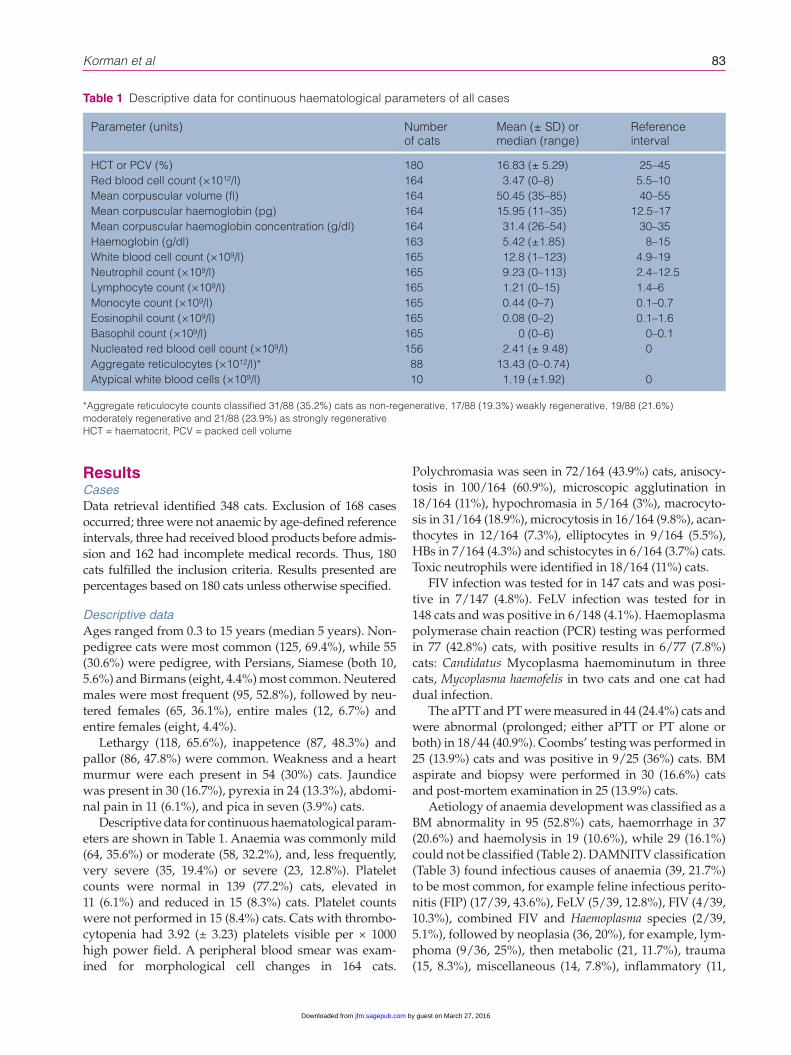
Descriptive data for continuous haematological param-eters are shown in Table 1. Anaemia was commonly mild (64, 35.6%) or moderate (58, 32.2%), and, less frequently, very severe (35, 19.4%) or severe (23, 12.8%). Platelet counts were normal in 139 (77.2%) cats, elevated in 11 (6.1%) and reduced in 15 (8.3%) cats. Platelet counts were not performed in 15 (8.4%) cats. Cats with thrombo-cytopenia had 3.92 (± 3.23) platelets visible per × 1000 high power field. A peripheral blood smear was exam-ined for morphological cell changes in 164 cats.
Polychromasia was seen in 72/164 (43.9%) cats, anisocy-tosis in 100/164 (60.9%), microscopic agglutination in 18/164 (11%), hypochromasia in 5/164 (3%), macrocyto-sis in 31/164 (18.9%), microcytosis in 16/164 (9.8%), acan-thocytes in 12/164 (7.3%), elliptocytes in 9/164 (5.5%), HBs in 7/164 (4.3%) and schistocytes in 6/164 (3.7%) cats. Toxic neutrophils were identified in 18/164 (11%) cats.
FIV infection was tested for in 147 cats and was posi-tive in 7/147 (4.8%). FeLV infection was tested for in 148 cats and was positive in 6/148 (4.1%). Haemoplasma polymerase chain reaction (PCR) testing was performed in 77 (42.8%) cats, with positive results in 6/77 (7.8%) cats: Candidatus Mycoplasma haemominutum in three cats, Mycoplasma haemofelis in two cats and one cat had dual infection.
The aPTT and PT were measured in 44 (24.4%) cats and were abnormal (prolonged; either aPTT or PT alone or both) in 18/44 (40.9%). Coombs’ testing was performed in 25 (13.9%) cats and was positive in 9/25 (36%) cats. BM aspirate and biopsy were performed in 30 (16.6%) cats and post-mortem examination in 25 (13.9%) cats.
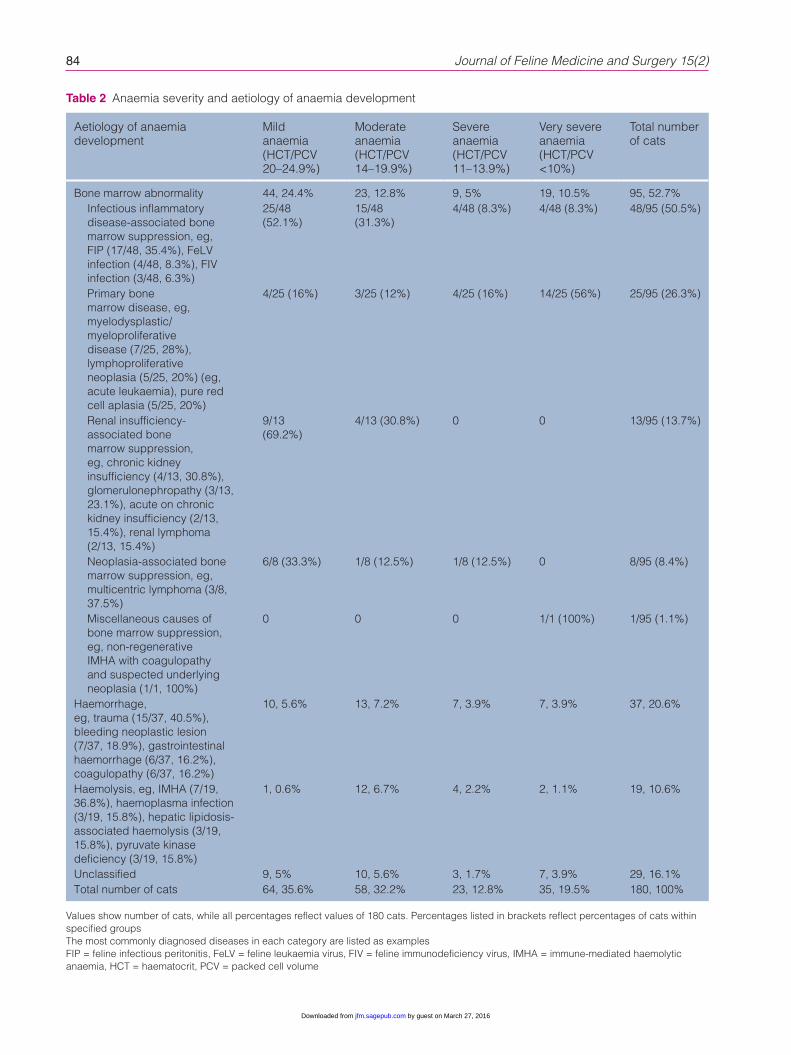
Aetiology of anaemia development was classified as a BM abnormality in 95 (52.8%) cats, haemorrhage in 37 (20.6%) and haemolysis in 19 (10.6%), while 29 (16.1%) could not be classified (Table 2). DAMNITV classi fication (Table 3) found infectious causes of anaemia (39, 21.7%) to be most common, for example feline infectious perito-nitis (FIP) (17/39, 43.6%), FeLV (5/39, 12.8%), FIV (4/39, 10.3%), combined FIV and Haemoplasma species (2/39, 5.1%), followed by neoplasia (36, 20%), for example, lym-phoma (9/36, 25%), then metabolic (21, 11.7%), trauma (15, 8.3%), miscellaneous (14, 7.8%), inflammatory (11,
Table 1 Descriptive data for continuous haematological parameters of all cases
Parameter (units) Number of cats
Mean (± SD) or median (range)
Reference interval
HCT or PCV (%) 180 16.83 (± 5.29) 25–45Red blood cell count (×1012/l) 164 3.47 (0–8) 5.5–10Mean corpuscular volume (fl) 164 50.45 (35–85) 40–55Mean corpuscular haemoglobin (pg) 164 15.95 (11–35) 12.5–17Mean corpuscular haemoglobin concentration (g/dl) 164 31.4 (26–54) 30–35Haemoglobin (g/dl) 163 5.42 (±1.85) 8–15White blood cell count (×109/l) 165 12.8 (1–123) 4.9–19Neutrophil count (×109/l) 165 9.23 (0–113) 2.4–12.5Lymphocyte count (×109/l) 165 1.21 (0–15) 1.4–6Monocyte count (×109/l) 165 0.44 (0–7) 0.1–0.7Eosinophil count (×109/l) 165 0.08 (0–2) 0.1–1.6Basophil count (×109/l) 165 0 (0–6) 0–0.1Nucleated red blood cell count (×109/l) 156 2.41 (± 9.48) 0Aggregate reticulocytes (×1012/l)* 88 13.43 (0–0.74) Atypical white blood cells (×109/l) 10 1.19 (±1.92) 0
*Aggregate reticulocyte counts classified 31/88 (35.2%) cats as non-regenerative, 17/88 (19.3%) weakly regenerative, 19/88 (21.6%) moderately regenerative and 21/88 (23.9%) as strongly regenerativeHCT = haematocrit, PCV = packed cell volume
by guest on March 27, 2016jfm.sagepub.comDownloaded from
84 Journal of Feline Medicine and Surgery 15(2)
Table 2 Anaemia severity and aetiology of anaemia development
Aetiology of anaemia development
Mild anaemia(HCT/PCV 20–24.9%)
Moderate anaemia(HCT/PCV 14–19.9%)
Severe anaemia(HCT/PCV 11–13.9%)
Very severe anaemia (HCT/PCV <10%)
Total number of cats
Bone marrow abnormality 44, 24.4% 23, 12.8% 9, 5% 19, 10.5% 95, 52.7%Infectious inflammatory disease-associated bone marrow suppression, eg, FIP (17/48, 35.4%), FeLV infection (4/48, 8.3%), FIV infection (3/48, 6.3%)
25/48 (52.1%)
15/48 (31.3%)
4/48 (8.3%) 4/48 (8.3%) 48/95 (50.5%)
Primary bone marrow disease, eg, myelodysplastic/myeloproliferative disease (7/25, 28%), lymphoproliferative neoplasia (5/25, 20%) (eg, acute leukaemia), pure red cell aplasia (5/25, 20%)
4/25 (16%) 3/25 (12%) 4/25 (16%) 14/25 (56%) 25/95 (26.3%)
Renal insufficiency-associated bone marrow suppression, eg, chronic kidney insufficiency (4/13, 30.8%), glomerulonephropathy (3/13, 23.1%), acute on chronic kidney insufficiency (2/13, 15.4%), renal lymphoma (2/13, 15.4%)
9/13 (69.2%)
4/13 (30.8%) 0 0 13/95 (13.7%)
Neoplasia-associated bone marrow suppression, eg, multicentric lymphoma (3/8, 37.5%)
6/8 (33.3%) 1/8 (12.5%) 1/8 (12.5%) 0 8/95 (8.4%)
Miscellaneous causes of bone marrow suppression, eg, non-regenerative IMHA with coagulopathy and suspected underlying neoplasia (1/1, 100%)
0 0 0 1/1 (100%) 1/95 (1.1%)
Haemorrhage,eg, trauma (15/37, 40.5%), bleeding neoplastic lesion (7/37, 18.9%), gastrointestinal haemorrhage (6/37, 16.2%), coagulopathy (6/37, 16.2%)
10, 5.6% 13, 7.2% 7, 3.9% 7, 3.9% 37, 20.6%
Haemolysis, eg, IMHA (7/19, 36.8%), haemoplasma infection (3/19, 15.8%), hepatic lipidosis-associated haemolysis (3/19, 15.8%), pyruvate kinase deficiency (3/19, 15.8%)
1, 0.6% 12, 6.7% 4, 2.2% 2, 1.1% 19, 10.6%
Unclassified 9, 5% 10, 5.6% 3, 1.7% 7, 3.9% 29, 16.1%Total number of cats 64, 35.6% 58, 32.2% 23, 12.8% 35, 19.5% 180, 100%
Values show number of cats, while all percentages reflect values of 180 cats. Percentages listed in brackets reflect percentages of cats within specified groupsThe most commonly diagnosed diseases in each category are listed as examplesFIP = feline infectious peritonitis, FeLV = feline leukaemia virus, FIV = feline immunodeficiency virus, IMHA = immune-mediated haemolytic anaemia, HCT = haematocrit, PCV = packed cell volume
by guest on March 27, 2016jfm.sagepub.comDownloaded from

Korman et al 85
6.1%), immune-mediated (11, 6.1%), anomalous (8, 4.4%), toxic (two, 1.1%) and vascular (one, 0.6%) dis-eases. DAMNITV classification was not possible in 22 (12.2%) cats. Commonly-diagnosed diseases are described in Tables 2 and 3.
Anaemia severity, according to aetiology of anaemia development and further BM abnormalities determined is shown in Table 2 and according to each DAMNITV classification in Table 3. Further descriptions of aetiology of anaemia development and DAMNITV classifications for selected clinical and laboratory features are shown in Table 4. Abnormal clotting times were uncommon, found in only 10% of cats and seen with haemorrhage by aetiol-ogy of anaemia development or neoplasia, by DAMNITV categorisation. Anaemia due to a coagulopathy was only identified in four cats, including one cat diagnosed with
disseminated intravascular coagulation and two cats with rodenticide toxicity.
Statistical analysisAnaemia severity was significantly associated with aeti-ology of anaemia development (χ2 = 19.9, P = 0.003). When BM abnormalities were removed from this analy-sis, the significant association was no longer present (χ2 = 5.815, P = 0.12), confirming that BM abnormalities were associated significantly with more severe anaemia. Anaemia severity was not associated significantly with DAMNITV classification (χ2 = 33.852, P = 0.153).
Most cats (112, 62.2%) survived to discharge, 55 (30.6%) were euthanased and 13 (7.2%) died. Anaemia severity was not associated significantly with survival to discharge (χ2 = 4.15, P = 0.248), whereas aetiology of
Table 3 Anaemia severity and DAMNITV classification
DAMNITV classification
Mild anaemia(HCT/PCV 20–24.9%)
Moderate anaemia(HCT/PCV 14–19.9%)
Severe anaemia(HCT/PCV 11–13.9%)
Very severe anaemia (HCT/PCV <10%)
Total number of cats
Anomalous, eg, hereditary coagulopathy (2/8, 25%), portovascular anomaly (2/8, 25%)
1, 0.6% 3, 1.7% 2, 1.1% 2, 1.1% 8, 4.4%
Metabolic, eg, chronic kidney insufficiency (4/21, 19%), hepatic lipidosis (4/21, 19%), acute on chronic kidney insufficiency (2/21, 9.5%)
12, 6.7% 8, 4.4% 1, 0.6% 0 21, 11.7%
Miscellaneous, eg, pure red cell aplasia not attributed to immune-mediated disease (3/14, 21.4%), aplastic anaemia (2/14, 14.3%)
4, 2.2% 2, 1.1% 4, 2.2% 4, 2.2% 14, 7.8%
Neoplastic, eg, lymphoma (9/36, 25%), carcinoma (3/36, 8.3%), haemangiosarcoma (2/36, 5.6%)
16, 8.9% 10, 5.6% 3, 1.6% 7, 3.9% 36, 20%
Infectious, eg, FIP (17/39, 43.6%), FeLV (5/39, 12.8%), FIV (4/39, 10.3%), combined FIV and Haemoplasma species (2/39, 5.1%)
13, 7.2% 15, 8.3% 5, 2.8% 6, 3.3% 39, 21.7%
Inflammation, eg, gastroduodenal ulceration associated with inflammatory bowel disease (2/11, 18.2%)
7, 3.9% 2, 1.1% 1, 0.6% 1, 0.6% 11, 6.1%
Immune-mediated, eg, IMHA (8/11, 72.7%)
0 5, 2.8% 2, 1.1% 4, 2.2% 11, 6.1%
Toxic, eg, rodenticide toxicity (2/2, 100%) 0 2, 1.1% 0 0 2, 1.1%Traumatic, eg, road traffic accident (11/15, 73.3%, dog attack (2/15, 13.3%)
6, 3.3% 5, 2.8% 2, 1.1% 2, 1.1% 15, 8.3%
Vascular, eg, dilated cardiomyopathy and congestive heart failure (1/1, 100%)
1, 0.6% 0 0 0 1, 0.6%
Unclassified 4, 2.2% 6, 3.3% 3, 1.7% 9, 5% 22, 12.2%Total number of cats 64, 35.6% 58, 32.2% 23, 12.8% 35, 19.4% 180, 100% (100%)
Values show number of cats, while all percentages reflect values of 180 cats, unless otherwise specifiedThe most commonly diagnosed diseases in each category are listed as examplesFIP = feline infectious peritonitis, FeLV = feline leukaemia virus, FIV = feline immunodeficiency virus, IMHA = immune-mediated haemolytic anaemia, HCT = haematocrit, PCV = packed cell volume
by guest on March 27, 2016jfm.sagepub.comDownloaded from

86 Journal of Feline Medicine and Surgery 15(2)
Table 4 Further descriptions of the aetiology of anaemia development and DAMNITV classifications for selected clinical and laboratory features
Clinical or laboratory feature Frequency within aetiology of anaemia development
Frequency within DAMNITV classification
Pica7, 3.9%
3/7, 42.8% BM abnormalities2/7, 28.6% Haemolysis1/7, 14.2% Haemorrhage1/7, 14.2% Unclassified
4/7, 57.1% Immune-mediated2/7, 28.6% Neoplasia1/7, 14.2% Metabolic
Jaundice30, 16.7%
11/24, 36.7% BM abnormalities9/24, 30% Haemolysis4/24, 13.3% Haemorrhage
4/22, 18.2% Metabolic4/22, 18.2% Neoplasia4/22, 18.2% Infectious3/22, 13.6% Anomalous3/22, 13.6% Inflammatory2/22, 9.1% Immune-mediated1/22, 4.5% Vascular1/22, 4.5% Traumatic
Pyrexia24, 13.3%
14/18, 77.8% BM abnormalities4/18, 22.2% Haemolysis
10/19, 53.6% Infectious6/19, 31.6% Neoplasia2/19, 10.5% Immune-mediated1/19, 5.3% Anomalous
Thrombocytopenia15, 8.3%
12/15, 80% BM abnormalities2/15, 13.3% Haemorrhage1/15, 6.7% Haemolysis
6/14, 42.9% Infectious5/14, 35.7% Neoplasia2/14, 14.3% Miscellaneous1/14, 7.1% Trauma
Increased number of HB7, 3.9%
2/4, 50% BM abnormalities2/4, 50% Haemolysis
2/6, 33.3% Neoplasia1/6, 16.7% Anomalous1/6, 16.7% Metabolic1/6, 16.7% Immune-mediated1/6, 16.7% Miscellaneous
Agglutination18, 10%
9/18, 50% BM abnormalities6/18, 33.3% Haemolysis
7/15, 46.7% Infectious5/15, 33.3% Immune-mediated1/15, 6.7% Miscellaneous1/15, 6.7% Metabolic1/15, 6.7% Neoplasia
Macrocytosis31, 17.2%
17/26, 65.3% BM abnormalities5/26, 19.2% Haemolysis4/26, 15.4% Haemorrhage
10/26, 38.5% Neoplasia6/26, 23.1% Immune-mediated3/26, 11.5% Metabolic2/26, 7.7% Inflammatory1/26, 3.8% Anomalous1/26, 3.8% Infectious1/26, 3.8% Miscellaneous1/26, 3.8% Toxic1/26, 3.8% Trauma
Microcytosis16, 8.9%
7/13, 53.8% BM abnormalities3/13, 23.1% Haemolysis3/13, 23.1% Haemorrhage
4/15, 26.7% Metabolic4/15, 26.7% Neoplastic4/15, 26.7% Infectious1/15, 6.7% Anomalous1/15, 6.7% Trauma1/15, 6.7% Toxic
Microcytosis and hypochromasia2, 1.1%
2/2, 100% BM abnormalities 2/2, 100% Infectious
Acanthocytes12, 6.7%
8/11, 72.7% BM abnormalities3/11, 27.3% Haemorrhage
4/10, 40% Neoplasia1/10, 10% Anomalous1/10, 10% Metabolic1/10, 10% Infectious1/10, 10% Inflammatory1/10, 10% Trauma1/10, 10% Miscellaneous
(Continued)
by guest on March 27, 2016jfm.sagepub.comDownloaded from

Korman et al 87
anaemia development was (χ2 = 6.070, P = 0.046) with 54/95 (56.8%) cats with BM abnormalities, 26/37 (70.3%) cats with haemorrhage and 16/19 (84.5%) cats with hae-molysis surviving to discharge. Significance was lost if BM abnormalities (χ2 = 1.301, P = 0.338) or haemolytic disorders (χ2 = 2.011, P = 0.171) were removed from sta-tistical analysis, but not if haemorrhagic disorders were removed (χ2 = 17.22, P = 0.01), indicating that cats with haemolysis were more likely to survive to discharge than cats with BM abnormalities. DAMNITV classifica-tion was also associated significantly with survival to
discharge (χ2 = 19.998, P = 0.010); all cats with immune-mediated disease, both cats with toxic and the one cat with vascular disease, 12/15 (80%) cats with trauma, 11/14 (78.6%) cats with miscellaneous disease, 8/11 (72.7%) cats with inflammatory disease, 24/39 (61.5%) cats with infectious disease, 12/21 (57.1%) cats with met-abolic disease and 4/8 (50%) cats with anomalous dis-eases surviving to discharge. Significance was lost if neoplastic disorders (χ2 = 11.671, P = 0.152) or immune-mediated disease (χ2 = 12.781, P = 0.102) were removed, suggesting that cats with neoplasia were less likely to
Clinical or laboratory feature Frequency within aetiology of anaemia development
Frequency within DAMNITV classification
Schistocytes6, 3.3%
3/4, 75% BM abnormalities1/4, 25% Haemorrhage
2/6, 33.3% Metabolic1/6, 16.7% Anomalous1/6, 16.7% Neoplasia1/6, 16.7% Trauma1/6, 16.7% Miscellaneous
Toxic neutrophils18, 10%
11/16, 68.8% BM abnormalities3/16, 18.8% Haemorrhage2/16, 12.5% Haemolysis
6/16, 37.5% Infectious3/16, 18.8% Neoplasia2/16, 12.5% Anomalous1/16, 6.3% Metabolic1/16, 6.3% Inflammatory1/16, 6.3% Immune-mediated1/16, 6.3% Miscellaneous1/16, 6.3% Toxic
Prolonged aPTT and PT10, 5.6%
5/10, 50% Haemorrhage3/10, 30% BM abnormalities2/10, 20% Haemolysis
4/9, 44.4% Neoplasia2/9, 22.2% Anomalous1/9, 11.1% Metabolic1/9, 11.1% Miscellaneous1/9, 11.1% Toxic
Coombs’ testing positivity9, 5%
6/9, 66.7% Haemolysis3/9, 33.3% BM abnormalities
7/9, 77.8% Immune-mediated2/9, 22.2% Infectious
Values show number of cats, while all percentages reflect values of 180 cats, unless otherwise specifiedHB = Heinz body, aPTT = activated partial thromboplastin time, PT = prothrombin time, BM = bone marrow
Table 4 (Continued)
Table 5 Significant variables identified in the most parsimonious Cox regression analysis model for the prediction of cats with anaemia
Variable Parameter estimate (β)(standard error)
P Exp (B)(95% confidence interval)
DAMNITV category* 0.011 Metabolic 0.094 (0.302) 0.757 1.098 (0.608–1.983)Neoplastic -0.054 (0.309) 0.861 0.947 (0.517–1.735)Infectious -1.283 (0.560) 0.022 0.277 (0.093–0.830)Inflammatory -1.279 (0.563) 0.023 0.278 (0.092–0.839)Immune-mediated -0.928 (0.482) 0.054 0.395 (0.154–1.018)Trauma -0.443 (0.351) 0.206 0.642 (0.323–1.276)Anomalous, toxic, miscellaneous and vascular
- - -
Age (years) 0.045 (0.026) 0.082 1.046 (0.994–1.100)
*Owing to the small numbers in some DAMNITV groups, cats with anomalous, toxic, vascular and miscellaneous diseases were considered one group to increase statistical power
by guest on March 27, 2016jfm.sagepub.comDownloaded from

88 Journal of Feline Medicine and Surgery 15(2)
survive and cats with immune-mediated disease were more likely to survive to discharge.
Cox regression analysis showed that the most parsi-monious model included only DAMNITV classification (P = 0.011) and age (P = 0.082) (Table 5). Cats with trauma had shorter survival times, followed by cats with neo-plastic, metabolic or infectious disease. Younger cats had longer survival times. A closely competing model included aetiology of anaemia development and age (Table 6). Here, cats with BM abnormalities had shorter survival times compared with anaemia due to haemoly-sis or haemorrhage. The estimated effect of age was simi-lar to that found in the DAMNITV model. In addition to these models, when all possible predictor variables were tested in isolation (as sole predictor), cats with elevated neutrophil (P = 0.043) or elevated monocyte counts (P = 0.029) had shorter survival times.
DiscussionThis study describes the historical and physical examina-tion findings, haematological data and causes of anaemia in a large group of anaemic cats. The effect of anaemia severity and cause on survival was also determined.
Clinical signs associated with anaemia in this popu-lation were vague and non-specific. Pica was very uncommon, seen in only 3.9% of cats and associated pre-dominantly with immune-mediated disease and was not associated significantly with anaemia severity (data not shown). An association between pica and immune-mediated disease has been described previously when 25% of cats with primary IMHA had pica.2
Jaundice was found in 16.7% of cats and was not always associated with haemolysis; only nine (5%) cats had both jaundice and evidence of haemolysis in the cur-rent study. Haemolysis alone rarely causes jaundice (only 10% cats with primary IMHA were jaundiced in a previous study2) owing to the large reserve capacity of the liver, although severe anaemia may cause jaundice due to hypoxic liver damage, impaired liver function and intrahepatic cholestasis.13 Jaundiced cases in the cur-rent study (data not shown) had diagnoses such as FIP, sepsis and cholangitis.
Haemoplasmas can induce infectious anaemia in cats.14,15 In our study population, haemoplasma infection was uncommon and lower than a previous study of UK cats.16 This may be because the study population com-prised referral cases, whereas haemoplasma infection is more commonly diagnosed in first-opinion practice, negating the need for referral.
Anaemia due to BM abnormalities was more com-mon than haemorrhage or haemolysis in this popula-tion; indeed, a large percentage (26.7%) of cats had secondary suppression of BM activity due to infectious inflammatory disease. Further analysis of specific dis-eases within each BM group and the effect on prognosis and survival was not performed in this study owing to the difficulty in obtaining a retrospective definitive diagnosis for each case that fits currently-recommended BM classification systems and the small numbers within some groups.17,18 Published studies describing aetiology of feline anaemia are uncommon. One study,19 pub-lished in German, described 100 anaemic cats in which anaemia of inflammatory disease (29%) was most com-monly diagnosed, followed by haemorrhage (24%), sim-ilar to our study. However, more cats were identified with haemolysis (24%) compared with our study (19, 10.6%). Differences in the prevalence of haemolytic causes of anaemia in these studies may arise, in part, owing to geographical variation in the prevalence of infectious agents that can be associated with haemolysis in cats, such as haemoplasmas.
When classified by DAMNITV, infectious causes of anaemia were most common and FIP was diagnosed fre-quently, possibly reflecting difficulties with ante-mortem diagnosis of FIP prompting referral. Anaemia is com-monly reported in FIP.20,21 In human medicine22,23 neo-plasia is a common cause of anaemia, as identified in this study. Pathogenesis of neoplasia-associated anaemia is often multifactorial — in our population it was most commonly associated with BM abnormalities.
Anaemia severity differed between aetiology of anae-mia development groups, with BM abnormalities associ-ated significantly with more severe anaemia. However, anaemia severity was not associated with DAMNITV classification. DAMNITV classification is based on a
Table 6 Significant variables identified in a closely competing Cox regression analysis model for prediction of survival time in cats with anaemia
Variable Parameter estimate (β)(standard error)
P Exp (B)(95% confidence interval)
Aetiology of anaemia development
0.018
Haemolysis -0.882 0.013 0.414 (0.206–0.832)Haemorrhage -0.499 0.046 0.607 (0.372–0.991)Bone marrow abnormalities - - -Age (years) 0.061 0.018 1.063 (1.010–1.119)
by guest on March 27, 2016jfm.sagepub.comDownloaded from

Korman et al 89
large number of groups, resulting in smaller numbers within each group compared with the aetiology of anae-mia development classification, possibly affecting statis-tical analysis. A notable finding of this study was that anaemia severity was not associated with survival to dis-charge, suggesting, importantly, that a clinical decision to treat an anaemic cat should not be based on anaemia severity alone.
Aetiology of anaemia development was associated significantly with survival to discharge as cats with hae-molysis were more likely to survive than cats with BM abnormalities. However, the range of survival times within each group was wide. Although cats with BM abnormalities were less likely to survive overall, cats with renal insufficiency-associated BM suppression typi-cally had long survival times (data not shown). Interestingly, all cats with primary BM disease due to immune-mediated causes survived to discharge, which is in agreement with other studies.24 The overall poor survival of cats with BM abnormalities may be owing to the large number of cats within the secondary infectious inflammatory disease-associated BM suppression group (48/95, 50.5%), in which 45.8% of cats in this group either died or were euthanased.
Cats with haemolysis were more likely to survive to discharge. This agrees with other investigations describ-ing cats with haemolytic anaemia of various causes hav-ing long survival times.2,25,26 DAMNITV classification was also associated with survival to discharge as cats with immune-mediated disease were more likely, and cats with neoplasia were less likely, to survive to dis-charge. In line with this, a previous study reported long median survival time in primary IMHA in cats.27
DAMNITV category appeared to be more strongly associated with survival than aetiology of anaemia development, and was included in the most parsimoni-ous model on Cox regression analysis. It may be more useful to determine DAMNITV classification for each anaemic cat, rather than aetiology of anaemia develop-ment, as this may give a better indication of survival. In the current study, cats with trauma, neoplasia, metabolic or infectious disease were more likely to die than cats in other categories. Although it could be thought that clas-sifying a patient according to aetiology of anaemia development does not necessarily require a definitive diagnosis, whereas DAMNITV classification often does, it is interesting that more cases were unclassified accord-ing to aetiology of anaemia development (16.1%) than DAMNITV (12.2%). The current study found that both systems provided information regarding survival to dis-charge and survival time, and so both are reported. An aetiological diagnosis is easier to obtain; however, DAMNITV classification appeared more significant in the final model of survival, suggesting the utilisation of DAMNITV classification for anaemic cats may be more useful.
On Cox regression analysis, younger cats had signifi-cantly longer survival times, indicating the usefulness of age as a prognostic indicator. However, therapeutic interventions were not investigated in this study and it is possible that for younger cats there was increased will-ingness to administer treatments (eg, blood transfusion) than for older cats, influencing this finding. Additionally, diseases associated with a better prognosis [eg, haemo-lytic anaemia, pure red cell aplasia (PRCA)] are also more common in younger cats. Interestingly, all cats in this study with PRCA and aplastic anaemia also survived to discharge (data not shown), which is in agreement with other studies.24,28 An alternative, and important, reason for the longer survival times identi-fied in younger cats is that regardless of health status, when followed for an extended time period, as in our study, younger cats will be more likely to live longer than older cats, who are more likely to die from other natural causes as they age further.
This study had a number of limitations. The study’s retrospective nature meant that not all parameters were available for all cases, for example, reticulocyte counts. Although a reticulocyte count would have provided fur-ther information for differentiating anaemia into regen-erative or non-regenerative categories, the lack of reticulocyte counts available for every patient prohibited this being used as a useful category. Both PCV and HCT may be affected by dehydration, which was not taken into account, and misclassification of anaemia severity may have occurred. Additionally, some cats that were anaemic on presentation but where dehydration increased PCV/HCT to within reference intervals will have been excluded by our inclusion criteria. It was decided to use PCV/HCT results for inclusion as if only cats having a full haematology profile, including Hb, performed on presentation had been included, an impor-tant population of cats presenting outside laboratory hours would be excluded from the study, including cats with trauma and haemorrhage. Also, the current study describes a referral population, which may bias the cases seen. Some cats with severe anaemia may not have been fit for travel to our referral centre, or cats with renal insufficiency-associated BM depression may not have been referred owing to growing confidence in managing these cats within general practice. A further limitation included case classification. Case placement within DAMNITV categories and subcategorisation within aeti-ology of anaemia development (eg, BM classifications) is subjective and some cases fit into multiple categories. In this study each case was reviewed and categorised by two board-certified internal medicine clinicians in an attempt to address the subjectivity of categorisation, but this is still a limitation. Additionally, classification of cases by the broader aetiology of anaemia development classes may have resulted in a false impression of sur-vival for some groups and, if larger population numbers
by guest on March 27, 2016jfm.sagepub.comDownloaded from
90 Journal of Feline Medicine and Surgery 15(2)
were available, further breakdown into subcategories may have revealed more significant prognostic indica-tors and survival data.
ConclusionsHistory and clinical signs of feline anaemia are often vague and non-specific. Anaemia arose most commonly as a result of BM abnormalities by aetiology of anaemia development or, if assessed by DAMNITV classification, infectious and neoplastic disease were more common. Anaemia severity did not affect survival, suggesting that a clinical decision to treat an anaemic cat should not be based on anaemia severity alone. Cats with haemolysis were more likely to survive to discharge than cats with BM abnormalities. Cats with neoplasia were less likely and cats with immune-mediated disease more likely to survive to discharge. Younger cats were also more likely to survive.
Funding This research received no specific grant from any funding agency in the public, commercial, or not-for-profit sectors.
Conflict of interest Rachel Korman and Andrea Harvey’s positions were funded by the Feline Advisory Bureau during the study period.
References 1 Ottenjann M, Weingart C, Arndt G and Kohn B. Character-
ization of the anemia of inflammatory disease in cats with abscesses, pyothorax, or fat necrosis. J Vet Intern Med 2006; 20: 1143–1150.
2 Kohn B, Weingart C, Eckmann V, Ottenjann M and Leibold W. Primary immune-mediated hemolytic anemia in 19 cats: diagnosis, therapy, and outcome (1998–2004). J Vet Intern Med 2006; 20: 159–166.
3 Wilson HE, Jasani S, Wagner TB, et al. Signs of left heart volume overload in severely anaemic cats. J Feline Med Surg 2010; 12: 904–909.
4 Castellanos I, Couto CG and Gray TL. Clinical use of blood products in cats: a retrospective study (1997–2000). J Vet Intern Med 2004; 18: 529–532.
5 Klaser DA, Reine NJ and Hohenhaus AE. Red blood cell transfusions in cats: 126 cases (1999). J Am Vet Med Assoc 2005; 226: 920–923.
6 Weingart C, Giger U and Kohn B. Whole blood transfu-sions in 91 cats: a clinical evaluation. J Feline Med Surg 2004; 6: 139–148.
7 Weingart C and Kohn B. Clinical use of a haemoglobin-based oxygen carrying solution (Oxyglobin) in 48 cats (2002–2006). J Feline Med Surg 2008; 10: 431–438.
8 Bonagura JD and Kirk RW. In: Bonagura JD (ed). Kirk’s current veterinary therapy: small animal practice. 12th ed. Philadelphia, London: WB Saunders, 1995, p 1395–1417.
9 Weiss DJ, Wardrop KJ and Schalm OW. In: Weiss DJ, Wardop KJ (eds). Schalm’s veterinary hematology. 6th ed. Oxford: Wiley-Blackwell, 2010, p 152–161.
10 Kohn B, Weingart C and Giger U. Haemorrhage in seven cats with suspected anticoagulant rodenticide intoxica-tion. J Feline Med Surg 2003; 5: 295–304.
11 Tasker S, Murray JK, Knowles TG and Day MJ. Coombs’, haemoplasma and retrovirus testing in feline anaemia. J Small Anim Pract 2010; 51: 192–199.
12 Rizzo F, Tappin SW and Tasker S. Thrombocytosis in cats: a retrospective study of 51 cases (2000–2005). J Feline Med Surg 2007; 9: 319–325.
13 Sherding RG. Feline jaundice. J Feline Med Surg 2000; 2: 165–169.
14 Tasker S, Caney SMA, Day MJ, et al. Effect of chronic FIV infection, and efficacy of marbofloxacin treatment, on ‘Candidatus Mycoplasma haemominutum’ infection. Microbes and Infection 2006; 8: 653–661.
15 Tasker S, Caney SMA, Day MJ, et al. Effect of chronic FIV infection, and efficacy of marbofloxacin treatment, on Myco-plasma haemofelis infection. Vet Microbiol 2006; 117: 169–179.
16 Sykes JE. Feline hemotropic mycoplasmas. Vet Clin North Am Small Anim Pract 2010; 40: 1157–1170.
17 Hisasue M, Okayama H, Okayama T, et al. Hematologic abnormalities and outcome of 16 cats with myelodysplas-tic syndromes. J Vet Intern Med. 2001; 15: 471–477.
18 Weiss DJ. Evaluation of dysmyelopoiesis in cats: 34 cases (1996–2005). J Am Vet Med Assoc 2006; 228: 893–897.
19 Kohn B. Erythrocyte studies in healthy and anaemic cats. Thesis, Habilitationsschrift, FU Berlin, 2001 [in German].
20 Sparkes AH, Gruffydd-Jones TJ and Harbour DA. Feline infectious peritonitis: a review of clinicopathological changes in 65 cases, and a critical assessment of their diag-nostic value. Vet Rec 1991; 129: 209–212.
21 Paltrinieri S, Grieco V, Comazzi S and Cammarata Parodi M. Laboratory profiles in cats with different pathological and immunohistochemical findings due to feline infec-tious peritonitis (FIP). J Feline Med Surg 2001; 3: 149–159.
22 Spivak JL, Gascon P and Ludwig H. Anemia management in oncology and hematology. Oncologist 2009; 14 (Suppl 1): 43–56.
23 Bohlius J, Tonia T and Schwarzer G. Twist and shout: one decade of meta-analyses of erythropoiesis-stimulating agents in cancer patients. Acta Haematol 2011; 125: 55–67.
24 Stokol T and Blue JT. Pure red cell aplasia in cats: 9 cases (1989–1997). J Am Vet Med Assoc 1999; 214: 75–79.
25 Kohn B and Fumi C. Clinical course of pyruvate kinase deficiency in Abyssinian and Somali cats. J Feline Med Surg 2008; 10: 145–153.
26 Barrs VR, Giger U, Wilson B, et al. Erythrocytic pyruvate kinase deficiency and AB blood types in Australian Abyssinian and Somali cats. Aust Vet J 2009; 87: 39–44.
27 Husbands BD SS and Weiss DJ. Idiopathic immune-mediated haemolytic anaemia (IMHA) in 25 cats. ACVIM Abstract 2002; 16: 350.
28 Weiss DJ. Aplastic anemia in cats – clinicopathological features and associated disease conditions 1996–2004. J Feline Med Surg 2006; 8: 203–206.
by guest on March 27, 2016jfm.sagepub.comDownloaded from