A retrospective cephalometric appraisal -
91
University of the Western Cape MSc Orthodontics Effects of premolar extraction on airway dimensions: A retrospective cephalometric appraisal Luzaan van Zyl 3048950 Supervisor Dr. Hudson http://etd.uwc.ac.za/
-
Upload
khangminh22 -
Category
Documents
-
view
1 -
download
0
Transcript of A retrospective cephalometric appraisal -

University of the Western Cape
MSc Orthodontics
Effects of premolar extraction on airway dimensions: A retrospective cephalometric appraisal
Luzaan van Zyl 3048950
Supervisor Dr. Hudson
http://etd.uwc.ac.za/

I
Effects of premolar extraction on airway dimensions: A retrospective cephalometric appraisal
Luzaan van Zyl
Keywords
Premolar Extraction
Bimaxillary Protrusion
Airway Dimension
Cephalometric Radiographs
http://etd.uwc.ac.za/
II
Abstract Aim: The aim of this study was to assess the effect of retraction of anterior teeth on pharyngeal airway dimensions, after orthodontic treatment of bimaxillary protrusion cases by means of the extraction of four premolars.
Method: A total of 88 lateral cephalometric radiograph pairs, consisting of a pre-treatment and post-treatment radiograph taken for orthodontic treatment of bimaxillary protrusion by means of extraction of four premolars, was used. The pharyngeal airway space, measured across three different levels, as well as the length of the maxilla and mandible were assessed for changes from pre-treatment to post-treatment. Pearson’s correlation coefficient was used to determine the degree to which the change in pharyngeal airway space was associated with the change in maxilla or mandible length.
Results: The pre-treatment average pharyngeal airway space measurements were recorded as 15.23mm for the Superior Pharyngeal Airway Space, 11.63mm for the Middle Pharyngeal Airway Space and 13.56mm for the Inferior Pharyngeal Airway Space. The average reduction in the pharyngeal airway space was noted as 1.21mm, 1.64mm and 2.23mm respectively. All with statistically significant P values of <0.001. The average maxilla and mandible lengths pre-treatment was recorded as 61.53mm and 85.89mm correspondingly. The average reduction in the maxilla and mandible lengths was measured at 0.65mm (P value 0.0154) and 2.00mm (P value 0.005). There was no correlation found between the change in pharyngeal airways and the change in maxilla or mandibular lengths (Correlation values ranged from -0.1191 to 0.0330). Further, no correlation was found between the change in pharyngeal airways and retraction of the maxillary and mandibular incisors (Correlation values ranged from -0.0128 to 0.0554). Conclusion: Practitioners should include pharyngeal airway measurements as part of routine lateral cephalometric analysis. This study suggests that practitioners who are considering extraction treatment with retraction of anterior teeth should consider the effect on the pharyngeal airway, and avoid unnecessary or excessive retraction of anterior teeth at all cost.
http://etd.uwc.ac.za/

III
Declaration
I, Luzaan van Zyl, declare that Effects of Premolar Extraction on Airway Dimensions: A Retrospective Cephalometric Appraisal is my work and it has not been submitted before for any degree or examination in any other university. I confirm that information derived from other sources has been indicated in the thesis and acknowledged as complete references. Luzaan van Zyl
July 2020
http://etd.uwc.ac.za/

IV
Acknowledgements To my husband, Shaun, for everything. To my family, Aré, Karin and Carli, for their unceasing love. To Dr. Hudson, for his guidance, immense knowledge and unparalleled support. And to all the bold researchers who embark on unchartered seas, who strives with great enthusiasm, great devotion and knows there is no effort without error, and who in the end knows triumph.
http://etd.uwc.ac.za/

V
Table of Contents Title Page -
Keywords I
Abstract II
Declaration III
Acknowledgements IV
Table of Contents V
List of Tables IX
List of Figures X
List of Abbreviations XI
Chapter 1: Introduction 1
Aim 3
Objectives 3
Hypothesis 3
Chapter 2: Literature Review 4
Orthodontics review 4
Extraction debate 4
Prevalence of extractions 4
Extraction decision: clinical guidelines 5
Extraction decision: cephalometric guidelines 5
Tweed 6
Steiner 6
Holdaway 6
Alvarez 6
Bimaxillary protrusion: 7
Aetiology 7
Clinical features 7
Cephalometric features 8
Medunsa norms 9
Treatment 9
http://etd.uwc.ac.za/

VI
Premolar extractions with anterior retraction: 9
Effect on facial profile 9
Effect on growth 10
Effect on teeth and dentoalveolar structures 11
Effect on maxilla and mandible 11
Effect on pharyngeal airway 12
Craniofacial growth 12
Pharyngeal growth 13
Craniofacial development and airways 14
Airway enhancing orthodontics 15
Airway dynamics 15
Airflow dynamics 16
Effects of reduced pharyngeal airway size: 17
Recued oxygen uptake 17
Obstructive sleep apnea 17
Mouth breathing 19
Radiographic imaging of the airway 19
Chapter 3: Methodology and Data 21
Study Design 21
Study Population 21
Inclusion Criteria 21
Exclusion Criteria 22
Sample Size 22
Radiographic Technique 23
Radiographic Tracing: 24
Dentofacial Cephalometric Tracing 24
Pharyngeal Cephalometric Tracing 26
Maxilla and Mandible Cephalometric Tracing 27
Orthodontic Treatment 28
Data Interpretation 29
Examiner Reliability 29
Statistical Analysis 30
Ethical Considerations 30
http://etd.uwc.ac.za/
VII
Chapter 4: Results 31
Reliability of results 31
Prevalence of bimaxillary protrusion 31
Cephalometric characteristics of study population 31
Orthodontic treatment: cephalometric changes from pre-treatment to post-treatment 32
Pharyngeal airway effect 33
Maxilla and mandibular length effect 34
Pharyngeal airway change comparison 34
Maxillary and mandibular incisor retraction comparisons 36
Chapter 5: Discussion 37
Prevalence of bimaxillary protrusion 37
Orthodontic treatment 37
Incisor retraction 37
Interincisal angle 38
Soft tissue profile 39
Comparison between nasiolabial change and upper/ lower lip protrusion 42
Pharyngeal airway space 42
Pre-treatment pharyngeal airway space 42
Change in pharyngeal airway space 44
Retraction protocol and airway 46
Factors affecting airway measurements 47
Maxilla and mandible length 48
Pre-treatment maxilla and mandible length 48
Change in maxilla and mandibular length 49
Factors affecting maxilla and mandible length measurements 51
Comparison between pharyngeal airway change and maxilla/ mandible length change 52
Comparison between pharyngeal airway change and maxilla/ mandible incisor retraction 53
Comparison between maxilla/ mandible length change and maxilla/ mandible incisor
retraction 54
Reliability of study 54
Method error 55
Frankfurt horizontal plane accuracy 55
Point A and Point B accuracy 55
http://etd.uwc.ac.za/

VIII
Limitations 56
Chapter 6: Conclusion and Recommendations 58
Conclusion 58
Recommendations 59
References 60
Appendix 76
A: Tweed Cephalometric Normal Range 76
B: Steiner Cephalometric Normal Range for Lower Incisor 77
C: Holdaway Cephalometric Normal Ratio 78
D: Alvarez A-line 79
http://etd.uwc.ac.za/

IX
List of Tables Table 3.1: Dentofacial Cephalometric Landmarks 24
Table 3.2: Dentofacial Cephalometric Measurements 25
Table 3.3: Pharyngeal Lateral Cephalometric Landmarks 26
Table 3.4: Measurements of the three pharyngeal sections 26
Table 3.5: Maxilla and Mandible Cephalometric Landmarks 27
Table 3.6: Anterior-Posterior Measurements of the Maxilla and Mandible 28
Table 4.1: Pre-treatment and post-treatment average value of cephalometric measurements of
the study population 31
Table 4.2: Retraction of maxillary and mandibular incisors from pre- to post-treatment
cephalometric measurements 32
Table 4.3: Cephalometric changes from pre- to post-treatment cephalometric
measurements 32
Table 4.4: Correlation between change in nasiolabial angle and change in maxillary incisor
measured to the S-line. P value in brackets 32
Table 4.5: Change in pharyngeal airway measurements in mm (comparison of pre-treatment
and post-treatment lateral cephalometric values) 33
Table 4.6: The average pharyngeal airway change calculated by obtaining an average linear
measurement (mm) difference across all three categories of the pre-treatment and post-
treatment comparisons 33
Table 4.7: Change in maxilla and mandibular length in mm (comparison of pre-treatment and
post-treatment lateral cephalometric values) 34
Table 4.8: Correlation between the change (pre- to posttreatment) in maxillary and
mandibular length and the change (pre- to post-treatment) in airway cephalometric
measurements. P value in brackets 34
Table 4.9: Correlation between maxillary and mandibular incisor retraction and the change in
airway from pre- to post-treatment cephalometric measurements. P value in brackets 35
Table 4.10: Correlation between maxillary and mandibular incisor retraction and the change
in maxillary and mandibular lengths from pre- to post-treatment cephalometric
measurements. P value in brackets 36
http://etd.uwc.ac.za/

X
List of Figures
Figure 3.1: Dentofacial Cephalometric Landmarks and Measurements 25
Figure 3.2: Pharyngeal Cephalometric Landmarks and Measurements 27
Figure 3.3: Anterior-Posterior Maxilla and Mandibular Landmarks and Measurements. 28
http://etd.uwc.ac.za/
XI
List of Abbreviations PAS Pharyngeal airway space
FMP Frankfurt-mandibular plane angle
IMP Incisor-mandibular plane angle
FMI Frankfurt-mandibular incisor angle
NB Nasion to point B
NA Nasion to point A
O2 Oxygen
CO2 Carbon dioxide
OSA Obstructive sleep apnea
Avg Average
SPAS Superior pharyngeal airway space
MPAS Middle pharyngeal airway space
IPAS Inferior pharyngeal airway space
FH Frankfort Horizontal
http://etd.uwc.ac.za/

1
CHAPTER 1 Introduction
Malocclusion is one of the most common oral disorders found. Orthodontic treatment
is sought for many reasons beyond the malocclusion itself, such as improvement in physical, social and psychological health (Zhang, et al., 2006).
The goal of orthodontic treatment, as defined by Proffit, is to create the best occlusal
relationship within the framework of acceptable facial aesthetics and stability of the end result. Dental arch morphology has been studied with the hope of defining proper goals for tooth position, aesthetics, function and even long-term stability (Proffit, et al., 2007).
Tooth extraction in the orthodontic field is still a debated topic, with a consensus on when to extract teeth still eluding the specialty. Extractions for orthodontic purposes are commonly advised in order to create more space for alignment of the crowded teeth, correct anterior-posterior discrepancies, camouflage orthognathic discrepancies as well as reduce incisor protrusion. The proposed benefits as well as negative sequelae should be carefully examined (Al Qahtani, 2016, Pliska, et al., 2016).
The decision to extract must be made on a decision of the patient’s total health, and not
just on the basis of malocclusion (Watson, 1980).
More recently the sequelae of dental extractions now include the effect on the patient’s airway. Orthodontic treatment as a modality alters dentofacial morphology of the patient and therefore it can be said that it should also affect the pharyngeal airway space (Pliska, et al., 2016)
The physiology of the upper airway consists of the external nose, vestibule, nasal valve
and airflow (Sahin-Yilmaz and Naclerio, 2011). Pharyngeal airway space (PAS) is described as the distance between the posterior and anterior wall of the pharynx, where the inferior border lies at the base of the tongue; and its width is dependent on the soft palate, tongue and hyoid bone position (Kulshrestha, et al., 2015; Ciavarella, et al., 2014).
A patent upper airway is essential for survival. In order to allow for the airway to remain
open, a fine balance exists between forces. At the airway wall, the local balance between tissue stresses acting radially towards the airway and airway pressure will determine if the airway dilates or narrows (Cheng, et al., 2014).
Orthodontic treatment can cause differences in the size of the oral cavity, influence the
size and function of the nasopharyngeal airway and affect breathing (Germec-Cakan, et al., 2011).
http://etd.uwc.ac.za/
2
Forward movement of the jaws and dentition due to orthodontics, such as functional appliances and rapid palatal expansion, may augment the airway dimensions. Whereas orthodontic treatments such as mandibular setback surgery, cervical headgear and retraction of anterior teeth leading to a reduced oral volume can all lead to a decrease in airway dimensions (Germec-Cakan, et al., 2011).
Changes in the size of the oral cavity due to different orthodontic treatments may affect
tongue position and therefore pharyngeal airway dimensions (Germec-Cakan, et al., 2011). The tongue position within the oral cavity also has an important function in many aspects, as it plays a role in ventilation, olfaction, speech production, retronasal appreciation of aromas, jaw postures and neck extensions. Normal tongue position at rest is recognized as the posture at which equilibrium is achieved (Tsuiki, et al., 2007). Altering the tongue in habitual position, whether it partly or totally occupies the mouth cavity, as well as a change in resting tongue position could have an effect on ventilation (Bourdiol, et al., 2010).
It important to note that the tongue is a unique structure with four pairs of intrinsic and
extrinsic muscles, all acting as a hydrostat, incompressible with constant volume (Saboisky, et al., 2015). Through muscular and connective tissue attachments, the tongue is attached to the hyoid bone. The posterior movement of the tongue, along with a change in hyoid position, thus results in narrowing of the airway (Germec-Cakan, et al., 2011)
Lateral cephalometric radiographs are considered part of routine diagnostic imaging for
orthodontic treatment. This provides valuable information regarding the sagittal and vertical relations of the craniofacial skeleton, soft tissue profile, dentition, pharynx and cervical vertebrae (Athanasiou, 1995). Thus, airway narrowing, or obstruction, as well as tongue position can easily be observed on lateral cephalometric radiographs (Bourdiol, et al., 2010).
It can be said that diagnosing upper airway obstruction should form part of routine
cephalometric analysis. Early diagnosis is critical, as changes in the dimensions of the airway could have health consequences, such as reduced quality of life and decreased life expectancy (Le, et al., 2019).
The problem that faces orthodontists is to safely choose extractions as a treatment
modality without affecting the pharyngeal airway space of the patient. This study was conducted in order to determine whether extraction treatment with the
retraction of anterior teeth, in bimaxillary protrusion patients, had a significant effect on the pharyngeal airway.
http://etd.uwc.ac.za/

3
Aim of the study
To assess the effect of retraction of anterior teeth on pharyngeal airway dimensions, after orthodontic treatment of bimaxillary protrusion cases by means of the extraction of four premolars.
Objectives of the study
1. To determine the pharyngeal airway space at predetermined levels. 2. Determine the effect of retraction of anterior teeth, by means of four premolar extractions,
on the pharyngeal airway space at the abovementioned predetermined levels. 3. Determine the length of the maxilla and mandible. 4. To determine the effect of retraction of anterior teeth, by means of four premolar
extractions, on the length of the maxilla and mandible. 5. Determine if any relation exists between changes in maxilla/mandible length and the
changes of pharyngeal airway space.
Hypothesis
It is hypothesised that there will be a change in pharyngeal airway space and
maxilla/mandible length after retraction of anterior teeth in bimaxillary protrusion cases treated with four premolar extractions.
http://etd.uwc.ac.za/
4
CHAPTER 2 Literature review
Orthodontics review
The specialty of orthodontics, which aims at enhancing dentofacial aesthetics along with creating the best occlusal relationship, has rightly found its place in an era of medicine where the enhancement of the quality of life takes precedence (Jayan and Kadu, 2018). Orthodontics is defined as the speciality in dentistry concerned with the treatment of irregularities of the teeth and jaws. It directly translates ‘ortho’ meaning straight and ‘odont’ meaning tooth, into straight teeth. Orthodontics further includes the study of facial growth, development of the dentition and occlusion, with diagnosis, interception and treatment of anomalies (Mitchell, 2007). Extraction debate
Dr Edward Angle, described as the “Father of Modern Orthodontics” separated the specialty from the other branches of dentistry in the late eighteenth century. Incredibly interesting to note is Angle’s uncompromising position against extraction of teeth. It was his credo that in order to obtain the best balance, harmony and proportions of the mouth in its relation to other features require that there is a full complement of teeth with each occupying its normal position (Asbell, 1990). It later became clear that in an effort to avoid extractions, expansion was advised. However, the arches could and did collapse after expansion, despite striving to produce ideal function. Thus, extraction as a treatment modality was reintroduced in the 1930’s and became commonplace in the orthodontic workplace (Proffit, 1994).
The debate on extraction versus non-extraction treatment modality in orthodontics started as early as the 1890’s when Dr. Calvin Case defended the discreet use of extraction as practical procedure (Asbell, 1990). He claimed that the only teeth that required extensive movement were the six maxillary anterior teeth. Removal of first premolars could thereby provide the space needed to align the anterior teeth. The buccal teeth were then aligned to occlude in order to meet the needs of function (Luecke and Johnston, 1992). The peak of this controversy was in 1911 at the annual meeting of the National Dental Association, where an extraction versus non-extraction debate ended with animosity (Asbell, 1990; Khanum, et al., 2018). Prevalence of extractions
The decision to extract teeth for orthodontic purposes has over the last decade undergone conceptual changes and seems to be susceptible to transition. Literature shows that
http://etd.uwc.ac.za/
5
during 1973 to 1977 approximately 85.71% of orthodontic cases were treated with some type of extraction protocol. This tendency decreased to a frequency of 45.45% in 2003 to 2007 (Janson, et al., 2014).
The balance towards a much more conservative approach when considering extractions was influenced by two major factors. The first factor was improved apparatus for modulating the expression of each patient’s inherent growth potential during childhood and adolescence, through functional appliances and extra-oral traction. Secondly, the discovery of bonded brackets avoided the use of interproximal space for bands, which lead to a mechanical increase in arch length and thus a greater need for extractions (Baumrind, et al.¸1996).
The prevalence when viewed from the different classes of dental occlusions, according to Dr. Case, extraction was necessary in 3% of class I cases, 5% in class II cases and nearly 0% in class III cases. This resulting in a total of 6-7% of cases that required extraction (Dardengo, et al., 2016). Extraction decision: clinical guidelines
The decision on whether extraction of permanent teeth as part of orthodontic treatment is necessary, a number of factors must be evaluated. These include aetiologic and morphologic features of the malocclusion, consideration of the relationships between arch size, occlusion, vertical control, aesthetics and lastly specific objectives of the treatment and the technique selected to accomplish the desired result (Weintraub, et al., 1989; Sharma, et al., 2014).
According to Dr. Chase (1964) the following rules are to be applied relative to extraction in orthodontics:
1. Never extract teeth for the purpose of making the orthodontic treatment easier to the practitioner. The malocclusion of the teeth can always, with the noted exceptions, be placed in arch alignment with normal occlusion.
2. All malocclusions caused by immature arches, the final development of the jaws and general growth should be accomplished first. As the mature arches will require all the teeth and their sustaining alveolar arches to harmonize the facial relations. Thus, every effort must be made in these cases not to extract, as to allow for the normal developing influence of the teeth on the associated bones.
3. Teeth should be extracted only for cases of excessive protrusions producing facial deformities and disharmonious facial profiles (Chase, 1964).
Extraction decision: cephalometric guidelines
Literature has shown that cephalometric radiograph analysis can be a very useful diagnostic tool in order to determine whether a case is to be treated as extraction or non-extraction. Tweed, Steiner and Holdaway suggested analysis with respect to whether extractions are indicated (Priewe, 1962).
http://etd.uwc.ac.za/
6
Tweed According to Tweed, a triangle is formed by intersecting lines; the first passes from the
lower border of the orbitale through porion (Frankfurt plane), the second passes along the length of the lower border of the mandible (mandibular plane) and the third passes through the long axis of the lower central incisor. An ideal measurement would be a Frankfurt-mandibular plane (FMP) angle of 25 degrees, incisor-mandibular plane (IMP) angle of 90 degrees and a Frankfurt-mandibular incisor (FMI) angle of 65 degrees. Tweed’s formula for treatment is as follows:
Non-extraction: FMI 65 degrees or greater with sufficient arch length Borderline: FMI 62 to 65 degrees and sufficient arch length Extraction: FMI 62 degrees or less (Priewe, 1962).
Steiner Cephalometric analysis according to Steiner relates the lower incisor to the Nasion-
Point B line (NB line). The normal measurement would be a linear measurement of 4mm of the incisal tip to NB line and an angular measurement of 25 degrees between NB line and the long axis of the lower incisor. Steiner advises that the lower incisor should be oriented according to the normal measurements in order to obtain a well-balanced face. Thus, a protrusive and protruded lower incisor would be indicative of extraction treatment and a restrusive and retruded lower incisor would indicate non-extraction treatment (Priewe, 1962).
Holdaway Holdaway proposed that the lower incisor and pogonion be related to each other by
reference to the NB line. The lower incisor and pogonion point are measured linearly to NB line; whereafter the two linear measurements are expressed as a ratio. Ideally a 1:1 ratio is desired. If the ratio has a 4mm difference, he suggests extraction as the treatment of choice, with a difference of 3mm being the limit for non-extraction (Priewe, 1962).
Alvarez A new concept presented by Alvarez (2001) suggests the use of the A-line to determine
whether the position of the maxillary incisors is deemed acceptable.
In order to correctly analyse the maxillary incisor position, the patient position when taking the cephalometric radiograph should be a natural head position. This is substantiated by the fact that people are viewed from the natural head position and that should be seen as the true horizontal. Alvarez claimed that it seems reasonable to base the diagnosis and treatment decisions on the same true horizontal plane as to avoid anatomical variations that can compromise the result. He claimed it is worthy to note that even Downs had misgivings about the variability of this anthropological standard (Alvarez, 2001).
http://etd.uwc.ac.za/
7
With the patient in the true horizontal position, a line is drawn parallel horizontal from point A to the soft tissue of the upper lip. This line is divided into thirds, and a line is drawn perpendicular down from the one third mark nearest point A. This perpendicular line is referred to as the A-line. The ideal maxillary incisor position relative to A-line, is when the facial surface of the tooth touches the A-line, or passes within 1mm of the surface (Alvarez, 2001). Bimaxillary protrusion
Aetiology
Bimaxillary protrusion is said to be caused by multiple factors. These include genetic predisposition, as well as environmental factors such as mouth breathing, tongue and lip habits and tongue volume (Lamberton, 1980).
A study conducted by Savage (1963) concluded that dental protrusion is the result of true genetic bimaxillary protrusion, assisted by a powerful tongue, growth of the obtuse angle of the mandible and the lip with soft tissue integument sufficient to hold the teeth in balance, without affecting the protrusion.
Posen (1972) assessed the position of the incisors relative to the perioral musculature and found a correlation. A significant relationship exists between the maximum strength and force of the lips and the position as well as angulation of the maxillary and mandibular incisors assume after eruption. He further observed that a change in tooth position to more normal position and angulation was accompanied by a change in perioral musculature (Posen, 1972).
McCann and Burden (1996) concluded that although the soft tissues did play the dominant role in the aetiology of bimaxillary protrusion, dental macrodontia may contribute to the proclination of incisors. Their study showed that on average, tooth size for the overall maxillary and mandibular dentition was 5.7% larger in patients that have bimaxillary protrusion than the control group (McCann and Burden, 1996).
Clinical features
Bimaxillary protrusions, also known as excessive protrusion, are characterized by a normal occlusion of the buccal teeth and nearly all of the cases show labial teeth in comparatively typical relation. The most pronounced feature therefore being the protrusive anterior teeth with protruding mouths and receding chins (Chase, 1964).
Clinical features include a protrusive anterior dentition, convex lower facial profile, procumbent lips and often lip incompetence with metalis strain and excessive gingival display (Solem, et al., 2013; Farow, et al., 1993). These patients typically have a vertical facial pattern, and decreased nasiolabial angle (Keating, 1985; Ismael, 2012).
http://etd.uwc.ac.za/
8
Bimaxillary protrusion is seen most commonly in the African-American and Asian populations, however it may be seen in any ethnic group. (Ismael, 2012, Solem, et al., 2013). This is corroborated by Wanjau et al. (2019) that suggested that patients categorized in the black racial group have protrusive upper and lower incisors. According to Shamlan and Aldrees (2015), thin and minimal lip protrusion is found in white Europeans, where more protrusion is found in those of Middle Eastern origin, with a greater thickness and protrusion of the lips among the Africans.
Protrusion of the dentoalveolar complex, with decreased interincisal angles, is
considered normal in the Chinese (120.8°), Zimbabweans (117°) and African Americans (119.2°). Increased facial convexity angles, portraying protrusion, is also found when comparing North American Caucasians to the African Americans and South African Blacks (Beukes, et al., 2007).
Cephalometric features Bimaxillary protrusive patients clinically have protrusive and proclined upper and
lower incisors, with a fully convex, disharmonious profile, substantiated by lateral cephalometric analysis. This includes a soft tissue and dentoalveolar analysis values that are not within the norm, and are classified as protrusive (Wang, et al., 2012; Mitra, et al., 2011).
The most commonly used cephalometric analysis to measure for bimaxillary protrusion, is the Steiner analysis. Steiner looks at the soft tissue analysis as well as the dentoalveolar analysis to determine if there is soft tissue, maxillary or mandibular incisor protrusion present (Proffit, et al., 2007).
The soft tissue measurements include the lips measured linearly anterior relative to the S-line as well as a decreased nasiolabial angle. The dentoalveolar analysis includes the maxillary incisor with an increased linear and angular measurement relative to the Nasion-Point A line, as well as the mandibular incisor with an increased linear and angular measurement relative the Nasion-Point B line (Proffit, et al., 2007).
It is important to note that cephalometric norms are different from one racial group to the next. Currently Steiner’s analysis includes norms for five racial groups, namely American white, American black, Chinese (Taiwan), Israeli and Japanese (Proffit, et al., 2007).
A study was done by Keating (1985) to assess the morphological features of bimaxillary protrusion and found a shorter cranial base, longer and more prognathic maxilla, smaller upper and lower face height, diverging facial planes and a procumbent soft tissue profile with a low lip line.
http://etd.uwc.ac.za/
9
Medunsa Norms As cephalometric values and norms became established, the accrual of the data
showed that the values for Africans differed from that of their Caucasian counterparts (Barter, et al. 1995).
It has been recorded that the cephalometric traits seen in these Africans frequently included bimaxillary dental and skeletal proclination, a larger arch length and a steeper mandibular plane (Beane, et al, 2003).
Therefore, Dawjee (2010) developed a cephalometric normal value set for the South African Black race group. This includes a Steiner ANB value of 5° (±2), with SNA 87 ° (±3) and SNB 82° (±3). The mean values for the upper incisor to the NA line is 22° (±6) and 7mm (±3), whilst the lower incisor to the NB line is 38° (±4) and 10mm (±2). The interincisal angle norm is 116° (±7). (Dawjee, 2010).
Treatment In the orthodontic profession, bimaxillary protrusion has been commonly associated
with removal of four premolars (usually first premolars) with retraction of the anterior teeth into the space created by die extractions, in order to correct the deformity (Bhatia, et al., 2016; Alqahtani, et al., 2019; Solem, et al., 2013; Chae, 2007; Celli, et al., 2007). A maximum anchorage approach is most commonly used, to allow for the most effective retraction of the anterior teeth, and thus reducing the protrusion maximally (Diels, et al., 1995).
First premolars as the teeth of choice when considering extractions, are justified since there are two premolars per quadrant, as well as their intermediate position in the arch, which facilitates easy correction of dentoalveolar protrusion (Dardengo, et al., 2016; Kumari and Fida, 2010).
Extractions are often the treatment of choice, even though these patients present with a class I molar relationship (Nasser, et al., 2019). Whilst it may seem unnecessary to consider extractions in such cases with nearly normal occlusions, studies have shown that widening the arches and retruding the anterior teeth only result in partial or no improvement. The strained management of the lips and unpleasant evidence of the teeth will remain. In the words of Dr. Chase: “who can say that it is wrong to remove teeth productive of facial deformities which cannot otherwise be corrected and whose absence is hardly noticeable?” (Chase, 1964). Premolar extractions with anterior retraction
Effect on facial profile
Facial appearance should always be considered when planning orthodontic treatment (Burrow, 2008). Angle strongly believed that once an ideal occlusion was achieved, the facial
http://etd.uwc.ac.za/
10
aesthetics would follow. Tweed challenged this concept by approaching the facial aesthetics first, showing that an ideal occlusion did not always result in facial balance (Rathod, et al., 2015).
Improvement of the soft tissue profile depends on many variables related to the anatomy of the face. This includes lip thickness, facial muscle activity and ethnicity (Solem, 2013). A study done by Solem (2013) showed that there was considerable variability in lip retraction seen after extraction of four premolars in bimaxillary protrusion cases. (Alqahtani, et al., 2019).
With extractions as a treatment modality, point A and point B on the maxilla and mandible, respectively, are affected (Kalwitzki, et al., 2011). The relationship between dentoalveolar movement and the change in soft tissue is complex and contingent on the movement of the soft tissue in all three planes of space (Solem, 2013). Where one would expect soft tissue to follow the skeletal tissue in a case like this, studies have shown that the change in skeletal point A and point B bears no reliable prediction on the soft tissue reactions (Kalwitzki, et al., 2011).
Literature has shown that extraction can have a positive effect on the profile of patients, when used to treat excessive protrusion (Burrow, 2008). It has been estimated that with extraction of premolars and retraction of the anterior teeth, the facial form is flattened by 2-3mm, and facial convexity is reduced by a mean angle of 1.3° when compared to non-extraction orthodontic treatment (Alqahtani, et al., 2019).
The removal of premolars is also said to have a greater effect on the facial profile when compared to extraction of molars in the treatment of bimaxillary protrusion cases. Staggers (1990) has shown that with the removal of premolars versus the removal of molars, a much greater maxillary incisor retraction effect can be seen. However, there is no significant increase in lip retraction in the maxilla. Looking at the mandible, premolar extractions versus molar extraction, yielded a greater incisor retraction as well as decrease in lip protrusion. Therefore, patients who present with protrusive, convex profiles, both retraction of the lips and reduction of soft tissue angle of facial convexity are desirable (Staggers, 1990).
Effect on growth Literature has made known that the presence of teeth and periodontal structures is
related to growth. Hence, the extraction of teeth has a direct impact on the growth alveolar processes and thus the maxilla and mandible. It can thus be concluded that point A and point B are affected by extractions (Kalwitzki, et al., 2011).
It has been documented that the maxilla and mandible however, react differently when it comes to extractions. After premolar extractions the maxillary growth was inhibited, however the same extractions had a much more pronounced effect on the mandibular growth. One theory for the disparity in the growth is that the mandible will frequently continue to grow, beyond
http://etd.uwc.ac.za/
11
maxillary growth completion, and the effect of extraction will be in action for a longer period. The extraction of four premolars is said to carry a risk of intermaxillary discoordination. (Kalwitzki, et al., 2011).
Effect on teeth and dentoalveolar structures
With the extraction of a premolar per quadrant, it is estimated that approximately 14mm of space is created per arch. With this space creation, it is possible to relieve crowding, as most arch length discrepancies rarely exceed 10mm (Staggers, 1990). However, the space created with extractions could be utilised not for alignment purposes only, but also for retraction of anterior teeth (Sharma, et al., 2014).
Alqahtani (2019) found in his study that the treatment of bimaxillary protrusion with extraction of premolars and retraction of anterior teeth, the maxillary incisors retroclined with a mean angle of 9.6% and the mandibular incisors retroclined with a mean angle of 9.65%.
It has been shown in literature that with the extraction of one premolar per quadrant, in the maxilla approximately 66,5% of the available extraction space was used for retraction of the anterior teeth. Whereas in the mandible, should one premolar be extracted per quadrant, only 56.3% of the available space was used for retraction of the anterior teeth (Sharma, et al., 2014).
Effect on maxilla and mandible
With extractions as a treatment modality, point A and point B on the maxilla and mandible, respectively, are affected. It has been shown that point A and point B will be in a more retruded position leading to a decreased maxillary and mandibular dentoalveolar size (Kalwitzki, et al., 2011).
It is widely accepted that constricted arch widths and decreased arch lengths are not an unusual outcome of extraction treatment. (Kumari and Fida, 2010). The arch length measurement is taken as the sum of the tooth widths from distal of the first molar around the arch perimeter to the distal of the opposing first molar (Heiser, et al., 2004).
This study continued, whereby the extraction effect was observed on the palate as well. Both the extraction and non-extraction group had almost identical palatal form before orthodontic treatment. The results showed that the palatal volume decreased as a consequence of premolar extractions in the maxilla. The palate form changed considerably in the extraction group. This is thought to be due to an alteration in tongue movement brought about by the anterior tooth retraction. It was concluded, that with the reduced anterior space for the tongue, it leads to a more posterior position of the tongue with increased tongue pressure (Heiser, et al., 2004).
http://etd.uwc.ac.za/
12
Effect on pharyngeal airway The effect of extractions combined with the retraction of incisors, in order to improve
on bimaxillary protrusion, will be reflected in the arch dimensions. The arches are said to be constricted and the intra oral volume will decrease (Larsen, et al.,2015). It is imperative to assume that with the change in arch dimension and altering of incisor and soft tissue position, it will affect the tongue position (Sharma, et al., 2014).
As the posterior one third of the tongue makes up the lower portion of the anterior border of the oropharynx (Arens and Marcus, 2004), it is understood that constricted dental arches, and therefore a decreased space between the cranial column and the mandibular corpus, might lead to a posterior postured tongue and soft palate (Mehta, et al., 2015). This results in the crowding of the oropharynx, and is the hypothetical link between dentoalveolar anatomy and the airway (Pliska, et al., 2016). These positional changes in the oral cavity have an influence on the pharyngeal airway (Nasser, et al., 2019). It is therefore said that jaw size and its spatial orientation has emerged as the key determinant of upper airway physiology (Jayan and Kadu, 2018).
Extractions of premolars with the retraction of the anterior teeth in orthodontic treatment which includes treatment modalities concerning contemporary orthodontic mechanics such as miniscrew temporary anchorage devices enabling bodily retraction of mandibular or maxillary incisors, all result in an even greater reduced intraoral volume change. These changes are said to have similar effects as mandibular set back surgery mandibular reduction orthognathic surgery, and thus affecting the pharyngeal airway space (Keum, et al., 2017).
Thus, as the functional space of the pharyngeal airway is affected by extractions, one can deduct that it thereby will affect breathing (Nasser, et al., 2019). Craniofacial growth
Craniofacial growth studies started in the 1970’s, where it was ascertained that the craniofacial complex increased in size well into adulthood (Rathod, et al., 2015). It would appear logical that the development of the maxilla and mandible would be of utmost importance if facial balance and airway development, rather than straight teeth, are the primary goals in orthodontics (Hang and Gelb, 2017).
Predicting dentofacial growth is essential to the profession of orthodontics, as it
influences the treatment outcome (Baumrind, 1991).
With the advent of radiographic imaging in orthodontics, it has contributed enormously to the understanding of craniofacial growth (Spassov and Pavlovic, 2016). There have been different techniques developed in order to predict the craniofacial growth. Tweed (1963) suggested the use of serial cephalometric superimpositions, Moorrees (1962) used mesh
http://etd.uwc.ac.za/
13
diagrams, Ricketts (1972) looked at arcial growth evaluations, Johnston (1975) made use of grids, and lastly Popovich and Thompson (1977) used craniofacial templates to account for the patient’s current stage of development.
According to literature, the maxillary and mandibular length increases significantly with growth (Fathi et al., 2017). It has been said that functional factors are the mediators that cause the bone to develop into its definitive shape and size and to occupy the location it does (Enlow and Hans, 1996).
The nasomaxillary complex growth is believed to be classified as remodelling, displacing forward and downward. The horizontal lengthening of the bony maxillary arch is produced by the remodelling at the maxillary tuberosity (Enlow and Hans, 1996).
The mandibular growth is said to be classified as remodelling as well, displacing forward and downward as it proceeds from the temporomandibular interface. The increase in arch length in the mandible occurs by means of deposits on the posterior surface of the lingual tuberosity and contiguous lingual side of the ramus (Enlow and Hans, 1996).
It has been shown that growth is complete approximately in 11-13 years for females and 15 years for males. In contrast, some studies have shown that the cranial base will not grow after the age of 7 years (Fathi et al., 2017). Pharyngeal growth
Craniofacial growth is said to result in pharyngeal growth, with an increase in length and volume of the upper airway (Hu, et al., 2015).
The pharyngeal airway is a space determined by the multitude of separate parts comprising its enclosing walls. The airway dimension and configuration are therefore a product of composite growth and development of many hard and soft tissues along its pathway (Enlow and Hans, 1996).
The airway is determined by the surrounding tissues, however those parts in turn are also dependant on the airway for maintenance of their own functional and anatomic positions. The airway is said to function as a keystone for the face, as it is strategically pivotal. The arch form of the orbits, nasal and oral sides of the palate, the maxillary arch, the sinuses and zygomatic arches are all subject to airway configuration (Enlow and Hans, 1996; Agostinho, et al., 2015).
The airway growth is vice versa also subject to hard tissue growth. It has been shown as the wings of the sphenoid expand during growth, as it causes the palate to drift forward. This directly affects the airway and allows for greater airway measurements (Goncalves et al., 2011, Taylor et al., 1996).
http://etd.uwc.ac.za/

14
The upper airway is also supported by the growth of the adenoid soft tissues, which are said to reach their maximum size by the age of 7 to 10 years. Thereafter these soft tissues progressively decrease in size, and is completely atrophied by the age of 12 to 14 years. Thus, the growth initially and the adenoid tissue atrophy later result in an increase in upper airway measurements. (Goncalves et al., 2011; Taylor et al., 1996; Tourne, 1991). This is substantiated by the airway distance as measured on a cephalometric radiograph, that has been shown to increase continuously from the age of 6 to the age of 17 (Mislik, et al., 2014).
The growth of the tissues of the pharyngeal airway itself must be differentiated. It has been reported that the pharyngeal structures will continue to grow between the ages of 8 and 18 years, with the peak around 13 years of age (Hu, et al., 2015); with the exception as stated by Taylor (et al., 1996) that the posterior pharyngeal wall shows very little change from the age 9 to 12 years.
When looking at the different pharyngeal airway sections, studies have shown that airway measured at the level of the palate increases significantly, whereas the airway at the uvula level underwent insignificant changes (Akcam, et al 2002). After this, a quiescent period for pharyngeal structures has been reported (Hakan and Palomo, 2011).
It can thus be stated that the upper airway dimensions are formed and matured in the early periods of growth. This is of high relevance to ensure the later physiological need of adequate airflow (Mislik, et al., 2014). Craniofacial development and airways
The oro- and naso-pharyngeal structures are said to play an indispensable role in growth and development of the craniofacial complex (Maurya, et al., 2019). The airway, mode of breathing and craniofacial formation is interrelated during growth and development, so much so that form can follow function and function can follow form. (Jayan and Kadu, 2018).
Significant relationships exist between craniofacial abnormalities and the pharyngeal dimensions and (Hakan and Palomo, 2011). The airway may be influenced by mandibular retrognathism/ hypoplastic mandible, mandibular tori, high arched palate, maxillary deficiency and inferior posteriorly placed hyoid bone (Jayan and Kadu, 2018).
Studies have shown that class I patients have a pharyngeal airway volume that is greater than seen in class II patients. It can be said that, when the mandible is in a normal anteroposterior position, the airway is greater (Alves, et al., 2012). This is confirmed by Zhang (et al., 2015) that showed that certain craniofacial patterns such as deficient mandibles and steep mandibular planes are related to smaller upper airway size.
According to orthotropists, a retrusive mandibular position results in excessive vertical facial growth which leads to downward and backward positioning of the mandible. This entails the stretching of lingual muscular attachment to the hyoid bone with resultant dorsal and
http://etd.uwc.ac.za/
15
inferior position of the hyoid bone. Both the excessive vertical facial height and displacement of the hyoid bone are predisposing factors for upper airway obstruction (Maurya, et al., 2019; Kiliaridis, et al., 1989; Mew, 2007).
If one deliberates the effect of craniofacial abnormalities concerning the maxilla, it has been shown in literature that a normal sized, but retro positioned maxilla, can also lead to narrowing of the nasopharynx and oropharynx (Diwakar, et al., 2015).
A study by Joseph (et al., 1998) evaluated the dimensions of the nasopharynx, oropharynx and hypopharynx. He found that patients with a hyperdivergent facial pattern showed a greater narrowing of the pharyngeal airway at the level of the soft palate (Joseph, et al., 1998).
Studies further support the relationship between respiratory mode and facial morphology (Grauer, et al., 2009; Malhorta, et al. 2012; Blum and McGowan, 2004), as function followed form, form will also follow function. In the long term the effect of airway obstruction can also have an effect on facial form. This includes an increase in lower face height and more vertical growth with mouth breathing as a co-factor (Woodside, et al., 1991). Airway enhancing orthodontics
The literature has abundant evidence regarding airway enhancing orthodontic procedures, such as functional appliances, rapid maxillary expansion and surgical mandibular and maxillary advancement (Jayan and Kadu, 2018). Literature has shown that airways can be dramatically improved with orthodontics, as much as 31% increase airway at the level of the palate, 23% increase at the angle of the mandible and 9% increase at the level of the hyoid bone (Hang and Gelb, 2017). However, airway constriction due to orthodontic procedures is not well documented or even considered. (Jayan and Kadu, 2018).
Airway dynamics
The airway dynamics of the patient is seldom factored during the decision-making process in orthodontics. The airway that is hierarchically the most important function for humans, therefore, it should also be a major consideration whilst striving to optimise dentofacial form, function and aesthetics. The impact of retraction orthodontics on oral volume and therefore airway space, merits consideration in the treatment decision process. (Jayan and Kadu, 2018).
The assessment of the upper airway is vital, because of its role in respiration, swallowing and pronunciation over malocclusion and the stability of orthodontic treatment outcomes (Nasser, et al., 2019; Ceylan and Oktay, 1995).
Various modalities exist that can be used to assess the airway, including nasal endoscopy, rhinomanometry, acoustic rhinometry, cephalometric analysis, computed
http://etd.uwc.ac.za/
16
tomography, magnetic resonance imaging and cone beam computed tomography (Nasser, et al., 2019).
When considering the airway dynamics, it is worth considering that the human pharynx is unique with its predisposition to collapsibility. In humans the tongue occupies a significant part of the oropharynx and the facial skeleton is situated directly below the frontal bone. Whilst in other mammals the tongue is restricted to the oral cavity and the facial skeleton is protruded away from the frontal bone. This is thought to be due to bipedalism and the erect posture in humans. It has been deduced that the spatial positioning and orientation of the jaws became important for head posture, breathing, phonation, deglutition and mastication (Jayan and Kadu, 2018).
It is well worth remarking that the pharyngeal wall is very deformable, as opposed to a much more rigid framework that support the nose, larynx and trachea. The cranial base and the mandible, are the structures that support the airway lumen (Strohl, et al., 2012).
Considering the importance of respiration to sustain life, it is noteworthy to mention that in contrast there are no muscles in the upper airway lumen that have a primary function of pharyngeal dilation (Arens and Marcus, 2004). Airflow dynamics
For airflow to exist, the pharyngeal airway should have patency first. The airway patency is maintained through the balance between opposing forces from factors that collapse the airway and forces that promote patency. The golden standard is a continuous positive airway pressure. The determinants that play a role in this balance of pressure are: the pharyngeal area supported by the craniofacial and soft tissue structures, compliance of collapsibility of the airway, negative intraluminal pressure within the airway transmitted from inspiratory muscles, pressure acting on the outside surface of the pharyngeal wall including tissue pressure and tongue, and lastly positive extra-luminal pressure from abduction force of pharyngeal dilator muscles (Aghoutan, et al., 2015).
The airflow dynamics of the pharynx should be well understood in order to understand the full effects of a reduced airway. Poiseuille’s law, or better known as the tube law, is defined as the velocity of a steady flow of a fluid through a narrow tube varies directly as the pressure and the fourth power of the radius of the tube and inversely as the length of the tube and the coefficient of viscosity (Merriam-Webster Inc, n.d.). Therefore, the airflow to the lungs is directly proportional to the air pressure as well as the radius of the airway. Airflow is related exponentially (radius4) to the radius of the airway, thus crucial to note that if the radius of airway is halved, it would result in 16-fold increase in airway resistance. Consequently, even the most modest of decreases in airway radius can result in distress due to lack of oxygen availability (Davies and Moores, 2003; Kahn Academy, n.d.).
http://etd.uwc.ac.za/
17
Effects of reduced pharyngeal airway size
Reduced oxygen uptake
In all humans, there is a dynamical balance between oxygen (O2) and carbon dioxide (CO2) during breathing (Akbudak and Mete, 2018). With a reduction of pharyngeal airway space, there will be a consequential reduced airflow (Mathur, et al., 2015). This airway obstruction will decrease the amount of oxygen delivered to the alveoli of the lung and directly results in a lowering of the amount of carbon dioxide leaving the alveoli (Akbudak and Mete, 2018).
The patient must therefore increase the speed of the airflow in order to maintain the required oxygen supply to the lungs and to get rid of the increased carbon dioxide remaining in the pulmonary arteries (Mathur, et al., 2015).
One method to increase the speed of airflow, is that one must increase one’s rate of breathing. Normal breathing rate is estimated at about 12 times per minute (Davies and Moores, 2003) with a breathing rate greater than 20 breaths per minute signalling reason for concern, and a breathing rate greater than 27 breaths per minute is a critical predictor of cardiac arrest (Cretikos, et al., 2008). With an increased minute ventilation rate, one can also exacerbate bronchospasms in patients with exercise induced asthma (Blum and McGowan, 2004).
Literature has shown that chronic airway obstruction interferes with pulmonary mechanisms, which places strain on the cardiac system to compensate for the decreased oxygen by increasing cardiac output (Sarkar, et al. 2017). This may result in cardiorespiratory complications such as moderate cardiac enlargement, right ventricular hypertrophy and pulmonary edema (Woodside, et al., 1991).
Chronic airway obstruction may also lead to structural remodelling of the pulmonary vascular bed with hypertrophy of the muscles of the medium and small sized pulmonary arteries (Blum and McGowan, 2004).
Obstructive sleep apnea
Obstructive sleep apnea (OSA) is a disorder characterised by repetitive episodes of pharyngeal airway collapse resulting in reduced airflow despite ongoing respiratory effort during sleep (Fairburn, et al., 2007), and is said to affect approximately 2-4% of middle-aged people (Fairburn, et al., 2007; Huynh, et al., 2014).
Literature has yet to prove the exact position and direction of airway collapse during sleep. However, they have concluded that the most common site of obstruction is at the level of the oropharynx with extension to the hypopharynx (Fairburn, et al., 2007).
http://etd.uwc.ac.za/
18
Obstructive sleep apnea is said to result from a combination of anatomic factors that predispose the airway to collapse during inspiration combined with an insufficient neuromuscular compensation during sleep to maintain patency (Mislik, et al., 2014).
Reduced pharyngeal airway dimensions have been proven as one of the leading causes of obstructive sleep apnea (Keum, et al., 2017). This reduced airway space is due to the structural narrowing of the pharynx and /or base of the tongue against the posterior pharyngeal wall (Fairburn, et al., 2007).
It has been shown that a pharyngeal airway space of less than 11mm was indicative of obstructive sleep apnea, whilst a measurement of less than 5mm measured along the Point B- Gonion line, as well as a mandibular plane-to-hyoid distance greater than 24mm had the greatest respiratory distress index (Tselnik and Pogrel, 2000).
If one is awake, the increased muscle tone will compensate for the narrowed airway. However, during sleep these protective reflexes become blunted, allowing the airway to collapse. Once the airway closes and the resistance to airflow increases apnea results. This arousal breaks the cycle from sleep and re-establishes airway patency (Fairburn, et al., 2007).
Other anatomical factors that are associated with obstructive sleep apnea include the size and position of the hard and soft tissue structures of the orofacial complex, including tongue size, soft palate length and thickness, mandibular length and plane angle, facial height and position of the hyoid bone (Al Qahtani, 2016) as well as enlarged tonsils, upper airway edema and obesity (Javaheri, et al., 2017).
Untreated OSA have been reported to have many health consequences. The temporary occlusion of the upper airway, in turn results in hypoxia, sleep fragmentation, snoring and chronic tiredness (Dempsey, et al., 2010; Keum, et al., 2017; Al Qahtani, 2016; Mathur, et al., 2015).
Obstructive sleep apnea has also been associated with neuropsychological impairment, metabolic and cardiovascular co-morbidities, sexual dysfunction and cause an increase in mortality (Aghoutan, et al., 2015). It is a risk factor for severe cardiovascular disease with increased risk of arterial hypertension, cardiac rhythm problems, cerebrovascular incidents and myocardial infarction (Barere, et al., 2016). It thus can lead to a reduced quality of life and decreased life expectancy (Le, et al., 2019). This is a potentially serious syndrome involving a decrease or complete stop in airflow (Dempsey, et al., 2010).
Very interesting to note, is a study done over a period of 26 years that showed that the re-opening of premolar extraction spaces alone can result in the elimination of obstructive sleep apnea. If one accepts that the retraction of anterior teeth might result in obstructive sleep apnea, would it not be important to know how much retraction will definitely affect the airway to produce obstructive sleep apnea? (Hang and Gelb, 2017).
http://etd.uwc.ac.za/
19
Mouth breathing
The presence of any obstacle in the nasal as well as pharyngeal respiratory system, will force a patient to breathe through their mouth. Thus, this altered mode of breathing known as mouth breathing, is commonly observed in patients with some degree of upper airway obstruction, whether it is temporary or permanent (Valcheva, et al., 2018).
Starting with a change in airway adequacy, a chain of interactions occurs in these patients. The airway adequacy causes a change in neuromuscular feedback, which leads to a change in craniocervical angulation to stretch the soft tissue covering the face and neck, with a morphologic change and ultimately a change in airway adequacy (Solow and Greve, 1984).
Mouth breathing thus will lead to a change in posture to allow for better airflow and thus adequate respiration (Barere, et al., 2016). This posture is typically seen as a lower position of the mandible with a lower position of the tongue and tonicity of the lower orofacial muscles (Sousa, et al., 2005). It is known as an improper rest oral posture (Hang and Gelb, 2017).
In order to hold the mandible in the mouth breathing position, it requires a much different muscle activity as compared to a closed mouth resting position, with minimal muscle effort. The increased muscle activity observed whilst the mouth is purposefully kept open, further results in an osteogenic, chondrogenic, periodontal and fibro genic pattern of signals causing a developmental response (Enlow and Hans, 1996). Thus, early diagnosis of disordered breathing is imperative, not only to prevent the health consequences, but also in order to promote normal facial development (Mislik, et al., 2014).
Health consequences associated with chronic mouth breathing and lack of lip competency or lip closure, is an increased risk for periodontal disease. Studies have reported significantly higher levels of plaque and gingival inflammation in patients who are mouth breathing (Demir, et al., 2013). This lack of lip closure, which leads to exaggerated evaporation of saliva impacts on the essential protective mechanisms in saliva against antimicrobial action. The loss of saliva is also said to have an effect on the self-cleansing of the mouth and can generate odoriferous volatile compounds (Motta, 2011).
Furthermore, studies have also shown that the lack of nasal breathing, can cause a loss of nasal humidification and leads to a change in lung surfactant, mucociliary clearance and decreased lung compliance (Blum and McGowan, 2004). Radiographic imaging of the airway
The American Dental Association has estimated that 20% to 50% of imaging diagnostic measures are unnecessary. Studies have suggested that excessive imaging does not influence treatment planning and thus should be avoided (Spassov and Pavlovic, 2016).
http://etd.uwc.ac.za/
20
Lateral cephalometric radiographs are considered to be part of the gold standard of diagnosis for orthodontics treatment. They are used to justify teeth extractions, and can be said to be the most critical diagnostic decision in orthodontics (Dincer, et al., 2013).
Lateral cephalometric radiographs, which are routine radiographs taken for orthodontic purposes, are thus a less expensive and available method for assessing upper airway structures. Literature has confirmed the liability of measurement of upper airway space on this two-dimensional view of lateral cephalometric radiographs. The measurements from lateral cephalometric radiographs were highly correlated to three-dimensional techniques such as computed tomography and magnetic resonance imaging (Fathi et al., 2017). It has been shown that there is up to a 92% accuracy when comparing lateral cephalometric radiographs to CT scans (Shastri, et al., 2015).
Literature has proven that lateral cephalometric radiographs provide a good diagnostic tool to determine the size of the pharyngeal airway (Preston, et al., 2004; Major, et al., 2006). This imaging is limited to two-dimensional depiction of the airway, nevertheless it does represent the critical and pivotal distances of airway patency (Mislik, et al., 2014). This suggests that a minimal sagittal dimension of the upper airway is required for a normal patency (Shastri, et al., 2015).
Guidelines on average physiologic airway dimensions in literature are scarce. Some studies have reported airway dimensions to average about 10-12mm at its shortest distance measured at the tongue and the pharyngeal wall, and 9-10mm at its shortest distance between the soft palate and the posterior pharyngeal wall (Mislik, et al., 2014).
http://etd.uwc.ac.za/
21
CHAPTER 3 Methodology and Data
Study Design
This is a retrospective cohort study in order to establish the effect of upper and lower premolar extractions in orthodontic treatment, on the pharyngeal airway space at predetermined levels. Study population
According to statistical advice, this study should comprise of a sample size of 88 cases. In these cases, the pre- and post-operative cephalometric radiographs were compared.
The radiographs were obtained from a private orthodontic practice in Randburg, Johannesburg covering the period from January 2011 to December 2019. All radiographs were taken as part of routine pre-treatment and post-treatment cephalometric radiographic records.
The pre- and post-operative cephalometric radiograph pairs amounting to a total of 88 cases were selected by means of a random sample selection. This was done by assigning a number to each patient’s cephalometric radiograph pairs. The cephalometric radiograph pairs that were used in this study were determined by using Excel to generate 88 random sample numbers that correlated to the number assigned to the radiograph pairs. Inclusion criteria 1. All patients were classified as skeletal class I as well as dental class I before orthodontic
treatment.
The patients were classified into the respective skeletal and dental classification by:
Steiner (ANB) analysis method to determine class I skeletal relationship, with cephalometric norms according to race (Medunsa norms).
Angle’s classification of malocclusion to determine dental class I relationship. 2. The patients were classified as bimaxillary protrusive.
The patients were classified bimaxillary protrusive by measurement on a lateral cephalogram by means of:
An increased horizontal distance from the most protrusive point of the upper lip to the Steiner S-line (UL-S) as well as
An increased horizontal distance from the most protrusive point of the lower lip to the Steiner S-line (LL-S).
According to the Steiner soft tissue analysis, a decreased value of the naso-labial angle.
http://etd.uwc.ac.za/

22
Steiner’s dentoalveolar analysis that showed a decrease in the interincisal angle.
According to Steiner dentoalveolar analysis, a patient was classified as bimaxillary protrusive if the following four criteria were met: o An increased linear measurement from the maxillary incisal tip to the NA line. o An increased angle between the long axis of the maxillary incisor and the NA line. o An increased linear measurement from the incisal tip of the mandibular incisor to
the NB line. o An increased angle between the long axis of the mandibular incisor and the NB line.
3. Patients chosen for this study must all have completed orthodontic treatment comprising of
upper and lower fixed appliances. 4. All patients must have been treated orthodontically by means of four premolar extractions
with a maximum anchorage space closure approach. The study required the removal of one premolar per dental quadrant. The removal of the first or second premolar per quadrant was not distinguished.
5. The gender of the patient was not be distinguished.
6. The patients were a minimum age of 16 years old.
7. All lateral cephalometric radiographs were of good quality without image distortions. Exclusion criteria 1. Patients with a medical history of naso-oro-pharyngeal obstruction, such as hyperplasia of
the tonsils or adenoids, as well as congenital abnormalities affecting the craniofacial region. 2. Patients with missing teeth (excluding third molars) or supernumerary teeth at pre-
treatment. 3. Patients with previous orthodontic or orthopaedic treatment. Sample size
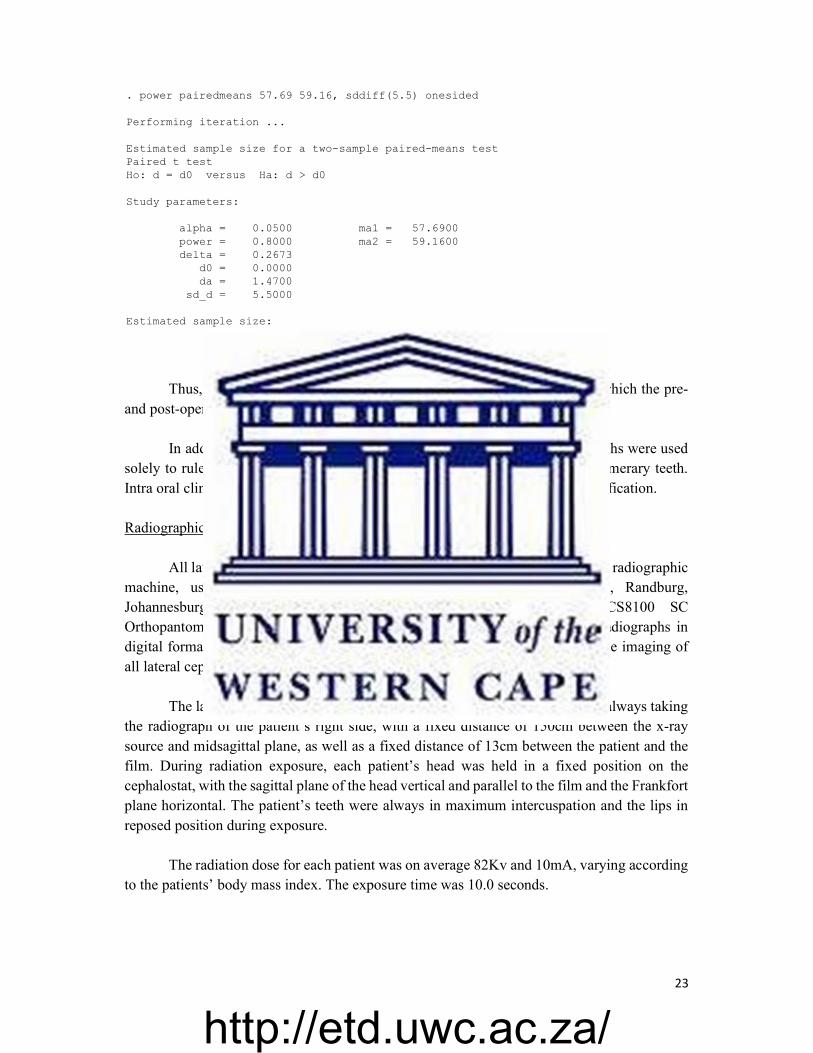
Based on data from Aldosari et al (2019) (1) and using the Vertical Airway Length, and choosing a statistical power of 80% with a 95% confidence level, the calculated sample size calculated is 88 patients.
http://etd.uwc.ac.za/
23
Thus, the selected sample size comprised a sample size of 88 cases, in which the pre- and post-operative cephalometric radiographs were be compared.
In addition to the lateral cephalometric radiographs, panoramic radiographs were used solely to rule out any dental anomalies, diagnose missing, impacted or supernumerary teeth. Intra oral clinical photographs were used to verify the dental malocclusion classification. Radiographic technique
All lateral cephalometric radiographs selected, were subjected to the same radiographic machine, using the same technique at the private orthodontic practice, Randburg, Johannesburg. All radiographs were taken using the CareStream CS8100 SC Orthopantomogram (serial number EDIG044) with all lateral cephalometric radiographs in digital format. Auxiliary staff, comprising of three oral hygienists, undertook the imaging of all lateral cephalograms.
The lateral cephalometric radiograph requirements were standardised by always taking the radiograph of the patient’s right side, with a fixed distance of 150cm between the x-ray source and midsagittal plane, as well as a fixed distance of 13cm between the patient and the film. During radiation exposure, each patient’s head was held in a fixed position on the cephalostat, with the sagittal plane of the head vertical and parallel to the film and the Frankfort plane horizontal. The patient’s teeth were always in maximum intercuspation and the lips in reposed position during exposure.
The radiation dose for each patient was on average 82Kv and 10mA, varying according to the patients’ body mass index. The exposure time was 10.0 seconds.
N = 88
Estimated sample size:
sd_d = 5.5000 da = 1.4700 d0 = 0.0000 delta = 0.2673 power = 0.8000 ma2 = 59.1600 alpha = 0.0500 ma1 = 57.6900
Study parameters:
Ho: d = d0 versus Ha: d > d0Paired t testEstimated sample size for a two-sample paired-means test
Performing iteration ...
. power pairedmeans 57.69 59.16, sddiff(5.5) onesided
http://etd.uwc.ac.za/
24
Radiographic tracing
The selected lateral cephalometric radiographs were calibrated with the cephalometric ruler as reference to allow for all adjustments to magnification of the image.
The principal investigator and gold standard recorded the landmark identification and cephalometric tracing and measurements on every 8th participant before the study commenced for inter-examiner reliability.
Thereafter, all lateral cephalometric radiographs were traced manually by the main investigator, in sittings of no more than 15 tracings per session. All cephalometric landmarks identifications were made using a standardized method, by manual tracing using a 4H 0.5mm pencil on acetate tracing paper.
Dentofacial Cephalometric Tracing
The dentofacial radiographic tracing was made on the pre-treatment lateral cephalometric radiograph to ensure that the patient met the inclusion criteria standards.
This includes the Steiner analysis to be used to confirm each patient’s skeletal relationship for confirmation of class I classification as well as confirmation of bimaxillary protrusion.
Table 3.1 lists the lateral cephalometric landmarks and Table 3.2 lists the dentofacial measurements. Figure 1 demonstrates the dentofacial landmarks and measurements.
Table 3.1: Dentofacial Cephalometric Landmarks
Variable Definition Nasion Most anterior point of the fronto-nasal suture in the midsagittal plane A Most posterior midline point in the concavity of the anterior maxilla,
between the anterior nasal spine and the prosthion (the most inferior point on the alveolar bone overlying the maxillary incisors)
B Most posterior midline point in the concavity of the mandible between the most superior point on the mandible between the most superior point on the alveolar bone overlying the lower incisors (infradentale) and pogonion
Soft tissue pogonion
Soft tissue point directly opposite hard tissue pogonion. Pogonion is the most anterior point on the bony chin.
S-line Steiner line drawn from soft-tissue pogonion to the midpoint of the S-shaped curve between subnasale and nasal tip
http://etd.uwc.ac.za/

25
Table 3.2: Dentofacial Cephalometric Measurements
Variable Definition ANB Angle between point A and B at Nasion UL-S Horizontal distance from the most protrusive point of the upper lip to the
Steiner S-line LL-S Horizontal distance from the most protrusive point of the lower lip to the
Steiner S-line NL Naso-labial angle measured between the line tangent to the base of the
nose and a line tangent to the upper lip IA Interincisal angle measured between the long axis of the upper and lower
incisors Mx NA Maxillary incisor to the NA line. Angular value between the long axis of
the maxillary incisor and NA, and the linear value in mm measured from the incisal tip of the maxillary incisor to NA
Md NB Mandibular incisor to the NB line. Angular value between the long axis of the mandibular incisor and NB, and the linear value in mm measured from the incisal tip of the mandibular incisor to NB
Figure 3.1: Dentofacial Cephalometric Landmarks and Measurements
http://etd.uwc.ac.za/

26
Pharyngeal Cephalometric Tracing
The pharynx consists of four different sections, namely the nasopharynx, velopharynx, glossopharynx and hypopharynx. Three sections will be identified radiographically and evaluated.
Table 3.3 lists all of the pharyngeal lateral cephalometric landmarks and Table 3.4 lists measurements of the pharyngeal airway. Figure 3.2 illustrates these landmarks, that will be used in the radiographic tracings.
Table 3.3: Pharyngeal Lateral Cephalometric Landmarks
Variable Definition Ba Lowermost point on anterior margin of foramen magnum SPPW Point of intersection of line from soft palate centre perpendicular to posterior
pharyngeal wall SPP Point of intersection of line from soft palate centre perpendicular to posterior
pharyngeal wall and posterior margin of soft palate U Tip of the uvula MPW Foot point of perpendicular line from point U to posterior pharyngeal wall TPPW Point of intersection of posterior pharyngeal wall and extension of line B-Go TB Point of intersection of base of the tongue and extension of line B-Go C3 Most anterior-inferior point of third vertebrae H Most superior and anterior point of hyoid bone
Table 3.4: Measurements of the three pharyngeal sections.
Airway measurement, mm Definition Velopharynx Distance between SPP and SPPW Oropharynx 1 Distance between U and MPW Oropharynx 2 Distance between TB and TPPW
http://etd.uwc.ac.za/
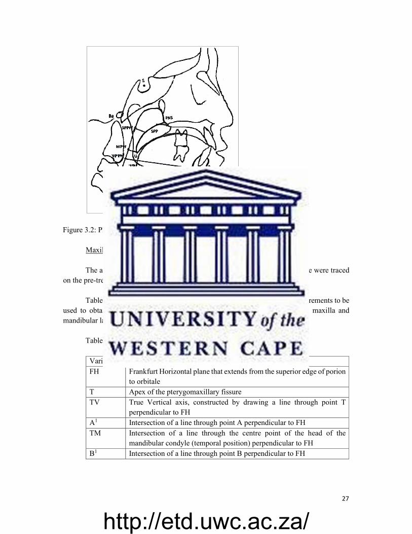
27
Figure 3.2: Pharyngeal Cephalometric Landmarks and Measurements Maxilla and Mandible Cephalometric Tracing
The anterior-posterior dimensions (or length) of the maxilla and mandible were traced
on the pre-treatment and post-treatment lateral cephalometric radiographs.
Table 3.5 lists the cephalometric landmarks and Table .3.6 lists the measurements to be used to obtain the length of the maxilla and mandible. Figure 3.3 shows the maxilla and mandibular landmarks and measurements.
Table 3.5: Maxilla and Mandible Cephalometric Landmarks
Variable Definition FH Frankfurt Horizontal plane that extends from the superior edge of porion
to orbitale T Apex of the pterygomaxillary fissure TV True Vertical axis, constructed by drawing a line through point T
perpendicular to FH A1 Intersection of a line through point A perpendicular to FH TM Intersection of a line through the centre point of the head of the
mandibular condyle (temporal position) perpendicular to FH B1 Intersection of a line through point B perpendicular to FH
http://etd.uwc.ac.za/

28
Table 3.6: Anterior-Posterior Measurements of the Maxilla and Mandible
Measurement Definition Maxillary length Measurement in mm along FH from A1 to TV Mandibular length Measurement in mm along FH from B1 to TM
Figure 3.3: Anterior-Posterior Maxilla and Mandibular Landmarks and Measurements.
Orthodontic treatment received by patients
All patients were treated with four premolar extractions in accordance to full fixed orthodontic treatment.
The TipEdge Plus bracket system was used on all cases. All cases were treated with 0.022 inch slot preadjusted appliances. Maximum anchorage was reinforced by the reciprocal anchorage engagement of the TipEdge bracket system. All cases were treated by the same operator.
http://etd.uwc.ac.za/
29
Data interpretation
The pharyngeal airway, consisting of three linear measurements (mm) of all the patients, were compared using the pre-treatment and post-treatment lateral cephalometric radiographs, as per the measurements listed in Table 3.2.
The total pharyngeal airway change was calculated by obtaining an average linear measurement (mm) difference across all three categories of the pre-treatment and post-treatment comparisons.
A total pharyngeal airway change with a positive value will indicate an increase in airway dimension, whereas a negative value indicates a loss of airway dimension.
The anterior-posterior linear measurement (mm) of the maxilla as well as the mandible,
of all patients, were compared using the pre-treatment and post-treatment lateral cephalometric radiographs, as per the measurements listed in Table 3.2.
A change in anterior posterior measurement of the maxilla and mandible with a positive value will indicate an increase in length whereas a negative value will indicate a loss of length.
The total pharyngeal airway change was compared to the change in anterior-posterior
measurements of the maxilla and of the mandible, to determine if a relationship exists. Examiner Reliability
Measurement bias towards tracing and landmark identification was avoided by having all tracings done by the main investigator for consistent tracing technique. Also, no more than 15 tracings of the lateral cephalometric radiographs were done at a time in order to avoid operator fatigue.
For inter-examiner reliability, ten randomly selected cephalometric radiograph pairs were selected. The principal investigator and gold standard will record the landmark identification and cephalometric tracing and measurements. The inter-examiner reliability (gold standard/accuracy) was done before the study commenced. The inter-examiner reliability was done on every 8th participant during the course of the study.
The 10 lateral cephalometric radiographs (taken as every 8th patient in the study) were selected by means of a random sample selection. This was done by selecting the 88 cephalometric radiograph pairs used in this study, using the number assigned to each patient’s cephalometric radiograph pairs with Excel to generate 10 random sample numbers to be used.
For intra-examiner reliability testing, ten randomly selected cephalometric radiograph pairs were selected. The same investigator did record the landmark identification and cephalometric tracing and measurements. These were done in two separate instances two weeks apart. An intraclass correlation assessment was utilized to test the reliability of the measurements.
http://etd.uwc.ac.za/
30
Statistical Analysis
All statistical tests will be performed using StataCorp. 2017. Stata Statistical Software: Release 15. College Station, TX: StataCorp LLC Quantitative.
Variables were described using means and standard deviations. The pre- and post-test mean values of the quantitative variables were compared using Student’s paired t-test. Statistical significance set at 5% and 95% confidence intervals were used to report the results.
Pearson’s correlation coefficient was used to determine the degree to which two variables are associated. Values less than 0.5 are indicative of poor reliability, values between 0.5 and 0.75 indicate moderate reliability, values between 0.75 and 0.9 indicate good reliability, and values greater than 0.90 indicate excellent reliability. Ethical Considerations
The study proposal was submitted to the Biomedical Research Ethics Committee of the
University of the Western Cape for approval. The project was registered by the BMREC as number BM20/3/4.
No direct patient evaluation was done in this study. Only patient records including lateral cephalometric radiographs, panoramic radiographs and intra oral clinical photos were used. All patients’, whose records were included in this study, gave written consent at the start of treatment of the private practise in Randburg, Johannesburg, that their records may be used for academic purposes.
All patient’s records, including the lateral cephalometric and panoramic radiographs, as well as clinical photographs were digitally renamed by assigning a number to each patient. No patient’s personal information was disclosed and anonymity could be ensured.
All clinical photographs were limited to intra-oral photographs only. Thus, the patient was unrecognizable and anonymity could be ensured.
http://etd.uwc.ac.za/

31
CHAPTER 4 Results
Reliability of results Reliability of lateral cephalometric tracings was calculated for inter-examiner and intra-examiner reliability using the intraclass correlation (ICC) formula. A good inter-observer agreement was demonstrated in all the measurements with ICC ranging from 0.56 to 0.97. Moreover, good intra-observer agreements with ICC ranging from 0.5 to 0.99 were obtained, which indicated good reliability. Prevalence of bimaxillary protrusion
According to the study population inclusion criteria, from a total of 3353 patients that have completed orthodontic treatment during the covered time period, 131 patients (3.9%) were classified as bimaxillary protrusive and had four premolars extraction treatment. Cephalometric characteristics of study population Table 4.1: Pre-treatment and post-treatment average value of cephalometric measurements of the study population
Cephalometric Measurement Average value pre-
treatment (SD)
Average value post-
treatment (SD)
ANB 4.26° (1.49) 4.47° (1.86)
Maxillary incisor to NA line (angular) 30.5° (5.61) 20.84° (6.53)
Maxillary incisor to NA line (linear) 5.33mm (1.81) 2.34mm (1.18)
Mandibular incisor to NB line (angular) 38.48° (5.66) 34.9° (6.48)
Mandibular incisor to NB line (linear) 6.34mm (2.46) 4.7mm (2.07)
Interincisal angle 106.39° (9.43) 118.74° (11.85)
Nasiolabial angle 86.51° (8.35) 94.92° (9.37)
S-line to upper lip 3.47mm (2.34) 1.93mm (2.37)
S-line to lower lip 4.41mm (2.68) 2.57mm (2.56)
http://etd.uwc.ac.za/

32
Orthodontic treatment: cephalometric changes from pre-treatment to post-treatment Table 4.2: Retraction of maxillary and mandibular incisors from pre- to post-treatment cephalometric measurements. Avg Angular
change (SD)
P value Avg Linear
change (SD)
P value
Maxillary incisor -9.67° (6.91) <0.001 -2.99mm (1.71) <0.001
Mandibular incisor -3.58° (6.16) <0.001 -1.64mm (1.51) <0.001
Table 4.3 Cephalometric changes from pre- to post-treatment cephalometric measurements.
Cephalometric Measurement Avg change (SD) P value
ANB 0.21° (1.09) 0.0739
Interincisal angle 12.35° (13.64) <0.001
Nasiolabial angle 8.41° (8.51) <0.001
S-line to upper lip -1.53mm (1.20) <0.001
S-line to lower lip -1.84mm (1.23) <0.001
*P value ≤0.05 is statistically significant
Table 4.4: Correlation between change in nasiolabial angle and change in maxillary incisor measured to the S-line. P value in brackets.
Change in Nasiolabial Angle (P value)
Change in S-line to upper lip -0.1379 (0.2000)
Change in S-line to lower lip -0.2720 (0.0103)
http://etd.uwc.ac.za/

33
Pharyngeal airway effect Table 4.5: Change in pharyngeal airway measurements in mm (comparison of pre-treatment and post-treatment lateral cephalometric values).
Variable Pre-treatment
(mm)
Post-treatment
(mm)
Change (mm)
Mean SD Mean SD Mean SD P Value
Velopharynx
(SPP-SPPW)
15.23 0.33 14.02 0.31 -1.21 2.45 <0.001
Oropharynx 1
(U-MPW)
11.63 3.26 9.99 2.87 -1.64 2.32 <0.001
Oropharynx 2
(TB-TPPW)
13.56 4.21 11.33 0.39 -2.23 2.81 <0.001
Table 4.6: The average pharyngeal airway change calculated by obtaining an average linear measurement (mm) difference across all three categories of the pre-treatment and post-treatment comparisons.
Variable Pre-treatment
(mm)
Post-treatment
(mm)
Change (mm)
Mean SD Mean SD Mean SD P Value
Average
airway size
13.47 2.88 11.78 2.65 -1.69 1.94 < 0.001
*P value ≤0.05 is statistically significant
http://etd.uwc.ac.za/
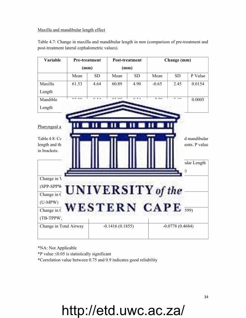
34
Maxilla and mandibular length effect Table 4.7: Change in maxilla and mandibular length in mm (comparison of pre-treatment and post-treatment lateral cephalometric values).
Variable Pre-treatment
(mm)
Post-treatment
(mm)
Change (mm)
Mean SD Mean SD Mean SD P Value
Maxilla
Length
61.53 4.64 60.89 4.90 -0.65 2.45 0.0154
Mandible
Length
85.89 8.54 83.89 9.54 --2.00 5.18 0.0005
Pharyngeal airway change comparison Table 4.8: Correlation between the change (pre- to post-treatment) in maxillary and mandibular length and the change (pre- to post-treatment) in airway cephalometric measurements. P value in brackets. Change in Maxillary Length
(P value)
Change in Mandibular Length
(P value)
Change in Velopharynx
(SPP-SPPW)
-0.1191 (0.2690) NA
Change in Oropharynx 1
(U-MPW)
-0.1625 (0.1305) NA
Change in Oropharynx 2
(TB-TPPW)
NA 0.0330 (0.7599)
Change in Total Airway
-0.1416 (0.1855) -0.0778 (0.4684)
*NA: Not Applicable *P value ≤0.05 is statistically significant *Correlation value between 0.75 and 0.9 indicates good reliability
http://etd.uwc.ac.za/

35
Table 4.9: Correlation between maxillary and mandibular incisor retraction and the change in airway from pre- to post-treatment cephalometric measurements. P value in brackets. Maxillary Incisor Retraction
(P value)
Mandibular Incisor Retraction
(P value)
Angular Linear Angular Linear
Change in
Velopharynx
(SPP-SPPW)
0.0183
(0.8651)
0.0554
(0.6059)
NA NA
Change in
Oropharynx 1
(U-MPW)
-0.0659
(0.5396)
-0.0258
(0.8100)
NA NA
Change in
Oropharynx 2
(TB-TPPW)
NA NA -0.0369
(0.7316)
-0.1635
(0.1257)
Change in Total
Airway
-0.1191
(0.2693)
-0.0400
(0.7097)
-0.0128
(0.9055)
-0.1274
(0.2341)
*NA: Not Applicable *P value ≤0.05 is statistically significant *Correlation value between 0.75 and 0.9 indicates good reliability
http://etd.uwc.ac.za/

36
Maxillary and mandibular incisor retraction comparisons Table 4.10: Correlation between maxillary and mandibular incisor retraction and the change in maxillary and mandibular lengths from pre- to post-treatment cephalometric measurements. P value in brackets. Maxillary Incisor Retraction
(P value)
Mandibular Incisor Retraction
(P value)
Angular Linear Angular Linear
Change in
Maxillary Length
-0.0457
(0.6708)
-0.2203
(0.0381)
NA NA
Change in
Mandibular Length
NA NA -0.0220
(0.8380)
0.0765
(0.4762)
*NA: Not Applicable *P value ≤0.05 is statistically significant *Correlation value between 0.75 and 0.9 indicates good reliability
http://etd.uwc.ac.za/
37
CHAPTER 5 Discussion
Prevalence of bimaxillary protrusion
The present study showed that 3.9% of the patients treated in this orthodontic practice,
who presented with bimaxillary protrusion, were treated by means of four premolar extractions and fixed orthodontic appliances with a maximum anchorage protocol. The prevalence of this type of bimaxillary protrusion treatment may not necessarily reflect the prevalence of this treatment approach in all orthodontic practices. This finding may be due to numerous limitations and other possible reasons, in that:
There was no external validity due to the study population selected. However, it could be said that the study may be indicative of a normal cross section of the population seeking orthodontic treatment.
The patients were all treated by one practitioner, which brings personal preference into the decision on what treatment approach was selected. As bimaxillary protrusion treatment is based on the facial appearance (fully convex, protruding teeth with procumbent upper and lower lip), practitioner views on what is considered unaesthetic could vary greatly and therefore change the treatment approach from extraction to non-extraction.
Another factor to consider is the number of patients with bimaxillary protrusion seeking orthodontic treatment. The reason for these patients seeking treatment, if they already have relatively straight teeth, is based on the individual perception of protrusion and what is accepted as aesthetic. This could explain why this study shows a very low percentage of patients with bimaxillary protrusion needing treatment.
Orthodontic treatment
Incisor retraction Table 4.2 shows the average amount of retraction of the maxillary and mandibular
incisors from pre-treatment to the post-treatment, after orthodontic treatment with four premolar extractions. The maxillary incisor showed an average retraction of 9.67 angular degrees and a linear measurement of 2.99mm. The lower incisor respectively showed 3.58 degrees and 1.64mm retraction. All of these retraction measurements are statistically significant with a P value of less than 0.001.
This is substantiated by Alqahtani (et al., 2019), who has found very similar results. The study found that with the extraction of premolars and retraction of anterior teeth with orthodontic treatment, the upper incisors retroclined by a mean angle of 9.6 degrees related to
http://etd.uwc.ac.za/
38
the palatal plane, and retracted a mean distance of 4.1 and 3.8mm using the A-Pog line. The results showed an equally pronounced lower incisor retraction effect. The lower incisors had a retroclination of a mean angle of 9.65 degrees in relation to the FMIA angle, and retracted a mean distance of 4.1 and 3.6mm in relation to the A-Pog line (Alqahtani, et al., 2019).
Another study showed the retraction effect on the incisors, when treating bimaxillary protrusion with premolar extraction. They found the maxillary incisor retroclined by 12.71 degrees and the mandibular incisor retroclined by 5.64 degrees, as well as 5.2mm (upper) and 3.22mm (lower) incisor retraction after orthodontic treatment (Bills, et al., 2005).
Solem et al. (2013) found that, when treating bimaxillary protrusive patients with four premolar extractions, the maxillary incisors retracted with an average of 4.16mm and the mandibular incisor with an average 3.39mm. The inclination of the maxillary incisor retroclined with an average of 13.18 degrees and the mandibular incisor with 8.75 degrees. The orthodontic retraction of the incisors was achieved with no skeletal anchorage (Solem et al., 2013).
The current study showed greater maxillary incisor post-operative changes when
compared to the mandibular incisor changes. These findings are substantiated by a study (Solem, et al., 2013) that also found the movement of mandibular incisors to be less and more varied. This may be attributed to the fact that the mandible is not ‘fixed’ as is the maxilla, and with retraction there could possibly be rotation of the mandible as well, which could influence the values.
One must also remember that an equilibrium zone among the forces created during
functions such as speech, mastication and swallowing combined with the action of the tongue, lips, cheeks and mouth floor, all determine the position of the tooth (Longhini, et al., 2017). Light sustained forces from the lips, cheeks and tongue determine tooth position (Proffit, et al., 2007). The smaller mandibular incisor movement observed in this study, can also thus be ascribed to the forces exerted by the tongue. It would stand to reason, that the orthodontic retraction forces applied on the incisors will lead to limited space for the tongue, and the tongue can resist the retraction effect of the incisors.
Interincisal angle Table 4.1 lists all the cephalometric changes seen from pre- to post-treatment. The
interincisal angle changed from a pre-treatment average measurement of 106.71° to a post treatment average measurement of 118.69°. The post-treatment average value being closer to both the Steiner and Medunsa norm for the interincisal angle.
This current study found the interincisal angle increased by 12.35° (p value <0.001, statistically significant) from the pre- to post-treatment cephalometric measurements (Table 4.3). Taking into consideration that the interincisal angle is formed by the maxillary and mandibular incisor positions, it can thus be said that the retraction observed in the incisors
http://etd.uwc.ac.za/

39
(Table 4.2) from pre- to post-treatment, will have a direct effect on the interincisal angle. This reduction in incisor proclination and protrusion along with the reduction in the interincisal angle is the desired effect from the treatment of bimaxillary protrusion by means of four premolar extractions. It treats the excessively proclined and protruded anterior teeth which is often the main complaint in bimaxillary patients seeking treatment.
The results from this study is validated by the findings of Kirschneck (et al., 2016) that showed an increase from an average interincisal angle of 127.5° to 130.5° when patients were treated with four premolar extractions, followed by retraction of the anterior teeth.
Bills et al. (2005) examined bimaxillary protrusion treatment by means of extraction orthodontic treatment, and found the interincisal angle to increase by 18.13 degrees. It is crucial to note that in this study by Bills et al. (2005) there was variable use of extraoral headgear, palatal buttons and transpalatal arches. All of the above lead to increased anchorage for retraction of the anterior teeth (Bills et al., 2005).
The interincisal increase in this study was greater than those found by Kirschneck (et al., 2016). This could possibly be attributed to a more effective maximum anchorage treatment approach utilised that allowed for more effective retraction of the maxillary and mandibular incisors. Since no extraoral anchorage, temporary anchorage devices or additional appliances like palatal buttons or transpalatal arches were used in this study, it clarifies the greater interincisal change observed by Bills et al. (2005).
This study, due to bracket choice for the orthodontic treatment, had a total anchorage demand that was lower than most conventional cases, thus, allowing for maximum anchorage to be achieved. This was achieved by means of differential tooth movement and bracket design which enabled variable anchorage. The anterior teeth being free to tip, comprise a light anchorage segment and can retract maximally (Parkhouse, 2009).
Soft tissue profile A harmonious soft tissue profile, which is an important goal in orthodontics, is
sometimes difficult to achieve, as soft tissues are highly variable in its thickness and tension (Kasai, 1998). The soft tissue response is further affected by the anatomy of the face, facial muscle activity and ethnicity (Solem, et al., 2013). Orthodontic treatment has been aiming at improving the lip form and soft tissue profile (Kasai, 1998).
The results in this study showed that the nasiolabial angle underwent a change from an
average measurement of 86.03° to an average of 94.41° (Table 4.1). The average value post-treatment is within the Steiner normal range of 90-110°. There was an increase of 8.41° (p value <0.001, statistically significant) seen in the nasiolabial angle from pre- to post-treatment in this study (Table 4.3). The current study further showed the upper and lower lip soft tissue changes from pre- to post-treatment. The average distance of the upper lip to the S-line measurement decreased by 1.53mm (p value <0.001, statistically significant) and the average
http://etd.uwc.ac.za/
40
distance of the lower lip to the S-line measurement decreased by 1.84mm (p value <0.001, statistically significant) (Table 4.3).
It can be said that this justifies the decision of extraction treatment, as the post-treatment
profile has shown to improve in both the soft tissue nasiolabial angle as well as the upper and lower lip position. However, one has to keep in mind that the nasiolabial angle is not only influenced by the lip, but also by the position and morphology of the nose itself (Kirschneck, et al., 2016).
It can be stated for this study that the maxillary incisor retraction (Table 4.2) seen has a direct effect on the increase seen in the nasiolabial angle (Table 4.3), as the upper lip is supported by the maxillary incisor.
This is confirmed by Lo and Hunter (1982) that showed an increase of 11.77° in the
nasiolabial angle from pre-treatment to post-treatment cephalometric analysis in patients treated with premolar extraction and retraction of anterior teeth. Lo and Hunter (1982) furthermore showed that the nasiolabial angle demonstrated a very high correlation with incisor retraction, with an estimated 1mm of retraction of the maxillary incisor resulting in a nasolabial angle increase of 1.63°. They found that the upper lip follows the incisors and subsequently pulls the subnasale areas forward and downward (Lo and Hunter, 1982).
Solem, et al (2013) stated that the lip changes as seen with anterior teeth retraction,
extended to the nasiolabial folds laterally, the columella superiorly and the mentolabial sulcus inferiorly. Their study showed that the upper lip retraction average was 2.26mm, which directly related to the nasiolabial angle (Solem, et al., 2013).
Alqahtani et al. (2019) reported on the soft tissue changes observed when treating
bimaxillary protrusive patients with premolar extractions. In their study a change in the nasiolabial angle was recorded from an average measurement of 98.1° to 104.7°, and thus an average increase of 6.6°. They further reported the upper lip protrusion to the S-line to have retracted by an average of 2mm and the lower lip an average of 2.7mm respectively (Alqahtani et al., 2019).
Ziliwu et al. (2008) examined the soft tissue changes in bimaxillary protrusive patients
treated with four premolar extractions. The upper lip protrusion decreased by 1.79mm and the lower lip protrusion was decreased by 3.6mm.
A systematic review of literature done by Leonardi et al. (2010) looking at bimaxillary
protrusive patients treated with premolar extractions found an average of 2.7mm retraction reported for upper lip retraction, and 2mm for the lower lip.
The increase seen in the nasiolabial angle in this study compared relatively well to the
literature stated, as the consensus across all the studies was an increase in the nasiolabial angle when bimaxillary protrusive patients were treated with premolar extraction. The decrease seen
http://etd.uwc.ac.za/
41
in the lip protrusiveness in this study, when compared to the literature, was lower than the results reported. This finding could be attributed to any of the myriad of factors affecting lip retraction.
Literature has concurred that there is a correlation between incisor retraction and lip
retraction (Konstantonis, et al., 2018; Kuhn, et al., 2016; Talass, et al., 1987). It is said by Hodgkinson, et al. (2019) that according to meta-analysis findings, the upper lip is expected to retract 0.7mm for every 1mm of upper incisor retraction, ranging from 0.3mm and 1mm between studies. The lower lip is also said to retract 0.7mm for every 1mm of upper incisor retraction, ranging from 0.1mm to 1.39mm, whilst for every 1mm of lower incisor retraction the lower lip retraction ranges from 0.8mm to 1.3mm (Hodgkinson, et al., 2019).
Ethnic differences in soft tissue to hard tissue movement also merit consideration.
Literature has shown racial variation in tissue response, with differences found when comparing ratios of soft to hard tissue movements across different races (Kolokitha and Chatzistavrou, 2012).
Park and Burstone (1986) stated that there is no linear ratio between the hard and soft
tissue retraction and the relationship is unpredictable. This is due to complex anatomy and structural support of the lip, as thinner lips will follow incisor retraction more closely than thicker lips, which, if very thick, may not change at all (Brock, et al., 2005). Lips with a pre-existing increased curve tend to tolerate retraction more, as opposed to thicker lips which curl more as the hard tissue support moves posteriorly (Arnett and Bergman, 1993).
When treating a patient’s soft tissue profile in orthodontics, there is uncertainty amongst many practitioners on how to treat the African black facial profile, as their normal facial features tend to be a naturally bimaxillary protrusive profile. The question is thus, to what aesthetic goals should they be treated? Do their aesthetic values conform to those of African heritage? And how much protrusion is considered acceptable? As society becomes more aesthetically inclined, it becomes vital to consider the patient’s opinion. The concept of beauty is subjective and one cannot simply treat to a set of norms (Farrow, et al., 1993).
According to Almutairi et al. (2015) bimaxillary protrusion has been found to be
associated with less attractive smiles by both dental professionals and laypeople. This is further corroborated by a study by Dawjee et al. (2010) which stated that the majority of African subjects with bimaxillary protrusion would want extraction orthodontic treatment to change their facial profile to a straighter profile ideal.
Thus, considering all the changes noted in the patients hard and soft tissues above, it
can be said that the soft tissue profile should have preference as the main treatment objective, as defined by the patient’s opinion on what he/she considers aesthetic, and that the tooth movement needed to obtain the necessary changes comes second (Holdaway, 1984).
http://etd.uwc.ac.za/
42
Comparison between nasiolabial change and upper/ lower lip protrusion
Considering the soft tissue changes, this study shows in Table 4.4 the correlation between the retraction of the upper lip and the increase in the nasiolabial angle. The change in the S-line to the upper lip had a correlation value of -0.1379 with P value 0.200. The change on the S-line to the lower lip had a correlation value of -0.2720 with P value 0.0103. Neither of the two correlations were statistically significant.
It can be concluded that the change observed at the most prominent point of the upper
lip, used as a cephalometric landmark in relation the S-line, is not a true indication of the angulation of the lip. As the angulation of the upper lip is used to create a tangent line, ignoring the prominence of the vermillion border, it explains why no correlation is to be found between the change in prominence of the upper lip and the changes in the nasiolabial angle.
Although no direct correlation is found, this study did find a statistically significant
change in the nasiolabial angle, S-line to upper lip and S-line to lower lip (Table 4.3).
Pharyngeal airway space
Pre-treatment pharyngeal airway space This study investigated three different measurements of the pharyngeal airway, namely
the velopharynx, oropharynx 1 and oropharynx 2 (Table 4.5 and Figure 3.2).
A study done by Nasser et al. (2019) reported measurements of the pharyngeal airway at three different levels in the antero-posterior dimension on cephalograms.
1. The superior posterior airway space from the soft palate to the posterior all of the airway.
2. The middle airway space, measured from the uvula to the posterior all of the airway. 3. The inferior airway space, measuring the airway along the Go-B line.
For the purpose of comparison, the velopharynx measurement in this study is equal to
the superior pharyngeal airway space (SPAS) as described by Nasser et al (2019). Respectively the oropharynx 1 is referred to the middle pharyngeal airway space (MPAS) and oropharynx 2 is referred to as the inferior pharyngeal airway space (IPAS).
This study determined the pharyngeal airway measurements at the pre-treatment lateral cephalometric radiographs as a baseline. The average measurement found at the SPAS was 15.23mm, at the MPAS it was 11.63mm and lastly the measurement at the IPAS was 13.56mm (Table 4.5).
As mentioned before, guidelines on average physiologic airway dimensions in literature are scarce. Mislik et al (2014) reported airway dimensions to average about 9-10mm at its shortest distance between the soft palate and the posterior pharyngeal wall (also known as the
http://etd.uwc.ac.za/
43
SPAS) and 10-12mm at its shortest distance measured between the tongue base and the pharyngeal wall (or the IPAS) (Mislik, et al., 2014).
Mani, et al. (2015) described normal upper pharyngeal airway space measured on a
lateral cephalometric radiograph for normo-divergent patients. The average superior pharyngeal airway measurement at 12.13mm and the average inferior pharyngeal airway measurement at 10.23mm (Mani, et al., 2015).
Daraza, et al (2017) also discussed upper pharyngeal airway norms. This study covered
a wide range of the population sample, including class I, II and III cases permitting they are healthy individuals with no medical concerns affecting the airway. They found the average SPAS measured 12.5 ±4.0mm, the MPAS measured 10.5 ±2.8mm and IPAS measured at 7.0 ±4.6mm (Daraza, et al., 2017).
It can thus be concluded that the results from the present study correlated well when
comparing the average measurements seen at the level of the uvula tip and tongue base (MPAS and IPAS), however this study did find a much greater baseline pharyngeal airway measurement at the level of the soft palate (SPAS). Only Sharma et al., (2014), showed values similar pre-treatment values to this study, with measurements of 15.03mm at SPAS, 13.16mm at MPAS and 12.1mm at IPAS.
This deviation seen from the literature could be due to the fact that all the patients included in this study are classified as class I. The literature has shown that class I patients have a pharyngeal airway volume that is greater than seen in class II patients. It can be concluded that when the mandible is in a normal anteroposterior position, the airway shows a greater antero-posterior dimension (Alves, et al., 2012; De Freitas, et al., 2006). Therefore, as the patients included in this study had to be classified as skeletal class I, there was no anterior-posterior discrepancy in the mandible that could affect the baseline pharyngeal airway measurements, in fact it may have led directly to a greater and fully patent pre-treatment airway as no risk factors were involved.
Literature has shown significantly different relationships between different dentofacial and craniofacial forms and the pharyngeal airway space exist (Mani, et al., 2015; Daraza, et al., 2017; Ung, et al., 1990; Coşkun and Kaya, 2018). It is said that patients with a hyperdivergent facial pattern showed a greater narrowing of the pharyngeal airway (Joseph, et al., 1998), with an average reduction of 2.93mm in the SPAS and 0.36mm in the IPAS when compared to normo-divergent patients (Mani, et al., 2015). Since no measurement was done on the divergence of the facial patterns, as well as no clinical examination to provide additional information on the dentofacial form of the patient, it could not be related to the expected pharyngeal airway space.
Predisposing factors to pharyngeal obstruction include allergies, environmental irritants and infections (Mani, et al., 2015). The only exclusion criteria for this study was patients with a medical history of naso-oro-pharyngeal obstruction, such as hyperplasia of the tonsils or
http://etd.uwc.ac.za/

44
adenoids, as well as congenital abnormalities affecting the craniofacial region. Thus, one cannot rule out that a patient could possibly have had active allergies or infection affecting the pharyngeal airway, whilst the pre- or post-treatment lateral cephalometric radiograph was taken.
Change in pharyngeal airway space
Sharma, et al (2014) showed that the removal of premolars had a detrimental effect on the oropharyngeal structures. Owing to the close relationship between the dentofacial structures and the pharynx, a mutual interaction has long been assumed (Sharma, et al., 2014).
This study looked at premolar extraction and retraction of anterior teeth in orthodontic treatment performed on bimaxillary protrusion patients, in order to establish if a relationship exists between removal of teeth and the pharyngeal airway space. Table 4.5 shows the changes seen in the pharyngeal airway space, from pre-treatment to post-treatment. The results included three different measurements of the pharyngeal airway.
This study has the following results:
The velopharynx (SPAS) showed an average reduction of 1.21mm in measurement with a statistically significant P value <0.001.
The oropharynx 1 (MPAS) showed an average reduction of 1.64mm in measurement with a statistically significant P value <0.001.
The oropharynx (IPAS) showed an average reduction of 2.23mm in measurement with a statistically significant P value <0.001.
Based on a similar study, Wang, et al (2012) reported a reduction in airway space at the
velopharynx and glossopharynx following retraction of anterior teeth. The average reduction of the superior, middle and inferior airway space is 0.56mm, 0.85mm and 1.63mm respectively (Wang, et al., 2012). It is worthy to note that a reduction was seen across all three measurements, as was the result in this study. However, the reduction measured in millimetres is much bigger in this study. The reason for the discrepancy seen between this study and Wang et al. (2012) is uncertain, as both studies have used the same protocols.
Germec-Cakan, et al (2011) reported a reduction of 2.1 ± 1.5mm in the middle airway
space (measured from the uvula to the posterior pharyngeal wall), and 3.8 ± 3.3mm in the inferior airway space (measured along the Go-B line) in a maximum anchorage space closure after orthodontic extractions.
Nasser, et al (2019) showed a decreased airway of 0.69mm in the superior airway space and a decrease of 0.66mm in die middle airway space, when examining adult bimaxillary protrusive patients who underwent orthodontic retraction of anterior teeth following extraction of all first premolars.
http://etd.uwc.ac.za/
45
Nuvusetty, et al (2016) found a significant narrowing of the pharyngeal airway behind the soft palate (SPP-SPPW), uvula (U-MPW) and at the base of the tongue (TB-TPPW) in Class I, bimaxillary protrusion patients treated with premolar extractions. The mean reduction found was 1mm, 2.4mm and 2.7mm respectively, or expressed as a reduction of 7.4%, 19.2% and 20.8% (Nuvusetty, et al., 2016).
A study by Hang and Gelb (2017), which discussed airway centric orthodontics, also found that bimaxillary protrusion patients treated with extractions had a reduction in airway space.
A study done by Bhatia (et al., 2016) showed that the extraction of the first premolars together with orthodontic retraction treatment in bimaxillary protrusive patients, had an influence on the pharyngeal airway. There was a statistically significant decrease at the SPAS, MPAS and IPAS. The mean reduction of the airway was 2.6mm (16.72%) for SPAS, 2,85mm (22.27%) for MPAS and 2.65mm (19.56%) for IPAS (Bhatia et al., 2016).
A study by Zheng et al., (2017) concluded that middle and inferior airway dimensions were diminished after extraction treatment. This study assessed the airflow in bimaxillary Class I patients, with first premolar extractions and maximum anchorage orthodontic treatment. After treatment the pharyngeal minimum cross section was diminished significantly, leading to a greater pressure gradient between the internal and external pressure from the surrounding tissues. This all contributes to an upper airway that is more collapsible (Zheng, et al., 2017).
Sharma et al., (2014) found that bimaxillary protrusive adolescents treated with first premolar extractions, coupled with orthodontic treatment to retract the incisors, resulted in a detrimental narrowing of the oropharyngeal structures. The study presented with an average reduction seen at the SPAS of 1.23mm, and 2.1mm at MPAS, with 1,47mm at IPAS (Sharma, et al., 2014).
It can be said that, when comparing the results from this study to previous literature, the SPAS reduction of 1.27mm is comparable to the range of 0.69-2.6mm seen across literature. The same goes for the MPAS reduction of 1.55mm that falls within the range in literature of 0.85-2.85mm, and the IPAS of 2.08mm within the literature of 1.63-3.8mm.
An interesting study by Zhang (et al., 2015) showed that the airway volume, height and cross-sectional area did not significantly change, however, and the sagittal airway dimensions decreased significantly in the middle and inferior pharyngeal airway. This confers with the present study.
Contradicting to the results obtained in this study, Stefanovic (et al., 2012) reported that extraction or non-extraction treatment would not directly affect the airway. Pliska (et al., 2016) also confirmed this contradiction, and showed that extraction treatment did not statistically constrict the volume of the upper airway. Further, Al Maaitah (et al., 2012) found insignificant change in the upper airway dimensions with orthodontic extraction followed by retraction.
http://etd.uwc.ac.za/
46
Retraction protocol and airway
One explanation as to why there was no significant change seen in the airway dimensions in the studies of Stefanovic (et al., 2012), Pliska (et al., 2016) and Al Maaitah (et al., 2012) could well be that the retraction of the anterior teeth was not of such an extent to affect the airway.
With regards to space closure, several techniques exist in orthodontics. The space closure approach could either be maximum anchorage or minimum anchorage, depending on desired effect (Felemban, et al., 2013). The closure of the extraction space can be achieved by means of retraction of the incisors (maximum anchorage), or premolar and molar protraction (minimum anchorage), or a combination of the two (moderate anchorage) (Proffit, et al., 2007). The retraction of the anterior teeth into the space, can be achieved either by means of elastics, powerchain, nickel-titanium springs or closing loops (Li, et al., 2018).
Germec-Cakan (et al., 2011) investigated the differences between retraction protocols with extraction on the airways. They found that with minimal anchorage control, an average mesial molar movement of 3mm, showed a mean increase of 1.5mm across the superior and middle airway space. Whereas in the group with maximum anchorage protocols, the middle and inferior airway reduced by 3mm (Germec-Cakan, et al., 2011).
Chen (et al., 2012) and Wang (et al., 2012) concluded that extraction treatment coupled with large retraction of the anterior teeth resulted in a reduction of the upper airway dimensions.
Adequate control of anchorage in orthodontics, however, is still a major concern. Anchorage is described as being comprised of units of resistance to movement. This can either be tooth anchorage or auxiliary anchorage (Hart, et al., 1992). Due to Newton’s third law, i.e. for every action there is an equal and opposite reaction, there are limitations in our ability to control all aspects of tooth movement. Therefore, in orthodontics the anchorage protocol applied, does not always yield the desired effect (Cope, 2005).
The same deduction was made by another study that looked at the effect of extractions and retraction of anterior teeth on the pharyngeal airway. They concluded that maximum anchorage was not obtained, and thus retraction may have been less effective and thereby did not significantly affect the pharyngeal airway (Hu, et al., 2015).
This study, due to bracket choice for the orthodontic treatment, allowed for maximum
anchorage to be achieved. This was achieved by means of differential tooth movement and by allowing the anterior teeth to tip, which resulted in a light anchorage segment that can be retracted maximally (Parkhouse, 2009).
Furthermore, facial patterns are also said to affect the retraction of teeth, whereby brachyfacial individuals are considered to be the least affected in comparison to mesofacial and dolichofacial patterns (Nasser, et al, 2019). This is explained by the fact that brachyfacial
http://etd.uwc.ac.za/
47
individuals have an underlying strong musculature pattern with a very strong natural muscular anchorage effect on the teeth, and thus will have difficulty moving teeth across long distances orthodontically. Conversely dolichofacial patients who have weaker muscles, will have a much greater capacity for tooth movement (Pepicello, et al., 2005).
Factors affecting airway measurements
Comparing the results from this study to previous studies, one can conclude that there
is more literature supporting the reduction of the pharyngeal airway space when anterior teeth are being retracted into premolar extraction spaces. However, the amount of pharyngeal airway space reduction seems to be highly variable from one study to the next.
One study proposed the idea that ethnic differences among patients may affect the severity of the reduction of the pharyngeal airway space (Nasser, et al, 2019).
In cephalometry, as much as standardisation techniques are applied when taking the
radiographs, constant head posture cannot be applied. Literature has proved that head posture can affect the size of the pharyngeal airway space. It has been estimated that a craniocervical inclination change of 10 degrees can have as much as a 4mm change in the pharyngeal airway (Muto, et al., 2008). This is supported by another study, that showed that head extension is linked to an increase in the pharyngeal airway space. They also found that the further superior the extension in the cervical spine, the greater the increase seen in the pharyngeal airway (Muto, et al., 2002).
The effects on the airway in this study could only be based on the cephalometric measurements in a two-dimensional capacity. A study by McNamara (1984) suggests that an opening of 5mm or less in the upper pharyngeal airway is an indicator of possible airway impairment. Further, with the help of Poiseuille’s law, one can determine to a certain extent the increase in airflow resistance that would occur with a reduction in the pharyngeal airway space.
Important to consider is the fact that airway resistance is not only related to airway size, but also to airway shape. Thus, even patients with adequate airway volume, but tortuous airway passages, the resistance could be severe enough to affect function (Aboudara, et al., 2009).
However, quantifying the proportions of oral and nasal breathing at repeated intervals is a prerequisite to any conclusion regarding respiratory function (Ung, et al., 1990). This is a limitation to this study as no clinical examination was done with a head-out body plethysmograph technique in order to obtain airflow measurements.
Pharyngeal airway changes related to growth changes cannot be included as a determinant affecting the airway space reduction in this study. The inclusion criteria of this study stated that only patients above 16 years of age were eligible. It is stated by Mislik (et al., 2014) that the upper airway dimensions are formed and matured in the early periods of growth.
http://etd.uwc.ac.za/
48
Further, as one would expect with growth to see an increase in airway space and not a reduction (Germec-Cakan, et al., 2011), it can be concluded, that due to the fact that the airway measurements across all three variables showed a reduction, growth could not have had any effect.
Further, growth of the mandible could also attribute to an increase in pharyngeal airway (Valiathan, et al., 2010). However, once again, growth would result in an increase in airway space and thus not be a contributing factor for airway reduction. It could nevertheless have had an effect on the amount of reduction seen in the pharyngeal airway, as growth would have lessened the reduction effect. This effect would be minimal in this study as all patients were above the age of 16 years, knowing that mandibular growth is complete.
Considering differences in gender, literature found that pharyngeal dimensions between males and females are generally small (Hanggi, et al., 2008). As gender was not distinguished in this study, it is important to note that gender is not said to have an effect.
Lastly this study also did not consider the patient’s body mass index changes from pre- to post-treatment. As literature shows that obesity and excessive soft tissue thickness can have a remarkable effect on the pharyngeal airway (Bollhalder, et al., 2013), this is a limitation to this study as weight gain was not recorded and thus can be a contributing factor to the changes seen in the pharyngeal airway measurements.
The size of the tongue, soft palate and parapharyngeal fat pads and the position of the
lateral pharyngeal walls have been shown to influence the upper airway morphology (Chen, et al., 2012). As this study had no clinical examination, there was no measurement of the tongue, soft palate, parapharyngeal fat pads or the position of the lateral pharyngeal wall, and thus is a limitation. This must be taken into account for the pre-treatment pharyngeal airway space measurements. Maxilla and mandible length
Pre-treatment maxilla and mandible length The present study looked at the anteroposterior or length measurement of the maxilla
and mandible. These measurements at the pre-treatment lateral cephalometric radiographs were used to determine baseline length. The average length of the maxilla measured 61.53mm and the average mandible at 85.89mm (Table 4.7).
This study based the anteroposterior length calculations on the Bimler Cephalometric analysis. This is based on a linear measurement of the maxilla from point A to the pterygomandibular fissure and the mandibular a linear measurement from point B to the centre of the condyle. Both the distance for the maxilla and mandible from their respective points are projected onto the Frankfurt horizontal reference line and measured.
http://etd.uwc.ac.za/

49
A study by Chang (1987) found that the anteroposterior measurement of the maxilla and mandible along the Frankfort horizontal plane is said to be a true measurement of the size of the size of the maxilla and mandible, as well as the relation to each other. Using this method, Huang (et al., 1998) found it to be more reliable, as it is less affected by the inclination of the reference plane. The Bimler analysis thus creates an analysis that assesses more detail, and allows for comparability (Turkdonmez, et al., 2014).
Harvold (1974) developed standards for the length of the maxilla and mandible derived from the Burlington growth study. The maxillary unit length is measured from the posterior border of the mandibular condyle to the anterior nasal spine. The mandibular length is also measured from the posterior border of the mandibular condyle, to the anterior point of the hard tissue chin (pogonion). He found the standard length of the maxilla for a male is 100mm and 93mm in females, and the mandibular length for a male 127mm and 119mm for females, if considering that growth is complete and the patient is 16 years of age and above. It could also be seen as a differential, irrespective of the age of the patient, whereby the mandible should always be 20-23mm longer than the maxilla to be balanced (Proffit, et al., 2007; McNamara, 1984).
There is insufficient data that reports on the maxillary and mandibular lengths as seen on cephalometric radiographs. Furthermore, there is no standardised cephalometric analysis to measure the lengths of the maxilla and mandible, which makes comparison impossible for this study.
Change in maxilla and mandibular length
The present study looked at premolar extraction and retraction of anterior teeth in orthodontic treatment received by bimaxillary protrusion patients, to establish if a relationship exists between removal of teeth and the length of the maxilla and mandible. Table 4.7 shows the changes seen in the length of the maxilla and mandible, from pre-treatment to post-treatment.
This study found that the maxilla had a decrease in length with an average measurement of 0.65mm and the mandible showed an average decrease in length of 2.00mm (Table 4.7). The P values for the changes were statistically significant, with values of 0.0154 and 0.0005 respectively.
Literature states that with extractions and subsequent retraction of anterior teeth, point A and point B on the maxilla and mandible, respectively, are affected. It has been shown that point A and point B will be in a more retruded position leading to a decreased maxillary and mandibular dentoalveolar size (Kalwitzki, et al., 2011).
This study, which investigated the effect of extraction orthodontic treatment and
retraction of anterior incisors, also showed a statistically significant reduction in the lengths of the maxilla and mandible. One can thus conclude and agree with Kalwitzki, as point A and
http://etd.uwc.ac.za/
50
point B were used in determining the maxilla and mandibular length measurements, one can conclude that the retraction of the anterior teeth has resulted in a more retruded position of these points.
No other studies examined the maxilla and mandible length changes associated with
premolar extractions and anterior teeth retraction. However, it can be compared to studies that investigated arch length changes and arch width changes as this will be the closest resemblance of the lengths of the jaws to this study.
A decrease in arch depth (measured as the shortest distance from a line distal of the first molars to the labial surface of the most anterior tooth of the arch) was found in both the maxilla and mandible in extraction cases (Kumari and Fida, 2010)
It has been reported that the mandibular arch length decreases with extraction treatment (McReynolds and Little, 1991; Paquette, et al., 1992; Shearn and Woods, 2000; Heiser, et al., 2004). The range of decreases seen due to premolar extraction, is reported to be 3.36mm when measured from the contact point of the central incisors to the midpoint connecting the distal contact points of the first molars (Ho and Kerr, 1987). Another study measuring the effect of premolar extractions on arch length, from the contact point between the central incisors to midpoint of the line connecting the mesial contacts of the first molars, found a decrease of 4.6mm (Boley, et al., 2003).
A study done by Al-Abdwani (et al., 2009) investigated the retraction of incisors and the effect on point B. In response to a 10-degree retraction of the mandibular incisors, a significant backward horizontal change was noted in point B (Al-Abdwani, et al., 2009). Similarly, in another study (Hosseinzadeh-Nik, et al., 2016), a backward movement was found in point B when anterior teeth were retracted, however, it was not significant. This backward movement, will affect the length measurement of the mandible.
Another aspect to consider, is the undesirable effect seen with extensive retraction of maxillary incisors which causes an increase in thickness of the buccal cortical bone. It is said to result from a lack of balance between bone resorption and neoformation, and depends on the amplitude, direction and quantity of movement, as well as changes in tooth tipping (Oliveira, et al., 2016).
This is confirmed by another study. They found that with the retraction of anterior teeth, significant remodelling of the alveolar bone was seen, with the bone measuring around the roots of the maxillary and mandibular anterior teeth approximately 1 to 2mm reduction in magnitude (Solem, et al., 2013).
Shrafeldin, et al. (2018) showed that extractions in orthodontics may lead to arch collapse, and thus a narrower arch width.
http://etd.uwc.ac.za/
51
Interestingly, a study done by Nascimento (et al., 2012) found evidence contradicting the ideas in this study which states that extractions with retraction of anterior leads to a reduced oral volume. They stated that when looking at intraoral volume capacity, the number of teeth had no influence (Nascimento, et al., 2012).
It is postulated in this study, the same conclusion that is found by Zheng (et al., 2017) that the decrease in maxilla and mandible lengths, will have an effect on the oral volume, forcing the tongue backward and thereby diminishing the airway in size.
Factors affecting maxilla and mandible length measurements This present study did not separate the results according to male and female patients
with regards to the maxilla and mandible lengths. As gender affects the size of the jaws, this factor would have an effect on the average length measurements obtained. The Harvold standard values for patients above the age of 16 years, shows that the average maxillary length is 100mm for men and 93mm for women, with a mandibular length of 127mm for men and 119mm for women (Proffit, et al., 2007).
However, the difference seen from pre- to post-treatment will not be influenced by
gender. This is due to the fact that the extraction of the premolars with the amount of retraction of anterior teeth, and thus arch changes, are not affected by gender. Literature does not show a direct difference in orthodontic tooth movement between males and females (Chisari, 2012).
The facial skeleton, including the length of the maxilla and mandible, reflect different
types of facial morphology described as straight, convex and concave. Patients with a straight profile have an average length of the mandible and maxilla in relation to age and a subspinal-nasion-supramental angle between 0 and 4 degrees. Patients with a convex profile show a subspinal-nasion-supramental angle more than 4 degrees, whereas patients with a concave profile show a subspinal-nasion-supramental angle less than 0 degrees (Macari, et al., 2014).
As this study did not include clinical examinations, the facial morphology of the
patients was not documented. This is a limitation of the study, as the different facial morphologies of the patients could affect the maxilla and mandibular length measurements on the pre-treatment records.
Growth would result in an increase in the size of the maxilla and mandible. According
to the Harvold standard values, the mean value of the maxillary length can increase by 18mm for men and 13mm for women between the age of 6 years to 16 years. Respectively, the mandibular length increases by an average of 28mm for men and 22mm for women (Proffit, et al., 2007). Saitoh (2004) stated that 60 percent of craniofacial development takes place during the first 4 years of life and 90 percent of craniofacial development is complete by the age of 12 years. The effect of growth would be minimal in this study as all patients were above the age of 16 years, knowing that growth is complete.
http://etd.uwc.ac.za/
52
Erroneous landmark identification on cephalometric radiographs would lead to different results and interfere with the reliability of the measurements. Point A and point B are both landmarks that are located at a curvature, which contribute to difficulty in identification. The validity of cephalometric distances thus depends on the validity of the individual landmarks involved (Durao, et al, 2014).
The Frankfurt horizontal plane was used in this study in order to determine the maxilla
and mandibular lengths. A change in this reference plane would have an effect on the measurements done along this plane. The accuracy of this reference plane is discussed under method error. Comparison between pharyngeal airway change and maxilla/mandible length change
As stated previously, no studies examined the maxilla and mandible length changes associated with premolar extractions and anterior teeth retraction.
The results from this study investigated the correlation between the difference in pharyngeal airway sizes and the difference in maxilla and mandibular lengths (Table 4.8). This study did not show a statistically significant correlation between the changes seen in the pharyngeal airway (including the three pharyngeal airway measurements and the total pharyngeal airway measurement) and the changes seen in the maxilla or mandible length.
It stands to reason that if the maxilla is retruded, so is the soft palate, and so if the
mandible is retruded, so is the tongue which is attached to the mandible (Hang and Gelb, 2017). The change in the maxillary length was correlated to the changes seen in the pharyngeal
airway at the level of velopharynx as well as oropharynx 1 (Also known as SPAS and MPAS). As the SPAS measures the pharyngeal airway from the centre of the soft palate and the MPAS from the tip of the uvula, both measurements directly relate to the maxilla. The correlation values respectively are -0.1191 and -0.1625, stipulating no correlation. P values were 0.2690 and 0.1305 respectively, showing no statistical significance.
The change in mandibular length was correlated to the change seen in the pharyngeal
airway at the level of oropharynx 2 (SPAS). The SPAS measure the pharyngeal airway from the anterior border of the airway where the Gonion-Point B line intersects and relates to the mandible. The results showed a correlation of 0.0330, stipulating no correlation. The P value was 0.7599, showing no statistical significance.
Lastly the change in the total airway value was correlated to the change in maxillary
and mandibular length. The correlation values are -0.1416 and -0.0778 respectively. It can thus be concluded that no correlation exists between the maxilla/mandible length changes and the changes in the pharyngeal airway. P values were 0.1855 and 00.4684 respectively, showing no statistical significance.
http://etd.uwc.ac.za/
53
Worthy to note is a study by Bollhalder (et al., 2013) which stated that it is not the absolute length of the jaws, but rather their position relative to the cranial base, which is an important determinant of the airway. This statement offers an explanation as to why in this study no correlation was found between the airway changes and the changes seen in the lengths of the maxilla and mandible.
Comparison between pharyngeal airway change and maxilla/mandible incisor retraction
It can be suggested that the retraction of the upper and lower incisors can be used as a predictive factor to determine the pharyngeal airway changes. As the incisors retract, the arches are said to be constricted and the intra oral volume will decrease (Larsen, et al.,2015). It is understood that this leads to a posterior postured tongue and soft palate (Mehta, et al., 2015) which results in the crowding of the oropharynx (Pliska, et al., 2016).
This study investigated the maxillary and mandibular incisor retraction (angular and linear) in relation to the changes in the pharyngeal airways (Table 4.9). The following correlations were observed:
Maxillary incisor angular retraction had a correlation value of 0.0183 to the change in velopharynx, with the linear retraction correlation value of 0.0554.
Maxillary incisor angular retraction had a correlation value of -0.0659 to the change in oropharynx 1, with the linear retraction correlation value of -0.0258.
Mandibular incisor angular retraction had a correlation value of -0.0369 to the change in oropharynx 2, with the linear retraction correlation value of -0.1635.
Maxillary incisor angular retraction had a correlation value of -0.1191 to the change in total airway, with the linear retraction correlation value of -0.0400.
Mandibular incisor angular retraction had a correlation value of -0.0128 to the change in total airway, with the linear retraction correlation value of -0.1274.
All of the correlations above had p values that showed no statistically significant correlations.
A study done by Bhatia (et al., 2016) showed a significant correlation between the
retraction distance of the lower incisor and the SPAS and IPAS.
Chen et al. (2012) stated that the retraction of incisors (in bimaxillary protrusive patients treated with orthodontic extraction treatment) lead to a decrease in oral volume, which in turn reduced the tongue’s space and resulted in a retracted tongue pressing on the soft palate and a diminution of the upper airway.
Chen et al. (2012) also found that airway dimension change has been correlated to
upper incisor retraction. It has been suggested that since the maxillary incisors are located above the mandibular incisor, retraction would not affect the pharyngeal airway as much when compared to the lower incisors.
http://etd.uwc.ac.za/
54
An explanation as to why this study did not find correlations between the changes in the airways and the incisor retractions, could be the fact that retraction of incisors does not have a direct relation to the airway. It can rather be said that the incisors can be retracted with no effect on the airway until a threshold is reached, whereafter if the retraction exceeds the threshold limit, the airways will only then be affected. One can postulate that beyond the threshold point, a direct correlation could be present between the incisor retraction and the airway effect. Comparison between maxilla/mandible length change and maxilla/mandible incisor retraction
Table 4.10 lists the correlation between the maxillary and mandibular incisor retraction to the maxillary and mandibular lengths, respectively. The maxillary incisor retraction (angular and linear) was correlated to the change in maxillary length, whereas the mandibular incisor retraction (angular and linear) was correlated to the change in mandibular length.
The maxillary incisor angular retraction had a correlation value of -0.0457, and the
linear retraction a correlation value of -0.2203, to the change in maxillary length. The mandibular incisor angular retraction had a correlation value of -0.0220, and the linear retraction a correlation value of 0.0765, to the change in mandibular length.
This study investigated the treatment of bimaxillary protrusion patients, where the goal
of the treatment outcome was to achieve less protrusion of the incisors. Incisor retraction can occur by means of angular (tipping) movements, or by means of linear (bodily) movements, or in almost all cases, a combination of the two. One could argue that with the angular change more buccal root torque was achieved, thus the dentoalveolar bone overlaying these roots which constitutes the anatomy for point A and point B, was not retruded. With the linear retraction (bodily movement) of the incisors one can expect that point A and point B will follow the backward movement of the roots of the teeth. However, one will always find there is a combination of angular and linear retraction movements. Some patients may have more tipping movement of the incisors and some will have more bodily movement of incisors, leading to variable change seen in points A and B.
As the maxilla and mandibular lengths were determined using point A and point B, this
may adequately explain why no direct correlation will be found between the retraction of the maxillary and mandibular incisors and the length changes in the maxilla and mandible.
Reliability of study
This is a retrospective appraisal study; therefore, the patient collective was chosen retrospectively and for availability of complete patient records. There is thus no missing data and no loss to follow-up that had to be addressed.
This study only included patients above the age of 16 years to rule out any growth changes influencing the results.
http://etd.uwc.ac.za/

55
Method error
Frankfurt horizontal plane accuracy
In cephalometric literature, there is abundant planes and reference planes, with the Frankfort horizontal (FH) plane as one of the most frequently documented. This plane was introduced in 1882 (Hofmann, et al., 2016; Pancherz and Gökbuget, 1996). This is drawn from porion, the most superior point of the outline of the external auditory meatus, to orbitale, the lowest point on the inferior orbital rim (Zebeib and Naini, 2014).
It has been documented that this cephalometric plane has its shortcomings with individual variability and difficulties in landmark identification. As this reference plane is intracranial, it may be significantly affected by landmark identification and location, with a high individual variation leading to erroneous findings (Zebeib and Naini, 2014). One study showed that the FH plane had a standard deviation of 0.7 degrees when compared between four different trained cephalometric tracers (Ricketts, et al., 1976).
Literature has reported that the cephalometric landmark Porion has very poor reproducibility and Orbitale is the most imprecise landmark (Pittayapat, et al., 2018). The porion has great transverse and sagittal anatomic variance, thus making it difficult to determine. The orbitale poses a transverse variation, due to the relatively flat lower border of the orbita (Hofmann, et al., 2016).
However, due to the fact that these landmarks are relatively far apart, it reduces the impact of measuring errors (Hofmann, et al., 2016). Reliability to identify landmarks can be enhanced if the landmark is in clear contrast to the adjacent structures and if it is stable and not affected by growth (Zebeib and Naini, 2014).
In an effort to avoid errors in the tracing of the FH plane, all tracings were made by one practitioner for this study. Ten randomly selected radiographs were re-traced two weeks after the initial tracing to assess intra-examiner reliability. Furthermore, as literature suggests that the anatomic porion is replaced by the ear-rod to improve reproducibility (Zebeib and Naini, 2014), this study used the ear-rod as the measurement of porion for exactly such reasons.
Another factor to take in consideration for this study is the use of the Frankfort horizontal plane. As this reference plane was not used in conjunction with any angular measurements, but rather a reference line onto which points were projected onto for a linear measurement, it would have a much lesser impact on the results.
Point A and Point B accuracy
It is suggested in literature that point A and B is one of the least identifiable landmarks. This is due to the fact that point A and B is a geometric landmark, subject to variation with radiographic technique. It is attributed to the fact that the radiographic exposure is uniform for
http://etd.uwc.ac.za/

56
the duration of the taking of the image, and as greater exposure is needed for posterior landmarks, the anterior landmarks are frequently dark and not as visible (Perillo, et al., 2000).
Difficulties arise in measuring dentofacial structures, as the representation of a three-
dimensional object such as the human skull, in a two-dimensional lateral cephalometric radiograph can result in errors of identification of anatomical landmarks. However, point A shows great agreement in an inter-rater reliability test, with point B having acceptable agreement (Grogger, et al., 2018).
As both Point A and point B are used in this study, the accuracy of identifying these
points are crucial to the outcome of the results. As these points are not subject to superimposition, and in literature shows good reliability, it can be argued that the accuracy of these landmarks is relatively high with fewer chance of error.
Further, in an effort to reduce error in identifying these landmarks, all tracings were
made by one practitioner for this study and an intra-examiner reliability test was conducted. Limitations
A limitation of this study was that there was no external validity due to the study population selected. However, it could be said that the study may be indicative of a normal cross section of the population seeking orthodontic treatment.
The use of lateral cephalometric radiographs in this study is a limitation, as errors due
to superimposition of reference points and double lines, still remain a problem. Although many studies have reported a very high accuracy when examining the pharyngeal airway (Preston, et al., 2004; Major, et al., 2006), one must consider that it is still a two-dimensional view and some loss of information may occur.
Furthermore, one has to consider the fact that airway resistance is not only related to
airway size, but also to airway shape (Aboudara, et al., 2009). The use of lateral cephalometric radiographs in this study has another limitation as it cannot provide information on the three-dimensional shape of the pharyngeal airway.
This study did not record ethnicity. This is considered a limitation of the study, as it has been documented in literature that there is racial variation in tissue response, with differences found when comparing ratios of soft to hard tissue movements across different races (Kolokitha and Chatzistavrou, 2012).
This study did not include a clinical examination of the patients. Therefore, the patient’s dentofacial form was not recorded. This is a limitation, as literature has shown significantly different relationships between different dentofacial and craniofacial forms and the pharyngeal airway space (Mani, et al., 2015; Daraza, et al., 2017; Ung, et al., 1990; Coşkun and Kaya, 2018). Further, dentofacial patterns are also said to affect the retraction of teeth (Nasser, et al,
http://etd.uwc.ac.za/
57
2019), and affect the maxilla and mandibular length measurements on the pre-treatment records.
The patient’s body mass index and presence of allergies or infections at the time of the
radiographic exposure, was not recorded. This could also be seen as a limitation, as all the factors above could influence the pharyngeal airway measurements.
This study did not include a head-out body plethysmograph technique in order to obtain
airflow measurements to assess airway patency. Thus, it cannot be quantified if and to which extent the airway patency was truly affected.
No follow up study was done to assess if any patient developed obstructive sleep apnea or mouth breathing after receiving extraction orthodontic treatment.
http://etd.uwc.ac.za/
58
CHAPTER 6 Conclusion
The field of orthodontics strives to normalise form and function, so that the function is optimised for life (Jayan and Kadu, 2018). The airway or the position of the mandible is rarely taken into consideration when diagnosing and deciding on an extraction case (Hang and Gelb, 2017). Recently airway health has been asserted as a primary consideration before starting orthodontic treatment (Nasser, et al, 2019). This study focused on assessing the effect of retraction of anterior teeth on pharyngeal airway dimensions, after orthodontic treatment of bimaxillary protrusion cases by means of the extraction of four premolars. There was a statistically significant reduction found in the pharyngeal airway space with the retraction of anterior teeth. An average reduction of 1.21mm was seen at the superior pharyngeal airway level, 1.64mm at the middle pharyngeal airway level and 2.23mm at the inferior pharyngeal airway level. A total average reduction of 1.69mm was seen for the pharyngeal airway. This study therefore concludes that retraction of anterior teeth as an orthodontic treatment modality for bimaxillary protrusive patients, does lead to a statistically significant reduction in the pharyngeal airway space.
Considering that sufficient anatomical dimensions of the airway must be present for normal respiration to occur (Mani, et al., 2015; Ceylan and Oktay, 1995), one can thus assume that the reduction on pharyngeal airway space does effect respiration and can have further health consequences, such as reduced quality of life and decreased life expectancy (Le, et al., 2019).
This raises the question and requests a recommendation on whether orthodontists can safely choose extractions as a treatment modality without affecting the pharyngeal airway space of the patient. .
http://etd.uwc.ac.za/
59
Recommendations
Extractions in orthodontics over the years were a result of to the following reasons: class I bimaxillary protrusion and excessive crowding, class II camouflage treatment and class III camouflage treatment. Perhaps shifting our view, and looking at bimaxillary patients in a different way, is to assess the mandible itself. Considering that, when using the aesthetic line (drawn from the tip of the nose to the soft tissue pogonion) to assess protrusion and aesthetics of the patient, it is dependent on the mandible position. Thus, the more retruded the mandible, the more pronounced the protrusion effect of the patient. If mandibular advancement is the treatment objective to properly position the mandible, would these patients still be considered protrusive? Thus, avoiding extractions with retraction and affecting the airway, with the added advantage that advancing the mandible will only increase airway patency. Orthodontist today still aim to avoid surgery and do so with camouflage treatment in attempt to reduce overjet in class II cases, by means of extractions and retraction of upper teeth. When considering these class II patients, they most likely already have a retruded mandible which stands to reason that the tongue is also retruded. With extraction and successive retraction of the maxillary anterior teeth, which have been shown to affect the airway, the question therefore begs: is camouflage treatment not affecting the airway negatively as well and should not be done? With regards to class III extractions, which includes removal of lower teeth only and subsequent retraction of anterior teeth in order to correct an overjet is no different in theory as the camouflage treatment in class II cases. The retraction of the lower anterior teeth stands to reason that it will affect the tongue and thereby the airway as well. Even generalised spacing, as another clinical scenario, should also be considered under the same scrutiny of the possible effects it may have. Closure of spaces should not be achieved by means of retraction. Spaces simply cannot be blindly closed and it be assumed that it will have no effect on the airway. Therefore, in orthodontics if we are to consider extractions for the purpose of retracting anterior teeth as part of orthodontic treatment, we need to indeed prove this technique to be safe and effective. Literature has not yet been able to prove how much retraction is ‘safe retraction’ before affecting the airway enough to cause subsequent problems. Until such a time when ‘safe retraction’ can be quantified, it should be deemed ‘unsafe retraction’, and how can we continue to do that? More studies are needed with regards to the orthodontic effect on the airway, especially 3D assessment, and long term follow up studies to ensure that we are doing no harm. It is time for orthodontists to become part of the health care profession and embrace airway health as the goal of treatment, as we are the gatekeepers to the airway.
http://etd.uwc.ac.za/
60
References Aboudara, C., Nielsen, I., Huang, J.C., Maki, K., Miller, A.J and Hatcher, D. (2009). Comparison of airway space with conventional lateral headfilms and 3-dimensional recosntruction from cone-beam computed tomography. American Journal of Orthodontics and Dentofacial Orthopedics. 135 (4), 468-479. Aghoutan, H., Alami, S., Diouny, S. and Bourzgui, F. (2015). Orthodontic Considerations in Obstructive Sleep Apnea- State of the Art. In: Motamedi, M.H.K A Textbook of Advanced Oral and Maxillofacial Surgery. 2nd ed. Croatia: InTech. 361-391. Agostinho, H.A., Furtado, I.A., Silva, F.S. and Ustrell Torrent, J. (2015). Cephalometric Evaluation of Children with Allergic Rhinitis and Mouth Breathing. Acta Medica Portuguesa. 28 (3), 316-321. Akbudak, I.H. and Mete, A. (2018). Pathophysiology of Apnea, Hypoxia, and Preoxygenation. In: Erbay, R.H. Tracheal Intubation. UK: InTech. 25-56. Akcam, M.O., Toygar, T.U. and Wada, T. (2002). Longitudinal Investigation of Soft Palate and Nasopharyngeal Airway Relations in Different Rotation Types. Angle Orthodontist. 72 (1), 521-526. Al-Abdwani, R., Moles, D.R. and Noar, J.H. (2009). Change of incisor inclination effects on points A and B. Angle Orthodontist. 79(3), 462-7. Al Maaitah, E., El Said, N. and Abu Alhaija, E.S. (2012). First premolar extraction effects on upper airway dimension in bimaxillary proclination patients. Angle Orthodontist. 82(5), 853–859. Almutairi, T.K., Albarakati, S.F. and Aldrees, A.M. (2015). Influence of bimaxillry protrusion on the perception of smile esthetics. Saudi Medical Journal. 36 (1), 87-93. Al Qahtani, N.D. (2016). Impact of different orthodontic treatment modalities on airway. Pakistan Journal of Medical Sciences. 32 (1), 249-252. Aldosari, M.A., Alqasir, A.M., Alqahtani, N.D., Almosa, N.A., Almoammar, K.A. and Albarakati, S.F., 2019. Evaluation of the airway space changes after extraction of four second premolars and orthodontic space closure in adult female patients with bimaxillary protrusion–A retrospective study. The Saudi Dental Journal. Alqahtani, N.D., Alshammari, R., Almoammar, K., Almosa, N., Almahdy, A. and Albarakati, S.F. (2019). Post-Orthodontic Cephalometric Variations in Bimaxillary Protrusion Cases Managed by Premolar Extraction - A Retrospective Study. Nigerian Journal of Clinical Practice. 22 (11), 1530-1538.
http://etd.uwc.ac.za/
61
Alvarez, A.T. (2001). The A Line: A New Guide for Diagnosis and Treatment Planning. Journal of Clinical Orthodontics. XXXV (9), 556-569. Alves, M., Franzotti, E.S., Baratieri, C., Nunes, L.K.F., Nojima, L.I. and Ruellas, A.C.O. (2012). Evaluation of pharyngeal airway space amongst different skeletal patterns. International Journal Oral Maxillofacial Surgery. 41 (1), 814-819. Arens, R. and Marcus, C.L. (2004). Pathophysiology of Upper Airway Obstruction: a Developmental Perspective. SLEEP. 27 (5), 997-1019. Arnett, G.W. and Bergman, R.T. (1993). Facial keys to orthodontic diagnosis and treatment planning. Part II. American Journal of Orthodontics and Dentofacial Orthopedics. 103, 395–411. Asbell, M.B. (1990). A brief history of orthodontics. American Journal of Orthodontics and Dentofacial Orthopedics. 98 (3), 206-213. Athanasiou, A.E (1995). Orthodontic Cephalometry. USA: Mosby-Wolfe. 21-62. Barere, F., Sapene, M., Mutel, Y., Raymond, N., Andrieux, A. and Forciolo, J. (2016). Relationship between obstructive sleep apnea and orthognathic surgery. Journal of Dentofacial Anomalies and Orthodontics. 19 (1), 204-226. Barter, M.A., Evans, W.G., Smit, G.L. and Becker, P.J. (1995). Cephalometric analysis of a Sotho-Tswana group. Journal of Dental Association of South Africa.50(11), 539-44. Baumrind, S., Korn, E.L., Boyd, R.L. and Maxwell, R. (1996). The decision to extract: Part 1 - Interclinician agreement. American Journal of Orthodontics and Dentofacial Orthopedics. 109 (3), 297-309. Beane, R.A., Reimann, G., Phillips, C. and Tulloch, C. (2003). A cephalometric comparison of black open-bite subjects and black normals. Angle Orthodontist. 73(3), 294-300. Beukes, S., Dawjee, S.M. and Hlongwa, P. (2007). Soft tissue profile analysis in a sample of South African Blacks with bimaxillary protrusion. South African Dental Journal. 62 (5), 206-212. Bhatia, S., Jayan, B.B. and Chopra, S.S. (2016). Effect of retraction of anterior teeth on pharyngeal airway and hyoid bone position in class I bimaxillary dentoalveolar protrusion. Medical Journal Armed Forces India. 72 (1), 17-23. Bills, D.A., Handelman, C.S. and BeGole, E.A. (2005). Bimaxillary dentoalveolar protrusion: Traits and orthodontic correction. Angle Orthodontist. 75 (3), 333-339.
http://etd.uwc.ac.za/
62
Blum, R.H. and McGowan, F.X. (2004). Chronic upper airway obstruction and cardiac dysfunction: anatomy, pathophysiology and anesthetic implications. Pediatric Anesthesia. 14 (1), 75-83. Boley, J.C., Mark, J.A., Sachdeva, R.S.C. and Buschang, P.H. (2003). Long-term stability of Class1 premolar extraction treatment. American Journal of Orthodontics and Dentofacial Orthopaedics. 124, 277-287. Bollhalder, J., Hanggi, M.P., Schatzle, M., Markic, G., Roos, M. and Peltomaki, T.A. (2013). Dentofacial and upper airway characteristics of mild and severe Class II division 1 subjects. European Journal of Orthodontics. 35 (1), 447-453. Bourdiol, P., Mishellany-Dutour, A., Abou-El-Karam, S., Nicolas, E. and Woda, A. (2010). Is the tongue position influenced by the palatal vault dimensions? Journal of Oral Rehabilitation. 37 (1), 100-106. Brock, R.A. II, Taylor, R.W. and Buschang, P.H. (2005). Ethnic differences in upper lip response to incisor retraction. American Journal of Orthodontics and Dentofacial Orthopedics. 127, 683–691. Burrow, S.J. (2008). To extract or not to extract: A diagnostic decision, not a marketing decision. American Journal of Orthodontics and Dentofacial Orthopedics. 133 (1), 341-342. Celli, D., Garcovich, D., Gasperoni, E. and Deli, R. (2007). Bimaxillary protrusion treated without extractions. Journal of Clinical Orthodontics. 41, 33-38. Ceylan, I. and Oktay, H. (1995). A study on the pharyngeal size in different skeletal patterns. American Journal of Orthodontics and Dentofacial Orthopedics. 108 (1), 69-75. Chae, J-M. (2007). Unusual extraction treatment of Class l bialveolar protrusion using microimplant anchorage. Angle Orthodontist. 77, 367-375. Chang, H.P. (1987). Assessment of anteroposterior jaw relationship. American Journal of Orthodontics and Dentofacial Orthopedics. 92, 117–122. Chase, C.S. (1964). The question of extraction in orthodontia. American Journal of Orthodontics. 50 (9), 660-691. Chen, Y., Hong, L.,Wang, C.L., Zhang, S.J., Cao, C.,Wei, F., Lv, T., Zhang, F. and Liu, D.X. (2012). Effect of large incisor retraction on upper airway morphology in adult bimaxillary protrusion patients. The Angle Orthodontist. 82(6), 964–970.
http://etd.uwc.ac.za/
63
Cheng, S., Brown, E.C., Hatt, A., Butler, J.E., Gandevia, S. and Bilston, L.E. (2014). Healthy humans with narrow upper airway maintain patency during quiet breathing by dilating the airway during inspiration. The Journal of Physiology. 592 (21), 4763-4774. Chisari, J.R. (2012). ‘Variables affecting orthodontic tooth movement’. MSc Thesis, University of Florida, United Sates of America. Ciavarella, D., Russo, L.L., Mastrovincenzo, M., Padalino, S., Montaruli, G., Giannatempo, G., Cassano, M., Laino, L. and Muzio, L.L. (2014). Cephalometric evaluation of tongue position and airway remodelling in children treated with swallowing occlusal contact intercept appliance (S.O.C.I.A.). International Journal of Paediatric Otorhinolaryngology. 78 (1), 1857-1860. Cope, J.B. (2005). Temporary Anchorage Devices in Orthodontics: a Paradigm Shift. Seminars in Orthodontics. 11 (1), 3-9. Coşkun, I. and Kaya, B. (2018). Cone beam computed tomography in orthodontics. Turkish Journal of Orthodontics. 31, 55-61. Cretikos, M.A., Bellomo, R., Hillman, K., Chen, J., Finfer, S. and Flabouris, A. (2008). Respiratory rate: the neglected vital sign. The Medical Journal of Australia. 188 (11), 657-659. Dardengo, C.S., Fernandes, L.Q.P. and Capelli, J. (2016). Frequency of orthodontic extraction. Dental Press Journal of Orthodontics. 21 (1), 54-59. Daraza, A., Delatte, M., Liistro, G. and Majzoub, Z. (2017). Cephalometrics of Pharyngeal Airway Sapce in Lebanese Adults. International Journal of Dentistry. Doi: 10.1155/2017/3959456. Davies, A. and Moores, C. (2003). Airflow in the Respiratory System. The Respiratory System: Basic Science and Clinical Conditions. 2nd ed. UK: Elsevier Limited. 41-59. Dawjee, S.M. (2010). ‘The introduction of a new lateral cephalometric method and its potential application in open bite deformities’. PhD Thesis, University of Limpopo, South Africa. Dawjee, S.M., Becker, P.J. and Hlongwa, P. (2010). Is orthodontics an option in the management of bimaxillary protrusion?. South African Dental Journal. 65 (9), 404-408. De Freitas, M.R., Alcazar, N.M.P.V., Janson, G., De Freitas, K.M.S. and Henriques, J.F.C. (2006). Upper and lower pharyngeal airways in subjects with Class I and Class II malocclusions and different growth patterns. American Journal of Orthodontics and Dentofacial Orthopedics. 130 (7), 42-45.
http://etd.uwc.ac.za/
64
Demir, U.L., Cetinkaya, B., Karaca, S. and Sigirli, D. (2013). The impacts of adenotonsillar hypertrophy on periodontal health in children: A prospective controlled pilot study. American Journal of Otolaryngology, Head and Neck Medicine and Surgery. 34 (1), 501-504. Dempsey, J.A., Veasey, S.C., Morgan, B.J. and O'Donnell, C.P. (2010). Pathophysiology of sleep apnea. Physiological Reviews. 90 (1), 47-112 Diels, R.M., Karla, V., De Loach, N., Powers, M. and Nelson, S.S. (1995). Changes in soft tissue profile of African Americans following extraction treatment. Angle Orthodontist. 65, 285-292. Dincer, B., Yetkiner, E., Aras, I., Attin, T. and Attin,R. (2013). Influence of lateral cephalometric radiographs on extraction decision in skeletal class I patients. Head & Face Medicine. 9 (36). Diwakar, R., Sidhu, M.S., Prabhakar, M., Grover, S. and Phogat, R. (2015). Three-dimensional evaluation of pharyngeal airway in individuals with varying growth patterns using cone beam computed tomography. Journal of Indian Orthodontic Society. 49 (22), 85-88. Durao, A.R., Bolstad, N., Pittayapat, P., Lambrichts, I., Ferreira, A.P. and Jacobs, R. (2014). Accuracy and reliability of 2D cephalometric analysis in orthodontics. Rev Port Estomatologica Medicina Dentaria Cir Maxilofac. 55 (3), 135-141. Enlow, D.H. and Hans, M.G (1996). Essentials of Facial Growth. USA: Saunders Company. Fairburn, S.C., Waite, P.D., Vilos, G., Harding, S.M., Bernreuter, W., Cure, J. and Cherala, S. (2007). Three-dimensional Changes in Upper Airways of Patients with Obstructive Sleep Apnea Following Maxillomandibular Advancement. Journal of Oral Maxillofacial Surgery. 65 (1), 6-12. Farow, A.L., Zarrinnia, K. and Azizi, K. (1993). Bimaxiilary protrusion in black Americans- an esthetic evaluation and the treatment considerations. American Journal of Orthodontics and Dentofacial Orthopedics. 104 (3), 240-250. Fathi, H., Mohammad-Rabei, E., Kabin, S., Baghban, A.A., Soheilifar, S. and Nouri, M. (2017). Craniofacial and Airway Growth in 9-11 Years Old Normal Dental Occlusion in Iranian Adolescents: A Longitudinal Cephalometric Study. Dental Hypotheses. 8 (1), 8-16. Felemban, N.H., Al-Sulaimani, F.F., Murshid, Z. and Hassan, A.H. (2013). En masse retraction versus two-step retraction of anterior teeth in extraction treatment of bimaxillary protrusion. Journal of Orthodontic Science. 2 (1), 28-37.
http://etd.uwc.ac.za/
65
Germec-Cakan, D., Taner, T. and Akan, S. (2011). Uvulo-glossopharyngeal dimensions in non-extraction, extraction with minimum anchorage, and extraction with maximum anchorage. European Journal of Orthodontics. 33(5), 515–520. Goncalves, R.C., Raveli, D.B. and Pinto, A.S. (2011). Effects of age and gender on upper airway, lower airway and upper lip growth. Brazilian Oral Research. 25 (3), 241-247. Grauer, D., Cevidanes, H., Styner, M.A., Ackerman, J.L. and Proffit, W.R. (2009). Pharyngeal airway volume and shape from cone-beam computed tomography: Relationship to facial morphology. American Journal of Orthodontics and Dentofacial Orthopedics. 136 (6), 805-814. Grogger, P., Sacher, C., Weber, S., Millesi, G. and Seemann, R. (2018). Identification of 'Point A' as the pravelant source of error in cephalometric analysis of lateral radiographs. International Journal Oral Maxillofacial Surgery. 47 (1), 1322-1329. Hakan, E.I. and Palomo, J.M. (2011). Airway volume for different dentofacial skeletal patterns. American Journal of Orthodontics and Dentofacial Orthopedics. 139 (6), 511-521. Hang, W.M. and Gelb, M. (2017). Airway centric TMJ philosophy. Journal of Craniomandibular & Sleep Practice. 35 (2), 68-78. Hanggi, M.P., Teuscher, U.M., Roos, M. and Peltomaki, T.A. (2008). Long-term changes in pharyngeal airway dimensions following activator-headgear and fixed appliance treatment. European Journal of Orthodontics. 30 (1), 598-605. Hart, A., Taft, L. and Greenberg, S.N. (1992). The effectiveness of differential moments in establishing and maintaining anchorage. American Journal of Orthodontics and Dentofacial Orthopedics. 102 (5), 434-442. Harvold, E.P. (1974). The activator in interceptive orthodontics. St. Louis. The C. V. Mosby Company. Heiser, W., Niederwanger, A., Bancher, B., Bittermann, G., Neunteufel, N. and Kulmer, S. (2004). Three-dimensional dental arch and palatal form changes after extraction and nonextraction treatment. Part 1. Arch length and area. American Journal of Orthodontics and Dentofacial Orthopedics. 126 (1), 71-81. Heiser, W., Niederwanger, A., Bancher, B., Bittermann, G., Neunteufel, N. and Kulmer, S. (2004). Three-dimensional dental arch and palatal form changes after extraction and nonextraction treatment. Part 2. Palatal volume and height. American Journal of Orthodontics and Dentofacial Orthopedics. 126 (1), 82-90.
http://etd.uwc.ac.za/
66
Heiser, W., Niederwanger, A., Bancher, B., Bittermann, G., Neunteufel, N. and Kulmer, S. (2004). Three-dimensional dental arch and palatal form changes after extraction and nonextraction treatment. Part 3. Transverse and sagittal palatal form. American Journal of Orthodontics and Dentofacial Orthopedics. 126 (1), 91-99. Ho, K.K. and Kerr, W.J.S. (1987). Arch dimensional changes during and following fixed appliance therapy. British Journal of Orthodontics. 14, 293-7. Hodgkinson, D., Firth, F.A. and Farella, M. (2019). Effect of incisor retraction on facial aesthetics. Journal of Orthodontics. 46 (SI), 49-53. Hofmann, E., Fimmers, R., Schmid, M., Hirschfelder, U., Detterbeck, A. and Hertrich, K. (2016). Landmarks of the Frankfort horizontal plane. Journal of Orofacial Orthopedics. 77 (1), 373-383. Holdaway, R.A. (1984). A soft-tissue cephalometric analysis and its use in orthodontic treatment planning. Part II. American Journal of Orthodontics. 85 (4), 279-293. Hosseinzadeh-Nik, T., Eftekhari, A., Shahroudi, A.S. and Kharrazifard, M.J. (2016). Changes of the Mandible after Orthodontic Treatment with and without Extraction of Four Premolars. Journal of Dentistry Tehran University of Medical Sciences. 13 (3), 199-206. Huang, W.J., Taylor, R.W. and Dasanayake, A.P. (1998). Determining cephalometric norms for Caucasians and African Americans in Birmingham. Angle Orthodontics. 68, 503–512. Hu, Z., Yin, X., Liao, J., Zhou, C., Yang, Z. and Zou, S. (2015). The effect of teeth extraction for orthodontic treatment on the upper airway: a systematic review. Sleep and Breathing. 19 (1), 441-451. Huynh, N.T., Emami, E., Helman, J.I. and Chervin, R.D. (2014). Interactions between sleep disorders and oral diseases. Oral Diseases. 20 (1), 236-245. Ismael, A.J. (2012). Cephalometric Characteristics of Bimaxillary Protrusion in Adolescents. Al-Rafidain Dental Journal. 12 (1), 135-141. Janson, G., Maria, F.R.T. and Bombonatti, R. (2014). Frequency evaluation of different extraction protocols in orthodontic treatment during 35 years. Progress in Orthodontics. 15 (51), 1-7. Javaheri, S., Barbe, F., Campos-Rodriguez, F., Dempsey, J.A., Khayat, R., Javaheri, S., Malhotra, A., Martinez-Garcia, M.A., Mehra, R., Pack, A.I., Polotsky, V.Y., Redline, S. and Somers, V.K. (2017). Sleep Apnea. Journal of the American College of Cardiology. 69 (7), 841-858.
http://etd.uwc.ac.za/
67
Jayan, B. and Kadu, A. (2018). Airway-focused orthodontics. Journal of Indian Orthodontic Society. 52, s23-s28. Johnston, L.E. (1975). A simplified approach to prediction. American Journal of Orthodontics. 67, 253-257. Joseph, A.A., Elbaum, J., Cisneros, G.J. and Eisig, S.B. (1998). A cephalometric comparative study of the soft tissue airway dimensions in persons with hyperdivergent and normodivergent facial patterns. Journal of Oral Maxillofacial Surgery. 56, 135–139. Kalwitzki, M., Godt, A. and Goz, G. (2011). Effect of extraction treatment on maxillary and mandibular saggittal development in growing patients. European Journal of Orthodontics. 33 (1), 544-550. Kasai, K. (1998). Soft tissue adaptability to hard tissues in facial profiles. American Journal of Orthodontics and Dentofacial Orthopedics. 113 (6), 674-684. Keating, P.J. (1985) Bimaxillary protrusion in the Caucasian: a cephalometric study of the morphological features. British Journal of Orthodontics. 12, 193-201. Keum, B., Choi, S., Choic, Y.J., Baik, H. and Lee, K. (2017). Effect of bodily retraction of mandibular incisors versus mandibular setback surgery on pharyngeal airway space: A comparative study. Korean Journal of Orthodontics. 47 (6), 344-352. Khan Academy. Flow and poiseuille's law in operation. Available: https://www.khanacademy.org/test-prep/mcat/physical-sciences-practice/physical-sciences-practice-tut/e/flow-and-poiseuille-s-law-in-operation. Last accessed 10th Mar 2019. Khanum, A., Prashanta, G.S., Mathew, S., Naidu, M. and Kumar, A. (2018). Extraction vs Non Extraction Controversy: A Review. Journal of Dental and Oro-facial Research. 14 (1), 41-48. Kiliaridis, S., Mejersjo, C. and Thilander, B. (1989). Muscle function and cranio-facial morphology: a clinical study in patients with myotonic dystrophy. European Journal of Orthodontics. 11, 131–138. Kirschneck, C., Proff, P., Reicheneder, C. and Lippold, C. (2016). Short-term effects of systematic premolar extraction on lip profile, vertical dimension and cephalometric parameters in borderline patients for extraction therapy- a retrospective cohort study. Clinical Oral Investigations. 20 (1), 865-874. Kolokitha, O. and Chatzistavrou, E. (2012). Factors influencing the accuracy of cephalometric prediction of soft tissue profile changes following orthognathic surgery. Journal of Oral Maxillofacial Surgery. 11 (1), 82-90.
http://etd.uwc.ac.za/
68
Konstantonis, D., Vasileiou, D. and Papageorgiou, S.N. (2018). Soft tissue changes following extraction vs. nonextraction orthodontic fixed appliance treatment: a systematic review and meta-analysis. European Journal of Oral Sciences. 126, 167–179. Kuhn, M., Markic, G. and Doulis, I. (2016). Effect of different incisor movements on the soft tissue profile measured in reference to a rough surfaced palatal implant. American Journal of Orthodontics and Dentofacial Orthopedics. 149, 349–357. Kulshrestha, R., Tandon, R., Singh, K. and Chandra, P. (2015). Analysis of pharyngeal airway space and tongue position in individuals with different body types and facial patterns: A cephalometric study. Journal of Indian Orthodontic Society. 49 (3), 139-144. Kumari, M. and Fida, M. (2010). Vertical facial and dental arch dimensional changes in extraction vs. non extraction orthodontic treatment. Journal of the College of Physicians and Surgeons Pakistan. 20 (1), 17-21. Larsen, A.j., Rindal, D.B., Hatch, J.P., Kane, S., Asche, S.E. and Carvalho, C. (2015). Evidence supports no relationship between obstructive sleep apnea and premolar extraction: an electronic health records review. Journal of Clinical Sleep Medicine. 11 (12), 1443-1448. Lamberton, C.N., Reichart, P.A. and Triratananimit, P. (1980) Bimaxillary protrusion as a pathologic problem in the Thai. American Journal of Orthodontics and Dentofacial Orthopedics. 77,320-329. Le, T.B., Moghaddam, M.G., Woodson, B.T. and Garcia, G.J.M. (2019). Airflow limitation in a collapsible model of the human pharynx: physical mechanisms studies with fluid-structure interaction simulations and experiments. Physiological Reports. 7 (10), e14099. Leonardi, R., Annunziata, A., Licciardello, V. and Barbato, E. (2010). Soft tissue changes following the extraction of premolars in nongrowing patients with bimaxillary protrusion. Angle Orthodontist. 80 (1), 211-216. Li, J., Zhao, Y., Li, H., Li, H. and Lei, L. (2018). Effects of force magnitude on torque control in the correction of bimaxillary protrusion with mass retraction. Journal of Orthodontic Science. 7 (1), 13-19. Lo, F.D. and Hunter, W.S. (1982). Changes in nasolabial angle related to maxillary incisor retraction. American Journal of Orthodontics. 82 (5), 384-391. Longhini, D., de Melo Rocha, C.O., Pereira, R., Adabo, G.L. and Arioli Filho, J.N. (2017). Neutral zone concept applied in implant-supported mandibular complete denture treatment of a retrognathic patient. Journal of Dental Implants. 7 (2), 59-63.
http://etd.uwc.ac.za/
69
Luecke, P. and Johnston, L.E. (1992). The effect of maxillary first premolar extraction and incisor retraction on mandibular position: Testing the central dogma of "functional orthodontics". American Journal of Orthodontics and Dentofacial Orthopedics. 101 (1), 4-12. Macari, A.T., Karam, I.A., Tabri, D., Sarieddine, D. and Hamdan, A. (2014). Correlation between the length and sagittal projection of the upper and lower jaw and the fundamental frequency. Journal of Voice. 28 (3), 291-296. Major, M.P., Flores-Mir, C. and Major, P.W. (2006). Assessment of lateral cephalometric diagnosis of adenoid hypertrophy and posterior upper airway obstruction: a systematic review. American Journal of Orthodontics and Dentofacial Orthopedics. 130, 700–708. Malhorta, S., Pandey, R.K., Nagar, A., Agarwal, S.P. and Gupta, V.K. (2012). The effect of mouth breathing on dentofacial morphology of growing child. Journal of Indian Society of Pedodontics and Preventative Dentistry. 30 (1), 27-31. Mani, P., Mathukumar, K., Krishnan, P. and Senthil Kumar, K.P. (2015). Upper and lower pharyngeal airway space in West-Tamil Nadu population. Journal of Pharmacy and Bioallied Sciences. 7 (2), 539-542. Mathur, R., Acharya, J., Kumar, M. and Singh, K. (2015). Management of Upper Airway Resistance Syndrome: A Systematic Literature Review. Advances in Human Biology. 5 (3), 1-5. Maurya, M.R.K., Kumar, P., Sharma, M. and Nehra, K. (2019). Cephalometric appraisal of the effects of orthodontic treatment on total airway dimensions in adolescents. Journal of Oral Biology and Crantofactal Research. 9, 51-56. Maurya, et al., 2019 McCann J, Burden DJ. (1996). An investigation of tooth size in Northern Irish people with bimaxillary dental protrusion. European Journal of Orthodontics.18, 617-621. McNamara, J.A. (1984). A method of cephalometric evaluation. American Journal of Orthodontics. 86 (6), 449-469. McReynolds, D.C. and Little, R.M. (1991). Mandibular second premolar extraction post retention evaluation of stability and relapse. Angle Orthodontist. 61, 133-44. Mehta,S., Lodha, S., Valiathan, A. and Urala, A. (2015). Mandibular morphology and pharyngeal airway space: a cephalometric study. APOS Trends in Orthodontics. 5 (1), 22-28. Merriam-Webster Inc. Poiseuille's Law. Available: https://www.merriam-webster.com/medical/Poiseuille%27s%20law. Last accessed 9th Mar 2019.
http://etd.uwc.ac.za/
70
Mew, J.R. (2007). Facial changes in identical twins treated by different orthodontic techniques. World Journal of Orthodontics. 8, 174–188. Mislik, B., Hanggi, M.P., Signorelli, L., Peltomaki, T.A. and Patcas, R. (2014). Pharyngeal airway dimensions: a cephalometric, growth-study-based analysis of physiological variations in children aged 6-17. European Journal of Orthodontics. 36 (1), 331-339. Mitchell, L. (2007). An introduction to orthodontics. 3rd ed. United Sates: Oxford University Press Inc. 2. Mitra, C.R., Londhe, B.S.M. and Kumar, C.P. (2011). A comparative evaluation of rate of space closure after extraction using E-chain and stretched modules in bimaxillary dentoalveolar protrusion cases. Medical Journal Armed Forces India. 67 (2), 152-156. Moorrees, C.F.A. and Lebret, L. (1962) The mesh diagram and cephalometrics. Angle Orthodontics.32, 214-231. Motta, L.J., Bachiega, J.C., Guedes, C.C., Laranja, L.T. and Bussadori, S.K. (2011). Association between halitosis and mouth breathing in children. Clinics. 66 (6), 939-942. Muto, T., Takeda, S., Kanazawa, M., Yamakazi, A., Fujiwara, Y. and Mizoguchi, I. (2002). The effect of head posture on the pharyngeal airway space (PAS). International Journal Oral Maxillofacial Surgery. 31 (1), 579-583. Muto, T., Yamazaki, A. and Takeda, S. (2008). A cephalometric evaluation of the pharyngeal airway space in patients with mandibular retrognathia and prognathia, and normal subjects. International Journal Oral Maxillofacial Surgery. 37 (1), 228-231. Nascimento, W.V., Cassiani, R.A. and Dantas, R.O. (2012). Gender Effect on Oral Volume Capicity. Dysphagia. 27 (1), 384-389. Nasser, A., Alshammari, R., Al-Jewair, T., Aldosari, M. and Sahar, A. (2019). Post-orthodontic pharyngeal airway changes following first premolar extraction and incisor retraction in bimaxillary protrusion patients- a retrospective study. Journal of Research in Medical and Dental Science. 7 (5), 29-38. Nuvusetty, B., Peddu, R., Prakash, A.S., Kalyani, M., Devikanth, L. and Chadalawada, D. (2016). Assessment of changes in pharyngeal airway size and hyoid bone position following orthodontic treatment of Class I bimaxillary dentoalveolar protrusion. Journal of Indian Orthodontic Society. 50, 215–221. Oliveira, T.M.F., Claudino, L.V., Mattos, C.T. and Sant'Anna, E.F. (2016). Maxillary dentoalveolar assessment following retraction of maxillary incisors: a preliminary study. Dental Press Journal of Orthodontics. 21 (5), 82-89.
http://etd.uwc.ac.za/

71
Pancherz, H. and Gökbuget, K. (1996). The reliability of the Frankfort Horizontal in roentgenographic cephalometry. European Journal of Orthodontics. 18 (1), 367-372. Park, Y.C. and Burstone, C.J. (1986). Soft-tissue profile: fallacies of hard-tissue standards in treatment planning. American Journal of Orthodontics and Dentofacial Orthopedics. 90. 52–62. Parkhouse, R (2009). Tip-Edge orthodontics and the plus bracket. 2nd ed. UK: Mosby Elsevier. 9-59. Paquette, D.E., Beattie, J.R. and Johnston, L.E. (1992). A long-term comparison of nonextraction and premolar extraction edgewise therapy in “borderline” Class II patients. American Journal of Orthodontics and Dentofacial Orthopaedics. 102, 1-14. Pepicello, A., Woods, M. and Briggs, C. (2005). The mandibular muscles and their importance in orthodontics: a contemporary review. American Journal of Orthodontics and Dentofacial Orthopedics. 128 (6), 774-780. Perillo, M.A., Beideman, R.W., Shofer, F.S., Jacobsson-Hunt, U., Higgins-Barber, K., Laster, L.L. and Ghafari, J.G. (2000). Effect of landmark identification on cephalometric measurements: guidelines for cephalometric analyses. Clinical Orthodontics. 3 (1), 29-36. Pittayapat, P., Jacobs, R., Bornstein, M.M., Odri, G.A., Lambrichts, I., Willems, G., Politis, C. and Olszewski, R. (2018). Three-dimensional Frankfort horizontal plane for #D cephalometry: a comparative assessment of conventional versus novel landmarks and horizontal planes. European Journal of Orthodontics. 40 (3), 239-248. Pliska, B.T., Tam, I.T., Lowe, A.A., Madson, A.M. and Almeida, F.R. (2016). Effect of orthodontic treatment on the upper airway volume in adults. American Journal of Orthodontics and Dentofacial Orthopedics. 150 (6), 937-944. Popovich, F. and Thompson, G.W. (1977). Craniofacial templates for orthodontic case analysis. American Journal of Orthodontics. 71, 406-420. Posen, A.L. (1972) The influence of maximum perioral and tongue force on the incisor teeth. Angle Orthodontics. 42, 285-310. Preston, C.B., Lampasso, J.D. and Tobias, P.V. (2004). Cephalometric Evaluation and Measurement of the Upper Airway. Seminars in Orthodontics. 10 (1), 3-15. Priewe, D.E. (1962). An evaluation of cephalometric analyis and extraction formulas for orthodontic treatment planning. American Journal of Orthodontics. 48 (6), 414-428.
http://etd.uwc.ac.za/
72
Proffit, W.R., Fields, H.W. and Sarver, D.M. (2007). Contemporary Orthodontics. 5th ed. St Louis: Mosby Elsevier. 1-6. Proffit, W.R. (1994). Forty-year review of extraction frequencies at a university orthodontic clinic. Angle Orthodontist. 64 (6), 407-414. Rathod, A.B., Araujo, E., Vaden, J.L., Behrents, R.G. and Oliver D.R. (2015). Extraction vs no treatment: Long-term facial profile changes. American Journal of Orthodontics and Dentofacial Orthopedics. 147 (5), 596-603. Ricketts, R.M. (1972). A principle of arcial growth of the mandible. Angle Orthodontics. 42, 368-386. Ricketts, R.M., Schulhof, R.J. and Bagha, L. (1976). Orientation- Sella-Nasion or Frankfort horizontal. European Journal of Orthodontics. 69 (6), 648-654. Saboisky, J.P., Luu, B.L., Butler, J.E. and Gandevia, S.C. (2015). Effect of tongue position and lung volume on voluntry maximal tongue protrusion force in humans. Journal of Respiratory Physiology and Neurobiology. 206 (1), 61-66. Sahin-Yilmaz, A. and Naclerio, R.M. (2011). Anatomy and Physiology of the Upper Airway. Proceedings of the American Thoracic Society. 8 (1), 31-39. Saitoh, K. (2004). Long‑term changes in pharyngeal airway morphology after mandibular setback surgery. American Journal of Orthodontics and Dentofacial Orthopaedics. 125, 556‑61. Sarkar, M., Niranjan, N. and Banyal, P.K. (2017). Mechanisms of hypoxemia. Lung India. 34 (1), 47-60. Savage, M. (1963) A dental investigation of Bantu children. Angle Orthodontics. 33, 105- 109. Shamlan, M.A. and Aldrees, A.M. (2015). Hard and soft tissue correlations in facial profiles: a canonical correlation study. Clinical, Cosmetic and Investigational Dentistry. 7 (1), 9-15. Shastri, D., Tandon, P., Nagar, A. and Singh, A. (2015). Cephalometric norms for the upper airway in a healthy North Indian population. Contemporary Clinical Dentistry. 6 (2), 183-188. Sharma, K., Shrivastav, S., Sharma, N., Hotwani, K. and Murrell, M.D. (2014). Effects of first premolar extraction on airway dimensions in young adolescents: A retrospective cephalometric appraisal. Contemporary Clinical Dentistry. 5 (2), 190-194.
http://etd.uwc.ac.za/

73
Shearn, B.N. and Woods, M.G. (2000). An occlusal and cephalometric analysis of lower first and second premolar extraction effects. American Journal of Orthodontics and Dentofacial Orthopaedics. 117, 351-361. Shrafeldin, A., Shahba, R.A., Hafez, H. and AbdelKader, H. (2018). Changes in Maxillary Dental Arch Parameters Concomitant to Maxillary 1st Premolar Extraction in Orthodontic Treatment of Angle Class II Dental Malocclusion. Biomedical Journal of Scientific and Technical Research. 5 (3), 4547-4553. Solem, R.C. (2013). ‘Three-dimensional hard and soft tissue change in the treatment of bimaxillary protrusion’, Masters of Science Thesis, University of Califronia, San Francisco. Solem, R.C., Marasco, R., Guiterrez-Pulido, L., Nielsen, I., Kim, S-H. and Nelson, G. (2013). Three-dimensional soft tissue and hard tissue changes in the treatment of bimaxillary protrusion. American Journal of Orthodontics and Dentofacial Orthopedics. 144 (2), 218-228. Solow, B. and Greve, E. (1984). Airway adequacy, head posture, and craniofacial morphology. American Journal of Orthodontics. 86 (3), 214-223. Sousa, J.B.R., Anselmo-Lima, W.T., Valera, F.C.P., Gallego, A.J. and Matsumoto, M.A.N. (2005). Cephalometric assessment of the mandibular growth pattern in mouth-breathing children. International Journal of Paediatric Otorhinolaryngology. 69 (1), 311-317. Spassov, A. and Pavlovic, D. (2016). History of imaging in orthodontics: A history of overdiagnosis? American Journal of Orthodontics and Dentofacial Orthopedics. 149 (5), 591. Staggers, J.A. (1990). A comparison of results of second molar and first premolar extraction treatment. American Journal of Orthodontics and Dentofacial Orthopedics. 98 (5), 430-436. Stefanovic, N., El, H., Chenin, D.L., Glisic, B. and Palomo, J.M. (2012). Three-dimensional pharyngeal airway changes in orthodontic patients treated with and without extractions. Orthodontic Craniofacial Research. 15, 1–10. Strohl, K.P., Butler, J.P. and Malhotra, A. (2012). Mechanical properties of the upper airway. Comprehensive Physiology. 2 (3), 1853-1872. Talass, M.F., Talass, L. and Baker, R.C. (1987). Soft-tissue profile changes resulting from retraction of maxillary incisors. American Journal of Orthodontics and Dentofacial Orthopedics. 91, 385–394. Taylor, M., Hans, M.G., Strohl, K.P., Nelson, S. and Broadbent, B.H. (1996). Soft tissue growth of the oropharynx. Angle Orthodontist. 66 (5), 393-400.
http://etd.uwc.ac.za/
74
Tourne, L.P.M. (1991). Growth of the pharynx and its physiologic implications. American Journal of Orthodontics and Dentofacial Orthopedics. 99 (2), 129-139. Tselnik, M. and Pogrel, M.A. (2000). Assessment of the Pharyngeal Airway Space After Mandibular Setback Surgery. Journal of Oral Maxillofacial Surgery. 58 (1), 282-285. Turkdonmez, C.O., Taner, L., Uzuner, D. and Gungor, K. (2014). Craniofacial Evaluation of Class I Turkish Adults: Bimler Analysis. Turkish Journal of Orthodontics. 26 (4), 269-176. Tsuiki, S., Handa, S. and Ohyama, K. (2007). Case Report: A simple method for evaluation of tongue position . Journal of Oral Rehabilitation. 34 (1), 304-310. Tweed, C.H. (1963). Treatment planning and therapy in the mixed dentition. American Journal of Orthodontics. 49, 881-906. Ung, N., Koenig, J., Shapiro, P.A., Shapiro, G. and Trask, G. (1990). A quantitative assessment of respiratory patterns and their effects on dentofacial development. American Journal of Orthodontics and Dentofacial Orthopedics. 98 (6), 523-532. Valcheva, Z., Arnautska, H., Dimova, M., Ivanova, G. and Atanasova, I. (2018). The role of mouth breathing on dentition development and formation. Journal of International Medical Association of Bulgaria. 24 (1), 1878-1882. Valiathan, M., El, H., Hans, M.G. and Palomo, M.J. (2010). Effects of extraction versus non-extraction treatment on oropharyngeal airway volume. Angle Orthodontist. 80, 1068–1074. Wang, Q., Jia, P., Anderson, N.K., Wang, L. and Lin, J. (2012). Changes of pharyngeal airway size and hyoid bone position following orthodontic treatment of class I bimaxillary protrusion. Angle Orthodontist. 82 (1), 115-121. Wanjau, J., Khan, M.I. and Sethusa, M.P.S. (2019). Applicability of the McNamara analysis in a sample of adult Black South Africans. South African Dental Journal. 74 (2), 87-92. Watson, W.G. (1980). An individual compass for extraction. American Journal of Orthodontics, 78, 111-3. Weintraub, J.A., Vig, P.S., Brown, C. and Kowalski, C.J. (1989). The prevalence of orthodontic extractions. American Journal of Orthodontics and Dentofacial Orthopedics. 96 (6), 462-466. Woodside, D.G., Linder-Aronson, S., Lundstrom, A. and McWilliam, J. (1991). Mandibular and maxillary growth after changed mode of breathing. American Journal of Orthodontics and Dentofacial Orthopedics. 100 (1), 1-18.
http://etd.uwc.ac.za/
75
Zebeib, A.M. and Naini, F.B. (2014). Variability of the inclination of anatomic horizontal reference planes of the craniofacial complex in relation to the horizontal line in orthognathic patients. American Journal of Orthodontics and Dentofacial Orthopedics. 146 (6), 740-747. Zhang, J., Chen, G., Li, W., Xu, T. and Gao, X. (2015). Upper Airway Changes after Orthodontic Extraction Treatment in Adults: A Preliminary Study using Cone Beam Computed Tomography. PLOS One. 10 (11), 1-15. Zhang, M., McGrath, C. and Hagg, U. (2006). The impact of malocclusion and its treatment on quality of life: a literature review. International Journal of Paediatric Dentistry. 16 (1), 381-387. Zheng, Z., Liu, H., Xu, Q., Wu, W., Du, L., Chen, H., Zhang, Y. and Liu, D. (2017). Computational fluid dynamics simulation of the upper airway response to large incisor retraction in adult class I bimaxillary protrusion patients. Scientific Reports. 7, srep45706. Ziliwu, Y., Hambali, T.S., Salim, J. and Mardiati, E. (2008). Soft tissue profile analysis using loi's methods for dentoalveolar bimaxillary protrusion cases before and after treatment with extractions of four first premolars. Padjadjaran Journal of Dentistry. 20 (2), 75-82.
http://etd.uwc.ac.za/
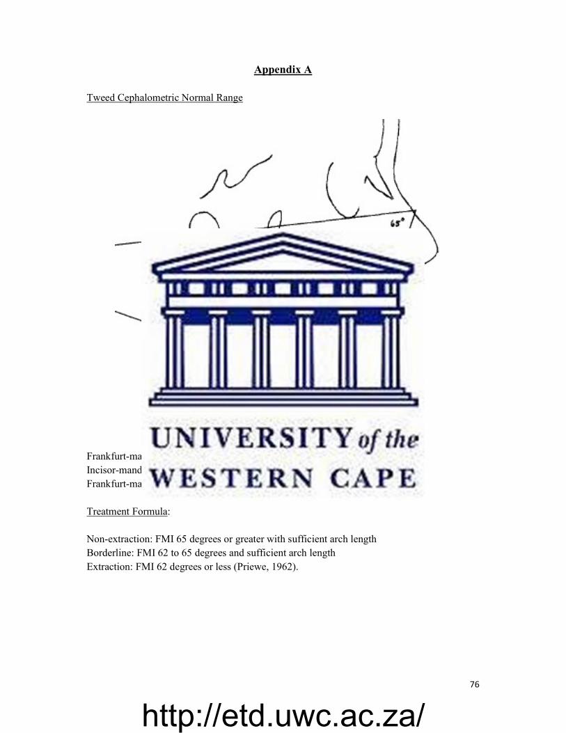
76
Appendix A Tweed Cephalometric Normal Range
Frankfurt-mandibular plane angle (FMP) 25 degrees Incisor-mandibular plane angle (IMP) 90 degrees Frankfurt-mandibular incisor angle (FMI)65 degrees. Treatment Formula: Non-extraction: FMI 65 degrees or greater with sufficient arch length Borderline: FMI 62 to 65 degrees and sufficient arch length Extraction: FMI 62 degrees or less (Priewe, 1962).
http://etd.uwc.ac.za/

77
Appendix B Steiner Cephalometric Normal Range for Lower Incisor
Linear measurement of 4mm from the lower incisal tip to NB line Angular measurement of 25 degrees between NB line and the long axis of the lower incisor Treatment Formula: Non-extraction: 4mm and 25 degrees / Less than 4mm and less than 25 degrees Extraction: Greater than 4mm and greater than 25 degrees (Priewe, 1962).
25°
4mm
http://etd.uwc.ac.za/

78
Appendix C Holdaway Cephalometric Normal Ratio
Ideally a 1:1 ratio is desired Treatment Formula: Non-extraction: 1:1 ratio Borderline: Ratio of 3mm difference Extraction: Ratio of 4mm difference (Priewe, 1962).
Po
http://etd.uwc.ac.za/

79
Appendix D Alvarez A-line
Ideal maxillary incisor position relative to A-line, is when the facial surface of the tooth touches the A-line (Alvarez, 2001).
http://etd.uwc.ac.za/