The neuropathology of CAG repeat diseases: review and update of genetic and molecular features
DIAGNOSTIC PATHOLOGY
Vet Pathol 45:626–633 (2008)
Neuropathology of Italian Cats in Feline SpongiformEncephalopathy Surveillance
B. IULINI, C. CANTILE, M. T. MANDARA, C. MAURELLA, G. R. LORIA, M. CASTAGNARO, C. SALVADORI,C. PORCARIO, C. CORONA, A. Z. PERAZZINI, A. MARONI, M. CARAMELLI, AND C. CASALONE
Centro Encefalopatie Animali, Istituto Zooprofilattico Sperimentale del Piemonte, Liguria e Valled’Aosta, Torino (BI, CM, CP, CCo, AZP, MCar, CCas); Department of Animal Pathology, Universityof Pisa, Pisa (CCan, CS); Department of Biopathological Veterinary Science, School of VeterinaryMedicine, University of Perugia, Perugia (MTM); Istituto Zooprofilattico Sperimentale della Sicilia,Palermo (GRL); Department of Public Health, Comparative Pathology and Veterinary Hygiene,
University of Padova, Padova (MCas); Italian Ministry of Health, Rome, Italy (AM)
Abstract. Feline spongiform encephalopathy (FSE) is a transmissible spongiform encephalopathyassociated with the consumption of feedstuffs contaminated with tissue from bovine spongiformencephalopathy-affected cattle and characterized by the accumulation in the central nervous system ofan abnormal isoform of the prion protein (PrPsc). Clinically, it presents as a progressive fatal neurologicsyndrome that is not easily distinguished from other feline neurologic conditions. Most cases of FSEhave been reported in England, where it was first detected in 1990, but a few cases have been reportedfrom other European countries. To identify possible cases of FSE in Italy, the Italian Ministry of Healthfunded a 2-year surveillance project during which the brains from 110 domestic cats with neurologicsigns were evaluated histologically for spongiform encephalopathy and immunohistochemically to detectPrPsc. Although no cases of FSE were found, the study proved useful in monitoring the Italian catpopulation for other neurologic diseases: neoplasia (21.8%), toxic-metabolic encephalopathy (18.2%),granulomatous encephalitis (15.5%), suppurative encephalitis (4.6%), trauma (3.6%), circulatorydisorders (3.6%), degeneration (2.7%), nonsuppurative encephalitis (2.7%), and neuromuscular diseases(1.8%). No histologic lesions were found in 20% of the brains, and samples from 5.5% of the cats wererejected as unsuitable.
Key words: Brain; cats; epidemiology; histology; prion diseases; prions.
Introduction
Feline spongiform encephalopathy (FSE) is atransmissible spongiform encephalopathy (TSE)caused by conversion of the cellular prion protein(PrPc) into an insoluble protease-resistant isoform(PrPsc).37 This fatal neurodegenerative disease isassociated with the consumption of bovine spongi-form encephalopathy (BSE)-contaminated food.Experimental data support a link between FSEand BSE, as mice inoculated with brain homoge-nates of FSE-affected cats had the incubation periodand lesion profile characteristics of the BSE strain.9
FSE was first identified in the UK in April 1990during the epizootic of BSE. Ninety cases havebeen identified in Great Britain,11 1 in NorthernIreland (Federal Register, 1997), 1 in Norway,1 1 inLiechtenstein (MAFF [2001] UK statistics on
incidence of BSE and related diseases http://www.defra.gov.uk/animalh/bse/othertses/index.html), 1 inPortugal (Silva JF, Correia JJ, Ribeiro J, Carmo S,Orge L: Feline spongiform encephalopathy: firstconfirmed case reported in Portugal, PRION 2006Abstract Book, Poster Session-Diagnosis [DIA-45]:392, 2006), and 2 in Switzerland (BVET-OVF-UFV, http://www.bvet.admin.ch/index.html?lang5it).5 Domestic cats with FSE are generally 4 to 9years of age (mean, 6.6 years).38 FSE has also beenreported in other felids,24 including the cheetah(Acinonyx jubatus),19 ocelot (Felis pardalis), puma(Felis concolor),36 lion (Felis leo), and tiger (Pantheratigris).16 The clinical signs and pathologic features ofthe disease are similar in all susceptible species ofFelidae.13,32
Clinically, FSE is characterized by progressiveneurologic signs such as locomotor dysfunction,
626
by guest on June 29, 2016vet.sagepub.comDownloaded from
abnormal behavior, and altered response to senso-rial stimulation. These clinical signs are not easilydistinguished from those of other feline neurologicsyndromes.13,32,37
FSE, like other TSEs, can be diagnosed onlypostmortem by histologic, immunohistochemical,or Western blot examination of brain tissue toidentify the typical spongiosis and PrPsc deposi-tion.23 Spongiosis is widespread in the brain andmost marked in the medial geniculate nucleus andthe basal nuclei.24,37 The degree of vacuolation inthe neuropil can be marked, progressing to statusspongiosis in some cases. Vacuolation is alsopresent in neuronal perikarya, particularly in theraphe nucleus, the dorsal motor nucleus of thevagus nerve, and the red nucleus. There isvacuolation in the white matter mainly in themedulla.37 Spongiosis is always accompanied byglial responses. PrPsc is typically detectablethroughout the brain of cats with FSE as smallaggregates, with punctate, particulate, and linearstrands of immunoreactivity.24
In 1998, Zanusso et al. reported the simultaneousoccurrence of spongiform encephalopathy in a 60-year-old Italian man and his cat. The prion strainin the brain of the cat was similar to that in theman, but distinct from the UK strain of BSE (newvariant Creutzfeldt-Jakob disease and its felinecounterpart).39 Because no BSE-related cases ofFSE had been detected in Italy, the Italian Ministryof Health funded a surveillance project to screenadult cats with untreatable neurologic disease forFSE.
Materials and Methods
In this multicenter study, the reference center forTransmissible Spongiform Encephalopathy was theCentro Encefalopatie Animali (CEA) – Istituto Zoo-profilattico Sperimentale (IZS) del Piemonte, Liguria eValle d’Aosta, which collaborated with the Universitiesof Pisa, Perugia, and Padua, and the IZS of Sicily andSardinia. The Italian Association of Veterinary Neurol-ogy (SINVet) kindly offered technical scientific support.Veterinary practitioners were introduced to the projectbefore its commencement at conferences and congresses,and their voluntary participation was requested. Duringthe 2-year project, 110 cats, older than 1 year with severeneurologic signs and poor prognosis, were submittedafter natural death or euthanasia by veterinary practi-tioners to the CEA and to the participating studycenters.
Neurologic signs were assessed using a standardclinical examination protocol. Details on age, sex,breed, history, clinical signs, serum biochemistry,ancillary investigations, and differential diagnosis wererecorded on a standardized form. At postmortem
examination, brains and major organs were sampled,fixed in 10% buffered formalin, and routinely processedfor histopathologic examination. Sections of brain fromrepresentative areas (medulla oblongata, pons, cerebel-lum, mesencephalon, diencephalon, and telencephalon),and from different spinal cord segments were stainedwith HE, periodic acid–Schiff (PAS), and Luxol fastblue. When necessary, immunohistochemistry wasperformed on selected central nervous system sectionsto establish an etiologic diagnosis. Feline coronavirusantigen was detected by immunohistochemistry withmonoclonal antibody FCV3-70 (Custom MonoclonalsInternational, West Sacramento, CA). In case ofneuromuscular disease, peripheral nerve samples werefixed in 2.5% glutaraldehyde and processed for electronmicroscopy. Ultrathin sections were stained with uranylacetate and lead citrate and examined with electronmicroscope. In 1 case, 15-mm-thick paraffin sections ofcerebellum were used for DNA extraction with DNeasyTissue Kit (QIAGEN, GmhH, Hilden, Germany).25 Apolymerase chain reaction (PCR) test was performed toamplify the entire open reading frame 2 of canine andfeline parvovirus (CPV-2/FPV), using the set of primersP33/34 (V. Martella, unpublished results). In some cases,the diagnostic workup included computed tomographyor magnetic resonance imaging. In suspected casesserologic tests for feline leukemia virus (FeLV) andfeline immunodeficiency virus (FIV) were carried out.
After morphologic or etiologic diagnosis, the studycenters sent the medulla oblongata (obex) samples andany other brain regions with vacuolar change to theCEA laboratories for histologic and immunohistochem-ical detection of prion disease. For immunohistochem-ical detection of PrPsc accumulation, deparaffinized andrehydrated sections were treated with 98% formic acidfor 20 minutes at room temperature, then autoclaved inbuffered citrate (pH 6.1) at 121uC for 30 minutes. Afterrinsing, the sections were incubated overnight at 4uCwith anti-PrP monoclonal antibody L42 (R-biopharmRIDA mbL42, Italia s.r.l. Cerro al Lambro, Milan,Italy) diluted 1 : 250. Subsequent antibody detection wascarried out using biotinylated goat anti-mouse second-ary antibody diluted 1 : 200 for 20 minutes (VectorLaboratories, Burlingame, CA) at room temperature,followed by the avidin–biotin–peroxidase complex(Vectastain ABC kit, Vector Laboratories). Immunore-activity was visualized using 3,39-diaminobenzidine aschromogen; the sections were counterstained withMayer’s hematoxylin counterstain. Paraffin sections ofmedulla oblongata of a cat with FSE (kind gift of A.Oevermann, University of Berne, Switzerland) wereused as the positive control.
Results
The most commonly recorded clinical signs wereataxia, altered grooming, hypersalivation, tremors,hyperesthesia and incoordination. The age of 93/110 cats was recorded. Mean age was 7.5 years; 67cats were 1 to 10 years of age; the remaining 26
Vet Pathol 45:5, 2008 Neuropathology of Italian Cats in Feline Spongiform Encephalopathy Surveillance 627
by guest on June 29, 2016vet.sagepub.comDownloaded from
were 10 to 20 years of age. Breed was recorded for94 cats. Most cats were of mixed breed (n 5 81); 9were Persian; 2 were Siamese; 1 was Soriano; 1 wasCertosino. Gender was recorded in all but 7 cats: 32intact females, 14 spayed females, 45 intact males,and 12 castrated males.No histologic lesions of FSE were found in any
cat; spongiform changes were present at the level ofthe obex in only 2 cases, which were diagnosedrespectively as hepatic and renal encephalopathy.All immunohistochemical examinations for PrPsc
were negative. Nevertheless, histologic lesions werepresent in 74.5% of the brain samples (Fig. 1). Thelesions were ascribed to a spectrum of neurologicdiseases that were classified in descending order offrequency as: neoplasia (21.8%), toxic-metabolic
encephalopathy (18.2%), granulomatous encepha-litis (15.5%), suppurative encephalitis (4.6%), trau-matic lesions (3.6%), circulatory disorders (3.6%),degenerative lesions (2.7%), nonsuppurative en-cephalitis (2.7%), and neuromuscular disease(1.8%). No histologic lesions were detected in20% of the samples; 5.5% were rejected asunsuitable.
Neoplasia
The 24 cases of neoplasia included 11 metastatictumors, 8 lymphomas, 4 meningiomas, and 1pituitary adenoma. Of the 11 metastatic tumors, 3were pulmonary carcinomas, 2 ceruminous carci-nomas, 2 mammary carcinoma, 1 lingual squamouscell carcinoma, 1 mast cell tumor, 1 cutaneous
Fig. 1. Diagram. Graphic representation of the diagnoses from examination of brain samples of 110 cats withneurologic signs.
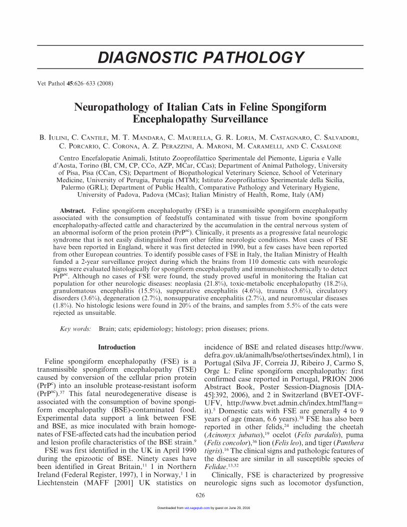
Fig. 2. Brain; cat. Neoplastic lymphocytes have perivascular orientation with neuroparenchymal infiltration.HE. Bar 5 150 mm.
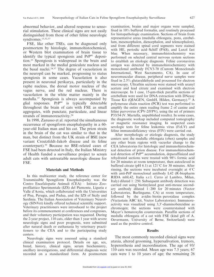
Fig. 3. Brain; cat. Transitional meningioma has whorls of neoplastic meningothelial cells separated by streamsof spindloid cells. HE. Bar 5 150 mm.
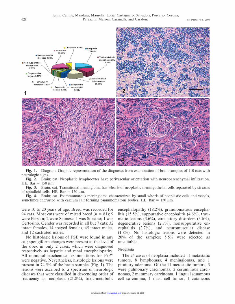
Fig. 4. Brain; cat. Psammomatous meningioma characterized by small whorls of neoplastic cells and vessels,sometimes encrusted with calcium salt forming psammomatous bodies. HE. Bar 5 150 mm.
628
Iulini, Cantile, Mandara, Maurella, Loria, Castagnaro, Salvadori, Porcario, Corona,Perazzini, Maroni, Caramelli, and Casalone Vet Pathol 45:5, 2008
by guest on June 29, 2016vet.sagepub.comDownloaded from
squamous cell carcinoma, and 1 renal lymphoma.One lymphoma formed a discrete mass that waslimited to the brain and composed of angiocentricneoplastic cells mainly in white matter. Tumor cellswere large with a round nucleus and a prominentcentral nucleolus. The other 7 lymphomas hadsimilar cytologic features and formed part ofmulticentric lymphoma (Fig. 2). All 8 cats withlymphoma tested negative for FIV antibodies andFeLV antigens.Two meningiomas were classified as transitional;
2 were psammomatous. Transitional meningiomas(Fig. 3) had features of both meningiotheliomatousand fibroblastic meningioma. In psammomatousmeningiomas (Fig. 4), neoplastic cells were ar-
ranged in whorls around small vessels or hyalinizedstructures with degenerated and mineralized wall(psammoma bodies).
The pituitary tumor consisted of sheets andtrabeculae of basophilic cells with a moderateamount of cytoplasm and large round oval nucleiwith a prominent nucleolus.
Toxic-metabolic disorders
The heterogeneous lesions in this categorywere ascribed to hepatic or renal encephalopathy(n 5 15) (Fig. 5), thiamine (vitamin B1) deficiency(n 5 1) (Fig. 6) and hippocampal necrosis (n 5 4)(Fig. 7). Hepatic encephalopathy was diagnosed onthe basis of degenerative changes in the brain
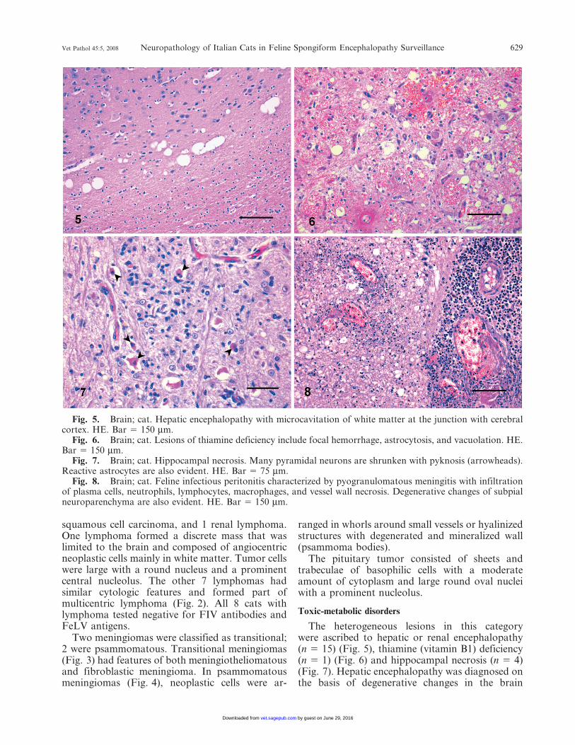
Fig. 5. Brain; cat. Hepatic encephalopathy with microcavitation of white matter at the junction with cerebralcortex. HE. Bar 5 150 mm.
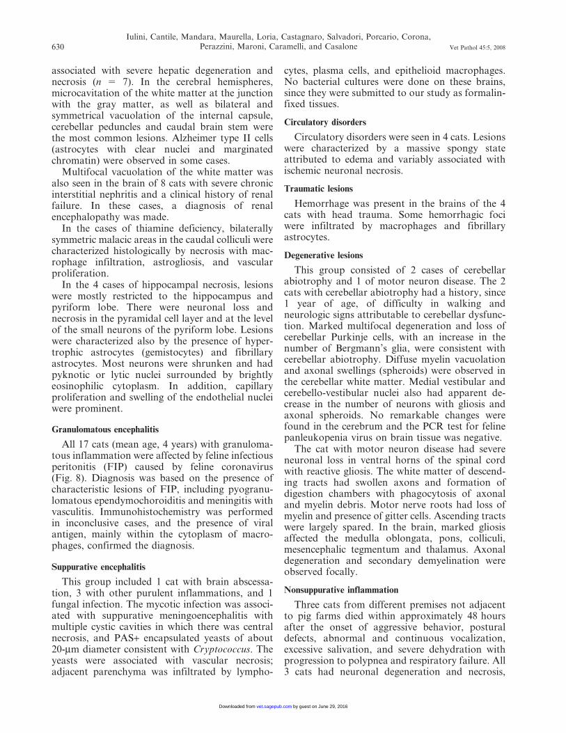
Fig. 6. Brain; cat. Lesions of thiamine deficiency include focal hemorrhage, astrocytosis, and vacuolation. HE.Bar 5 150 mm.
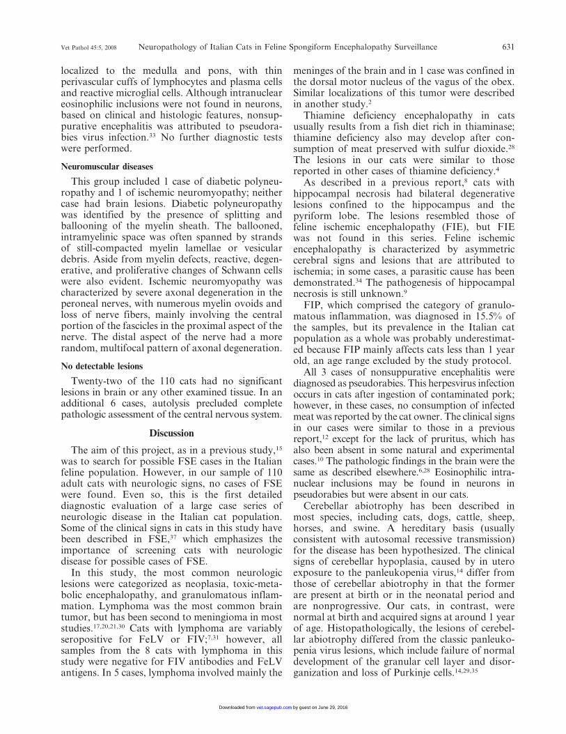
Fig. 7. Brain; cat. Hippocampal necrosis. Many pyramidal neurons are shrunken with pyknosis (arrowheads).Reactive astrocytes are also evident. HE. Bar 5 75 mm.
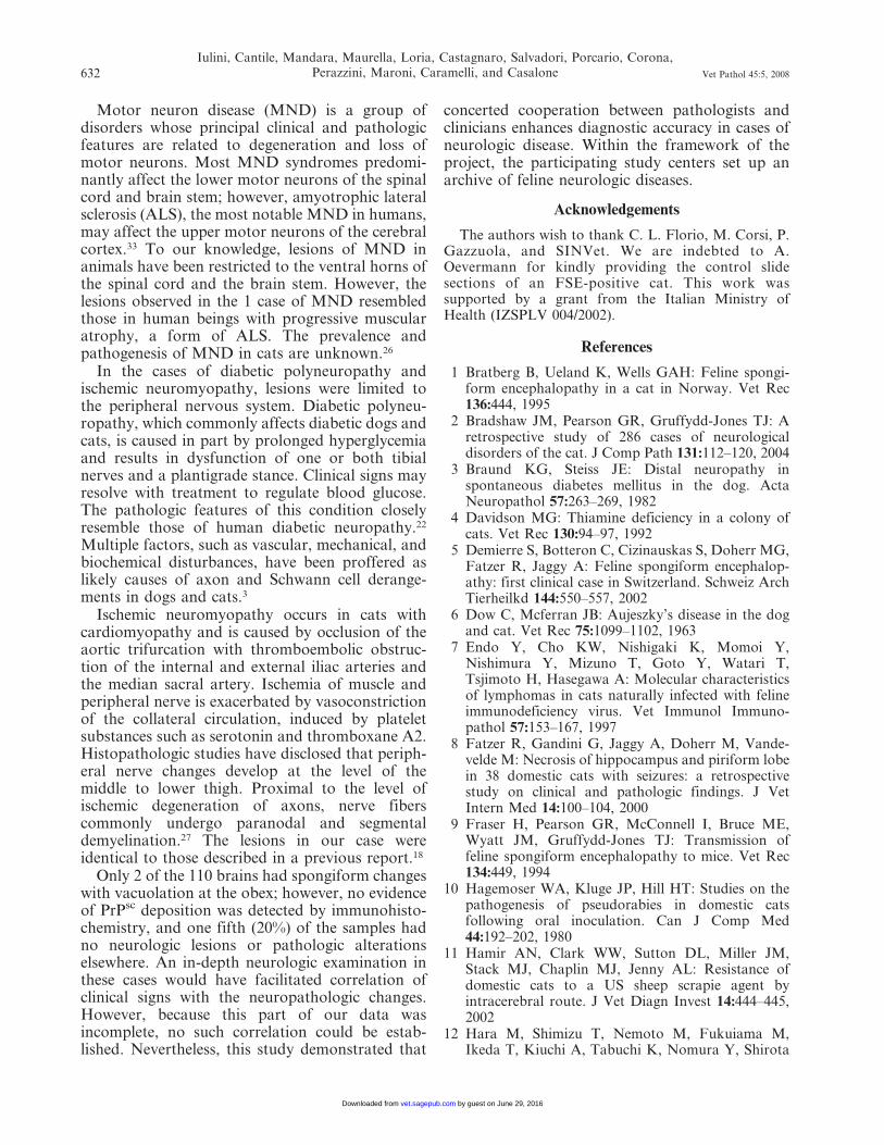
Fig. 8. Brain; cat. Feline infectious peritonitis characterized by pyogranulomatous meningitis with infiltrationof plasma cells, neutrophils, lymphocytes, macrophages, and vessel wall necrosis. Degenerative changes of subpialneuroparenchyma are also evident. HE. Bar 5 150 mm.
Vet Pathol 45:5, 2008 Neuropathology of Italian Cats in Feline Spongiform Encephalopathy Surveillance 629
by guest on June 29, 2016vet.sagepub.comDownloaded from
associated with severe hepatic degeneration andnecrosis (n 5 7). In the cerebral hemispheres,microcavitation of the white matter at the junctionwith the gray matter, as well as bilateral andsymmetrical vacuolation of the internal capsule,cerebellar peduncles and caudal brain stem werethe most common lesions. Alzheimer type II cells(astrocytes with clear nuclei and marginatedchromatin) were observed in some cases.Multifocal vacuolation of the white matter was
also seen in the brain of 8 cats with severe chronicinterstitial nephritis and a clinical history of renalfailure. In these cases, a diagnosis of renalencephalopathy was made.In the cases of thiamine deficiency, bilaterally
symmetric malacic areas in the caudal colliculi werecharacterized histologically by necrosis with mac-rophage infiltration, astrogliosis, and vascularproliferation.In the 4 cases of hippocampal necrosis, lesions
were mostly restricted to the hippocampus andpyriform lobe. There were neuronal loss andnecrosis in the pyramidal cell layer and at the levelof the small neurons of the pyriform lobe. Lesionswere characterized also by the presence of hyper-trophic astrocytes (gemistocytes) and fibrillaryastrocytes. Most neurons were shrunken and hadpyknotic or lytic nuclei surrounded by brightlyeosinophilic cytoplasm. In addition, capillaryproliferation and swelling of the endothelial nucleiwere prominent.
Granulomatous encephalitis
All 17 cats (mean age, 4 years) with granuloma-tous inflammation were affected by feline infectiousperitonitis (FIP) caused by feline coronavirus(Fig. 8). Diagnosis was based on the presence ofcharacteristic lesions of FIP, including pyogranu-lomatous ependymochoroiditis and meningitis withvasculitis. Immunohistochemistry was performedin inconclusive cases, and the presence of viralantigen, mainly within the cytoplasm of macro-phages, confirmed the diagnosis.
Suppurative encephalitis
This group included 1 cat with brain abscessa-tion, 3 with other purulent inflammations, and 1fungal infection. The mycotic infection was associ-ated with suppurative meningoencephalitis withmultiple cystic cavities in which there was centralnecrosis, and PAS+ encapsulated yeasts of about20-mm diameter consistent with Cryptococcus. Theyeasts were associated with vascular necrosis;adjacent parenchyma was infiltrated by lympho-
cytes, plasma cells, and epithelioid macrophages.No bacterial cultures were done on these brains,since they were submitted to our study as formalin-fixed tissues.
Circulatory disorders
Circulatory disorders were seen in 4 cats. Lesionswere characterized by a massive spongy stateattributed to edema and variably associated withischemic neuronal necrosis.
Traumatic lesions
Hemorrhage was present in the brains of the 4cats with head trauma. Some hemorrhagic fociwere infiltrated by macrophages and fibrillaryastrocytes.
Degenerative lesions
This group consisted of 2 cases of cerebellarabiotrophy and 1 of motor neuron disease. The 2cats with cerebellar abiotrophy had a history, since1 year of age, of difficulty in walking andneurologic signs attributable to cerebellar dysfunc-tion. Marked multifocal degeneration and loss ofcerebellar Purkinje cells, with an increase in thenumber of Bergmann’s glia, were consistent withcerebellar abiotrophy. Diffuse myelin vacuolationand axonal swellings (spheroids) were observed inthe cerebellar white matter. Medial vestibular andcerebello-vestibular nuclei also had apparent de-crease in the number of neurons with gliosis andaxonal spheroids. No remarkable changes werefound in the cerebrum and the PCR test for felinepanleukopenia virus on brain tissue was negative.
The cat with motor neuron disease had severeneuronal loss in ventral horns of the spinal cordwith reactive gliosis. The white matter of descend-ing tracts had swollen axons and formation ofdigestion chambers with phagocytosis of axonaland myelin debris. Motor nerve roots had loss ofmyelin and presence of gitter cells. Ascending tractswere largely spared. In the brain, marked gliosisaffected the medulla oblongata, pons, colliculi,mesencephalic tegmentum and thalamus. Axonaldegeneration and secondary demyelination wereobserved focally.
Nonsuppurative inflammation
Three cats from different premises not adjacentto pig farms died within approximately 48 hoursafter the onset of aggressive behavior, posturaldefects, abnormal and continuous vocalization,excessive salivation, and severe dehydration withprogression to polypnea and respiratory failure. All3 cats had neuronal degeneration and necrosis,
630
Iulini, Cantile, Mandara, Maurella, Loria, Castagnaro, Salvadori, Porcario, Corona,Perazzini, Maroni, Caramelli, and Casalone Vet Pathol 45:5, 2008
by guest on June 29, 2016vet.sagepub.comDownloaded from
localized to the medulla and pons, with thinperivascular cuffs of lymphocytes and plasma cellsand reactive microglial cells. Although intranucleareosinophilic inclusions were not found in neurons,based on clinical and histologic features, nonsup-purative encephalitis was attributed to pseudora-bies virus infection.33 No further diagnostic testswere performed.
Neuromuscular diseases
This group included 1 case of diabetic polyneu-ropathy and 1 of ischemic neuromyopathy; neithercase had brain lesions. Diabetic polyneuropathywas identified by the presence of splitting andballooning of the myelin sheath. The ballooned,intramyelinic space was often spanned by strandsof still-compacted myelin lamellae or vesiculardebris. Aside from myelin defects, reactive, degen-erative, and proliferative changes of Schwann cellswere also evident. Ischemic neuromyopathy wascharacterized by severe axonal degeneration in theperoneal nerves, with numerous myelin ovoids andloss of nerve fibers, mainly involving the centralportion of the fascicles in the proximal aspect of thenerve. The distal aspect of the nerve had a morerandom, multifocal pattern of axonal degeneration.
No detectable lesions
Twenty-two of the 110 cats had no significantlesions in brain or any other examined tissue. In anadditional 6 cases, autolysis precluded completepathologic assessment of the central nervous system.
Discussion
The aim of this project, as in a previous study,15
was to search for possible FSE cases in the Italianfeline population. However, in our sample of 110adult cats with neurologic signs, no cases of FSEwere found. Even so, this is the first detaileddiagnostic evaluation of a large case series ofneurologic disease in the Italian cat population.Some of the clinical signs in cats in this study havebeen described in FSE,37 which emphasizes theimportance of screening cats with neurologicdisease for possible cases of FSE.In this study, the most common neurologic
lesions were categorized as neoplasia, toxic-meta-bolic encephalopathy, and granulomatous inflam-mation. Lymphoma was the most common braintumor, but has been second to meningioma in moststudies.17,20,21,30 Cats with lymphoma are variablyseropositive for FeLV or FIV;7,31 however, allsamples from the 8 cats with lymphoma in thisstudy were negative for FIV antibodies and FeLVantigens. In 5 cases, lymphoma involved mainly the
meninges of the brain and in 1 case was confined inthe dorsal motor nucleus of the vagus of the obex.Similar localizations of this tumor were describedin another study.2
Thiamine deficiency encephalopathy in catsusually results from a fish diet rich in thiaminase;thiamine deficiency also may develop after con-sumption of meat preserved with sulfur dioxide.28
The lesions in our cats were similar to thosereported in other cases of thiamine deficiency.4
As described in a previous report,8 cats withhippocampal necrosis had bilateral degenerativelesions confined to the hippocampus and thepyriform lobe. The lesions resembled those offeline ischemic encephalopathy (FIE), but FIEwas not found in this series. Feline ischemicencephalopathy is characterized by asymmetriccerebral signs and lesions that are attributed toischemia; in some cases, a parasitic cause has beendemonstrated.34 The pathogenesis of hippocampalnecrosis is still unknown.9
FIP, which comprised the category of granulo-matous inflammation, was diagnosed in 15.5% ofthe samples, but its prevalence in the Italian catpopulation as a whole was probably underestimat-ed because FIP mainly affects cats less than 1 yearold, an age range excluded by the study protocol.
All 3 cases of nonsuppurative encephalitis werediagnosed as pseudorabies. This herpesvirus infectionoccurs in cats after ingestion of contaminated pork;however, in these cases, no consumption of infectedmeat was reported by the cat owner. The clinical signsin our cases were similar to those in a previousreport,12 except for the lack of pruritus, which hasalso been absent in some natural and experimentalcases.10 The pathologic findings in the brain were thesame as described elsewhere.6,28 Eosinophilic intra-nuclear inclusions may be found in neurons inpseudorabies but were absent in our cats.
Cerebellar abiotrophy has been described inmost species, including cats, dogs, cattle, sheep,horses, and swine. A hereditary basis (usuallyconsistent with autosomal recessive transmission)for the disease has been hypothesized. The clinicalsigns of cerebellar hypoplasia, caused by in uteroexposure to the panleukopenia virus,14 differ fromthose of cerebellar abiotrophy in that the formerare present at birth or in the neonatal period andare nonprogressive. Our cats, in contrast, werenormal at birth and acquired signs at around 1 yearof age. Histopathologically, the lesions of cerebel-lar abiotrophy differed from the classic panleuko-penia virus lesions, which include failure of normaldevelopment of the granular cell layer and disor-ganization and loss of Purkinje cells.14,29,35
Vet Pathol 45:5, 2008 Neuropathology of Italian Cats in Feline Spongiform Encephalopathy Surveillance 631
by guest on June 29, 2016vet.sagepub.comDownloaded from
Motor neuron disease (MND) is a group ofdisorders whose principal clinical and pathologicfeatures are related to degeneration and loss ofmotor neurons. Most MND syndromes predomi-nantly affect the lower motor neurons of the spinalcord and brain stem; however, amyotrophic lateralsclerosis (ALS), the most notable MND in humans,may affect the upper motor neurons of the cerebralcortex.33 To our knowledge, lesions of MND inanimals have been restricted to the ventral horns ofthe spinal cord and the brain stem. However, thelesions observed in the 1 case of MND resembledthose in human beings with progressive muscularatrophy, a form of ALS. The prevalence andpathogenesis of MND in cats are unknown.26
In the cases of diabetic polyneuropathy andischemic neuromyopathy, lesions were limited tothe peripheral nervous system. Diabetic polyneu-ropathy, which commonly affects diabetic dogs andcats, is caused in part by prolonged hyperglycemiaand results in dysfunction of one or both tibialnerves and a plantigrade stance. Clinical signs mayresolve with treatment to regulate blood glucose.The pathologic features of this condition closelyresemble those of human diabetic neuropathy.22
Multiple factors, such as vascular, mechanical, andbiochemical disturbances, have been proffered aslikely causes of axon and Schwann cell derange-ments in dogs and cats.3
Ischemic neuromyopathy occurs in cats withcardiomyopathy and is caused by occlusion of theaortic trifurcation with thromboembolic obstruc-tion of the internal and external iliac arteries andthe median sacral artery. Ischemia of muscle andperipheral nerve is exacerbated by vasoconstrictionof the collateral circulation, induced by plateletsubstances such as serotonin and thromboxane A2.Histopathologic studies have disclosed that periph-eral nerve changes develop at the level of themiddle to lower thigh. Proximal to the level ofischemic degeneration of axons, nerve fiberscommonly undergo paranodal and segmentaldemyelination.27 The lesions in our case wereidentical to those described in a previous report.18
Only 2 of the 110 brains had spongiform changeswith vacuolation at the obex; however, no evidenceof PrPsc deposition was detected by immunohisto-chemistry, and one fifth (20%) of the samples hadno neurologic lesions or pathologic alterationselsewhere. An in-depth neurologic examination inthese cases would have facilitated correlation ofclinical signs with the neuropathologic changes.However, because this part of our data wasincomplete, no such correlation could be estab-lished. Nevertheless, this study demonstrated that
concerted cooperation between pathologists andclinicians enhances diagnostic accuracy in cases ofneurologic disease. Within the framework of theproject, the participating study centers set up anarchive of feline neurologic diseases.
Acknowledgements
The authors wish to thank C. L. Florio, M. Corsi, P.Gazzuola, and SINVet. We are indebted to A.Oevermann for kindly providing the control slidesections of an FSE-positive cat. This work wassupported by a grant from the Italian Ministry ofHealth (IZSPLV 004/2002).
References
1 Bratberg B, Ueland K, Wells GAH: Feline spongi-form encephalopathy in a cat in Norway. Vet Rec136:444, 1995
2 Bradshaw JM, Pearson GR, Gruffydd-Jones TJ: Aretrospective study of 286 cases of neurologicaldisorders of the cat. J Comp Path 131:112–120, 2004
3 Braund KG, Steiss JE: Distal neuropathy inspontaneous diabetes mellitus in the dog. ActaNeuropathol 57:263–269, 1982
4 Davidson MG: Thiamine deficiency in a colony ofcats. Vet Rec 130:94–97, 1992
5 Demierre S, Botteron C, Cizinauskas S, Doherr MG,Fatzer R, Jaggy A: Feline spongiform encephalop-athy: first clinical case in Switzerland. Schweiz ArchTierheilkd 144:550–557, 2002
6 Dow C, Mcferran JB: Aujeszky’s disease in the dogand cat. Vet Rec 75:1099–1102, 1963
7 Endo Y, Cho KW, Nishigaki K, Momoi Y,Nishimura Y, Mizuno T, Goto Y, Watari T,Tsjimoto H, Hasegawa A: Molecular characteristicsof lymphomas in cats naturally infected with felineimmunodeficiency virus. Vet Immunol Immuno-pathol 57:153–167, 1997
8 Fatzer R, Gandini G, Jaggy A, Doherr M, Vande-velde M: Necrosis of hippocampus and piriform lobein 38 domestic cats with seizures: a retrospectivestudy on clinical and pathologic findings. J VetIntern Med 14:100–104, 2000
9 Fraser H, Pearson GR, McConnell I, Bruce ME,Wyatt JM, Gruffydd-Jones TJ: Transmission offeline spongiform encephalopathy to mice. Vet Rec134:449, 1994
10 Hagemoser WA, Kluge JP, Hill HT: Studies on thepathogenesis of pseudorabies in domestic catsfollowing oral inoculation. Can J Comp Med44:192–202, 1980
11 Hamir AN, Clark WW, Sutton DL, Miller JM,Stack MJ, Chaplin MJ, Jenny AL: Resistance ofdomestic cats to a US sheep scrapie agent byintracerebral route. J Vet Diagn Invest 14:444–445,2002
12 Hara M, Shimizu T, Nemoto M, Fukuiama M,Ikeda T, Kiuchi A, Tabuchi K, Nomura Y, Shirota
632
Iulini, Cantile, Mandara, Maurella, Loria, Castagnaro, Salvadori, Porcario, Corona,Perazzini, Maroni, Caramelli, and Casalone Vet Pathol 45:5, 2008
by guest on June 29, 2016vet.sagepub.comDownloaded from
K, Une Y: A natural case of Aujeszky’s disease in acat in Japan. J Vet Med Sci 53:947–949, 1991
13 Hewicker-Trautwein M, Bradley R: Portrait oftransmissible feline spongiform encephalopathy. In:Prions in Humans and Animals, eds. Hornlimann B,Riesner D, and Kretzschmar H, pp. 271–274. Walterde Gruyter, Berlin, 2006
14 Johnson RH, Margolis G, Kilham L: Identity offeline ataxia virus with feline panleukopenia virus.Nature 214:175–177, 1967
15 Kelly DF, Wells GAH, Haritani M, Higgins RJ,Jeffrey M: Neuropathological findings in cats withclinically suspect but histologically unconfirmedfeline spongiform encephalopathy. Vet Rec156:472–477, 2005
16 Kirkwood JK, Cunningham AA: Epidemiologicalobservations on spongiform encephalopathies incaptive wild animals in the British Isles. Vet Rec135:296–303, 1994
17 Koestner A, Bilzer T, Fatzer R, Schulman FY,Summers BA, Van Winkle TJ: Histological Classi-fication of Tumors of the Nervous System ofDomestic Animals, vol. 5, pp. 27–30. Armed ForcesInstitute of Pathology American Registry of Pathol-ogy, Washington, DC, 1999
18 Langelier KM: Ischemic neuromyopathy associatedwith steel pellet BB shot aortic obstruction in a cat.Can Vet J 23:187–189, 1982
19 Lezmi S, Bencsik A, Monks E, Petit T, Baron T:First case of feline spongiform encephalopathy in acaptive cheetah born in France: PrPsc analysis invarious tissues revealed unexpected targeting ofKidney and adrenal gland. Histochem Cell Biol119:415–422, 2003
20 Marioni-Henry K, Vite CH, Newton AL, VanWinkle TJ: Prevalence of diseases of the spinal cordof cats. J Vet Intern Med 18:851–858, 2004
21 MeutenDJ: Tumors inDomestic Animals, 5th ed., pp.697–738. Iowa State University Press, Ames, IA, 2002
22 Mizisin AP, Nelson RW, Sturges BK, Vernau KM,Lecouteur RA, Williams DC, Burgers ML, SheltonGD: Comparable myelinated nerve pathology infeline and human diabetes mellitus. Acta Neuro-pathol 113:431–442, 2007
23 Pearson GR, Wyatt JM, Gruffydd-Jones TJ, Hope J,Chong A, Higgins RJ, Scott AC, Wells GA: Felinespongiform encephalopathy: fibril and PrP studies.Vet Rec 131:307–310, 1992
24 Ryder SJ, Wells GAH, Bradshaw JM, Pearson GR:Inconsistent detection of PrP in extraneural tissuesof cats with feline spongiform encephalopathy. VetRec 148:437–441, 2001
25 Schatzberg SJ, Haley NJ, Barr SC, Parrish C,Steingold S, Summers BA, de Lahunta A, KornegayJN, Sharp NJH: Polymerase chain reaction (PCR)amplification of parvovirus DNA from the brains of
dogs and cats with cerebellar hypoplasia. J VetIntern Med 17:538–544, 2003
26 Shelton GD, Hopkins AL, Ginn PE, de Lahunta A,Cummings JF, Berryman FC, Hansen L: Adult-onset motor neuron disease in three cats. J Am VetMed Assoc 212:1271–1275, 1998
27 Summers BA, Cummings JF, de Lahunta A:Veterinary Neuropathology, p. 99, Mosby-YearBook, St Louis, MO, 1995
28 Summers BA, Cummings JF, de Lahunta A:Veterinary Neuropathology, pp. 249, 280. Mosby-Year Book, St Louis, MO, 1995
29 Taniyama H, Takayanagi S, Izumisawa Y, KotaniT, Kaji Y, Okada H, Matsukawa K: Cerebellarcortical atrophy in a kitten. Vet Pathol 31:710–713,1994
30 Troxel MT, Vite CH, Massicotte C, McLear RC,Van Winkle TJ, Glass EN, Tiches D, Dayrell-HartB: Magnetic resonance imaging features of felineintracranial neoplasia: retrospective analysis of 46cats. J Vet Intern Med 18:176–189, 2004
31 Troxel MT, Vite CH, Van Winkle TJ, Newton AL,Tiches D, Dayrell-Hart B, Kapatkin AS, Shofer FS,Steinberg SA: Feline intracranial neoplasia: retro-spective review of 160 cases (1985–2001). J VetIntern Med 17:850–859, 2003
32 Wells GAH, Wilesmith JW: Bovine spongiformencephalopathy and related diseases. In: PrionBiology and Diseases, ed. Prusiner S, 2nd ed., pp.595–628. Cold Spring Harbor Laboratory Press,New York, NY, 2004
33 Williams DB, Windebank AJ: Motor neuron dis-ease. In: Peripheral Neuropathy, ed. Dyck PJ andThomas PK, pp. 1028–1050. WB Saunders Co,Philadelphia, PA, 1993
34 Williams KJ, Summers BA, DeLahunta A: Cerebro-spinal cuterebriasis in cats and its association withfeline ischemic encephalopathy. Vet Pathol35:330–343, 1998
35 Willoughby K, Kelly DF: Hereditary cerebellardegeneration in three full sibling kittens. Vet Rec151:295–298, 2002
36 Willoughby K, Kelly DF, Lyon DG, Wells GAH:Spongiform encephalopathy in a captive puma. VetRec 131:431–434, 1992
37 Wyatt JM, Pearson GR, Smerdon TN, Gruffydd-Jones TJ, Wells GAH, Wilesmith JW: Naturallyoccurring scrapie-like spongiform encephalopathy infive domestic cats. Vet Rec 129:233–236, 1991
38 Wyatt SJ, Pearson GR, Gruffydd-Jones TJ: Felinespongiform encephalopathy. Feline Pract 21:7–9,1993
39 Zanusso G, Nardelli E, Rosati A, Fabrizi G, FerrariS, Carteri A, De Simone F, Rizzuto N, Monaco S:Simultaneous occurrence of spongiform encephalop-athy in a man and his cat in Italy. Lancet352:1116–1117, 1998
Request reprints from Dr. C. Cristina, CEA – Istituto Zooprofilattico Sperimentale del Piemonte, Liguria e Valled’Aosta, Via Bologna 148, 10154 Torino (Italy). E-mail: [email protected].
Vet Pathol 45:5, 2008 Neuropathology of Italian Cats in Feline Spongiform Encephalopathy Surveillance 633
by guest on June 29, 2016vet.sagepub.comDownloaded from
Copyright © 2022 FDOKUMEN