Monocyte- and macrophage-mediated immune reactions against Eimeria bovis
14
This article appeared in a journal published by Elsevier. The attached copy is furnished to the author for internal non-commercial research and education use, including for instruction at the authors institution and sharing with colleagues. Other uses, including reproduction and distribution, or selling or licensing copies, or posting to personal, institutional or third party websites are prohibited. In most cases authors are permitted to post their version of the article (e.g. in Word or Tex form) to their personal website or institutional repository. Authors requiring further information regarding Elsevier’s archiving and manuscript policies are encouraged to visit: http://www.elsevier.com/copyright
-
Upload
independent -
Category
Documents
-
view
5 -
download
0
Transcript of Monocyte- and macrophage-mediated immune reactions against Eimeria bovis

This article appeared in a journal published by Elsevier. The attachedcopy is furnished to the author for internal non-commercial researchand education use, including for instruction at the authors institution
and sharing with colleagues.
Other uses, including reproduction and distribution, or selling orlicensing copies, or posting to personal, institutional or third party
websites are prohibited.
In most cases authors are permitted to post their version of thearticle (e.g. in Word or Tex form) to their personal website orinstitutional repository. Authors requiring further information
regarding Elsevier’s archiving and manuscript policies areencouraged to visit:
http://www.elsevier.com/copyright

Author's personal copy
Monocyte- and macrophage-mediated immune reactions againstEimeria bovis
Anja Taubert a,*, Jan Hillern Behrendt a, Anke Suhwold a, Horst Zahner a, Carlos Hermosilla a,b
a Institute of Parasitology, Justus Liebig University Giessen, Rudolf-Buchheim-Str. 2, 35392 Giessen, Germanyb Royal Veterinary College, Department of Pathology and Infectious Diseases, Herts AL9 7TA, United Kingdom
1. Introduction
Eimeriosis in cattle is an important enteric protozoanparasitosis causing economic losses and severe clinicaldisease in calves (Fitzgerald, 1980; Daugschies et al., 1998).So far, innate immune responses of ruminant hosts against
Eimeria spp. have hardly been investigated, although thesereactions may be crucial for the outcome of a primaryinfection with respect to the severity of the disease and aneffective induction of adaptive immunity. The first line ofdefence against invading pathogens is represented byprofessional phagocytes, such as macrophages, dendriticcells and polymorphonuclear neutrophils (PMNs). PMNhave recently been identified as effector cells against one ofthe most pathogenic Eimeria species in cattle, Eimeria bovis
(Behrendt et al., 2008). Hardly anything, however, is
Veterinary Parasitology 164 (2009) 141–153
A R T I C L E I N F O
Article history:
Received 17 February 2009
Received in revised form 28 May 2009
Accepted 2 June 2009
Keywords:
Eimeria
Innate immune reaction
Macrophage
Monocyte
Cytokines
Chemokines
Oxidative burst
Phagocytosis
A B S T R A C T
Innate immune reactions conducted by macrophages may affect the outcome of primary
infections and are crucial for the transition to adaptive immune responses. In bovine
coccidiosis little is known on early monocyte/macrophage-mediated responses. We
therefore investigated in vivo, in vitro and ex vivo reactions of monocytes and macrophages
against Eimeria bovis, one of the most pathogenic Eimeria species in cattle. Macrophages
significantly infiltrate the gut mucosa of E. bovis-infected calves, particularly after
challenge infection. Furthermore, peripheral monocytes of infected animals, as precursor
cells of macrophages, exhibited enhanced ex vivo phagocytic and oxidative burst activities.
Enhanced levels of both activities were found early after infection and towards the end of
first merogony. In vitro exposure of macrophages to sporozoites led to phagocytosis of the
pathogen, whilst monocytes failed to do so. Phagocytosis occurred independently of the
viability of the sporozoites, indicating that active invasion by the parasites was negligible.
Phagocytosis occurred in the absence of immune serum, but could clearly be enhanced by
addition of immune serum, suggesting macrophage-derived antibody-dependent
cytotoxicity. Furthermore, co-culture of macrophages with sporozoites and stimulation
with merozoite I antigen induced distinct levels of cytokine and chemokine gene
transcription. Thus, the transcription of genes encoding for IFN-g, IL-12, TNF-a, IL-6,
CXCL1, CXCL8, CXCL10 and COX-2 was upregulated after sporozoite encounter. In contrast,
soluble merozoite I antigen only induced the gene transcription of IL-6 and IL-12 and failed
to upregulate IFN-g and TNF-a gene transcripts. In monocytes, IFN-g and CXCL10 were
found upregulated, all other immunoregulatory molecules tested were not affected. In
summary, our results strongly suggest that macrophage-mediated, innate immune
reactions play an important role in the early immune response to E. bovis infections in
calves.
� 2009 Elsevier B.V. All rights reserved.
* Corresponding author. Tel.: +49 641 9938475; fax: +49 641 9938469.
E-mail address: [email protected] (A. Taubert).
Contents lists available at ScienceDirect
Veterinary Parasitology
journal homepage: www.elsev ier .com/ locate /vetpar
0304-4017/$ – see front matter � 2009 Elsevier B.V. All rights reserved.
doi:10.1016/j.vetpar.2009.06.003
Author's personal copy
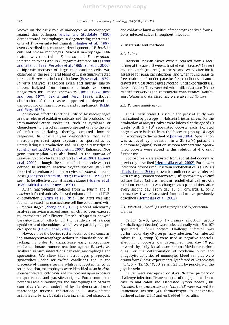
known on the early role of monocytes or macrophagesagainst this pathogen. Friend and Stockdale (1980)demonstrated macrophages in degenerating macromer-onts of E. bovis-infected animals. Hughes et al. (1987)even described macromeront development of E. bovis incultured bovine monocytes. Mucosal macrophage infil-tration was reported in E. tenella- and E. acervulina-infected chickens and in E. separata-infected rats (Troutand Lillehoi, 1993; Vervelde et al., 1996; Shi et al., 2000).A biphasic increase of large mononuclear cells wasobserved in the peripheral blood of E. nieschulzi-infectedrats and E. maxima-infected chickens (Rose et al., 1979).In vitro analyses suggested avian and murine macro-phages isolated from immune animals as potentphagocytes for Eimeria sporozoites (Rose, 1974; Roseand Lee, 1977; Bekhti and Pery, 1989), althoughelimination of the parasites appeared to depend onthe presence of immune serum and complement (Bekhtiand Pery, 1989).
Additional effector functions utilised by macrophagesare the release of oxidative radicals and the production ofimmunomodulatory molecules, such as cytokines orchemokines, to attract immunocompetent cells to the siteof infection initiating, thereby, acquired immuneresponses. In vitro analyses demonstrate that avianmacrophages react upon exposure to sporozoites byupregulating NO production and iNOS gene transcription(Lillehoj and Li, 2004; Dalloul et al., 2007). Enhanced iNOSgene transcription was also found in the mucosa ofEimeria-infected chickens and rats (Shi et al., 2001; Laurentet al., 2001), although, the source of this molecule was notdefined. In addition, reactive oxygen species (ROS) arereported as enhanced in leukocytes of Eimeria-infectedhosts (Ovington and Smith, 1992; Prowse et al., 1992) andseem to be effective against Eimeria stages (Hughes et al.,1989; Michalski and Prowse, 1991).
Avian macrophages isolated from E. tenella and E.
maxima-infected animals showed enhanced IL-1 and TNF-a production (Byrnes et al., 1993). The latter was alsofound increased in a macrophage cell line co-cultured withE. tenella stages (Zhang et al., 1995). Recent microarrayanalyses on avian macrophages, which had been exposedto sporozoites of different Eimeria subspecies showedparasite-induced effects on the synthesis of variouscytokines and chemokines, which were partially subspe-cies specific (Dalloul et al., 2007).
However, for the bovine system detailed data concern-ing monocyte/macrophage actions in eimeriosis are stilllacking. In order to characterise early macrophage-mediated, innate immune reactions against E. bovis, weanalysed in vitro interactions between macrophages andsporozoites. We show that macrophages phagocytisesporozoites under serum-free conditions and in thepresence of immune serum, whilst monocytes fail to doso. In addition, macrophages were identified as an in vitro-source of several cytokines and chemokines upon exposureto sporozoites and parasite-antigens. Furthermore, thepotential role of monocytes and macrophages in parasitecontrol in vivo was underlined by the demonstration ofmacrophage mucosal infiltration in E. bovis-infectedanimals and by ex vivo data showing enhanced phagocytic
and oxidative burst activities of monocytes derived from E.
bovis-infected calves throughout infection.
2. Materials and methods
2.1. Calves
Holstein Friesian calves were purchased from a localfarmer at the age of 2 weeks, treated with Baycox1 (Bayer)and Halocur1 (Intervet) in the second week after birth,assessed for parasitic infections, and when found parasitefree, maintained under parasite-free conditions in auto-claved stainless steel cages (Woetho) until experimental E.
bovis infection. They were fed with milk substitute (HemoMischfutterwerke) and commercial concentrates (Raiffei-sen). Water and sterilized hay were given ad libitum.
2.2. Parasite maintenance
The E. bovis strain H used in the present study wasmaintained by passages in Holstein Friesian calves. For theproduction of oocysts, calves were infected at the age of 10weeks with 5 � 104 sporulated oocysts each. Excretedoocysts were isolated from the faeces beginning 18 daysp.i. according to the method of Jackson (1964). Sporulationwas achieved by incubation in a 2% (w/v) potassiumdichromate (Sigma) solution at room temperature. Sporu-lated oocysts were stored in this solution at 4 8C untilfurther use.
Sporozoites were excysted from sporulated oocysts aspreviously described (Hermosilla et al., 2002). For in vitro
infections bovine umbilical vein endothelial cells (BUVEC)(Taubert et al., 2006), grown to confluence, were infectedwith freshly isolated sporozoites (106 sporozoites/75 cm2
culture flask). Culture medium (endothelial cell growthmedium, PromoCell) was changed 24 h p.i. and thereafterevery second day. From day 18 p.i. onwards, E. bovis
merozoites I were harvested from culture as previouslydescribed (Hermosilla et al., 2002).
2.3. Infections, bleedings and necropsies of experimental
animals
Calves (n = 3; group 1 = primary infection, group2 = challenge infection) were infected orally with 5 � 104
sporulated E. bovis oocysts. Challenge infection wasperformed on day 40 after primary infection. Non-infectedcalves (n = 3, group 3) were used as negative controls.Shedding of oocysts was determined from day 18 p.i.onwards by daily faecal examination (McMaster techni-que). For the determination of oxidative burst andphagocytic activities of monocytes blood samples weredrawn from E. bovis experimentally infected calves on days�1, 1, 5, 7, 13, 15, 18, 20, 22 and 25 p.i. by puncture of thejugular vein.
Calves were necropsied on days 26 after primary orchallenge infection. Tissue samples of the jejunum, ileum,caecum and colon and associated lymph nodes (Lnn.
jejunales, Lnn. ileocaecales and Lnn. colici) were excised forimmediate fixation (4% formaldehyde in phosphate-buffered saline, 24 h) and embedded in paraffin.
A. Taubert et al. / Veterinary Parasitology 164 (2009) 141–153142

Author's personal copy
2.4. Immunohistochemistry
5 mm cross-sections of formalin-fixed tissues weredeparaffinied according to standard histological procedures.Endogenous peroxidase was inactivated in 0.5% H2O2
(30 min, room temperature, Roth). Samples were washedfor 5 min in Tris-buffered saline (TBS) and treated withprotease (protease type 24, 5 min, 37 8C, Sigma). Proteaseactivity was stopped by dipping the slides in 4 8C TBS. Tissuesamples were then probed with monoclonal mouse anti-human monocyte/macrophage-specific antibodies (1:100,60 min, 37 8C, humidity chamber, MAC387, Serotec), whichcross-react with bovine cells (Gutierrez et al., 1999). Afterrinsing three times in TBS (5 min), samples were incubatedin sheep anti-mouse IgG conjugated with peroxidase (1:50,30 min, 37 8C, humidity chamber, NA 931, AmershamPharmacia Biotech Europe). After three washings in TBS(5 min), reactions were visualised by adding substrate(0.048 g DAB, Fluka, and 800 ml 3% H2O2 in 80 ml imidazolebuffer, 3–5 min). After rinsing three times in TBS (5 min) andonce in Aqua dest (5 min), the tissue samples werecounterstained for 15 s in Papanicolaou solution (1:10,Merck), washed in tap water (5 min), dehydrated accordingto standard procedures and mounted in Aquatex1 (Merck).
Immunostained macrophages present in gut mucosawere counted in 10 randomly chosen vision fields (200�magnification) per sample.
2.5. Detection of the ex vivo phagocytic and oxidative burst
activities of monocytes
Phagocytic and oxidative burst activities of monocyteswere determined by using Phagotest1and Phagoburst1 kits(ORPEGEN-Pharma), according to Moussay et al. (2006). Alltests were performed in duplicates. Four ml of heparinisedblood were mixed with 36 ml distilled water (40 s, shaking)to lyse erythrocytes, supplemented with 10� Hank’s buffer(Gibco) and pelleted (10 min, 400 � g). After washing (10 mlPBS/EDTA, 10 min, 400� g) cells were transferred toV-shaped microtitre plates (2� 105 cells/well, Nunc) andcentrifuged (4 8C, 200� g, 7 min).
For ex vivo quantification of phagocytic activity cellswere suspended in 100 ml ice-cold autologous plasma.After addition of 10 ml FITC-labelled Escherischia coli pre-opsonized with human serum (provided with the kit), cellswere incubated for 10 min at 37 8C (shaking water bath) oron ice (=negative control). After transferring the plates onice, the quenching of surface-bound bacteria, fixation andpermeabilisation of cells was performed according to themanufacturer’s instructions.
For ex vivo quantification of the inducible oxidativeburst activity, cells were suspended in 100 ml ice-cold PBS,supplemented with either 10 ml non-labelled E. coli,phorbol-12-myristate 13 acetate solution (PMA, 8.1 mM,ORPEGEN-Pharma; =positive control) or PBS (=negativecontrol) and incubated at 37 8C (shaking water bath). After10 min, 10 ml dihydrorhodamine 123 substrate solutionwere added and cells were incubated for further 20 min(37 8C, shaking water bath). After transferring the platesonto ice, cells were fixed and permeabilised according tothe manufacturer’s instructions.
In both assays PBS-EDTA were then added to the wells(4 8C, 5 min) to recover plastic-adherent cells. Cells werecounterstained with ‘‘DNA-staining solution’’ (providedwith the kits) and analysed by flow cytometry (FCM;FACScalibur, BD Biosciences).
2.6. Isolation and cultivation of bovine monocytes and
macrophages
For both, monocytes and macrophages, PBMC had to beisolated in advance. Therefore, 18 ml of blood, substitutedwith 2 ml 3.8% citric acid, were mixed with 17 ml of 0.9%NaCl and applied on the top of 12 ml Ficoll-paque(density = 1.077 g/l, Biochrom) in 50 ml centrifugationtubes (Nunc). After centrifugation (45 min, 400 � g) thelymphocyte/monocyte layer was collected and the cellswere washed three times (10 min, 400 � g, 4 8C) in RPMI.Using the trypan blue (Sigma) exclusion test, viable cellswere counted in a Neubauer haemocytometer chamber.
Bovine monocytes were isolated as described byGoddeeris et al. (1986). If not stated differently, we usedmonocytes of infected animals. In brief, 7.5 � 107 PBMCwere allowed to adhere (1 h, 37 8C) to 75 cm2 tissue plasticflasks (Greiner), previously coated with 2% sterile gelatinesolution (2 h, 37 8C, thereafter dried) and autologousplasma (1 h, 37 8C, thereafter washed twice with RPMI1640/1% penicillin/1% streptomycin, Sigma). Non-adher-ent PBMC were removed and monocytes were washedwith prewarmed RPMI 1640/1% penicillin/1% streptomy-cin. Monocytes were detached (5–10 min in 10 mM EDTAin Mg2+- and Ca2+-free Hank’s solution, room tempera-ture), washed (10 min, 400 � g, 4 8C) and resuspended in4 8C RPMI 1640/1% penicillin/1% streptomycin. The cellswere kept on ice until use and counted in a Neubauerhaemocytometer chamber.
Bovine macrophages were prepared according to Jungiet al. (1996). If not stated differently, we used macrophagesof infected animals. PBMC were sealed in Teflon bags(20 ml, 5 � 106 PBMC/ml) as described by Jungi et al.(1996) and cultured for 7–8 days at 37 8C in a humidifiedatmosphere of 5% CO2. The medium was Iscove’s modifiedDulbecco’s Medium (IMDM Glutamax1, Sigma) containing100 IU/ml penicillin, 100 mg/ml streptomycin, 1% (v/v)non-essential amino acids for minimal essential medium(MEM; Gibco), 0.4% (v/v) vitamin solution for MEM(Gibco), 1 mM sodium pyruvate (Gibco), 2.5 mM ampho-tericin B (Gibco), 50 mM 2-mercaptoethanol (Gibco) and20% foetal calf serum (FCS; Biowest). From the cell mixture,macrophages were purified by selective adherence tomicrotitre plate wells for 4 h.
2.7. Determination of monocyte or macrophage-mediated
elimination of E. bovis sporozoites in vitro
In a corresponding series of experiments we controlledthe elimination of the parasites from the incubationmedium in the presence of monocytes or macrophages.The respective cell types were incubated with freshlyisolated vital or heat-inactivated (60 8C, 30 min) E. bovis
sporozoites and either supplemented with plain mediumRPMI 1640 or immune serum (1:30). After 4 h incubation
A. Taubert et al. / Veterinary Parasitology 164 (2009) 141–153 143
Author's personal copy
the total number of remaining sporozoites was counted ina Neubauer chamber. Sporozoite viability was estimatedby trypan blue exclusion.
For the detection of intracellular sporozoites in macro-phages, freshly isolated E. bovis sporozoites were stainedwith the fluorescence dye 5(6)-carboxyfluorescein diacetatesuccinimidyl ester (CFSE, Invitrogen) according to Hermo-silla et al. (2008). Sporozoites (106/ml) were suspended inthe dye (2.5 mM CFSE in PBS) by gently shaking andincubated for 10 min (37 8C, 5% CO2 atmosphere). In order tostop the labelling process, an equal volume of PBS with 10%FCS was added and CFSE-labelled sporozoites (sporozoi-tesCFSE) were washed four times (400� g, 10 min) in PBS,resuspended in PBS/10% FCS and protected from light untiluse. SporozoitesCFSE were co-cultured with macrophages for4 h. The samples were analysed by flow cytometry (FCM)according to Hermosilla et al. (2008).
2.8. Sera and E. bovis merozoite I antigen (EbAg)
Immune serum was obtained from a calf after primary(4 � 104 E. bovis oocysts) and challenge (3 � 104 E. bovis
oocysts) infection.
E. bovis merozoites I, collected from culture as describedpreviously (Hermosilla et al., 2002), were homogenised byrepeated freezing and thawing followed by sonication(20 kHz, 5 s � 15 s pulses) on ice. After centrifugation(11,000 � g, 4 8C, 20 min) the supernatants were passedthrough 0.2 mm sterile filters (Renner). Protein concentra-tion was determined using the Bradford method (Bradford,1976). The antigen (EbAg) was stored at �80 8C untilfurther use.
2.9. In vitro stimulation of monocytes/macrophages and
isolation and reverse transcription of total RNA
For RT-PCR analyses, monocytes/macrophages wereeither exposed to freshly isolated E. bovis sporozoitesat a ratio of 1:1 or incubated in EbAg (10 mg/mlfinal concentration) for 4 h (37 8C, 5% CO2). Cellsincubated in plain medium served as negative control.Stimulation of macrophages with LPS (lipopolysacchar-ides, Escherischia coli, O111:B4, 1 mg/ml, Sigma)and bovine recombinant IFN-g (100 U/ml, kindlydonated by R. Steiger, Novartis Pharma) was used aspositive control.
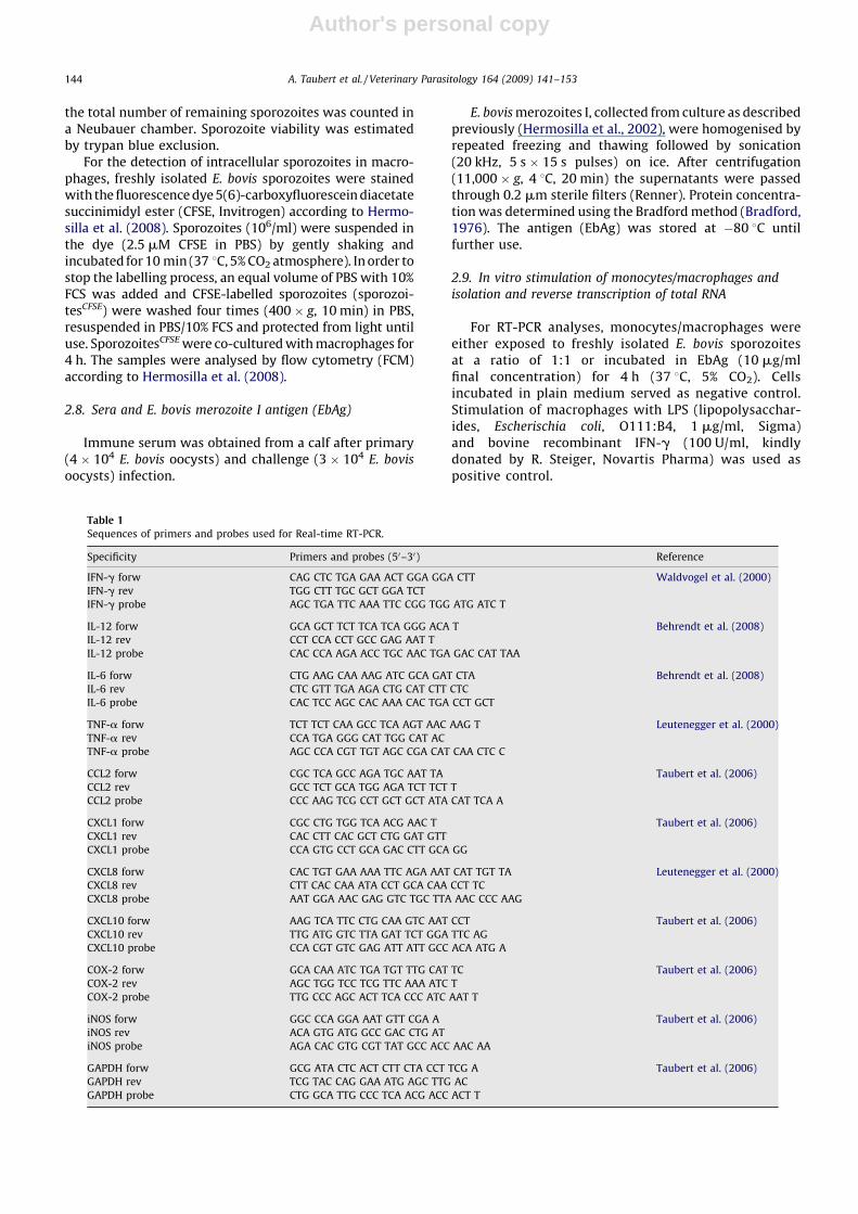
Table 1
Sequences of primers and probes used for Real-time RT-PCR.
Specificity Primers and probes (50–30) Reference
IFN-g forw CAG CTC TGA GAA ACT GGA GGA CTT Waldvogel et al. (2000)
IFN-g rev TGG CTT TGC GCT GGA TCT
IFN-g probe AGC TGA TTC AAA TTC CGG TGG ATG ATC T
IL-12 forw GCA GCT TCT TCA TCA GGG ACA T Behrendt et al. (2008)
IL-12 rev CCT CCA CCT GCC GAG AAT T
IL-12 probe CAC CCA AGA ACC TGC AAC TGA GAC CAT TAA
IL-6 forw CTG AAG CAA AAG ATC GCA GAT CTA Behrendt et al. (2008)
IL-6 rev CTC GTT TGA AGA CTG CAT CTT CTC
IL-6 probe CAC TCC AGC CAC AAA CAC TGA CCT GCT
TNF-a forw TCT TCT CAA GCC TCA AGT AAC AAG T Leutenegger et al. (2000)
TNF-a rev CCA TGA GGG CAT TGG CAT AC
TNF-a probe AGC CCA CGT TGT AGC CGA CAT CAA CTC C
CCL2 forw CGC TCA GCC AGA TGC AAT TA Taubert et al. (2006)
CCL2 rev GCC TCT GCA TGG AGA TCT TCT T
CCL2 probe CCC AAG TCG CCT GCT GCT ATA CAT TCA A
CXCL1 forw CGC CTG TGG TCA ACG AAC T Taubert et al. (2006)
CXCL1 rev CAC CTT CAC GCT CTG GAT GTT
CXCL1 probe CCA GTG CCT GCA GAC CTT GCA GG
CXCL8 forw CAC TGT GAA AAA TTC AGA AAT CAT TGT TA Leutenegger et al. (2000)
CXCL8 rev CTT CAC CAA ATA CCT GCA CAA CCT TC
CXCL8 probe AAT GGA AAC GAG GTC TGC TTA AAC CCC AAG
CXCL10 forw AAG TCA TTC CTG CAA GTC AAT CCT Taubert et al. (2006)
CXCL10 rev TTG ATG GTC TTA GAT TCT GGA TTC AG
CXCL10 probe CCA CGT GTC GAG ATT ATT GCC ACA ATG A
COX-2 forw GCA CAA ATC TGA TGT TTG CAT TC Taubert et al. (2006)
COX-2 rev AGC TGG TCC TCG TTC AAA ATC T
COX-2 probe TTG CCC AGC ACT TCA CCC ATC AAT T
iNOS forw GGC CCA GGA AAT GTT CGA A Taubert et al. (2006)
iNOS rev ACA GTG ATG GCC GAC CTG AT
iNOS probe AGA CAC GTG CGT TAT GCC ACC AAC AA
GAPDH forw GCG ATA CTC ACT CTT CTA CCT TCG A Taubert et al. (2006)
GAPDH rev TCG TAC CAG GAA ATG AGC TTG AC
GAPDH probe CTG GCA TTG CCC TCA ACG ACC ACT T
A. Taubert et al. / Veterinary Parasitology 164 (2009) 141–153144

Author's personal copy
Total RNA was isolated from monocytes and macro-phages using the RNeasy1Mini Kit (Qiagen) according to themanufacturer’s instructions. To minimize contaminationwith genomic DNA and to achieve reliable photometricmeasurements of the RNA, an on-column-DNase I treatment(Qiagen) was applied during total RNA isolation followingthe manufacturer’s instructions. RNA was controlled forintegrity by electrophoresis on a 1% agarose gel. Since on-column-DNase I treatment was not absolutely efficient, theRNA (1 mg) was additionally treated with 1 U RNase-freeDNase I (30 min, 37 8C; Roche). DNase I was inactivatedafterwards by heating (65 8C, 6 min). Total RNA probes werestored at �80 8C until use. For cDNA synthesis M-MLV-reverse-transcriptase (Gibco) was used. DNase I-treatedtotal RNA was mixed with 5 ml 5� RT-buffer (250 mM Tris–HCl, pH 8.3, 375 mM KCl, 15 mM MgCl2), 2 ml DTT (0.1 M),2 ml hexanucleotides (62.5 A260/ml; all Hoffmann La Roche),1 ml dNTPs (10 mM, MBI Fermentas) and 1 ml M-MLV-reverse transcriptase (200 U/ml). The reaction was carriedout in a final volume of 25 ml at 37 8C for 60 min. Afteraddition of 185 ml TE buffer (10 mM Tris–HCl, pH 8, 1 mMEDTA) the sample was stored at �208 C until further use.
2.10. Real-time PCR for the relative quantification of IFN-g,
IL-12, IL-6, TNF-a, CXCL1, CXCL8, CXCL10, CCL2, COX-2, iNOS
and GAPDH cDNAs
The relative quantification of IFN-g, IL-12, IL-6, TNF-a,CXCL1, CXCL8, CXCL10, CCL2, COX-2, iNOS and glycer-aldehyde 3-phosphate dehydrogenase (GAPDH) genetranscripts was done by real-time PCR applying TaqMan1
probes. The sequences of primers (MWG Biotech) andprobes (Eurogentec) are depicted in Table 1. Probeswere labelled at the 50-end with the reporter dye FAM(6-carboxyfluorescein) and at the 30-end with the
quencher dye TAMRA (6-carboxytetramethylrhodamine).PCR amplification was performed employing an automatedfluorometer (ABI PRISMTM 5700 Sequence DetectionSystem, Applied Biosystems) using 96-well optical plates.Samples were analysed in duplicate. For PCR 5 ml cDNA(corresponding to 25 ng total RNA) were used in a 25 ml PCRreaction mixture containing 12.5 ml TaqMan1 PCR MasterMix (Eurogentec), 300 nM of each primer and 200 nM probe.Amplification conditions were the same for all targetsassayed: one cycle at 50 8C for 2 min, one cycle at 95 8C for10 min, 45 cycles at 95 8C for 15 s and at 60 8C for 60 s.
Semi-quantitative analyses used the comparative CT
method (DDCT method) according to the instructions of theABI PRISMTM 5700 Sequence Detector manufacturer andreported as n-fold differences in comparison to the respec-tive medium control (after normalising the samples referr-ing to their corresponding housekeeping gene GAPDH).
2.11. Statistical analyses
Statistical analyses used the programme package BMDPfor XP, Release 8.1 (Dixon, 1993). For the description of the
Fig. 1. Macrophages in gut tissues of Eimeria bovis primary and challenge
infected calves. Tissue samples of the jejunum, ileum, caecum and colon
of non-infected (white bars), E. bovis primary (26 days p.i., grey bars) and
challenge (26 days after challenge, black bars) infected calves were fixed
in paraformaldehyde and embedded in paraffin. Cross-sections were
probed with anti-macrophage antibodies and macrophages present in the
tissue were counted. Arithmetic means of three calves each and standard
deviations.
Fig. 2. Oxidative burst and phagocytosis activity of monocytes isolated ex
vivo from Eimeria bovis infected calves. Monocytes were isolated from
infected calves (n = 3) before and on various days after infection. Oxidative
burst activity (a) was determined by flow cytometry estimating the
oxidation of dihydrorhodamine 123 fluorogen stimulated by Escherichia coli
phagocytosis. The general phagocytic activity (b) was estimated by flow
cytometry determining the uptake of FITC-labelled E. coli. Incubations were
performed at 37 8C (black symbols) or on ice (controls, grey symbols).
Arithmetic means of three monocyte donors, minimum, maximum.
A. Taubert et al. / Veterinary Parasitology 164 (2009) 141–153 145
Author's personal copy
data arithmetic means were calculated. To describe thevariability of the data standard deviations were used. Assome of the original data were not normally distributed(skewed to the right), if necessary, logarithmic transfor-mations were performed to obtain an approximate normaldistribution of the values. Data concerning infiltration ofmacrophages into parasitized tissue (Fig. 1), ex vivo
oxidative burst and phagocytosis activities of monocytes(Fig. 2) and macrophage-mediated phagocytosis of CFSE-stained sporozoites (Fig. 6) were analysed by two-factorialanalysis of variance (ANOVA) with repeated measures(BMDP2V). For data sets dealing with monocyte andmacrophage-mediated elimination of sporozoites (Figs. 3,5 and 6) statistical analyses were performed by applyingone-factorial ANOVA with repeated measures (BMDP2V)followed by the Student–Newman–Keuls test for pairwisetreatment comparison. For the gene transcription data sets(Figs. 7 and 8) as well as for the data concerning titrationaleffects of macrophage-mediated elimination of sporo-zoites (Fig. 4) co-culture/stimulation conditions werecompared by t-test for dependent samples (BMDP3D).Differences were regarded as significant at a level ofp � 0.05.
3. Results
3.1. Macrophages infiltrate E. bovis infected gut mucosa and
accumulate in lymph nodes
E. bovis-infected calves showed significantly moremacrophages in the gut wall than naıve animals (Fig. 1).A significant increase in macrophage numbers wasapparent in both primary and challenge infected animals(both p < 0.01). In challenged calves macrophagecounts significantly exceeded those of primary infectedanimals (p < 0.01). Macrophage infiltration occurredin all gut samples tested in comparable proportions(Fig. 1).
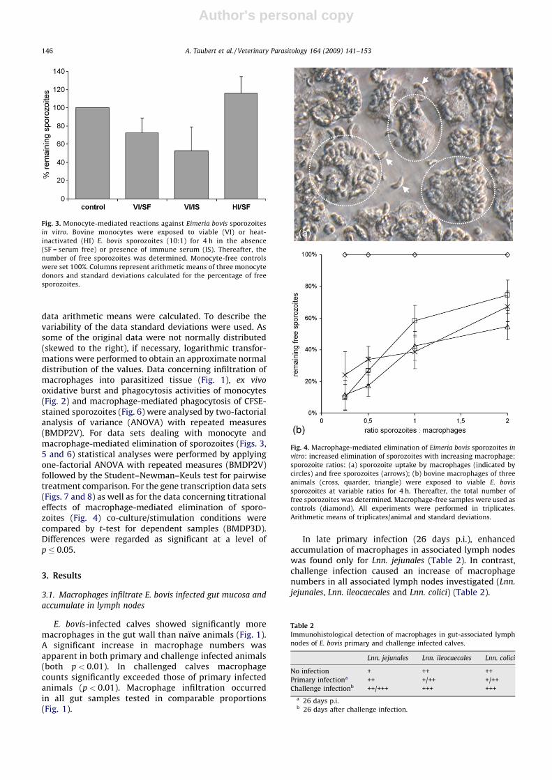
In late primary infection (26 days p.i.), enhancedaccumulation of macrophages in associated lymph nodeswas found only for Lnn. jejunales (Table 2). In contrast,challenge infection caused an increase of macrophagenumbers in all associated lymph nodes investigated (Lnn.
jejunales, Lnn. ileocaecales and Lnn. colici) (Table 2).
Fig. 3. Monocyte-mediated reactions against Eimeria bovis sporozoites
in vitro. Bovine monocytes were exposed to viable (VI) or heat-
inactivated (HI) E. bovis sporozoites (10:1) for 4 h in the absence
(SF = serum free) or presence of immune serum (IS). Thereafter, the
number of free sporozoites was determined. Monocyte-free controls
were set 100%. Columns represent arithmetic means of three monocyte
donors and standard deviations calculated for the percentage of free
sporozoites.
Fig. 4. Macrophage-mediated elimination of Eimeria bovis sporozoites in
vitro: increased elimination of sporozoites with increasing macrophage:
sporozoite ratios: (a) sporozoite uptake by macrophages (indicated by
circles) and free sporozoites (arrows); (b) bovine macrophages of three
animals (cross, quarder, triangle) were exposed to viable E. bovis
sporozoites at variable ratios for 4 h. Thereafter, the total number of
free sporozoites was determined. Macrophage-free samples were used as
controls (diamond). All experiments were performed in triplicates.
Arithmetic means of triplicates/animal and standard deviations.
Table 2
Immunohistological detection of macrophages in gut-associated lymph
nodes of E. bovis primary and challenge infected calves.
Lnn. jejunales Lnn. ileocaecales Lnn. colici
No infection + ++ ++
Primary infectiona ++ +/++ +/++
Challenge infectionb ++/+++ +++ +++
a 26 days p.i.b 26 days after challenge infection.
A. Taubert et al. / Veterinary Parasitology 164 (2009) 141–153146
Author's personal copy
3.2. Monocytes display enhanced oxidative burst and
phagocytic activities during E. bovis infection
Monocytes were analysed directly ex vivo from wholeblood samples, i.e., without performing any furtherisolation techniques or stimulation. Data generated onday �1 p.i., which reflect the situation of non-infectedanimals, revealed low phagocytic and oxidative burstactivities, whilst a biphasic upregulation of the generalphagocytic and oxidative burst activities of monocyteswas observed when compared to the negative controlincubated at 4 8C (Fig. 2). Oxidative burst activity wasenhanced already 1 day after infection, most probablyreflecting the phase of sporozoite transmigration throughthe epithelium of the ileum and invasion of lymphaticendothelial cells. A second peak was detected at days 13–18 p.i., corresponding to the phase of macromerontmaturation and the release of first generation merozoites.Highest values occurred on day 15 p.i. when, by means,more than 50% of monocytes showed increased oxidativeburst activity.
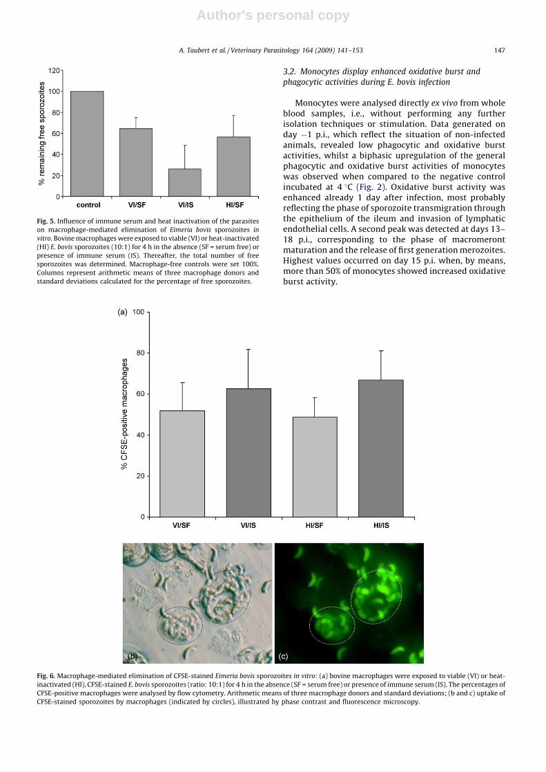
Fig. 5. Influence of immune serum and heat inactivation of the parasites
on macrophage-mediated elimination of Eimeria bovis sporozoites in
vitro. Bovine macrophages were exposed to viable (VI) or heat-inactivated
(HI) E. bovis sporozoites (10:1) for 4 h in the absence (SF = serum free) or
presence of immune serum (IS). Thereafter, the total number of free
sporozoites was determined. Macrophage-free controls were set 100%.
Columns represent arithmetic means of three macrophage donors and
standard deviations calculated for the percentage of free sporozoites.
Fig. 6. Macrophage-mediated elimination of CFSE-stained Eimeria bovis sporozoites in vitro: (a) bovine macrophages were exposed to viable (VI) or heat-
inactivated (HI), CFSE-stained E. bovis sporozoites (ratio: 10:1) for 4 h in the absence (SF = serum free) or presence of immune serum (IS). The percentages of
CFSE-positive macrophages were analysed by flow cytometry. Arithmetic means of three macrophage donors and standard deviations; (b and c) uptake of
CFSE-stained sporozoites by macrophages (indicated by circles), illustrated by phase contrast and fluorescence microscopy.
A. Taubert et al. / Veterinary Parasitology 164 (2009) 141–153 147

Author's personal copy
Fig. 7. Transcription of genes encoding for CXCL1, CXCL8, CXCL10, CCL2, IL-6 and iNOS in monocytes after exposure to Eimeria bovis sporozoites or
merozoite-antigen. Bovine monocytes were exposed to E. bovis sporozoites (ratio: 1:1), merozoite-antigen (Mero-Ag, 10 mg/ml) or medium alone for 4 h.
Total RNA was isolated, reversely transcribed into cDNA and probed with real-time RT-PCR systems for the detection of the respective mRNA equivalents.
Arithmetic means of three monocyte donors and standard deviations.
A. Taubert et al. / Veterinary Parasitology 164 (2009) 141–153148

Author's personal copy
Fig. 8. Transcription of genes encoding for IFN-g, IL-12, TNF-a, IL-6, CXCL1, CXCL8, CXCL10 and COX-2 in macrophages after exposure to Eimeria bovis
sporozoites or merozoite-antigen. Bovine macrophages were exposed to E. bovis sporozoites (ratio: 1:1), merozoite I antigen (Mero-Ag, 10 mg/ml) or
medium alone for 4 h. Stimulation with LPS (1 mg/ml) and bovine recombinant IFN-g (1000 U/ml) was used as positive control. Total RNA was isolated,
reversely transcribed into cDNA and probed with real-time RT-PCR systems for the detection of the respective mRNA equivalents. Arithmetic means of three
macrophage donors and standard deviations.
A. Taubert et al. / Veterinary Parasitology 164 (2009) 141–153 149

Author's personal copy
Similar time course dynamics were detected in the caseof phagocytic activity of monocytes, however, peaksoccurred delayed by 5 days p.i. and at days 18–27 p.i.However, the negative controls performed by 4 8C showedincreased values at days 12–18 p.i. So far, we have noplausible explanation for this phenomenon.
For both activities ANOVA showed significant differ-ences when calculating dynamics of time (for burst activityp < 0.0001, for phagocytic activity p < 0.001) and whencomparing test with control samples represented by theinteraction (for both, burst activity and phagocytic activityp = 0.0001). Stimulation of monocytes with PMA, servingas positive control, led to consistently enhanced values ofoxidative burst activity throughout E. bovis infection,which varied with ongoing infection (data not shown).
3.3. Monocytes fail to phagocytise E. bovis sporozoites in vitro
but are invaded by them
In vitro exposure of E. bovis sporozoites to monocytesresulted in moderate, but significant reduction of freesporozoites in the medium (p < 0.05, Fig. 3). This reactionwas slightly enhanced by the supplementation of immuneserum (differences compared to negative control were alsosignificant, p < 0.05), however, the differences betweenserum-free conditions and supplementation of immuneserum were insignificant. To clarify whether the reductionin numbers of free sporozoites was owing to phagocytosisor to active cell invasion by the parasite, sporozoites killedby heat inactivation were exposed to monocytes. Heat-inactivated sporozoites were not significantly eliminatedfrom plain medium when compared to the cell-freecontrols (Fig. 3), but the values differed significantly fromthose of PMN incubated with viable sporozoites in thepresence or absence of immune serum (both p < 0.01).These data suggest that the parasite had disappeared fromthe medium because it had actively invaded the mono-cytes.
3.4. Macrophages phagocytise E. bovis sporozoites in vitro
Macrophages co-incubated with sporozoites werefound tightly loaded with parasites after 4 h (Fig. 4a).Extracellular sporozoites appeared fully vital and active.Elimination of sporozoites from the medium increasedsignificantly (p < 0.05) with increasing macrophage–spor-ozoite-ratios (Fig. 4b).
Co-culture of macrophages and sporozoites in theabsence of serum resulted in a significant reduction(p < 0.05) of the parasites (35.6%) from the medium(Fig. 5). This effect was significantly enhanced to 73.6%(p < 0.05 compared to cell-free control, p < 0.01 comparedto serum-free samples) by immune serum suggestingopsonisation of the parasites. Heat-inactivated sporozoitesin a serum-free milieu were eliminated from the mediumto a similar degree as viable parasites, indicating thatactive cell invasion by sporozoites played a negligible role(Fig. 5).
The data were confirmed by flow cytometry analysesusing CFSE-stained parasites. This staining does not affectthe parasite viability. As observed microscopically (Fig. 6b
and c), the sporozoitesCFSE accumulated in the macro-phages irrespective of heat inactivation of the sporozoites(Fig. 6a). However, in this assay the fraction of CFSE-positive macrophages was only moderately enhanced bysupplementation of immune serum and, in consequence,the differences were barely not significant (p = 0.08)(Fig. 6a).
3.5. Exposure of monocytes and macrophages to E. bovis
sporozoites or to merozoite I antigen (EbAg) leads to
differential upregulation of immunoregulatory molecule gene
transcription
Monocytes reacted upon exposure to E. bovis sporo-zoites by enhanced IFN-g gene transcription (Fig. 7). TNF-a, IL-6, CCL2, CXCL8, COX-2 and iNOS gene transcriptswere induced rather weakly (Fig. 7). There was noupregulation of IL-12 and CXCL1 mRNAs (data not shown).Transcripts encoding for IFN-g and CXCL10 were morestrongly induced after stimulation with merozoite Iantigen (Fig. 7). However, the differences described abovewere not significant (EbAg vs. sporozoites: CCL2: p = 0.083;CXCL8: p = 0.067, CXCL10: p = 0.069; sporozoites vs.control: IFN-g: 0.079, IL-6: p = 0.074, IL-8: p = 0.089).
Co-culture of macrophages with sporozoites andstimulation with EbAg induced distinct levels of cytokineand chemokine gene transcription (Fig. 8). Thus, IFN-g(p < 0.01) and IL-6 (p < 0.05) mRNAs were significantlyenhanced by viable parasites when compared to mediumcontrols, whilst reactions concerning IL-12 and TNF-awere not significant. Soluble antigen induced the genetranscription of IL-6 (p = 0.065) and IL-12 (p < 0.01) butfailed to upregulate IFN-g and TNF-a gene transcripts(Fig. 8). In the case of IFN-g, differences between exposureto sporozoites and stimulation with EbAg were highlysignificant (p < 0.001).
Co-culture with sporozoites significantly enhancedCXCL1 (p < 0.01), CXCL8 (p < 0.05), CXCL10 (p < 0.05)and COX-2 (p < 0.01) gene transcription, whilst stimula-tion with EbAg influenced these mRNAs only moderately(n.s. for CXCL1 and CXCL10, p < 0.05 for CXCL8 andp < 0.05 for COX-2) (Fig. 8). However, the differencesbetween exposure to sporozoites and stimulation withEbAg were significant only for CXCL10 (p < 0.001) andbarely not significant for CXCL1 (p = 0.066) and COX-2(p = 0.083). The transcription of genes encoding for iNOS orCCL2 was neither induced by sporozoites nor by EbAg (datanot shown).
4. Discussion
Macrophage-based, early innate immune reactionsagainst cattle Eimeria spp. have scarcely been investigatedso far, although, the first encounter between parasite andthe innate part of the immune system should be decisivefor the subsequent outcome of the infection. In this workwe have focused on monocyte- and macrophage-mediatedimmune reactions against E. bovis in vivo and in vitro. Wefound enhanced general phagocytic and oxidative reactiv-ities of monocytes obtained from calves experiencingexperimental E. bovis infections. Macrophages were shown
A. Taubert et al. / Veterinary Parasitology 164 (2009) 141–153150

Author's personal copy
to accumulate in the gut mucosa of E. bovis infected animals.Direct exposure of macrophages to E. bovis sporozoites in
vitro resulted in elimination of the parasite from themedium and in upregulated transcription of genes encodingfor various immunoregulatory molecules. These resultssuggest macrophages as anti-parasitic effector cells andactive mediators of immune responses against E. bovis.
The in vivo relevance of macrophages was underlinedby a significant infiltration of these immune cells into theintestinal mucosa of infected animals, a phenomenon, thatis also documented for other Eimeria infections (Trout andLillehoi, 1993; Vervelde et al., 1996; Shi et al., 2000). AlsoFriend and Stockdale (1980) observed macrophagesinfiltrating gut tissue of E. bovis infected animals, but theyfound them mainly in degrading macromeronts.
Macrophage infiltration depends on adequate chemo-tactic signals. PMN, which are generally accepted as theearliest immune cells to be involved in inflammatoryprocesses, have recently been identified as an early sourceof immunomodulatory molecules upon encounter with E.
bovis sporozoites (Behrendt et al., 2008), including TNF-aand CCL3, which are of relevance with respect tomacrophage infiltration and activation.
The observation of additional macrophage accumula-tion in the gut tissue of challenged calves in combinationwith the sporozoite opsonising efficacy of immune serumemphasizes the in vivo relevance of these cells inabrogating challenge infections. This is in agreement withreports on avian and murine Eimeria infections which alsoshow enhanced in vitro anti-sporozoite phagocytic activityof macrophages isolated from immune animals (Rose,1974; Rose and Lee, 1977; Bekhti and Pery, 1989) andincreased macrophage-mediated antibody dependentcytotoxicity (Bekhti and Pery, 1989).
Monocytes of E. bovis infected calves exhibited bipha-sically increased, general phagocytic and oxidative burstactivities coinciding with periods of time when E. bovis
stages most probably are not yet or not longer situatedintracellularly and, consequently, should be accessible forimmune cells. Comparable time courses have beendescribed for PMN activities in the course of E. bovis
infection (Behrendt et al., 2008). It is noteworthy, that theproportions of monocytes involved in these reactions arefar lower than those of PMN. Interestingly, Rose et al.(1979) reported on a biphasic increase of large mono-nuclear cells in the peripheral blood of E. nieschulzi infectedrats and E. maxima infected chickens suggesting a commonsituation for Eimeria infections. However, in in vitro
experiments monocytes failed to effectively phagocytiseheat-inactivated sporozoites, although the fact, thatsporozoite uptake was enhanced by supplementation ofimmune serum, argues for the potential ability ofmonocytes for antibody-dependent phagocytosis.
Furthermore, monocytes were identified as a source ofIFN-g and TNF-a, i.e., molecules involved in macrophageactivation. In addition, monocytes may attract NK cells andactively initiate adaptive immune reactions in E. bovis
infected hosts as they showed enhanced gene transcriptionof CXCL10 after stimulation with EbAg, a chemokine thatacts on NK cells (Muller et al., 2001; Lande et al., 2003) andT cells (Taub et al., 1993).
Primary bovine macrophages phagocytised sporozoiteseven at serum-free conditions, indicating their ability tofight against these parasitic stages in the first encounter.Sporozoite uptake occurred irrespective of the viability ofthe parasite, as heat-inactivated sporozoites and viableones were both phagocytised. Thus, active invasion by theparasite cannot be excluded, but appears of minorimportance. Hughes et al. (1987) reported on developmentof E. bovis macromeronts in a macrophage-like cell line. Inour experiments we could not observe development ofsporozoites, neither in a bovine macrophage cell line(BoMac, unpublished data) nor in primary bovine macro-phages, but the cells were only cultured for up to 8 days.Owing to the fact, that parasite invasion seemed rather arare event, we judged reactions induced by invasion asneglectable.
Macrophages reacted upon exposure to viable spor-ozoites by upregulation of IFN-g and IL-12 mRNAs and, inconsequence, can play an active role in activating NK cells(Subauste et al., 1992; Trinchieri and Scott, 1995;Trinchieri, 1995, 1998a,b; Biron et al., 1999) and in thetransition of innate to adaptive immune reactions as thesecytokines are well known to trigger Th1 associatedimmune responses. Th1 dominated responses haverecently been reported for E. bovis infected calves inprepatency (Taubert et al., 2008). Similar situations arewell known in other Eimeria infections (Rose et al., 1989,1991b; Smith and Hayday, 2000; Shi et al., 2001) and seemto be a key feature of control (for review, see Ovington andSmith, 1992). IFN-g, which was more strongly induced inmacrophages after stimulation with EbAg, may addition-ally activate other macrophages, which, in turn, cangenerate reactive oxygen intermediates, that have beenreported to kill sporozoites of Eimeria spp. (Hughes et al.,1989; Michalski and Prowse, 1991). In addition, thedetrimental effect of IFN-g on intracellular Eimeria spp.replication is well documented (Hughes et al., 1989; Kogutand Lange, 1989a,b; Rose et al., 1991a; Ovington andSmith, 1992) with NO being assumed as the effectormolecule (Ovington and Smith, 1992). However, incontrast to sporozoite-triggered reactions of avian macro-phages (Dalloul et al., 2007), bovine macrophages failed toupregulate gene transcripts for iNOS. Even prestimulationof macrophages with IFN-g, as successfully used in studieson Babesia bovis (Stich et al., 1998), did not lead to iNOSinduction by sporozoites or EbAg (unpublished data).
In general and in contrast to PMN-mediated reactions(Behrendt et al., 2008), co-culture of macrophages withsporozoites led to stronger reactions than stimulation withEbAg as measured on the transcriptional level. Besidescytokines, sporozoite encounter also induced upregulationof different chemokines, such as CXCL1, CXCL8 andCXCL10, in macrophages. In all likelihood, the attractionof both, cells of the innate immune system and T cells tothe site of infection should be the consequence.
In summary, the presented data emphasize the role ofmacrophages and respective precursor cells in E. bovis
induced immune reactions. Enhanced phagocytic andoxidative burst activities and increased macrophagedensities in gut mucosa of E. bovis infected calves indicatein vivo relevance of these cells. In vitro analyses show both
A. Taubert et al. / Veterinary Parasitology 164 (2009) 141–153 151
Author's personal copy
antibody-dependent and -independent phagocytosis ofsporozoites and point at parasite induced gene transcrip-tion of immunoregulatory molecules, that influence boththe chemotaxis of cells of the innate and adaptive immunesystem and the development of Th1 dominated immuneresponses.
Acknowledgements
We greatly acknowledge D. Werling (Royal VeterinaryCollege, London) for his help in macrophage isolation andculture. We also thank Brigitte Hofmann and BirgitReinhardt for their excellent technical assistance in cellculture. We further thank K. Failing (Justus LiebigUniversity Giessen) for his support in statistical analysesof the data.
This work was supported by the German ResearchFoundation (DFG; project TA 291/1-2).
References
Behrendt, J.H., Hermosilla, C., Hardt, M., Failing, K., Zahner, H., Taubert, A.,2008. PMN-mediated immune reactions against Eimeria bovis. Vet.Parasitol. 151, 97–109.
Bekhti, K., Pery, P., 1989. In vitro interactions between murine macro-phages and Eimeria falciformis sporozoites. Res. Immunol. 140, 697–709.
Biron, C.A., Nguyen, K.B., Pien, G.C., Cousens, L.P., Salazar-Mather, T.P.,1999. Natural killer cells in antiviral defense: function and regulationby innate cytokines. Annu. Rev. Immunol. 17, 189–220.
Bradford, M.M., 1976. A rapid and sensitive method for the quantitation ofmicrogram quantities of protein utilizing the principle of protein–dyebinding. Anal. Biochem. 72, 248–254.
Byrnes, S., Eaton, R., Kogut, M., 1993. In vitro interleukin-1 and tumornecrosis factor-alpha production by macrophages from chickensinfected with either Eimeria maxima or Eimeria tenella. Int. J. Parasitol.23, 639–645.
Dalloul, R.A., Bliss, T.W., Hong, Y.H., Ben-Chouikha, I., Park, D.W., Keeler,C.L., Lillehoj, H.S., 2007. Unique responses of the avian macrophage todifferent species of Eimeria. Mol. Immunol. 44, 558–566.
Daugschies, A., Burger, H.J., Akimaru, M., 1998. Apparent digestibility ofnutrients and nitrogen balance during experimental infection ofcalves with Eimeria bovis. Vet. Parasitol. 77, 93–102.
Fitzgerald, P.R., 1980. The economic impact of coccidiosis in domesticanimals. Adv. Vet. Sci. Comp. Med. 24, 121–143.
Friend, S.C., Stockdale, P.H., 1980. Experimental Eimeria bovis infection incalves: a histopathological study. Can. J. Comp. Med. 44, 129–140.
Goddeeris, B.M., Baldwin, C.L., ole-MoiYoi, O., Morrison, W.I., 1986.Improved methods for purification and depletion of monocytes frombovine peripheral blood mononuclear cells. Functional evaluation ofmonocytes in responses to lectins. J. Immunol. Methods 89, 165–173.
Gutierrez, M., Forster, F.I., McConnell, S.A., Cassidy, J.P., Pollock, J.M.,Bryson, D.G., 1999. The detection of CD2+, CD4+, CD8+ and WC1+ Tlymphocytes, B cells and macrophages in fixed and paraffinembedded bovine tissue using a range of antigen recovery and signalamplification techniques. Vet. Immunol. Immunopathol. 71, 321–334.
Hermosilla, C., Barbisch, B., Heise, A., Kowalik, S., Zahner, H., 2002.Development of Eimeria bovis in vitro: suitability of several bovine,human and porcine endothelial cell lines, bovine fetal gastrointest-inal, Madin–Darby bovine kidney (MDBK) and African green monkeykidney (VERO) cells. Parasitol. Res. 88, 301–307.
Hermosilla, C., Stamm, I., Taubert, A., Lutz, K., Zahner, H., Menge, C., 2008.Fluorescent Eimeria bovis sporozoites and meront stages in vitro: ahelpful tool to study parasite–host cell interactions. Parasitol. Res.102, 777–786.
Hughes, H.P., Speer, C.A., Kyle, J.E., Dubey, J.P., 1987. Activation of murinemacrophages and a bovine monocyte cell line by bovine lymphokinesto kill the intracellular pathogens Eimeria bovis and Toxoplasma gondii.Infect. Immun. 55, 784–791.
Hughes, H.P., Boik, R.J., Gerhardt, S.A., Speer, C.A., 1989. Susceptibility ofEimeria bovis and Toxoplasma gondii to oxygen intermediates and anew mathematical model for parasite killing. J. Parasitol. 75, 489–497.
Jackson, A.R., 1964. The isolation of variable coccidial sporozoites. Para-sitology 54, 87–93.
Jungi, T.W., Thony, M., Brcic, M., Adler, B., Pauli, U., Peterhans, E., 1996.Induction of nitric oxide synthase in bovine mononuclear phagocytesis differentiation stage-dependent. Immunobiology 195, 385–400.
Kogut, M.H., Lange, C., 1989a. Recombinant interferon-gamma inhibitscell invasion by Eimeria tenella. J. Interferon Res. 9, 67–77.
Kogut, M.H., Lange, C., 1989b. Interferon-gamma-mediated inhibition ofthe development of Eimeria tenella in cultured cells. J. Parasitol. 75,313–317.
Lande, R., Giacomini, E., Grassi, T., Remoli, M.E., Iona, E., Miettinen, M.,Julkunen, I., Coccia, E.M., 2003. IFN-alpha beta released by Mycobac-terium tuberculosis-infected human dendritic cells induces theexpression of CXCL10: selective recruitment of NK and activated Tcells. J. Immunol. 170, 1174–1182.
Laurent, F., Mancassola, R., Lacroix, S., Menezes, R., Naciri, M., 2001.Analysis of chicken mucosal immune response to Eimeria tenellaand Eimeria maxima infection by quantitative reverse transcrip-tion-PCR. Infect. Immun. 69, 2527–2534.
Leutenegger, C.M., Alluwaimi, A.M., Smith, W.L., Perani, L., Cullor, J.S.,2000. Quantitation of bovine cytokine mRNA in milk cells of healthycattle by real-time TaqMan polymerase chain reaction. Vet. Immunol.Immunopathol. 77, 275–287.
Lillehoj, H.S., Li, G., 2004. Nitric oxide production by macrophages sti-mulated with Coccidia sporozoites, lipopolysaccharide, or interferon-gamma, and its dynamic changes in SC and TK strains of chickensinfected with Eimeria tenella. Avian Dis. 48, 244–253.
Michalski, W.P., Prowse, S.J., 1991. Superoxide dismutases in Eimeriatenella. Mol. Biochem. Parasitol. 47, 189–195.
Moussay, E., Stamm, I., Taubert, A., Baljer, G., Menge, C., 2006. Escherichiacoli Shiga toxin 1 enhances il-4 transcripts in bovine ileal intraepithe-lial lymphocytes. Vet. Immunol. Immunopathol. 113, 367–382.
Muller, K., van, Z.G., Hansen, B., Laufs, H., Jahnke, N., Solbach, W., Laskay,T., 2001. Chemokines, natural killer cells and granulocytes in the earlycourse of Leishmania major infection in mice. Med. Microbiol. Immu-nol. 190, 73–76.
Ovington, K.S., Smith, N.C., 1992. Cytokines, free radicals and resistance toEimeria. Parasitol. Today 8, 422–426.
Prowse, S.J., Michalski, W.P., Fahey, K.J., 1992. Enhanced H2O2 releasefrom immune chicken leucocytes following infection with Eimeriatenella. Immunol. Cell Biol. 70 (Pt 1), 41–48.
Rose, M.E., 1974. Immune responses in infections with coccidia: macro-phage activity. Infect. Immun. 10, 862–871.
Rose, M.E., Lee, D.L., 1977. Interactions in vitro between sporozoites ofEimeria tenella and host peritoneal exudate cells: electron microsco-pal observations. Z. Parasitenkd. 54, 1–7.
Rose, M.E., Hesketh, P., Ogilvie, B.M., 1979. Peripheral blood leucocyteresponse to coccidial infection: a comparison of the response in ratsand chickens and its correlation with resistance to reinfection. Immu-nology 36, 71–79.
Rose, M.E., Wakelin, D., Hesketh, P., 1989. Gamma interferon controlsEimeria vermiformis primary infection in BALB/c mice. Infect. Immun.57, 1599–1603.
Rose, M.E., Smith, A.L., Wakelin, D., 1991a. Gamma interferon-mediatedinhibition of Eimeria vermiformis growth in cultured fibroblasts andepithelial cells. Infect. Immun. 59, 580–586.
Rose, M.E., Wakelin, D., Hesketh, P., 1991b. Interferon-gamma-mediatedeffects upon immunity to coccidial infections in the mouse. ParasiteImmunol. 13, 63–74.
Shi, M.Q., Huther, S., Burkhardt, E., Zahner, H., 2000. Immunity in ratsagainst Eimeria separata: oocyst excretion, effects on endogenousstages and local tissue response after primary and challenge infec-tions. Parasitol. Res. 86, 891–898.
Shi, M.Q., Hirzmann, J., Dafa’alla, T.H., Zahner, H., 2001. In vivo expressionprofiles of cytokine and iNOS mRNAs in rats infected with Eimeriaseparata. Vet. Parasitol. 97, 131–140.
Smith, A.L., Hayday, A.C., 2000. Genetic dissection of primary and sec-ondary responses to a widespread natural pathogen of the gut,Eimeria vermiformis. Infect. Immun. 68, 6273–6280.
Stich, R.W., Shoda, L.K., Dreewes, M., Adler, B., Jungi, T.W., Brown, W.C.,1998. Stimulation of nitric oxide production in macrophages byBabesia bovis. Infect. Immun. 66, 4130–4136.
Subauste, C.S., Dawson, L., Remington, J.S., 1992. Human lymphokine-activated killer cells are cytotoxic against cells infected with Toxo-plasma gondii. J. Exp. Med. 176, 1511–1519.
Taub, D.D., Lloyd, A.R., Conlon, K., Wang, J.M., Ortaldo, J.R., Harada, A.,Matsushima, K., Kelvin, D.J., Oppenheim, J.J., 1993. Recombinanthuman interferon-inducible protein 10 is a chemoattractant forhuman monocytes and T lymphocytes and promotes T cell adhesionto endothelial cells. J. Exp. Med. 177, 1809–1814.
A. Taubert et al. / Veterinary Parasitology 164 (2009) 141–153152

Author's personal copy
Taubert, A., Zahner, H., Hermosilla, C., 2006. Dynamics of transcription ofimmunomodulatory genes in endothelial cells infected with differentcoccidian parasites. Vet. Parasitol. 142, 214–222.
Taubert, A., Hermosilla, C., Suhwold, A., Zahner, H., 2008. Antigen-inducedcytokine production in lymphocytes of Eimeria bovis primaryand challenge infected calves. Vet. Immunol. Immunopathol.,doi:10.1016/j.vetimm.2008.09.003.
Trinchieri, G., 1995. Natural killer cells wear different hats: effector cellsof innate resistance and regulatory cells of adaptive immunity and ofhematopoiesis. Semin. Immunol. 7, 83–88.
Trinchieri, G., 1998a. Immunobiology of interleukin-12. Immunol. Res. 17,269–278.
Trinchieri, G., 1998b. Proinflammatory and immunoregulatory functionsof interleukin-12. Int. Rev. Immunol. 16, 365–396.
Trinchieri, G., Scott, P., 1995. Interleukin-12: a proinflammatory cytokinewith immunoregulatory functions. Res. Immunol. 146, 423–431.
Trout, J.M., Lillehoi, H.S., 1993. Evidence of a role for intestinal CD8+lymphocytes and macrophages in transport of Eimeria acervulinasporozoites. J. Parasitol. 79, 790–792.
Vervelde, L., Vermeulen, A.N., Jeurissen, S.H., 1996. In situ characterizationof leucocyte subpopulations after infection with Eimeria tenella inchickens. Parasite Immunol. 18, 247–256.
Waldvogel, A.S., Hediger-Weithaler, B.M., Eicher, R., Zakher, A., Zar-lenga, D.S., Gasbarre, L.C., Heussler, V.T., 2000. Interferon-gammaand interleukin-4 mRNA expression by peripheral blood mono-nuclear cells from pregnant and non-pregnant cattle seropositivefor bovine viral diarrhea virus. Vet. Immunol. Immunopathol. 77,201–212.
Zhang, S., Lillehoj, H.S., Ruff, M.D., 1995. Chicken tumor necrosis-likefactor. I. In vitro production by macrophages stimulated withEimeria tenella or bacterial lipopolysaccharide. Poult. Sci. 74,1304–1310.
A. Taubert et al. / Veterinary Parasitology 164 (2009) 141–153 153