Monocyte heterogeneity underlying phenotypic changes in monocytes according to SIV disease stage

l l l l 7 ~ l l l l l l l l i I ~ l n Boll.el L)i\clr\e I:?h(+?75 1YY.5 Crohn’ \ KL Colitis Foundation of America, Inc.
The Effect of Immunosuppressive Agents on Monocyte Generation and Cytokine Expression
“Levinus A. Dieleman, ?Kenneth W. Beagley, and ?Charles 0. Elson
*L)i\i.vion (4’ G~rstrortitrrology, Free University of Amsterdam, The Netherlands; ?Department of Pathology, Royal iVe\c.c.u.stlr Hospi tu l , Nrvtw~srle , N S W , Australia; and tDivision of Gustroenterology University of Alabama,
Birmingham, Alabama, U.S .A.
Summary: l h e purpose of this study was to determine the effect of’ immunosuppressive drugs on the production of inflammatory cytokines and on the generation of macro- phages from bone marrow precursors. Lipopolysaccha- ride - s t i m ii I a te d peripheral blood mononuc lear cells IPBMCs) were cultured in the presence of therapeutic concentrations of 6-mercaptopurine (6-MP). methotrex- atc ( M T X ) , or prednisolone. The effect of these drugs on the expre\sion of interleukin (IL)-lp, IL-8, and tumor necrosis factor-a (TNF-a) was studied using bioassays and Northern analysis. The generation of monocytes/ macrophages from precursors in the bone marrow was examined by culturing murine bone marrow cells with macrophage colony-stimulating factor (M-CSF) in the
Ulcerative colitis and Crohn’s disease are inflam- matory bowel diseases (1BDs) characterized by chronic inflammation, with remissions and relapses of active inflammation and clinical disease. Despite the fact that every limb of the immune system is activated, no microbial or dietary factor has been found to explain the pathogenesis of the disease (I). A number of agents have been empirically used in the treatment of patients with IBD. The treatment of refractory cases has used immunosuppressive drugs such as 6-mercaptopurine (6-MP) and, more recently, low-dose pulse methotrexate (MTX; 2-5). Both of these drugs have been found to be effica- cious in refractory disease, but their mechanism(s) of action remains unclear. Patients can relapse
Address correspondence and reprint requests to Dr. C. 0. Elson. Divi\ion of Gastroenterology, University of Alabama at Birmingham. UAB Stat ion, Birmingham, AL 35294-0007, U.S.A.
Manuscript received April 14, 1995; accepted August 3, 1995.
presence or absence of these agents. 6-MP and MTX did not inhibit the production or messenger RNA (mRNA) expression of the macrophage-derived cytokines, al- though prednisolone did. Both 6-MP and MTX substan- tially decreased the generation of macrophages from bone marrow precursors. We conclude that the beneficial ef- fects of these immunosuppressive agents might be due to the suppression of the generation of monocytes in the bone marrow precursors, secondarily diminishing the production of cytokines, eicosanoids, and the production of free radicals. Key Words: Bone marrow-derived mac- rophages-Interleukin- l-Interleukin-bInterleukin-8- 6-Mercaptopurine-Methotrexate-Mononuclear cells- Prednisolone-Tumor necrosis factor.
fairly rapidly when these drugs are stopped, raising questions as to whether they are anti-inflammatory rather than immunosuppressive.
Cytokines are potent immunoregulatory proteins that are produced by a variety of cells, especially lymphoid cells. They have been implicated as im- portant mediators of inflammation and immunity and likely play an important role in the pathogenesis of IBD. An increased production or mRNA expres- sion or both of monocyte/macrophage-derived cy- tokines such as interleukin (IL)-lP and IL-6 has been found in both inflamed gut mucosa and by peripheral blood mononuclear cells (PBMCs) ob- tained from IBD patients with active disease (6-10). Recently tumor necrosis factor-cx (TNF-a) and IL- 8, cytokines also produced mainly by macrophages, were found to be increased in active lesions and in serum of IBD patients (1 1-13) and reverted toward normal during remission. Because of the increased expression of these “inflammatory” cytokines in
266

EFFECTS OF IMMUNOSUPPRESSIVES ON MONOCYTES 26 7
active IBD, we asked whether 6-MP and MTX in- hibit their production. In addition, we assessed whether these drugs could inhibit the generation of macrophages from bone marrow precursors and therefore reduce the total number of cells available for recruitment into inflammatory lesions.
MATERIALS AND METHODS
Mononuclear Cell Cultures
All tissue-culture reagents were obtained from Gibco Labs, Grand Island, NY, U.S.A., except where stated. PBMCs from healthy donors were separated on Ficoll-Hypaque gradients (14), washed and resuspended in RPMI 1640 with 10% fetal calf serum (FCS) supplemented with 100 U/ml penicillin, 100 mg/ml streptomycin, 2 mM L-glu- tamine, and 25 mM HEPES buffer (complete me- dium). Cells were cultured in sterile polypropylene tubes at 2.5 x lo6 cellslml at 37°C with 5% CO, atmosphere. In the first series of experiments, PBMCs were cocultured for 24 h in the presence or absence of 10 pg/ml Escherichia coli lipopolysac- charide (LPS) and various drugs such as predniso- lone (100, 500, 1,000 ng/ml), 6-MP (50, 100, 500 ngl ml), and MTX (50, 100, 500 ng/ml). These doses bracket the therapeutic range of these drugs in plasma (15-17). After 24-h incubation, supernatants were collected for bioassays. All drugs and LPS were obtained from Sigma Chemical Co., St. Louis, MO, U.S.A., and were dissolved and diluted in me- dia to the desired final concentration.
In a second series of experiments, cells were pre- incubated with the various drugs overnight, fol- lowed by a 24-h LPS stimulation, after which su- pernatants were harvested. Cell cultures containing MTX were performed in folate-free RPMI 1640 plus 10% FCS (Irvine Scientific, Santa Ana, CA, U.S.A.).
Bioassa ys
IL-1 Bioassay
IL-1 p activity was measured in a proliferation as- say using the murine T-helper cell clone D10 M in the presence of excess amounts of recombinant hu- man IL-2 (100 U/ml) and 3 pg/ml concanavalin A (Con A; 18). D10 M cells were cocultured with the samples for 72 h. A standard curve was constructed by using human recombinant IL-lp, which was
kindly provided by Dr. M. P. Everson (University of Alabama at Birmingham). Proliferation was quantitated in this and subsequent assays by using the MTT colorimetric assay (19). Based on the ab- sorbance readings from the standard curve, the amount of JL-lP activity was quantitated in units per milliliter, in which a unit was defined as the concentration of IL-1p that resulted in a 50% of maximum absorbance reading.
IL-6 Bioassay
IL-6 activity was measured as a function of the proliferation of the IL-6-dependent mouse hybrid- oma cell line B9.9, which responds only to IL-6 (20). A standard curve was constructed with recom- binant human IL-6, which was a gift from Dr. T. Hirano (Osaka University, Osaka, Japan). The re- sults were quantitated as described for the IL-1 bio- assay and expressed in units per milliliter.
TNF-a Bioassay
TNF-a activity was measured using the TNF-a- sensitive murine leukemia cell line WEHI 164 clone 13 (21). WEHI 164 clone 13 cells were incubated with the samples for 48 h. The standard curve was generated with human recombinant TNF-a, which was purchased from R&D Systems (Minneapolis, MN, U.S.A.). The amount of TNF-a was quanti- tated as inhibition units, in which a unit was defined as the TNF-a concentration that resulted in 50% inhibition of maximum absorbance reading, using the MTT colorimetric assay. Because MTX was di- rectly toxic for WEHI 164 clone 13 cells, it was removed from the samples by Centricon-10 filtra- tion (Amicon, Beverly, MA, U.S.A.) before the bioassay.
R N A Preparation and Northern Blot Analysis
At selected times after LPS stimulation, 10 X lo6 PBMCs were pelleted for RNA extraction. Total RNA was prepared using the acid guanidinium thio- cyanate-phenol-chloroform extraction method as described by Chomczynski and Sacchi (22). Equal amounts of total RNA (10 pg/lane) were fraction- ated on a 2.2 M formaldehyde 1% agarose gel and transferred to a MagnaGraph nylon membrane (MSI, Westboro, MA, U.S.A.) in 20x saline plus 0.015 M sodium citrate, pH 7.0 (SSC) and baked under vacuum at 80°C for 2 h. Filters were prehy-
Inflammatory Bowel Diseases, Vol. 1 , No. 4 , 1995

268 L . A . DIELEMAN ET AL.
bridized for 1 h at 65°C in 1% sodium dodecyl sul- fate (SDS) and 1 M NaCI. Hybridizations were per- formed at 65°C overnight in 1% SDS, 1 M NaCl, 5% dextran sulfate, and 100 mglml of sheared salmon sperm DNA, in which 32P-labeled complementary DNA (cDNA) probe for IL-1p was added, which had been oligolabeled with 10 mCi/ml [32P]dCTP (deoxycytidine triphosphate; Amersham Corp., Ar- lington Heights, IL, U.S.A.) to a specific activity of 4-8 x 10’ dpm/mg DNA. Filters were washed twice with 2xSSC containing 1% SDS at 65°C. Filters were exposed at -70°C to Kodak XAR film using an intensifying screen. Sizes of mRNA were esti- mated from a 0.24-9.5 kb RNA ladder (BRL Life Technologies, Gaithersburg, MD, U.S.A.) and the positions of 28s and 18s ribosomal RNA bands vi- sualized by ethidium bromide staining under UV illumination. Blots were subsequently stripped of 32P-labeled IL-1 p cDNA by boiling for 5 min at 95°C in diethylpyrocarbonate (DEPC)-treated H,O and rehybridized with 32P-labeled IL-6, TNF-a, IL-8, and p-actin cDNA, respectively. Densities of the specific cytokine mRNA bands were quantitated using laser densitometry (LKB Ultrascan XL, Bro- mma, Sweden).
Molecular Probes
The human IL-1p cDNA probe, a 0.6-kb BamHI- Smal fragment (23) and the IL-6 cDNA probe, a 1.2-kb EcoRI fragment (24) were obtained from ATCC, Rockville, MD, U.S.A. The human TNF-a cDNA probe, a 0.8-kb EcoRI fragment (25), was kindly provided by Dr. A. Singh (Genentech, Inc., Cambridge, MA, U.S.A.). The human IL-8 cDNA probe, a 0.5-kb fragment (26), was a gift from Dr. J . J. Oppenheim (National Cancer Institute, Freder- ick, MD, U.S.A.). The p-actin cDNA probe, a 2.1- kb fragment (27), was kindly provided by Dr. T. A. Rado (U.A.B., Birmingham, AL, U.S.A.).
Bone Marrow Macrophages
Bone marrow cells were obtained from femurs of either BALB/c or C57BL/6 mice (Jackson Labora- tories, Bar Harbor, ME, U.S.A.) and were cultured in 100-mm tissue culture plates (Falcon, Becton Dickinson, Lincoln Park, NJ, U.S.A.) in Dulbec- CO’S modified Eagle’s medium (DMEM) supple- mented with 20% FCS, 2 mM glutamine, 100 U/ml penicillin, 100 pg/ml streptomycin, 25 mM HEPES buffer, and 20% (vol/vol) L-cell conditioned media
(LCM). Drugs were added at the indicated concen- trations. LCM was obtained from the supernatants of confluent L-929 fibroblast monolayers and served as a source of M-CSF. Cultures were incu- bated for 7 days in the presence or absence of ther- apeutic concentrations of the drugs mentioned. Af- ter 7 days, almost all cells had become adherent and were harvested by scraping the plates with a rubber policeman and counted. These cells were close to 100% macrophages, as determined by morphology, adherence, nonspecific esterase staining, and sur- face marker immunofluorescence staining with FITC-labeled Mac- 1 and biotinylated F4/80 anti- bodies.
Flow Cytometric Analysis
Biotinylated rat anti-mouse macrophage F4/80, FITC-labeled rat anti-MAC- 1 , and phycoerythrin- avidin were gifts from Dr. John Eldridge, Birming- ham, AL, U.S.A. Harvested macrophages were stained with FITC-labeled anti Mac-1 or biotiny- lated anti F4/80 antibodies for 30 min at 4°C. In the latter instance, cells were washed and then incu- bated with PE-avidin for 30 min at 4°C. Negative controls consisted of cells without stain and cells incubated with PE-avidin only. Stained cells were analyzed by using a fluorescence-activated cell sorter (FACS Analyzer; Becton Dickinson, Moun- tain View, CA, U.S.A.).
Statistical Analysis
Because of rather large variations of cytokine production in supernatants of LPS-stimulated PBMCs between individuals, the cytokine concen- trations in the presence of drugs were normalized for each experiment to percentage of activity rela- tive to the concentration in cultures of LPS-stim- ulated PBMCs in the absence of drugs for the same individual. Values were expressed as the mean and standard deviation of five to eight individual exper- iments for each drug concentration. Statistical sig- nificance was assessed using a nonparametric Mann-Whitney test, in which sets of data of each drug concentration were compared to the data of the stimulated PBMCs in the absence of drugs.
RESULTS
LPS stimulated the production of IL-1p and 1L-6 10- to 20-fold in 24-h cell cultures compared with
Inflammatory Bowel Diseases, Vol. 1. No. 4, 1995

EFFECTS OF IMMUNOSUPPRESSIVES ON MONOCYTES 269
cultures not receiving LPS. Likewise TNF-a pro- duction was stimulated fivefold to 10-fold (data not shown). Coculturing PBMCs with LPS in the pres- ence of various concentrations of prednisone showed a very significant decrease of IL-lp, IL-6, and TNF-a production at all doses, whereas neither 6-MP nor MTX had any effect (Figs. 1-3). At the tested dilutions of the supernatants from PBMC, MTX was the only drug that had a direct toxic effect on the bioassay cells, and this was limited to WEHI 164 clone 13 cells. MTX was therefore removed from the supernatants by filtration, but this did not change the results of the bioassay. MTX had no direct effect on the other bioassays at the tested supernatant dilutions, nor did any of the other drugs. A cytotoxic effect of the drugs on PBMCs was also excluded, using the trypan-blue exclusion assay (data not shown): cell viability of PBMCs was -95% in the absence or presence of drugs.
Northern analysis of mRNA from LPS- stimulated PBMCs cocultured with the drugs re- flected the results obtained by the bioassays [i.e., prednisolone alone showed a dose-dependent downregulation of mRNA expression for IL-lp and IL-6; Fig. 41. The mRNA expression of TNF-a and IL-8 was decreased also in a dose-dependent fash- ion by prednisolone, whereas the other drugs had no effect (Fig. 5) . IL-8 bioreactivity could not be measured because no bioassay was available.
It seemed possible that longer exposure to the drugs before the activating signal might reveal ef- fects of the drugs not seen when both were deliv- ered together. Accordingly, PBMCs were preincu- bated with the drugs overnight, followed by 24-h LPS stimulation. These results were similar to
150 150
IT T T
those obtained in 24-h cocultures when LPS and drug were both added at the start [i.e., prednisolone inhibited the production or mRNA expression or both of all four cytokines, whereas the other drugs showed no effect (data not shown)].
Murine bone marrow stem cells differentiate into a pure macrophage population after 7 days' culture in the presence of 20% (vol/vol) L 929 supernatant, as shown by nonspecific esterase staining, morphol- ogy, and flow cytometry (data not shown). The ad- dition of 500 ng/ml prednisolone to such bone mar- row cultures resulted in -50% reduction of bone marrow macrophage production, compared with the control production. 6-MP and MTX caused a 77 and 51% reduction, respectively, at 100 ng/ml and 50 ng/ml, respectively (Table 1). At the tested drug concentrations, the cell viability remained between 80 and 90% (data not shown).
DISCUSSION
The natural history of ulcerative colitis and Crohn's disease is characterized by recurrent epi- sodes of active disease with periods of disease re- mission. Anti-inflammatory drugs such as cortico- steroids, sulfasalazine, and 5-acetylsalicylic acid (5- ASA) are effective at suppressing disease activity in most patients. However, -10-15% of patients have complicated or refractory disease or are corticoste- roid dependent. These two groups of patients re- quire other therapy, often doing well with immuno- suppressive drugs. Treatment with azathioprine or its metabolite 6-MP resulted in a 70% response rate in refractory Crohn's disease (2). Recent reports have shown the successful use of low-dose MTX in
500 100 50 0 500 100 50 0 1000 500 100 0
methotrexate(ng/ml) 6-mercaptopurine(nglml) prednisolonefnglml)
FIG. 1. Effect of methotrexate, 6-mercaptopurine (6-MP) and prednisolone on interleukin (IL)-lp production by peripheral blood mononuclear cells (PBMCs) costimulated with lipopolysaccha- ride (LPS). IL-1 p concentrations were measured by bioassay of supernatants from 24-h cultured PBMCs using an IL-1-dependent D 10 M murine cell line. Values were normalized to percentage of activity in cultures of LPS-stimulated PBMCs in the absence of drugs and represent the mean and standard deviation of five experiments. ' p < 0.001.
Inflammatory Bowel Diseases, Val. I , No. 4, 1995

270 L . A . DIELEMAN ET AL.
150 1 150 1 150 1
500 100 50 0 500 100 50 0 1000 500 100 0
methotrexate(nglm1) 6-mercaptopurine( nglml) prednisolone(nglm1)
FIG. 2. Effect of methotrexate, 6-mercaptopurine (6-MP), and prednisolone on interleukin-6 (IL-6) production by peripheral blood mononuclear cells (PBMCs) costimulated with lipopoly- saccharide (LPS). IL-6 concentrations were measured by bioassay of supernatants from 24-h cultured PBMCs using an IL-6dependent 69.9 murine cell line. Values were normalized to percentage of activity in cultures of LPS-stimulated PBMCs in the absence of drugs and repre- sent the mean and standard deviation of five experiments. ' p < 0.001,
refractory IBD patients, indicating a significant im- provement of disease activity scores and a reduc- tion of the steroid dose ( 4 3 . Despite their clinical efficacy, the primary mechanism of action of 6-MP and MTX remains unknown.
MTX and its polyglutamyl derivatives are inhib- itors of dihydrofolate reductase, thymidine syn- thase, and other enzymes involving DNA, RNA, and protein synthesis of immune and nonimmune cells (28). Long-term MTX therapy reduces the ac- tivity of folate-dependent enzymes, whereas short- term exposure to MTX has no effect (29). Recently MTX has been reported also to have antiinflamma- tory effects on neutrophils. By inhibiting the syn- thesis of 5-aminoimidazole-4-carboxamide ribonu- cleotide (AICAR) transformylase, an enzyme re-
15~1 -
quired for de novo purine synthesis, MTX can induce the release of adenosine, a potent antiinflam- matory substance, which can inhibit neutrophil ad- hesion (30). Another antiinflammatory effect is the inhibition of LBT4 release by neutrophils (31).
6-MP is the active metabolite of azathioprine (32). It is converted to thioinosinic acid by hypoxanthine guanine phosphoribosyl transferase (HGPRT), which inhibits purine synthesis and results in the inhibition of cell proliferation in the DNA-synthetic phase of the cell cycle (33). It seems to act primarily on proliferating cells, such as T cells, shortly after antigenic encounter (34).
During active IBD, the inflamed intestinal mu- cosa contains a large number of activated neutro- phils, lymphocytes, and macrophages. Many of
150 1 150
li z 100 100
g 100 !- - CI
0 u 50 50 E 50
8
.- CI
0 0 0 500 100 5 0 0 500 100 50 0 1000 500 100 0
methotrexate( nglml) 6-mercaptopurine(ng/ml) prednisolone( nglml)
FIG. 3. Effect of methotrexate, 6mercaptopurine (6-MP), and prednisolone on tumor necrosis factor-a (TNF-a) production by peripheral blood mononuclear cells (PBMCs) costimulated with lipopolysaccharide (LPS). TNF-a concentrations were measured by bioassay of supernatants from 24-h cultured PBMCs using a TNF-a-responsive WEHl 164 clone 13 murine cell line. Values were normalized to percentage of activity in cultures of LPS-stimulated PBMCs in the absence of drugs and represent the mean and standard deviation of five experiments. ' p < 0.001.
Inflammutow Bowel Diseases, Vol. I . No. 4, 1995
EFFECTS OF IMMUNOSUPPRESSIVES ON MONOCYTES 271
LPS+MTX 500 ng/ml
LPS+6-MP 500 ng/ml
LPS+pred 100 nglml
LPS+pred 500 ng/ml
LPS+pred 1000 ng/ml
LPS 10 pg/ml
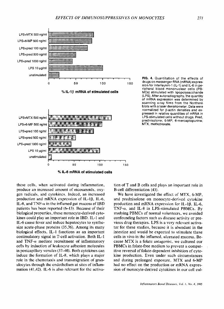
unstimulated FIG. 4. Quantitation of the effects of s 0 50 100 drugs on messenger RNA (mRNA) expres- sion for interleukin-1 (IL-1) and IL-6 in pe- ripheral blood mononuclear cells (PB- MCs) stimulated with lipopolysaccharide (LPS). After autoradiography, the quantity of mRNA expression was determined by scanning x-ray films from the Northern blots with a laser densitometer. Data were normalized for p-actin densities and ex- pressed in relative quantities of mRNA in LPS-stimulated cells without drugs. Pred, prednisolone; 6-MP, 6-mercaptopurine; MTX, methotrexate.
50
% IL-lp mRNA of stimulated cells
LPS+MTX 500 nglml
LPS+6-MP 500 n g h l
LPS+pred 100 ng/ml
LPS+pred 500 nglml
LPS+pred 1000 nglml
LPS 10 pg/ml
I " " I " " I
0 50 100 150
% IL-6 mRNA of stimulated cells
these cells, when activated during inflammation, produce an increased amount of eicosanoids, oxy- gen radicals, and cytokines. Indeed, an increased production and mRNA expression of IL-lp, IL-6, IL-8, and TNF-a in the inflamed gut mucosa of IBD patients has been reported (6-13). Because of their biological properties, these monocyte-derived cyto- kines could play an important role in IBD. IL-1 and IL-6 cause fever and induce hepatocytes to synthe- size acute-phase proteins (35,36). Among its many biological effects, IL-1 functions as an important costimulatory signal in T-cell activation. Both IL- 1 and TNF-a mediate recruitment of inflammatory cells by induction of leukocyte adhesion molecules in postcapillary venules (37-40). Both cytokines can induce the formation of IL-8, which plays a major role in the chemotaxis and transmigration of gran- ulocytes through the endothelium at sites of inflam- mation (41,42). IL-6 is also relevant for the activa-
tion of T and B cells and plays an important role in B-cell differentiation (43).
We have investigated the effect of MTX, 6-MP, and prednisolone on monocyte-derived cytokine production and mRNA expression for IL-lp, IL-6, TNF-a, and IL-8 in LPS-stimulated PBMCs. By studying PBMCs of normal volunteers, we avoided confounding factors such as disease activity or pre- vious drug therapies. LPS is a very relevant activa- tor for these studies, because it is abundant in the intestine and would be expected to stimulate these cells in vivo in the inflamed, ulcerated mucosa. Be- cause MTX is a folate antagonist, we cultured our PBMCs in folate-free medium to prevent a compet- itive reversal of folate-dependent inhibition of cyto- kine production. Even under such circumstances and during prolonged exposure, MTX and 6-MP had no effect on the production or mRNA expres- sion of monocyte-derived cytokines in our cell cul-
Inflammatory Bowel Diseases, Vol. l , No. 4 , 1995

272 L . A . DIELEMAN ET AL.
LPS+ MTX 500 nglml
LPS+6-MP 500 ng/ml
LPS+pred 100 nglml
LPS+pred 500 nglml
LPS+pred 1000 nglml
LPS 10 pglml
unstimulated
0 50 100 150
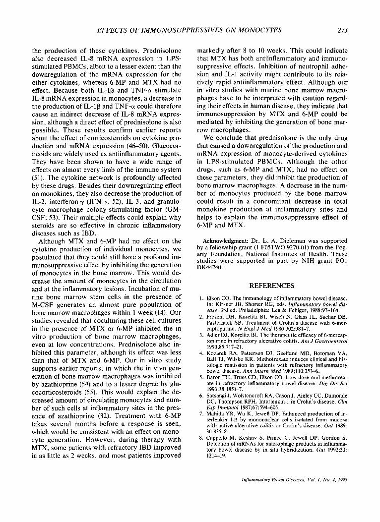
'YO TNF-a mRNA of stimulated cells FIG. 5. Quantit tion of the eff cts of
LPS+ MTX 500 n g h l
LPS+6-MP 500 nglml
drugs on messenger RNA (mRNA) expres- sion for tumor necrosis factor-a (TNF-a) and interleukin-8 (IL-8) in peripheral blood mononuclear cells (PBMCs) stimu- lated with lipopolysaccharide (LPS). For method, see legend to Fig. 4.
LPS+pred 100 nglml
LPS+pred 500 ng/ml
LPS+pred 1000 nglml
LPS 10 pglml
unstimulated
0 50 100 150
'Yo IL-8 mRNA of stimulated cells
tures. However, we did see a MTX-induced inhibi- tion of the proliferation of the IL-1-dependent T-helper cell line DlOM (data not shown). These findings are in accordance with in vitro experiments by Segal et al. (44). Recently Brody et al. (45)
TABLE 1. in vitro production of murine bone marrow-derived macrophages
Cell number D w ( X IO6)a Control (%)
No drug 10 ? 1.0 100 Prednisolone, 500 ndml 4.2' t 0.9 44b
Methotrexate, 50 nm/ml 4.7b ? 1.1 49b 6-Mercaptopurine, 100 nglml 2.2' 2 1.0 23'
Fresh bone marrow cells were cocultured with L 929 super- natant as a source of macrophage colony-stimulating factor (M- CSF) in the presence or absence of drugs. Results are expressed as mean and standard deviation of the total number of viable cells and as percentage of the cell number of bone marrow mac- rophages cultured in the absence of drugs (controls).
a Mean t standard deviation of the total number of viable cells after 7 days' culture. ' Significant difference from controls, p < 0.001.
showed that MTX could indeed block the binding of IL-lP to its IL-1 receptor on monocytes, lympho- cytes, and granulocytes. This would contribute to its anti-inflammatory effect.
In our studies, prednisolone affected all mono- cyte-derived cytokines and decreased their produc- tion significantly. This effect appeared to be due to a decreased mRNA expression for these cytokines, as shown by the Northern blot analysis. The inhi- bition of mRNA was less pronounced in compari- son with the inhibition of cytokine production. To- gether, this suggests both a transcriptional and post- transcriptional inhibitory effect of prednisolone on
Inflummrctory Bowel Diseuses, Vol. I , No. 4, 1995
EFFECTS OF IMMUNOSUPPRESSIVES ON MONOCYTES 273
the production of these cytokines. Prednisolone also decreased IL-8 mRNA expression in LPS- stimulated PBMCs, albeit to a lesser extent than the downregulation of the mRNA expression for the other cytokines, whereas 6-MP and MTX had no effect. Because both IL-lp and TNF-a stimulate IL-8 mRNA expression in monocytes, a decrease in the production of IL-1p and TNF-a could therefore cause an indirect decrease of IL-8 mRNA expres- sion, although a direct effect of prednisolone is also possible. These results confirm earlier reports about the effect of corticosteroids on cytokine pro- duction and mRNA expression (46-50). Glucocor- ticoids are widely used as antiinflammatory agents. They have been shown to have a wide range of effects on almost every limb of the immune system (51). The cytokine network is profoundly affected by these drugs. Besides their downregulating effect on monokines, they also decrease the production of IL-2, interferon-y (IFN-y; 52), IL-3, and granulo- cyte macrophage colony-stimulating factor (GM- CSF; 53). Their multiple effects could explain why steroids are so effective in chronic inflammatory diseases such as IBD.
Although MTX and 6-MP had no effect on the cytokine production of individual monocytes, we postulated that they could still have a profound im- munosuppressive effect by inhibiting the generation of monocytes in the bone marrow. This would de- crease the amount of monocytes in the circulation and at the inflammatory lesions. Incubation of mu- rine bone marrow stem cells in the presence of M-CSF generates an almost pure population of bone marrow macrophages within 1 week (14). Our studies revealed that coculturing these cell cultures in the presence of MTX or 6-MP inhibited the in vitro production of bone marrow macrophages, even at low concentrations. Prednisolone also in- hibited this parameter, although its effect was less than that of MTX and 6-MP. Our in vitro study supports earlier reports, in which the in vivo gen- eration of bone marrow macrophages was inhibited by azathioprine (54) and to a lesser degree by glu- cocorticosteroids (55) . This would explain the de- creased amount of circulating monocytes and num- ber of such cells at inflammatory sites in the pres- ence of azathioprine (32). Treatment with 6-MP takes several months before a response is seen, which would be consistent with an effect on mono- cyte generation. However, during therapy with MTX, some patients with refractory IBD improved in as little as 2 weeks, and most patients improved
markedly after 8 to 10 weeks. This could indicate that MTX has both antiinflammatory and immuno- suppressive effects. Inhibition of neutrophil adhe- sion and IL-1 activity might contribute to its rela- tively rapid antiinflammatory effect. Although our in vitro studies with murine bone marrow macro- phages have to be interpreted with caution regard- ing their effects in human disease, they indicate that immunosuppression by MTX and 6-MP could be mediated by inhibiting the generation of bone mar- row macrophages.
We conclude that prednisolone is the only drug that caused a downregulation of the production and mRNA expression of monocyte-derived cytokines in LPS-stimulated PBMCs. Although the other drugs, such as 6-MP and MTX, had no effect on these parameters, they did inhibit the production of bone marrow macrophages. A decrease in the num- ber of monocytes produced by the bone marrow could result in a concomitant decrease in total monokine production at inflammatory sites and helps to explain the immunosuppressive effect of 6-MP and MTX.
Acknowledgment: Dr. L. A. Dieleman was supported by a fellowship grant (1 FOSTWO 9270-01) f rom t h e Fog- arty Foundation, National Insti tutes of Health. These studies were s u p p o r t e d in p a r t b y NIH g r a n t PO1 DK44240.
REFERENCES
1.
2.
3.
4.
5 .
6.
7.
8.
Elson CO. The immunology of inflammatory bowel disease. In: Kirsner JB, Shorter RG, eds. Inflammatory bowel dis- ease. 3rd ed. Philadelphia: Lea & Febiger, 1988:97-164. Present DH, Korelitz BI, Wisch N , Glass JL, Sachar DB, Pasternack SB. Treatment of Crohn’s disease with 6-mer- captopurine. N Engl J Med 1980;302:981-7. Adler DJ, Korelitz BI. The therapeutic efficacy of 6-mercap- topurine in refractory ulcerative colitis. Am J Gastroenterol
Kozarek RA, Patterson DJ, Geelfand MD, Botoman VA, Ball TJ, Wilske KR. Methotrexate induces clinical and his- tologic remission in patients with refractory inflammatory bowel disease. Ann Intern Med 1989;110:353-6. Baron TH, Truss CD, Elson CO. Low-dose oral methotrex- ate in refractory inflammatory bowel disease. Dig Dis Sci 1993;38:1851-7. Satsangi J , Wolstencroft RA, Cason J , Ainley CC, Dumonde DC, Thompson RPH. Interleukin-1 in Crohn’s disease. Clin Exp Immunol 1987;67:594-605. Mahida YR, Wu K, Jewell DP. Enhanced production of in- terleukin 1-@ by mononuclear cells isolated from mucosa with active ulc5rative colitis or Crohn’s disease. Gut 1989; 30:83543. Cappello M, Keshav S, Prince C, Jewell DP, Gordon S. Detection of mRNAs for macrophage products in inflamma- tory bowel disease by in situ hybridization. Gut 1992;33: 121419.
1990;85:717-21.
Inflammatory Bowel Diseases, Vol. I , No . 4, 1995

2 74 L . A . DIELEMAN ET AL.
9. Isaacs KI, Sartor RB, Haskill S . Cytokine messenger RNA profiles in inflammatory bowel disease mucosa detected by polymerase chain reaction amplification. Gastroenterology 1992; 103: 1587-95,
10. Suzuki Y, Saito H , Kasanuki J, Kishimoto T, Tamura Y, Yoshida S. Significant increase of interleukin-6 production in blood mononuclear leukocytes obtained from patients with active inflammatory bowel disease. Life Sci 1990;47:
11. MacDonald TT, Hutchings P, Choy MY, Murch S, Cooke A. Tumor necrosis factor-alpha and interferon-gamma produc- tion measured at the single cell level in normal and inflamed human intestine. Clin Exp Immunol 1990;81:301-5.
12. Murch SH, Lamkin VA, Savage MO, Walker-Smith JA, MacDonald TT. Serum concentrations of tumor necrosis factor alpha in childhood chronic inflammatory bowel dis- ease. Gut 1991;32:913-17.
13. lzzo RS, Witkon K , Chen AI, Hadjiyane C, Weinstein MI, Pellecchia C. Interleukin-8 and neutrophil markers in co- lonic mucosa from patients with ulcerative colitis. Am J Gastroenterol 1992;87: 1447-52.
14. Lee KC, Wong M. Functional heterogeneity of culture- grown bone marrow-derived macrophages. J Immunoll980; 125:86-95.
15. Boyum A. Isolation of leukocytes from human blood. A two- phase system for removal of red cells with methylcellulose as erythrocyte-aggregating agent. Scand J Clin Lab Invest
16. Frey SM, Walker C, Frey FJ, de Weck AL. Pharmacokinet- ics and pharmacodynamics of three different prednisolone prodrugs: effect on circulating lymphocyte subsets and func- tion. J lmmunol 1984;133:2479-87.
17. Bertino JR. Chemical action and pharmacology of meth- otrexate, azathioprine and cyclophosphamide in man. Ar- thritis Rheum 1973; 16:79-84.
18. Hopkins SJ, Humphreys M. Simple, sensitive and specific bioassay of interleukin-I. J Zmmunol Methods 1989;120: 271-6.
19. Mosmann TR. Rapid colorimetric assay for cellular growth and survival: application to proliferation and cytotoxicity assays. J Immunol Methods 1983;65:55-63.
20. Aarden LA, De Groot ER, Schaap OL, Lansdorp PM. Pro- duction of hybridoma growth factor by human monocytes. Eur J Zmmunol 1987;17:1411-l6.
21. Espevik T, Nissen Meyer J . A highly sensitive cell line, WEHI 164 clone 13, for measuring cytotoxic factorltumor necrosis factor from human monocytes. J Zmmunol Methods
22. Chomczynski P, Sacchi N. Single-step method of RNA iso- lation by acid guanidinium thiocyanate-phenol-chloroform extraction. Anal Biochem 1987;162: 156-9.
23. Baldari C, Murray JA, Ghiara P, Cesaremi G, Galeotti CL. A novel leader peptide which allows efficient secretion of a fragment of human interleukin- I beta in Saccharomyces cer- evisiae. EMBO J 1987;6:229-34.
24. Wong GG, Witek-Giannotti J , Hewick RM, Clark SC, Ogawa M. Interleukin-6: identification as a hematopoietic colony-stimulating factor. Behring Insf Mitt 1988;83:40-7.
25. Pennica D, Nedwin GE, Haflick JS, et al. Human tumor necrosis factor: precursor structure, expression and homol- ogy to lymphotoxin. Nature 1984;312:724-9.
26. Matsushima K, Morishita K, Yoshimura T, et al. Molecular cloning of a human monocyte-derived neutrophil chemotac- tic factor (MDNCF) and the induction of MDNCF mRNA by interleukin-1 and tumor necrosis factor. J Exp Med 1988;
27. Okayama H, Berg P. A cDNA cloning vector that permits expression of cDNA inserts in mammalian cells. Mol Cell Biol 1983:3:280-9.
2193-7.
1968 ;2 1 (SUPPI 97):9-29.
1986 ;95 :99- 105.
167: 1883-93.
28. Galivan J. Evidence for the cytotoxic activity of methotrex- ate polyglutamates. Mol Pharmacol 1980;17: 105-10.
29. Morgan SC, Baggott JE, Altz-Smith M. Folate status of RA patients receiving long-term low dose MTX therapy. Arrhri- tis Rheum 1987;30: 1348-56.
30. Cronstein BN, Eberle MA, Gruber HE, Levin R1. Meth- otrexate inhibits neutrophil function by stimulating adeno- sine release from connective tissue cells. Proc Nail Acad Sci
31. Leroux JL, Damon M, Chavis C, de Paulet AC, Blotman F. Effects of a single dose of methotrexate on 5- and 12- lipoxygenase products in patients with rheumatoid arthritis. J Rheumatol 1992;19:8634.
32. Elion GB. Biochemistry and pharmacology of purine ana- logues. Fed Proc 1967;26:898-904.
33. Bach JF. The mode of action of immunosuppressive agents. Front Biol 175;41:1-374.
34. Winkelstein A. The effects of azathioprine and 6-MP on im- munity. J Immunopharmacol 1979;1:429-54.
35. Gauldie J , Richards C, Harmish D, Lasdorp P, Bauman H. Interferon-p2-B cell stimulatory factor type 2 shares identity with monocyte-derived hepatocyte-stimulating factor and regulates the major acute phase protein response in liver cells. Proc Nail Acad Sci USA 1987;84:7251-5.
USA 199 1 ;244 1-244.
36. Dinarello CA. Interleukin-I. Rev Infect Dis 1984;6:51-95. 37. Bevilacqua MP, Stengelin S, Gimbrone MA Jr, Seed B. En-
dothelial leukocyte adhesion molecule I. An inducible recep- tor for neutrophils related to complement regulatory pro- teins and lectins. Science 1989;243: 1160-3.
38. Dustin ML, Springer TA. Lymphocyte function-associated antigen-1 (LFA-I) interaction with intercellular adhesions molecule-I (ICAM-I) is one of at least three mechanisms of lymphocyte adhesion to cultured endothelial cells. J Cell Biol 1988;107:321-3 1.
39. Osborn L, Hession C, Tizard R, et al. Direct expression cloning of vascular cell adhesion molecule I , a cytokine- induced endothelial protein that binds to lymphocytes. Cell 1989;59: 1203-1 1.
40. Tracey KJ, Vlassara H , Cerami A. Cachectin/tumor necrosis factor. Lancer 19893: 11224.
41. Yoshimura T, Matsushima K, Oppenheim JJ, Leonard EJ. Neutrophil chemotactic factor produced by lipopolysaccha- ride (LPS)-stimulated human blood mononuclear leuko- cytes: partial characterization and separation from interleu- kin-I (IL-I). J Immunol 1987;139:788-93.
42. Huber AR, Kunkel SL, Todd RF, Weiss SJ. Regulation of transendothelial neutrophil migration by endogenous inter- leukin-8. Science 1991 ;254:99-105.
43. Akira S, Hirano T , Taga T, Kishimoto T. Biology of multi- functional cytokines: IL-6 and related molecules (1L-l and TNF). FASEB J 1990;4:2860-7.
44. Segal R, Yaron M, Tartakovsky B. Methotrexate: mecha- nism of action in rheumatoid arthritis. Semin Arthritis Rheum 1990;20: 190-200.
45. Brody M, Bohm M, Bauer R. Mechanism of action of meth- otrexate: experimental evidence that methotrexate blocks the binding of interleukin 1 beta to the interleukin 1 receptor on target cells. Eur J Clin Chem Clin Biochem 1993;31:667- 74.
46. Lew W, Oppenheim JJ, Matsushima K. Analysis of the sup- pression of IL-la and IL-1p production in human peripheral blood mononuclear adherent cells by a glucocorticoid hor- mone. J Immunol 1988;140:1895-1902.
47. Lee SW, Tsou AP, Chan H, et al. Glucocorticoids selec- tively inhibit the transcription of the interleukin-I p gene and decrease the stability of interleukin-lp mRNA. Proc Nail Acad Sci USA 1988;85:1204-8.
48. Beutler B, Krochin N, Milsark IW, Luedke C, Cerami A. Control of cachectin (tumor necrosis factor) synthesis:
Inflammatory Bowel Diseases, Vol. I . No. 4, 1995

EFFECTS OF IMMUNOSUPPRESSIVES ON MONOCYTES 2 75
mechanisms of endotoxin resistance. Science 1986;232:977- 90.
49. Mukaida N, Shiroo M, Matsushima K. Genomic structure of the human monocyte-derived neutrophil chemotactic factor 1L-8. J Immunol l989;143: 1366-71.
50. Niida N , Murata Y , Suzuki K, et al. Increased interleukin-6 (IL-6) synthesis by peripheral blood mononuclear cells from patients with ulcerative colitis, but not with Crohn’s disease. Gastroenterology 1990;98: A466.
51. Cupps TR, Fauci AS. Corticosteroid-mediated immunoreg- ulation in man. Immunol Rev 1982;65:133-5.
52. Arya SK, Wong-Staal F , Gallo RC. Dexamethasone-
mediated inhibition of human T cell growth factor and y-in- terferon messenger RNA. J Immunol 1984;133:273-6.
53. Culpepper JA, Lee F. Regulation of IL-3 expression by glu- cocorticoids in cloned murine T lymphocytes. J Immunol
54. Van Furth R, Gassrnann AE, Diesselhoff-Den Dulk MMC. The effect of azathioprine (Imuran) on the cell cycle of promonocytes and the production of monocytes in the bone marrow. J Exp Med 1975;141:53146.
5 5 . Thompson J , van Furth R. The effect of glucocorticosteroids on the proliferation and kinetics of promonocytes and mono- cytes of the bone marrow. J Exp Med 1973;137:10-21.
1985;135:3191-7.
lnflomrnarory Bowel Diseases. Vol. I , N o . 4 , I995
Copyright © 2022 FDOKUMEN