The human C-type lectin CLECSF8 is a novel monocyte/macrophage endocytic receptor
Upload
independentCategory
view
0download
0
of January 9, 2015.This information is current as
InflammationLungInvolved in Airway Hyperreactivity and
Mouse Monocyte-Derived Chemokine Is
Jose-Carlos Gutierrez-RamosE. I. Proudfoot, Anthony J. Coyle, David Gearing andJia, Gary Yu, Barry Dussault, Christine A. Powers, Amanda Jose-Angel Gonzalo, Yang Pan, Clare M. Lloyd, Gui-Quan
http://www.jimmunol.org/content/163/1/4031999; 163:403-411; ;J Immunol
Referenceshttp://www.jimmunol.org/content/163/1/403.full#ref-list-1
, 31 of which you can access for free at: cites 44 articlesThis article
Subscriptionshttp://jimmunol.org/subscriptions
is online at: The Journal of ImmunologyInformation about subscribing to
Permissionshttp://www.aai.org/ji/copyright.htmlSubmit copyright permission requests at:
Email Alertshttp://jimmunol.org/cgi/alerts/etocReceive free email-alerts when new articles cite this article. Sign up at:
Print ISSN: 0022-1767 Online ISSN: 1550-6606. Immunologists All rights reserved.Copyright © 1999 by The American Association of9650 Rockville Pike, Bethesda, MD 20814-3994.The American Association of Immunologists, Inc.,
is published twice each month byThe Journal of Immunology
by guest on January 9, 2015http://w
ww
.jimm
unol.org/D
ownloaded from
by guest on January 9, 2015
http://ww
w.jim
munol.org/
Dow
nloaded from
Mouse Monocyte-Derived Chemokine Is Involved in AirwayHyperreactivity and Lung Inflammation
Jose-Angel Gonzalo,1* Yang Pan,1† Clare M. Lloyd, 1* Gui-Quan Jia,* Gary Yu, †
Barry Dussault,* Christine A. Powers,‡ Amanda E. I. Proudfoot,‡ Anthony J. Coyle,*David Gearing,† and Jose-Carlos Gutierrez-Ramos2*
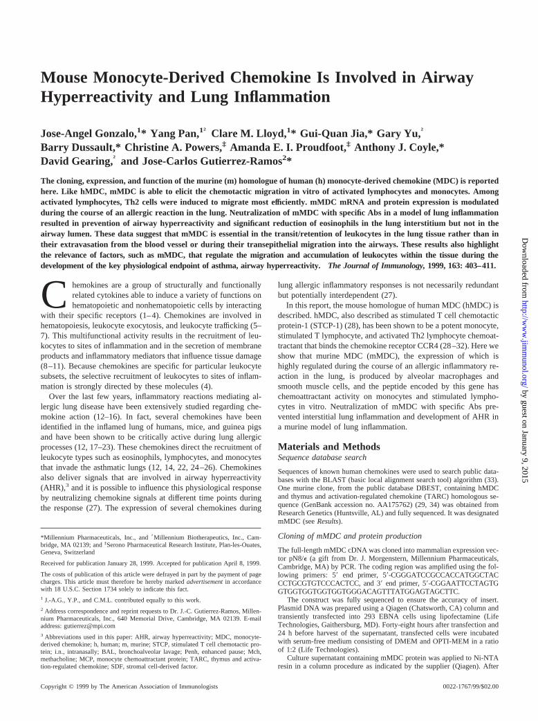
The cloning, expression, and function of the murine (m) homologue of human (h) monocyte-derived chemokine (MDC) is reportedhere. Like hMDC, mMDC is able to elicit the chemotactic migration in vitro of activated lymphocytes and monocytes. Amongactivated lymphocytes, Th2 cells were induced to migrate most efficiently. mMDC mRNA and protein expression is modulatedduring the course of an allergic reaction in the lung. Neutralization of mMDC with specific Abs in a model of lung inflammationresulted in prevention of airway hyperreactivity and significant reduction of eosinophils in the lung interstitium but not in theairway lumen. These data suggest that mMDC is essential in the transit/retention of leukocytes in the lung tissue rather than intheir extravasation from the blood vessel or during their transepithelial migration into the airways. These results also highlightthe relevance of factors, such as mMDC, that regulate the migration and accumulation of leukocytes within the tissue during thedevelopment of the key physiological endpoint of asthma, airway hyperreactivity. The Journal of Immunology,1999, 163: 403–411.
Chemokines are a group of structurally and functionallyrelated cytokines able to induce a variety of functions onhematopoietic and nonhematopoietic cells by interacting
with their specific receptors (1–4). Chemokines are involved inhematopoiesis, leukocyte exocytosis, and leukocyte trafficking (5–7). This multifunctional activity results in the recruitment of leu-kocytes to sites of inflammation and in the secretion of membraneproducts and inflammatory mediators that influence tissue damage(8–11). Because chemokines are specific for particular leukocytesubsets, the selective recruitment of leukocytes to sites of inflam-mation is strongly directed by these molecules (4).
Over the last few years, inflammatory reactions mediating al-lergic lung disease have been extensively studied regarding che-mokine action (12–16). In fact, several chemokines have beenidentified in the inflamed lung of humans, mice, and guinea pigsand have been shown to be critically active during lung allergicprocesses (12, 17–23). These chemokines direct the recruitment ofleukocyte types such as eosinophils, lymphocytes, and monocytesthat invade the asthmatic lungs (12, 14, 22, 24–26). Chemokinesalso deliver signals that are involved in airway hyperreactivity(AHR),3 and it is possible to influence this physiological responseby neutralizing chemokine signals at different time points duringthe response (27). The expression of several chemokines during
lung allergic inflammatory responses is not necessarily redundantbut potentially interdependent (27).
In this report, the mouse homologue of human MDC (hMDC) isdescribed. hMDC, also described as stimulated T cell chemotacticprotein-1 (STCP-1) (28), has been shown to be a potent monocyte,stimulated T lymphocyte, and activated Th2 lymphocyte chemoat-tractant that binds the chemokine receptor CCR4 (28–32). Here weshow that murine MDC (mMDC), the expression of which ishighly regulated during the course of an allergic inflammatory re-action in the lung, is produced by alveolar macrophages andsmooth muscle cells, and the peptide encoded by this gene haschemoattractant activity on monocytes and stimulated lympho-cytes in vitro. Neutralization of mMDC with specific Abs pre-vented interstitial lung inflammation and development of AHR ina murine model of lung inflammation.
Materials and MethodsSequence database search
Sequences of known human chemokines were used to search public data-bases with the BLAST (basic local alignment search tool) algorithm (33).One murine clone, from the public database DBEST, containing hMDCand thymus and activation-regulated chemokine (TARC) homologous se-quence (GenBank accession no. AA175762) (29, 34) was obtained fromResearch Genetics (Huntsville, AL) and fully sequenced. It was designatedmMDC (seeResults).
Cloning of mMDC and protein production
The full-length mMDC cDNA was cloned into mammalian expression vec-tor pN8/e (a gift from Dr. J. Morgenstern, Millennium Pharmaceuticals,Cambridge, MA) by PCR. The coding region was amplified using the fol-lowing primers: 59 end primer, 59-CGGGATCCGCCACCATGGCTACCCTGCGTGTCCCACTCC, and 39 end primer, 59-CGGAATTCCTAGTGGTGGTGGTGGTGGTGGGACAGTTTATGGAGTAGCTTC.
The construct was fully sequenced to ensure the accuracy of insert.Plasmid DNA was prepared using a Qiagen (Chatsworth, CA) column andtransiently transfected into 293 EBNA cells using lipofectamine (LifeTechnologies, Gaithersburg, MD). Forty-eight hours after transfection and24 h before harvest of the supernatant, transfected cells were incubatedwith serum-free medium consisting of DMEM and OPTI-MEM in a ratioof 1:2 (Life Technologies).
Culture supernatant containing mMDC protein was applied to Ni-NTAresin in a column procedure as indicated by the supplier (Qiagen). After
*Millennium Pharmaceuticals, Inc., and†Millennium Biotherapeutics, Inc., Cam-bridge, MA 02139; and‡Serono Pharmaceutical Research Institute, Plan-les-Ouates,Geneva, Switzerland
Received for publication January 28, 1999. Accepted for publication April 8, 1999.
The costs of publication of this article were defrayed in part by the payment of pagecharges. This article must therefore be hereby markedadvertisementin accordancewith 18 U.S.C. Section 1734 solely to indicate this fact.1 J.-A.G., Y.P., and C.M.L. contributed equally to this work.2 Address correspondence and reprint requests to Dr. J.-C. Gutierrez-Ramos, Millen-nium Pharmaceuticals, Inc., 640 Memorial Drive, Cambridge, MA 02139. E-mailaddress: [email protected] Abbreviations used in this paper: AHR, airway hyperreactivity; MDC, monocyte-derived chemokine; h, human; m, murine; STCP, stimulated T cell chemotactic pro-tein; i.n., intranasally; BAL, bronchoalveolar lavage; Penh, enhanced pause; Mch,methacholine; MCP, monocyte chemoattractant protein; TARC, thymus and activa-tion-regulated chemokine; SDF, stromal cell-derived factor.
Copyright © 1999 by The American Association of Immunologists 0022-1767/99/$02.00
by guest on January 9, 2015http://w
ww
.jimm
unol.org/D
ownloaded from

washing with 25 mM imidazole, bound C-terminal-6xHis-tagged mMDCprotein was eluted with 250 mM imidazole. Fractions containing recom-binant mMDC protein were pooled and dialyzed against PBS.
Anti-mMDC Ab generation
Rabbit polyclonal Abs against murine mMDC were prepared according tostandard methods (35). This Ab was generated against a 15-aa peptidecorresponding to the N-terminal region of mMDC. The sequence of thepeptide was GPYGANMEDSVCCRD. Rabbit serum was first depleted ofanti-human IgG Abs by passage over a human IgG column, and anti-mMDC Abs were purified from the flow-through on an affinity columnusing the same peptide or using mMDC recombinant protein depending onthe preparation (Research Genetics, Huntsville, AL).
Mice and in vivo procedures
C57BL/6J mice 8–10 wk old were purchased from The Jackson Laboratory(Bar Harbor, ME) and kept in the Millennium Pharmaceuticals, Inc., spe-cific pathogen-free mouse facility. The mouse model of lung inflammationused here consists of two phases: sensitization (OVA, 0.1 mg/mouse i.p. onday 0) (Sigma, St. Louis, MO) and induction of the response (2% OVA for5 min intranasally (i.n.) on day 8 and 1% OVA for 20 min i.n. on days15–21) (19) (Fig. 4A). PBS (i.p. and/or i.n.) was administered to mice as anegative control. For the blocking experiments, mice also received 20mg/mouse of neutralizing polyclonal Abs against mMDC. This Ab was ad-ministered i.v. 30 min before OVA provocation either on days 8 and 15 oron day 8 and days 15–21 (Fig. 4A). OVA-treated control mice were in-jected with the same amount of rabbit Ig control Ab (Rb Ig) at the sametime points indicated during treatment (Dako, Carpinteria, CA). Threehours after OVA administration on day 15 or day 21, mice were sacrificedby CO2 asphyxiation and analyzed for lung inflammation and AHR.
Bronchoalveolar lavage (BAL) was performed as described (19). AHRwas expressed as enhanced pause (Penh), a calculated value, which corre-lates with measurements of airway resistance, impedance, and intrapleuralpressure in the same mouse (36):Penh5 (Te/Tr 2 1) 3 (Pef/Pif) (Te,expiration time;Tr, relaxation time;Pef, peak expiratory flow;Pif, peakinspiratory flow3 0.67 5 coefficient). The relaxation time is the time ittakes for the box pressure to change from a maximum to a user-definedpercentage of the maximum. Here,Tr measurement begins at the maximumbox pressure and ends at 40%. AHR was measured 3 h after the last Agchallenge by recording respiratory pressure curves by whole-body pleth-ysmography (Buxco Electronics, Sharon, CT) in response to inhaledmethacholine (Mch; Aldrich Chemical, Milwaukee, WI) as described (27).
Peritoneal recruitment assays in vivo with mMDC protein were per-formed after injection of 800ml i.p. of mMDC recombinant protein-con-taining conditioned medium or control conditioned medium. At differenttime points after injection (0, 1, 2, 4, and 6 h), peritoneal leukocytes werecollected and enumerated. In one series of blocking experiments, micewere injected i.v. either with 20mg/mouse of anti-mMDC neutralizing Abor with Ab control, 30 min before mMDC recombinant protein-containingconditioned medium. Peritoneal lavage was performed 2 h after chemokineinjection.
Immunohistochemical phenotyping and quantitation ofleukocytes
Total BAL cell and peritoneal cell counts were performed, and aliquots(5 3 105 cells/slide) were pelleted onto glass slides by cytocentrifugation.To determine the number of eosinophils and neutrophils, slides werestained with Wright-Giemsa stain (Fisher Diagnostics, Pittsburgh, PA).T lymphocytes, B lymphocytes, and mononuclear phagocytes were iden-tified by Thy 1.2 (53-2.1) (PharMingen, San Diego, CA), IgM (II/41)(Phar-Mingen), and Moma 2 (BioSource International, Camarillo, CA)staining, respectively, as described (14). Percentage of eosinophils, lym-phocytes, neutrophils, and macrophages was determined by counting theirnumber in eight high power fields (340 magnification; total area, 0.5 mm2)per area randomly selected and dividing this number by the total number ofcells per high power field. To obtain the absolute number of each leukocytesubtype in the lavage, these percentages were multiplied by the total num-ber of cells recovered from the BAL fluid.
Lung sections from the different experimental groups of mice were pre-pared as described (14). Briefly, lungs were fixed in 10% neutral bufferedformalin (J.T. Baker, Phillipsburg, NJ) and paraffin embedded. Sections (4mm) were stained with hematoxylin and eosin according to standard pro-tocols. An estimation of the percentage of each leukocyte subtype withinthe infiltrate in OVA 1 anti-mMDC Ab-treated mice or OVA1 rabbitIg-treated controls was made by counting 200 cells in one randomly se-
lected peribronchiolar infiltrate and determining the number of eosinophils.Quantitation of leukocytes both in BAL fluid and in lung sections wasperformed in a blinded fashion.
In vitro chemotaxis
The in vitro migration of leukocytes to recombinant mMDC through anendothelial cell layer was evaluated in duplicate as described (19). Theendothelial cells in transwell inserts were washed once with serum-freemedium, and 23 105 leukocytes (BM cells from C57BL6/J mice, Th1 orTh2 polarized lymphocytes, eosinophils from IL-5 transgenic mice (37), orStaphylococcus aureusenterotoxin B (SEB)-stimulated (10mg/ml, 12 h)or unstimulated lymph node cells from C57BL6/J mice) were added in 0.1ml of serum-free medium. After a 2-h incubation, the Transwells wereremoved and the number of cells per well was counted in the FACScan bypassing each sample for a constant predetermined time period (contami-nating endothelial cells were gated out). In the blocking experiments, leu-kocytes were preincubated with either 1 or 10mg of anti-mMDC Ab orcontrol Ab at 37°C for 15 min before their addition to the Transwell inserts.
Generation of the HEK/mCCR4 cell line
Murine CCR4 was cloned from a mouse thymus cDNA library by PCRusing primers based on the mouse CCR4 sequence (38). The full codingsequence of mCCR4 was subcloned into the mammalian cell expressionvector pcDNA3.1zeo (Invitrogen). Stable cell lines were generated follow-ing transfection of the expression vector into HEK-293 cells using thecalcium phosphate transfection system (Life Technologies) according tothe manufacturer’s instructions. Positive clones were selected with zeocin(100 mg/ml) (Invitrogen), and clones expressing high levels of mCCR4were identified by binding to125I-labeled human TARC (Amersham).
Calcium mobilization
The ability of mMDC to activate CCR4 was determined by adding 5, 25,and 50 aliquots of the conditioned medium containing mMDC to 13 106
fura-2-loaded HEK/mCCR4 cells for each measurement, as previously de-scribed (39). Desensitization experiments were conducted by adding 50mlof the conditioned medium containing mMDC, followed by the addition ofdifferent concentrations of human TARC or hMDC 60 s later.
Th1-Th2 cell polarization
Mice expressing the transgene for the DO11.10ab-TCR, which recognizesresidues 323–339 of chicken OVA in association with I-Ad (40), wereprovided by Dr. D. Loh (Washington University, St. Louis, MO). OVA-specific TCR-transgenic CD41 T cells were isolated from the spleen(.97% purity) by using mouse CD41 T cell subset enrichment columns(R&D Systems, Minneapolis, MN) and cultured in complete RPMI 1640with OVA323–339(1 mg/ml) and mitomycin C-treated splenocytes. For Th1phenotype development, recombinant murine IL-12 (40 ng/ml) (Endogen,Cambridge, MA) and neutralizing anti-IL-4 Ab (11B11, 20mg/ml, R&DSystems) were added and for Th2 phenotype development recombinantmurine IL-4 (50 ng/ml) and anti-IL12 (TOSH-2, 10mg/ml, Endogen) wereused. Cells were cultured for three rounds of antigenic stimulations underpolarizing conditions. To determine that cells were differentiated, 23 105
cells were activated on immobilized anti-CD3 mAb (2C11, 10mg/ml,PharMingen) in the presence of human IL-2 (10 U/ml) (Endogen) for 48 h.IL-4, IL-5, and IFN-g levels were determined by specific ELISA (Endo-gen). In general, Th2 cells produced high levels of IL-4 and IL-5 but littleIFN-g, whereas Th1 cells produced high levels of IFN-g but little IL-4 andIL-5 (Th2 cells produced 100–300 ng/ml IL-4, 50–150 ng/ml IL-5, and,20 pg/ml IFN-g; Th1 cells produced 7,000–15,000 ng/ml IFN-g). Theviability of Th1 and Th2 cells was.95%. Subsequently, these cell wereused for both CCR4 expression analysis and in vitro migration assays.
Measurement of mMDC protein by immunohistochemistry
mMDC protein expression was determined in both normal and inflamedmouse lung tissue with a polyclonal rabbit anti-mMDC Ab using a mod-ified avidin/biotin staining method as described (14). Sections were over-laid with 20% normal donkey serum in PBS for 15 min and then incubatedovernight at 4°C with anti-mMDC Ab diluted 1:750 in PBS with 0.1%BSA and 0.1% sodium azide. Endogenous peroxide was subsequentlyblocked by incubation for 20 min in methanol containing 0.3% hydrogenperoxide. Nonspecific staining due to cross-reaction with endogenous avi-din or biotin was blocked by incubation with avidin solution followed bybiotin solution, both for 20 min. Bound Ab was visualized by incubation
404 mMDC IN LUNG ALLERGIC INFLAMMATION
by guest on January 9, 2015http://w
ww
.jimm
unol.org/D
ownloaded from

with biotinylated anti-rabbit Ig diluted in 10% normal mouse serum PBSand then with streptavidin peroxidase complex prepared according to themanufacturer’s instructions (both from Dako) and incubated for 1 h each.Finally, slides were flooded with peroxidase substrate solution for 10 minbefore counterstaining with hematoxylin. Control slides with the followingwere included: 1) staining with normal rabbit Ig instead of primary Ab, 2)omission of biotinylated anti-rat Ig, and 3) omission of streptavidin com-plex. In addition, competitive inhibition of the Ab was accomplished bypreincubation of Ab with the peptide (100-fold excess) for 45 min at 37°Cbefore incubation with tissue sections.
Measurement of mMDC mRNA expression
mMDC mRNA expression in normal murine tissues was analyzed byNorthern blot analysis.32P-labeled full-length mMDC cDNA was used toprobe a murine multiple tissue Northern blot (Clontech Laboratories, PaloAlto, CA). An actin control probe was used to hybridize to the same blotsto ensure that all lanes were equally loaded.
Total RNA from the lungs of OVA-treated mice or control littermatesat different time points was extracted by the single-step method using RNASTAT-60 (Tel-Test, Inc., Friendswood, TX).
mMDC mRNA expression during lung allergic inflammation was de-termined by Multiprobe RNase protection assay as described (27). A464-bp mMDC probe was derived by PCR using the following primers:59-GCTCTCGTCCTTCTTGCTGTCC-39and 59-AGGGGATGGAGGAGGTGAGTAAAGGTG-39. The identity and quantity of each mRNA spe-cies in the original RNA sample was determined based on the signal in-tensities given by the appropriately sized, protected probe fragment bands.Values were created by expressing mMDC up-regulation relative to itsexpression in normal tissue. The sample loading was normalized by thehousekeeping gene, GADPH, which is included in each template set.
Results and DiscussionIdentification of mMDC gene
Several mouse chemokine-like sequences were identified aftersearching the public database DBEST. One clone contained anovel full-length CC chemokine, which was designated mMDCbased on its pattern of tissue expression, chemotactic specificity invitro, and receptor usage. Nucleotide and amino acid sequence ofthis clone and homology analysis are not shown because, duringthe preparation of our manuscript, a novel mouse chemokinenamed ABCD-1 showing the same gene and protein sequence wasreported (41).
The mouse chromosomal location of mMDC was determined byusing a panel of backcross progeny of C57BL/6JMus musculusandMus spretusmice. The mapping results indicated that mMDCis located on mouse chromosome 2 (data not shown).
Anti-mMDC polyclonal Ab reacting with recombinant protein
Recombinant mMDC protein was produced in 293 EBNA cells bytransient transfection as described inMaterials and Methods. Pu-rification of 6xHis-tagged mMDC protein was made following theNi-NTA affinity purification method (Qiagen). Affinity-purifiedrabbit polyclonal Abs generated against a 15-aa peptide corre-sponding to the N-terminal region of mMDC were shown to rec-ognize a specific band in a Western blot against mMDC. Using thispolyclonal antiserum, a single 8-kDa band was detected from themMDC-transfected supernatant mentioned above but not in con-trol supernatant (data not shown). This polyclonal Ab did notcross-react with murine macrophage-inflammatory protein-1a,murine monocyte chemoattractant protein (MCP)-1, mMCP-5,murine eotaxin, or stromal cell-derived factor -1a (SDF-1a) pro-tein (data not shown). Furthermore, as shown later, in vitro leu-kocyte migration in response to either SDF-1a or MCP-5 was notaffected by the anti-mMDC polyclonal Ab. Because it is knownthat in general the N-terminal regions of chemokines, such as IL-8and MCP-1, contain receptor binding sites, it is likely that this Abmasked the receptor binding site on mMDC. This hypothesis isdemonstrated below, where the neutralizing capabilities of the Abpreparation are shown.
In vitro chemotactic responses of leukocytes to mMDC
To evaluate the chemotactic function of mMDC on resting leuko-cytes, neutrophils, lymphocytes, and monocytes were isolatedfrom C57BL/6J mouse bone marrow and subjected to in vitrotransendothelial migration assays (42). Fig. 1A shows that the con-ditioned medium containing mMDC (but not the control condi-tioned medium) or purified mMDC protein induced the migrationof monocytes. This migration was similar to that provoked byMCP-5 (Fig. 1A) and was three times smaller than that induced bySDF-1 (data not shown), which were used as positive controls.Neither bone marrow neutrophils from wild-type mice nor eosin-ophils from IL-5 transgenic mice (37) migrated in response tomMDC (data not shown). No effect on resting bone marrow lym-phocytes in response to mMDC was detected in the same set ofmigration assays in which SDF-1 was the positive control (Fig.1A). However, when lymph node T lymphocytes were stimulatedin vitro with the superantigenStaphylococcus aureusenterotoxinB for 12 h (43), these cells were able to migrate in response tomMDC, showing a 3-fold increase in chemotactic index whencompared with that observed in unstimulated cells (Fig. 1B). Tofurther evaluate chemotactic function of mMDC on activated Tlymphocyte subclasses, IL-2-stimulated Th1 and Th2 polarizedcells were used in chemotaxis assays (Fig. 1B). Our data clearlyshowed that although mMDC induced both Th1 and Th2 cell mi-gration, the chemotactic index of Th2 cells in response to mMDCwas significantly higher than that observed in Th1 cells (Fig. 1B).
To attribute unambiguously the migration of monocytes andstimulated lymphocytes to the mMDC chemotactic activity withinthe serum-free supernatant from mMDC-transfected cells or to pu-rified mMDC protein, this chemokine was blocked in vitro withaffinity-purified polyclonal Abs raised against an mMDC peptide(seeMaterials and Methods). Specific anti-mMDC Ab was able toneutralize almost all of the monocyte and lymphocyte chemotacticactivity of the purified mMDC protein or in the mMDC-condi-tioned medium (Fig. 1,A andB). Both in vitro chemotaxes inducedby SDF-1a and by MCP-5 were unaffected by the anti-mMDC Ab(Fig. 1, A andB).
In vivo chemotactic responses to mMDC
To determine the efficacy of mMDC in vivo, serum-free superna-tant from mMDC-transfected cells was injected into the perito-neum of mice. Maximal peritoneal leukocyte accumulation in re-sponse to mMDC was detected 2 h after injection (data not shown).At this time point, quantitation of leukocyte subtypes revealed1-fold increase in monocyte numbers after mMDC injection (Fig.1C). Peritoneal monocyte numbers (detected by Moma 2-positivecell staining) in the mMDC-treated mice and IL-8-treated controllittermates were (2156 20) 3 103 and (1406 12) 3 103, respec-tively. This mMDC-induced monocyte accumulation in vivo cor-relates well with the in vitro data shown above. No differences inthe total number of leukocytes or in the number of each cell typewere detected when PBS-treated mice and control medium-in-jected mice were compared (Fig. 1C).
No discernible increase in peritoneal lymphocytes were detectedfollowing i.p. mMDC administration in the experimental group ofmice when compared with PBS- or control medium-treated mice(Fig. 1C). Stimulated lymphocytes, but not resting lymphocytes,migrate in in vitro assays in response to mMDC (Fig. 1), suggest-ing that resting lymphocytes must be activated before respondingto this chemokine and that mMDC is not able to mediate thisactivation.
The specificity of the migratory response to mMDC within thetissue culture supernatant injected was also confirmed in vivo by
405The Journal of Immunology
by guest on January 9, 2015http://w
ww
.jimm
unol.org/D
ownloaded from

using anti-mMDC neutralizing Ab. No monocyte accumulationwas detected after coinjection of the mMDC protein-containingsupernatant and the specific neutralizing Ab against this (Fig. 1C).
Ca21 flux in CCR4-transfectant cells and MDC cross-desensitization assays
Taken together, the previous data indicate that mMDC displayschemotactic activities on monocytes and stimulated T lympho-cytes. Because of the homology between mMDC and the humanchemokine MDC (29), also described as STCP-1 (28), mMDCcould be considered as the mouse homologue. In fact, like mMDC,hMDC is a potent chemoattractant for monocytes and stimulated Tlymphocytes (28–30, 32). In addition, hMDC has been describedas a functional ligand for the chemokine receptor CCR4 (30). Cor-responding to the highest expression of hCCR4 on Th2 lympho-
cytes vs Th1 lymphocytes, hMDC is much more active on Th2cells than on Th1 cells (31). Similarly, mouse Th2-polarized lym-phocytes, which also show higher levels of expression of CCR4than Th1 cells by PCR (data not shown), respond more readily tomMDC in in vitro migration assays than do Th1 cells (Fig. 1B).
To confirm that mMDC utilizes the CCR4 receptor, calciummobilization in response to mMDC was evaluated in HEK-293cells transfected with mCCR4. Fig. 2 shows that calcium flux wasinduced in these cells following stimulation by conditioned me-dium containing mMDC. No calcium flux was detected when con-trol conditioned medium was used on CCR4-transfected cells (datanot shown). Mock transfectants or untransfected HEK-293 cellsalso did not show calcium flux following mMDC stimulation (datanot shown). This indicates that, as shown for hMDC, mMDC bindsCCR4 with functional consequences. Furthermore, mMDC was
FIGURE 1. mMDC-induced leukocyte migration.A, Chemotactic activity of mMDC on mouse leukocytes in vitro. Bone marrow lymphocytes andmonocytes were subjected to chemotaxis to different dilutions of either the conditioned medium containing mMDC (filled bars) or the control medium (openbars), or to different concentrations of purified mMDC (50 ng/ml or 200 ng/ml; gray bars). Eotaxin (200 ng/ml) was used as negative control for monocytemigration. SDF-1a (200 ng/ml) and MCP-5 (200 ng/ml) were used as positive controls for lymphocyte and monocyte migration, respectively (gray bars).These bars represent the percentage of leukocyte migration; error bars indicate the mean for three representative experiments. Hatched bars representblockage of mMDC-induced monocyte migration by the anti-mMDC Ab as well as MCP-51 anti-mMDC Ab and SDF-11 anti-mMDC Ab controls.B,Staphylococcus aureusenterotoxin B-stimulated lymph node cells or in vitro polarized Th1 and Th2 lymphocytes were subjected to the same migrationprotocol. SDF-1a was used as a positive control for lymphocyte migration. Results are expressed as chemotactic index (the ratio between the number ofcells that migrated to the sample and the number that migrated to negative control). Error bars indicate the mean for three representative experiments.Hatched bars represent blockage of mMDC-induced lymphocyte migration by anti-mMDC Ab.p, Significant difference between chemotaxis induced byconditioned medium containing mMDC or by the control medium; Student’st test (p , 0.01). C, mMDC-induced recruitment of leukocytes to theperitoneum. Peritoneal exudate was collected 2 h after injection of either mMDC-containing conditioned medium in the peritoneum of the experimentalmice (filled symbols) or control conditioned medium or PBS in the peritoneum of the control littermates (open symbols). Thirty minutes before injectionof mMDC-containing conditioned medium, experimental mice were injected i.v. with 20mg/mouse of either anti-mMDC Ab (filled symbols) or Ab control(gray symbols). Each dot represents one individual mouse analyzed (5 mice per control group, 10 mice per experimental group). The bar in each panelrepresents the mean of the total number of cells of the leukocyte type indicated.
406 mMDC IN LUNG ALLERGIC INFLAMMATION
by guest on January 9, 2015http://w
ww
.jimm
unol.org/D
ownloaded from
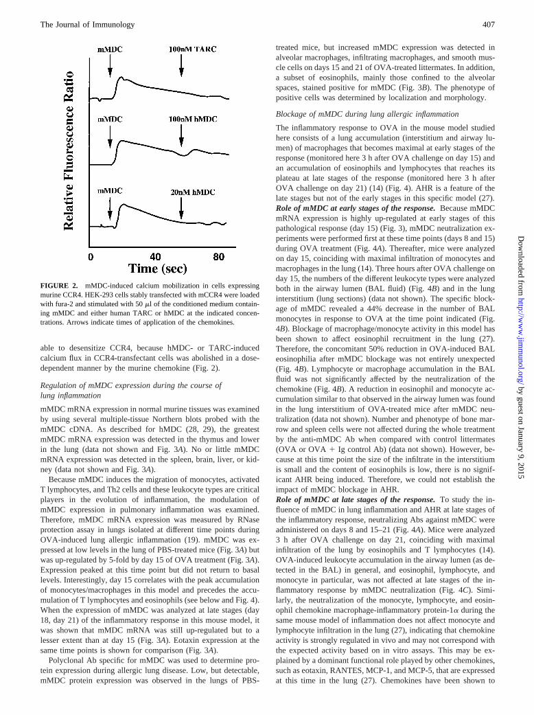
able to desensitize CCR4, because hMDC- or TARC-inducedcalcium flux in CCR4-transfectant cells was abolished in a dose-dependent manner by the murine chemokine (Fig. 2).
Regulation of mMDC expression during the course oflung inflammation
mMDC mRNA expression in normal murine tissues was examinedby using several multiple-tissue Northern blots probed with themMDC cDNA. As described for hMDC (28, 29), the greatestmMDC mRNA expression was detected in the thymus and lowerin the lung (data not shown and Fig. 3A). No or little mMDCmRNA expression was detected in the spleen, brain, liver, or kid-ney (data not shown and Fig. 3A).
Because mMDC induces the migration of monocytes, activatedT lymphocytes, and Th2 cells and these leukocyte types are criticalplayers in the evolution of inflammation, the modulation ofmMDC expression in pulmonary inflammation was examined.Therefore, mMDC mRNA expression was measured by RNaseprotection assay in lungs isolated at different time points duringOVA-induced lung allergic inflammation (19). mMDC was ex-pressed at low levels in the lung of PBS-treated mice (Fig. 3A) butwas up-regulated by 5-fold by day 15 of OVA treatment (Fig. 3A).Expression peaked at this time point but did not return to basallevels. Interestingly, day 15 correlates with the peak accumulationof monocytes/macrophages in this model and precedes the accu-mulation of T lymphocytes and eosinophils (see below and Fig. 4).When the expression of mMDC was analyzed at late stages (day18, day 21) of the inflammatory response in this mouse model, itwas shown that mMDC mRNA was still up-regulated but to alesser extent than at day 15 (Fig. 3A). Eotaxin expression at thesame time points is shown for comparison (Fig. 3A).
Polyclonal Ab specific for mMDC was used to determine pro-tein expression during allergic lung disease. Low, but detectable,mMDC protein expression was observed in the lungs of PBS-
treated mice, but increased mMDC expression was detected inalveolar macrophages, infiltrating macrophages, and smooth mus-cle cells on days 15 and 21 of OVA-treated littermates. In addition,a subset of eosinophils, mainly those confined to the alveolarspaces, stained positive for mMDC (Fig. 3B). The phenotype ofpositive cells was determined by localization and morphology.
Blockage of mMDC during lung allergic inflammation
The inflammatory response to OVA in the mouse model studiedhere consists of a lung accumulation (interstitium and airway lu-men) of macrophages that becomes maximal at early stages of theresponse (monitored here 3 h after OVA challenge on day 15) andan accumulation of eosinophils and lymphocytes that reaches itsplateau at late stages of the response (monitored here 3 h afterOVA challenge on day 21) (14) (Fig. 4). AHR is a feature of thelate stages but not of the early stages in this specific model (27).Role of mMDC at early stages of the response.Because mMDCmRNA expression is highly up-regulated at early stages of thispathological response (day 15) (Fig. 3), mMDC neutralization ex-periments were performed first at these time points (days 8 and 15)during OVA treatment (Fig. 4A). Thereafter, mice were analyzedon day 15, coinciding with maximal infiltration of monocytes andmacrophages in the lung (14). Three hours after OVA challenge onday 15, the numbers of the different leukocyte types were analyzedboth in the airway lumen (BAL fluid) (Fig. 4B) and in the lunginterstitium (lung sections) (data not shown). The specific block-age of mMDC revealed a 44% decrease in the number of BALmonocytes in response to OVA at the time point indicated (Fig.4B). Blockage of macrophage/monocyte activity in this model hasbeen shown to affect eosinophil recruitment in the lung (27).Therefore, the concomitant 50% reduction in OVA-induced BALeosinophilia after mMDC blockage was not entirely unexpected(Fig. 4B). Lymphocyte or macrophage accumulation in the BALfluid was not significantly affected by the neutralization of thechemokine (Fig. 4B). A reduction in eosinophil and monocyte ac-cumulation similar to that observed in the airway lumen was foundin the lung interstitium of OVA-treated mice after mMDC neu-tralization (data not shown). Number and phenotype of bone mar-row and spleen cells were not affected during the whole treatmentby the anti-mMDC Ab when compared with control littermates(OVA or OVA 1 Ig control Ab) (data not shown). However, be-cause at this time point the size of the infiltrate in the interstitiumis small and the content of eosinophils is low, there is no signif-icant AHR being induced. Therefore, we could not establish theimpact of mMDC blockage in AHR.Role of mMDC at late stages of the response.To study the in-fluence of mMDC in lung inflammation and AHR at late stages ofthe inflammatory response, neutralizing Abs against mMDC wereadministered on days 8 and 15–21 (Fig. 4A). Mice were analyzed3 h after OVA challenge on day 21, coinciding with maximalinfiltration of the lung by eosinophils and T lymphocytes (14).OVA-induced leukocyte accumulation in the airway lumen (as de-tected in the BAL) in general, and eosinophil, lymphocyte, andmonocyte in particular, was not affected at late stages of the in-flammatory response by mMDC neutralization (Fig. 4C). Simi-larly, the neutralization of the monocyte, lymphocyte, and eosin-ophil chemokine macrophage-inflammatory protein-1a during thesame mouse model of inflammation does not affect monocyte andlymphocyte infiltration in the lung (27), indicating that chemokineactivity is strongly regulated in vivo and may not correspond withthe expected activity based on in vitro assays. This may be ex-plained by a dominant functional role played by other chemokines,such as eotaxin, RANTES, MCP-1, and MCP-5, that are expressedat this time in the lung (27). Chemokines have been shown to
FIGURE 2. mMDC-induced calcium mobilization in cells expressingmurine CCR4. HEK-293 cells stably transfected with mCCR4 were loadedwith fura-2 and stimulated with 50ml of the conditioned medium contain-ing mMDC and either human TARC or hMDC at the indicated concen-trations. Arrows indicate times of application of the chemokines.
407The Journal of Immunology
by guest on January 9, 2015http://w
ww
.jimm
unol.org/D
ownloaded from

coordinately activate different cellular and molecular pathways in-volved in the pathophysiology of asthma. Thus, MCP-1, which isnot a predominantly lymphocytic or eosinophilic chemokine, di-
minishes both lymphocyte-derived inflammatory mediators and Tcell and eosinophil recruitment to the lung of mice subjected to thesame OVA model (27). Minimal OVA-induced eosinophil and T
FIGURE 3. mMDC mRNA and protein expression in mouse.A, mMDC mRNA expression by RPA in the lung of OVA-treated mice. Inflamed lungswere obtained from mice subjected to OVA treatment 3 h after Ag challenge at the time points indicated (days 0, 8, 15, 18, and 21). mMDC mRNAexpression in normal and inflamed murine lung (first five lanes) and normal kidney (final lane) is shown (left). Results from using a GAPDH control probeare shown in thelower portion of the figure. Each full bar in theright panelrepresents the mean level of mMDC mRNA expression from five mice at thetime points indicated (3 h after OVA challenge) during treatment. Eotaxin mRNA expression at the same time points is also shown for comparison (leftpanel; open bars inright panel). Values are expressed as fold increase in mMDC or eotaxin expression, respectively, over that in PBS-treated lungs(designated a value of 1). Although mMDC is more highly expressed than eotaxin at time 0, both chemokines were normalized to 1 to better illustrate theexpression pattern during disease. Values are also expressed as mean1 SEM.B, mMDC protein expression in the lung of OVA-treated mice. Sections wereprepared from lungs isolated on day 15 (i), 21 (ii), or 0 (iii, left panel) of OVA treatment and were stained with a polyclonal Ab that recognizes mMDC.Positive staining was detected with an avidin-biotin peroxidase staining system that resulted in a brown reaction product. Sections were counterstained withhematoxylin (blue) for contrast. Protein expression was detected in macrophages (iv) and smooth muscle cells (vi) on day 15 as well as in a proportion ofinfiltrating eosinophils on day 21 (v) (as indicated by arrows). Preincubation of the anti-mMDC Ab with the immunizing peptide (iii, right panel) orirrelevant control Ab did not show any staining.
408 mMDC IN LUNG ALLERGIC INFLAMMATION
by guest on January 9, 2015http://w
ww
.jimm
unol.org/D
ownloaded from

lymphocyte accumulation is detected in the lung interstitium afterthe neutralization of the monocytic chemokine MCP-5, whereasBAL eosinophil and T lymphocyte numbers are not affected underthe same conditions (27). Interestingly, the examination of tissuesections of OVA-treated mice after blockage of mMDC revealed adifferential role for the chemokine mMDC in eosinophil accumu-lation in the lung interstitium. During this phase of the response,there is a substantial interstitial infiltrate that develops in perivas-cular and peribronchiolar areas, which was inhibited by 70% afterthe administration of anti-mMDC Ab (Fig. 5). This reduction ininfiltrate size was seen to be due to a significant decrease in num-bers of eosinophils within the infiltrate (Fig. 5). OVA-inducedlymphocyte accumulation is not markedly affected by mMDC neu-tralization. This finding deserves further comment; despite the sig-nificant chemotaxis in vitro of both Th2 and activated lymphocytesto mMDC, no obvious effect on lymphocyte recruitment is ob-served in vivo during OVA challenge following mMDC neutral-ization. This could be due to the number of Ag-specific Th2 cellsthat migrate to the lung in this active immunization model. Thesecells represent a small fraction of the total lymphocyte accumula-tion, which makes it difficult to detect a reduction in the total cellnumber. In fact, we are currently studying a lung inflammationmodel based on the adoptive transfer of TCR-transgenic Th2 cellsinto mice. In this model, Ag-specific Th2 cells can be monitoredfollowing their arrival to the lung interstitium and airway lumenduring the response. Our results show that mMDC is responsible
for 80% of the Th2 cells that migrate to this organ. However,Ag-specific Th2 cells represent,15% of all the T cells present inthe lung in this adoptive transfer model (C. M. Lloyd et al., manu-script in preparation). These findings suggest that the percentage ofTh2 cells in the active immunization model used here could besignificantly lower and therefore difficult to detect. Alternatively,this could also be explained by the fact that mMDC might influ-ence a small subset of Ag-specific cells by promoting their reten-tion in a specific interstitial location, a phenotype that will be dif-ficult to substantiate in this model. These findings suggest thatmMDC is instrumental in the accumulation of eosinophils within thelung interstitium during allergic eosinophilia by altering the traffick-ing/retention of the eosinophils and monocytes through the lung in-terstitium rather than affecting their extravasation from the blood ves-sels or their exit to airway lumen.
Role of mMDC in the induction of AHR
To determine whether the reduction of inflammation in the lunginterstitium, but not in the airway lumen, following mMDC neu-tralization is associated with changes in airway function, AHR wasevaluated in OVA-treated mice after mMDC blockage. Fig. 6
FIGURE 4. Leukocyte accumulation in the airways after mMDC block-age during lung allergic inflammation.A, mMDC neutralization was per-formed daily before each aerosolized provocation with OVA either on days8 and 15 or on days 8 and 15–21. BAL eosinophil, monocyte, lymphocyte,and macrophage accumulation was evaluated 3 h after OVA administrationon day 15 (B) or 21 (C). Each dot represents a single PBS or OVA1 rabbitIg control Ab (Rb Ig)-treated mouse (open symbol) or a single OVA1anti-mMDC Ab-treated mouse (filled symbol). Bars represent the mean ofeach group. One representative experiment of three, with 10 mice pergroup, is shown. Significant difference between control and test groups ofmice was determined using the Student’st test (p, 0.001).
FIGURE 5. Leukocyte accumulation in the lung after mMDC blockageduring lung allergic inflammation.A, Representative sections of lungs iso-lated from mice treated with OVA1 rabbit Ig control Ab (Rb Ig) or OVA1 anti-mMDC Ab are shown.B, Semiquantitative scoring system was usedto estimate the size of lung infiltrates, where15 signifies a large wide-spread infiltrate around the majority of vessels and bronchioles, and11signifies a small number of inflammatory foci. Each dot represents a singleOVA 1 anti-mMDC Ab-treated mouse (F) or OVA 1 rabbit Ig-treatedcontrol (E). An estimation of the percentage of eosinophils within theinfiltrate in OVA 1 anti-mMDC Ab-treated mice (filled bar) or OVA1rabbit Ig-treated controls (open bar) was made by counting 200 cells in onerandomly selected peribronchiolar infiltrate and determining the number ofeosinophils present (B). Values are expressed as the mean1 SEM.
409The Journal of Immunology
by guest on January 9, 2015http://w
ww
.jimm
unol.org/D
ownloaded from

shows that mMDC neutralization inhibits the development of AHRin this experimental group of mice when compared with OVAcontrol-treated mice. This decrease correlates with the 70% reduc-tion in leukocyte accumulation, mainly eosinophils, in the lunginterstitium of OVA-treated mice after mMDC blockage. This sug-gests a correlation between the location of the inflammatory cellsin the lung and the establishment of AHR. Evidence indicates thateosinophils accumulated in the airways of asthmatic patients dic-tate the severity of disease (44). However, it has also been reportedthat the development of AHR depends on the recruitment of eo-sinophils to the mouse bronchial submucosa, but not to the airways(45). In addition, the blockage of the chemokine MCP-5, whichaffects interstitial eosinophil recruitment but not airway eosino-philia, abrogates AHR (27). Likewise, a recent clinical study hasfound no significant correlation between the degree of AHR andthe number of inflammatory cells in sputum or BAL (46).
Concluding remarks
The murine model studied here has allowed us to dissect the pre-dominant features of allergic lung disease, namely, cellular inflam-mation and bronchial hyperreactivity. Disease development neces-sitates the migration of leukocytes from the peripheral circulationthrough the vascular endothelium, across the lung interstitium, andinto the airway lumen via the bronchial epithelium. We have pre-viously determined that chemokines act in a tightly controlled,coordinated fashion to direct migration of leukocytes through theinterstitium (as observed in histology) and into the airway lumen(as observed in lavage). The data presented here indicate that thepresence of eosinophils (and, indeed, other inflammatory cells) inthe airway lumen is not sufficient to induce the AHR that is char-acteristic of asthmatic processes. Rather, our data indicate that eo-sinophils and other inflammatory cells have to be localized withinthe peribronchiolar submucosa or in perivascular regions (as de-tected in sections of the lung interstitium) to induce these delete-rious effects.
Our data support the notion that the chemokine mMDC is crit-ical for the retention/trafficking of eosinophils and other leukocytesand for their proper localization to these areas in the lung during
the development of an allergic reaction. During preparation of ourmanuscript, a mouse novel chemokine named ABCD-1 showingthe same nucleotide and amino acid sequence was reported (41).This recent study describes ABCD-1 as the first activated T cellchemokine produced in large amounts by activated B cells. Thissuggests the possible implication of mMDC/STCP-1/ABCD-1during a T cell-dependent B cell-humoral response.
The finding that mMDC is able to induce the recruitment ofeffector or regulator leukocytes during the allergic reaction makesthe findings mentioned above even more relevant for the develop-ment of disease.
Finally, although there is no synteny between the chromosomallocation for mMDC and hMDC loci (mouse chromosome 2 (datanot shown) and human chromosome 16 (29) respectively), basedon sequence homologies, pattern of tissue expression, receptor us-age, and functional activities, mMDC may be considered the mu-rine homologue of hMDC/STCP-1.
AcknowledgmentsWe thank D. Wen, J. Tian, T. Nguyen, T. Delaney, and N. Bikkal forskilled technical assistance; R. Buser for the construction of the HEK-293cell line; and M. Melzer for editorial assistance.
References1. Kameyoshi, Y., A. Dorschner, A. I. Mallet, E. Christophers, and J. M. Schroder.
1992. Cytokine RANTES released by thrombin-stimulated platelets is a potentattractant for human eosinophils.J. Exp. Med. 176:587.
2. Mackay, C. R. 1996. Chemokine receptors and T cell chemotaxis.J. Exp. Med.184:799.
3. Kita, H., and G. J. Gleich. 1996. Chemokines active on eosinophils: potentialroles in allergic inflammation.J. Exp. Med. 183:2421.
4. Rollins, B. J. 1997. Chemokines.Blood 90:909.5. Miller, M. D., and M. S. Krangel. 1992. Biology and biochemistry of the che-
mokines: a family of chemotactic and inflammatory cytokines.Crit. Rev. Immu-nol. 12:17.
6. Baggiolini, M., B. Dewald, and B. Moser. 1994. Interleukin-8 and related che-motactic cytokines: CXC and CC chemokines.Adv. Immunol. 55:97.
7. Peled, A., J.-A. Gonzalo, C. Lloyd, and J. C. Gutierrez-Ramos. 1997. The che-motactic cytokine eotaxin acts as a granulocyte-macrophage colony stimulatingfactor during lung inflammation.Blood 91:1909.
8. Gleich, G. J., N. A. Flavahan, T. Fujisawa, and P. M. Vanhoutte. 1988. Theeosinophil as a mediator of damage to respiratory epithelium: a model for bron-chial hyperreactivity.J. Allergy Clin. Immunol. 81:776.
9. Koch, A. E., S. L. Kunkel, L. A. Harlow, B. Johnson, H. L. Evanoff,G. K. Haines, M. D. Burdick, R. M. Pope, and R. M. Strieter. 1992. Enhancedproduction of monocyte chemoattractant protein-1 in rheumatoid arthritis.J. Clin.Invest. 90:772.
10. Adams, D. H., M. E. Russell, W. W. Hancock, M. H. Sayegh, L. R. Wyner, andM. J. Karnovsky. 1993. Chronic rejection in experimental cardiac transplantation:studies in the Lewis-F344 model.Immunol. Rev. 134:5.
11. Lloyd, C. M., A. W. Minto, M. E. Dorf, A. Proudfoot, T. N. C. Wells,D. J. Salant, and J.-C. Gutierrez-Ramos. 1997. RANTES and monocyte chemoat-tractant protein-1 (MCP-1) play an important role in the inflammatory phase ofcrescentic nephritis but only MCP-1 is involved in crescent formation and inter-stitial fibrosis.J. Exp. Med. 185:1371.
12. Jose, P. J., D. A. Griffiths-Johnson, P. D. Collins, D. T. Walsh, R. Moqbel,N. F. Totty, O. Truong, J. J. Hsuan, and T. J. Williams. 1994. Eotaxin: a potenteosinophil chemoattractant cytokine detected in a guinea pig model of allergicairways inflammation.J. Exp. Med. 179:881.
13. Collins, P. D., S. Marleau, D. A. Griffiths-Johnson, P. J. Jose, and T. J. Williams.1995. Cooperation between interleukin-5 and the chemokine eotaxin to induceeosinophil accumulation in vivo.J. Exp. Med. 182:1169.
14. Gonzalo, J. A., C. M. Lloyd, L. Kremer, E. Finger, C. Martinez-A,M. H. Siegelman, M. Cybulski, and J. C. Gutierrez-Ramos. 1996. Eosinophilrecruitment to the lung in a murine model of allergic inflammation: the role of Tcells, chemokines and endothelial adhesion receptors.J. Clin. Invest. 98:2332.
15. MacLean, J. A., R. Ownbey, and A. D. Luster. 1996. T cell-dependent regulationof eotaxin in antigen-induced pulmonary eosinophil.J. Exp. Med. 184:1461.
16. Humbles, A. A., D. M. Conroy, S. Marleau, S. M. Rankin, R. T. Palframan,A. E. I. Proudfoot, T. N. C. Wells, D. Li, P. K. Jeffery, D. A. Griffiths-Johnson,et al. 1997. Kinetics of eotaxin generation and its relationship to eosinophil ac-cumulation in allergic airways disease: analysis in a guinea pig model in vivo.J. Exp. Med. 186:601.
17. Ying, S., L. Taborda-Barata, Q. Meng, M. Humbert, and A. B. Kay. 1995. Thekinetics of allergen-induced transcription of messenger RNA for monocyte che-motactic protein-3 and RANTES in the skin of human atopic subjects: relation-ship to eosinophil, T cell, and macrophage recruitment.J. Exp. Med. 181:2153.
FIGURE 6. Inhibition of OVA-induced AHR after mMDC blockage.Results are shown as the mean6 SEM for Penh before (open bars) andafter (filled bars) Mch provocation (n 5 10, three independent experi-ments). Mice were exposed to an aerosol of Mch for 1 min, and airwayconstriction was evaluated for the next 5 min. Mice treated with PBS orOVA 1 rabbit Ig were used as controls for OVA-treated littermates inwhich mMDC was blocked from days 8 to 21. Significant difference be-tween control and test groups of mice was determined using the Student’st test (p, 0.01) and is indicated by an asterisk.
410 mMDC IN LUNG ALLERGIC INFLAMMATION
by guest on January 9, 2015http://w
ww
.jimm
unol.org/D
ownloaded from

18. Dahinden, C. A., T. Geiser, T. Brunner, V. von Tscharner, D. Caput, P. Ferrara,A. Minty, and M. Baggiolini. 1994. Monocyte chemotactic protein 3 is mosteffective basophil- and eosinophil-activating chemokine.J. Exp. Med. 179:751.
19. Gonzalo, J.-A., G.-Q. Jia, V. Aguirre, D. Friend, A. J. Coyle, N. A. Jenkins,G. S. Lin, H. Katz, A. Litchman, N. Copeland, et al. 1996. Mouse eotaxin ex-pression parallels eosinophil accumulation during lung allergic inflammation butit is not restricted to a Th2-type response.Immunity. 4:1.
20. Ponath, P. D., S. Qin, T. W. Post, J. Wang, L. Wu, N. P. Gerard, W. Newman,C. Gerard, and C. R. Mackay. 1996. Molecular cloning and characterization of ahuman eotaxin receptor expressed selectively on eosinophils.J. Exp. Med. 183:2437.
21. Alam, R., J. York, M. Boyars, S. Stafford, A. J. Grant, J. Lee, P. Forsythe,T. Simm, and N. Ida. 1996. Increased MCP-1, RANTES, and MIP-1a in bron-choalveolar lavage fluid of allergic asthmatic patients.Am. J. Respir. Crit. CareMed. 153:1398.
22. Jia, G. Q., J. A. Gonzalo, C. Lloyd, L. Kremer, L. Lu, C. Martinez, B. K. Wershil,and J. C. Gutierrez-Ramos. 1996. Distinct expression and function of the novelmouse chemokine monocyte chemotactic protein-5 in lung allergic.J. Exp. Med.184:1939.
23. Uguccioni, M., P. Loetscher, U. Forssmann, B. Dewald, H. Li, S. H. Lima, Y. Li,B. Kreider, G. Garotta, M. Thelen, and M. Baggiolini. 1996. Monocyte chemo-tactic protein 4 (MCP-4), a novel structural and functional analogue of MCP-3and eotaxin.J. Exp. Med. 183:2379.
24. Rothenberg, M. E., A. D. Luster, C. M. Lilly, J. M. Drazen, and P. Leder. 1995.Constitutive and allergen-induced expression of eotaxin mRNA in the guinea piglung. J. Exp. Med. 181:1211.
25. Garcia-Zepeda, E. A., C. Combadiere, M. E. Rothenberg, M. N. Sarafi,F. Lavigne, Q. Hamid, P. M. Murphy, and A. D. Luster. 1996. Human monocytechemoattractant protein (MCP)-4 is a novel CC chemokine with activities onmonocytes, eosinophils, and basophils induced in allergic and nonallergic in-flammation that signals through the CC chemokine receptors (CCR)-2 and -3.J. Immunol. 157:5613.
26. Kips, J. C., E. Palmans, A. E. I. Proudfoot, M. D. Tyers, A. J. Coyle,T. N. C. Wells, and R. A. Pauwels. 1997. The effect of Met-RANTES on theallergen induced airway eosinophilia in an in vivo mouse model.Am. J. Respir.Crit. Care Med. 155:A733.
27. Gonzalo, J. A., C. M. Lloyd, D. Wen, J. P. Albar, T. N. Wells, A. Proudfoot,C. Martinez-A, M. Dorf, T. Bjerke, A. J. Coyle, and J. C. Gutierrez-Ramos. 1998.The coordinated action of CC chemokines in the lung orchestrates allergic in-flammation and airway hyperresponsiveness.J. Exp. Med. 188:157.
28. Chang, M.-S., J. McNinch, C. Elias III, C. L. Manthey, D. Grosshans, T. Meng,T. Boone, and D. P. Andrew. 1997. Molecular cloning and functional character-ization of a novel CC chemokine, stimulated T cell chemotactic protein (STCP-1)that specifically acts on activated T lymphocytes.J. Biol. Chem. 272:25229.
29. Godiska, R., D. Chantry, C. J. Raport, S. Sozzani, P. Allavena, D. Leviten,A. Mantovani, and P. W. Gray. 1997. Human macrophage-derived chemokine(MDC), a novel chemoattractant for monocytes, monocyte-derived dendriticcells, and natural killer cells.J. Exp. Med. 185:1595.
30. Imai, T., D. Chantry, C. J. Raport, C. L. Wood, M. Nishimura, R. Godiska,O. Yoshie, and P. W. Gray. 1998. Macrophage-derived chemokine is a functionalligand for the CC chemokine receptor 4.J. Biol. Chem. 273:1764.
31. Bonecchi, R., G. Bianchi, P. P. Bordignon, D. D’Ambrosio, R. Lang, A. Borsatti,S. Sozzani, P. Allavena, P. A. Gray, A. Mantovani, and F. Sinigaglia. 1998.
Differential expression of chemokine receptors and chemotactic responsivenessof type 1 T helper cells (Th1s) and Th2s.J. Exp. Med. 187:129.
32. Andrew, D. P., M. S. Chang, J. McNinch, S. T. Wathen, M. Rihanek, J. Tseng,J. P. Spellberg, and C. G. Elias 3rd. 1998. STCP-1 (MDC) CC chemokine actsspecifically on chronically activated Th2 lymphocytes and is produced by mono-cytes on stimulation with Th2 cytokines IL-4 and IL-13.J. Immunol. 161:5027.
33. Altschul, S. F., W. Gish, W. Miller, E. W. Myers, and D. J. Lipman. 1990. Basiclocal alignment search tool.J. Mol. Biol. 215:403.
34. Imai, T., T. Yoshida, M. Baba, M. Nishimura, M. Kakizaki, and O. Yoshie. 1996.Molecular cloning of a novel T cell-directed CC chemokine expressed in thymusby signal sequence trap using Epstein-Barr virus vector.J. Biol. Chem. 271:21514.
35. Harlow, E., and D. Lane. 1988.Antibodies: A Laboratory Manual.Cold SpringHarbor Lab. Press, Plainview, NY.
36. Gelfand, E. W., and C. G. Irvin. 1997. T lymphocyte: setting the tone of theairways.Nat. Med. 3:382.
37. Tominaga, A., S. Takaki, N. Koyama, S. Katoh, R. Matsumoto, M. Migita,Y. Hitoshi, Y. Hosoya, S. Yamauchi, Y. Kanai, et al. 1991. Transgenic miceexpressing a B cell growth and differentiation factor gene (interleukin 5) developeosinophilia and autoantibody production.J. Exp. Med. 173:429.
38. Hoogewerf, A., D. Black, A. E. Proudfoot, T. N. Wells, and C. A. Power. 1996.Molecular cloning of murine CC CKR-4 and high affinity binding of chemokinesto murine and human CC CKR-4.Biochem. Biophys. Res. Commun. 218:337.
39. Lusti-Narasimhan, M., C. A. Power, B. Allet, S. Alouani, K. B. Bacon,J. J. Mermod, A. E. Proudfoot, and T. N. Wells. 1995. Mutation of Leu25 andVal27 introduces CC chemokine activity into interleukin-8.J. Biol. Chem. 270:2716.
40. Murphy, K. M., A. B. Heimberger, and D. Y. Loh. 1990. Induction by antigen ofintrathymic apoptosis of CD41CD81 TCR10 thymocytes in vivo.Science 250:1720.
41. Schaniel, C., E. Pardali, F. Sallusto, M. Speletas, C. Ruedl, T. Shimizu, T. Seidl,J. Andersson, F. Melchers, A. G. Rolink, and P. Sideras. 1998. Activated murineB lymphocytes and dendritic cells produce a novel CC chemokine which actsselectively on activated T cells.J. Exp. Med. 188:451.
42. Carr, M. W., S. J. Roth, E. Luther, S. S. Rose, and T. A. Springer. 1994. Mono-cyte chemoattractant protein 1 acts as a T-lymphocyte chemoattractant.Proc.Natl. Acad. Sci. USA 91:3652.
43. Gonzalo, J. A., A. Gonzalez-Garcia, E. Baixeras, N. Zamzami, R. Tarazona,R. Rappuoli, C. Martinez-A, and G. Kroemer. 1994. Pertussis toxin interfereswith superantigen-induced deletion of peripheral T cells without affecting T cellactivation in vivo.J. Immunol. 152:4291.
44. Djukanovic, R., W. R. Roche, J. W. Wilson, C. R. Beasley, O. P. Twentyman,R. H. Howarth, and S. T. Holgate. 1990. Mucosal inflammation in asthma.Am.Rev. Respir. Dis. 142:434.
45. Eum, S.-Y., S. Haile, J. Lefort, M. Huerre, and B. B. Vargaftig. 1995. Eosinophilrecruitment into the respiratory epithelium following antigenic challenge in hy-per-IgE mice is accompanied by interleukin 5-dependent bronchial hyperrespon-siveness.Proc. Natl. Acad. Sci. USA 92:12290.
46. Crimi, E., A. Spanevello, M. Neri, P. W. Ind, G. A. Rossi, and V. Brusasco. 1998.Dissociation between airway inflammation and airway hyperresponsiveness inallergic asthma.Am. J. Respir. Crit. Care Med. 157:4.
411The Journal of Immunology
by guest on January 9, 2015http://w
ww
.jimm
unol.org/D
ownloaded from
Copyright © 2022 FDOKUMEN