A Liver Capsular Network of Monocyte-Derived Macrophages ...

The human C-type lectin CLECSF8 is a novelmonocyte/macrophage endocytic receptor
Ignacio Arce, Laura Martınez-Munoz, Pedro Roda-Navarro and Elena Fernandez-Ruiz
Unidad de Biologıa Molecular, Hospital Universitario de la Princesa, Universidad Autonoma deMadrid, Madrid, Spain
Cell surface lectin receptors play important roles in the function of macrophages. Herein, wehave identified and characterized the human orthologue of the mouse Mcl/Clecsf8. HumanCLECSF8 codes for a type II membrane glycoprotein of 215 amino acids that belongs to thehuman calcium-dependent lectin family (C-type lectin). The cytoplasmic tail of CLECSF8lacks consensus signaling motifs and its extracellular region shows a single carbohydraterecognition domain (CRD). The CLECSF8 gene has been localized on the telomeric region ofthe NK gene complex on chromosome 12p13 close to MINCLE. CLECSF8 mRNA shows amonocyte/macrophage expression pattern. Biochemical analysis of CLECSF8 on transientlytransfected cells showed a glycoprotein of 30 kDa. Cross-linking of the receptor leads to arapid internalization suggesting that CLECSF8 constitutes and endocytic receptor.
Key words: C-type lectin / NK gene complex / Macrophages/monocytes / Endocytosis / Receptor
Received 27/5/03Revised 17/10/03Accepted 31/10/03
[DOI 10.1002/eji.200324230]
Abbreviations: aa: Amino acids M D : Macrophage NKC:Natural killer gene complex CRD: Carbohydrate recognitiondomain BAC: Bacterial artificial chromosome EST:Expressed sequence tag UTR: Untranslated region ORF:Open reading frame RT: Reverse transcription DIC: Differ-ential interference contrast
1 Introduction
Macrophages (M ¶ ) and related cells are phagocytic cellswidely distributed throughout the body. They are found inthe lymphoid organs, liver, lungs, gastrointestinal tract,central nervous system, bones, synovia and skin. Theirfunctions include clearance of apoptotic cells, cytokineproduction, hematopoiesis, bone resorption, antigenuptake, processing and transport, and neuro-endocrineregulation. Activated M ¶ are recruited to sites of infec-tion or injury by immune or inflammatory stimuli and playa crucial role in acute and chronic inflammation, tissuerepair, immunopathology and neoplasia [1].
Some specialized surface receptors of M ¶ belong to theCa2+-dependent (C-type) animal lectin family that medi-ate both, pathogen recognition and cell-cell interactionsusing structurally related carbohydrate-recognitiondomains (CRD) [2, 3]. Within this diversified family thereare two groups of C-type lectins mainly expressed in M ¶and dendritic cells (DC): type I proteins with multipleCRD such as the mannose receptor (MR) and type II
transmembrane proteins with a single extracellular CRDsuch as the M ¶ galactose-/N-acetylgalactosamine-specific lectin (Mgl1 and Mgl2 and its human homolo-gous HML1) [4, 5]. MR participates in foreign particlerecognition and uptake [6] and the HML is expressed onactivated tumoricidal M ¶ and immature DC, allowingspecific recognition of carbohydrates on the tumor cellsand internalization of glycosylated antigens [4, 7–9].Recently, two M ¶ -restricted lectin receptor genes havebeen cloned and characterized: the M ¶ inducible C-typelectin (MINCLE) and the murine (m) M ¶ -restricted lectinMcl/Mpcl. mMcl is expressed in resident peritoneal M ¶whereas MINCLE is expressed in M ¶ in response to LPS,TNF- § , IL-6 or IFN- + [10, 11]. The functions of thesegenes have not been determined so far.
The C-type lectin receptor genes mMgl1 and mMgl2,together with other C-type lectins (mAsgr1and mAsgr2),are clustered on mouse chromosome 11 and theirhuman orthologues on the homologous region of humanchromosome 17p12-p13, forming a cluster of group II C-type lectins [5, 12]. Other C-type lectin receptor genes,such as MINCLE and Mcl genes, have been localizedlinked to the Natural Killer gene complex (NKC) onmouse chromosome 6 [10, 11], in a region homologousto human chromosome 12p12-p13. This genetic region,the NKC, encodes C-type lectin molecules that arehighly relevant to NK cell functions, including the Ly-49family, the CD94/NKG2 family and the NKG2D receptor[13]. In addition, the NKC includes other non-NK-relatedmolecules expressed on the myeloid-cell subpopulationas DECTIN-1, CLEC-1, DCIR and DLEC [14–18].
210 I. Arce et al. Eur. J. Immunol. 2004. 34: 210–220
© 2004 WILEY-VCH Verlag GmbH & Co. KGaA, Weinheim www.eji.de
The NKC region is conserved in mouse, rat and humans,and during the last few years great effort has been under-taken to characterize orthologue genes among thesespecies. Herein we describe the identification of a cDNAencoding the human orthologue of mouse Mcl/Clecsf8, aC-type lectin gene that localizes in the telomeric regionof the NKC and is expressed in monocytes and M ¤ . Inaddition, we present the biochemical analysis of the pro-tein and demonstrate its ability to be internalized.
2 Results and discussion
2.1 Identification of a novel member of thehuman C-type lectin receptor gene family
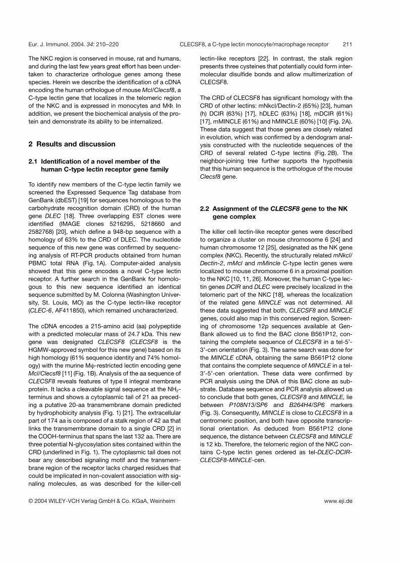
To identify new members of the C-type lectin family wescreened the Expressed Sequence Tag database fromGenBank (dbEST) [19] for sequences homologous to thecarbohydrate recognition domain (CRD) of the humangene DLEC [18]. Three overlapping EST clones wereidentified (IMAGE clones 5216295, 5218660 and2582768) [20], which define a 948-bp sequence with ahomology of 63% to the CRD of DLEC. The nucleotidesequence of this new gene was confirmed by sequenc-ing analysis of RT-PCR products obtained from humanPBMC total RNA (Fig. 1A). Computer-aided analysisshowed that this gene encodes a novel C-type lectinreceptor. A further search in the GenBank for homolo-gous to this new sequence identified an identicalsequence submitted by M. Colonna (Washington Univer-sity, St. Louis, MO) as the C-type lectin-like receptor(CLEC-6, AF411850), which remained uncharacterized.
The cDNA encodes a 215-amino acid (aa) polypeptidewith a predicted molecular mass of 24.7 kDa. This newgene was designated CLECSF8 (CLECSF8 is theHGMW-approved symbol for this new gene) based on itshigh homology (61% sequence identity and 74% homol-ogy) with the murine M ¶ -restricted lectin encoding geneMcl/Clecsf8 [11] (Fig. 1B). Analysis of the aa sequence ofCLECSF8 reveals features of type II integral membraneprotein. It lacks a cleavable signal sequence at the NH2-terminus and shows a cytoplasmic tail of 21 aa preced-ing a putative 20-aa transmembrane domain predictedby hydrophobicity analysis (Fig. 1) [21]. The extracellularpart of 174 aa is composed of a stalk region of 42 aa thatlinks the transmembrane domain to a single CRD [2] inthe COOH-terminus that spans the last 132 aa. There arethree potential N-glycosylation sites contained within theCRD (underlined in Fig. 1). The cytoplasmic tail does notbear any described signaling motif and the transmem-brane region of the receptor lacks charged residues thatcould be implicated in non-covalent association with sig-naling molecules, as was described for the killer-cell
lectin-like receptors [22]. In contrast, the stalk regionpresents three cysteines that potentially could form inter-molecular disulfide bonds and allow multimerization ofCLECSF8.
The CRD of CLECSF8 has significant homology with theCRD of other lectins: mNkcl/Dectin-2 (65%) [23], human(h) DCIR (63%) [17], hDLEC (63%) [18], mDCIR (61%)[17], mMINCLE (61%) and hMINCLE (60%) [10] (Fig. 2A).These data suggest that those genes are closely relatedin evolution, which was confirmed by a dendogram anal-ysis constructed with the nucleotide sequences of theCRD of several related C-type lectins (Fig. 2B). Theneighbor-joining tree further supports the hypothesisthat this human sequence is the orthologue of the mouseClecsf8 gene.
2.2 Assignment of the CLECSF8 gene to the NKgene complex
The killer cell lectin-like receptor genes were describedto organize a cluster on mouse chromosome 6 [24] andhuman chromosome 12 [25], designated as the NK genecomplex (NKC). Recently, the structurally related mNkcl/Dectin-2, mMcl and mMincle C-type lectin genes werelocalized to mouse chromosome 6 in a proximal positionto the NKC [10, 11, 26]. Moreover, the human C-type lec-tin genes DCIR and DLEC were precisely localized in thetelomeric part of the NKC [18], whereas the localizationof the related gene MINCLE was not determined. Allthese data suggested that both, CLECSF8 and MINCLEgenes, could also map in this conserved region. Screen-ing of chromosome 12p sequences available at Gen-Bank allowed us to find the BAC clone B561P12, con-taining the complete sequence of CLECSF8 in a tel-5’-3’-cen orientation (Fig. 3). The same search was done forthe MINCLE cDNA, obtaining the same B561P12 clonethat contains the complete sequence of MINCLE in a tel-3’-5’-cen orientation. These data were confirmed byPCR analysis using the DNA of this BAC clone as sub-strate. Database sequence and PCR analysis allowed usto conclude that both genes, CLECSF8 and MINCLE, liebetween P108N13/SP6 and B264H4/SP6 markers(Fig. 3). Consequently, MINCLE is close to CLECSF8 in acentromeric position, and both have opposite transcrip-tional orientation. As deduced from B561P12 clonesequence, the distance between CLECSF8 and MINCLEis 12 kb. Therefore, the telomeric region of the NKC con-tains C-type lectin genes ordered as tel-DLEC-DCIR-CLECSF8-MINCLE-cen.
Eur. J. Immunol. 2004. 34: 210–220 CLECSF8, a C-type lectin monocyte/macrophage receptor 211
© 2004 WILEY-VCH Verlag GmbH & Co. KGaA, Weinheim www.eji.de

Fig. 1. (A) cDNA and deduced amino acid sequence of CLECSF8. The Kozak-like translational start site is underlined. The startcodon and the polyadenylation-like signal are in bold letters. The putative transmembrane domain is boxed and shaded, the con-served cysteines are shown in bold letters, and the N-linked putative glycosylation sites are underlined. Numbers at the left sideindicate the nucleotide position relative to the translation initiation site. Numbers at the right side indicate aa position relative tothe start methionine. Black arrows indicate exon boundaries. The CLECSF8 nucleotide sequence has been submitted to theGenBank database under the accession number AY297446. (B) Alignment of amino acid sequence of mouse and humanCLECSF8. Numbers indicate the amino acid position relative to the initiation methionine. Conserved amino acid between bothsequences are shaded.
2.3 Genomic structure and alternative splicing ofthe CLECSF8 gene
The genomic structure of CLECSF8 was obtained bydirect comparison of the CLECSF8 cDNA with the geno-mic sequence of B561P12 clone. It reveals the sameorganization as the type II C-type lectin genes previouslydescribed [18, 23, 27–29]. The full length CLECSF8 isencoded by six exons, as represented in Fig. 1, withexon I encoding 9 amino acids (aa) of the cytoplasmicdomain, exon II encoding 31 aa mainly in the transmem-
brane domain, exon III encoding 37 aa in the stalk regionand exon IV-VI encoding the CRD domain. The analysisof the spliced acceptor and donor sequences matchedthe consensus (GT-AG) rule for splicing (data not shown).
RT-PCR experiments were performed on total RNA fromfreshly isolated PBMC using specific primers flanking theCLECSF8 ORF. A major 724-bp and a minor 572-bpband were obtained (data not shown). Sequencing ofboth PCR products revealed that the 724-bp band corre-sponded to the complete amplified ORF, while the 572-
212 I. Arce et al. Eur. J. Immunol. 2004. 34: 210–220
© 2004 WILEY-VCH Verlag GmbH & Co. KGaA, Weinheim www.eji.de

Fig. 2. Analysis of the CRD of CLECSF8. (A) Alignment of several CRD of group II C-type lectins. Numbers indicate the positionrelative to the first amino acid shown. Amino acids conserved among several sequences are shaded. (B) Neighbor-joining dendo-gram of several group II lectins CRD. Numbers indicate bootstrap values.
Fig. 3. Physical localization of the CLECSF8 and MINCLEgenes to the NKC. The upper line extending across repre-sents the chromosome 12p12.3–13.2 region. Positions ofthe centromere and telomere are shown. The gene andgenetic marker names are written above the line. Thegenetic distance in cM is indicated below the chromosomeline (Genethon map). In the lower line lectin genes areshaded.
bp band showed a deletion of 152 bp. These data indi-cated that the 572-bp band could correspond to an alter-native spliced mRNA (CLECSF8b) that is the result ofexon IV deletion that disrupts the established ORF andgenerates a premature stop codon. The translation ofthis spliced mRNA would lead to a protein lacking theCRD (data not shown).
2.4 Tissue and cellular distribution of CLECSF8transcripts
In order to characterize the distribution of CLECSF8gene expression we performed Northern blot analysis onpolyadenylated RNA from human spleen, lymph node,thymus, appendix, peripheral blood leukocytes, bonemarrow and fetal liver. Two bands of approximately 2.4and 1.2 kb were detected in bone marrow and peripheralblood leukocytes, and faintly in spleen (Fig. 4A). This tis-sue expression pattern, together with the fact that Mclgene is expressed in murine M ¶ , suggested thatCLECSF8 was expressed in human M ¶ . Therefore, cellu-lar distribution of CLECSF8 in hematopoietic cells wasfurther investigated by RT-PCR on total RNA from sev-eral sources. Expression of CLECSF8 was weakly
detected in monocytes, M ¶ derived in vitro from bloodmonocytes (MoM ¶ ) and in the monomyelocytic cell lineHL-60 (Fig. 4A and B). No expression was detected onother cell lines from hematopoietic origin such as Jurkat,K562, Peer, NKL, Raji or U937. CLECSF8 was alsodetected in PBL and faintly in Langerhans cells (LC)
Eur. J. Immunol. 2004. 34: 210–220 CLECSF8, a C-type lectin monocyte/macrophage receptor 213
© 2004 WILEY-VCH Verlag GmbH & Co. KGaA, Weinheim www.eji.de

Fig. 4. Expression of CLECSF8. (A) Northern blot analysis of CLECSF8 expression on total RNA from several immune tissues (leftpanel) and HL-60 cells and M ¶ derived in vitro from peripheral blood monocytes treated or not with IL-6 for 6 h (right panel). (B)RT-PCR on RNA from several cell lines and cell populations, freshly isolated monocytes, monocytes treated or not for 6 h withseveral stimuli and M ¶ : MoM ¶ , M ¶ derived in vitro from peripheral blood monocytes; PM ¶ , peritoneal M ¶ from a patient withperitonitis caused by Pseudomonas aeruginosa; RAM ¶ , SF M ¶ from a patient with rheumatoid arthritis; GM ¶ , SF M ¶ from apatient with gout.
whereas no expression has been detected in immatureDC or in other hematopoietic cell populations. To test ifCLECSF8 could be expressed on peritoneal M ¶ (PM ¶ ),as it was described for the mouse Mcl gene [11], we ana-lyzed RNA of PM ¶ obtained from the effluent of a patientreceiving peritoneal dialysis treatment, which presenteda peritonitis caused by a Pseudomonas aeruginosainfection. In addition, M ¶ isolated from synovium fluid(SF) of patients with rheumatoid arthritis (RAM ¶ ) or gout(GM ¶ ) were also analyzed. CLECSF8 displayed the high-est expression in PM ¶ , whereas RAM ¶ were slightlypositive and GM ¶ were negative. The low expressionlevel of CLECSF8 mRNA in normal tissues, and its up-regulation in the case of infection (see PM ¶ ), led us totest whether CLECSF8 could be an inducible gene as itwas described for MINCLE [10]. For this purpose, HL-60cells were incubated with different stimuli as PMA, LPSand IL-6 and MoM ¶ with IL-6. In both cases, only IL-6induced the CLECSF8 mRNA expression and this induc-tion was only slightly detected by Northern blot after 12-day exposure suggesting a low abundance of this mRNA(Fig. 4A). In addition, treatment of monocytes with vari-ous stimuli showed that IL-6, TNF- § , IL-10 and IFN- + up-regulated CLECSF8 mRNA, whereas LPS down-
regulated it. Strikingly, freshly isolated monocytes fromperipheral blood had higher levels of CLECSF8 expres-sion than those maintained in culture for 6 h without anystimulus or those isolated from buffy-coats. Thus,CLECSF8 seems to be down-regulated when mono-cytes are cultured in vitro or lack of their natural environ-ment, but the expression level can be maintained or evenup-regulated with the described cytokines.
Therefore, the expression of CLECSF8 appears to beconfined to monocytes and M ¶ , and can be up-regulated by IL-6, TNF- § , IFN- + and IL-10. In agreementwith these data, CLECSF8 is expressed in M ¶ from aperitoneal infection and RA, where the M ¶ are activatedby autocrine inflammatory stimuli, whereas in M ¶ fromgout, CLECSF8 is not expressed. This finding is in accor-dance with recent evidences suggesting that in gout theM ¶ are implicated in the control of inflammation byremoving apoptotic neutrophils and generation of anti-inflammatory rather than pro-inflammatory cytokines[30]. However, further studies are necessary to demon-strate a correlation among the inflammatory activity ofM ¶ and their CLECSF8 expression level.
214 I. Arce et al. Eur. J. Immunol. 2004. 34: 210–220
© 2004 WILEY-VCH Verlag GmbH & Co. KGaA, Weinheim www.eji.de

Fig. 5. Biochemical analysis of CLECSF8-FLAG molecule in transfected 293 cells. (A) Western blot analysis of 293 cell lysatestransfected with CLECSF8-FLAG or the vector alone ( ¶ ) under reducing (R) and non-reducing (NR) conditions. Molecular masssizes are shown on the right. (B) Western blot analysis of immunoprecipitates of 293 cells transfected with CLECSF8-FLAG or thevector alone under reducing conditions treated or not with N-glycanase.
2.5 Subcellular localization and biochemicalcharacterization of the CLECSF8 protein
To study the expression of the CLECSF8 protein, 293cells were transiently transfected with a FLAG-taggedCLECSF8 construct or with a control plasmid, and thetransfectants were analyzed by immunofluorescencemicroscopy and flow cytometry. CLECSF8-FLAG couldbe detected in the surface of transiently transfected cellsby both methods (data not shown). Also, CLECSF8-FLAG-transfected cells were lysed under reducing andnon-reducing conditions and analyzed by Western blotwith an anti-FLAG mAb (Fig. 5A). Under reducing condi-tions a specific band of a molecular mass of approxi-mately 30 kDa was detected together with other unspe-cific bands also visible in the control lane, while undernon-reducing conditions specific bands of approxi-mately 60 kDa, 90 kDa, and higher molecular weight,suggested that this receptor could form multimers stabi-lized by intermolecular disulfide bonds. These disulfidebonds might be established by the cysteine residuespresent in the stalk region of CLECSF8, as it wasdescribed for other lectins [31]. In addition, CLECSF8-FLAG was immunoprecipitated from transiently trans-fected cell lysates and treated with N-glycanase. Theremoval of N-linked carbohydrate residues resulted in aprotein with a reduced molecular mass demonstratingthat at least one of the previously predicted N-glycosylation sites is functional (Fig. 5B).
2.6 CLECSF8 is an endocytic receptor
Many surface lectins expressed in M ¶ and DC are impli-cated in antigen uptake and internalization [32–35]. Totest whether CLECSF8 may function as an endocyticreceptor, we performed internalization assays in 293cells transiently transfected with a CLECSF8-FLAG con-struct. CLECSF8-FLAG was cross-linked with anti-FLAGmAb, and its surface expression was measured by flowcytometry after allowing or not allowing internalization at37°C. The results showed that the surface expression ofCLECSF8 is significantly reduced from 38% to 13% asresult of its internalization (Fig. 6).
We also confirmed our results by immunofluorescenceand confocal microscopy analysis. CLECSF8-FLAG sur-face proteins on 293 transiently transfected cells werelabeled with the anti-FLAG mAb and cross-linked with ananti-mouse IgG1-FITC mAb. Cells were allowed to inter-nalize the cross-linked receptor at 37°C and then wereincubated again with an anti-mouse IgG1-rhodamine.Therefore, the receptor molecules that remained on thesurface of the cell would be visualized in yellow whereasthose receptors that were previously internalized wouldstain only green (Fig. 7). After incubation at 37°C most ofthe receptor molecules were internalized while at 4°Cmost of them remained on the cell surface. These resultsindicate that CLECSF8 is internalized after its cross-linking.
M ¶ are phagocytic cells involved in the uptake of anti-gens at the site of tissue damage or infection. Many C-type lectins expressed by M ¶ play a role in this process.
Eur. J. Immunol. 2004. 34: 210–220 CLECSF8, a C-type lectin monocyte/macrophage receptor 215
© 2004 WILEY-VCH Verlag GmbH & Co. KGaA, Weinheim www.eji.de

Fig. 6. Flow cytometry surface expression analysis of 293 cells transfected with CLECSF8-FLAG incubated with a control anti-body, anti-MHC-I, anti-transferrin receptor and anti-FLAG allowing or not internalization at 37°C for 30 min. Data shown in thebar diagram represent the mean and standard deviation from five independent experiments. Histograms show the FACS profileof one representative experiment.
Fig. 7. Internalization assays of CLECSF8 analyzed by confocal microscopy. Green color shows CLECSF8 receptor internalizedand noninternalized. Red shows CLECSF8 receptor remaining in the membrane at the cell surface. In the merged images thegreen color represents the internalized receptor and the colocalization of red and green (yellow) shows the remaining receptor inthe membrane. (A) Confocal laser scanning micrographs showing a horizontal section of the cells. DIC images of the field areshown on the right. (B) Confocal laser scanning micrographs showing horizontal projections of the same field. Colocalization his-tograms of green and red signals corresponding to these images are shown on the right.
216 I. Arce et al. Eur. J. Immunol. 2004. 34: 210–220
© 2004 WILEY-VCH Verlag GmbH & Co. KGaA, Weinheim www.eji.de

The expression pattern of CLECSF8 is associated withactivated M ¶ , and together with its internalization ability,suggests that this receptor may be involved in antigenuptake at the site of infection, either for clearance of theantigen or for processing and further presentation to Tcells. The identification and characterization of the grow-ing number of lectin receptors expressed in M ¶ wouldallow a better understanding of their roles in innate andacquired immunity and disease.
3 Concluding remarks
We identified a novel C-type lectin gene, CLECSF8, thehuman orthologue of mouse Mcl, coding for a type IItransmembrane protein with an extracellular CRD andwithout signaling motifs in the cytoplasmic domain.CLECSF8 presents the same genomic structure as otherrelated lectins and one alternative spliced isoform wasdetected. The CLECSF8 gene is located close toMINCLE on the telomeric region of the NKC and bothgenes have opposite transcriptional orientation.CLECSF8 mRNA is expressed in PBL, bone marrow,spleen and, within the hematopoietic lineage, in mono-cytes and M ¶ . It is also detected in the monomyelocyticcell line HL-60. The expression of this receptor shows tobe up-regulated by IL-6, TNF- § , IFN- + and IL-10. There-fore, these data indicate that CLECSF8 is a novel mem-ber of the C-type lectin receptors that is internalized afterits cross-linking, suggesting a potential role in antigenuptake in activated monocytes and M ¶ .
4 Materials and methods
4.1 Identification of EST
The human Expressed Sequence Tag database from Gen-Bank (dbEST) [19] (http://www.ncbi.nlm.nih.gov/dbEST/index.html) was screened using the tblastn algorithm in theBLAST 2.0 program [36] available at the National Center forBiotechnology Information (http://www.ncbi.nlm.nih.gov/BLAST) with the CRD of the human DLEC protein sequence(accession number NP–569708) as query. This identifiedthree overlapping EST IMAGE clones: 5216295, 5218660and 2582768, provided by the Human Genome MappingProject Resource Center (HGMP, MRC, GB).
4.2 RNA isolation
Total RNA was isolated from cells with Ultraspec reagent(Biotecx Labs, Houston, TX) according to the manufacturer’sinstructions. The complete ORF was obtained by performingRT-PCR on PBMC total RNA using specific primers for the 5’and 3’ UTR sequences deduced from the EST sequences
(CLECSF81: 5’ GAA CAA ATT CTT GCC GTC CTG ACC 3’and CLECSF82: 5’ TCC ATC ACA AGG ACC ACT TTC TGAG 3’). The PCR products obtained in two independent reac-tions were cloned in pCR2.1-TOPO and sequenced. DNAsequencing was performed using d-rhodamine terminatorcycle sequencing kit and an ABI PRISM 337 DNA sequencer(Perkin Elmer-Applied Biosystems, CA). Sequence align-ments were carried out using the BLAST and the ClustalW atthe Baylor College of Medicine search server(http://dot.imgen.bcm.tmc.edu:9331/multi-align/options/clustalw.html). The transmembrane domain was assignedusing the SOSUI program available at the ExPASy Proteom-ics tools (http://www.expasy.ch/tools). A phylogenetic treebased on the CRD sequences was constructed using theneighbor-joining method [37] and the computer programsNJBOOT (kindly provided by Dr. M. Nei). The bootstrappinganalysis for tree topology [38] was used to evaluate the sta-tistical significance of each internal branch of the tree.
4.3 Genomic structure
The genomic structure and the intron lengths of theCLECSF8 gene were directly deduced from the sequence ofthe genomic BAC clone RPCI11–561P12 (GenBank acces-sion number AC092746).
4.4 Physical localization of the CLECSF8 and MINCLEgenes
BAC clones from the Roswell Park Center Institute (RPCI)libraries constructed by de Jong and colleagues [39] wereprovided by the BAC/PAC Resource (www.chori.org/bacpac). The BAC-based physical map is availableat http://www.hpcgg.org/Sequence/human/seq–ready–-maps.html. For each gene, PCR reactions were performedwith specific primers within a single exon (CLECSF83: 5’GGA GCA CCT GGA ACT GTT GTC C 3’ and CLECSF84: 5’CGT GCT GAT GGT CAT CAG ATG G 3’ for CLECSF8 gene,and MINCLE1: 5’ GGG AGC CCA ACA ACA TAG CTA CC 3’and MINCLE2: 5’ TGT GGC CAT GTT CTT GCT CTT CC 3’for MINCLE gene). The PCR was performed for 35 cycleswith an annealing temperature of 68°C. The PCR productswere visualized on 1% agarose gel electrophoresis andsequenced.
4.5 Cell preparation
Cells were grown in RPMI 1640 medium with Glutamax-1(Gibco/BRL, USA) supplemented with penicillin (100 U/ml),streptomycin (100 mg/ml) and 10% fetal calf serum (FCS).PBMC were purified from human peripheral blood of healthydonors or buffy-coats by Ficoll centrifugation (Lymphoprep,Nycomed, Oslo, Norway). NK cells were obtained fromPBMC cultured in the presence of irradiated RPMI 8866 cellline. After one week, NK cells were purified by immunode-
Eur. J. Immunol. 2004. 34: 210–220 CLECSF8, a C-type lectin monocyte/macrophage receptor 217
© 2004 WILEY-VCH Verlag GmbH & Co. KGaA, Weinheim www.eji.de

pletion with anti-CD3-, anti-CD19- and anti-CD14-coatedmagnetic beads (Dynal, Oslo, Norway). Surface phenotypeof obtained NK cells analyzed by flow cytometry yieldedG 96% CD56+ CD3– purity. DC cultures were obtained from
peripheral blood monocytes. Briefly, PBMC in RPMI 164010% FCS were allowed to adhere to 6-well plates (BectonDickinson, NJ). After 1 h at 37°C the nonadherent cells wereremoved and the adherent monocytes were cultured inRPMI 1640 10% FCS with 200 ng/ml GM-CSF (NovartisPharma, Basel, Switzerland) and 10 ng/ml IL-4 (R&D Sys-tems, MN). DC were tested by flow cytometry for CD14–,CD80+ and CD83– expression. LC were obtained fromperipheral blood monocytes as described for DC but adding10 ng/ml of TGF- g (R&D Systems) to the cytokine cocktailand tested for the expression of E-cadherin. M ¶ wereobtained from adherent PBMC cultured with RPMI 1640with 10% FCS and 200 ng/ml GM-CSF for 6 days. M ¶ weretested by flow cytometry for CD14+, CD11b+ and CD16+
expression. The NKL cell line [40] was grown in the presenceof 100 U/ml human recombinant IL-2 (Hoffman-La Roche,NJ). HEK293T cells [41], where grown in Dulbecco’s modi-fied Eagle’s medium (DMEM), supplemented with 10% FCS,2 mM L-glutamine and 50 ? g/ml gentamycin. Cells weretreated 6 h with 2000 U/ml of IL-6 (PrepoTech, NJ), 250 U/mlIFN- + (R&D Systems), 100 ng/ml IL-10 (R&D Systems),100 ng/ml LPS from Escherichia coli 055:B5 (Sigma, St.Louis, MO) or 250 U/ml TNF- § (Strathmann Biotec AG,Hamburg, Germany).
M ¶ from effluents in dialysis fluid from a patient sufferingperitonitis caused by Pseudomonas aeruginosa undergoingcontinuous ambulatory peritoneal dialysis were isolated bycentrifugation of dialysis fluid. Of the cells obtained, 90%were M ¶ as determined by flow cytometry, and were cul-tured for 2 days with 200 ng/ml GM-CSF. SF M ¶ wereobtained from two patients with RA and gout, respectively,by Ficoll centrifugation and the adherent cells were cultured5 days with 200 ng/ml GM-CSF.
The study was approved by the ethics committee of HospitalUniversitario de la Princesa, and oral informed consent wasobtained from all donors.
4.6 Biochemical analysis
To generate the CLECSF8-FLAG construct, CLECSF8 cDNAwas prepared by PCR using a 3’ primer that removes thestop codon and adds the FLAG (DYKDDDDK) epitope. ThePCR product obtained was cloned in the expression vectorpCR3 (Invitrogen BV, De Schelp, The Netherlands) andsequenced. HEK293T cells were transiently transfected withpCR3-CLECSF8-FLAG or pCR3-empty plasmid using stan-dard calcium phosphate protocol. Forty-eight hours aftertransfection, cells were lysed in 1% Nonidet-P40 (NP-40)lysis buffer. Samples were subjected to 10% or 12% sodiumdodecyl sulfate polyacrylamide gel electrophoresis under
non-reducing and reducing conditions (5% 2-ME), trans-ferred to nitrocellulose membranes and probed with M2mAb in Western blot using a chemiluminescent kit (Super-Signal West Pico, IL). For N-glycosidase treatment,CLECSF8-FLAG was immunoprecipitated from cell lysateswith Sepharose-PA (Pharmacia Biotech, Piscataway, NJ)coupled to the anti-FLAG M2 mAb (Sigma, St. Louis, MO)and further treated with N-glycosidase F (Roche DiagnosticsGmbH, Mannheim, Germany) in PBS buffer with 0.5% SDS,1% NP-40, 50 mM g -mercaptoethanol and 50 mM EDTA at37°C overnight.
4.7 Flow cytometry analysis, immunofluorescence andconfocal analysis
For flow cytometry analysis experiments CLECSF8-FLAGtransiently transfected HEK293T cells were incubated at 4°Cfor 20 min with the control antibody (X63), anti-MHC-I (W6/32), anti-transferrin receptor (T2/12) or 5 ? g/ml anti-FLAGM2 mAb in PBS. Cells were washed and set or not at 37°Cfor 30 min to allow internalization. Cells were fixed in PBSwith 4% paraformaldehyde and then labeled with rabbitanti-mouse IgG1-FITC mAb (Dako, Denmark).
For immunofluorescence and confocal analysis CLECSF8-FLAG transiently transfected HEK293T cells were seeded onfibronectin-coated glass plates and allowed to grow over-night. The following morning, plates were incubated at 4°Cfor 20 min with 5 ? g/ml anti-FLAG M2 mAb in PBS and thencross-linked with 10 ? g/ml of rabbit anti-mouse IgG1-FITCmAb. Cells were washed and set or not at 37°C for 30 min toallow internalization. Cells were fixed in PBS with 4% para-formaldehyde and then labeled with Rhodamine-X goat anti-mouse IgG (H+L) mAb (Molecular Probes, OR). Series ofoptical sections were obtained with a Leica TCS-SP confo-cal laser scanning unit equipped with Ar and He/Ne laserbeams and attached to a Leica DMIRBE inverted epifluores-cence microscope (Leica Microsystems), using a 63× oilimmersion objective. Colocalization histograms wereobtained using the Leica Confocal Software.
4.8 Expression studies
Standard Northern blot analysis was performed using both,a Multiple Tissue Northern blot membrane (Clontech, PaloAlto, CA) and total RNA (10 ? g/lane) isolated from differentsources. The complete cDNA of CLECSF8 was used as aprobe. Reverse transcription was performed with 1 ? g oftotal RNA using the RNA PCR core Kit (Perkin Elmer, GR).PCR was performed for 35 cycles with an annealing temper-ature of 64°C. Primers CLECSF81 and CLECSF82 (seeabove) flanking the ORF of CLECSF8 were used for the PCRreaction. For g -actin control amplification, specific primerswere used in the same PCR conditions with the exception ofthe 60°C annealing temperature (5’ ATC TGG CAC CACCAC CTT CTA CAA TGA GCT GCC 3’ as sense primer and 5’
218 I. Arce et al. Eur. J. Immunol. 2004. 34: 210–220
© 2004 WILEY-VCH Verlag GmbH & Co. KGaA, Weinheim www.eji.de

CGT CAT ACT CCT GCT TGC TGA TCC ACA TCT TGC 3’ asantisense primer).
Acknowledgements: This work was supported by grantsfrom the Spanish Government (CAM 08.3/003/2001 and FIS02/0520) to E.F.-R. P.R.-N. is recipient of a fellowship fromCAM. We are very grateful to Dr. M. Lopez-Cabrera, Dr. E.Lara-Pezzi and O. Barreiro for helpful discussion and criticalreading of the manuscript, and Dr. M. Yanez-Mo for helpwith confocal analysis. Also to Dr. I. Gonzalez-Alvaro and Dr.C. Domınguez-Jimenez for synovium fluid samples supplyand advice, and M. Ramirez-Huesca for technical assis-tance.
References
1 Lewis, C. and McGee, J. O. D., The Macrophage: The NaturalImmune System, IRL Press at Oxford University Press, Oxford1992.
2 Drickamer, K., Ca2+ dependent carbohydrate-recognitiondomains in animal proteins. Curr. Opin. Struc. Biol. 1993. 3:393–400.
3 Feizi, T., Carbohydrate-mediated recognition systems in innateimmunity. Immunol. Rev. 2000. 173: 79–88.
4 Suzuki, N., Yamamoto, K., Toyoshima, S., Osawa, T. and Iri-mura, T., Molecular cloning and expression of cDNA encodinghuman macrophage C-type lectin. Its unique carbohydrate bind-ing specificity for Tn antigen. J. Immunol. 1996. 156: 128–135.
5 Tsuiji, M., Fujimori, M., Ohashi, Y., Higashi, N., Onami, T. M.,Hedrick, S. M. and Irimura, T., Molecular cloning and character-ization of a novel mouse macrophage C-type lectin, mMGL2,which has a distinct carbohydrate specificity from mMGL1. J.Biol. Chem. 2002. 277: 28892–28901.
6 Stahl, P. D. and Ezekowitz, R. A., The mannose receptor is apattern recognition receptor involved in host defense. Curr. Opin.Immunol. 1998. 10: 50–55.
7 Yamamoto, K., Ishida, C., Shinohara, Y., Hasegawa, Y.,Konami, Y., Osawa, T. and Irimura, T., Interaction of immobi-lized recombinant mouse C-type macrophage lectin with glyco-peptides and oligosaccharides. Biochemistry 1994. 33:8159–8166.
8 Denda-Nagai, K., Kubota, N., Tsuiji, M., Kamata, M. and Iri-mura, T., Macrophage C-type lectin on bone marrow-derivedimmature dendritic cells is involved in the internalization of glyco-sylated antigens. Glycobiology 2002. 12: 443–50.
9 Higashi, N., Fujioka, K., Denda-Nagai, K., Hashimoto, S.,Nagai, S., Sato, T., Fujita, Y., Morikawa, A., Tsuiji, M., Miyata-Takeuchi, M., Sano, Y., Suzuki, N., Yamamoto, K., Matsu-shima, K. and Irimura, T., The macrophage C-type lectin spe-cific for galactose/N-acetylgalactosamine is an endocytic recep-tor expressed on monocyte-derived immature dendritic cells. J.Biol. Chem. 2002. 277: 20686–20693.
10 Matsumoto, M., Tanaka, T., Kaisho, T., Sanjo, H., Copeland, N.G., Gilbert, D. J., Jenkins, N. A. and Akira, S., A novel LPS-inducible C-type lectin is a transcriptional target of NF-IL6 inmacrophages. J. Immunol. 1999. 163: 5039–5048.
11 Balch, S. G., McKnight, A. J., Seldin, M. F. and Gordon, S.,Cloning of a novel C-type lectin expressed by murine macro-phages. J. Biol. Chem. 1998. 273: 18656–18664.
12 Tsuiji, M., Fujimori, M., Seldin, M. F., Taketo, M. M. andIrimura, T., Genomic structure and chromosomal location of themouse macrophage C-type lectin gene. Immunogenetics 1999.50: 67–70.
13 Yokoyama, W. M. and Plougastel, B. F., Immune functionsencoded by the natural killer gene complex. Nat. Rev. Immunol.2003. 3: 304–316.
14 Hermanz-Falcon, P., Arce, I., Roda-Navarro, P. andFernandez-Ruiz, E., Cloning of human DECTIN-1, a novel C-type lectin-like receptor gene expressed on dendritic cells.Immunogenetics 2001. 53: 288–295.
15 Colonna, M., Samaridis, J. and Angman, L., Molecular charac-terization of two novel C-type lectin-like receptors, one of whichis selectively expressed in human dendritic cells. Eur. J. Immunol.2000. 30: 697–704.
16 Sobanov, Y., Bernreiter, A., Derdak, S., Mechtcheriakova, D.,Schweighofer, B., Duchler, M., Kalthoff, F. and Hofer, E., Anovel cluster of lectin-like receptor genes expressed in mono-cytic, dendritic and endothelial cells maps close to the NK recep-tor genes in the human NK gene complex. Eur. J. Immunol. 2001.31: 3493–3503.
17 Bates, E. E. M., Fournier, N., Garcia, E., Valladeau, J., Durand,I., Pin, J.-J., Zurawski, S. M., Patel, S., Abrams, J. S., Lebec-que, S., Garrone, P. and Saeland, S., APCs express DCIR, anovel C-type lectin surface receptor containing an immunorecep-tor tyrosine-based inhibitory motif. J. Immunol. 1999. 163:1973–1983.
18 Arce, I., Roda-Navarro, P., Montoya, M. C., Hernanz-Falcon,P., Puig-Kroger, A. and Fernandez-Ruiz, E., Molecular andgenomic characterization of human DLEC, a novel member of theC-type lectin receptor gene family preferentially expressed onmonocyte-derived dendritic cells. Eur. J. Immunol. 2001. 31:2733–2740.
19 Boguski, M. S., Lowe, T. M. J. and Tolstochev, C. M., dbEST-database for “expressed sequence tags”. Nat. Genet. 1993. 4:332–333.
20 Lennon, G. G., Auffray, C., Polymeropoulos, M. and Soares,M. B., The I.M.A.G.E. consortium: An integrated molecular analy-sis of genomes and their expression. Genomics 1996. 33:151–152.
21 Kyte, J. and Doolittle, R. F., A simple method for displaying thehydropathic character of a protein. J. Mol. Biol. 1982. 157:105–132.
22 Moretta, A., Bottino, C., Vitale, M., Pende, D., Cantoni, C.,Mingari, M. C., Biassoni, R. and Moretta, L., Activating recep-tors and coreceptors involved in human natural killer cell-mediated cytolysis. Annu. Rev. Immunol. 2001. 19: 197–223.
23 Ariizumi, K., Shen, G.-L., Shikano, S., Ritter III, R., Zukas, P.,Edelbaum, D., Morita, A. and Takashima, A., Cloning of a sec-ond dendritic cell-associated C-type lectin (Dectin-2) and itsalternative spliced isoforms. J. Biol. Chem. 2000. 275:11957–11963.
24 Brown, M. G., Scalzo, A., Matsumoto, K. and Yokoyama, W.M., The natural killer gene complex: a genetic basis for under-standing natural killer cell function and innate immunity. Immunol.Rev. 1997. 155: 53–65.
25 Renedo, M., Arce, I., Montgomery, K., Roda-Navarro, P., Lee,E., Kucherlapati, R. and Fernandez-Ruiz, E., A sequence-ready physical map of the region containing the human naturalkiller gene complex on chromosome 12p12.3-p13.2. Genomics2000. 65: 129–136.
26 Fernandes, M. J., Finnegan, A. A., Siracusa, L. D., Brenner, C.,Iscove, N. N. and Calabretta, B., Characterization of a novel
Eur. J. Immunol. 2004. 34: 210–220 CLECSF8, a C-type lectin monocyte/macrophage receptor 219
© 2004 WILEY-VCH Verlag GmbH & Co. KGaA, Weinheim www.eji.de

receptor that maps near the Natural Killer gene complex: demon-stration of carbohydrate binding and expression in hematopoieticcells. Cancer Res. 1999. 59: 2709–2717.
27 Bezouska, K., Crichlow, G. V., Rose, J. M., Taylor, M. E. andDrickamer, K., Evolutionary conservation of intron position in asubfamily of genes encoding carbohydrate-recognition domains.J. Biol. Chem. 1991. 266: 11604–11609.
28 Rodrıguez, A., Carretero, M., Glienke, J., Bellon, T., Ramırez,A., Lehrach, H., Francis, F. and Lopez-Botet, M., Structure ofthe human CD94 C-type lectin gene. Immunogenetics 1998. 47:305–309.
29 Roda-Navarro, P., Arce, I., Renedo, M., Montgomery, K.,Kucherlapati, R. and Fernandez-Ruiz, E., Human KLRF1, anovel member of the killer cell lectin-like receptor gene family:molecular characterization, genomic structure, physical mappingto the NK gene complex and expression analysis. Eur. J. Immu-nol. 2000. 30: 568–576.
30 Haskard, D. O. and Landis, R. C., Interactions between leuko-cytes and endothelial cells in gout: lessons from a self-limitinginflammatory response. Arthritis Res. 2002. 4: 91–97.
31 Sancho, D., Santis, A. G., Alonso-Lebrero, J. L., Viedma, F.,Tejedor, R. and Sanchez-Madrid, F., Functional analysis ofligand-binding and signal transduction domains of CD69 andCD23 C-type lectin leukocyte receptors. J. Immunol. 2000. 165:3868–3875.
32 Sallusto, F., Cella, M., Danieli, C. and Lanzavecchia, A., Den-dritic cells use macropinocytosis and the mannose receptor toconcentrate macromolecules in the major histocompatibilitycomplex class II compartment: downregulation by cytokines andbacterial products. J. Exp. Med. 1995. 182: 389–400.
33 Jiang, W., Swiggard, W. J., Heufler, C., Peng, M., Mirza, A.,Steinman, R. M. and Nussenzweig, M. C., The receptor DEC-205 expressed by dendritic cells and thymic epithelial cells isinvolved in antigen processing. Nature 1995. 375: 151–155.
34 Brown, G. D. and Gordon, S., Immune recognition. A new recep-tor for beta-glucans. Nature 2001. 413: 36–37.
35 Willment, J. A., Gordon, S. and Brown, G. D., Characterizationof the human Beta -glucan receptor and its alternatively splicedisoforms. J. Biol. Chem. 2001. 276: 43818–43823.
36 Altschul, S. F., Stephen, F., Madden, T. L., Schäffer, A. A.,Zhang, J., Zhang, Z., Miller, W. and Lipman, D. J., GappedBLAST and PSI-BLAST: a new generation of protein databasesearch programs. Nucleic Acids Res. 1997. 25: 3389–3402.
37 Saitou, N. and Nei, M., The neighbor-joining method. A newmethod for reconstructing phylogenetic trees. Mol. Biol. Evol.1987. 4: 406–425.
38 Felsenstein, J., Confidence limits of phylogenies: an approachusing the bootstrap. Evolution 1985. 39: 783–795.
39 Ioannou, P. A., Amemiya, C. T., Garnes, J., Kroisel, P. M., Shi-zuya, H., Chen, C., Batzer, M. A. and de Jong, P. J., A new bac-teriophage P1-derived vector for the propagation of large humanDNA fragments. Nat. Genet. 1994. 6: 84–89.
40 Robertson, M. J., Cochran, K. J., Cameron, C., Le, J.-M.,Tantravahi, R. and Ritz, J., Characterization of a cell line, NKL,derived from an aggresive human natural killer cell leukemia. Exp.Hematol. 1996. 24: 406–415.
41 DuBridge, R. B., Tang, P., Hsia, H. C., Leong, P. M., Miller, J. H.and Calos, M. P., Analysis of mutation in human cells by using anEpstein-Barr virus shuttle system. Mol. Cell Biol. 1987. 7:379–387.
Correspondence: Elena Fernandez-Ruiz, Unidad de Bio-logıa Molecular. Hospital Universitario de la Princesa. c/Diego de Leon 62, E-28006 Madrid, SpainFax: +34-91-520-2374e-mail: efernandez.hlpr — salud.madrid.org
220 I. Arce et al. Eur. J. Immunol. 2004. 34: 210–220
© 2004 WILEY-VCH Verlag GmbH & Co. KGaA, Weinheim www.eji.de
Copyright © 2022 FDOKUMEN