Mouse Monocyte-Derived Chemokine Is Involved in Airway Hyperreactivity and Lung Inflammation
Upload
independentCategory
view
2download
0
RESEARCH PAPER
Identification and profilingof CXCR3–CXCR4chemokine receptorheteromer complexesAO Watts1, MMH van Lipzig1, WC Jaeger4, RM Seeber4, M van Zwam2,J Vinet2, MMC van der Lee3, M Siderius1, GJR Zaman3*,HWGM Boddeke2, MJ Smit1, KDG Pfleger4,5, R Leurs1 and HF Vischer1
1Leiden/Amsterdam Center for Drug Research, Division of Medicinal Chemistry, Faculty of
Science, VU University Amsterdam, Amsterdam, The Netherlands, 2Department of Medical
Physiology, University Medical Center Groningen, University of Groningen, Groningen, The
Netherlands, 3Molecular Pharmacology & DMPK, MSD, Merck Research Laboratories, Oss, The
Netherlands, 4Laboratory for Molecular Endocrinology-GPCRs, Western Australian Institute for
Medical Research (WAIMR) and Centre for Medical Research, The University of Western
Australia, Nedlands, Australia, and 5Dimerix Bioscience Pty Ltd, Nedlands, Australia
CorrespondenceDr Henry F Vischer,Leiden/Amsterdam Center forDrug Research, Division ofMedicinal Chemistry, Facultyof Science, VU UniversityAmsterdam, De Boelelaan 1083,1081 HV Amsterdam, TheNetherlands. E-mail:h.f.vischer@vu.nl----------------------------------------------------------------
*Present address: NetherlandsTranslational Research CenterB.V., Molenstraat 110, 5342 CCOss, The Netherlands.----------------------------------------------------------------
Keywordschemokine receptor; GPCR;heteromerization; b-arrestin;radioligand binding; CXCR3;CXCR4----------------------------------------------------------------
Received23 July 2012Revised29 September 2012Accepted29 October 2012
BACKGROUND AND PURPOSEThe C-X-C chemokine receptors 3 (CXCR3) and C-X-C chemokine receptors 4 (CXCR4) are involved in various autoimmunediseases and cancers. Small antagonists have previously been shown to cross-inhibit chemokine binding to CXCR4, CCchemokine receptors 2 (CCR2) and 5 (CCR5) heteromers. We investigated whether CXCR3 and CXCR4 can form heteromericcomplexes and the binding characteristics of chemokines and small ligand compounds to these chemokine receptorheteromers.
EXPERIMENTAL APPROACHCXCR3–CXCR4 heteromers were identified in HEK293T cells using co-immunoprecipitation, time-resolved fluorescenceresonance energy transfer, saturation BRET and the GPCR-heteromer identification technology (HIT) approach. Equilibriumcompetition binding and dissociation experiments were performed to detect negative binding cooperativity.
KEY RESULTSWe provide evidence that chemokine receptors CXCR3 and CXCR4 form heteromeric complexes in HEK293T cells. Chemokinebinding was mutually exclusive on membranes co-expressing CXCR3 and CXCR4 as revealed by equilibrium competitionbinding and dissociation experiments. The small CXCR3 agonist VUF10661 impaired binding of CXCL12 to CXCR4, whereassmall antagonists were unable to cross-inhibit chemokine binding to the other chemokine receptor. In contrast, negativebinding cooperativity between CXCR3 and CXCR4 chemokines was not observed in intact cells. However, using the GPCR-HITapproach, we have evidence for specific b-arrestin2 recruitment to CXCR3-CXCR4 heteromers in response to agoniststimulation.
CONCLUSIONS AND IMPLICATIONSThis study indicates that heteromeric CXCR3–CXCR4 complexes may act as functional units in living cells, which potentiallyopen up novel therapeutic opportunities.
AbbreviationsCFP, cyan fluorescent protein; CXCL10, C-X-C chemokine ligand 10; CXCL11, C-X-C chemokine ligand 11; CXCL12,C-X-C chemokine ligand 12; CXCL9, C-X-C chemokine ligand 9; CXCR3, C-X-C chemokine receptor 3; CXCR4, C-X-Cchemokine chemokine receptor 4; eBRET, extended BRET; EYFP, enhanced yellow fluorescent protein; FCS, fetal calfserum; GABAB2, GABA-B receptor 2; HA, haemagglutinin epitope tag; HIT, heteromer identification technology; IP,immunoprecipitation; Rluc, Renilla luciferase; RT, room temperature; trFRET, time-resolved FRET
BJP British Journal ofPharmacology
DOI:10.1111/bph.12064www.brjpharmacol.org
1662 British Journal of Pharmacology (2013) 168 1662–1674 © 2012 The AuthorsBritish Journal of Pharmacology © 2012 The British Pharmacological Society

Introduction
Chemokines are secreted chemoattractant proteins of8–14 kDa that direct immune cell migration through interac-tion with GPCRs. Over 40 chemokines and 20 chemokinereceptors have been identified in humans, forming a regula-tory system in which many of these receptors promiscuouslybind multiple chemokines and vice versa (Scholten et al.,2012a). In addition, chemokine receptors can also form bothhomo- and heteromers (Scholten et al., 2012a). The relativeoccurrence of such GPCR complexes and transiency,however, is still a matter of debate (Dorsch et al., 2009; Hernet al., 2010). Importantly, heteromers between the CC chem-okine receptors 2 (CCR2), 5 (CCR5) and C-X-C chemokinereceptor 4 (CXCR4) can only bind a single chemokine withhigh affinity (El-Asmar et al., 2005; Springael et al., 2006;Sohy et al., 2007; 2009). This negative binding cooperativitybetween the heteromerized chemokine receptors is notlimited to the cognate chemokines of both receptors, butextends to synthetic allosteric antagonists of these receptors(Sohy et al., 2007; 2009). Binding of these antagonists to onereceptor allosterically inhibits chemokine binding to theother receptor within the heteromer, resulting in a cross-inhibition of intracellular signalling, and in vitro and in vivochemotaxis (Sohy et al., 2007; 2009). Besides inhibitingone chemokine receptor by targeting its heteromeric partner,chemokine receptor heteromers may be therapeutically tar-geted by heteromer-selective ligands and/or bivalent ligandsthat simultaneously bind both receptors in a dimer, as previ-ously described for, for opioid receptor heteromers (Waldhoeret al., 2005; Mathews et al., 2008; Jahnichen et al., 2010;Tanaka et al., 2010). Because GPCR heteromers are likelyexpressed on a more limited subset of cell types as comparedwith the individual GPCRs, heteromer-selective therapeuticagents have the potential to display improved efficacy andtoxicity profiles.
C-X-C chemokine receptor 3 (CXCR3) and CXCR4 areexpressed on activated T-cells, natural killer cells, dendriticcells and cancer cells (Vandercappellen et al., 2008; Viola andLuster, 2008; Fulton, 2009). CXCR4 plays an essential role inhaematopoiesis, leukocyte homing/retention in secondarylymphoid tissues and recruitment to sites of inflammation, inresponse to local concentrations of C-X-C chemokine ligand12 (CXCL12; Moser and Loetscher, 2001). Additionally,CXCR4 and CXCL12 are upregulated in tumours by, forexample hypoxia, and mediate angiogenesis, proliferation,invasion and metastasis (Vandercappellen et al., 2008; Violaand Luster, 2008; Fulton, 2009). Expression of CXCR3 and itsligands C-X-C chemokine ligand 9 (CXCL9), C-X-C chemok-ine ligand 10 (CXCL10) and C-X-C chemokine ligand 11(CXCL11) is induced under inflammatory conditions, andhas been implicated in autoimmune disease, graft-versus-hostdisease and transplant rejection (Lacotte et al., 2009). Moreo-ver, CXCR3 is also involved in cancer proliferation andmetastasis (Vandercappellen et al., 2008; Viola and Luster,2008; Fulton, 2009).
In the present study, we show that CXCR3 and CXCR4form heteromers at the cell surface. Negative binding coop-erativity between CXCR3 and CXCR4 agonists was detectedin membrane preparations of cells that co-express both recep-
tors, but not on intact cells. However, b-arrestin2 recruitmentspecifically to CXCR3–CXCR4 heteromers was shown inliving cells using the GPCR heteromer identification technol-ogy (GPCR-HIT) approach (Mustafa and Pfleger, 2011; Seeet al., 2011; Mustafa et al., 2012).
Methods
MaterialsThe small CXCR3 ligands VUF10085 [(Johnson et al.,2007) and VUF10661 (N-(6-amino-1-((2,2-diphenyl-ethyl)amino)-1-oxohexan-2-yl)-2-(4-oxo-4-phenylbutanoyl)-1,2,3,4-tetrahydro-isoquinoline-3-carboxamide] have been pre-viously described as AMG 487 (Johnson et al., 2007) andcompound 2 (Stroke et al., 2006), respectively, and weresynthesized at VU University Amsterdam (Scholten et al.,2012b). TAK-779 (N,N-dimethyl-N-[4-[[[2-(4-methylphenyl)-6,7-dihydro-5H-benzocyclohepten-8-yl]carbonyl]-amino]benzyl]-tetrahydro-2H-pyran-4-aminium chloride) was ob-tained through the AIDS Research and Reference ReagentProgram, Division of AIDS, NIAID, NIH (Baba et al., 1999).AMD3100 (1,1’-[1,4-phenylenebis-(methylene)]-bis-1,4,8,11-tetraazacyclotetra-decane) was obtained from Sigma-Aldrich(St-Louis, MO, USA). CXCL10 [immunoprecipitation (IP-10)],CXCL11 (I-TAC), and CXCL12 (SDF-1a) were from Peprotech(Rocky Hill, NJ, USA).
DNA constructsHuman CXCR3 (Verzijl et al., 2008) and CXCR4 were taggedat the N-terminus with FLAG (DYKDDDDK) or haemaggluti-nin epitope tag (HA; YPYDVPDYA) using PCR-based methodsand subcloned in the pcDEF3 expression plasmid (a gift fromDr. Langer, Robert Wood Johnson Medical School, Piscata-way, NJ, USA). The receptor fusion proteins CXCR3-Renillaluciferase (Rluc), CXCR3-enhanced yellow fluorescentprotein (EYFP), CXCR4-Rluc, and CXCR4-EYFP have beenpreviously described (Vischer et al., 2008). CXCR3-Rluc8 wasconstructed by substitution of Rluc with the optimized Rluc8variant as previously described (Nijmeijer et al., 2010).CXCR4-Rluc8 and b-arrestin2-Venus constructs have beendescribed previously (See et al., 2011). For sensitized emissionFRET studies, CXCR3, CXCR4 and GABAB2 constructs weresubcloned into the pECFP-N1 vector (Clontech LaboratoriesInc., Mountain View, CA, USA) and the pVenus-N1 vector.The latter plasmid was constructed from pECFP-N1 by sub-stitution of the sequence encoding for cyan fluorescentprotein (CFP) with Venus DNA, which was obtained frompcDNA3.1-eCFP-exchange protein activated by cAMP (EPAC)-Venus (a gift from Dr. Jalink, The Netherlands Cancer Insti-tute, Amsterdam, The Netherlands). All generated constructswere verified by DNA sequencing.
Cell culture and transfectionHEK293T cells were cultured in DMEM containing 10% fetalcalf serum (FCS), 100 units·mL-1 penicillin and 100 mg·mL-1
streptomycin (PAA Laboratories GmbH, Cölbe, Germany)were maintained at 37°C in a humidified atmosphere con-taining 5% CO2. For saturation BRET, time-resolved FRET(trFRET), IP and radioligand-binding studies, HEK293T cells
BJPCXCR3–CXCR4 heteromers
British Journal of Pharmacology (2013) 168 1662–1674 1663

were transfected with indicated amounts of plasmid DNAusing 25-kDa linear polyethylenimine (Polysciences, Eppel-heim, Germany) as described previously (Verzijl et al., 2008).Transfected DNA was held constant by adjusting the totalamount of DNA with the empty vector pcDEF3. For GPCR-HIT assays, HEK293FT cells were maintained at 37°C in 5%CO2 and complete media [DMEM containing 0.3 mg·mL-1
glutamine, 100 IU·mL-1 penicillin and 100 mg·mL-1 strepto-mycin (Gibco, Grand Island, NY, USA)] supplemented with10% FCS and 400 mg·mL-1 Geneticin (Gibco). Transient trans-fections were carried out 24 h after seeding about 550 000cells per well of a 6-well plate. Genejuice (Novagen, SanDiego, CA, USA) transfection reagent was used according tothe manufacturer’s instructions. Cells were harvested with0.05% Trypsin-EDTA (Gibco).
Saturation BRETHEK293T cells were cultured and transfected in 96-wellplates. GPCR-EYFP and GPCR-Rluc expression and saturationBRET between these receptors were measured as describedpreviously (Vischer et al., 2008).
Time-resolved FRET (TrFRET)HEK293T cells were transfected with plasmid DNA encodingN-terminally tagged CXCR3 and/or CXCR4. TrFRET wasmeasured on a Novostar plate reader (BMG LabTechnologies,Offenburg, Germany) 48 h post-transfection, as describedpreviously (van Rijn et al., 2006) with minor modifications.Briefly, following incubation with both 0.8 nM Eu3+-labelledanti-HA and 13 nM XL665-labelled anti-Flag antibodies(CisBio Bioassays, 30204 Bagnols/Ceze Cedex, France), thecells were washed and resuspended in PBS (107 cells·mL-1).Next, 50 mL of each sample was dispensed in triplicate in awhite-walled 384-well microtiter plate.
Sensitized emission FRETImaging of HEK293T cells expressing receptor CFP or Venusfusion proteins was performed on a Leica AOBS_TCS SP2confocal laser scanning microscope using a 63¥ NA 1.4 oil-immersion objective (Leica Microsystems, Rijswijk, The Neth-erlands) and the 458 nm line of an Ar/Kr laser. FRET wasimaged by detecting sensitized emission (van Rheenen et al.,2004). FRET efficiency was determined by background sub-traction, bleed-through correction, and correction of inten-sity (Jalink and Van Rheenen, 2009). Values were measuredby scaling all samples to the same level of CXCR3-CFP/CXCR3-Venus followed by detecting the intensity at differentregions of interest on the cell membrane.
Co-immunoprecipitation and immunoblottingHEK293T cells were transfected with plasmid DNA encodingN-terminally tagged receptors. 24 h after transfection, celllysates were prepared and IP with anti-HA-agarose antibody(clone HA-7, Sigma-Aldrich) was performed according tomanufacturer’s instructions. Immunoprecipitated proteinwas eluted from anti-HA-agarose antibody by incubationwith 6¥ sample buffer (0.35 M Tris.HCl, pH 6.8, 10.3% SDS,30% glycerol, 0.6 M dithiothreitol, 180 mM bromophenolblue; all chemicals were obtained from Sigma-Aldrich) atroom temperature (RT) for 5 min. Protein samples wereresolved by 12% SDS-PAGE and transferred to polyvinylidene
fluoride membranes (AppliChem, Darmstadt, Germany).Blots were probed with the primary antibodies rat anti-HA(Roche, Indianapolis, IN, USA) or mouse anti-FLAG (Sigma-Aldrich) followed by HRP-conjugated secondary antibodies(Thermo Scientific, Rockford, IL, USA and Bio-Rad, Rich-mond, CA, USA) and visualized with an enhanced chemilu-minescent reagent (Thermo Scientific, Rockford, IL, USA).
Radioligand bindingPreparation of HEK293T cell membrane fractions and [125I]-chemokine competition binding were performed as describedpreviously (Verzijl et al., 2008). Cell membrane fractionswere prepared 48 h post-transfection. Dissociation of [125I]-chemokine was determined using infinite dilution (Chris-topoulos et al., 1997). To this end, membranes were incubatedin binding buffer with approximately 100 pM of radioligandat RT for 60 min, followed by centrifugation. The pellet wasthen resuspended in 3% of the supernatant volume and 3 mLwas dispensed per well of a 96-well plate. At the indicated timepoints, 300 mL of binding buffer or 100 nM unlabelled chem-okine or VUF10661 solution was added to the membranesuspension. Whole-cell binding experiments were performedessentially as previously described (Scholten et al., 2012b).Briefly, 24 h after transfections, cells were collected andseeded into poly-L-lysine coated 48-well plates. The next day,culture medium was replaced with ice-cold binding buffercontaining 50 pM [125I]-CXCL10 or [125I]-CXCL12 in theabsence or presence of 100 nM unlabelled chemokines.After 4 h the cells were washed, solubilized, and collectedto measure bound radioligand in a Wallac Compugammacounter (Perkin Elmer Life Sciences, Waltham, MA, USA).
GPCR-HITAs described previously (See et al., 2011), HEK293FT cells weretransiently transfected with cDNA encoding a GPCR fusedto Rluc8 (GPCR-Rluc8) and b-arrestin2 fused to Venus(b-arrestin2-Venus), along with a second GPCR that wasuntagged with respect to BRET signalling, or empty vector.DNA amounts of 0.1, 0.3 and 0.1 mg per well of a 6-well platewere used for GPCR-Rluc8, b-arrestin2-Venus, and untaggedGPCR or empty vector, respectively. Forty-eight hours post-transfection, cells were incubated at 37°C, 5% CO2 for 2 h with30 mM EnduRen (Promega, Madison, WI, USA) in Freestyle293medium with 25 mM HEPES (Gibco) to ensure substrate equi-librium was reached. BRET measurements were taken at 37°Cusing the VICTOR Light plate reader with Wallac 1420 soft-ware (Perkin Elmer Life Sciences). Filtered light emissions weresequentially measured at 400–475 and 520–540 nm. The BRETsignal was calculated by subtracting the ratio of 520–540 nmemission over 400–475 nm emission for a vehicle-treated cellsample from the same ratio for a second aliquot of the samecells treated with agonist, as described previously (Pflegeret al., 2006a,b). In this calculation, the vehicle-treated cellsample represents the background, eliminating the require-ment for measuring a donor-only control sample (Pfleger et al.,2006a,b). For these BRET kinetic assays, the final pretreatmentreading is presented at the zero time point (time of ligand/vehicle addition). The situation where addition of ligandspecific for the untagged GPCR results in a ligand-inducedBRET signal indicates b-arrestin binding specifically to a heter-omer complex (See et al., 2011).
BJP AO Watts et al.
1664 British Journal of Pharmacology (2013) 168 1662–1674

Data analysisStatistical analysis as well as nonlinear regression analysis ofsaturation BRET and radioligand binding data was performedusing Prism 4.03 (GraphPad Software Inc, San Diego, CA,USA).
Results
Identification of CXCR3–CXCR4 heteromersPhysical interactions between CXCR3 and CXCR4 wereassessed by co-immunoprecipitation of differentially N-terminal epitope-tagged receptors from HEK293T cellsco-expressing HA-CXCR3 and FLAG-CXCR4. To this end, celllysates were subjected to IP with anti-HA beads. Both lysatesand immunoprecipitates were resolved by SDS-PAGE andprobed with anti-HA or anti-FLAG antibodies. Detectionof a FLAG-immunoreactive band at 39 kDa demonstratedco-immunoprecipitation of co-expressed (i.e. co) FLAG-CXCR4 with HA-CXCR3 (Figure 1A lower panel), indicatingthat CXCR3 and CXCR4 are physically associated in a ligand-independent manner. Mixing of cells expressing eitherHA-CXCR3 or FLAG-CXCR4 (i.e. mix) prior to solubilizationand anti-HA IP did not result in FLAG immunoreactivityfollowing SDS-PAGE immunoblotting, ruling out the forma-tion of non-specific HA-CXCR3/FLAG-CXCR4 aggregates
during the solubilization process (Figure 1A lower panel).FLAG-CXCR4 was undetectable in direct immunoblots oflysates produced from cells expressing FLAG-CXCR4 in theabsence (mix) or presence of HA-CXCR3 (co), which may bethe consequence of low FLAG-CXCR4 expression levels (datanot shown). In contrast, HA-CXCR3 was detected in lysate aswell as anti-HA IP immunoblots prepared from both mixedand co-expressed cell samples (Figure 1A upper panel). Todemonstrate the presence of HA-CXCR3/FLAG-CXCR4 heter-omers on the cell surface, trFRET was measured on intact cellsin parallel with the co-immunoprecipitation experiments.Intact cells co-expressing HA-CXCR3 and FLAG-CXCR4 (i.e.co), and mixed cells expressing either HA-CXCR3 or FLAG-CXCR4 (i.e. mix), were labelled with Eu3+-conjugated anti-HAand XL665-conjugated anti-FLAG antibodies. A higher trFRETsignal was observed for cells co-expressing both receptors incomparison with cells expressing either HA-CXCR3 or FLAG-CXCR4, which were mixed prior to antibody incubation. Thisindicates that CXCR3 and CXCR4 indeed exist in close prox-imity (<10 nm) on the surface of living cells that co-expressboth receptors (Figure 1B). Because of poor expression of theFLAG-CXCR3, as determined by ELISA (data not shown),the FLAG-CXCR3/HA-CXCR4 combination could not beinvestigated in either co-immunoprecipitation or trFRETexperiments.
Next, sensitized emission FRET using confocal laser scan-ning microscopy was used to visualize CXCR3-CXCR4 heter-omers. CXCR3 and CXCR4 are colocalized at the cell surfaceand exist as heteromers as revealed by FRET between ECFPand Venus fluorophores that are fused to these receptors(Figure 2A). In contrast, CXCR3 and GABAB2 do not formheteromers as revealed by significantly lower FRET levels(Figure 2B), even though both receptors are colocalized at thecell surface (Figure 2A).
To determine relative propensities of CXCR3 and CXCR4to form homo- and heteromers, cells were cotransfected witha constant amount of BRET donor constructs CXCR3-Rlucor CXCR4-Rluc, and increasing amounts of BRET acceptorconstructs CXCR3-EYFP or CXCR4-EYFP. Hyperbolic BRETsignals were observed between CXCR4-Rluc and CXCR4-EYFP(Figure 3A), CXCR4-Rluc and CXCR3-EYFP (Figure 3B),CXCR3-Rluc and CXCR4-EYFP (Figure 3C), and CXCR3-Rlucand CXCR3-EYFP (Figure 3D). Saturation of the BRET signalwith increasing EYFP/Rluc ratios indicates that close proxim-ity (<10 nm) between Rluc and EYFP is due to specific inter-actions between receptor-Rluc and receptor-EYFP fusionconstructs, and not the consequence of random collisionsbetween BRET partners (Mercier et al., 2002). Saturated BRET(BRETmax) was higher between CXCR4-Rluc and CXCR4-EYFPin comparison with the other combinations. BRETmax wascomparable between CXCR4-Rluc and CXCR3-EYFP, andCXCR3-Rluc and CXCR3-EYFP, whereas the CXCR3-Rluc andCXCR4-EYFP combination yielded the lowest BRETmax value.The BRET50 value of a BRET saturation curve is the EYFP/Rlucratio resulting in half-maximal BRET, and is considered to bea measure of the relative affinity of the interacting proteinsfor each other. Comparable BRET50 values were obtainedfor all CXCR3 and CXCR4 homo- and heteromer combina-tions, indicating that CXCR3 and CXCR4 have comparablepropensities to form homo- and heteromeric complexes(Table 1).
B
Mix Co
0.0
0.5
1.0
1.5
2.0 ***
tr-F
RE
T (
fold
ove
r m
ix)
A
4035
4035
Mix Co Mix Co
lysate IP: HA
WB: HA
WB: FLAG
Figure 1CXCR3 and CXCR4 form heteromers. HEK293T cells were transfectedwith HA-CXCR3 and/or FLAG-CXCR4 (500 ng/106 cells). Cellsexpressing HA-CXCR3 were collected and mixed (1:1) with cellsexpressing FLAG-CXCR4 (i.e. mix), whereas cells co-expressingHA-CXCR3 and FLAG-CXCR4 were mixed (1:1) with cells trans-fected with the empty vector pcDEF3 (i.e. co). (A) For co-immunoprecipitation experiments, cells were solubilized and lysateswere immunoprecipitated with anti-HA beads, and both lysates andimmunoprecipitates were resolved by SDS-PAGE and immunoblottedwith anti-HA (top) or anti-FLAG antibodies (bottom). Immunoblotsshown are from a representative experiment performed three times.(B) For trFRET analysis, cell surface expressed HA-CXCR3 and FLAG-CXCR4 were labelled with Eu3+-conjugated anti-HA and XL665-conjugated anti-Flag antibodies, respectively, and trFRET wasdetermined by measuring emission at 665 nm 100 ms after excitationof Eu3+ at 337 nm. Specific trFRET between GPCR heteromers is givenby the trFRETco/trFRETmix ratio. Pooled data from five independentexperiments are shown. ***Cotransfected cells emitted a significantlyhigher FRET signal in comparison to the mix control (P < 0.0001).WB, Western blot.
BJPCXCR3–CXCR4 heteromers
British Journal of Pharmacology (2013) 168 1662–1674 1665

The recruitment of b-arrestin2 to CXCR3 (Figure 4A) andCXCR4 (Figure 4B) was assessed using BRET with Rluc8-tagged receptors co-expressed with b-arrestin2-Venus.CXCL11 resulted in clear b-arrestin2-Venus recruitment toCXCR3-Rluc8 and no CXCL12-induced signal was observed(Figure 4A). Additionally, the CXCL11-induced b-arrestin2recruitment to CXCR3 was not affected by co-stimulationwith CXCL12 (Figure 4A). CXCL11 was used in preference toCXCL10 in these experiments as the CXCL10-inducedb-arrestin2-Venus recruitment to CXCR3-Rluc8 was veryweak compared with that induced by CXCL11 (data notshown), as shown previously (Scholten et al., 2012b). In con-trast, CXCL12 resulted in clear b-arrestin2-Venus recruitment
to CXCR4-Rluc8, with no ligand-induced signal observedwith CXCL11 (Figure 4B). Combined treatment with bothCXCL11 and CXCL12 in this case resulted in a similar BRETprofile to that observed with CXCL12 alone (Figure 4B). Inthe GPCR-HIT configuration of CXCR4-Rluc8, b-arrestin2-Venus and CXCR3 (Figure 4C), the CXCL12-induced BRETsignal for b-arrestin2-Venus recruitment to CXCR4-Rluc8 wassmaller and more transient than that observed in the absenceof co-expressed non-BRET-tagged CXCR3. Such a change inprofile was observed previously when comparing b-arrestin2-Venus recruitment to CXCR4-Rluc8 with and withoutuntagged CCR2 (See et al., 2011). A very weak signal was nowobserved with CXCL11, but most interesting was the BRETsignal observed with both CXCL11 and CXCL12 with thisconfiguration, which was substantially stronger than thatobserved with CXCL12 alone (Figure 4C). As published pre-viously for other chemokine receptor combinations, this maybe indicative of b-arrestin2 recruitment being facilitated byboth types of receptor in the complex being in active confor-mations (See et al., 2011). Alternatively, the proximity of thedonor and acceptor in these complexes may be sufficientlyclose to enable detection of changes in donor–acceptor dis-tance and/or relative orientation resulting from both recep-tors being stabilized in active conformations instead of justone. Both scenarios are consistent with b-arrestin2 recruit-ment specifically to the CXCR3-CXCR4 heteromer.
CXCR3 and CXCR4 agonists displaynegative binding cooperativity on membranesco-expressing CXCR3 and CXCR4The CXCR3 chemokine CXCL10 and small CXCR3 agonistVUF10661 (Stroke et al., 2006; Scholten et al., 2012b)
BA
CF
PV
enu
s
*
CXCR3-Venus GABAB2-Venus
CXCR3-CFP
CXCR4-Venus
CX
CR
3-C
FP
/C
XC
R3-
Ven
us
CX
CR
3-C
FP
/G
AB
AB
2-V
enu
s
CX
CR
3-C
FP
/C
XC
R4-
Ven
us
0
25
50
75
100
125 *** ***
**
FR
ET
effi
cien
cy(%
)
FR
ET
eff
icie
ncy
Figure 2CXCR3–CXCR4 heteromers are present on the cell surface. (A) HEK293T cells were cotransfected with CXCR3–CFP and either CXCR3-Venus (leftpanels), CXCR4-Venus (middle panels), or GABAB2-Venus (right panels). CXCR3–CFP fluorescence images are shown in the upper panels,receptor-Venus fluorescence images are shown in the middle panels, whereas sensitized emission FRET is shown in the bottom panels.(B) Quantification of FRET efficiency from the sensitized emission FRET images.
Table 1Saturation bioluminescence resonance energy transfer betweenCXCR3 and CXCR4
BRET50 BRETmax
CXCR4-Rluc/CXCR4-EYFP 2.73 � 0.09 1.06 � 0.011
CXCR4-Rluc/CXCR3-EYFP 3.58 � 0.17 0.35 � 0.005
CXCR3-Rluc/CXCR4-EYFP 4.11 � 0.14 0.18 � 0.002
CXCR3-Rluc/CXCR3-EYFP 1.97 � 0.09 0.33 � 0.004
HEK293T cells were transfected with a constant amount ofCXCR3-Rluc or CXCR4-Rluc DNA (150 ng/106 cells) and increas-ing amounts of CXCR3-EYFP or CXCR4-EYFP DNA (10–2200 ng/106 cells). BRET50 and BRETmax values � SE were determined byfitting pooled data from three or more independent experi-ments to a single binding site isotherm.
BJP AO Watts et al.
1666 British Journal of Pharmacology (2013) 168 1662–1674

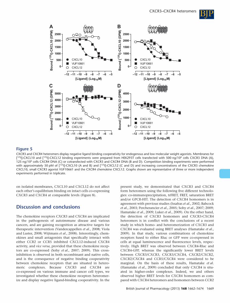
inhibited binding of [125I]-CXCL10 to CXCR3-expressingmembranes in a concentration-dependent manner underequilibrium conditions (Figure 5A), which is not affectedby the co-expression of CXCR4 (Figure 5B and Table 2). Asexpected, the CXCR4 chemokine CXCL12 was unable to dis-place [125I]-CXCL10 from membranes expressing only CXCR3(Figure 5A). However, CXCL12 competed with [125I]-CXCL10for binding to membranes co-expressing CXCR3 with CXCR4(Figure 5B). This heterologous transinhibition of [125I]-CXCL10 binding to CXCR3/CXCR4 co-expressing mem-branes by CXCL12 was only partial in comparison withhomologous inhibition by CXCL10 (i.e. 70 � 8% and 100%displacement, respectively), which corresponded to theanticipated number of CXCR3 and CXCR4 being associatedas homo- and heteromers. Conversely, in membranesexpressing CXCR4 alone (Figure 5C), [125I]-CXCL12 was dis-placed by unlabelled CXCL12, but not by the CXCR3 ago-nists CXCL10 or VUF10661. However, on co-expression ofCXCR3 with CXCR4, both CXCL10 and VUF10661 inhibited[125I]-CXCL12 binding to CXCR4 (Figure 5D). In agreementwith the expected presence of both CXCR3/CXCR4 homo-and heteromers, CXCL10 and VUF10661 could only partiallydisplace [125I]-CXCL12 (i.e. 53 � 6% and 75 � 3% displace-
ment, respectively). Although the lower CXCR3 expressionlevels in comparison with CXCR4 might explain whyCXCL10 is less effective in inhibiting CXCL12 binding toCXCR3/CXCR4 membranes than the reverse, this explana-tion is not supported by the more effective displacement of[125I]-CXCL12 by VUF10661.
The small-molecule CXCR3 antagonists VUF10085(Johnson et al., 2007) and TAK-779 (Baba et al., 1999) inhib-ited [125I]-CXCL10 binding to membranes expressing CXCR3alone (Figure 6A) or together with CXCR4 (Figure 6B). Calcu-lated pIC50 values of both CXCR3 antagonists were notaffected by the co-expression of CXCR4 (Table 2). The CXCR4antagonist AMD3100 did not affect [125I]-CXCL10 binding toCXCR3-expressing membranes (Figure 6A). In contrast to thenegative binding cooperativity between CXCL10 and CXCL12at the CXCR3-CXCR4 heteromer, AMD3100 did not transin-hibit [125I]-CXCL10 binding to membranes co-expressingCXCR3 and CXCR4 (Figure 6B). Similarly, [125I]-CXCL12binding to CXCR4 in membranes expressing this receptoralone (Figure 6C) or in combination with CXCR3 (Figure 6D)was inhibited by AMD3100 with similar potencies (Table 2),whereas VUF10085 and TAK-779 could not inhibit [125I]-CXCL12 binding to either membrane preparation.
0 20 40 60 80
0.00
0.25
0.50
0.75
1.00
1.25
1.50
CXCR4-EYFP/CXCR4-Rluc
BR
ET
ratio
0 10 20 30 40 50
0.00
0.10
0.20
0.30
0.40
0.50
CXCR3-EYFP/CXCR3-Rluc
BR
ET
ratio
0 10 20 30 40 50
0.00
0.10
0.20
0.30
0.40
CXCR3-EYFP/CXCR4-Rluc
BR
ET
ratio
0 20 40 60 80
0.00
0.05
0.10
0.15
0.20
0.25
CXCR4-EYFP/CXCR3-Rluc
BR
ET
ratio
A B
C D
Figure 3Hetero- and homomerization of CXCR3 and CXCR4. HEK293T cells were transiently cotransfected with a constant amount (150 ng/106 cells) ofCXCR4-Rluc (A and B) or CXCR3-Rluc (C and D) DNA and increasing amounts of CXCR3-EYFP (B and D) or CXCR4-EYFP (A and C) (0–2200 ng/106
cells). Saturation curves were obtained by measuring BRET ratio as function of acceptor/donor ratio (i.e. EYFP/Rluc). Data were obtained from atleast three independent experiments each performed in triplicate. Curves were fitted using nonlinear regression, assuming a single binding site.
BJPCXCR3–CXCR4 heteromers
British Journal of Pharmacology (2013) 168 1662–1674 1667

CXCR3-CXCR4 heteromerization altersligand-binding kineticsTransinhibition of CXCL12 equilibrium binding to CXCR4by both CXCL10 and VUF10661 suggests an allosteric modeof action between agonist-occupied CXCR3 and CXCR4,rather than steric hindrance between these agonists to bindreceptor heteromers. To confirm allosteric interactionsbetween the CXCR3 and CXCR4 ligand binding sites, [125I]-CXCL12 dissociation rates from CXCR4 and CXCR3-CXCR4heteromers were determined in the absence or presence ofCXCR3 agonists, using an infinite radioligand dilutionapproach (Christopoulos et al., 1997). To this end, mem-branes were pre-equilibrated with [125I]-CXCL12, and free[125I]-CXCL12 was removed by centrifugation. Dissociationkinetics of bound [125I]-CXCL12 was measured upon 100-folddilution in binding buffer in the absence or presence of unla-belled chemokines. Dissociation of [125I]-CXCL12 from mem-branes expressing CXCR4 alone was significantly acceleratedin the presence of 100 nM unlabelled CXCL12 as comparedwith the basal dissociation rate in binding buffer (Figure 7Aand Table 3). This could be due to negative binding co-operativity within CXCR4 homomers, but could also bedue to direct competition with [125I]-CXCL12 that preventsre-binding of dissociated [125I]-CXCL12. In agreement withequilibrium binding (Figure 5C), both CXCR3 agonists didnot affect CXCL12 dissociation in the absence of CXCR3(Figure 7A). Interestingly, CXCR3 co-expression acceleratedbasal CXCL12 dissociation from CXCR4, which was furtheraccelerated by 100 nM CXCL10 or 10 mM VUF10661(Figure 7B and Table 3). The very rapid dissociation of [125I]-CXCL10 from CXCR3 in the absence or presence of CXCR4did not allow the measurement of accelerated effects of unla-belled ligands within this short time frame (data not shown).
Negative binding cooperativity betweenCXCR3 and CXCR4 chemokines is notapparent in intact cellsHigh-affinity agonist binding to chemokine receptorsrequires the coupling of G proteins (De Lean et al., 1980;Springael et al., 2006), as revealed by decreased membranebinding of CXCL12 to CXCR4 and CXCL10 to CXCR3 uponG protein uncoupling using GTPgS and pertussis toxin,respectively (Cox et al., 2001; Nijmeijer et al., 2010). Hence,negative cooperativity observed in equilibrium bindingexperiments on isolated membrane fractions between twoagonists that interact with different receptors, may be theconsequence of G protein scavenging, if these receptorscouple to the same and limited pool of G proteins (Chabreet al., 2009; Birdsall, 2010). In contrast, to our observations
CXCR3-Rluc8 + -arrestin2-Venus
–10 0 10 20 30 40 50 60
–0.04
–0.02
0.00
0.02
0.04
0.06
0.08
0.10
0.12
Time following agonist addition (min)
Lig
and
-in
du
ced
BR
ET
sig
nal
CXCL11CXCL12CXCL11 + CXCL12
CXCR4-Rluc8 + -arrestin2-Venus
–10 0 10 20 30 40 50 60
–0.04
–0.02
0.00
0.02
0.04
0.06
0.08
0.10
0.12
Time following agonist addition (min)
Lig
and
-in
du
ced
BR
ET
sig
nal
CXCL11CXCL12CXCL11 + CXCL12
CXCR4-Rluc8 + -arrestin2-Venus + CXCR3
–10 0 10 20 30 40 50 60
–0.04
–0.02
0.00
0.02
0.04
0.06
0.08
0.10
0.12
Time following agonist addition (min)
Lig
and
-in
du
ced
BR
ET
sig
nal
CXCL11CXCL12CXCL11 + CXCL12
A
B
C
Figure 4Evidence for the CXCR3-CXCR4 heteromer recruiting b-arrestin2using the GPCR-HIT assay. eBRET kinetic profiles (Pfleger et al.,2006a) were generated with live HEK293FT cells co-expressingCXCR3-Rluc8 and b-arrestin2-Venus (A), CXCR4-Rluc8 andb-arrestin2-Venus (B) or CXCR4-Rluc8, b-arrestin2-Venus and CXCR3(C). These cells were treated with 100 nM CXCL11, CXCL12 or both.Data are mean � SEM of three independent experiments.�
BJP AO Watts et al.
1668 British Journal of Pharmacology (2013) 168 1662–1674
on isolated membranes, CXCL10 and CXCL12 do not affecteach other’s equilibrium binding on intact cells co-expressingCXCR3 and CXCR4 at comparable levels (Figure 8).
Discussion and conclusions
The chemokine receptors CXCR3 and CXCR4 are implicatedin the pathogenesis of autoimmune disease and variouscancers, and are gaining recognition as attractive targets fortherapeutic intervention (Vandercappellen et al., 2008; Violaand Luster, 2008; Wijtmans et al., 2008). Interestingly, chem-okines and small antagonists that specifically interact witheither CCR2 or CCR5 inhibited CXCL12-induced CXCR4activity, and vice versa, provided that these chemokine recep-tors are co-expressed (Sohy et al., 2007; 2009). This cross-inhibition is observed in both recombinant and native cells,and is the consequence of negative binding cooperativitybetween chemokine receptors that are situated in hetero-meric complexes. Because CXCR3 and CXCR4 areco-expressed on various immune and cancer cell types, weinvestigated whether these chemokine receptors heteromer-ize and display negative ligand-binding cooperativity. In the
present study, we demonstrated that CXCR3 and CXCR4form heteromers using the following five different technolo-gies: co-immunoprecipitation, trFRET, FRET, saturation BRETand/or GPCR-HIT. The detection of CXCR4 homomers is inagreement with previous studies (Issafras et al., 2002; Babcocket al., 2003; Percherancier et al., 2005; Sohy et al., 2007; 2009;Hamatake et al., 2009; Luker et al., 2009). On the other hand,the detection of CXCR3 homomers and CXCR3-CXCR4heteromers is in conflict with the conclusions of a recentstudy in which homo- and heteromerization of CXCR3 andCXCR4 was evaluated using BRET analyses (Hamatake et al.,2009). In that study, various combinations of chemokinereceptors fused to either Rluc or GFP were co-expressed incells at equal luminescence and fluorescence levels, respec-tively. High BRET was observed between CXCR4-Rluc andCXCR4-GFP, whereas the significantly lower BRET levelsbetween CXCR3/CXCR3, CXCR3/CXCR4, CXCR2/CXCR2,CXCR2/CXCR4 and CCR5/CXCR4 were considered to bemarginal. On the basis of these results, Hamatake et al.(Hamatake et al., 2009) concluded that only CXCR4 is situ-ated in higher-order complexes. Indeed, we and othersobserved higher BRET levels for CXCR4 homomers as com-pared with CXCR4 heteromers and homomers between CCR2
A
–12 –11 –10 –9 –8 –7 –6 –5
0
500
1000
1500
2000
2500
CXCL10VUF10661CXCL12
[Ligand] (Log10M)
Bo
un
d [
125I]
-CX
CL
10 (
DP
M)
–12 –11 –10 –9 –8 –7 –6 –5
0
500
1000
1500
2000
2500
CXCL10VUF10661CXCL12
[Ligand] (Log10M)
Bo
un
d [
125I]
-CX
CL
10 (
DP
M)
–12 –11 –10 –9 –8 –7 –6 –5
0
1000
2000
3000
4000
5000
CXCL10
VUF10661CXCL12
[Ligand] (Log10M)
Bo
un
d [
125I]
-CX
CL
12 (
DP
M)
–12 –11 –10 –9 –8 –7 –6 –5
0
1000
2000
3000
4000
CXCL10VUF10661CXCL12
[Ligand] (Log10M)
Bo
un
d [
125I]
-CX
CL
12 (
DP
M)
B
C D
Figure 5CXCR3 and CXCR4 heteromers display negative ligand binding cooperativity for endogenous and low molecular weight agonists. Membranes for[125I]-CXCL10 and [125I]-CXCL12 binding experiments were prepared from HEK293T cells transfected with 500 ng/106 cells CXCR3 DNA (A),125 ng/106 cells CXCR4 DNA (C) or cotransfected with CXCR3 and CXCR4 DNA (B and D). Competition binding experiments were performedwith approximately 50 pM of [125I]-CXCL10 (A and B) and [125I]-CXCL12 (C and D) and increasing concentrations of the CXCR3 chemokineCXCL10, small CXCR3 agonist VUF10661 and the CXCR4 chemokine CXCL12. Graphs shown are representative of three or more independentexperiments performed in triplicate.
BJPCXCR3–CXCR4 heteromers
British Journal of Pharmacology (2013) 168 1662–1674 1669

(Percherancier et al., 2005), CCR5 (Sohy et al., 2009) andCXCR3 in saturation BRET experiments. However, BRETlevels per se cannot be used as a quantitative measure of therelative number of complexes being formed between differentreceptor combinations, as BRET is not only determined bynumbers of complexes, but also heavily depends on the closeproximity and relative orientation between the BRET donor(i.e. Rluc) and acceptor (i.e. GFP or EYFP) proteins withineach of the different receptor complexes. On the other hand,the relative propensity of receptors to form complexes can beextracted from saturation BRET analysis as the BRET50 value,which corresponds to the BRET acceptor/donor ratio of recep-tor fusion proteins resulting in 50% of the saturated BRETsignal (Mercier et al., 2002). The BRET50 values were compa-rable for CXCR3–CXCR3, CXCR3–CXCR4 and CXCR4–CXCR4 complexes in our experiments, suggesting thatCXCR3 and CXCR4 have comparable probability to formhomo- and heteromers. Likewise, comparable BRET50 valueshave been observed for homo- and heteromers betweenchemokine receptors CCR2 and CCR5 (El-Asmar et al., 2005;Springael et al., 2006), CCR2 and CXCR4 (Percherancier et al.,2005; Sohy et al., 2007), CCR5 and CXCR4 (Contentoet al., 2008; Sohy et al., 2009), CXCR1 and CXCR2 (Wilsonet al., 2005), and CXCR4 and CXCR7 (Levoye et al., 2009).Importantly, chemokine receptor homo- and heteromeriza-tion were confirmed in these studies by other biophysical,biochemical and/or pharmacological evidence.
Negative binding cooperativity has been described withinheteromers of CCR2, CCR5 or CXCR4 (El-Asmar et al., 2005;Springael et al., 2006; Sohy et al., 2007; 2009). In this study,we showed that negative binding cooperativity also occurs
within CXCR3–CXCR4 heteromers, as binding of the chem-okines CXCL10 and CXCL12 is mutually exclusive on mem-branes co-expressing these receptors when using traceconcentrations of radiolabelled chemokine (100 pM). Moreo-ver, inhibition of [125I]-CXCL12 binding to CXCR4 by thesmall CXCR3 agonist VUF10661 suggests that the observednegative binding cooperativity is not due to steric hindrance,but rather results from agonist-induced conformationalchanges transmitted from one receptor to the other withinCXCR3–CXCR4 heteromers. Such transmission of conforma-tional changes across receptor pairs has been directly shownfor the norepinephrine-occupied a2A-adrenergic receptorupon morphine binding to the associated m-opioid receptorwithin the heteromer using intramolecular FRET analysis(Vilardaga et al., 2008). Hence, agonist binding to CXCR3constrains CXCR4 to a conformation with lower affinity forCXCL12 and vice versa. Negative binding cooperativitywithin CXCR3–CXCR4 heteromers results in partial reduc-tion in radiolabelled chemokine binding in equilibriumbinding experiments, which reflects the proportion ofCXCR3-CXCR4 heteromers relative to homomers of bothreceptor types. This is in agreement with the comparablepropensities of CXCR3 and CXCR4 to form homo- and heter-omers as observed in our saturation BRET analyses. Interest-ingly, negative binding cooperativity within CXCR3–CXCR4heteromers is limited to agonists, which contrasts withprevious observations on CCR2–CXCR4 and CCR5–CXCR4heteromers, in which small antagonists cross-inhibited bothchemokine binding and chemokine-induced in vitro and invivo activity (Sohy et al., 2007; 2009). This discrepancy maybe related to distinct interfaces between various chemokine
Table 2Binding parameters of ligands on membranes expressing CXCR3 and/or CXCR4
Radioligand Displacer CXCR3 CXCR4 CXCR3 + CXCR4
[125I]-CXCL10 CXCL10 9.6 � 0.1 ND 9.6 � 0.1
CXCL12 ND ND 8.9 � 0.1
VUF10661 7.5 � 0.1 ND 7.3 � 0.1
VUF10085 7.1 � 0.1 ND 7.2 � 0.1
TAK-779 5.7 � 0.1 ND 5.7 � 0.1
AMD3100 ND ND ND
Bmax (fmol·mg–1) 0.20 � 0.02 ND 0.26 � 0.04
[125I]-CXCL12 CXCL10 ND ND 8.8 � 0.2
CXCL12 ND 9.5 � 0.1 9.5 � 0.2
VUF10661 ND ND 6.9 � 0.3
VUF10085 ND ND ND
TAK-779 ND ND ND
AMD3100 ND 6.8 � 0.2 7.3 � 0.1
Bmax (fmol·mg–1) ND 1.58 � 0.15 1.08 � 0.14
Binding parameters of the CXCR3 ligands CXCL10 (chemokine), VUF10661 (small agonist), TAK-779 and VUF10085 (antagonists) and theCXCR4 ligands CXCR4L12 (chemokine) and AMD3100 (antagonist) for their cognate receptors were determined in the absence and presenceof CXCR4 and CXCR3, respectively. pIC50 values were determined using displacement of [125I]-CXCL10 and [125I]-CXCL12 from membranepreparations of HEK293T cells transfected with CXCR3, CXCR4 or both receptors. pIC50 values are given as averages � SEM of two or moreindependent experiments performed in triplicate.ND: value could not be determined.
BJP AO Watts et al.
1670 British Journal of Pharmacology (2013) 168 1662–1674

receptor pairs, resulting in different efficiencies with which aconformational change in one protomer is conveyed to theother. However, antagonist binding to the m-opioid receptordid not induce conformational changes across heteromericreceptor pairs as revealed by an unaltered intramolecular
FRET signal in the a2A-adrenergic receptor (Vilardaga et al.,2008), which is in line with our observations.
The validity of negative binding cooperativity detected inmembrane preparations expressing two GPCRs has recentlybeen questioned (Chabre et al., 2009). Based on a catalytic
B
C D
A
–10 –9 –8 –7 –6 –5 –4
0
1000
2000
3000
4000
VUF10085TAK-779AMD3100
Bo
un
d [
125I]
-CX
CL
10 (
DP
M)
–10 –9 –8 –7 –6 –5 –4
0
1000
2000
3000
4000
VUF10085
TAK-779
AMD3100
Bo
un
d [
125I]
-CX
CL
10 (
DP
M)
–10 –9 –8 –7 –6 –5 –4
0
1000
2000
3000
4000
5000
VUF10085
TAK-779
AMD3100
[Ligand] (Log10M) [Ligand] (Log10M)
[Ligand] (Log10M) [Ligand] (Log10M)
Bo
un
d [
125I]
-CX
CL
12 (
DP
M)
–10 –9 –8 –7 –6 –5 –4
0
2000
4000
6000
VUF10085
TAK-779
AMD3100
Bo
un
d [
125I]
-CX
CL
12 (
DP
M)
Figure 6Low molecular weight antagonists of CXCR3 and CXCR4 do not have negative binding cooperativity with endogenous agonists. Membranes for[125I]-CXCL10 and [125I]-CXCL12 binding experiments were prepared from HEK293T cells transfected with 500 ng/106 cells CXCR3 DNA (A),125 ng/106 cells CXCR4 DNA (C) or cotransfected with CXCR3 and CXCR4 DNA (B and D). Competition binding experiments were performedwith approximately 50 pM of [125I]-CXCL10 (A and B) and [125I]-CXCL12 (C and D) and increasing concentrations of the CXCR3 chemokineantagonists VUF10085 and TAK-779 and the CXCR4 antagonist AMD3100. Graphs shown are representative of three or more independentexperiments performed in triplicate.
A B
0 20 40 60 80 100
0
25
50
75
100
125
Time (min)
Bou
nd [12
5 I]-
CX
CL1
2(%
spe
cific
bin
ding
)
0 20 40 60 80 100
0
25
50
75
100
125
Time (min)
Bou
nd [12
5 I]-
CX
CL1
2(%
spe
cific
bin
ding
) VUF10661CXCL12CXCL10Buffer
Figure 7Heteromerization of CXCR3 and CXCR4 increases the dissociation rate of CXCL12. [125I]-CXCL12 dissociation half-life was determined in HEK293Tmembranes expressing CXCR4 (A) alone or (B) in combination with CXCR3, in the absence (asterisk with dotted line) and presence of the CXCR3endogenous agonist CXCL10 (open circles), the small CXCR3 agonist VUF10661 (open squares), and the CXCR4 chemokine CXCL12 (closedcircles). Representative graphs of three or more independent experiments performed in triplicate are shown.
BJPCXCR3–CXCR4 heteromers
British Journal of Pharmacology (2013) 168 1662–1674 1671

model, Chabre et al. (Chabre et al., 2009) argued that Gprotein coupling to an agonist-occupied receptor is almostirreversible in equilibrium binding assays on membranepreparations, as free GTP is not available to occupy the emptynucleotide binding pocket in the G protein upon GDP releasein this experimental setup. Because the pool of shared Gproteins might be smaller than the total number of cognatereceptors, this agonist-induced or constitutive G protein scav-enging by one of the receptor subtypes results in a permanentdepletion of G-proteins from other receptors (Nijmeijer et al.,2010). As a consequence, the latter GPCRs often display lowagonist binding affinity, which is observed as apparentnegative binding cooperativity between two agonists ofco-expressed GPCRs, but in fact does not necessarily requirereceptor heteromerization. In this respect, the absence of
negative binding cooperativity between antagonists andchemokines on CXCR3/CXCR4 membrane preparationsmight also be explained as further support for the ‘G proteinscavenging’ model. Sohy et al. (Sohy et al., 2009) confirmednegative binding cooperativity between CCR2, CCR5 andCXCR4 chemokine receptor heteromers in both recombinantand native intact cells in which GTP is readily available in thecytoplasm. In contrast, CXCL10 and CXCL12 did not affecteach other’s binding to intact HEK293T cells that co-expressCXCR3 and CXCR4. Indeed, negative binding cooperativityon membrane preparations may involve heteromeric CXCR3-CXCR4 complexes present in intracellular compartmentsthat are not accessible in binding assays on intact cells.However, the presence of CXCR3-CXCR4 heteromers on thecell surface as detected by (tr)FRET and GPCR-HIT assays isnot in support of this hypothesis. Furthermore it could beargued that the results of the GPCR-HIT studies, again in livecells, are also more consistent with a lack of negative coop-erativity as we observe greater BRET signals with dual agonisttreatment, although we have previously suggested that thesefindings could be reconciled because the significant confor-mational changes believed to result in negative binding co-operativity may also influence the ability of the heteromerto recruit b-arrestin2 (See et al., 2011). The contradictionbetween intact cells and isolated membranes indeed suggestsG protein scavenging in the latter format as proposed byChabre et al. (Chabre et al., 2009). Yet, this ‘G protein scav-enging’ model cannot explain the increased dissociation rateof CXCL12 from membranes co-expressing CXCR3 andCXCR4 in the presence of CXCR3 agonists. This decreasedaffinity of CXCR4 for bound CXCL12 can only be explainedby allosteric interactions between CXCR4 and CXCR3upon binding of agonists to the latter, as initial binding ofradiolabelled CXCL12 takes place in the absence of theseCXCR3 agonists, and is consequently not hampered byG-protein availability. To distinguish direct allosteric interac-
Table 3[125I]-CXCL12 dissociation half-life from CXCR4 is decreased in thepresence of CXCR3
t1/2 � SEM (min)
CXCR4 CXCR3 + CXCR4
Buffer 93 � 15 56 � 8
100 nM CXCL10 81 � 9 32 � 8#
100 nM CXCL12 12 � 1* 24 � 6*
100 nM VUF10661 83 � 25 16 � 3*
Results shown are average � SEM of three or more independentexperiments performed in triplicate.*t1/2 differs significantly from t1/2 in presence of vehicle(P < 0.05).#t1/2 differs significantly from t1/2 in membranes expressingCXCR4 alone (P < 0.05).
Vehic
le
CXCL10
CXCL12
0
20
40
60
80
100
120
Bo
un
d [12
5 I]-C
XC
L1
0(%
sp
ec
ific
bin
din
g)
Vehic
le
CXCL10
CXCL12
0
20
40
60
80
100
120
Bo
un
d [12
5 I]-C
XC
L1
2(%
sp
ec
ific
bin
din
g)
A B
Figure 8Chemokines display no negative binding cooperativity on intact cells. Binding of approximately 50 pM [125I]-CXCL10 (A) or [125I]-CXCL12 (B), inthe absence or presence of 100 nM unlabelled CXCL10 or CXCL12, was measured on intact HEK293T cells transiently transfected with 500 ngCXCR3 DNA/106 cells (open bars), 125 ng CXCR4 DNA/106 cells (hatched bars), or cotransfected with CXCR3 and CXCR4 DNA (closed bars).Graphs show mean � SEM of two or more independent experiments performed in triplicate.
BJP AO Watts et al.
1672 British Journal of Pharmacology (2013) 168 1662–1674

tions between these receptors from downstream crosstalkevents such as G protein scavenging (Vischer et al., 2011),intramolecular FRET approaches might be applied on CXCR3and CXCR4 in the future (Vilardaga et al., 2008). At present,however, we cannot explain the observed discrepancy inCXCR3/CXCR4 binding cooperativity.
In summary, we show that CXCR3 and CXCR4 formheteromers at the cell surface using multiple experimentalapproaches. CXCR3 co-expression increased the dissociationof CXCL12 from CXCR4 membranes, which is further accel-erated by CXCR3 agonists. Although specific CXCR3 andCXCR4 agonists inhibit each other’s equilibrium binding onisolated membranes as well, this apparent negative bindingcooperativity was not observed on intact cells.
Acknowledgements
This work was supported by the Top Institute Pharma (projectnumber D1-105: the GPCR forum). KDGP is an AustralianResearch Council (ARC) Future Fellow (FT100100271) andWCJ is supported by ARC Project Grant DP120101297. Wethank Dr. S. Jähnichen and Razia Alimahomed (Leiden/Amsterdam Center for Drug Research, Division of MedicinalChemistry, Faculty of Science, VU University Amsterdam) fortechnical assistance.
Conflicts of interest
In addition to being Head of the Laboratory for MolecularEndocrinology–GPCRs, Western Australian Institute forMedical Research and Centre for Medical Research, Universityof Western Australia, K.D.G.P. is Chief Scientific Officer ofDimerix Bioscience Pty Ltd, a spin-out company of the Uni-versity of Western Australia that has been assigned the rightsto the ‘GPCR-HIT’ technology. K.D.G.P. has a minor share-holding in Dimerix.
ReferencesBaba M, Nishimura O, Kanzaki N, Okamoto M, Sawada H, Iizawa Yet al. (1999). A small-molecule, nonpeptide CCR5 antagonist withhighly potent and selective anti-HIV-1 activity. Proc Natl Acad SciU S A 96: 5698–5703.
Babcock GJ, Farzan M, Sodroski J (2003). Ligand-independentdimerization of CXCR4, a principal HIV-1 coreceptor. J Biol Chem278: 3378–3385.
Birdsall NJ (2010). Class A GPCR heterodimers: evidence frombinding studies. Trends Pharmacol Sci 31: 499–508.
Chabre M, Deterre P, Antonny B (2009). The apparent cooperativityof some GPCRs does not necessarily imply dimerization. TrendsPharmacol Sci 30: 182–187.
Christopoulos A, Lanzafame A, Ziegler A, Mitchelson F (1997).Kinetic studies of co-operativity at atrial muscarinic M2 receptorswith an ‘infinite dilution’ procedure. Biochem Pharmacol 53:795–800.
Contento RL, Molon B, Boularan C, Pozzan T, Manes S, Marullo Set al. (2008). CXCR4-CCR5: a couple modulating T cell functions.Proc Natl Acad Sci U S A 105: 10101–10106.
Cox MA, Jenh CH, Gonsiorek W, Fine J, Narula SK, Zavodny PJet al. (2001). Human interferon-inducible 10-kDa protein andhuman interferon-inducible T cell alpha chemoattractant areallotopic ligands for human CXCR3: differential binding to receptorstates. Mol Pharmacol 59: 707–715.
De Lean A, Stadel JM, Lefkowitz RJ (1980). A ternary complexmodel explains the agonist-specific binding properties of theadenylyl cyclase-coupled b-adrenergic receptor. J Biol Chem 255:7108–7117.
Dorsch S, Klotz KN, Engelhardt S, Lohse MJ, Bunemann M (2009).Analysis of receptor oligomerization by FRAP microscopy. NatMethods 6: 225–230.
El-Asmar L, Springael JY, Ballet S, Andrieu EU, Vassart G,Parmentier M (2005). Evidence for negative binding cooperativitywithin CCR5-CCR2b heterodimers. Mol Pharmacol 67: 460–469.
Fulton AM (2009). The chemokine receptors CXCR4 and CXCR3 incancer. Curr Oncol Rep 11: 125–131.
Hamatake M, Aoki T, Futahashi Y, Urano E, Yamamoto N,Komano J (2009). Ligand-independent higher-ordermultimerization of CXCR4, a G-protein-coupled chemokinereceptor involved in targeted metastasis. Cancer Sci 100: 95–102.
Hern JA, Baig AH, Mashanov GI, Birdsall B, Corrie JE, Lazareno Set al. (2010). Formation and dissociation of M1 muscarinic receptordimers seen by total internal reflection fluorescence imaging ofsingle molecules. Proc Natl Acad Sci U S A 107: 2693–2698.
Issafras H, Angers S, Bulenger S, Blanpain C, Parmentier M,Labbe-Jullie C et al. (2002). Constitutive agonist-independent CCR5oligomerization and antibody-mediated clustering occurring atphysiological levels of receptors. J Biol Chem 277: 34666–34673.
Jahnichen S, Blanchetot C, Maussang D, Gonzalez-Pajuelo M,Chow KY, Bosch L et al. (2010). CXCR4 nanobodies (VHH-basedsingle variable domains) potently inhibit chemotaxis and HIV-1replication and mobilize stem cells. Proc Natl Acad Sci U S A 107:20565–20570.
Jalink K, Van Rheenen J (2009). FilterFRET: quantitative imaging ofsensitized emission. In: Gadella TW, Jr (ed.). Fret and FlimTechniques, Vol. 33. Academic Press: Burlington, MA, pp. 289–349.
Johnson M, Li AR, Liu J, Fu Z, Zhu L, Miao S et al. (2007).Discovery and optimization of a series of quinazolinone-derivedantagonists of CXCR3. Bioorg Med Chem Lett 17: 3339–3343.
Lacotte S, Brun S, Muller S, Dumortier H (2009). CXCR3,inflammation, and autoimmune diseases. Ann N Y Acad Sci 1173:310–317.
Levoye A, Balabanian K, Baleux F, Bachelerie F, Lagane B (2009).CXCR7 heterodimerizes with CXCR4 and regulatesCXCL12-mediated G protein signaling. Blood 113: 6085–6093.
Luker KE, Gupta M, Luker GD (2009). Imaging chemokine receptordimerization with firefly luciferase complementation. FASEB J 23:823–834.
Mathews JL, Fulton BS, Negus SS, Neumeyer JL, Bidlack JM (2008).In vivo characterization of (–)(–)MCL-144 and (+)(–)MCL-193:isomeric, bivalent ligands with mu/kappa agonist properties.Neurochem Res 33: 2142–2150.
Mercier JF, Salahpour A, Angers S, Breit A, Bouvier M (2002).Quantitative assessment of beta 1- and beta 2-adrenergic receptor
BJPCXCR3–CXCR4 heteromers
British Journal of Pharmacology (2013) 168 1662–1674 1673

homo- and heterodimerization by bioluminescence resonanceenergy transfer. J Biol Chem 277: 44925–44931.
Moser B, Loetscher P (2001). Lymphocyte traffick control bychemokines. Nat Immunol 2: 123–128.
Mustafa S, Pfleger KD (2011). G protein-coupled receptor heteromeridentification technology: identification and profiling of GPCRheteromers. J Lab Autom 16: 285–291.
Mustafa S, See HB, Seeber RM, Armstrong SP, White CW, Ventura Set al. (2012). Identification and profiling of novel alpha1A-adrenoceptor-CXC chemokine receptor 2 heteromer. J Biol Chem287: 12952–12965.
Nijmeijer S, Leurs R, Smit MJ, Vischer HF (2010). The Epstein-Barrvirus-encoded G protein-coupled receptor BILF1 hetero-oligomerizeswith human CXCR4, scavenges Galphai proteins, and constitutivelyimpairs CXCR4 functioning. J Biol Chem 285: 29632–29641.
Percherancier Y, Berchiche YA, Slight I, Volkmer-Engert R,Tamamura H, Fujii N et al. (2005). Bioluminescence resonanceenergy transfer reveals ligand-induced conformational changes inCXCR4 homo- and heterodimers. J Biol Chem 280: 9895–9903.
Pfleger KD, Dromey JR, Dalrymple MB, Lim EM, Thomas WG,Eidne KA (2006a). Extended bioluminescence resonance energytransfer (eBRET) for monitoring prolonged protein-proteininteractions in live cells. Cell Signal 18: 1664–1670.
Pfleger KD, Seeber RM, Eidne KA (2006b). Bioluminescenceresonance energy transfer (BRET) for the real-time detection ofprotein–protein interactions. Nat Protoc 1: 337–345.
van Rheenen J, Langeslag M, Jalink K (2004). Correcting confocalacquisition to optimize imaging of fluorescence resonance energytransfer by sensitized emission. Biophys J 86: 2517–2529.
van Rijn RM, Chazot PL, Shenton FC, Sansuk K, Bakker RA, Leurs R(2006). Oligomerization of recombinant and endogenouslyexpressed human histamine H(4) receptors. Mol Pharmacol 70:604–615.
Scholten D, Canals M, Maussang D, Roumen L, Smit M,Wijtmans M et al. (2012a). Pharmacological modulation ofchemokine receptor function. Br J Pharmacol 165: 1617–1643.
Scholten D, Canals M, Wijtmans M, de Munnik S, Nguyen P,Verzijl D et al. (2012b). Pharmacological characterisation of asmall-molecule agonist for the chemokine receptor CXCR3. Br JPharmacol 166: 898–911.
See HB, Seeber RM, Kocan M, Eidne KA, Pfleger KD (2011).Application of G protein-coupled receptor-heteromer identificationtechnology to monitor beta-arrestin recruitment to Gprotein-coupled receptor heteromers. Assay Drug Dev Technol 9:21–30.
Sohy D, Parmentier M, Springael JY (2007). Allosterictransinhibition by specific antagonists in CCR2/CXCR4heterodimers. J Biol Chem 282: 30062–30069.
Sohy D, Yano H, de Nadai P, Urizar E, Guillabert A, Javitch JA et al.(2009). Hetero-oligomerization of CCR2, CCR5, and CXCR4 andthe protean effects of ‘selective’ antagonists. J Biol Chem 284:31270–31279.
Springael JY, Le Minh PN, Urizar E, Costagliola S, Vassart G,Parmentier M (2006). Allosteric modulation of binding propertiesbetween units of chemokine receptor homo- and hetero-oligomers.Mol Pharmacol 69: 1652–1661.
Stroke IL, Cole AG, Simhadri S, Brescia MR, Desai M, Zhang JJ et al.(2006). Identification of CXCR3 receptor agonists in combinatorialsmall-molecule libraries. Biochem Biophys Res Commun 349:221–228.
Tanaka T, Nomura W, Narumi T, Masuda A, Tamamura H (2010).Bivalent ligands of CXCR4 with rigid linkers for elucidation of thedimerization state in cells. J Am Chem Soc 132: 15899–15901.
Vandercappellen J, Van Damme J, Struyf S (2008). The role of CXCchemokines and their receptors in cancer. Cancer Lett 267:226–244.
Verzijl D, Storelli S, Scholten DJ, Bosch L, Reinhart TA, Streblow DNet al. (2008). Noncompetitive antagonism and inverse agonism asmechanism of action of nonpeptidergic antagonists at primate androdent CXCR3 chemokine receptors. J Pharmacol Exp Ther 325:544–555.
Vilardaga JP, Nikolaev VO, Lorenz K, Ferrandon S, Zhuang Z,Lohse MJ (2008). Conformational cross-talk betweenalpha2A-adrenergic and mu-opioid receptors controls cell signaling.Nat Chem Biol 4: 126–131.
Viola A, Luster AD (2008). Chemokines and their receptors: drugtargets in immunity and inflammation. Annu Rev PharmacolToxicol 48: 171–197.
Vischer HF, Nijmeijer S, Smit MJ, Leurs R (2008). Viral hijacking ofhuman receptors through heterodimerization. Biochem Biophys ResCommun 377: 93–97.
Vischer HF, Watts AO, Nijmeijer S, Leurs R (2011). Gprotein-coupled receptors: walking hand-in-hand, talkinghand-in-hand? Br J Pharmacol 163: 246–260.
Waldhoer M, Fong J, Jones RM, Lunzer MM, Sharma SK, Kostenis Eet al. (2005). A heterodimer-selective agonist shows in vivorelevance of G protein-coupled receptor dimers. Proc Natl Acad SciU S A 102: 9050–9055.
Wijtmans M, Verzijl D, Leurs R, de Esch IJ, Smit MJ (2008). Towardssmall-molecule CXCR3 ligands with clinical potential.ChemMedChem 3: 861–872.
Wilson S, Wilkinson G, Milligan G (2005). The CXCR1 and CXCR2receptors form constitutive homo- and heterodimers selectively andwith equal apparent affinities. J Biol Chem 280: 28663–28674.
BJP AO Watts et al.
1674 British Journal of Pharmacology (2013) 168 1662–1674
Copyright © 2022 FDOKUMEN