Isolated human astrocytes are not susceptible to infection by M- and T-tropic HIV-1 strains despite...
13
Isolated Human Astrocytes Are Not Susceptible to Infection by M- and T- Tropic HIV-1 Strains Despite Functional Expression of the Chemokine Receptors CCR5 and CXCR4 AGNE ` S BOUTET, 1 * HASSAN SALIM, 2 YASSINE TAOUFIK, 1 PIERRE-MARIE LLEDO, 2 JEAN-DIDIER VINCENT, 2 JEAN-FRANC ¸ OIS DELFRAISSY, 1 AND MARC TARDIEU 1 1 Laboratoire d’immunologie antivirale syste ´mique et ce ´re ´brale, Inserm E 0109, Faculte ´ de Me ´decine Paris-Sud, Le Kremlin-Bice ˆtre Cedex, France 2 CNRS, Institut de Neurobiologie Alfred-Fessard, UPR 2197, Gif-sur-Yvette Cedex, France. KEY WORDS chemokine receptors; CD4; HIV; astrocytes; microglial cells; development ABSTRACT Within the brain, HIV-1 targets the microglia and astrocytes. Previous studies have reported that viral entry into astrocytes is independent of CD4, in contrast to microglia. We aimed to determine whether chemokine receptors play a role in mediating CD4-independent HIV-1 entry into astrocytes. We found that embryonic astrocytes and microglial cells express CCR5, CCR3, and CXCR4 transcripts. Intracel- lular calcium levels in astrocytes were found to increase following application of RAN- TES, MIP-1b (CCR5-agonist), SDF-1a (CXCR4-agonist), but not eotaxin (CCR3-agonist). In microglial cells, eotaxin was also able to modulate internal calcium homeostasis. CD4 was not present at the cell surface of purified astrocytes but CD4 mRNA could be detected by RT-PCR. Neither HIV-1 9533 (R5 isolate) nor HIV-1 LAI (X4 isolate) penetrated into purified astrocytes. In contrast, mixed CNS cell cultures were infected by HIV-1 9533 and this was inhibited by anti-CD4 mAb in 4/4 tested cultures and by anti-CCR5 mAb in 2/4. Thus, the HIV-1 R5 strain requires CD4 to penetrate into brain cells, suggesting that CCR5 cannot be used as the primary receptor for M-tropic HIV-1 strains in astrocytes. Moreover, inconstant inhibition of HIV-1 entry by anti-CCR5 mAb supports the existence of alternative coreceptors for penetration of M-tropic isolates into brain cells. GLIA 34:165–177, 2001. © 2001 Wiley-Liss, Inc. INTRODUCTION The cellular tropism displayed by different human im- munodeficiency virus type 1 (HIV-1) isolates is related to their use of specific chemokine receptors that mediate HIV-1 entry in conjunction with CD4. Virus strains that infect primary T cells and transformed T-cell lines (T- tropic strains) use the a-chemokine receptor CXCR4 (X4 strains), while those that replicate in primary T cells and macrophages (M-tropic strains) typically use the b-che- mokine receptor CCR5 (R5 strains) (Berger et al., 1998; Miller and Meucci, 1999). Other chemokine receptors such as CCR2b, CCR3, CCR8, CCR9, CX 3 CR1, and APJ in addition to some orphan receptor such as GPR1, GPR15, and STRL33 have been shown to bind HIV and SIV envelopes (for review, see Berger et al., 1999). In vivo, the contribution of these alternative receptors to infection and pathogenesis remains obscure. HIV-1 invades the Abbreviations used: AIDS, acquired immunodeficiency syndrome; FCS, fetal calf serum; FITC, fluorescein isothiocyanate; GAPDH, glyceraldehyde phospho- dehydrogenase; GFAP, glial fibrillary acidic protein; GPCR, G protein-coupled receptor; HIV-1, human immunodeficiency virus type 1; LTR, long terminal repeat; MDM, monocyte-derived macrophage; MIP, macrophage inflammatory protein; PBMC, peripheral blood mononuclear cell; PBS, phosphate-buffered saline; PTX, pertussis toxin; RANTES, regulated on activation normal T ex- pressed and secreted; SDF, stromal cell-derived factor; SIV, simian immunode- ficiency virus. Grant sponsor: Institut National de la Recherche Me ´dicale (INSERM; Grant number: 96012; Grant sponsor: Centre National de la Recherche Scientifique (CNRS); Grant sponsor: Agence Nationale de Recherche sur le SIDA (ANRS); Grant sponsor: Universite ´ Paris-Sud. *Correspondence to: Dr. Agne `s Boutet. Laboratoire d’immunologie antivirale sys- te ´mique et ce ´re ´brale, Inserm E 0109, Faculte ´ de Me ´decine Paris-Sud, 63, rue Gabriel Pe ´ri, 94276 Le Kremlin-Bice ˆtre Cedex, France. E-mail: [email protected] Received 22 September 2000; Accepted 23 February 2001 GLIA 34:165–177 (2001) © 2001 Wiley-Liss, Inc.
-
Upload
independent -
Category
Documents
-
view
2 -
download
0
Transcript of Isolated human astrocytes are not susceptible to infection by M- and T-tropic HIV-1 strains despite...
Isolated Human Astrocytes Are NotSusceptible to Infection by M- and T-
Tropic HIV-1 Strains Despite FunctionalExpression of the Chemokine Receptors
CCR5 and CXCR4AGNES BOUTET,1* HASSAN SALIM,2 YASSINE TAOUFIK,1
PIERRE-MARIE LLEDO,2 JEAN-DIDIER VINCENT,2JEAN-FRANCOIS DELFRAISSY,1 AND MARC TARDIEU1
1Laboratoire d’immunologie antivirale systemique et cerebrale, Inserm E 0109, Faculte deMedecine Paris-Sud, Le Kremlin-Bicetre Cedex, France
2CNRS, Institut de Neurobiologie Alfred-Fessard, UPR 2197, Gif-sur-Yvette Cedex, France.
KEY WORDS chemokine receptors; CD4; HIV; astrocytes; microglial cells; development
ABSTRACT Within the brain, HIV-1 targets the microglia and astrocytes. Previousstudies have reported that viral entry into astrocytes is independent of CD4, in contrastto microglia. We aimed to determine whether chemokine receptors play a role inmediating CD4-independent HIV-1 entry into astrocytes. We found that embryonicastrocytes and microglial cells express CCR5, CCR3, and CXCR4 transcripts. Intracel-lular calcium levels in astrocytes were found to increase following application of RAN-TES, MIP-1b (CCR5-agonist), SDF-1a (CXCR4-agonist), but not eotaxin (CCR3-agonist).In microglial cells, eotaxin was also able to modulate internal calcium homeostasis. CD4was not present at the cell surface of purified astrocytes but CD4 mRNA could bedetected by RT-PCR. Neither HIV-19533 (R5 isolate) nor HIV-1LAI (X4 isolate) penetratedinto purified astrocytes. In contrast, mixed CNS cell cultures were infected by HIV-19533and this was inhibited by anti-CD4 mAb in 4/4 tested cultures and by anti-CCR5 mAbin 2/4. Thus, the HIV-1 R5 strain requires CD4 to penetrate into brain cells, suggestingthat CCR5 cannot be used as the primary receptor for M-tropic HIV-1 strains inastrocytes. Moreover, inconstant inhibition of HIV-1 entry by anti-CCR5 mAb supportsthe existence of alternative coreceptors for penetration of M-tropic isolates into braincells. GLIA 34:165–177, 2001. © 2001 Wiley-Liss, Inc.
INTRODUCTION
The cellular tropism displayed by different human im-munodeficiency virus type 1 (HIV-1) isolates is related totheir use of specific chemokine receptors that mediateHIV-1 entry in conjunction with CD4. Virus strains thatinfect primary T cells and transformed T-cell lines (T-tropic strains) use the a-chemokine receptor CXCR4 (X4strains), while those that replicate in primary T cells andmacrophages (M-tropic strains) typically use the b-che-mokine receptor CCR5 (R5 strains) (Berger et al., 1998;Miller and Meucci, 1999). Other chemokine receptorssuch as CCR2b, CCR3, CCR8, CCR9, CX3CR1, and APJin addition to some orphan receptor such as GPR1,GPR15, and STRL33 have been shown to bind HIV andSIV envelopes (for review, see Berger et al., 1999). In vivo,
the contribution of these alternative receptors to infectionand pathogenesis remains obscure. HIV-1 invades the
Abbreviations used: AIDS, acquired immunodeficiency syndrome; FCS, fetalcalf serum; FITC, fluorescein isothiocyanate; GAPDH, glyceraldehyde phospho-dehydrogenase; GFAP, glial fibrillary acidic protein; GPCR, G protein-coupledreceptor; HIV-1, human immunodeficiency virus type 1; LTR, long terminalrepeat; MDM, monocyte-derived macrophage; MIP, macrophage inflammatoryprotein; PBMC, peripheral blood mononuclear cell; PBS, phosphate-bufferedsaline; PTX, pertussis toxin; RANTES, regulated on activation normal T ex-pressed and secreted; SDF, stromal cell-derived factor; SIV, simian immunode-ficiency virus.
Grant sponsor: Institut National de la Recherche Medicale (INSERM; Grantnumber: 96012; Grant sponsor: Centre National de la Recherche Scientifique(CNRS); Grant sponsor: Agence Nationale de Recherche sur le SIDA (ANRS);Grant sponsor: Universite Paris-Sud.
*Correspondence to: Dr. Agnes Boutet. Laboratoire d’immunologie antivirale sys-temique et cerebrale, Inserm E 0109, Faculte de Medecine Paris-Sud, 63, rue GabrielPeri, 94276 Le Kremlin-Bicetre Cedex, France. E-mail: [email protected]
Received 22 September 2000; Accepted 23 February 2001
GLIA 34:165–177 (2001)
© 2001 Wiley-Liss, Inc.
central nervous system (CNS) early after systemic infec-tion and causes severe neurological complications thatcan be observed in the late stages of acquired immuno-deficiency syndrome (AIDS). Cells that are productivelyinfected are resident activated microglia and macro-phages that have transmigrated through the blood–brainbarrier (Watkins et al., 1990; Lee et al., 1993; Tardieu,1999). In astrocytes, HIV-1 infection is restricted to theexpression of early regulatory gene products (e.g., Nefand Rev), suggesting that HIV-1 establishes a nonproduc-tive infection in these cells (Saito et al., 1994; Tornatore etal., 1994; Ranki et al., 1995). Such an infection mightcontribute to the persistence of the virus in the brain. Invitro studies have reported that, in contrast to microglia(Jordan et al., 1991), HIV-1 infection of astrocytes is CD4-independent (Harouse et al., 1989; Tornatore et al., 1991).Whether chemokine receptors play a role in mediatingCD4-independent entry into astrocytes is still an openquestion.
The expression of chemokine receptors in brain hasbeen widely investigated but has produced controversialresults in astrocytes. Immunocytochemical studies onhealthy human brain or brain with HIV-encephalitishave revealed the presence of CXCR4, CCR5, and CCR3in astrocytes and microglial cells (Rottman et al., 1997;Lavi et al., 1997; Vallat et al., 1998; Sanders et al., 1998;Ghorpade et al., 1998). Futhermore, functional expres-sion of CXCR4 and CCR5, but not CCR3, has been dem-onstrated on cultured fetal astrocytes (Klein et al., 1999;Andjelkovic et al., 1999) and adult microglia (Albright etal., 1999) by microphotometry. In contrast, Sabri et al.(1999) failed to detect coreceptor at the cell surface ofcultured fetal astrocytes, although mRNAs of CXCR4 andCCR5 in addition to transcripts of the orphan receptorsSTRL33 and APJ were present. These observations sug-gest a heterogeneous expression of chemokine receptorsamong subpopulations of astrocytes or variations depen-dent on the stage of development.
HIV entry and the role of chemokine receptors as core-ceptor have been investigated mainly in microglia amongCNS cells. T-tropic viruses that use CXCR4 penetratethem relatively inefficiently (He et al., 1997; Shieh et al.,1998). The penetration of brain-derived HIV isolates canbe mediated by CCR3 and CCR5, together with CD4 (Heet al., 1997; Shieh et al., 1998; Albright et al., 1999) butGhorpade et al. (1998) failed to inhibit viral entry of bothM-tropic and brain-derived strains by anti-CCR5 and an-ti-CCR3 mAbs in microglia. This suggests that microgliaexpress additional chemokine receptors that function asHIV coreceptor. Recently, Albright et al. (1999) reportedthat some HIV-1 strains isolated from the CNS can useCCR8 in transfected cell lines and Jinno et al. (1998)showed expression of CCR8 in a glioma cell line and itsinvolvement in viral entry.
To further study the chemokine receptors used ascofactors for HIV-1 entry into human astrocytes, weinvestigated the functional expression of CXCR4,CCR5 and CCR3 in highly purified embryonic astrocytecultures. The results were compared with those ob-tained from microglial cultures tested in parallel. Viral
penetration into astrocytes was quantified by measur-ing early steps in HIV reverse transcription by semi-quantitative PCR and was compared with infection ofmixed CNS cells or MDM with the same HIV-1 isolates.
MATERIALS AND METHODSCell Preparation And Culture
Primary CNS cell cultures
Primary mixed CNS cell cultures were prepared aspreviously described (Janabi et al., 1996) from prosen-cephalon and spinal cord of 8–10-week-old human em-bryos obtained after elective abortion (performed in fullcompliance with both French National Ethics and localethics committee guidelines). Briefly, brain tissue wasdissected, trypsinized and the cell suspension was dis-tributed at a density of 106 cells/35-mm culture wellcoated with collagen in MCDB medium (Seromed,France), supplemented with 5% fetal calf serum (FCS),antibiotics (105 U/L penicillin, 0.1 g/L streptomycin),and 2 mM glutamine. Cultures were placed in a 10%CO2-humidified incubator at 37°C. The medium wasremoved 48 h after plating and changed by one-halfevery 3–4 days. Cultures consisted of nerve cell adhe-sion molecule- and neurofilament-positive neuronalclusters lying on a monocellular layer containing 50–70% glial fibrillary acidic protein (GFAP)-positive as-trocytes and 30–50% CD68/KiM-7-positive microglialcells as described (Janabi et al., 1998).
Purified cultures of microglia or astrocytes
Ten to 15 days after plating, circular shaking ofprimary CNS cell cultures allowed removal of micro-glial cells, which were transferred and selected by ad-herence. These cultures were subsequently grown for 3weeks to 2 months and contained more than 98% ofCD68/KiM-7-positive cells (Janabi et al., 1996) (Fig. 1).The remaining adherent cells were trypsinized, seededinto 175-cm2 flask and passaged 3–4 times to obtainpurified cultures of astrocytes. Before each trypsiniza-tion, astrocyte cultures were submitted to an addi-tional circular shaking to avoid microglial cell contam-ination. Three-week-old (passage 3) astrocyte cultureswere .99% pure, as determined by GFAP immuno-staining, and contained no CD68/KiM-7-positive cells(Janabi et al., 1996) (Fig. 1).
Preparation of human macrophages
Human PBMC from healthy HIV-seronegative do-nors were isolated by Ficoll-Hypaque gradient centrif-ugation. Monocytes were selected by a 2 hour adher-ence in Lab-tek tissue-culture chamber/slides (OSI,France) at 37°C in a 10% CO2 atmosphere. Monocytesdifferentiated into macrophages for 15 days in culture
166 BOUTET ET AL.
in RPMI-1640 medium (Seromed) containing 10% FCS,antibiotics and glutamine. Monocyte-derived macro-phage (MDM) cultures contained more than 95% ofCD68/KiM-7-positive cells.
Materials
Recombinant human b chemokines MIP-1a, MIP-1b,RANTES, eotaxin and the a chemokine SDF-1a were
purchased from R&D Systems (Oxon, UK) and used at100 ng/ml for photometric experiments. Bordetella per-tussis toxin (PTX; 250 ng/ml) was obtained from Sigma(St. Quentin Fallavier, France). Monoclonal Ab toCCR5 (2D7; PharMingen, San Diego, CA; Wu et al.,1997), CCR3 (7B11; AIDS Research & Reference Re-agent Program, National Institutes of Health [NIH],Rockville, MD), and CXCR4 (12G5; R&D Systems; En-dres et al., 1996) were used to inhibit HIV-1 infection.HIV-blocking anti-CD4 mAb (Leu3a) was purchased
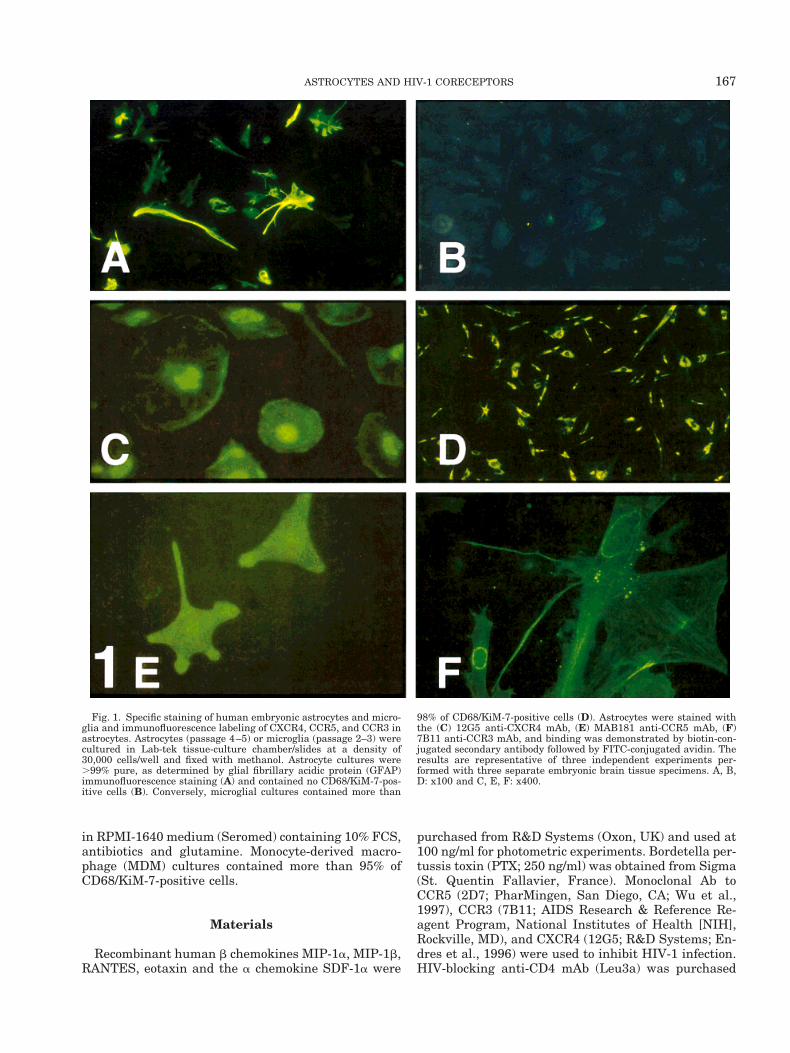
Fig. 1. Specific staining of human embryonic astrocytes and micro-glia and immunofluorescence labeling of CXCR4, CCR5, and CCR3 inastrocytes. Astrocytes (passage 4–5) or microglia (passage 2–3) werecultured in Lab-tek tissue-culture chamber/slides at a density of30,000 cells/well and fixed with methanol. Astrocyte cultures were.99% pure, as determined by glial fibrillary acidic protein (GFAP)immunofluorescence staining (A) and contained no CD68/KiM-7-pos-itive cells (B). Conversely, microglial cultures contained more than
98% of CD68/KiM-7-positive cells (D). Astrocytes were stained withthe (C) 12G5 anti-CXCR4 mAb, (E) MAB181 anti-CCR5 mAb, (F)7B11 anti-CCR3 mAb, and binding was demonstrated by biotin-con-jugated secondary antibody followed by FITC-conjugated avidin. Theresults are representative of three independent experiments per-formed with three separate embryonic brain tissue specimens. A, B,D: x100 and C, E, F: x400.
167ASTROCYTES AND HIV-1 CORECEPTORS
from Becton Dickinson (San Jose, CA). Anti-CD4 andanti-CCR5 mAb were dialyzed against PBS overnightat 4°C to remove sodium azide. Immunolabeling exper-iments were performed with anti-CCR5 mAb MAB181obtained from R&D System.
HIV-1 Strains
The HIV-1 T-tropic isolate LAI, passed through apermanent T-cell line and known to use CXCR4 (Sim-mons et al., 1996), was kindly provided by Dr. Barre-Sinoussi (Institut Pasteur, Paris, France). The labora-tory-adapted M-tropic strain HIV-19533 (a gift of Dr.Molina and Dr. Sinet) was grown in MDM. Before use,virus supernatants were treated with 50 U/ml RNase-free DNase I (Roche Diagnostic, Germany) for 30 minat room temperature to remove contaminant viralDNA. The HIV-19533 strain was characterized regard-ing the coreceptor usage by infection of MDM in thepresence of various inhibitors of chemokine receptors.Positive entry of the virus into MDM was confirmed bycomparison of viral DNA levels in cells after exposureto infectious HIV-19533 or to heat-inactivated HIV-19533(2 h pi: 0.8 6 0.08 vs 0.016 0.01 HIV DNA units;infectious vs heat-inactivated virus; n 5 2). HIV-19533infection of macrophages was inhibited by anti-CCR5(2D7; 0.2 mg/ml; 8 h postinfection: 2 6 0.3 vs 5.8 6 0.8HIV DNA units, 2D7-treated vs control; n 5 2) andanti-CD4 (Leu3a; 12.5 mg/ml; 8 h pi: 1.7 6 0.8 vs 5.8 60.8 HIV DNA units, Leu3a-treated vs control; n 5 2)mAb, but neither by eotaxin (100 ng/ml; 8 h pi: 12.4 64 vs 5.8 6 0.8 HIV DNA units, eotaxin-treated vscontrol; n 5 2) nor by anti-CXCR4 mAb (12G5; 20mg/ml; 8 h pi: 11.2 6 2 vs 5.8 6 0.8 HIV DNA units,12G5-treated vs control; n 5 2). We considered a sig-nificant difference between values of both conditionswhen one-log-separated. This supports the identifica-tion of the M-tropic HIV9533 strain as a R5 isolate.
HIV-1 Infection of Brain Cells
Astrocytes seeded in 12-well plates (5 3 105 cells/well), or primary mixed CNS cells were incubated over-
night (12 h) with viral inoculum (25 ng of p24 gagantigen/well) at 37°C, washed several times and keptin culture until lysis. Cells were also pulsed with heat-inactivated virus (1 h at 56°C) to determine baselinecontamination of DNA in the viral stocks. For inhibi-tion experiments, cells were preincubated for 45 minbefore infection with monoclonal antibodies describedabove. DNA was extracted using the Qiamp blood kit(Qiagen, Germany) at regular times after viral expo-sure (12, 18 h) and R/U5-PCR assay was performed toassess HIV penetration.
Semiquantitative PCR of HIV-1 DNA
Semiquantification of DNA was carried out usingkinetic polymerase chain reaction–enzyme-linked im-munosorbent assay (PCR-ELISA), as reported by Ta-oufik et al. (1997). The oligonucleotide primer pair spe-cific for the R/U5 region of the LTR was used to assessviral entry (Table 1). This primer pair flanks sequenceswithin the first region of the viral DNA synthesizedduring reverse transcription, as previously described(Zack et al., 1990). PCR was performed on 10-ml DNAsamples. Aliquots of PCR reactions were sampled everythree cycles and transferred onto avidin-coated micro-plates. The 59-biotinylated PCR products were incu-bated in microplates and denatured with NaOH. Thecaptured strand was hybridized with a digoxigenin-labeled probe and the bound probe was detected usingalkaline phosphatase-coupled anti-digoxigenin anti-body. Chemiluminescent substrate for alkaline phos-phatase (CSPD, Roche Diagnostic) plus luminescenceenhancer (Sapphire; Tropix, Bedford, MA) were added;luminescence was measured using Micro Beta Plus(Wallac, Turku, Finland). The amounts of ampliconsquantified by enzyme-linked immunosorbent assay(ELISA) were compared by linear regression with anexternal scale of DNA at the cycles where signals werein the linear portion of the revelation assay. GAPDHwas amplified and quantified for each DNA prepara-tion to correct for variability in DNA recovery. Resultswere expressed as relative DNA units (calculated as
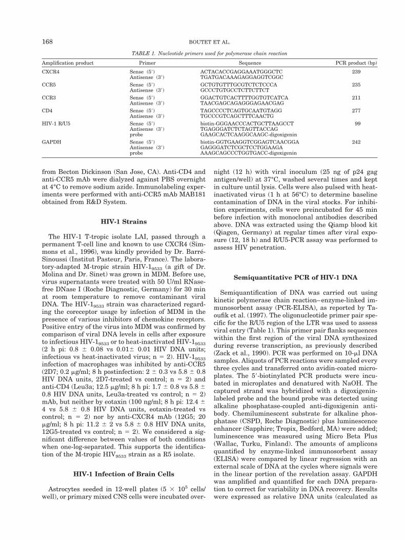
TABLE 1. Nucleotide primers used for polymerase chain reaction
Amplification product Primer Sequence PCR product (bp)
CXCR4 Sense (59) ACTACACCGAGGAAATGGGCTC 239Antisense (39) TGATGACAAAGAGGAGGTCGGC
CCR5 Sense (59) GCTGTGTTTGCGTCTCTCCCA 235Antisense (39) GCCCTGTGCCTCTTCTTCT
CCR3 Sense (59) GGACTGTCACTTTTGGTGTCATCA 211Antisense (39) TAACGAGCAGAGGGAGAACGAG
CD4 Sense (59) TAGCCCCTCAGTGCAATGTAGG 277Antisense (39) TGCCCGTCAGCTTTCAACTG
HIV-1 R/U5 Sense (59) biotin-GGGAACCCACTGCTTAAGCCT 99Antisense (39) TGAGGGATCTCTAGTTACCAGprobe GAAGCACTCAAGGCAAGC-digoxigenin
GAPDH Sense (59) biotin-GGTGAAGGTCGGAGTCAACGGA 242Antisense (39) GAGGGATCTCGCTCCTGGAAGAprobe AAAGCAGCCCTGGTGACC-digoxigenin
168 BOUTET ET AL.
the ratio of HIV DNA to GAPDH, both in Ag/ml) and arepresented as the average of duplicate determinations.
Immunostaining
Astrocytes or microglia cultured for 3 weeks to 2months were seeded in Lab-tek tissue culture chamber/slides at a density of 30,000 cells/well and fixed ineither methanol or 2% paraformaldehyde. Monocytespurified from PBMC were differentiated into macro-phages for 15 days in vitro. Cells were incubated 1.5hours at room temperature with either anti-GFAP (2mg/ml), anti-CD68 (KiM-7; 2 mg/ml), anti-CXCR4(12G5; 20 mg/ml), anti-CCR5 (MAB181; 10 mg/ml), oranti-CCR3 (7B11; 20 mg/ml) mAb. Immunostainingwas demonstrated by secondary biotin-conjugated an-tibody and FITC-avidin (Vector, CA) and FITC-labeledcell cultures were analyzed by inverted microscopy(Zeiss, Germany).
RT-PCR Analysis
RNA was isolated from 106 astrocytes, microglialcells, or MDM, using the method of Chomczynsky andSacchi (1987), as modified in the RNAxis™ system(Genaxis, France). Total RNA was treated with 25 U ofRNase-free DNase I (Roche Diagnostic) for 60 min atroom temperature in the presence of 10 mM MgCl2,with subsequent inactivation at 70°C for 15 min. cDNAsynthesis was performed in a 20-ml volume containing5 ml of RNA, 3.2 mg of random primer p(dN)6, 1.6 mg ofoligo(dT)15 primer, 1 mM (each) dNTP, 50 U of RNaseinhibitor, and 20 U of avian myeloblastosis virus-reverse transcriptase (first-strand cDNA synthesis,Roche Diagnostic). PCR amplifications were performedin 50 ml reactions containing 4 ml of cDNA, 13 PCRbuffer, 1.25 U Platinum™ Taq DNA Polymerase (Gibco-BRL, France), 0.2 mM of each dNTP (Promega, Madi-son, WI), 1.5 mM MgCl2, and 0.25 pmol of each primer(Table 1), by using a PTC-100 thermal cycler (MJ Re-search, Watertown, MA; 10 min at 94°C; 42 cycles of30 s at 94°C, 1 min at 60°C, and 1 min at 72°C; and 5min at 72°C). RT-PCR products were analyzed on a 2%agarose gel in the presence of ethidium bromide. Con-trol cDNA reactions without reverse transcriptasewere run in parallel to verify the absence of contami-nation of genomic DNA.
Calcium Flux Assay
Changes in the intracellular calcium concentrations([Ca21]i) were monitored using the fluorescent probeIndo-1 pentaacetoxymethylester (Indo-1 AM, Molecu-lar Probes, Eugene, OR) according to the techniquedescribed by Grynkiewicz et al. (1985). Astrocytes ormicroglia, plated on coverslips, were used for 3 weeksto 2 months. Cells were labeled with 5 mM Indo-1 for 30
min at 37°C. Medium was then removed, and cells werestimulated with chemokines for 90 s in solution con-taining 5 mM CaCl2 as previously reported (Lannuzelet al., 1995). Samples were excited at 355 nm withcontinuous stirring and the Indo-1 fluorescence wasmeasured as a function of time at both 405 nm and 490nm, using an inverted microscope (Nikon Diaphot, Ja-pan) fitted with epifluorescence (340 glycerol immer-sion fluorescent objective) and equipped for micro-fluorimetry (PhoCal system, Life Science Resources,Cambridge, UK). Results are expressed as the ratio ofvalues obtained at the two emission wavelengths as afunction of time (seconds).
RESULTSExpression of Functional Chemokine Receptors
on Glial Cells
Astrocytes and microglia express CXCR4, CCR5,and CCR3 mRNA
RT-PCR were performed using specific primers forCXCR4, CCR5, and CCR3 on 8–19 purified cultures ofastrocytes and microglia obtained from different brainspecimens. Total RNA from 106 glial cells was treatedwith DNase before reverse transcription, to eliminatecontaminating genomic DNA. CXCR4, CCR5, andCCR3 transcripts were detected in both glial cell types(Fig. 2). The identity of the transcripts was confirmedby DNA sequencing of the PCR products. However,mRNA expression was not constant in the differentcultures tested. In astrocytes, RT-PCR were positive in16/19 cultures for CXCR4, but in only 3/17 for CCR5.Seven out of 9 cultures of microglia were positive forCXCR4 mRNA, and 3 out of 8 for CCR5 mRNA. Finally,CCR3 mRNA was present in 4/16 different astrocytecultures and in 5/9 for microglia. MDM, isolated from 3separate adult donors, were used as positive controlsfor the 3 transcripts (Fig. 2) and invariably expressedthe tested messengers, except CCR3 (Ponath et al.,1996).
Detection of CXCR4, CCR5, and CCR3 protein
To establish that astrocytes expressed CXCR4,CCR5, and CCR3 protein, cells in highly purified as-trocyte cultures were stained with specific anti-CXCR4(12G5), anti-CCR5 (MAB181) and anti-CCR3 (7B11)mAb. Figure 1 demonstrated positive immunostainingfor CXCR4 (98% labeled astrocytes), for CCR5 (62%labeled astrocytes), and for CCR3 (41% labeled astro-cytes). CCR5-, CCR3- and CXCR4-positive astrocytesdisplay typical morphology of astrocytes: either star-shaped (CCR5 and CCR3 staining: Fig. 1E,F) or proto-plasmic (CXCR4 staining: Fig. 1C) type. Both morphol-ogies are visible in purified cultures of astrocytes(Fig. 1A).
169ASTROCYTES AND HIV-1 CORECEPTORS
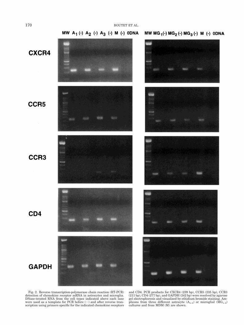
Fig. 2. Reverse transcription-polymerase chain reaction (RT-PCR)detection of chemokine receptor mRNA in astrocytes and microglia.DNase-treated RNA from the cell types indicated above each lanewere used as a template for PCR before (2) and after reverse tran-scription using primers specific for the indicated chemokine receptors
and CD4. PCR products for CXCR4 (239 bp), CCR5 (235 bp), CCR3(211 bp), CD4 (277 bp), and GAPDH (242 bp) were resolved by agarosegel electrophoresis and visualized by ethidium bromide staining. Am-plicons from three different astrocyte (A1–3) or microglial (MG1–3)cultures and from MDM (M) are shown.
170 BOUTET ET AL.
SDF-1a, MIP-1b, and RANTES induce transientelevation of [Ca21]i in astrocytes and microglia
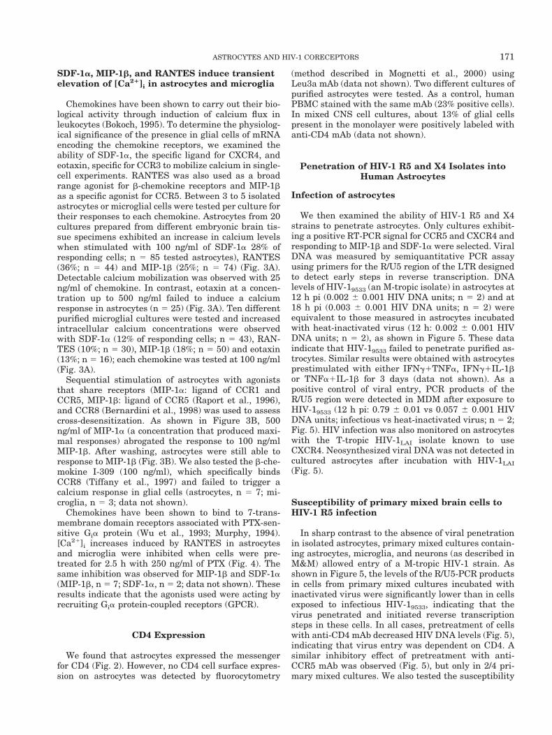
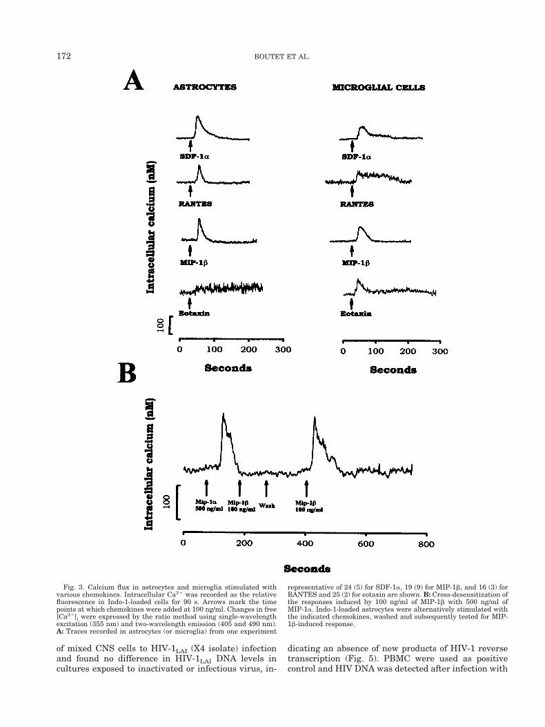
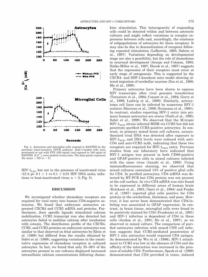
Chemokines have been shown to carry out their bio-logical activity through induction of calcium flux inleukocytes (Bokoch, 1995). To determine the physiolog-ical significance of the presence in glial cells of mRNAencoding the chemokine receptors, we examined theability of SDF-1a, the specific ligand for CXCR4, andeotaxin, specific for CCR3 to mobilize calcium in single-cell experiments. RANTES was also used as a broadrange agonist for b-chemokine receptors and MIP-1bas a specific agonist for CCR5. Between 3 to 5 isolatedastrocytes or microglial cells were tested per culture fortheir responses to each chemokine. Astrocytes from 20cultures prepared from different embryonic brain tis-sue specimens exhibited an increase in calcium levelswhen stimulated with 100 ng/ml of SDF-1a 28% ofresponding cells; n 5 85 tested astrocytes), RANTES(36%; n 5 44) and MIP-1b (25%; n 5 74) (Fig. 3A).Detectable calcium mobilization was observed with 25ng/ml of chemokine. In contrast, eotaxin at a concen-tration up to 500 ng/ml failed to induce a calciumresponse in astrocytes (n 5 25) (Fig. 3A). Ten differentpurified microglial cultures were tested and increasedintracellular calcium concentrations were observedwith SDF-1a (12% of responding cells; n 5 43), RAN-TES (10%; n 5 30), MIP-1b (18%; n 5 50) and eotaxin(13%; n 5 16); each chemokine was tested at 100 ng/ml(Fig. 3A).
Sequential stimulation of astrocytes with agoniststhat share receptors (MIP-1a: ligand of CCR1 andCCR5, MIP-1b: ligand of CCR5 (Raport et al., 1996),and CCR8 (Bernardini et al., 1998) was used to assesscross-desensitization. As shown in Figure 3B, 500ng/ml of MIP-1a (a concentration that produced maxi-mal responses) abrogated the response to 100 ng/mlMIP-1b. After washing, astrocytes were still able toresponse to MIP-1b (Fig. 3B). We also tested the b-che-mokine I-309 (100 ng/ml), which specifically bindsCCR8 (Tiffany et al., 1997) and failed to trigger acalcium response in glial cells (astrocytes, n 5 7; mi-croglia, n 5 3; data not shown).
Chemokines have been shown to bind to 7-trans-membrane domain receptors associated with PTX-sen-sitive Gia protein (Wu et al., 1993; Murphy, 1994).[Ca21]i increases induced by RANTES in astrocytesand microglia were inhibited when cells were pre-treated for 2.5 h with 250 ng/ml of PTX (Fig. 4). Thesame inhibition was observed for MIP-1b and SDF-1a(MIP-1b, n 5 7; SDF-1a, n 5 2; data not shown). Theseresults indicate that the agonists used were acting byrecruiting Gia protein-coupled receptors (GPCR).
CD4 Expression
We found that astrocytes expressed the messengerfor CD4 (Fig. 2). However, no CD4 cell surface expres-sion on astrocytes was detected by fluorocytometry
(method described in Mognetti et al., 2000) usingLeu3a mAb (data not shown). Two different cultures ofpurified astrocytes were tested. As a control, humanPBMC stained with the same mAb (23% positive cells).In mixed CNS cell cultures, about 13% of glial cellspresent in the monolayer were positively labeled withanti-CD4 mAb (data not shown).
Penetration of HIV-1 R5 and X4 Isolates intoHuman Astrocytes
Infection of astrocytes
We then examined the ability of HIV-1 R5 and X4strains to penetrate astrocytes. Only cultures exhibit-ing a positive RT-PCR signal for CCR5 and CXCR4 andresponding to MIP-1b and SDF-1a were selected. ViralDNA was measured by semiquantitative PCR assayusing primers for the R/U5 region of the LTR designedto detect early steps in reverse transcription. DNAlevels of HIV-19533 (an M-tropic isolate) in astrocytes at12 h pi (0.002 6 0.001 HIV DNA units; n 5 2) and at18 h pi (0.003 6 0.001 HIV DNA units; n 5 2) wereequivalent to those measured in astrocytes incubatedwith heat-inactivated virus (12 h: 0.002 6 0.001 HIVDNA units; n 5 2), as shown in Figure 5. These dataindicate that HIV-19533 failed to penetrate purified as-trocytes. Similar results were obtained with astrocytesprestimulated with either IFNg1TNFa, IFNg1IL-1bor TNFa1IL-1b for 3 days (data not shown). As apositive control of viral entry, PCR products of theR/U5 region were detected in MDM after exposure toHIV-19533 (12 h pi: 0.79 6 0.01 vs 0.057 6 0.001 HIVDNA units; infectious vs heat-inactivated virus; n 5 2;Fig. 5). HIV infection was also monitored on astrocyteswith the T-tropic HIV-1LAI isolate known to useCXCR4. Neosynthesized viral DNA was not detected incultured astrocytes after incubation with HIV-1LAI(Fig. 5).
Susceptibility of primary mixed brain cells toHIV-1 R5 infection
In sharp contrast to the absence of viral penetrationin isolated astrocytes, primary mixed cultures contain-ing astrocytes, microglia, and neurons (as described inM&M) allowed entry of a M-tropic HIV-1 strain. Asshown in Figure 5, the levels of the R/U5-PCR productsin cells from primary mixed cultures incubated withinactivated virus were significantly lower than in cellsexposed to infectious HIV-19533, indicating that thevirus penetrated and initiated reverse transcriptionsteps in these cells. In all cases, pretreatment of cellswith anti-CD4 mAb decreased HIV DNA levels (Fig. 5),indicating that virus entry was dependent on CD4. Asimilar inhibitory effect of pretreatment with anti-CCR5 mAb was observed (Fig. 5), but only in 2/4 pri-mary mixed cultures. We also tested the susceptibility
171ASTROCYTES AND HIV-1 CORECEPTORS
of mixed CNS cells to HIV-1LAI (X4 isolate) infectionand found no difference in HIV-1LAI DNA levels incultures exposed to inactivated or infectious virus, in-
dicating an absence of new products of HIV-1 reversetranscription (Fig. 5). PBMC were used as positivecontrol and HIV DNA was detected after infection with
Fig. 3. Calcium flux in astrocytes and microglia stimulated withvarious chemokines. Intracellular Ca21 was recorded as the relativefluorescence in Indo-1-loaded cells for 90 s. Arrows mark the timepoints at which chemokines were added at 100 ng/ml. Changes in free[Ca21]i were expressed by the ratio method using single-wavelengthexcitation (355 nm) and two-wavelength emission (405 and 490 nm).A: Traces recorded in astrocytes (or microglia) from one experiment
representative of 24 (5) for SDF-1a, 19 (9) for MIP-1b, and 16 (3) forRANTES and 25 (2) for eotaxin are shown. B: Cross-desensitization ofthe responses induced by 100 ng/ml of MIP-1b with 500 ng/ml ofMIP-1a. Indo-1-loaded astrocytes were alternatively stimulated withthe indicated chemokines, washed and subsequently tested for MIP-1b-induced response.
172 BOUTET ET AL.
HIV-1LAI, but not in the presence of inactivated virus(12 h pi: 9.1 6 1 vs 0.1 6 0.01 HIV DNA units; infec-tious vs heat-inactivated virus; n 5 2; Fig. 5).
DISCUSSION
We investigated whether chemokine receptors arerequired for viral entry into human CD4-negative as-trocytes. We found that embryonic astrocytes ex-pressed CXCR4 and CCR5 mRNA and proteins. Fur-thermore, their specific ligands stimulated calciummobilization. CCR3 transcript was also detected butastrocytes failed to mobilize calcium after incubationwith eotaxin. The distribution profile of the CXCR4,CCR5, and CCR3 proteins on embryonic astrocytes wassimilar to that observed on fetal astrocytes by Klein etal. (1999) but differed from the results obtained bySabri et al. (1999), suggesting variations in the consti-tutive expression of chemokine receptors in culturedastrocytes. In fact, we found that only 25–36% of theastrocytes present in our cultures displayed increasedintracellular calcium concentrations following chemo-
kine stimulation. This heterogeneity of respondingcells could be detected within and between astrocytecultures and might reflect variations in receptor ex-pression between cells and, accordingly, the existenceof subpopulations of astrocytes for these receptors. Itmay also be due to desensitization of receptors follow-ing repeated stimulation (Lefkowitz, 1993; Sabroe etal., 1997). Variations depending on developmentalstage are also a possibility, but the role of chemokinesin neuronal development (Araujo and Cotman, 1993;Halks-Miller et al., 1997; Horuk et al., 1997) suggeststhat the expression of their receptors must occur atearly stage of ontogenesis. This is supported by theCXCR4- and SDF-1-knockout mice model showing al-tered migration of cerebellar neurons (Zou et al., 1998;Ma et al., 1998).
Primary astrocytes have been shown to expressHIV transcripts after viral genomic transfection(Tornatore et al., 1994; Conant et al., 1994; Gorry etal., 1999; Ludwig et al., 1999). Similarly, astrocy-toma cell lines can be infected by numerous HIV-1isolates (Harouse et al., 1989; Neumann et al., 1995).In contrast, studies reporting HIV-1 entry into pri-mary human astrocytes are scarce (Nath et al., 1995;Sabri et al., 1999). We observed that the M-tropicHIV-19533 strain infected MDM via CCR5 but did notpenetrate purified CCR5-positive astrocytes. In con-trast, in primary mixed brain cell cultures, neosyn-thesized viral DNA was detected after exposure toHIV-19533 and DNA levels were reduced with anti-CD4 and anti-CCR5 mAb, indicating that these tworeceptors are required for HIV-19533 entry. Previousstudies from our laboratory have indicated thatHIV-1 antigen was present in both CD68-positiveand GFAP-positive cells in mixed cultures infectedwith the same virus (Janabi et al., 1998). Usingimmunofluorescence staining, we observed thatmixed cultures contained 13% of positive glial cellsfor CD4. In purified astrocytes, CD4 mRNA was de-tected by RT-PCR but CD4 protein was not presentat the cell surface. In vivo CD4 mRNA was also foundto be expressed in different areas of human brain(Erickson et al., 1991; Omri et al., 1994) and Funkeet al. (1987) reported glial cells positive for CD4protein in the cerebellum, thalamus, and pons. How-ever, it has never been demonstrated that CD4-la-beling was associated to GFAP expression. In con-trast, in brain tissue, microglial cells were found tobe positively stained for CD4 (Peudenier et al., 1991)and HIV-1 infection is dependent of CD4 in thesecells (Jordan et al., 1991; He et al., 1997), as weobserved in mixed culture. The comparison of puri-fied astrocytes infection with mixed CNS cell infec-tion suggests that CCR5-mediated penetration ofHIV-1 into astrocytes would required CD4 antigen.As demonstrated by Wu et al. (1996), HIV-1 attach-ment to CCR5 was low in the absence of CD4 and theaffinity of the interaction was increased in the pres-ence of soluble CD4. Furthermore, Speck et al. (1999)demonstrated that CD4 provided in trans, induced
Fig. 4. Astrocytes and microglial cells respond to RANTES by thepertussis toxin-sensitive GPCR pathway. Indo-1-loaded cells werepretreated or not with PTX (250 ng/ml) and exposed to 100 ng/ml ofRANTES. [Ca21]i were plotted versus time. The data points representthe mean 6 SD (n 5 5).
173ASTROCYTES AND HIV-1 CORECEPTORS
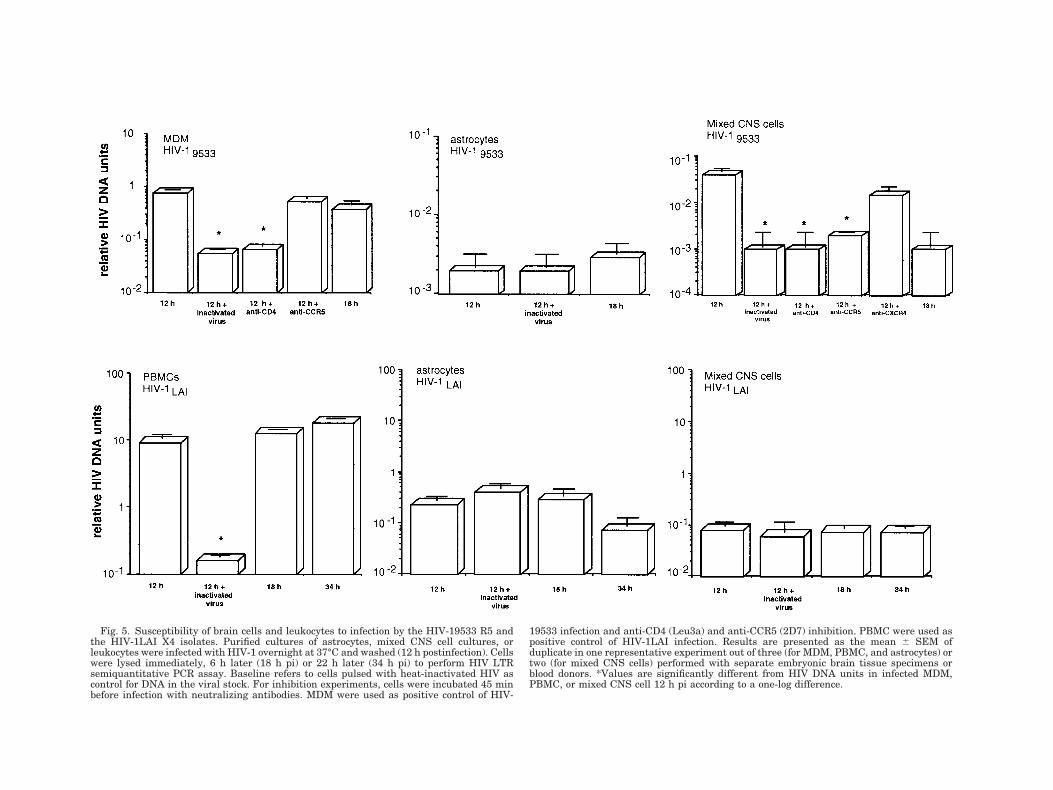
Fig. 5. Susceptibility of brain cells and leukocytes to infection by the HIV-19533 R5 andthe HIV-1LAI X4 isolates. Purified cultures of astrocytes, mixed CNS cell cultures, orleukocytes were infected with HIV-1 overnight at 37°C and washed (12 h postinfection). Cellswere lysed immediately, 6 h later (18 h pi) or 22 h later (34 h pi) to perform HIV LTRsemiquantitative PCR assay. Baseline refers to cells pulsed with heat-inactivated HIV ascontrol for DNA in the viral stock. For inhibition experiments, cells were incubated 45 minbefore infection with neutralizing antibodies. MDM were used as positive control of HIV-
19533 infection and anti-CD4 (Leu3a) and anti-CCR5 (2D7) inhibition. PBMC were used aspositive control of HIV-1LAI infection. Results are presented as the mean 6 SEM ofduplicate in one representative experiment out of three (for MDM, PBMC, and astrocytes) ortwo (for mixed CNS cells) performed with separate embryonic brain tissue specimens orblood donors. *Values are significantly different from HIV DNA units in infected MDM,PBMC, or mixed CNS cell 12 h pi according to a one-log difference.
HIV-1 infection of primary human CD4-negative as-trocytes. In mixed CNS cell cultures, one can hypoth-esize that microglial cells supplied CD4 in trans toallow R5 HIV-1 penetration of astrocytes. The repli-cation of HIV-1 M-tropic isolate on CD4-negativecells is however possible but required adaptationprocesses. Kolchinsky et al. (1999) demonstrated theadaptation of M-tropic isolate replication on CD4-negative canine cells expressing human CCR5 withconcomitant changes in the V2 loop conformation ofgp120.
The ability of HIV to use CXCR4 in the absence ofCD4 in human fetal astrocytes has been described fora T-cell line adapted (TCLA) HIV-2 but not for pri-mary X4 HIV-2 (Reeves et al., 1999). In our study,the TCLA HIV-1LAI did not infect CXCR4-expressingastrocytes, suggesting that CD42 astrocytes proba-bly display variable susceptibility to HIV X4 straininfection. This might be related to viral strains bear-ing a fusion-competent Env conformation whichwould normally be expected to follow CD4 binding(Chan and Kim, 1998).
Since HIV-19533 infection was inhibited by anti-CCR5 mAb in only 1/2 mixed brain cell cultures tested,this R5 viral isolate might use alternative coreceptors.APJ, GPR1 and CCR8 have been shown to supportHIV-1 entry and are expressed in human CNS or brain-derived cells (Edinger et al., 1998; Jinno et al., 1998;Shimizu et al., 1999). Our data suggest that CCR8 wasnot present on astrocytes or microglia because I-309,its more potent agonist, did not induce Ca21 responsesin these cells, but the involvement of APJ and GPR1remains possible. The ability of an M-tropic HIV-1 iso-late to use additional coreceptors expressed on glialcells may lead to the biological selection for particularHIV-1 variants within the brain (Power et al., 1994;Chan et al., 1999) and the emergence of neurotropismor neurovirulence (Gabuzda et al., 1998).
In conclusion, whereas CCR5 mediated viral entryinto mixed CNS cell cultures in conjunction with CD4,our results suggest that CCR5 cannot function as pri-mary receptor for M-tropic HIV-1 strains in astrocytes.In vivo, HIV-1 might use CD4 supplied in trans bymicroglial cells to penetrate astrocytes. However, itcannot be ruled out that infection of astrocytes mightoccur through alternative mechanisms such as endocy-tosis (Hao and Lyman, 1999) or cell-to-cell transfer, assuggested by Kohleisen et al. (1992).
ACKNOWLEDGMENTS
The authors are grateful to Nazila Janabi for helpfuldiscussions, to Isabelle Peguillet for advice on quanti-tative PCR, and to Juliana Croitoru and Bruno Vaslinfor their contribution to the flow cytometry experi-ments.
REFERENCES
Albright AV, Shieh JT, Itoh T, Lee B, Pleasure D, O’Connor MJ, DomsRW, Gonzalez-Scarano F. 1999. Microglia express CCR5, CXCR4,and CCR3, but of these, CCR5 is the principal coreceptor for humanimmunodeficiency virus type 1 dementia isolates. J Virol 73:205–213.
Andjelkovic AV, Kerkovich D, Shanley J, Pulliam L, Pachter JS. 1999.Expression of binding sites for b chemokines on human astrocytes.Glia 28:225–235.
Araujo DM, Cotman CW. 1993. Trophic effects of interleukin-4, -7 and-8 on hippocampal neuronal cultures: potential involvement of glial-derived factors. Brain Res 600:49–55.
Berger EA, Doms RW, Fenyo EM, Korber BT, Littman DR, Moore JP,Sattentau QJ, Schuitemaker H, Sodroski J, Weiss RA. 1998. A newclassification for HIV-1 [letter]. Nature 391:240.
Berger EA, Murphy PM, Farber JM. 1999. Chemokine receptors asHIV-1 coreceptors: roles in viral entry, tropism and disease. AnnuRev Immunol 17:657–700.
Bernardini G, Hedrick J, Sozzani S, Luini W, Spinetti G, Weiss M,Menon S, Zlotnik A, Mantovani A, Santoni A, Napolitano M. 1998.Identification of the CC chemokines TARC and macrophage inflam-matory protein-1 b as novel functional ligands for the CCR8 recep-tor. Eur J Immunol 28:582–588.
Bokoch GM. 1995. Chemoattractant signaling and leukocyte activa-tion. Blood 86:1649–1660.
Chan DC, Kim PS. 1998. HIV entry and its inhibition. Cell 93:681–684.
Chan SY, Speck RF, Power C, Gaffen SL, Chesebro B, Goldsmith MA.1999. V3 recombinants indicate a central role for CCR5 as a core-ceptor in tissue infection by human immunodeficiency virus type 1.J Virol 73:2350–2358.
Chomczynsky P, Sacchi N. 1987. Single-step method of RNA isolationby acid guanidinium thiocyanate-phenol-chloroform extraction.Ann Biochem 162:156–159.
Conant K, Atwood WJ, Traub R, Tornatore C, Major EO. 1994. Anincrease in p50/p65 NF-kB binding to the HIV-1 LTR is not suffi-cient to increase viral expression in the primary human astrocyte.Virology 205:586–590.
Endres MJ, Clapham PR, Marsh M, Ahuja M, Turner JD, McKnightA, Thomas JF, Stoebenau-Haggarty B, Choe S, Vance PJ, Wells TN,Power CA, Sutterwala SS, Doms RW, Landau NR, Hoxie JA. 1996.CD4-independent infection by HIV-2 is mediated by fusin/CXCR4.Cell 87:745–756.
Erickson JD, Trojanowski JQ, Eiden LE. 1991. Regional distributionand partial molecular characterization of CD4-related mRNA inhuman brain and peripheral tissues. Mol Brain Res 10:23–31.
Funke I, Hahn A, Rieber EP, Weiss E, Riethmuller G. 1987. Thecellular receptor (CD4) of the human immunodeficiency virus isexpressed on neurons and glial cells in human brain. J Exp Med165:1230–1235.
Gabuzda D, He J, Ohagen A, Vallat AV. 1998. Chemokine receptors inHIV-1 infection of the central nervous system. Semin Immunol10:203–213.
Ghorpade A, Xia MQ, Hyman BT, Persidsky Y, Nukuna A, Bock P,Che M, Limoges J, Gendelman HE, Mackay CR. 1998. Role of theb-chemokine receptors CCR3 and CCR5 in human immunodefi-ciency virus type 1 infection of monocytes and microglia. J Virol72:3351–3361.
Gorry PR, Howard JL, Churchill MJ, Anderson JL, Cunningham A,Adrian D, McPhee DA, Purcell DF. 1999. Diminished production ofhuman immunodeficiency virus type 1 in astrocytes results frominefficient translation of gag, env, and nef mRNAs despite efficientexpression of Tat and Rev. J Virol 73:352–361.
Grynkiewicz G, Poenie M, Tsien RY. 1985. A new generation of Ca21
indicators with greatly improved fluorescence properties. J BiolChem 260:3440–3450.
Halks-Miller M, Hesselgesser J, Miko IJ, Horuk R. 1997. Chemokinereceptors in developing human brain. Methods Enzymol 288:27–38.
Hao HN, Lyman WD. 1999. HIV infection of fetal human astrocytes:the potential role of a receptor-mediated endocytic pathway. BrainRes 823:24–32.
Harouse JM, Kunsch C, Hartle HT, Laughlin MA, Hoxie JA, WigdahlB, Gonzalez-Scarano F. 1989. CD4-independent infection of humanneural cells by human immunodeficiency virus type 1. J Virol 63:2527–2533.
He J, Chen Y, Farzan M, Choe H, Ohagen A, Gartner S, Busciglio J,Yang X, Hofmann W, Newman W, Mackay CR, Sodroski J, GabuzdaD. 1997. CCR3 and CCR5 are co-receptors for HIV-1 infection ofmicroglia. Nature 385:645–649.
175ASTROCYTES AND HIV-1 CORECEPTORS
Horuk R, Martin AW, Wang Z, Schweitzer L, Gerassimides A, Guo H,Lu Z, Hesselgesser J, Perez HD, Kim J, Parker J, Hadley TJ, PeiperSC. 1997. Expression of chemokine receptors by subsets of neuronsin the central nervous system. J Immunol 158:2882–2890.
Janabi N, Chabrier S, Tardieu M. 1996. Endogenous nitric oxideactivates prostaglandin F2a production in human microglial cellsbut not in astrocytes: a study of interactions between eicosanoids,nitric oxide, and superoxide anion (O2
2) regulatory pathways. J Im-munol 157:2129–2135.
Janabi N, Di Stefano M, Wallon C, Hery C, Chiodi F, Tardieu M. 1998.Induction of human immunodeficiency virus type 1 replication inhuman glial cells after proinflammatory cytokines stimulation: ef-fect of IFNg, IL1b, and TNFa on differentiation and chemokineproduction in glial cells. Glia 23:304–315.
Jinno A, Shimizu N, Soda Y, Haraguchi Y, Kitamura T, Hoshino H.1998. Identification of the chemokine receptor TER1/CCR8 ex-pressed in brain-derived cells and T cells as a new coreceptor forHIV-1 infection. Biochem Biophys Res Commun 243:497–502.
Jordan CA, Watkins BA, Kufta C, Dubois-Dalcq M. 1991. Infection ofbrain microglial cells by human immunodeficiency virus type 1 isCD4 dependent. J Virol 65:736–742.
Klein RS, Williams KC, Alvarez-Hernandez X, Westmoreland S, ForceT, Lackner AA, Luster AD. 1999. Chemokine receptor expressionand signaling in macaque and human fetal neurons and astrocytes:implications for the neuropathogenesis of AIDS. J Immunol 163:1636–1646.
Kohleisen B, Neumann M, Herrmann R, Brack-Werner R, Krohn KJ,Ovod V, Ranki A, Erfle V. 1992. Cellular localization of Nef ex-pressed in persistently HIV-1-infected low-producer astrocytes[published erratum appears in AIDS 1993 Mar;7(3):following 444].AIDS 6:1427–1436.
Kolchinsky P, Mirzabekov T, Farzan M, Kiprilov E, Cayabyab M,Mooney LJ, Choe H, Sodroski J. 1999. Adaptation of a CCR5-using,primary human immunodeficiency virus type 1 isolate for CD4-independent replication. J Virol 73:8120–8126.
Lannuzel A, Lledo PM, Lamghitnia HO, Vincent JD, Tardieu M. 1995.HIV-1 envelope proteins gp120 and gp160 potentiate NMDA-in-duced [Ca21]i increase, alter [Ca21]i homeostasis and induce neu-rotoxicity in human embryonic neurons. Eur J Neurosci 7:2285–2293.
Lavi E, Strizki JM, Ulrich AM, Zhang W, Fu L, Wang Q, O’Connor M,Hoxie JA, Gonzalez-Scarano F. 1997. CXCR-4 (Fusin), a co-receptorfor the type 1 human immunodeficiency virus (HIV-1), is expressedin the human brain in a variety of cell types, including microgliaand neurons. Am J Pathol 151:1035–1042.
Lee SC, Hatch WC, Liu W, Kress Y, Lyman WD, Dickson DW. 1993.Productive infection of human fetal microglia by HIV-1. Am JPathol 143:1032–1039.
Lefkowitz RJ. 1993. G protein-coupled receptor kinases. Cell 74:409–412.
Ludwig E, Ceccherini-Silberstein F, van Empel J, Erfle V, NeumannM, Brack-Werner R. 1999. Diminished rev-mediated stimulation ofhuman immunodeficiency virus type 1 protein synthesis is a hall-mark of human astrocytes. J Virol 73:8279–8289.
Ma Q, Jones D, Borghesani PR, Segal RA, Nagasawa T, Kishimoto T,Bronson RT, Springer TA. 1998. Impaired B-lymphopoiesis, myelo-poiesis, and derailed cerebellar neuron migration in CXCR4- andSDF-1-deficient mice. Proc Natl Acad Sci USA 95:9448–9453.
Miller RJ, Meucci O. 1999 AIDS and the brain: is there a chemokineconnection? Trends Neurosci 22:471–479.
Mognetti B, Moussa M, Croitoru J, Menu E, Dormont D, Roques P,Chaouat G. 2000. HIV-1 co-receptor expression on trophoblasticcells from early placentas and permissivity to infection by severalHIV-1 primary isolates. Clin Exp Immunol 119:486–492.
Murphy PM. 1994. The molecular biology of leukocyte chemoattrac-tant receptors. Annu Rev Immunol 12:593–633.
Nath A, Hartloper V, Furer M, Fowke KR. 1995. Infection of humanfetal astrocytes with HIV-1: viral tropism and the role of cell to cellcontact in viral transmission. J Neuropathol Exp Neurol 54:320–330.
Neumann M, Felber BK, Kleinschmidt A, Froese B, Erfle V, PavlakisGN, Brack-Werner R. 1995. Restriction of human immunodefi-ciency virus type 1 production in a human astrocytoma cell line isassociated with a cellular block in Rev function. J Virol 69:2159–2167.
Omri B, Crisanti P, Alliot F, Marty MC, Rutin J, Levallois C, PrivatA, Pessac B. 1994. CD4 expression in neurons of the central nervoussystem. Int Immunol 6:377–385.
Peudenier S, Hery C, Heng Ng K, Tardieu M. 1991. HIV receptorswithin the brain: a study of CD4 and MHC-II on human neurons,astrocytes and microglial cells. Res Virol 142:145–149.
Ponath PD, Qin S, Ringler DJ, Clark-Lewis I, Wang J, Kassam N,Smith H, Shi X, Gonzalo JA, Newman W, Gutierrez-Ramos JC,Mackay CR. 1996. Cloning of the human eosinophil chemoattrac-tant, eotaxin. Expression, receptor binding, and functional proper-ties suggest a mechanism for the selective recruitment of eosino-phils. J Clin Invest 97:604–612.
Power C, McArthur JC, Johnson RT, Griffin DE, Glass JD, PerrymanS, Chesebro B. 1994. Demented and nondemented patients withAIDS differ in brain-derived human immunodeficiency virus type 1envelope sequences. J Virol 68:4643–4649.
Ranki A, Nyberg M, Ovod V, Haltia M, Elovaara I, Raininko R,Haapasalo H, Krohn K. 1995. Abundant expression of HIV Nef andRev proteins in brain astrocytes in vivo is associated with dementia.AIDS 9:1001–1008.
Raport CJ, Gosling J, Schweickart VL, Gray PW, Charo IF. 1996.Molecular cloning and functional characterization of a novel humanCC chemokine receptor (CCR5) for RANTES, MIP-1b, and MIP-1a.J Biol Chem 271:17161–17166.
Reeves JD, Hibbitts S, Simmons G, McKnight A, Azevedo-Pereira JM,Moniz-Pereira J, Clapham PR. 1999. Primary human immunodefi-ciency virus type 2 (HIV-2) isolates infect CD4-negative cells viaCCR5 and CXCR4: comparison with HIV-1 and simian immunode-ficiency virus and relevance to cell tropism in vivo. J Virol 73:7795–7804.
Rottman JB, Ganley KP, Williams K, Wu L, Mackay CR, Ringler DJ.1997. Cellular localization of the chemokine receptor CCR5. Corre-lation to cellular targets of HIV-1 infection. Am J Pathol 151:1341–1351.
Sabri F, Tresoldi E, Di Stefano M, Polo S, Monaco MC, Verani A, FioreJR, Lusso P, Major EO, Chiodi F, Scarlatti G. 1999. Nonproductivehuman immunodeficiency virus type 1 infection of human fetalastrocytes: independence from CD4 and major chemokine receptors.Virology 264:370–384.
Sabroe I, Williams TJ, Hebert CA, Collins PD. 1997. Chemoattractantcross-desensitization of the human neutrophil IL-8 receptor in-volves receptor internalization and differential receptor subtyperegulation. J Immunol 158:1361–1369.
Saito Y, Sharer LR, Epstein LG, Michaels J, Mintz M, Louder M,Golding K, Cvetkovich TA, Blumberg BM. 1994. Overexpression ofnef as a marker for restricted HIV-1 infection of astrocytes inpostmortem pediatric central nervous tissues. Neurology 44:474–481.
Sanders VJ, Pittman CA, White MG, Wang G, Wiley CA, Achim CL.1998. Chemokines and receptors in HIV encephalitis. AIDS 12:1021–1026.
Shieh JT, Albright AV, Sharron M, Gartner S, Strizki J, Doms RW,Gonzalez-Scarano F. 1998. Chemokine receptor utilization by hu-man immunodeficiency virus type 1 isolates that replicate in micro-glia. J Virol 72:4243–4249.
Shimizu N, Soda Y, Kanbe K, Liu HY, Jinno A, Kitamura T, HoshinoH. 1999. An orphan G protein-coupled receptor, GPR1, acts as acoreceptor to allow replication of human immunodeficiency virustypes 1 and 2 in brain-derived cells. J Virol 73:5231–5239.
Simmons G, Wilkinson D, Reeves JD, Dittmar MT, Beddows S, WeberJ, Carnegie G, Desselberger U, Gray PW, Weiss RA, Clapham PR.1996. Primary, syncytium-inducing human immunodeficiency virustype 1 isolates are dual-tropic and most can use either Lestr orCCR5 as coreceptors for virus entry. J Virol 70:8355–8360.
Speck RF, Esser U, Penn ML, Eckstein DA, Pulliam L, Chan SY,Goldsmith MA. 1999. A trans-receptor mechanism for infection ofCD4-negative cells by human immunodeficiency virus type 1. CurrBiol 9:547–550.
Taoufik Y, Lantz O, Wallon C, Charles A, Dussaix E, Delfraissy JF.1997. Human immunodeficiency virus gp120 inhibits interleu-kin-12 secretion by human monocytes: an indirect interleukin-10-mediated effect. Blood 89:2842–2848.
Tardieu M. 1999. HIV-1-related central nervous system diseases.Curr Opin Neurol 12:377–381.
Tiffany HL, Lautens LL, Gao JL, Pease J, Locati M, Combadiere C,Modi W, Bonner TI, Murphy PM. 1997. Identification of CCR8: ahuman monocyte and thymus receptor for the CC chemokine I-309.J Exp Med 186:165–170.
Tornatore C, Nath A, Amemiya K, Major EO. 1991. Persistent humanimmunodeficiency virus type 1 infection in human fetal glial cellsreactivated by T-cell factor(s) or by the cytokines tumor necrosisfactor alpha and interleukin-1 b. J Virol 65:6094–6100.
Tornatore C, Chandra R, Berger JR, Major EO. 1994. HIV-1 infectionof subcortical astrocytes in the pediatric central nervous system.Neurology 44:481–487.
Vallat AV, De Girolami U, He J, Mhashilkar A, Marasco W, Shi B,Gray F, Bell J, Keohane C, Smith TW, Gabuzda D. 1998. Localiza-
176 BOUTET ET AL.

tion of HIV-1 co-receptors CCR5 and CXCR4 in the brain of childrenwith AIDS. Am J Pathol 152:167–178.
Watkins BA, Dorn HH, Kelly WB, Armstrong RC, Potts BJ, Michaels F,Kufta CV, Dubois-Dalcq M. 1990. Specific tropism of HIV-1 for micro-glial cells in primary human brain cultures. Science 249:549–553.
Wu D, LaRosa GJ, Simon MI. 1993. G protein-coupled signal trans-duction pathways for interleukin-8. Science 261:101–103.
Wu L, Gerard NP, Wyatt R, Choe H, Parolin C, Ruffing N, Borsetti A,Cardoso AA, Desjardin E, Newman W, Gerard C, Sodroski J. 1996.CD4-induced interaction of primary HIV-1 gp120 glycoproteinswith the chemokine receptor CCR-5 [see comments]. Nature 384:179–183.
Wu L, LaRosa G, Kassam N, Gordon CJ, Heath H, Ruffing N, Chen H,Humblias J, Samson M, Parmentier M, Moore JP, Mackay CR.1997. Interaction of chemokine receptor CCR5 with its ligands:multiple domains for HIV-1 gp120 binding and a single domain forchemokine binding. J Exp Med 186:1373–1381.
Zack JA, Arrigo SJ, Weitsman SR, Go AS, Haislip A, Chen IS. 1990.HIV-1 entry into quiescent primary lymphocytes: molecular analy-sis reveals a labile, latent viral structure. Cell 61:213–222.
Zou YR, Kottmann AH, Kuroda M, Taniuchi I, Littman DR. 1998.Function of the chemokine receptor CXCR4 in haematopoiesisand in cerebellar development [see comments]. Nature 393:595–599.
177ASTROCYTES AND HIV-1 CORECEPTORS