SDF-1/CXCR4 Signaling Preserves Microvascular Integrity and Renal Function in Chronic Kidney Disease
11
SDF-1/CXCR4 Signaling Preserves Microvascular Integrity and Renal Function in Chronic Kidney Disease Li-Hao Chen 1 , Suzanne L. Advani 1 , Kerri Thai 1 , M. Golam Kabir 1 , Manish M. Sood 2 , Ian W. Gibson 3 , Darren A. Yuen 1 , Kim A. Connelly 1 , Philip A. Marsden 1 , Darren J. Kelly 4 , Richard E. Gilbert 1 , Andrew Advani 1 * 1 Keenan Research Centre for Biomedical Science and Li Ka Shing Knowledge Institute of St. Michael’s Hospital, Toronto, Ontario, Canada, 2 Ottawa Hospital Research Institute, University of Ottawa, Ottawa, Ontario, Canada, 3 Health Sciences Centre, University of Manitoba, Winnipeg, Manitoba, Canada, 4 Department of Medicine, St. Vincent’s Hospital, Melbourne, Victoria, Australia Abstract The progressive decline of renal function in chronic kidney disease (CKD) is characterized by both disruption of the microvascular architecture and the accumulation of fibrotic matrix. One angiogenic pathway recently identified as playing an essential role in renal vascular development is the stromal cell-derived factor-1a (SDF-1)/CXCR4 pathway. Because similar developmental processes may be recapitulated in the disease setting, we hypothesized that the SDF-1/CXCR4 system would regulate microvascular health in CKD. Expression of CXCR4 was observed to be increased in the kidneys of subtotally nephrectomized (SNx) rats and in biopsies from patients with secondary focal segmental glomerulosclerosis (FSGS), a rodent model and human correlate both characterized by aberration of the renal microvessels. A reno-protective role for local SDF- 1/CXCR4 signaling was indicated by i) CXCR4-dependent glomerular eNOS activation following acute SDF-1 administration; and ii) acceleration of renal function decline, capillary loss and fibrosis in SNx rats treated with chronic CXCR4 blockade. In contrast to the upregulation of CXCR4, SDF-1 transcript levels were decreased in SNx rat kidneys as well as in renal fibroblasts exposed to the pro-fibrotic cytokine transforming growth factor b (TGF-b), the latter effect being attenuated by histone deacetylase inhibition. Increased renal SDF-1 expression was, however, observed following the treatment of SNx rats with the ACE inhibitor, perindopril. Collectively, these observations indicate that local SDF-1/CXCR4 signaling functions to preserve microvascular integrity and prevent renal fibrosis. Augmentation of this pathway, either purposefully or serendipitously with either novel or existing therapies, may attenuate renal decline in CKD. Citation: Chen L-H, Advani SL, Thai K, Kabir MG, Sood MM, et al. (2014) SDF-1/CXCR4 Signaling Preserves Microvascular Integrity and Renal Function in Chronic Kidney Disease. PLoS ONE 9(3): e92227. doi:10.1371/journal.pone.0092227 Editor: Niels Olsen Saraiva Ca ˆmara, Universidade de Sao Paulo, Brazil Received December 13, 2013; Accepted February 19, 2014; Published March 17, 2014 Copyright: ß 2014 Chen et al. This is an open-access article distributed under the terms of the Creative Commons Attribution License, which permits unrestricted use, distribution, and reproduction in any medium, provided the original author and source are credited. Funding: These studies were supported by grants from CIHR (grant MOP-97791) and the Canadian Diabetes Association (CDA, OG-3-10-2949-AA) to Dr. Advani. Mr. Chen was supported by a CIHR Frederick Banting and Charles Best Graduate Scholarship and a Banting and Best Diabetes Centre Graduate Studentship. Dr. Advani is a Canadian Diabetes Association Clinician Scientist and this work was supported, in part, by the Canadian Diabetes Association. Dr. Gilbert is a Canada Research Chair in Diabetes Complications. Dr. Yuen is a KRESCENT New Investigator and Canadian Diabetes Association Clinician Scientist. The funders had no role in study design, data collection and analysis, decision to publish, or preparation of the manuscript. Competing Interests: REG and AA report having received funds through their institution from Servier to assist with patient care. KAC reports having received honoraria from Servier for CME talks. * E-mail: [email protected] Introduction The progressive decline of renal function in chronic kidney disease (CKD) is characterized by both fibrotic scarring of the kidney and obliteration of the renal microvessels, these two pathogenetic hallmarks commonly occurring in tandem and enjoying a reciprocal relationship. On the one hand, microvascu- lar loss may occur as a result of the occlusive actions of accumulating matrix proteins, whereas on the other hand the same process may itself contribute to organ fibrosis and progressive renal decline by predisposing the kidney to hypoxic injury [1]. Coupled with an increasing appreciation for the pivotal role that angiogenic factors may play in renal development [2], homeostasis [3] and disease [4,5], preservation of the glomerular and peritubular capillary architecture is thus a desirable charac- teristic of both existing and novel renoprotective therapies [6]. One pathway that has recently emerged as playing an essential role in renal vascular development is the stromal cell-derived factor-1a (SDF-1)/CXCR4 pathway [7]. SDF-1 is a CXC chemokine and the principal ligand for its cognate receptor, CXCR4, a seven transmembrane domain G-protein coupled receptor and the most prevalent chemokine receptor found in endothelial cells [8]. While originally defined for its role in maintenance of the hematopoietic stem cell niche and B-cell lymphopoiesis [9], the near ubiquitous tissue distribution of SDF-1 and its rapid degradation in blood indicate the capacity for much broader intra-organ specific functions [10]. The fundamental nature of the SDF-1/CXCR4 relationship is attested to by the development of identical defects in vasculogenesis and organo- genesis that occur in the absence of either gene [11]. However, as with other angiogenic pathways [4,5,12], the role that SDF-1/ CXCR4 signaling may play in the adult kidney appears to be context-dependent. For instance, studies in acute kidney injury support a reno-protective function for SDF-1/CXCR4 [13], whereas CXCR4-mediated hyperproliferation may actually con- tribute to the development of certain glomerular diseases [14]. PLOS ONE | www.plosone.org 1 March 2014 | Volume 9 | Issue 3 | e92227
Transcript of SDF-1/CXCR4 Signaling Preserves Microvascular Integrity and Renal Function in Chronic Kidney Disease
SDF-1/CXCR4 Signaling Preserves Microvascular Integrityand Renal Function in Chronic Kidney DiseaseLi-Hao Chen1, Suzanne L. Advani1, Kerri Thai1, M. Golam Kabir1, Manish M. Sood2, Ian W. Gibson3,
Darren A. Yuen1, Kim A. Connelly1, Philip A. Marsden1, Darren J. Kelly4, Richard E. Gilbert1,
Andrew Advani1*
1 Keenan Research Centre for Biomedical Science and Li Ka Shing Knowledge Institute of St. Michael’s Hospital, Toronto, Ontario, Canada, 2 Ottawa Hospital Research
Institute, University of Ottawa, Ottawa, Ontario, Canada, 3 Health Sciences Centre, University of Manitoba, Winnipeg, Manitoba, Canada, 4 Department of Medicine, St.
Vincent’s Hospital, Melbourne, Victoria, Australia
Abstract
The progressive decline of renal function in chronic kidney disease (CKD) is characterized by both disruption of themicrovascular architecture and the accumulation of fibrotic matrix. One angiogenic pathway recently identified as playingan essential role in renal vascular development is the stromal cell-derived factor-1a (SDF-1)/CXCR4 pathway. Because similardevelopmental processes may be recapitulated in the disease setting, we hypothesized that the SDF-1/CXCR4 system wouldregulate microvascular health in CKD. Expression of CXCR4 was observed to be increased in the kidneys of subtotallynephrectomized (SNx) rats and in biopsies from patients with secondary focal segmental glomerulosclerosis (FSGS), a rodentmodel and human correlate both characterized by aberration of the renal microvessels. A reno-protective role for local SDF-1/CXCR4 signaling was indicated by i) CXCR4-dependent glomerular eNOS activation following acute SDF-1 administration;and ii) acceleration of renal function decline, capillary loss and fibrosis in SNx rats treated with chronic CXCR4 blockade. Incontrast to the upregulation of CXCR4, SDF-1 transcript levels were decreased in SNx rat kidneys as well as in renalfibroblasts exposed to the pro-fibrotic cytokine transforming growth factor b (TGF-b), the latter effect being attenuated byhistone deacetylase inhibition. Increased renal SDF-1 expression was, however, observed following the treatment of SNx ratswith the ACE inhibitor, perindopril. Collectively, these observations indicate that local SDF-1/CXCR4 signaling functions topreserve microvascular integrity and prevent renal fibrosis. Augmentation of this pathway, either purposefully orserendipitously with either novel or existing therapies, may attenuate renal decline in CKD.
Citation: Chen L-H, Advani SL, Thai K, Kabir MG, Sood MM, et al. (2014) SDF-1/CXCR4 Signaling Preserves Microvascular Integrity and Renal Function in ChronicKidney Disease. PLoS ONE 9(3): e92227. doi:10.1371/journal.pone.0092227
Editor: Niels Olsen Saraiva Camara, Universidade de Sao Paulo, Brazil
Received December 13, 2013; Accepted February 19, 2014; Published March 17, 2014
Copyright: � 2014 Chen et al. This is an open-access article distributed under the terms of the Creative Commons Attribution License, which permitsunrestricted use, distribution, and reproduction in any medium, provided the original author and source are credited.
Funding: These studies were supported by grants from CIHR (grant MOP-97791) and the Canadian Diabetes Association (CDA, OG-3-10-2949-AA) to Dr. Advani.Mr. Chen was supported by a CIHR Frederick Banting and Charles Best Graduate Scholarship and a Banting and Best Diabetes Centre Graduate Studentship. Dr.Advani is a Canadian Diabetes Association Clinician Scientist and this work was supported, in part, by the Canadian Diabetes Association. Dr. Gilbert is a CanadaResearch Chair in Diabetes Complications. Dr. Yuen is a KRESCENT New Investigator and Canadian Diabetes Association Clinician Scientist. The funders had no rolein study design, data collection and analysis, decision to publish, or preparation of the manuscript.
Competing Interests: REG and AA report having received funds through their institution from Servier to assist with patient care. KAC reports having receivedhonoraria from Servier for CME talks.
* E-mail: [email protected]
Introduction
The progressive decline of renal function in chronic kidney
disease (CKD) is characterized by both fibrotic scarring of the
kidney and obliteration of the renal microvessels, these two
pathogenetic hallmarks commonly occurring in tandem and
enjoying a reciprocal relationship. On the one hand, microvascu-
lar loss may occur as a result of the occlusive actions of
accumulating matrix proteins, whereas on the other hand the
same process may itself contribute to organ fibrosis and
progressive renal decline by predisposing the kidney to hypoxic
injury [1]. Coupled with an increasing appreciation for the pivotal
role that angiogenic factors may play in renal development [2],
homeostasis [3] and disease [4,5], preservation of the glomerular
and peritubular capillary architecture is thus a desirable charac-
teristic of both existing and novel renoprotective therapies [6].
One pathway that has recently emerged as playing an essential
role in renal vascular development is the stromal cell-derived
factor-1a (SDF-1)/CXCR4 pathway [7]. SDF-1 is a CXC
chemokine and the principal ligand for its cognate receptor,
CXCR4, a seven transmembrane domain G-protein coupled
receptor and the most prevalent chemokine receptor found in
endothelial cells [8]. While originally defined for its role in
maintenance of the hematopoietic stem cell niche and B-cell
lymphopoiesis [9], the near ubiquitous tissue distribution of SDF-1
and its rapid degradation in blood indicate the capacity for much
broader intra-organ specific functions [10]. The fundamental
nature of the SDF-1/CXCR4 relationship is attested to by the
development of identical defects in vasculogenesis and organo-
genesis that occur in the absence of either gene [11]. However, as
with other angiogenic pathways [4,5,12], the role that SDF-1/
CXCR4 signaling may play in the adult kidney appears to be
context-dependent. For instance, studies in acute kidney injury
support a reno-protective function for SDF-1/CXCR4 [13],
whereas CXCR4-mediated hyperproliferation may actually con-
tribute to the development of certain glomerular diseases [14].
PLOS ONE | www.plosone.org 1 March 2014 | Volume 9 | Issue 3 | e92227

Although reactivation of ontogenetic pathways is a common
response of cells, tissues and organisms to a variety of injurious
insults [15], the function of the developmentally essential SDF-1/
CXCR4 pathway in CKD is unclear. Accordingly, in the present
study we sought to combine studies conducted in experimental
animals, cultured cells and human biopsy tissue to define the role
of SDF-1/CXCR4 signaling in CKD, focusing on the bidirec-
tional relationship between renal fibrosis and microvascular loss.
Materials and Methods
Ethics statementHuman biopsy studies were approved by the Institutional
Research Board of the Health Sciences Centre, University of
Manitoba. All patients gave written informed consent and the
study was performed in accordance with the Declaration of
Helsinki. All animal work was conducted according to the
Canadian Council on Animal Care Guidelines. The specific
experimental protocol, ACC 166, was approved by the Animal
Care Committee of St. Michael’s Hospital.
Human studiesLocalization of CXCR4 and SDF-1 was determined in kidney
sections from patients who had undergone nephrectomy for
tumor, with tissue removed from the opposite pole [16]. For gene
expression studies, kidney tissue was obtained from patients with
either secondary focal segmental glomerulosclerosis (FSGS) or
time zero live kidney donors.
AnimalsStudy 1. Expression of CXCR4 and SDF-1 was determined in
the kidneys of sham (n = 6) and subtotally nephrectomized (SNx)
(n = 8) rats after 8 weeks. Subtotal (5/6) nephrectomy or sham surgery
was performed, in female Fischer 344 rats (F344, Charles River,
Montreal, Quebec) aged 8 weeks, as previously described [17].
Study 2. For the study of chronic CXCR4 antagonism,
female F344 rats aged 8 weeks underwent sham or subtotal
nephrectomy surgery. Two days later, animals were randomized
to receive either vehicle (PBS) or AMD3100 (1 mg/kg/day,
Cayman Chemical, Ann Arbor, MI) s.c. and were followed for 8
weeks (sham, PBS n = 18, AMD3100 n = 12; SNx, PBS n = 14,
AMD3100 n = 15). Glomerular filtration rate (GFR) was deter-
mined by FITC-inulin clearance [18]. Urine protein excretion was
determined after 24 h metabolic caging. Systolic blood pressure
(SBP) was measured with a 2F micro-manometer (Model SPR-838
Millar Instruments, Houston, TX) and analysed using Chart
Software v5.6 (AD Instruments, NSW, Australia).
Study 3. In Study 3, we examined the effect of acute SDF-1
administration on glomerular signaling. Eight week old female
F344 rats were first randomized to receive either PBS or
AMD3100 (1 mg/kg) s.c. Four hours later, recombinant rat
SDF-1 (10 mg/kg [19]; PeproTech, Rocky Hill, NJ) or vehicle
(PBS) was delivered to the kidneys via the abdominal aorta (n = 6/
group). To achieve this, the abdominal aorta was dissected, the
right kidney was removed and the descending aorta was ligated
distal and transiently ligated proximal to the renal artery. Either
PBS or SDF-1 was delivered via an 18 G angiocath, circulation
into the left kidney was then restored for 30 min before flushing
the kidney with heparin (100 U), followed by 1 mL of PBS, with
perfusion-exsanguination facilitated by severance of the external
jugular vein. The kidney was then removed and glomeruli isolated
by differential sieving [20].
Study 4. To determine whether either CXCR4 or SDF-1
mRNA were altered with ACE inhibition, real-time PCR was
performed on mRNA isolated from the kidneys of sham (n = 8)
and SNx (n = 7) Sprague Dawley rats after 12 weeks or SNx rats
treated with the ACE inhibitor perindopril (8 mg/L in drinking
water) (n = 8). The clinical characteristics of these rats have been
previously described [21].
ImmunohistochemistryImmunohistochemistry was performed as previously described
[4,16,22] with antibodies in the following concentrations: SDF-1
1:25 (R&D Systems, Minneapolis, MN), CXCR4 1:50 (Abcam,
Cambridge, MA), collagen IV 1:100 (Southern Biotech, Birming-
ham, AL) and JG-12 1:1000 (Bender Medsystems GbdH, Vienna,
Austria). For quantitation of JG-12 and collagen IV immuno-
staining, kidney sections were scanned with the Aperio ScanScope
system (Aperio Technologies Inc., Vista, CA) and analyzed using
ImageScope (Aperio Technologies Inc.). Glomerular endothelial
(JG-12) immunostaining was determined in 30 glomerular profiles
from each rat kidney section [4,23]. For estimation of peritubular
JG-12 and tubulointerstitial collagen IV, the proportional area of
positive immunostaining (excluding glomeruli) was determined in
10 randomly selected cortical fields (x100 magnification).
Glomerulosclerosis IndexA minimum of 50 glomeruli were examined in PAS-stained
kidney sections from each rat. The degree of sclerosis was
subjectively graded on a scale of 0 to 4 as previously described [4].
Fluorescent microangiographyFluorescent microangiography (FMA) was performed in n$3
rats/group as previously described [18]. Briefly, the abdominal
aorta was ligated proximal to the renal artery and distally at the
level of the aortic bifurcation and 1 ml of heparinized saline
followed by 1 ml of 3% KCl were delivered, before perfusion with
100 ml 0.9% saline. A pre-warmed (40uC) agarose-fluorescent
microbead mixture (1% low melting point agarose [Sigma] and
10% 0.02 mm fluospheres [Invitrogen, Carlsbad, CA]) was then
delivered via an 18G angiocath. After infusion, the rat was cooled
on ice and the kidney removed and fixed in 10% NBF.
Subsequently, 200 mm thick kidney cross-sections were washed
in PBS overnight and embedded in 95% 2,29-thiodiethanol
(Sigma). Serial images were collected with a confocal microscope
(Leica TCS SL, Leica, Richmond Hill, ON) across the z-stack
(0.8141 mm steps) in 6 glomeruli/rat. Glomerular capillary volume
was calculated using ImageJ version 1.39 (National Institutes of
Health, Bethesda, MD). Three dimensional reconstructions were
generated using Neurolucida (MBF Bioscience, Williston, VT).
Cell cultureIn vitro experiments were conducted in NRK-49F renal
fibroblasts (ATCC, Manassas, VA) and human umbilical vein
endothelial cells (HUVECs) [4,23]. NRK-49F cells were treated
with 10 ng/ml recombinant rat transforming growth factor b(TGF-b) (R&D Systems) for 24 h, with or without pre-treatment
with the histone deacetylase (HDAC) inhibitor vorinostat (5 mM)
(Exclusive Chemistry, Obninsk, Russia) for 4 h. HUVECs were
pre-incubated with 20 mM LY294002 (LC Laboratories, Woburn,
MA), 1 mM AMD3100 or vehicle (0.1% DMSO) for 30 min
before the addition of recombinant human SDF-1 (100 ng/ml)
(R&D Systems) (or 1% BSA) for 30 min.
ImmunoblottingImmunoblotting was performed as previously described [23]
with antibodies in the following concentrations: phospho-eNOS
SDF-1/CXCR4 in Chronic Kidney Disease
PLOS ONE | www.plosone.org 2 March 2014 | Volume 9 | Issue 3 | e92227

Ser1177 1:1000 (Cell Signaling, Danvers, MA), total eNOS 1:2500
(BD Transduction Laboratories, Lexington, KY). Densitometry
was performed using Image J.
Real-time PCRRNA was isolated from homogenized rat kidney tissue using
TRIzol reagent (Life Technologies, Grand Island, NY). Total
RNA (4 mg) was treated with RQ1 DNAse (1 U/ml) (Promega).
For in vitro experiments, RNA isolation and DNase treatment of
cultured cell extracts were performed using RNAspin Mini (GE
Healthcare, Buckinghamshire, UK). DNase treated RNA (4 mg)
was reverse-transcribed in a final volume of 25 ml using 0.5 ml
AMV-RT (Roche Diagnostics, Laval, Quebec) in the manufac-
turer’s buffer containing 1 mmol/L dNTPs, 0.5 ml RNase
inhibitor (Roche) and 2 mg random hexamers (Amersham). Total
RNA was extracted from human tissue using a Paradise Plus
Reagent System (Arcturus, Mountain View, CA). SYBR green
based real time PCR was performed on an ABI Prism 7900 HT
Fast PCR System (Applied Biosystems, Foster City, CA) using the
following primer sequences: rCXCR4, forward ATCATCTC-
CAAGCTGTCACACTCC, reverse GTGATGGAGATCCAC-
TTGTGCAC; rSDF-1, forward GCTCTGCATCAGTGACGG-
TAAG, reverse TGGCGACATGGCTCTCAAA; rTGF-b, for-
ward CACCCGCGTGCTAATGGT, reverse TGTGTGATGT-
CTTTGGTTTTGTCA; rRPL13a, forward GATGAACACCA-
ACCCGTCTC, reverse CACCATCCGCTTTTTCTTGT;
r18S, forward ATGTGGTGTTGAGGAAAGCAGAC, reverse
GGATCTTGTATTGTCGTGGGTTCTG; hCXCR4, forward
TGACGGACAAGTACAGGCTGC, reverse CCAGAAGGGA-
AGCGTGATGA; hSDF-1, forward AATTCTCAACACTC-
CAAACTGTGC, reverse TGCACACTTGTCTGTTGTTGT-
TC; hRPL32, forward CAACATTGGTTATGGAAGCAACA,
reverse TGACGTTGTGGACCAGGAACT. Expression of the
housekeeping genes did not differ between groups. Data analysis
was performed using Applied Biosystems Comparative CT
method.
StatisticsData are expressed as means 6 SEM except numerical
proteinuria data which are presented as geometric mean 6/4
tolerance factor. Statistical significance was determined by one-
way ANOVA with a Newman-Keuls post-hoc comparison or
Student’s t-test where appropriate. Statistical analyses were
performed using GraphPad Prism 5 for Mac OS X (GraphPad
Software Inc., San Diego, CA).
Results
CXCR4 and SDF-1 localization in the adult kidneyIn our first experiments, we set out to define the sites of
expression of SDF-1 and CXCR4 in the adult kidney. This initial
immunostaining survey revealed constitutive expression of both
SDF-1 and CXCR4 protein distributed prominently, although not
exclusively, within the renal glomerulus (Figure 1). SDF-1 protein
was notable within interstitial fibroblasts, podocytes, arteriolar
smooth muscle and endothelial cells, epithelial cells of Bowman’s
capsule and scattered distal tubular cells, with weak, focal
immunostaining within renal glomerular endothelial cells
(Figure 1A-C). CXCR4 protein was present in glomerular
podocytes and endothelial cells of the glomerular and peritubular
capillaries (Figure 1D-F).
Altered expression of SDF-1/CXCR4 in subtotallynephrectomized rats
To elucidate the role of SDF-1/CXCR4 signaling in CKD we
first determined gene expression of both receptor and ligand in the
kidneys of rats that had undergone subtotal nephrectomy (SNx), a
well-established model of progressive renal fibrosis. In these
experiments, we observed an approximate two-fold increase in
CXCR4 mRNA in the kidneys of SNx rats in comparison to
sham-operated animals (Figure 2A). By way of contrast, SDF-1
mRNA was reduced in SNx kidneys (Figure 2B).
In exploring potential mechanisms that may mediate the
downregulation of SDF-1 in SNx kidneys we considered the
chemokine’s prominent presence within interstitial fibroblasts and
the sensitivity of these cells to the pro-fibrotic growth factor, TGF-
b. TGF-b mRNA was increased .50% in the kidneys of SNx rats
in comparison to sham animals (Figure 2C), whereas exposure of
cultured NRK-49F renal fibroblasts to recombinant TGF-bresulted in an approximate 80% reduction in SDF-1 mRNA
(Figure 2D). Consistent with an emerging recognition for the
importance of post-translational protein acetylation in regulating
the cellular response to TGF-b [24], SDF-1 downregulation was
Figure 1. Immunostaining for SDF-1 (A–C) and CXCR4 (D–F) in adult human kidney. There is focal tubular (A and C) and glomerularpodocyte staining for SDF-1. The thick arrows mark scattered positively staining glomerular and peritubular capillary endothelial cells; the thin arrowsmark positively staining interstitial fibroblasts (C). (A and D) Original magnification x160. (B, C, E and F) Original magnification x400.doi:10.1371/journal.pone.0092227.g001
SDF-1/CXCR4 in Chronic Kidney Disease
PLOS ONE | www.plosone.org 3 March 2014 | Volume 9 | Issue 3 | e92227

attenuated by pre-treatment of fibroblasts with the histone
deacetylase (HDAC) inhibitor, vorinostat (Figure 2D).
Chronic CXCR4 antagonism accelerates renal decline insubtotally nephrectomized rats
Having identified a dysregulation in SDF-1/CXCR4 expression
in the kidneys of SNx rats, we next sought to determine the role of
this pathway in the chronically ischemic kidney. Sham and SNx
rats were therefore randomized to receive either vehicle (PBS) or
the CXCR4 antagonist AMD3100 (1 mg/kg/day s.c.) for eight
weeks. AMD3100 is a non-peptide, highly specific antagonist of
CXCR4 (IC50 for calcium flux 5726190 nM vs. .100 mM for
CCR1, CCR2b, CXCR3, CCR4, CCR5 and CCR7 [25]) that
binds to the receptor through three primary acid residues Asp171
(AspIV:20), Asp262 (AspVI:23) and Glu288 (GluVII:06) in an
irreversible or slowly reversible manner [26]. At the end of the
eight week study period, systolic blood pressure (SBP) and urine
protein excretion were increased while GFR was decreased in SNx
rats relative to sham animals (Table 1). Change in each of these
parameters was augmented in SNx rats receiving AMD3100, with
a rise in SBP and urine protein and decrease in GFR relative to
vehicle-treated SNx rats (Table 1). AMD3100 had no effect on
SBP, urine protein excretion or GFR in sham rats (Table 1).
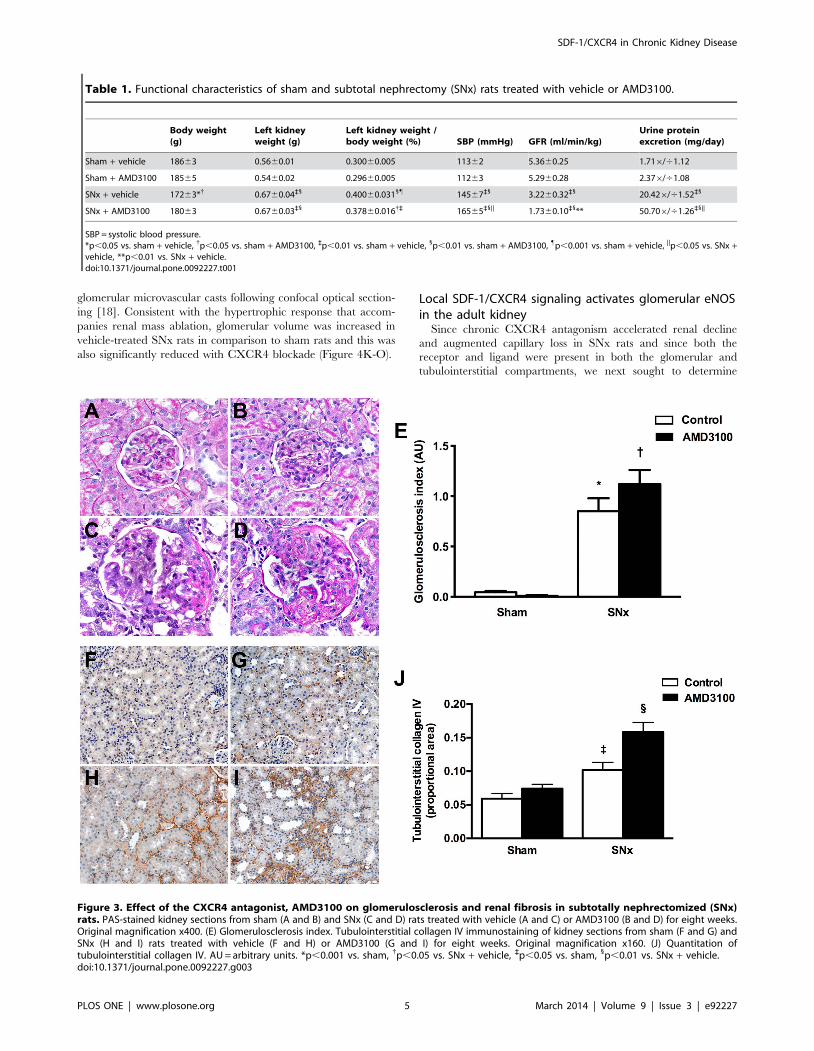
Examination of kidney sections by light microscopy revealed that
SNx surgery was associated with an expected increase in
glomerulosclerosis (Figure 3A–E) and tubulointerstitial collagen
IV deposition (Figure 3F–J), with both of these indicators of renal
fibrosis being augmented with CXCR4 antagonism in SNx rats
(Figure 3).
CXCR4 blockade accelerates capillary loss in subtotallynephrectomized rats
As CXCR4 was detectable in the endothelial cells of both the
glomerular and peritubular capillaries we next sought to determine
the effect of CXCR4 antagonism on both capillary density and
capillary volume in sham and SNx rats. To assess the density of the
glomerular and peritubular capillaries, kidney sections were
immunostained with the monoclonal antibody JG-12 that binds
to endothelial cells of capillaries but not lymphatics in rat kidneys
[27]. Endothelial immunostaining was reduced in both the
glomerular and peritubular compartments eight weeks after SNx
surgery, with a further reduction in capillary density noted in
kidney sections of AMD3100-treated SNx rats (Figure 4A–J). To
determine whether glomerular capillary volume was also affected,
we used the novel technique of fluorescent microangiography
(FMA) that allows the generation of virtual three dimensional
Figure 2. CXCR4 and SDF-1 mRNA in sham-operated and subtotally nephrectomized (SNx) rats. CXCR4 expression is increased in thekidneys of SNx rats (A), whereas SDF-1 is reduced (B). The kidneys of SNx rats also demonstrated an upregulation of the pro-fibrotic growth factor,transforming growth factor-b (TGF-b) (C). In cultured NRK-49F renal fibroblasts, recombinant TGF-b downregulated SDF-1 mRNA, with this effectbeing attenuated by HDAC inhibition with vorinostat (D). *p,0.001 vs. sham, {p,0.05 vs. sham, `p,0.01 vs. sham, 1p,0.001 vs. control, "p,0.001 vs.TGF-b.doi:10.1371/journal.pone.0092227.g002
SDF-1/CXCR4 in Chronic Kidney Disease
PLOS ONE | www.plosone.org 4 March 2014 | Volume 9 | Issue 3 | e92227
glomerular microvascular casts following confocal optical section-
ing [18]. Consistent with the hypertrophic response that accom-
panies renal mass ablation, glomerular volume was increased in
vehicle-treated SNx rats in comparison to sham rats and this was
also significantly reduced with CXCR4 blockade (Figure 4K-O).
Local SDF-1/CXCR4 signaling activates glomerular eNOSin the adult kidney
Since chronic CXCR4 antagonism accelerated renal decline
and augmented capillary loss in SNx rats and since both the
receptor and ligand were present in both the glomerular and
tubulointerstitial compartments, we next sought to determine
Table 1. Functional characteristics of sham and subtotal nephrectomy (SNx) rats treated with vehicle or AMD3100.
Body weight(g)
Left kidneyweight (g)
Left kidney weight /body weight (%) SBP (mmHg) GFR (ml/min/kg)
Urine proteinexcretion (mg/day)
Sham + vehicle 18663 0.5660.01 0.30060.005 11362 5.3660.25 1.716/41.12
Sham + AMD3100 18565 0.5460.02 0.29660.005 11263 5.2960.28 2.376/41.08
SNx + vehicle 17263*{ 0.6760.04`1 0.40060.0311" 14567`1 3.2260.32`1 20.426/41.52`1
SNx + AMD3100 18063 0.6760.03`1 0.37860.016{` 16565`1|| 1.7360.10`1** 50.706/41.26`1||
SBP = systolic blood pressure.*p,0.05 vs. sham + vehicle, {p,0.05 vs. sham + AMD3100, `p,0.01 vs. sham + vehicle, 1p,0.01 vs. sham + AMD3100, "p,0.001 vs. sham + vehicle, ||p,0.05 vs. SNx +vehicle, **p,0.01 vs. SNx + vehicle.doi:10.1371/journal.pone.0092227.t001
Figure 3. Effect of the CXCR4 antagonist, AMD3100 on glomerulosclerosis and renal fibrosis in subtotally nephrectomized (SNx)rats. PAS-stained kidney sections from sham (A and B) and SNx (C and D) rats treated with vehicle (A and C) or AMD3100 (B and D) for eight weeks.Original magnification x400. (E) Glomerulosclerosis index. Tubulointerstitial collagen IV immunostaining of kidney sections from sham (F and G) andSNx (H and I) rats treated with vehicle (F and H) or AMD3100 (G and I) for eight weeks. Original magnification x160. (J) Quantitation oftubulointerstitial collagen IV. AU = arbitrary units. *p,0.001 vs. sham, {p,0.05 vs. SNx + vehicle, `p,0.05 vs. sham, 1p,0.01 vs. SNx + vehicle.doi:10.1371/journal.pone.0092227.g003
SDF-1/CXCR4 in Chronic Kidney Disease
PLOS ONE | www.plosone.org 5 March 2014 | Volume 9 | Issue 3 | e92227

Figure 4. Effect of CXCR4 antagonism with AMD3100 on capillary loss in subtotally nephrectomized (SNx) rats. (A–D) Glomerularendothelial (JG-12) immunostaining of kidney sections from sham (A and B) and SNx (C and D) rats treated with vehicle (A and C) or AMD3100 (B andD) for eight weeks. Original magnification x400. (E) Quantitation of glomerular JG-12. (F–I) Peritubular JG-12 immunostaining of kidney sections fromsham (F and G) and SNx (H and I) rats treated with vehicle (F and H) or AMD3100 (G and I). Original magnification x160. (J) Quantitation of peritubularJG-12. (K–N) Fluorescent microangiography (FMA) images of glomeruli from sham (K and L) and SNx (M and N) rats treated with vehicle (K and M) orAMD3100 (L and N) for eight weeks. (O) Glomerular capillary volume. *p,0.001 vs. sham, {p,0.05 vs. SNx + vehicle, `p,0.01 vs. sham, 1p,0.01 vs.SNx + vehicle.doi:10.1371/journal.pone.0092227.g004
SDF-1/CXCR4 in Chronic Kidney Disease
PLOS ONE | www.plosone.org 6 March 2014 | Volume 9 | Issue 3 | e92227
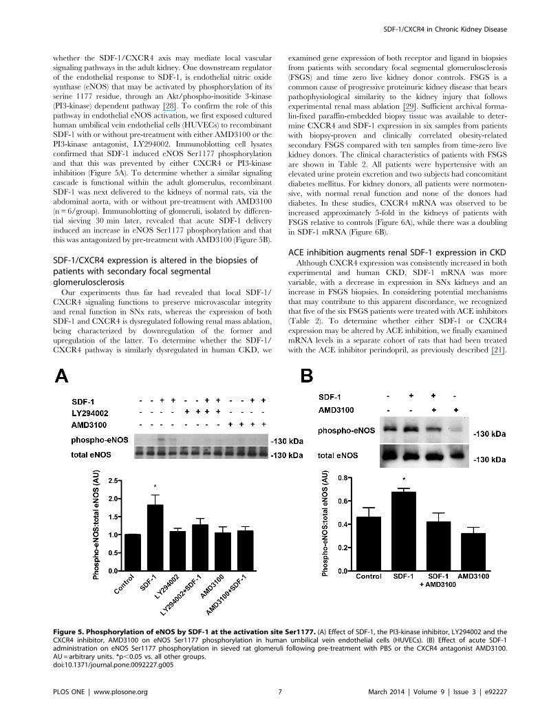
whether the SDF-1/CXCR4 axis may mediate local vascular
signaling pathways in the adult kidney. One downstream regulator
of the endothelial response to SDF-1, is endothelial nitric oxide
synthase (eNOS) that may be activated by phosphorylation of its
serine 1177 residue, through an Akt/phospho-inositide 3-kinase
(PI3-kinase) dependent pathway [28]. To confirm the role of this
pathway in endothelial eNOS activation, we first exposed cultured
human umbilical vein endothelial cells (HUVECs) to recombinant
SDF-1 with or without pre-treatment with either AMD3100 or the
PI3-kinase antagonist, LY294002. Immunoblotting cell lysates
confirmed that SDF-1 induced eNOS Ser1177 phosphorylation
and that this was prevented by either CXCR4 or PI3-kinase
inhibition (Figure 5A). To determine whether a similar signaling
cascade is functional within the adult glomerulus, recombinant
SDF-1 was next delivered to the kidneys of normal rats, via the
abdominal aorta, with or without pre-treatment with AMD3100
(n = 6/group). Immunoblotting of glomeruli, isolated by differen-
tial sieving 30 min later, revealed that acute SDF-1 delivery
induced an increase in eNOS Ser1177 phosphorylation and that
this was antagonized by pre-treatment with AMD3100 (Figure 5B).
SDF-1/CXCR4 expression is altered in the biopsies ofpatients with secondary focal segmentalglomerulosclerosis
Our experiments thus far had revealed that local SDF-1/
CXCR4 signaling functions to preserve microvascular integrity
and renal function in SNx rats, whereas the expression of both
SDF-1 and CXCR4 is dysregulated following renal mass ablation,
being characterized by downregulation of the former and
upregulation of the latter. To determine whether the SDF-1/
CXCR4 pathway is similarly dysregulated in human CKD, we
examined gene expression of both receptor and ligand in biopsies
from patients with secondary focal segmental glomerulosclerosis
(FSGS) and time zero live kidney donor controls. FSGS is a
common cause of progressive proteinuric kidney disease that bears
pathophysiological similarity to the kidney injury that follows
experimental renal mass ablation [29]. Sufficient archival forma-
lin-fixed paraffin-embedded biopsy tissue was available to deter-
mine CXCR4 and SDF-1 expression in six samples from patients
with biopsy-proven and clinically correlated obesity-related
secondary FSGS compared with ten samples from time-zero live
kidney donors. The clinical characteristics of patients with FSGS
are shown in Table 2. All patients were hypertensive with an
elevated urine protein excretion and two subjects had concomitant
diabetes mellitus. For kidney donors, all patients were normoten-
sive, with normal renal function and none of the donors had
diabetes. In these studies, CXCR4 mRNA was observed to be
increased approximately 5-fold in the kidneys of patients with
FSGS relative to controls (Figure 6A), while there was a doubling
in SDF-1 mRNA (Figure 6B).
ACE inhibition augments renal SDF-1 expression in CKDAlthough CXCR4 expression was consistently increased in both
experimental and human CKD, SDF-1 mRNA was more
variable, with a decrease in expression in SNx kidneys and an
increase in FSGS biopsies. In considering potential mechanisms
that may contribute to this apparent discordance, we recognized
that five of the six FSGS patients were treated with ACE inhibitors
(Table 2). To determine whether either SDF-1 or CXCR4
expression may be altered by ACE inhibition, we finally examined
mRNA levels in a separate cohort of rats that had been treated
with the ACE inhibitor perindopril, as previously described [21].
Figure 5. Phosphorylation of eNOS by SDF-1 at the activation site Ser1177. (A) Effect of SDF-1, the PI3-kinase inhibitor, LY294002 and theCXCR4 inhibitor, AMD3100 on eNOS Ser1177 phosphorylation in human umbilical vein endothelial cells (HUVECs). (B) Effect of acute SDF-1administration on eNOS Ser1177 phosphorylation in sieved rat glomeruli following pre-treatment with PBS or the CXCR4 antagonist AMD3100.AU = arbitrary units. *p,0.05 vs. all other groups.doi:10.1371/journal.pone.0092227.g005
SDF-1/CXCR4 in Chronic Kidney Disease
PLOS ONE | www.plosone.org 7 March 2014 | Volume 9 | Issue 3 | e92227

These experiments revealed that CXCR4 mRNA was increased in
SNx rats and, while reduced with perindopril-treatment, remained
significantly elevated relative to sham rats (Figure 7A). Moreover,
SDF-1 mRNA was markedly increased in the kidneys of
perindopril-treated SNx rats when compared with either sham
animals or SNx rats treated with vehicle, consistent with the
change in gene expression observed in human FSGS biopsies
(Figure 7B).
Discussion
Despite existing therapies and regardless of the underlying
etiology, renal function continues to decline in the majority of
people with CKD. Over recent years, the almost universal failure
of novel anti-albuminuric therapies to meaningfully impact on
clinical outcomes has encouraged investigators to direct their
attention to pathogenetic processes that commonly occur during
the later stages of CKD development. These pathogenetic
processes include renal fibrosis and the associated disruption in
the microvascular architecture, acting together and compounding
the deleterious effects of one another in the relentless progression
towards end-stage renal disease. In the present study, we identified
a novel role for SDF-1/CXCR4 signaling in this reciprocal
relationship, observing that local SDF-1/CXCR4 signaling
preserves microvascular integrity and attenuates fibrogenesis,
whereas the pro-fibrotic growth factor TGF-b, overelaborated in
the CKD setting, downregulates SDF-1 expression. Augmentation
of SDF-1/CXCR4 signaling by novel or existing agents, either
purposefully or serendipitously, may thus slow the progression of
renal decline in CKD.
SDF-1 signaling through CXCR4 promotes cell survival [30],
migration [28], and proliferation [31], favoring neo-angiogenesis
both through direct effects and through the creation of a
permissive microenvironment that facilitates the actions of the
angiogenic factor, vascular endothelial growth factor (VEGF)
[32,33]. In the present study we observed that i) CXCR4 is
expressed on the surface of endothelial cells in both the glomerular
and peritubular compartments, ii) chronic CXCR4 blockade
accelerates capillary loss in rats with CKD and iii) local SDF-1
delivery induces glomerular eNOS activation in a CXCR4-
dependent manner. Moreover, accelerated disruption of the
microvascular architecture with chronic CXCR4 blockade was
associated with augmented renal decline and progressive renal
fibrosis, analogous to the exacerbation of cardiac dysfunction with
chronic CXCR4 blockade in the post-myocardial infarction setting
[34]. Collectively, these observations highlight a hitherto unrec-
ognized role for local SDF-1/CXCR4 signaling in preserving
microvascular integrity and preventing coincident renal fibrosis in
CKD.
In light of the detrimental effects observed with chronic
CXCR4 blockade, the consistent upregulation of CXCR4 in the
kidneys of SNx rats and in biopsies from patients with secondary
FSGS is likely indicative of a compensatory response. CXCR4
upregulation has previously been described as a feature of a
hypoxia-related glomerulopathy in patients with hypertensive
nephrosclerosis [35]. Although both CXCR4 and SDF-1 are
recognized as being hypoxia-responsive genes [36], their expres-
sion patterns were not indistinguishable in rat and human CKD.
Whereas CXCR4 was consistently upregulated in both rats and
humans with CKD, SDF-1 expression was more variable, the
chemokine being notably downregulated in SNx kidneys of both
female and male rats. In considering plausible mediators for SDF-
1 downregulation in rats with CKD, we recognized the reciprocal
relationship between fibrogenesis and capillary loss and hypoth-
Ta
ble
2.
Clin
ical
char
acte
rist
ics
of
pat
ien
tsw
ith
seco
nd
ary
foca
lse
gm
en
tal
glo
me
rulo
scle
rosi
s(F
SGS)
.
Pa
tie
nt
Ag
e(y
ea
rs)
We
igh
t(k
g)
Se
x(M
/F)
Hy
pe
rte
nsi
on
(Y/N
)A
CE
i/A
RB
Dia
be
tes
(Y/N
)U
rin
ep
rote
ine
xcr
eti
on
Se
rum
cre
ati
nin
e(m
mo
l/L
)
11
31
02
.6M
YEn
alap
ril
10
mg
o.d
.N
Pro
tein
:cre
atin
ine
rati
o4
00
mg
/mm
ol
83
23
41
02
.6M
YR
amip
ril
10
mg
o.d
.N
24
hu
rin
ep
rote
ine
xcre
tio
n1
.87
g/2
4h
30
0
35
51
06
.6M
YR
amip
ril
10
mg
o.d
.Y
Pro
tein
:cre
atin
ine
rati
o6
79
mg
/mm
ol
10
8
43
41
04
.5F
YN
oN
Pro
tein
:cre
atin
ine
rati
o3
38
mg
/mm
ol
76
51
71
54
.9M
YEn
alap
ril
10
mg
o.d
.Y
Pro
tein
:cre
atin
ine
rati
o1
18
mg
/mm
ol
53
63
81
16
MY
Enal
apri
l2
0m
go
.d.
NP
rote
in:c
reat
inin
era
tio
12
0m
g/m
mo
l9
8
do
i:10
.13
71
/jo
urn
al.p
on
e.0
09
22
27
.t0
02
SDF-1/CXCR4 in Chronic Kidney Disease
PLOS ONE | www.plosone.org 8 March 2014 | Volume 9 | Issue 3 | e92227

esized that decreased SDF-1 expression may occur as a
consequence of the fibrotic process itself. TGF-b is a pro-sclerotic
cytokine implicated in the fibrogenic response of many organs,
including the kidney. Consistent with previous studies in oral
myofibroblasts [37] and mimicking the changes in gene expression
observed in the kidneys of SNx rats, exposure of cultured renal
fibroblasts to recombinant TGF-b resulted in a marked downreg-
ulation in SDF-1 expression.
The post-translational modification of proteins, by the addition
or removal of functional groups, is a common mechanism for
controlling protein behaviour, perhaps most readily appreciated
when considering the essential role that (de)phosphorylation plays
in cellular homeostasis. More recently, post-translational protein
modification through the addition or removal of acetyl groups has
been recognized to rival phosphorylation in its diversity of
substrates and the functional pathways affected [38]. (De)acetyla-
tion of proteins on lysine residues is regulated by the opposing
actions of groups of enzymes called histone acetyltransferases and
histone deacetylases (HDACs). In recent times, increasing
evidence has begun to emerge for a pivotal role for HDACs in
mediating both the progression of renal fibrosis and the response
to TGF-b itself. For instance, work from our own group showed
that HDAC inhibition attenuated glomerular matrix accumulation
in diabetic mice [23], whereas acetylation may alter the cellular
response to TGF-b through affecting Smad 2/3 activity [39],
Smad7 stability [39] and/or the actions of downstream transcrip-
tion factors [40] among other processes. Confirming that TGF-bmediated SDF-1 downregulation is under the regulatory control of
protein acetylation, the decrement in SDF-1 transcript levels
induced by TGF-b was attenuated by pre-treatment of cells with
the HDAC inhibitor, vorinostat.
One of the challenges restricting the translation of promising
experimental observations to the clinic is the limited ability of
rodent models to recapitulate human disease. For instance, we
observed that whereas SDF-1 expression was downregulated in
SNx rats, the opposite effect occurred in biopsies from patients
with secondary FSGS. This superficial discordance encouraged us
to consider the differences between the human disease and
experimental model. To account for the confounding effects of
medication usage among patients, we therefore examined SDF-1
expression patterns in historical samples from SNx rats that had or
had not been treated with the ACE inhibitor, perindopril [21]. In
contrast to vehicle treatment, ACE inhibition, previously shown to
decrease the overelaboration of TGF-b in SNx rats [41], resulted
in a marked upregulation in SDF-1. Augmentation of SDF-1
activity may thus represent a novel mechanism by which renin
angiotensin system blockade preserves the renal vasculature [42].
Figure 6. Real-time PCR for CXCR4 (A) and SDF-1 (B) mRNA in biopsies from time zero live kidney donors (Control, n = 10) andpatients with secondary focal segmental glomerulosclerosis (FSGS, n = 6). AU = arbitrary units. *p,0.001, {p,0.01.doi:10.1371/journal.pone.0092227.g006
Figure 7. Real-time PCR for CXCR4 (A) and SDF-1 (B) mRNA in kidneys from sham rats, subtotally nephrectomized (SNx) rats andSNx rats treated with perindopril. *p,0.001 vs. sham, {p,0.05 vs. sham, `p,0.05 vs. SNx.doi:10.1371/journal.pone.0092227.g007
SDF-1/CXCR4 in Chronic Kidney Disease
PLOS ONE | www.plosone.org 9 March 2014 | Volume 9 | Issue 3 | e92227

Although SDF-1/CXCR4 interaction was originally considered
a monogamous relationship, more recent evidence suggests that
this is not the case. CXCR4 also acts as a receptor for the HIV
envelope receptor glycoprotein gp120 [43] and for the small
protein ubiquitin [44], while SDF-1 may also bind to CXCR7.
The renal actions of CXCR7 are complex with reports that the
receptor signals in ‘‘renal multipotent progenitors’’ [45] while also
functioning as a scavenger protein for SDF-1 [46]. Similarly,
under some alternative conditions renal SDF-1/CXCR4 may play
a potentially detrimental role through promoting podocyte
proliferation [14], inflammatory cell recruitment [47] or, in the
case of Shiga-toxin induced injury, endothelial phenotypic switch
[48]. Thus, as with other angiogenic mediators [4,5], the role of
renal SDF-1/CXCR4 is likely to be contextual, varying according
to stage of development and the underlying injurious insult. The
collective in vitro, acute and chronic in vivo and human correlative
studies herein described indicate a reno-protective function for this
pathway in CKD.
In summary, SDF-1/CXCR4 signaling is not only important
for renal vascular development but the same system also plays a
pivotal role in preserving microvascular integrity in CKD.
Augmentation of this pathway by novel or existing therapies
may attenuate renal fibrosis and slow the progression of renal
decline in CKD.
Acknowledgments
The authors thank Ms. Bridgit Bowskill, Ms. Bailey Stead, Ms. Christine
Kuliszewski, Ms. Krystina Vecchio and Ms. Katrina Zefkic for their
excellent technical assistance.
Author Contributions
Conceived and designed the experiments: LHC DAY KAC PAM REG
AA. Performed the experiments: LHC SLA KT MGK. Analyzed the data:
LHC SLA KT MGK AA. Contributed reagents/materials/analysis tools:
MMS IWG DJK. Wrote the paper: AA.
References
1. Fine LG, Orphanides C, Norman JT (1998) Progressive renal disease: the
chronic hypoxia hypothesis. Kidney Int Suppl 65: S74–78.
2. Eremina V, Sood M, Haigh J, Nagy A, Lajoie G, et al. (2003) Glomerular-
specific alterations of VEGF-A expression lead to distinct congenital and
acquired renal diseases. J Clin Invest 111: 707–716.
3. Eremina V, Jefferson JA, Kowalewska J, Hochster H, Haas M, et al. (2008)
VEGF inhibition and renal thrombotic microangiopathy. N Engl J Med 358:
1129–1136.
4. Advani A, Kelly DJ, Advani SL, Cox AJ, Thai K, et al. (2007) Role of VEGF in
maintaining renal structure and function under normotensive and hypertensive
conditions. Proc Natl Acad Sci U S A 104: 14448–14453.
5. Jeansson M, Gawlik A, Anderson G, Li C, Kerjaschki D, et al. (2011)
Angiopoietin-1 is essential in mouse vasculature during development and in
response to injury. J Clin Invest 121: 2278–2289.
6. Fogo AB (2005) New capillary growth: a contributor to regression of sclerosis?
Curr Opin Nephrol Hypertens 14: 201–203.
7. Takabatake Y, Sugiyama T, Kohara H, Matsusaka T, Kurihara H, et al. (2009)
The CXCL12 (SDF-1)/CXCR4 axis is essential for the development of renal
vasculature. J Am Soc Nephrol 20: 1714–1723.
8. Gupta SK, Lysko PG, Pillarisetti K, Ohlstein E, Stadel JM (1998) Chemokine
receptors in human endothelial cells. Functional expression of CXCR4 and its
transcriptional regulation by inflammatory cytokines. J Biol Chem 273: 4282–
4287.
9. Nagasawa T, Kikutani H, Kishimoto T (1994) Molecular cloning and structure
of a pre-B-cell growth-stimulating factor. Proc Natl Acad Sci U S A 91: 2305–
2309.
10. Janowski M (2009) Functional diversity of SDF-1 splicing variants. Cell Adh
Migr 3: 243–249.
11. Tachibana K, Hirota S, Iizasa H, Yoshida H, Kawabata K, et al. (1998) The
chemokine receptor CXCR4 is essential for vascularization of the gastrointes-
tinal tract. Nature 393: 591–594.
12. Yuen DA, Stead BE, Zhang Y, White KE, Kabir MG, et al. (2012) eNOS
deficiency predisposes podocytes to injury in diabetes. J Am Soc Nephrol 23:
1810–1823.
13. Stokman G, Stroo I, Claessen N, Teske GJ, Florquin S, et al. (2010) SDF-1
provides morphological and functional protection against renal ischaemia/
reperfusion injury. Nephrol Dial Transplant 25: 3852–3859.
14. Ding M, Cui S, Li C, Jothy S, Haase V, et al. (2006) Loss of the tumor
suppressor Vhlh leads to upregulation of Cxcr4 and rapidly progressive
glomerulonephritis in mice. Nat Med 12: 1081–1087.
15. Floege J, Smeets B, Moeller MJ (2009) The SDF-1/CXCR4 axis is a novel
driver of vascular development of the glomerulus. J Am Soc Nephrol 20: 1659–
1661.
16. Advani A, Gilbert RE, Thai K, Gow RM, Langham RG, et al. (2009)
Expression, localization, and function of the thioredoxin system in diabetic
nephropathy. J Am Soc Nephrol 20: 730–741.
17. Yuen DA, Connelly KA, Advani A, Liao C, Kuliszewski MA, et al. (2010)
Culture-modified bone marrow cells attenuate cardiac and renal injury in a
chronic kidney disease rat model via a novel antifibrotic mechanism. PLoS One
5: e9543.
18. Advani A, Connelly KA, Yuen DA, Zhang Y, Advani SL, et al. (2011)
Fluorescent microangiography is a novel and widely applicable technique for
delineating the renal microvasculature. PLoS ONE 6: e24695.
19. Kanki S, Segers VF, Wu W, Kakkar R, Gannon J, et al. (2011) Stromal cell-
derived factor-1 retention and cardioprotection for ischemic myocardium. Circ
Heart Fail 4: 509–518.
20. Burlington H, Cronkite EP (1973) Characteristics of cell cultures derived from
renal glomeruli. Proc Soc Exp Biol Med 142: 143–149.
21. Kelly DJ, Hepper C, Wu LL, Cox AJ, Gilbert RE (2003) Vascular endothelial
growth factor expression and glomerular endothelial cell loss in the remnant
kidney model. Nephrol Dial Transplant 18: 1286–1292.
22. Advani A, Kelly DJ, Cox AJ, White KE, Advani SL, et al. (2009) The (Pro)reninreceptor: site-specific and functional linkage to the vacuolar H+-ATPase in the
kidney. Hypertension 54: 261–269.
23. Advani A, Huang Q, Thai K, Advani SL, White KE, et al. (2011) Long-TermAdministration of the Histone Deacetylase Inhibitor Vorinostat Attenuates
Renal Injury in Experimental Diabetes through an Endothelial Nitric OxideSynthase-Dependent Mechanism. Am J Pathol 178: 2205–2214.
24. Yuan H, Reddy MA, Sun G, Lanting L, Wang M, et al. (2013) Involvement of
p300/CBP and epigenetic histone acetylation in TGF-beta1-mediated genetranscription in mesangial cells. Am J Physiol Renal Physiol 304: F601–613.
25. Fricker SP, Anastassov V, Cox J, Darkes MC, Grujic O, et al. (2006)
Characterization of the molecular pharmacology of AMD3100: a specific
antagonist of the G-protein coupled chemokine receptor, CXCR4. BiochemPharmacol 72: 588–596.
26. Rosenkilde MM, Gerlach LO, Jakobsen JS, Skerlj RT, Bridger GJ, et al. (2004)
Molecular mechanism of AMD3100 antagonism in the CXCR4 receptor:transfer of binding site to the CXCR3 receptor. J Biol Chem 279: 3033–3041.
27. Kim YG, Suga SI, Kang DH, Jefferson JA, Mazzali M, et al. (2000) Vascular
endothelial growth factor accelerates renal recovery in experimental thromboticmicroangiopathy. Kidney Int 58: 2390–2399.
28. Sameermahmood Z, Balasubramanyam M, Saravanan T, Rema M (2008)
Curcumin modulates SDF-1alpha/CXCR4-induced migration of human retinalendothelial cells (HRECs). Invest Ophthalmol Vis Sci 49: 3305–3311.
29. Johnson RJ (1997) What mediates progressive glomerulosclerosis? The
glomerular endothelium comes of age. Am J Pathol 151: 1179–1181.
30. Yano T, Liu Z, Donovan J, Thomas MK, Habener JF (2007) Stromal cellderived factor-1 (SDF-1)/CXCL12 attenuates diabetes in mice and promotes
pancreatic beta-cell survival by activation of the prosurvival kinase Akt. Diabetes
56: 2946–2957.
31. De Falco V, Guarino V, Avilla E, Castellone MD, Salerno P, et al. (2007)
Biological role and potential therapeutic targeting of the chemokine receptor
CXCR4 in undifferentiated thyroid cancer. Cancer Res 67: 11821–11829.
32. Kanda S, Mochizuki Y, Kanetake H (2003) Stromal cell-derived factor-1alpha
induces tube-like structure formation of endothelial cells through phosphoino-
sitide 3-kinase. J Biol Chem 278: 257–262.
33. Grunewald M, Avraham I, Dor Y, Bachar-Lustig E, Itin A, et al. (2006) VEGF-induced adult neovascularization: recruitment, retention, and role of accessory
cells. Cell 124: 175–189.
34. Dai S, Yuan F, Mu J, Li C, Chen N, et al. (2010) Chronic AMD3100antagonism of SDF-1alpha-CXCR4 exacerbates cardiac dysfunction and
remodeling after myocardial infarction. J Mol Cell Cardiol 49: 587–597.
35. Neusser MA, Lindenmeyer MT, Moll AG, Segerer S, Edenhofer I, et al. (2010)Human nephrosclerosis triggers a hypoxia-related glomerulopathy. Am J Pathol
176: 594–607.
36. Ceradini DJ, Kulkarni AR, Callaghan MJ, Tepper OM, Bastidas N, et al. (2004)Progenitor cell trafficking is regulated by hypoxic gradients through HIF-1
induction of SDF-1. Nat Med 10: 858–864.
37. Daly AJ, McIlreavey L, Irwin CR (2008) Regulation of HGF and SDF-1expression by oral fibroblasts—implications for invasion of oral cancer. Oral
Oncol 44: 646–651.
38. Kouzarides T (2000) Acetylation: a regulatory modification to rival phosphor-
ylation? EMBO J 19: 1176–1179.
SDF-1/CXCR4 in Chronic Kidney Disease
PLOS ONE | www.plosone.org 10 March 2014 | Volume 9 | Issue 3 | e92227

39. Simonsson M, Kanduri M, Gronroos E, Heldin CH, Ericsson J (2006) The DNA
binding activities of Smad2 and Smad3 are regulated by coactivator-mediatedacetylation. J Biol Chem 281: 39870–39880.
40. Chabane N, Li X, Fahmi H (2009) HDAC4 contributes to IL-1-induced
mPGES-1 expression in human synovial fibroblasts through up-regulation ofEgr-1 transcriptional activity. J Cell Biochem 106: 453–463.
41. Gilbert RE, Wu LL, Kelly DJ, Cox A, Wilkinson-Berka JL, et al. (1999)Pathological expression of renin and angiotensin II in the renal tubule after
subtotal nephrectomy. Implications for the pathogenesis of tubulointerstitial
fibrosis. Am J Pathol 155: 429–440.42. Remuzzi A, Gagliardini E, Sangalli F, Bonomelli M, Piccinelli M, et al. (2006)
ACE inhibition reduces glomerulosclerosis and regenerates glomerular tissue ina model of progressive renal disease. Kidney Int 69: 1124–1130.
43. Feng Y, Broder CC, Kennedy PE, Berger EA (1996) HIV-1 entry cofactor:functional cDNA cloning of a seven-transmembrane, G protein-coupled
receptor. Science 272: 872–877.
44. Saini V, Marchese A, Majetschak M (2010) CXC chemokine receptor 4 is a cell
surface receptor for extracellular ubiquitin. J Biol Chem 285: 15566–15576.
45. Mazzinghi B, Ronconi E, Lazzeri E, Sagrinati C, Ballerini L, et al. (2008)
Essential but differential role for CXCR4 and CXCR7 in the therapeutic
homing of human renal progenitor cells. J Exp Med 205: 479–490.
46. Naumann U, Cameroni E, Pruenster M, Mahabaleshwar H, Raz E, et al. (2010)
CXCR7 functions as a scavenger for CXCL12 and CXCL11. PLoS One 5:
e9175.
47. Chu PY, Zatta A, Kiriazis H, Chin-Dusting J, Du XJ, et al. (2011) CXCR4
antagonism attenuates the cardiorenal consequences of mineralocorticoid excess.
Circ Heart Fail 4: 651–658.
48. Petruzziello-Pellegrini TN, Yuen DA, Page AV, Patel S, Soltyk AM, et al. (2012)
The CXCR4/CXCR7/SDF-1 pathway contributes to the pathogenesis of Shiga
toxin-associated hemolytic uremic syndrome in humans and mice. J Clin Invest
122: 759–776.
SDF-1/CXCR4 in Chronic Kidney Disease
PLOS ONE | www.plosone.org 11 March 2014 | Volume 9 | Issue 3 | e92227