Insulin receptor activation by proinsulin preserves synapses ...
14
ARTICLE OPEN Insulin receptor activation by proinsulin preserves synapses and vision in retinitis pigmentosa Alonso Sánchez-Cruz 1,2,3 , Alberto Hernández-Pinto 1 , Concepción Lillo 4 , Carolina Isiegas 1 , Miguel Marchena 1 , Ignacio Lizasoain 2,3 , Fátima Bosch 5,6 , Pedro de la Villa 7,8 , Catalina Hernández-Sánchez 1,6 ✉ and Enrique J. de la Rosa 1 ✉ © The Author(s) 2022 Synaptic loss, neuronal death, and circuit remodeling are common features of central nervous system neurodegenerative disorders. Retinitis pigmentosa (RP), the leading cause of inherited blindness, is a group of retinal dystrophies characterized by photoreceptor dysfunction and death. The insulin receptor, a key controller of metabolism, also regulates neuronal survival and synaptic formation, maintenance, and activity. Indeed, deficient insulin receptor signaling has been implicated in several brain neurodegenerative pathologies. We present evidence linking impaired insulin receptor signaling with RP. We describe a selective decrease in the levels of the insulin receptor and its downstream effector phospho-S6 in retinal horizontal cell terminals in the rd10 mouse model of RP, as well as aberrant synapses between rod photoreceptors and the postsynaptic terminals of horizontal and bipolar cells. A gene therapy strategy to induce sustained proinsulin, the insulin precursor, production restored retinal insulin receptor signaling, by increasing S6 phosphorylation, without peripheral metabolic consequences. Moreover, proinsulin preserved photoreceptor synaptic connectivity and prolonged visual function in electroretinogram and optomotor tests. These findings point to a disease-modifying role of insulin receptor and support the therapeutic potential of proinsulin in retinitis pigmentosa. Cell Death and Disease (2022)13:383 ; https://doi.org/10.1038/s41419-022-04839-0 INTRODUCTION Neurodegenerative disorders are complex pathological conditions that involve, among other processes, synaptic loss and neuronal cell death leading to deterioration of neuronal structure and function. According to the 2016 Global Burden of Disease report [1], neurodegenerative diseases are the second leading cause of death and a major cause of disability. The development of strategies to cure or at least delay the progression of neurode- generative diseases has been hindered by their diverse etiology and complex nature, and by the limited regenerative capacity of neurons. There is thus an urgent need for effective medical interventions. As part of the central nervous system (CNS), the retina shares multiple pathophysiological features with the brain [2]. Despite their diverse etiology, retinal neurodegenerative diseases, like those affecting the brain, are characterized by synaptic failure and neuronal cell death [3]. Retinitis pigmentosa (RP) comprises a group of hereditary retinal neurodegenerative conditions with a complex genetic etiology. To date more than 60 genes and 3,000 mutations have been implicated in RP, and over 300 genes associated with inherited retinal dystrophies (https:// sph.uth.edu/retnet/disease.htm) have been identified. RP is characterized by primary dysfunction and death of photoreceptor cells followed by reactive gliosis and remodeling of the retinal structure, resulting in vision loss and eventual blindness [3, 4]. RP is categorized as a rare disease (prevalence 1/3,500-4,000), but accounts for most cases of hereditary blindness. Gene therapy would be the ideal definitive treatment, and one such therapy has been recently approved for a related retinal dystrophy [5]. However, the complexity and diversity of the mutations under- lying RP necessitate the development of alternative therapeutic approaches, particularly those independent of the causative mutation, as well as palliative treatments. The insulin receptor (INSR), traditionally considered a key peripheral metabolic regulator, is increasingly viewed as an important modulator of neuronal cell survival, and synaptic formation, maintenance, and activity [6–8]. Since initial reports described widespread INSR expression in the CNS, including the retina [9–13], INSR signaling has been implicated in a growing number of CNS functions. In addition to regulating feeding behavior and peripheral metabo- lism, central INSR signaling is involved in memory formation and cognitive functions [6, 7, 14]. The mechanisms underpinning the non-metabolic actions of INSR are being gradually unraveled. At Received: 23 September 2021 Revised: 4 April 2022 Accepted: 5 April 2022 1 Department of Molecular Biomedicine, Centro de Investigaciones Biológicas Margarita Salas (CSIC), C/ Ramiro de Maeztu 9, 28040 Madrid, Spain. 2 Neurovascular Research Unit, Department of Pharmacology and Toxicology and Instituto Universitario de Investigació n en Neuroqui ́ mica, Facultad de Medicina, Universidad Complutense de Madrid, Ciudad Universitaria, 28040 Madrid, Spain. 3 Instituto de Investigación Hospital 12 de Octubre (IMAS12), 28041 Madrid, Spain. 4 Cell Biology and Pathology Department, University of Salamanca, Institute for Biomedical Research of Salamanca (IBSAL), Institute of Neurosciences of Castilla y León (INCYL), C/ Pintor Fernando Gallego,1, 37007 Salamanca, Spain. 5 Center of Animal Biotechnology and Gene Therapy and Department of Biochemistry and Molecular Biology, School of Veterinary Medicine, Universitat Autònoma de Barcelona, 08193 Bellaterra, Spain. 6 Centro de Investigación Biomédica en Red de Diabetes y Enfermedades Metabólicas Asociadas (CIBERDEM), ISCIII, 28034 Madrid, Spain. 7 Department of System Biology, Facultad de Medicina, Universidad de Alcalá, 28805 Alcalá de Henares, Spain. 8 Instituto Ramón y Cajal de Investigación Sanitaria, ISCIII, 28034 Madrid, Spain. ✉ email: [email protected]; [email protected] Edited by Professor Nicolas Bazan. www.nature.com/cddis Official journal of CDDpress 1234567890();,:
-
Upload
khangminh22 -
Category
Documents
-
view
0 -
download
0
Transcript of Insulin receptor activation by proinsulin preserves synapses ...

ARTICLE OPEN
Insulin receptor activation by proinsulin preserves synapses andvision in retinitis pigmentosaAlonso Sánchez-Cruz1,2,3, Alberto Hernández-Pinto1, Concepción Lillo 4, Carolina Isiegas1, Miguel Marchena1, Ignacio Lizasoain2,3,Fátima Bosch5,6, Pedro de la Villa7,8, Catalina Hernández-Sánchez 1,6✉ and Enrique J. de la Rosa 1✉
© The Author(s) 2022
Synaptic loss, neuronal death, and circuit remodeling are common features of central nervous system neurodegenerative disorders.Retinitis pigmentosa (RP), the leading cause of inherited blindness, is a group of retinal dystrophies characterized by photoreceptordysfunction and death. The insulin receptor, a key controller of metabolism, also regulates neuronal survival and synaptic formation,maintenance, and activity. Indeed, deficient insulin receptor signaling has been implicated in several brain neurodegenerativepathologies. We present evidence linking impaired insulin receptor signaling with RP. We describe a selective decrease in the levelsof the insulin receptor and its downstream effector phospho-S6 in retinal horizontal cell terminals in the rd10 mouse model of RP, aswell as aberrant synapses between rod photoreceptors and the postsynaptic terminals of horizontal and bipolar cells. A gene therapystrategy to induce sustained proinsulin, the insulin precursor, production restored retinal insulin receptor signaling, by increasing S6phosphorylation, without peripheral metabolic consequences. Moreover, proinsulin preserved photoreceptor synaptic connectivityand prolonged visual function in electroretinogram and optomotor tests. These findings point to a disease-modifying role of insulinreceptor and support the therapeutic potential of proinsulin in retinitis pigmentosa.
Cell Death and Disease (2022) 13:383 ; https://doi.org/10.1038/s41419-022-04839-0
INTRODUCTIONNeurodegenerative disorders are complex pathological conditionsthat involve, among other processes, synaptic loss and neuronalcell death leading to deterioration of neuronal structure andfunction. According to the 2016 Global Burden of Disease report[1], neurodegenerative diseases are the second leading cause ofdeath and a major cause of disability. The development ofstrategies to cure or at least delay the progression of neurode-generative diseases has been hindered by their diverse etiologyand complex nature, and by the limited regenerative capacity ofneurons. There is thus an urgent need for effective medicalinterventions. As part of the central nervous system (CNS), theretina shares multiple pathophysiological features with the brain[2]. Despite their diverse etiology, retinal neurodegenerativediseases, like those affecting the brain, are characterized bysynaptic failure and neuronal cell death [3]. Retinitis pigmentosa(RP) comprises a group of hereditary retinal neurodegenerativeconditions with a complex genetic etiology. To date more than 60genes and 3,000 mutations have been implicated in RP, and over300 genes associated with inherited retinal dystrophies (https://sph.uth.edu/retnet/disease.htm) have been identified. RP is
characterized by primary dysfunction and death of photoreceptorcells followed by reactive gliosis and remodeling of the retinalstructure, resulting in vision loss and eventual blindness [3, 4]. RPis categorized as a rare disease (prevalence 1/3,500-4,000), butaccounts for most cases of hereditary blindness. Gene therapywould be the ideal definitive treatment, and one such therapy hasbeen recently approved for a related retinal dystrophy [5].However, the complexity and diversity of the mutations under-lying RP necessitate the development of alternative therapeuticapproaches, particularly those independent of the causativemutation, as well as palliative treatments. The insulin receptor(INSR), traditionally considered a key peripheral metabolicregulator, is increasingly viewed as an important modulator ofneuronal cell survival, and synaptic formation, maintenance, andactivity [6–8]. Since initial reports described widespread INSRexpression in the CNS, including the retina [9–13], INSR signalinghas been implicated in a growing number of CNS functions. Inaddition to regulating feeding behavior and peripheral metabo-lism, central INSR signaling is involved in memory formation andcognitive functions [6, 7, 14]. The mechanisms underpinning thenon-metabolic actions of INSR are being gradually unraveled. At
Received: 23 September 2021 Revised: 4 April 2022 Accepted: 5 April 2022
1Department of Molecular Biomedicine, Centro de Investigaciones Biológicas Margarita Salas (CSIC), C/ Ramiro de Maeztu 9, 28040 Madrid, Spain. 2Neurovascular Research Unit,Department of Pharmacology and Toxicology and Instituto Universitario de Investigacion en Neuroquimica, Facultad de Medicina, Universidad Complutense de Madrid, CiudadUniversitaria, 28040 Madrid, Spain. 3Instituto de Investigación Hospital 12 de Octubre (IMAS12), 28041 Madrid, Spain. 4Cell Biology and Pathology Department, University ofSalamanca, Institute for Biomedical Research of Salamanca (IBSAL), Institute of Neurosciences of Castilla y León (INCYL), C/ Pintor Fernando Gallego,1, 37007 Salamanca, Spain.5Center of Animal Biotechnology and Gene Therapy and Department of Biochemistry and Molecular Biology, School of Veterinary Medicine, Universitat Autònoma de Barcelona,08193 Bellaterra, Spain. 6Centro de Investigación Biomédica en Red de Diabetes y Enfermedades Metabólicas Asociadas (CIBERDEM), ISCIII, 28034 Madrid, Spain. 7Department ofSystem Biology, Facultad de Medicina, Universidad de Alcalá, 28805 Alcalá de Henares, Spain. 8Instituto Ramón y Cajal de Investigación Sanitaria, ISCIII, 28034 Madrid, Spain.✉email: [email protected]; [email protected] by Professor Nicolas Bazan.
www.nature.com/cddis
Official journal of CDDpress
1234567890();,:

the neuronal level, INSR is involved in the control of synapticfunction, through regulation of neurotransmitter receptor traffick-ing, and in synapse maintenance and dendritic arbor formation[8, 15]. Downregulation of INSR signaling has been implicated inseveral neurodegenerative diseases [6], particularly Alzheimer’sdisease [16–19], and INSR stimulation proposed as a potentialtreatment for neurodegenerative disorders [20].We previously showed that INSR stimulation in the embryonic
retina promotes neuronal differentiation and downregulatesdevelopmental cell death [21, 22], and that the insulin precursorproinsulin can slow RP progression [23–25]. In the present study,we investigated the role of INSR in retinal neurodegeneration andsought to characterize the neuroprotective role of proinsulin as aputative INSR ligand. We present the first evidence linking INSRdownregulation with retinal neurodegeneration, and provideinsight into the multifaceted neuroprotective role of INSR. Weemployed a gene therapy strategy to produce sustained increasesin systemic proinsulin levels without peripheral metabolicconsequences. Proinsulin treatment restored INSR signaling asmeasured by S6 phosphorylation, preserved photoreceptorsynaptic connectivity, and more importantly, extended visualfunction in a murine model of RP.
MATERIALS AND METHODSAnimalsThe rd10 mouse is an autosomal recessive homozygous mutant carrying amutation, identical to one of those found in human patients, in thephosphodiesterase 6b (Pde6brd10/rd10) on a C57BL/6J background [26]. Bothrd10 and wild type (WT) control mice of the same background wereobtained from The Jackson Laboratory (Bar Harbor, ME, USA). All animalswere housed and handled in accordance with the 3Rs principle, the ARVOstatement for the Use of Animals in Ophthalmic and Vision Research,European Union guidelines, and those of the local ethics committees of theCSIC and the Comunidad de Madrid (Spain). Mice were bred in the CIB corefacilities on a 12/12-h light/dark cycle. Light intensity was maintained at3–5 lx. Males and females were included in the study. The number ofanimals was selected to ensure statistical significance applying the EthicalGuidelines for Statistical Practice of the American Statistical Association.The animals were assigned randomly to each experimental group withoutany prior consideration.
Generation and administration of adeno-associated viralvectorsRecombinant AAV serotype 2/1 viral vectors bearing cDNA from thehuman proinsulin (hPi) gene under control of the cytomegaloviruspromoter (AAV-hPi) or without human proinsulin cDNA (AAV-null) weregenerated in the Center for Animal Biotechnology and Gene Therapy atthe Universitat Autònoma de Barcelona, as previously described [27]. TheAAV system requires ~7 days to achieve sustained levels of humanproinsulin. Therefore, to ensure that human proinsulin was expressed fromearly stages of the degenerative process, the rd10 mice had to be injectedat P10-P12. We chose intramuscular vs intraocular injection to preventfurther retinal damage caused by the forced opening of the eyes which arestill close at the injection time. rd10 mice received a single intramuscularinjection of 7.2 × 10¹¹ vector genomes/kg body weight of AAV-hPi or AAV-null at P10–12. The total vector dose was distributed equally between thegastrocnemius muscles of both hind limbs.
Measurement of proinsulin, insulin, and glycaemiaSerum, eye, and retinal levels of human proinsulin and serum levels ofhuman insulin were measured using human Pi and human insulin ELISAkits (EZHPI-15K and EZHI-14K, respectively; Millipore, Darmstadt, Germany)according to the manufacturer’s instructions and as described previously[25]. Glycaemia was directly measured in blood samples using theGlucocart™ Gmeter kit (A. Menarini Diagnostics Ltd., Berkshire, UK).
RNA isolation and RT-PCRTotal RNA from tissues was isolated using Trizol reagent (Invitrogen). Thereverse transcriptase reaction (RT) was typically performed with 2 µg RNA,
the Superscript III Kit, and random primers (all from ThermoFisherScientific, Waltham, MA), followed by amplification with the 2X PANGEA-Long PCR Master Mix (Canvax Biotech, Córdoba, Spain). For conventionalPCR mouse Insr was amplified using the following primers: sense, 5′-GGCCAGTGAGTGCTGCTCATGC-3′ (inside exon 10); antisense, 5′-TGTGGTGGCTGTCACATTCC-3’ (inside exon 12). Mouse β-actin wasamplified using the following primers: sense, 5′-AAGGCCAACCGTGAAAA-GAT-3’; antisense, 5′-GTGGTACGACCAGAGGCATAC-3’. For quantitative (q)PCR before performing the RT reaction, total RNA was treated with DNAse I(ThermoFisher Scientific) to eliminate potential contaminating DNA. RT wasperformed with 1 µg RNA and also with the Superscript III Kit and randomprimers (ThermoFisher). qPCR was performed with the ABI Prism 7900HTSequence Detection System using Taq-Man Universal PCR Master Mix, no-AmpEThrase UNG, and Taqman assays (all from ThermoFisher): for Rps6(Mm02342456_g1); for Insr (Mm01211875_m1) and for Tbp(Mm01277042_m1).
Immunofluorescence and image analysisAnimals were euthanized and their eyes enucleated and fixed for 50min infreshly prepared 4% (w/v) paraformaldehyde in Sörensen’s phosphatebuffer (SPB) (0.1 M, pH 7.4), and then cryoprotected by incubation inincreasing concentrations of sucrose [final concentration, 50% (w/v) inSPB]. The eyes were then embedded in Tissue-Tek OCT (Sakura Finetec,Torrance, CA, USA) and snap frozen in dry-ice cold isopentane. Equatorialsections (12 µm) were cut on a cryostat and mounted on Superfrost Plusslides (ThermoFisher Scientific), dried at room temperature, and stored at−20 °C until the day of the assay.Before performing further analyses, slides were dried at room
temperature. After rinsing in PBS and permeation with 0.2% (w/v) TritonX-100 in PBS, sections were incubated with blocking buffer [5% (v/v)normal goat serum, 1% (w/v) Tween-20, 1 M glycine in PBS] for 1 h andthen incubated overnight at 4 °C with primary antibodies (Table S1) dilutedin blocking buffer. Sections incubated in the absence of primary antibodywere used as specificity controls. After rinsing in PBS and incubation withthe appropriate secondary antibodies (Table S1), sections were stainedwith DAPI (4´,6-diamidino-2-phenylindole; Sigma-Aldrich Corp., St. Louis,MO, USA) and cover slipped with Fluoromount-G (ThermoFisher Scientific).For GluA2 and mGluR6 immunostaining, an antigen retrieval step wasperformed prior to incubation in blocking buffer. To this end, sections wereincubated in citrate buffer [10mM sodium citrate, 0.05% (w/v) Tween-20,pH 6.0] in boiling water for 10min. After cooling to room temperature,sections were rinsed with PBS and then incubated in freshly prepared 0.2%(w/v) sodium borohydride in PBS before continuing with the blockingreaction described above.Sections were analyzed using a laser confocal microscope (TCS SP5 and
TCS SP8; Leica Microsystems, Wetzlar, Germany). In all cases, retinalsections to be compared were stained and imaged under identicalconditions. To measure the area occupied by INSR and NF-M immunos-taining, images were converted to black and white and analyzed with Fijisoftware. To quantify the number of horizontal cell tips with associatedpS6Ser240/244 punctate staining, over 200 horizontal cell tips per animal in 4retinal areas were analyzed. Similarly, for synapse quantification, over 200rod ribbons per animal in 4 retinal areas were assessed for associatedGluA2 (horizontal cell postsynaptic terminal) or mGluR6 (bipolar cellpostsynaptic terminal) punctate immunostaining. To measure the levels ofS6 ribosomal protein in the OPL, the mean intensity of S6 fluorescence wasquantified in 4 predefined 10 µm2 areas for each image. Four images permouse were analyzed. To evaluate photoreceptor preservation in thewhole retina, three sections per retina were analyzed: for each section, sixareas in a nasotemporal sequence were photographed [28] (Fig. S1). ONLand INL thickness were measured in three random positions for eachimage. To evaluate the preservation of the outer segments sections threesections per retina were analyzed: for each section, two areas werephotographed (T1 and T6) and cone and rod OS were measured in threerandom positions for each image. The value for each retina was consideredthe mean of T1 and T6 areas. When possible image analysis was performedby a researcher blinded to the experimental conditions.
Transmission electron microscopyAnimals were euthanized and their eyes enucleated and fixed overnight infreshly prepared 2% (w/v) paraformaldehyde and 2% (w/v) glutaraldehydein sodium cacodylate buffer (0.1 M, pH 7.4). After dissection of the corneaand lens, optic cups were post-fixed for 1 h with 1% (w/v) OsO4 and 1% (w/v) K3Fe(CN)6 in ultrapure water, dehydrated through a graded series of
A. Sánchez-Cruz et al.
2
Cell Death and Disease (2022) 13:383

ethanol solutions and embedded in Epoxy EMbed-812 resin (EMS, ElectronMicroscopy Sciences). Semi-thin (0.5 μm) and ultra-thin sections wereobtained using an Ultracut E ultramicrotome (Leica). Semi-thin sectionswere stained with Toluidine Blue and mounted with Entellan and imageswere obtained using a light microscope (Axio Observer Z1, Zeiss) with 20×and 100× oil immersion objectives. Ultra-thin sections were contrastedwith uranyl acetate and lead citrate, and analyzed at the NUCLEUS electronmicroscopy facility at the University of Salamanca using a Tecnai SpiritTwin 120 kv electron microscope with a CCD Gatan Orius SC200D camerawith DigitalMicrograph™ software. The proportions of the different types ofsynapses were quantified in ultra-thin sections from 3 mice of eachgenotype by analyzing between 58 and 86 synapses per mouse.
ERG recordings and optomotor testsMice were handled and ERGs performed as previously described [29].Measurements and data analysis were performed by two independentobservers, both of them blind to experimental condition. Mice weremaintained in darkness overnight. The next day the animals wereanesthetized in scotopic conditions with ketamine (50mg/kg; Ketolar,Pfizer, New York, NY) and medetomidine (0.3 mg/kg; Domtor, OrionCorporation, Espoo, Finland), and their pupils dilated with a drop oftropicamide (Alcon, Fort Worth, TX, USA). Then, the ground electrode waslocated parallel to the tail of the animal and the reference electrode wasplaced in the mouth. Methocel (Colorcon, Harleysville, PA, USA) wasapplied to the cornea to avoid drying and the corneal electrode was placedin contact with the Methocel. After ERG recording, sedation wasinterrupted with atipamezol (1 mg/kg; Antisedan, Orion Corporation).ERG responses were recorded using a device designed by Dr. P. de la Villa(Universidad de Alcala, Madrid, Spain). ERG recordings were first obtainedin scotopic conditions and the ERG signals were amplified and bandfiltered between 0.3 and 1000 Hz (CP511 Preamplifier, Grass Instruments,Quincy, MA, USA) and digitized to 10 kHz using a PowerLab acquisitiondata card (AD Instruments Ltd., Oxfordshire, UK). Graphical representationsof the signals recorded and luminous stimuli control were performed withScope v6.4 PowerLab software (AD Instruments, Oxford, UK). ERG waveamplitudes were measured off-line and the results averaged. Waveamplitude analysis was performed using the MATLAB application.An optomotor device was built based on the design proposed by Prusky
et al. [30]. Measurements and data analysis were performed by twoindependent observers, both of them blind to experimental condition.Mice were placed in the center of a square array of computer monitors thatdisplayed stimulus gratings, and then monitored using an overheadinfrared television camera placed above the testing chamber. The teststarted with the most easily visible stimulus, with a spatial frequency of0.088 cycles/degree, a temporal frequency of 0.88 Hz, and a normalizedcontrast of 1. Contrast sensitivity was calculated as the inverse of contrastthreshold, and was measured at distinct spatial frequencies ranging from0.022 to 0.355 cycles/degree (see below Fig. 8B). The Vision Egg tool wasused for light stimulation. Stimuli consisted of vertical black/white bars(gratings) moving through the screens.
Cell death analysisCell death in retinal sections was assessed by TUNEL assay (DeadEndFlurometric TUNEL System, Promega, Madison, WI, USA, G3250) followingthe manufacturer’s instructions.
Statistical analysisStatistical analysis was performed with GraphPad Prism software 8.0(GraphPad Software Inc., La Jolla, CA, USA). No samples were discardedfrom the analysis.To compare two groups, normality was assessed for each group using
the Shapiro-Wilk normality test. Normally distributed data were analyzedusing an unpaired two-tailed T-test, and non-normally distributed datausing a two-tailed Mann Whitney U-test. Homocedasticity for all data setswas assessed employing F test. When variance between data sets wassignificantly different Welch’s correction was applied to the T-test.Unpaired T-tests were used to compare insulin receptor and pS6Ser240/244
levels between WT and rd10 animals, and to assess rod-horizontal and rod-bipolar connectivity in AAV-null and AAV-hPi mice.Analysis of variables over time was achieved using a 1-way ANOVA.
Dunnett’s multiple comparison test was used to compare values atdifferent time points with those at a specific time point. Accordingly,serum, eye and retinal human proinsulin levels at different time
points were compared with initials values using Dunnett’s multiplecomparison test.Comparisons of two variables were performed using a 2-way ANOVA,
followed by Sidak’s multiple comparison test when a significant interactionbetween both variables was detected. Thus, Sidak’s multiple comparisontest was used to compare the different synaptic morphologies betweenWT and rd10 animals, and between AAV-null and AAV-hPi mice.Differences in histological parameters (ONL/INL ratio), ERG wave ampli-tude, and optomotor test results between AAV-null mice and AAV-hPi micewere assessed using a 2-way ANOVA.In all cases, statistical significance was set at p ≤ 0.05.
RESULTSInsulin receptor expression and signaling in WT and rd10mouse retinasThe insulin receptor gene (Insr) is expressed in mammals as twodifferentially spliced mRNA isoforms that differ by the presence ofa small exon (exon 11) encoding 12 amino acids at the C-terminalof the α-subunit [31, 32]. The two isoforms (INSR-A and INSR-B)have distinct biochemical and functional properties and tissuedistributions [32]. To first characterize the possible role of INSRsignaling in the dystrophic retina, we evaluated the retinalexpression of each isoform in both physiological and pathologicalconditions: retinal Insr-a and Insr-b expression was analyzed byreverse transcriptase polymerase chain reaction (RT-PCR) in WTmice and in the Pde6brd10/rd10 (rd10) mouse model of RP [26].Retinal RNA extracts were obtained at different stages frompostnatal day 15 (P15) (i.e., before the appearance of evidentretinal degeneration) up to P60 (after rod loss). The Insr-a isoform,which lacks exon 11, was the only isoform detected in both WTand rd10 retinas at all stages analyzed, as well as in the four adultWT brain areas analyzed (cerebellum, brainstem, remainder of thetelencephalon-diencephalon and olfactory bulb) (Fig. 1A). Con-versely, key glucoregulatory tissues, such as the liver and adiposetissue, preferentially expressed the Insr-b isoform, which containsexon 11.INSR tissue distribution was visualized by immunofluorescence
using the anti-INSR β-subunit antibody C19 (see Table S1). Thespecificity of this antibody in neural tissue has been previouslyconfirmed in Insr-knockout mice [33, 34]. We found that INSR waswidely distributed in the WT retina, in accordance with its versatilerole in the CNS. Interestingly, we observed prominent expressionin the outer plexiform layer (OPL) and the retinal nerve fiber layer(NFL) (Fig. 1B). The OPL is a synaptic layer in which photoreceptor,horizontal, and bipolar cell axons and dendrites connect. Doubleimmunostaining for INSR and calbindin, a marker of horizontalcells, revealed that INSR expression in the OPL was restricted to asubset of calbindin-positive fibers (Fig. 1C). By contrast, weobserved no colocalization of INSR and PKC-α, which labels ON-bipolar cells (Fig. S2A). To determine the type (axon or dendrite) ofINSR-positive horizontal cell processes, we performed immunos-taining for neurofilament M (NF-M), which is selectively expressedby the axons [35, 36] (Fig. 1D, E). INSR expression in the OPL wasrestricted to NF-M-positive horizontal cell axons (Fig. 1D). NF-M-positive ganglion cell axons also showed robust INSR expression(Fig. S2B). Moreover, triple co-immunostaing for INSR, calbindinand NF-M confirmed the foremost location of INSR to horizontalcell axons (Fig. 1F). Horizontal cells receive rod and cone inputsseparately; while their axon terminals receive inputs from rods,their dendrites collect input from cone pedicles [36–39]. Therefore,our results indicate preferential expression of INSR in thehorizontal cell axons that synapse with rod spherules.Next, we sought to characterize the pattern of INSR expression
associated with retinal dystrophy. In early disease stages, beforethe onset of any significant disturbances in rod cells (P16),comparable global patterns of INSR immunostaining wereobserved in WT and rd10 retinas (Fig. S3A, B). However, duringthe course of rod degeneration (P21–P23) we observed a selective
A. Sánchez-Cruz et al.
3
Cell Death and Disease (2022) 13:383

and progressive decrease in INSR immunostaining in horizontalcell axons in the rd10 retina (Fig. 2). This decrease was not due tothe loss of horizontal cells, since similar numbers of calbindin-positive cells were observed in WT and rd10 retinas at these ages(Fig. S4A, B), nor due to degeneration of horizontal cell axons, asevidenced by the preservation of NF-M immunostaining in theOPL (Fig. 2C, F). Moreover, INSR downregulation was specific tohorizontal cell axons; we observed no significant changes in INSRimmunostaining in ganglion cell axons (Fig. 2B, E).We next investigated whether the selective decrease in INSR
expression in horizontal cell axons had consequences at the levelof local INSR signaling. Of the multiple downstream effectors ofINSR signaling, we specifically focused on ribosomal protein S6. Arecent study [40] reported robust pS6Ser240/244 immunostaining inretinal horizontal and ganglion cells, where we observedprominent INSR expression. Immunostaining of WT retinasfor pS6Ser240/244 corroborated the aforementioned labeling of
horizontal and ganglion cell bodies (Fig. 3A, upper panels and Fig.S4C). Moreover, a closer examination of pS6Ser240/244 staining inthe OPL revealed profuse punctate labeling in close apposition tothe horizontal cell terminal tips (Fig. 3A, upper panels). Interest-ingly, pS6Ser240/244 immunostaining in the rd10 retina revealedsimilar staining of horizontal cell bodies, but a dramatic decreasein punctate labeling (Fig. 3A, lower panels). Double immunostain-ing for pS6Ser240/244 and calbindin showed a decrease in thenumber of calbindin-positive tips due to retraction of horizontalterminal fibers caused by photoreceptor loss [3, 4]. Moreover, inthe rd10 retina half of the remaining horizontal cell terminal tipswere devoid of pS6Ser240/244 labeling (Fig. 3A, lower panels, andFig. 3B). This decrease was not due to lower levels of total S6ribosomal protein, since similar staining for total S6 was observedin the OPL of WT and rd10 retinas (Fig. S4D, E).Taken together, our results indicate that in parallel with the
degeneration of rod photoreceptors, the terminals of their post-
Fig. 1 Insulin receptor expression in the mouse retina. A RT-PCR of WT and rd10 mouse retinas at the indicated ages, and WT adult brainregions and peripheral tissues. Forward and reverse PCR primers corresponded to exons 10 and 12, respectively, of Insr. CB, cerebellum; BS,brain stem; TE, remainder of telencephalon and diencephalon; OB, olfactory bulb; M, muscle; L, liver; A, adipose tissue. ß-actin was used as aloading control. B–F Representative images of P21 retinal sections from WTmice. B Image of a retinal section immunostained for INSR (green).Nuclei are stained with DAPI (blue). C–F Magnified image of the OPL showing double (C–E) or triple (F) immunostaining for the indicatedmarkers. In F, asterisks indicate cell bodies, arrows axons and arrow heads dendrites of horizontal cells. ONL Outer nuclear layer, OPL Outerplexiform layer, INL Inner nuclear layer, IPL Inner plexiform layer; NFL Nerve fiber layer. Scale bars: 90 μm (B), 45 μm (C–E) and 13 μm (F).
A. Sánchez-Cruz et al.
4
Cell Death and Disease (2022) 13:383

synaptic horizontal cell partners undergo a putative process ofinsulin resistance, as evidenced by the decreases in INSR levelsand pS6Ser240/244 signaling. This process may resemble the centraland peripheral insulin resistance that occurs in brain neurode-generative conditions and in type 2 diabetes [6, 41].
Analysis of synaptic ultrastructure in the OPL of WT and rd10mouse retinasGiven the marked downregulation of INSR expression andsignaling in horizontal cell axons and terminal tips thataccompanies rod degeneration, together with the role of INSR in
Fig. 2 Downregulation of insulin receptor expression in the rd10 mouse retina. A, D Representative images of P21 (A) and P23 (D) retinalsections from WT and rd10mice co-immunostained for INSR (green) and neurofilament-M (NF-M, red). Nuclei are stained with DAPI (blue). ONLOuter nuclear layer, OPL Outer plexiform layer, INL Inner nuclear layer, IPL Inner plexiform layer, NFL Nerve fiber layer. Scale bar: 66 μm.B, E Quantification of the area of INSR-positive staining in P21 (B) and P23 (E) WT and rd10 retinal sections. The area corresponding to INSRimmunostaining was normalized to that of neurofilament-M within the same region to correct for potential variations among retinal sections,and to the INSR/NF-M ratio in corresponding WT sections (=1.0). C, F Quantification of the area of NF-M-positive staining in P21 (C) and P23(F) WT and rd10 retinal sections expressed relative to WT levels (=1.0). Data are presented as the mean+ SEM. n= 4–5 mice, 4 images perretina. *p ≤ 0.05 (unpaired-T test in B and C and unpaired-T test with Welch’s correction in E and F).
A. Sánchez-Cruz et al.
5
Cell Death and Disease (2022) 13:383

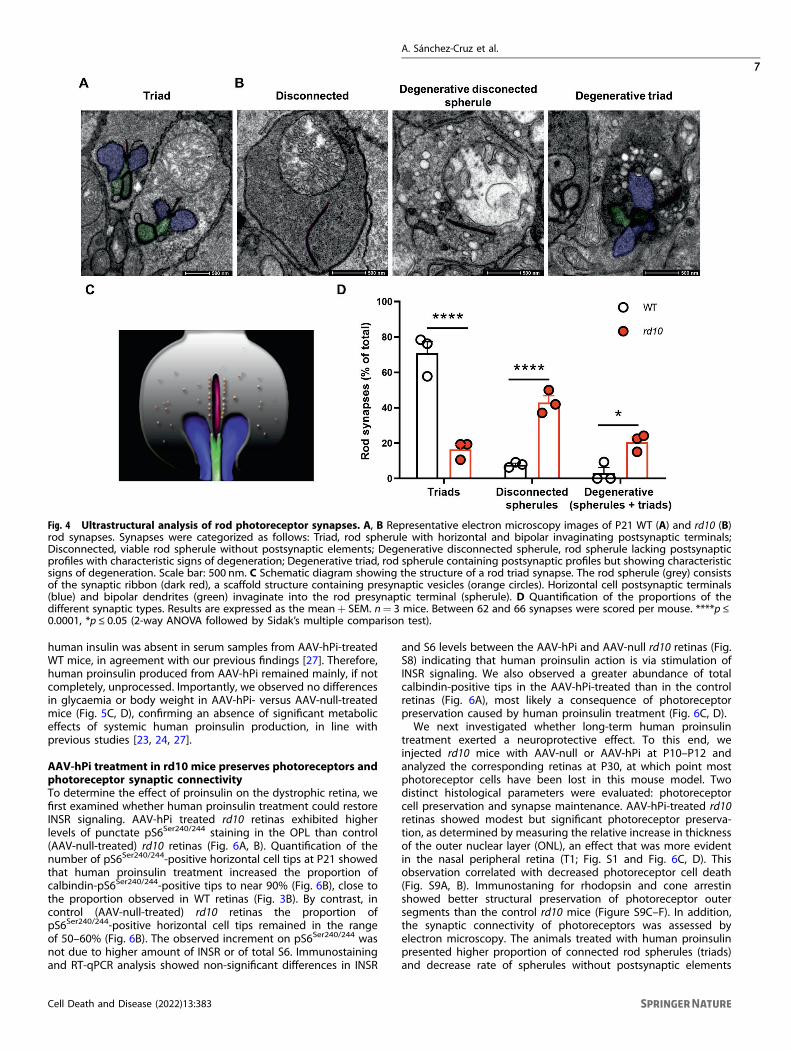
synapse formation and maintenance [8, 14, 15], we nextinvestigated whether changes in INSR expression coincided withalterations in synaptic structure in the OPL. Our analyses focusedon rod synapses, since cone survival remains uncompromised inthe early stages of degeneration in the rd10 retina [42]. The OPLcontains so-called triad synapses, formed by the presynaptic rodspherule, two lateral horizontal and one or two central bipolarpostsynaptic terminals invaginating in close apposition to thesynaptic ribbon (Fig. 4A, C). Using electron microscopy, weexamined WT and rd10 retinas in the early stages of degeneration(P21), when a large proportion of rod photoreceptor cells persistbut signs of degeneration are also evident (Fig. S5). In WT retinas,most of the rod synapses (70%) corresponded to characteristictriad synapses (Fig. 4A, D). Conversely, in rd10 retinas at the sametimepoint, triad synapses accounted for less than 20% of allsynapses (Fig. 4D). Moreover, in rd10 retinas more than 40% of rodterminals lacked any postsynaptic element while in WT retinas thiswas observed in only 8% of rod terminals (Fig. 4B, D). Despite theabsence of postsynaptic partners, these disconnected rodspherules had a viable appearance, as evidenced by intactmitochondria and electro-lucent content. In these disconnectedspherules the synaptic ribbon, when present, was attached to anunusually flat cell membrane with no signs of any postsynapticinvagination (Fig. 4B). By contrast, the degenerative rod spherules(accounting for 20% and 3% of rod terminals in rd10 and WTretinas, respectively) were highly vacuolated, with electro-densecontent and aberrant mitochondria (Fig. 4B, D). Among thedegenerated rod spherules, we observed both disconnectedterminals and triad synapses (Fig. 4B), suggesting that disconnec-tion is not a necessary step before degeneration. Furthermore,both genotypes showed similar numbers of dyads (18.65 ± 4.12%for WT vs 20.60 ± 3.01% for rd10), most likely due to themisalignment of one of the postsynaptic elements with the planeof section [43].Immunostaining using specific markers for photoreceptor
ribbon (Ribeye and Ctbp2) and horizontal (GluA2) and bipolar
(mGluR6) postsynaptic terminal tips also revealed synapticdisconnection of rod photoreceptors in the rd10 retina (Fig. S6A,B).
Systemically-provided human proinsulin reaches the retinaand exerts no metabolic effectThe results described above suggest a possible link betweendeficient INSR signaling, synaptic disconnection in rod photo-receptors, and the visual impairment characteristic of RP. Todetermine whether deficient INSR signaling is a disease-modifyingfactor in RP, and therefore a potential therapeutic target, weemployed a gene therapy strategy to enhance INSR signaling.Proinsulin, the insulin precursor, is an INSR-A selective ligand [44]with a low metabolic profile, likely due to its poor affinity for theINSR-B isoform [32, 44]. We have previously demonstrated theneuroprotective potential of proinsulin during retinal develop-ment and degeneration [23–25, 45]. Given that treatment of RP inhumans would entail long-term administration, we selectedproinsulin to activate INSR-A, the predominant INSR isoformexpressed in the retina (Fig. 1A).To achieve sustained production of Pi, we built upon our
previous experience with a recombinant AAV2/1 expressing thehuman proinsulin (hPi) coding region (Fig. 5; AAV-hPi) [24, 27]. Wefirst evaluated human proinsulin production following intramus-cular administration of AAV-hPi. rd10 mice received a singleinjection of AAV-hPi into the gastrocnemius muscle at P10 toinduce human proinsulin production before the onset ofdegeneration (around P18). In AAV-hPi-treated rd10 mice humanproinsulin serum levels were uniformly sustained within eachindividual mouse for up to 5 months (the maximum follow-upperiod) (Fig. 5B). Moreover, human proinsulin was detected inwhole eye and in retinal extracts as early as 1 week post-injectionand up to 7 weeks post-injection (the maximum follow-up period)(Fig. S7A). Conversely, human proinsulin was not detected at anytimepoint in serum samples, whole eye, or retinal extracts frommice injected with the control vector (AAV-null). Moreover, mature
Fig. 3 Downregulation of insulin receptor signaling in the rd10 mouse retina. A Representative images of the OPL of P23 retinal sectionsfrom WT and rd10 mice co-immunostained for pS6Ser240/244 (green) and calbindin (red). Asterisks indicate horizontal cell bodies. Insets showamplification (2.5X) of the indicated area. Scale bar: 6 μm. B Quantification of the number of horizontal terminal tips (calbindin+ puncta) withapposite pS6Ser240/244 labeling. Data are presented as the mean+ SEM. n= 4 mice, 4 images. Over 200 calbindin+ tips were scored per retina.***p ≤ 0.001 versus WT (unpaired T-test).
A. Sánchez-Cruz et al.
6
Cell Death and Disease (2022) 13:383
human insulin was absent in serum samples from AAV-hPi-treatedWT mice, in agreement with our previous findings [27]. Therefore,human proinsulin produced from AAV-hPi remained mainly, if notcompletely, unprocessed. Importantly, we observed no differencesin glycaemia or body weight in AAV-hPi- versus AAV-null-treatedmice (Fig. 5C, D), confirming an absence of significant metaboliceffects of systemic human proinsulin production, in line withprevious studies [23, 24, 27].
AAV-hPi treatment in rd10 mice preserves photoreceptors andphotoreceptor synaptic connectivityTo determine the effect of proinsulin on the dystrophic retina, wefirst examined whether human proinsulin treatment could restoreINSR signaling. AAV-hPi treated rd10 retinas exhibited higherlevels of punctate pS6Ser240/244 staining in the OPL than control(AAV-null-treated) rd10 retinas (Fig. 6A, B). Quantification of thenumber of pS6Ser240/244-positive horizontal cell tips at P21 showedthat human proinsulin treatment increased the proportion ofcalbindin-pS6Ser240/244-positive tips to near 90% (Fig. 6B), close tothe proportion observed in WT retinas (Fig. 3B). By contrast, incontrol (AAV-null-treated) rd10 retinas the proportion ofpS6Ser240/244-positive horizontal cell tips remained in the rangeof 50–60% (Fig. 6B). The observed increment on pS6Ser240/244 wasnot due to higher amount of INSR or of total S6. Immunostainingand RT-qPCR analysis showed non-significant differences in INSR
and S6 levels between the AAV-hPi and AAV-null rd10 retinas (Fig.S8) indicating that human proinsulin action is via stimulation ofINSR signaling. We also observed a greater abundance of totalcalbindin-positive tips in the AAV-hPi-treated than in the controlretinas (Fig. 6A), most likely a consequence of photoreceptorpreservation caused by human proinsulin treatment (Fig. 6C, D).We next investigated whether long-term human proinsulin
treatment exerted a neuroprotective effect. To this end, weinjected rd10 mice with AAV-null or AAV-hPi at P10–P12 andanalyzed the corresponding retinas at P30, at which point mostphotoreceptor cells have been lost in this mouse model. Twodistinct histological parameters were evaluated: photoreceptorcell preservation and synapse maintenance. AAV-hPi-treated rd10retinas showed modest but significant photoreceptor preserva-tion, as determined by measuring the relative increase in thicknessof the outer nuclear layer (ONL), an effect that was more evidentin the nasal peripheral retina (T1; Fig. S1 and Fig. 6C, D). Thisobservation correlated with decreased photoreceptor cell death(Fig. S9A, B). Immunostaning for rhodopsin and cone arrestinshowed better structural preservation of photoreceptor outersegments than the control rd10 mice (Figure S9C–F). In addition,the synaptic connectivity of photoreceptors was assessed byelectron microscopy. The animals treated with human proinsulinpresented higher proportion of connected rod spherules (triads)and decrease rate of spherules without postsynaptic elements
Fig. 4 Ultrastructural analysis of rod photoreceptor synapses. A, B Representative electron microscopy images of P21 WT (A) and rd10 (B)rod synapses. Synapses were categorized as follows: Triad, rod spherule with horizontal and bipolar invaginating postsynaptic terminals;Disconnected, viable rod spherule without postsynaptic elements; Degenerative disconnected spherule, rod spherule lacking postsynapticprofiles with characteristic signs of degeneration; Degenerative triad, rod spherule containing postsynaptic profiles but showing characteristicsigns of degeneration. Scale bar: 500 nm. C Schematic diagram showing the structure of a rod triad synapse. The rod spherule (grey) consistsof the synaptic ribbon (dark red), a scaffold structure containing presynaptic vesicles (orange circles). Horizontal cell postsynaptic terminals(blue) and bipolar dendrites (green) invaginate into the rod presynaptic terminal (spherule). D Quantification of the proportions of thedifferent synaptic types. Results are expressed as the mean+ SEM. n= 3 mice. Between 62 and 66 synapses were scored per mouse. ****p ≤0.0001, *p ≤ 0.05 (2-way ANOVA followed by Sidak’s multiple comparison test).
A. Sánchez-Cruz et al.
7
Cell Death and Disease (2022) 13:383

(disconnected) than the control mice (Fig. 6E, F). These resultswere confirmed by immunostaining of rod-bipolar and rod-horizontal synapses (Fig. S6C–F). As expected, photoreceptorpreservation in AAV-hPi-treated retinas led to an increase in thenumber of ribbons (Fig. S6C, D, lower panels). Moreover, humanproinsulin reduced the proportion of disconnected postsynapticterminals of rod cells [i.e. those lacking either a GluA2-positivehorizontal postsynaptic terminal (Fig. S6C, E) or a mGluR6-positivebipolar postsynaptic terminal (Fig. S6D, F)] in agreement with theabove electron microscopy results (Fig. 6E, F). These results reveala novel effect of proinsulin: preservation of the synapticconnectivity of rod cells with their postsynaptic second-orderneuronal partners. This observation suggests a second distinct roleof human proinsulin that may be partially independent of itseffects on photoreceptor cell survival revealed here (Fig. S9) aswell as in our previous studies [23–25]. Moreover, the increase inthe number of photoreceptor synapses induced by proinsulintreatment that we previously observed in the P23H rat model ofRP [24] is most likely a consequence not only of the rescue ofphotoreceptor cells but also of the preservation of their synapticcontacts, as described here.
AAV-hPi treatment in rd10 mice preserves visual functionFinally, in AAV-hPi-treated rd10 mice we assessed whether humanproinsulin preserved visual function, which is the most clinicallyrelevant outcome for a potential RP treatment. Electroretino-graphic (ERG) recordings were performed in dim and daylight
conditions every 10 days between P30 and P60 to evaluate rod-and cone-mediated light responses. Mixed light responses of theWT animals experience small increase in ERG amplitudes from P30to P60, however the light response of AAV-null animals was almostnull at P50 (Fig. 7B). Of note, a remaining b-mixed wave was stillobserved in the P60 treated mouse (Fig. 7B). Analysis of differentERG waves showed that AAV-hPi-treated rd10 mice displayedbetter defined and more prominent ERG waves than their AAV-null-treated counterparts (Fig. 7B). Waves corresponding to rods(b-scotopic wave), cones (b-photopic wave), and both photo-receptors (a-mixed and b-mixed waves) were of a significantlygreater amplitude in AAV-hPi-treated than AAV-null-treated rd10mice (Fig. 7C). ERG recordings thus confirmed that humanproinsulin treatment preserved visual function, consistent withthe aforementioned preservation of photoreceptor cells and theirsynapses. Optokinetic testing further confirmed partial preserva-tion of the light response in AAV-hPi-treated rd10 retinas. Inaddition to measuring the retinal response to light, this visualbehavior test evaluates the function of other components of thevisual system, namely optic nerve transmission and visualintegration in the brain [46]. Mice instinctively respond to rotatingvertical bars with characteristic movement of their heads in thesame direction as the rotation of the bar (Fig. 8A, B). AAV-hPi-treated rd10 mice showed greater contrast sensitivity than AAV-null-treated counterparts at P40 (Fig. 8C). Moreover, at P50, age atwhich all control mice have lost the optokinetic response, two outof six proinsulin-treated rd10 showed significant contrast
Fig. 5 AAV-hPi treatment results in sustained human proinsulin production. A rd10 mice received a single intramuscular injection of AAV-hPi or AAV-null at P10 and were analyzed at different timepoints post-injection. B Long-term monitoring of serum human proinsulin levels asdetermined by ELISA in individual rd10 mice injected with AAV-hPi. Human proinsulin levels in AAV-null mice were under the detectionthreshold of the assay (0.5 pM). C, D Long-term monitoring of glucose levels (C) and body weight (D) in AAV-hPi or AAV-null injected rd10mice. Data are presented as the mean+ SEM. n= 4 mice in B and 4–6 mice per group in C and D.
A. Sánchez-Cruz et al.
8
Cell Death and Disease (2022) 13:383

sensitivity (Fig. 8D). Together, our results support the disease-modifying potential of proinsulin, which can prolong visualfunction in an animal model of RP and therefore constitutes aworthwhile candidate therapy for retinal dystrophies.
DISCUSSIONThis study describes the downregulation of retinal INSR levels andlocal signaling during the early stages of retinal neurodegenerationin the rd10 mouse model of RP, together with concomitant
Fig. 6 Effect of AAV-hPi administration on insulin receptor signaling and mouse retina structure. rd10mice received a single intramuscularinjection of AAV-null or AAV-hPi at P12 and retinas were analyzed for pS6Ser240/244 expression (A, B), photoreceptor preservation (C, D) andsynapse maintenance (E, F). A Representative images of the OPL in P21 retinal sections co-immunostained for pS6Ser240/244 (green) andcalbindin (red). Asterisks indicate horizontal cell bodies. Insets show amplification (3X) of the indicated area. Scale bar: 11 μm. B Quantificationof the number of horizontal terminal tips (calbindin+ puncta) with apposite pS6Ser240/244 labeling. Data are presented as the mean+ SEM. n=4 mice, 4 images. Over 200 calbindin+ tips per retina were analyzed. *p ≤ 0.05 (unpaired T-test with Welch’s correction). C Representativeimages of P30 retinal sections showing the T1 region from AAV-null- and AAV-hPi-treated rd10 mice. Blue color corresponds to DAPI staining.ONL, outer nuclear layer; INL, inner nuclear layer. Scale bar: 70 μm. D ONL and INL thickness were measured in equatorial sectionscorresponding to 6 retinal areas, following a nasotemporal sequence (T1–T6 as defined in Fig. S1). Plot shows the mean+ SEM. n= 5 mice,3 sections per retina, 6 regions, 3 measurements per region. E Representative electron microscopy images of P30 AAV-null- and AAV-hPi-treated rd10 mice. Arrow heads indicate triads, asterisks disconnected spherules, yellow arrow degenerative spherule and white arrow dyad.Scale bar: 1 μm. F Quantification of the proportions of the different synaptic types. Results are expressed as the mean+ SEM. n= 3 mice.Between 58 and 86 synapses were scored per mouse. ****p ≤ 0.0001 (Sidak’s multiple comparisons test after 2-way ANOVA).
A. Sánchez-Cruz et al.
9
Cell Death and Disease (2022) 13:383
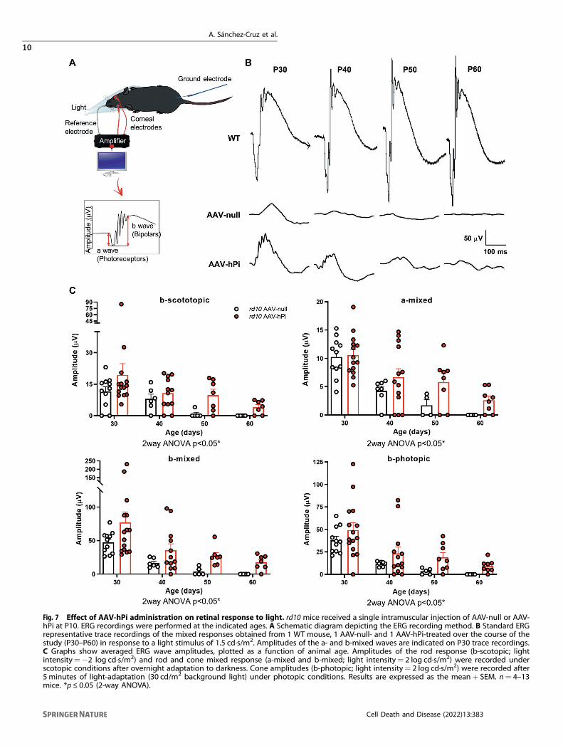
Fig. 7 Effect of AAV-hPi administration on retinal response to light. rd10 mice received a single intramuscular injection of AAV-null or AAV-hPi at P10. ERG recordings were performed at the indicated ages. A Schematic diagram depicting the ERG recording method. B Standard ERGrepresentative trace recordings of the mixed responses obtained from 1 WT mouse, 1 AAV-null- and 1 AAV-hPi-treated over the course of thestudy (P30–P60) in response to a light stimulus of 1.5 cd·s/m2. Amplitudes of the a- and b-mixed waves are indicated on P30 trace recordings.C Graphs show averaged ERG wave amplitudes, plotted as a function of animal age. Amplitudes of the rod response (b-scotopic; lightintensity=−2 log cd·s/m2) and rod and cone mixed response (a-mixed and b-mixed; light intensity= 2 log cd·s/m2) were recorded underscotopic conditions after overnight adaptation to darkness. Cone amplitudes (b-photopic; light intensity= 2 log cd·s/m2) were recorded after5 minutes of light-adaptation (30 cd/m2 background light) under photopic conditions. Results are expressed as the mean+ SEM. n= 4–13mice. *p ≤ 0.05 (2-way ANOVA).
A. Sánchez-Cruz et al.
10
Cell Death and Disease (2022) 13:383

disruption of photoreceptor triad synapses. We provide proof ofconcept of the neuroprotective effect of INSR stimulation with theinsulin precursor proinsulin, which is selective for the INSR-A isoformfound in the retina. Gene therapy using an AAV induced sustainedproduction of circulating proinsulin, which reached the retina,restored local INSR signaling, and exerted neuroprotective effectson retinal structure and visual function, without affecting peripheralmetabolic parameters. Proinsulin treatment attenuated both photo-receptor cell loss and synaptic disconnection, and prolonged visualfunction, highlighting the potential of proinsulin as a candidatetherapy for RP.Studies over the past two decades have broadened the scope of
INSR activity far beyond the peripheral metabolic role initiallyascribed to this receptor. The versatility of this receptor is alsoimplied by its widespread expression in the CNS. At the neuronallevel, INSR has been implicated in synaptic plasticity, dendriticoutgrowth, and cell survival [8, 14, 15]. The Insr-a splice variant, whichis predominantly expressed in different areas of the brain as well asthe retina, is the most ancient and promiscuous isoform, andparticipates in insulin, proinsulin, and IGF-II signaling [31, 44].Interestingly, many of the signaling pathways in which INSR-A isinvolved are similar to those mediated by growth factor receptors,and in some ways this isoform resembles the ancestral INSR
expressed in invertebrates and low vertebrates [7, 47, 48]. Thepresent findings confirm the previously described wide distributionof INSR in the WT retina [49, 50]. However, we also describe for thefirst time more intense INSR immunostaining in horizontal andganglion cell axons than in other retinal structures. Interestingly,analysis of INSR expression during retinal degeneration in the rd10mouse revealed local downregulation of INSR, specifically inhorizontal cell axons. This was accompanied by a decrease in thephosphorylation of ribosomal protein S6 in horizontal cell terminals,indicating impaired INSR signaling. Notably, horizontal cell axonterminals receive input from the rod photoreceptors, the maintargets of degeneration in RP. A recent study by Agostinone et al.[40] reported specific decreases in pS6Ser240/244 in ganglion cellsfollowing transection of their axons, but no alterations in pS6Ser240/244
in horizontal cells. In the present study, we observed selectivedownregulation of INSR expression and impaired signaling in thehorizontal cells with which the damaged rod photoreceptorssynapse. It remains unclear whether this finding is mechanisticallylinked to the decreases in pS6Ser240/244 described by Agostinone et al.[40]. However, our results are consistent with the proposals by otherauthors that INSR signaling may vary in a neuronal activity-dependent manner [8, 51, 52], and suggest that INSR expressionand signaling in horizontal cells may depend on rod input.
Fig. 8 Effect of AAV-hPi administration on optokinetic response. rd10 mice received a single intramuscular injection of AAV-null or AAV-hPiat P10. The optomotor test was performed at the indicated ages. A, B Schematic depicting the optomotor test (A); 5 different spatialfrequencies were tested (0.022–0.355 cycles/degree) and the contrast of the moving bars was adjusted (from 100% to 5%) to determinecontrast sensitivity (B). C, D Optokinetic responses recorded at the indicated ages in AAV-null- and AAV-hPi treated rd10 mice. Contrastsensitivity is represented as a function of spatial frequency. Data are presented as the mean+ SEM. n= 6 mice. P40 (****p ≤ 0.0001) and P50(*p ≤ 0.05) (2-way ANOVA).
A. Sánchez-Cruz et al.
11
Cell Death and Disease (2022) 13:383

Interestingly, in rd10 retinas INSR downregulation and consequentimpairment of INSR signaling coincided with the presence ofdisconnected but otherwise apparently viable rod terminals. Altera-tions in retinal synaptic circuitry as part of retinal remodeling duringphotoreceptor degeneration have been acknowledged for morethan two decades [3, 53–55]. De-afferentiation of second-orderneurons has typically been described in advanced stages ofdegeneration as a consequence of photoreceptor loss [3, 53–55].However, here we describe early disconnection events affecting upto 50% of apparently viable rod spherules during the initial stages ofdegeneration. Although further studies will be required to clarify theconsequences of this disconnection, it does not appear to necessarilyprecede rod death, as we detected degenerative triads containingthe presynaptic rod spherule and horizontal and bipolar terminals.These observations have direct implications for the development ofneuroprotective therapies and could open a new line of research intointerventions aimed at preserving rod connectivity.Our findings demonstrate the potential of proinsulin as a
synaptoprotective factor. In rd10 mice treated with AAV-hPi,systemically produced proinsulin reached the retina and restoredINSR signaling, as determined by measuring pS6Ser240/244 levels.Concomitantly, proinsulin treatment reduced the number ofdisconnected rod presynaptic terminals, suggesting a role of INSRsignaling in photoreceptor synaptic connectivity. Our results are inline with those of the seminal study by Chiu et al. 2008 [8], whoused optic tectal neurons in living Xenopus tadpoles todemonstrate that INSR signaling maintains both synaptic contactsand the branches on which they lie. Moreover, in the aforemen-tioned study by Agostinone et al. [40] stimulation of INSR signalingwith insulin promoted regeneration of the dendritic arbors ofretinal ganglion cells after axonal injury.Deficient local INSR signaling associated with RP may be a
general feature of neurodegenerative diseases, irrespective ofperipheral insulin resistance [reviewed in [6]]. Several studies havedescribed decreased INSR expression and/or attenuation of theactivation states of INSR signaling molecules in affected brainregions in patients with Alzheimer’s [17–19] and Parkinson’s [56–58] diseases. We show that pharmacological stimulation of INSRsignaling with Pi has a disease-modifying effect over the course ofretinal degeneration, in line with the beneficial effects of INSRstimulation with insulin reported in other neurodegenerativediseases of the brain and retina [6, 40, 41]. Moreover, thewidespread expression of INSR in the retina suggests that synapticmaintenance promoted by INSR signaling is only one of severalretinal processes regulated by INSR. Indeed, we and others havedemonstrated the neuroprotective effects of INSR signalingmolecules on photoreceptor survival [28, 59, 60].Our results provide a novel mechanism to account for our
previous descriptions of the neuroprotective effects of proinsulinon retinal dystrophy [23–25] and cognitive impairment [27], andfurther support the validity of proinsulin-mediated stimulation ofINSR as a candidate therapy for neurodegenerative conditions ofthe CNS.
DATA AVAILABILITYAll analyzed datasets are included in the manuscript and SI Appendix.
REFERENCES1. Collaborators GBDN. Global, regional, and national burden of neurological dis-
orders, 1990-2016: a systematic analysis for the Global Burden of Disease Study2016. Lancet Neurol 2019;18:459–80.
2. de La Rosa EJ, Hernandez-Sanchez C. CNS Targets for the Treatment of RetinalDystrophies: A Win–Win Strategy. Therapies for retinal degeneration: Targetingcommon processes. de la Rosa EJ, Cotter TG Editors. Royal Society of Chemistry;2019. p. 277.
3. Pfeiffer RL, Marc RE, Jones BW. Persistent remodeling and neurodegeneration inlate-stage retinal degeneration. Prog Retin Eye Res. 2020;74:100771.
4. Cuenca N, Fernandez-Sanchez L, Campello L, Maneu V, De la Villa P, Lax P, et al.Cellular responses following retinal injuries and therapeutic approaches forneurodegenerative diseases. Prog Retin Eye Res. 2014;43:17–75.
5. Russell S, Bennett J, Wellman JA, Chung DC, Yu ZF, Tillman A, et al. Efficacy andsafety of voretigene neparvovec (AAV2-hRPE65v2) in patients with RPE65-mediated inherited retinal dystrophy: A randomised, controlled, open-label,phase 3 trial. Lancet 2017;390:849–60.
6. Arnold SE, Arvanitakis Z, Macauley-Rambach SL, Koenig AM, Wang HY, Ahima RS,et al. Brain insulin resistance in type 2 diabetes and Alzheimer disease: Conceptsand conundrums. Nat Rev Neurol. 2018;14:168–81.
7. Banks WA, Owen JB, Erickson MA. Insulin in the brain: There and back again.Pharm Ther. 2012;136:82–93.
8. Chiu SL, Cline HT. Insulin receptor signaling in the development of neuronalstructure and function. Neural Dev. 2010;5:7.
9. de la Rosa EJ, Bondy CA, Hernandez-Sanchez C, Wu X, Zhou J, Lopez-Carranza A,et al. Insulin and insulin-like growth factor system components gene expressionin the chicken retina from early neurogenesis until late development and theireffect on neuroepithelial cells. Eur J Neurosci. 1994;6:1801–10.
10. Havrankova J, Roth J, Brownstein M. Insulin receptors are widely distributed inthe central nervous system of the rat. Nature 1978;272:827–9.
11. Marks JL, Porte D Jr, Stahl WL, Baskin DG. Localization of insulin receptor mRNA inrat brain by in situ hybridization. Endocrinology 1990;127:3234–6.
12. Rodrigues M, Waldbillig RJ, Rajagopalan S, Hackett J, LeRoith D, Chader GJ.Retinal insulin receptors: Localization using a polyclonal anti-insulin receptorantibody. Brain Res. 1988;443:389–94.
13. Unger J, McNeill TH, Moxley RT 3rd, White M, Moss A, Livingston JN. Distribution ofinsulin receptor-like immunoreactivity in the rat forebrain. Neuroscience1989;31:143–57.
14. Gralle M. The neuronal insulin receptor in its environment. J Neurochem.2017;140:359–67.
15. Lee CC, Huang CC, Hsu KS. Insulin promotes dendritic spine and synapse for-mation by the PI3K/Akt/mTOR and Rac1 signaling pathways. Neuropharmacology2011;61:867–79.
16. Moloney AM, Griffin RJ, Timmons S, O’Connor R, Ravid R, O’Neill C. Defects in IGF-1 receptor, insulin receptor and IRS-1/2 in Alzheimer’s disease indicate possibleresistance to IGF-1 and insulin signalling. Neurobiol Aging. 2010;31:224–43.
17. Rivera EJ, Goldin A, Fulmer N, Tavares R, Wands JR, de la Monte SM. Insulin andinsulin-like growth factor expression and function deteriorate with progression ofAlzheimer’s disease: link to brain reductions in acetylcholine. J Alzheimers Dis.2005;8:247–68.
18. Steen E, Terry BM, Rivera EJ, Cannon JL, Neely TR, Tavares R, et al. Impaired insulinand insulin-like growth factor expression and signaling mechanisms in Alzhei-mer’s disease-is this type 3 diabetes? J Alzheimers Dis. 2005;7:63–80.
19. Talbot K, Wang HY, Kazi H, Han LY, Bakshi KP, Stucky A, et al. Demonstrated braininsulin resistance in Alzheimer’s disease patients is associated with IGF-1 resis-tance, IRS-1 dysregulation, and cognitive decline. J Clin Invest. 2012;122:1316–38.
20. Chapman CD, Schioth HB, Grillo CA, Benedict C. Intranasal insulin in Alzheimer’sdisease: Food for thought. Neuropharmacology 2018;136:196–201.
21. Hernandez-Sanchez C, Lopez-Carranza A, Alarcon C, de La Rosa EJ, de Pablo F.Autocrine/paracrine role of insulin-related growth factors in neurogenesis: localexpression and effects on cell proliferation and differentiation in retina. Proc NatlAcad Sci USA. 1995;92:9834–8.
22. Valenciano AI, Corrochano S, de Pablo F, de la Villa P, de la Rosa EJ. Proinsulin/insulin is synthesized locally and prevents caspase- and cathepsin-mediated celldeath in the embryonic mouse retina. J Neurochem. 2006;99:524–36.
23. Corrochano S, Barhoum R, Boya P, Arroba AI, Rodriguez-Muela N, Gomez-VicenteV, et al. Attenuation of vision loss and delay in apoptosis of photoreceptorsinduced by proinsulin in a mouse model of retinitis pigmentosa. Invest Oph-thalmol Vis Sci. 2008;49:4188–94.
24. Fernandez-Sanchez L, Lax P, Isiegas C, Ayuso E, Ruiz JM, de la Villa P, et al.Proinsulin slows retinal degeneration and vision loss in the P23H rat model ofretinitis pigmentosa. Hum Gene Ther. 2012;23:1290–300.
25. Isiegas C, Marinich-Madzarevich JA, Marchena M, Ruiz JM, Cano MJ, de la Villa P,et al. Intravitreal injection of proinsulin-loaded microspheres delays photo-receptor cell death and vision loss in the rd10 mouse model of retinitis pig-mentosa. Invest Ophthalmol Vis Sci. 2016;57:3610–8.
26. Chang B, Hawes NL, Pardue MT, German AM, Hurd RE, Davisson MT, et al. Twomouse retinal degenerations caused by missense mutations in the beta-subunitof rod cGMP phosphodiesterase gene. Vis Res. 2007;47:624–33.
27. Corpas R, Hernandez-Pinto AM, Porquet D, Hernandez-Sanchez C, Bosch F, Ortega-Aznar A, et al. Proinsulin protects against age-related cognitive loss through anti-inflammatory convergent pathways. Neuropharmacology 2017;123:221–32.
28. Sanchez-Cruz A, Villarejo-Zori B, Marchena M, Zaldivar-Diez J, Palomo V, Gil C,et al. Modulation of GSK-3 provides cellular and functional neuroprotection in therd10 mouse model of retinitis pigmentosa. Mol Neurodegener. 2018;13:19.
A. Sánchez-Cruz et al.
12
Cell Death and Disease (2022) 13:383

29. Sánchez-Cruz A, Méndez AC, Lizasoain I, de la Villa P, de la Rosa EJ, Hernández-Sánchez C. Tlr2 gene deletion delays retinal degeneration in two geneticallydistinct mouse models of retinitis pigmentosa. Int J Mol Sci. 2021;22:7815.
30. Prusky GT, Alam NM, Beekman S, Douglas RM. Rapid quantification of adult anddeveloping mouse spatial vision using a virtual optomotor system. Invest Oph-thalmol Vis Sci. 2004;45:4611–6.
31. Hernandez-Sanchez C, Mansilla A, de Pablo F, Zardoya R. Evolution of the insulinreceptor family and receptor isoform expression in vertebrates. Mol Biol Evol.2008;25:1043–53.
32. Belfiore A, Malaguarnera R, Vella V, Lawrence MC, Sciacca L, Frasca F, et al. Insulinreceptor isoforms in physiology and disease: An updated view. Endocr Rev.2017;38:379–431.
33. Bruning JC, Gautam D, Burks DJ, Gillette J, Schubert M, Orban PC, et al. Role ofbrain insulin receptor in control of body weight and reproduction. Science2000;289:2122–5.
34. Dixon-Salazar TJ, Fourgeaud L, Tyler CM, Poole JR, Park JJ, Boulanger LM. MHCclass I limits hippocampal synapse density by inhibiting neuronal insulin receptorsignaling. J Neurosci. 2014;34:11844–56.
35. Peichl L, Gonzalez-Soriano J. Unexpected presence of neurofilaments in axon-bearing horizontal cells of the mammalian retina. J Neurosci. 1993;13:4091–100.
36. Peichl L, Gonzalez-Soriano J. Morphological types of horizontal cell in rodent retinae:a comparison of rat, mouse, gerbil, and guinea pig. Vis Neurosci. 1994;11:501–17.
37. Feigenspan A, Babai N. Functional properties of spontaneous excitatory currentsand encoding of light/dark transitions in horizontal cells of the mouse retina. EurJ Neurosci. 2015;42:2615–32.
38. Kolb H. Organization of the outer plexiform layer of the primate retina: Electronmicroscopy of Golgi-impregnated cells. Philos Trans R Soc Lond B Biol Sci.1970;258:261–83.
39. Kolb H. The connections between horizontal cells and photoreceptors in the retinaof the cat: Electron microscopy of Golgi preparations. J Comp Neurol. 1974;155:1–14.
40. Agostinone J, Alarcon-Martinez L, Gamlin C, Yu WQ, Wong ROL, Di Polo A. Insulinsignalling promotes dendrite and synapse regeneration and restores circuitfunction after axonal injury. Brain 2018;141:1963–80.
41. Holscher C. Brain insulin resistance: Role in neurodegenerative disease andpotential for targeting. Expert Opin Investig Drugs. 2020;29:333–48.
42. Zhao L, Zabel MK, Wang X, Ma W, Shah P, Fariss RN, et al. Microglial phagocytosisof living photoreceptors contributes to inherited retinal degeneration. EMBO MolMed. 2015;7:1179–97.
43. Wang T, Pahlberg J, Cafaro J, Frederiksen R, Cooper AJ, Sampath AP, et al. Acti-vation of rod input in a model of retinal degeneration reverses retinal remodelingand induces formation of functional synapses and recovery of visual signaling inthe adult retina. J Neurosci. 2019;39:6798–810.
44. Malaguarnera R, Sacco A, Voci C, Pandini G, Vigneri R, Belfiore A. Proinsulin bindswith high affinity the insulin receptor isoform A and predominantly activates themitogenic pathway. Endocrinology 2012;153:2152–63.
45. Hernandez-Sanchez C, Mansilla A, de la Rosa EJ, de Pablo F. Proinsulin in develop-ment: New roles for an ancient prohormone. Diabetologia 2006;49:1142–50.
46. Abdeljalil J, Hamid M, Abdel-Mouttalib O, Stephane R, Raymond R, Johan A, et al.The optomotor response: A robust first-line visual screening method for mice. VisRes. 2005;45:1439–46.
47. Belfiore A, Frasca F, Pandini G, Sciacca L, Vigneri R. Insulin receptor isoforms andinsulin receptor/insulin-like growth factor receptor hybrids in physiology anddisease. Endocr Rev. 2009;30:586–623.
48. Chan SJ, Steiner DF. Insulin Through the Ages: Phylogeny of a Growth Promotingand Metabolic Regulatory Hormone 1. Am Zool. 2000;40:213–22.
49. Gosbell AD, Favilla I, Baxter KM, Jablonski P. Insulin receptor and insulin receptorsubstrate-I in rat retinae. Clin Exp Ophthalmol. 2000;28:212–5.
50. Rajala RV, Wiskur B, Tanito M, Callegan M, Rajala A. Diabetes reduces autopho-sphorylation of retinal insulin receptor and increases protein-tyrosine phospha-tase-1B activity. Invest Ophthalmol Vis Sci. 2009;50:1033–40.
51. Clarke DW, Mudd L, Boyd FT Jr, Fields M, Raizada MK. Insulin is released from ratbrain neuronal cells in culture. J Neurochem. 1986;47:831–6.
52. Hori K, Yasuda H, Konno D, Maruoka H, Tsumoto T, Sobue K. NMDA receptor-dependent synaptic translocation of insulin receptor substrate p53 via proteinkinase C signaling. J Neurosci. 2005;25:2670–81.
53. Jones BW, Kondo M, Terasaki H, Lin Y, McCall M, Marc RE. Retinal remodeling. JpnJ Ophthalmol. 2012;56:289–306.
54. Lewis GP, Linberg KA, Fisher SK. Neurite outgrowth from bipolar and horizontal cellsafter experimental retinal detachment. Invest Ophthalmol Vis Sci. 1998;39:424–34.
55. Strettoi E, Pignatelli V, Rossi C, Porciatti V, Falsini B. Remodeling of second-orderneurons in the retina of rd/rd mutant mice. Vis Res. 2003;43:867–77.
56. Moroo I, Yamada T, Makino H, Tooyama I, McGeer PL, McGeer EG, et al. Loss ofinsulin receptor immunoreactivity from the substantia nigra pars compactaneurons in Parkinson’s disease. Acta Neuropathol. 1994;87:343–8.
57. Takahashi M, Yamada T, Tooyama I, Moroo I, Kimura H, Yamamoto T, et al. Insulinreceptor mRNA in the substantia nigra in Parkinson’s disease. Neurosci Lett.1996;204:201–4.
58. Timmons S, Coakley MF, Moloney AM, ON C. Akt signal transduction dysfunctionin Parkinson’s disease. Neurosci Lett. 2009;467:30–5.
59. Punzo C, Kornacker K, Cepko CL. Stimulation of the insulin/mTOR pathwaydelays cone death in a mouse model of retinitis pigmentosa. Nat Neurosci.2009;12:44–52.
60. Rajala A, Tanito M, Le YZ, Kahn CR, Rajala RV. Loss of neuroprotective survivalsignal in mice lacking insulin receptor gene in rod photoreceptor cells. J BiolChem. 2008;283:19781–92.
ACKNOWLEDGEMENTSThe authors thank Cayetana Murillo, María D. Hernández-Fuentes, the staff at the CIBanimal and electron microscopy facilities and CIB and UCM microscopy facilities fortechnical support; Nuria Forns and Ryan Steel for setting the conditions for theoptomotor test and for synapse immunostaining; Noemi Alvarez Lindo for designingthe diagrams; and José A. Esteban for supplying the anti-Glu2A antibody. ASC wasrecipient of a UCM predoctoral fellowship (CT45/15).
AUTHOR CONTRIBUTIONSAS-C, AH-P, CI, and MM performed experimental procedures. CL carried out andanalyzed the electron microscopy studies. PdlV designed the functional studiesand analyzed the data. FB designed and generated the AAV vectors. EJdlR and CH-S designed and supervised the biological experiments. EJdlR and CH-S analyzedand discussed the results. EJdlR, CH-S, IL, CL, and PdlV provided Funding. AS-C,AH-P, CL, CI, IL, PdlV, FB, EJdlR, and CH-S contributed to the writing and editing ofthe manuscript. All authors read and approved the final manuscript.
FUNDINGThis work was supported by grants from the Spanish MINECO (SAF2016-75681-R toEJdlR, PID2019-109506RB-100 to EJdlR and CHS, and PI18-0754 to PdlV), the Institutode Salud Carlos III and co-financed by the European Development Regional Fund “Away to make Europe”/“Investing in your future” (ERDF/ESF) (PI20/00535 to IL and PI18/01536 to CL), the Instituto de Salud Carlos III RETICS RD16/0019/0009 and the MadridRegional Government B2017/BMD-3688 to IL.
COMPETING INTERESTSThe authors declare no competing interests.
ETHICS STATEMENTAnimal handling and experimental procedures followed the 3Rs principles, and wererevised by the Animal Welfare Committee of the Centro de Investigaciones Biológicas(CIB) and the Ethics Committee of the Consejo Superior de Investigaciones Científicas(CSIC) and authorized by the Comunidad de Madrid (Ref: PROEX 287/194), inaccordance with the Spanish legislation and the European Union (EU) Directive 2010/63/EU for animal research.
CONSENT FOR PUBLICATIONAll authors consent for the publication of this study.
ADDITIONAL INFORMATIONSupplementary information The online version contains supplementary materialavailable at https://doi.org/10.1038/s41419-022-04839-0.
Correspondence and requests for materials should be addressed to CatalinaHernández-Sánchez or Enrique J.dela Rosa.
Reprints and permission information is available at http://www.nature.com/reprints
Publisher’s note Springer Nature remains neutral with regard to jurisdictional claimsin published maps and institutional affiliations.
A. Sánchez-Cruz et al.
13
Cell Death and Disease (2022) 13:383

Open Access This article is licensed under a Creative CommonsAttribution 4.0 International License, which permits use, sharing,
adaptation, distribution and reproduction in anymedium or format, as long as you giveappropriate credit to the original author(s) and the source, provide a link to the CreativeCommons license, and indicate if changes were made. The images or other third partymaterial in this article are included in the article’s Creative Commons license, unlessindicated otherwise in a credit line to the material. If material is not included in thearticle’s Creative Commons license and your intended use is not permitted by statutoryregulation or exceeds the permitted use, you will need to obtain permission directlyfrom the copyright holder. To view a copy of this license, visit http://creativecommons.org/licenses/by/4.0/.
© The Author(s) 2022
A. Sánchez-Cruz et al.
14
Cell Death and Disease (2022) 13:383