Differential effects of JNK1 and JNK2 inhibition on murine steatohepatitis and insulin resistance
Upload
khangminh22Category
view
0download
0
J U N E 2 0 0 3 , V O L . 6 1 , N O . 6
© 2003 Van Zuiden Communications B.V. All rights reserved.
194
A B S T R A C T
It is well known that obesity is associated with insulinresistance and an increased risk for type 2 diabetes mellitus.Formerly it was postulated that increased lipolysis andconsequently free fatty acid (FFA) production, from withtriglycerides overloaded fat cells, would disrupt glucosehomeostasis via Randle’s hypothesis. Lipodystrophy,however, also leads to insulin resistance. Recently it hasbecome clear that adipose tissue functions as an endocrineorgan and secretes numerous proteins in response to avariety of stimuli. These secreted proteins exert a pleiotropiceffect. The proteins that are involved in glucose and fatmetabolism and hence can influence insulin resistanceare discussed in this paper. They include leptin, resistin,adiponectin, acylation-stimulating protein, tumour necrosisfactor-� and interleukin-6. The stimuli for production andthe site and mechanism of action in relation to insulinresistance will be discussed. None of these proteins are,however, without controversy with regard to their mechanismof action. Furthermore, some of these proteins may influenceeach other via common signalling pathways. A theory ispresented to link the interrelationship between these adipocytesecretory products and their effect on insulin resistance.
L I S T O F A B B R E V I A T I O N S
Acrp 30 Complement-related protein 30
AgRP Agouti-related protein
AMPK Adenosine monophosphate kinase
ADD1/SREBP Adipocyte determination and differentiation
factor/sterol regulatory element-binding
protein
aP2 Fatty acid-binding protein
apM1 Adipose most abundant gene transcript-1
ASP Acylation-stimulating protein
ATP Adenosine triphosphate
BAT Brown adipose tissue
BMI Body mass index
CART Cocaine-amphetamine-related transcript
C/EBP CCAAT (is piece of DNA)/enhancer-binding
proteins
CNS Central nervous system
COS cells Monkey cells immortalised with simian
V40 virus
CRH Corticotropin-releasing hormone
Cys Cysteine
DAG Diacetylglycerol
DM Diabetes mellitus
DNA Deoxyribonucleic acid
FAS Fatty acid synthase
FFA Free fatty acid
FIZZ Found in inflammatory zone
Gdp 28 Gelatin-binding protein
GLUT-4 Glucose transporter-4
IL-6 Interleukin-6
IRS-1 Insulin receptor substrate-1
JAK Janus kinase
�-MSH Alpha-melanocyte-stimulating hormone
mRNA Messenger ribonucleic acid
NEFA Non-esterified fatty acids
NPY Neuropeptide Y
PEPCK Phospo-enolpyruvate carboxykinase
PI3K Phosphatidylinositol-3 phosphate
POMC Pro-opiomelanocortin
Ob-Rb Long isoform of the leptin receptor
RELM Resistine-like molecule
PPAR-� Peroxisome proliferator-activated receptor �
RXR Retinoid X receptor
STAT Signal transducers and activators of
transcription
TG Triglycerides
TNF-� Tumour necrosis factor alpha
TZDs Thiazolidinediones
WAT White adipose tissue
Adipose tissue as an endocrine organ:impact on insulin resistance
I.M. Jazet*, H. Pijl, A.E. Meinders
Department of General Internal Medicine (C1-r-45), Leiden University Medical Centre, Albinusdreef 2,2333 ZA Leiden, the Netherlands, tel.: +31 (0)71-526 43 85, fax: +31 (0)71-524 81 40,
e-mail: [email protected], * corresponding author
R E V I E W

Jazet, et al. Adipose tissue as an endocrine organ: impact on insulin resistance.
J U N E 2 0 0 3 , V O L . 6 1 , N O . 6
195
I N T R O D U C T I O N
Type 2 diabetes mellitus is a chronic disease characterised
by insulin resistance of the muscle, liver and adipose tissue
and an impaired function of the �-cell of the pancreas.1
The incidence of type 2 diabetes mellitus (type 2 DM) has
increased dramatically over the last decades. Nowadays it is
the most frequently occurring metabolic disease, affecting
over 140 million people worldwide with an expected rise
to about 300 million patients in 2025.2 Epidemiological
studies assessing the explanation for this explosion point
to an excess caloric intake over metabolic demand and
decreased physiological activity as plausible causes. A chronic
imbalance between energy intake and energy expenditure
eventually leads to obesity, a condition predisposing to
insulin resistance and type 2 DM. Of type 2 diabetic patients,
80% are obese as defined by a body mass index >27 kg/m2.3
In the past, adipose tissue was merely viewed as a passive
organ for storing excess energy in the form of triglycerides.
Recently, however, it has become clear that the adipocyte
actively regulates the pathways responsible for energy
balance and that this function is controlled by a complex
network of hormonal and neuronal signals.
To discuss all the adipocyte secretory products (table 1)
and all their effects is beyond the scope of this paper. In
this review we will focus on the function of the adipocyte
in relation to insulin resistance and obesity. First the
differentiation process of the adipocyte will be discussed.
Then some of the adipocyte secretory products that are
involved in energy balance regulation and their function
will be considered. Finally, some interactions between
adipocyte-derived factors that could be involved in inducing
insulin resistance will be described.
A D I P O C Y T E D I F F E R E N T I A T I O N
There are two forms of adipose tissue: white adipose tissue
(WAT) and brown adipose tissue (BAT). BAT serves
primarily to dissipate energy, which is done via uncoupling
protein 1 (UCP-1) in the mitochondria of BAT. Adult
humans only have a small amount of BAT. WAT stores
energy in the form of triglycerides. It has recently become
evident that WAT also secretes a vast amount of so-called
adipocytokines, which are involved in maintaining energy
homeostasis. This will be discussed in this article.
In humans, the formation of white adipose tissue (WAT)
begins during late embryonic development, with a rapid
expansion shortly after birth as a result of increased fat
cell size as well as fat cell numbers. Even in adults the
potential to generate new fat cells persists. The origin of
the adipose cell and adipose tissue are still poorly under-
stood. Our current understanding indicates that a
Table 1Proteins secreted by adipocytes
MOLECULE EFFECT
Leptin* Feedback effect on hypothalamic energy regulation; maturation of reproductive function
Resistin* Appears to impair insulin sensitivity
Adiponectin* Improves insulin sensitivity if administered to rodent models of insulin resistance; improves fatty acid transport and utilisation
Adipsin* Required for the synthesis of ASP, possible link between activation of the complement pathway and adiposetissue metabolism
ASP* Activates diacylglycerol acyltransferase, inhibits hormone sensitive lipase, stimulates GLUT-4 translocation to the cell surface
TNF-�* Mediator of the acute phase response. Inhibits lipogenesis, stimulates lipolysis and impairs insulin-induced glucose uptake, thus leading to insulin resistance and weight loss
IL-6* Increases hepatic glucose production and triglyceride synthesis, role in insulin resistance unclear
PAI-1 Potent inhibitor of the fibrinolytic system
Tissue factor Initiator of the coagulation cascade
Angiotensinogen Regulator of blood pressure and electrolyte homeostasis
PGI2 and PGF2� Implicated in inflammation and blood clotting, ovulation and menstruation, acid secretion
TGF-� Regulates growth and differentiation of numerous cell types
IGF-1 Stimulates cell proliferation and mediates many of the effects of growth hormone
MIF Involved in proinflammatory processes and immunoregulation
aP2 Involved in intracellular trafficking and targeting of fatty acids
Agouti Might be involved in inducing insulin resistance through increasing intracellular free calcium concentrations
* Proteins discussed in this article.

J U N E 2 0 0 3 , V O L . 6 1 , N O . 6
Jazet, et al. Adipose tissue as an endocrine organ: impact on insulin resistance.
196
pluripotent stem cell precursor gives rise to a mesenchymal
precursor cell, which has the potential to differentiate
along mesodermal lineages of myoblast, chondroblast,
osteoblast and adipocyte (figure 1).4 Given appropriate
stimuli the preadipocyte undergoes clonal expansion and
subsequent terminal differentiation into a mature adipocyte.
In vitro, adipogenesis follows an orderly and well-
characterised temporal sequence.4,5 Initially there is
growth arrest of proliferating preadipocytes induced by
the addition of a prodifferentiative hormonal mixture
(including insulin, a glucocorticoid, an agent that elevates
cAMP levels and foetal bovine serum). Growth arrest is
followed by one or two rounds of cell division, known as
clonal expansion. At about the second day after differentiation
induction there is a second, permanent period of growth
arrest. Growth-arrested cells are committed to becoming
adipocytes and begin to express late markers of adipocyte
differentiation at day 3. Cells eventually become spherical,
accumulate fat droplets and become terminally differentiated
adipocytes by day 5 to 7.
Most of the changes that occur during adipocyte differ-
entiation take place at the gene expression level. Several
reports4,5 have attempted to schematise the stages of
adipocyte differentiation as we have here in figure 1.
Cell type
Characteristics
Gene expression
Timetable
Very early
Stem cell
Pluripotent Multipotential:chondroblastosteoblastmyoblast
Determined:growth arrestpostconfluent mitosesclonal expansion
Terminal differentiation
Mesenchymalprecursor cell
Preadipocyte
Mature adipocyte
DNAreplication
Celldivision
Growtharrest
Confluence
Adipocyte specific gene expression
= 1 Day Prodifferentiativestimuli
Fat droplet formation
Early Intermediate Late
LPL
C/EBP �
C/EBP �
PPAR-�
C/EBP-�
ADD-1/SREBP-1
Figure 1Addition of mitogens and hormonal stimuli to 3T3-L1 cells leads to a cascade of transcriptional events that account forthe expression of most proteins-mediating adipocyte functionSee text on the first three pages of this review for explanation.
Three major classes of transcription factors that directly
influence fat cell development have been identified: the
peroxisome proliferator-activated receptor-� (PPAR-�),
CCAAT/enhancer binding proteins (C/EBPs) and the
basic helix-loop-helix family (ADD1/SREBP-1c).
The C/EBPs belong to the basic-leucine zipper class of
transcription factors which function through homodimeric
and heterodimeric complexes with C/EBP family members.
Six isoforms have been identified with varying tissue
distribution. C/EBP �, � and � are expressed in both
white and brown adipose tissue and are involved in the
regulation of adipogenesis.5
The peroxisome proliferator-activated receptor (PPAR)
belongs to the nuclear hormone receptor family. Three
isotypes have been identified thus far, PPAR �, � and �,
each with a different tissue distribution, ligand and
metabolic action. All PPARs form a heterodimer with the
retinoid X receptor (RXR) and bind to a PPAR-RXR
response element on the DNA. Their actions upon ligand
binding, however, are completely different. PPAR-� exists
as three isoforms, �1,�2 and �3. PPAR-�2 is highly expressed
in adipose tissue. The thiazolidinediones (a new class of
oral blood glucose lowering drugs), which are high-affinity
synthetic ligands for PPAR-�, strongly induce adipogenesis
and activate the expression of multiple genes encoding
Jazet, et al. Adipose tissue as an endocrine organ: impact on insulin resistance.
J U N E 2 0 0 3 , V O L . 6 1 , N O . 6
197
for proteins involved in lipid and glucose metabolism.6,7
Adipocyte determination and differentiation factor 1
(ADD1) and sterol regulatory element binding protein 1c
(SREBP-1), which are rodent and human homologues
respectively, belong to the basic helix-loop-helix (bHLH)
family of transcription factors. ADD1/SREBP-1c is
expressed in brown adipose tissue, the liver, WAT and the
kidney.5 The expression of ADD1/SREBP-1c is increased
early during adipocyte differentiation.4,5 The protein seems
to exert its adipogenic effect through upregulation of PPAR-�.
Furthermore the protein might be involved in the production
of an endogenous ligand for PPAR-�.8 In addition to its effect
on adipogenesis, ADD1/SREBP-1c clearly stimulates many
genes involved in fatty acid and cholesterol metabolism.9
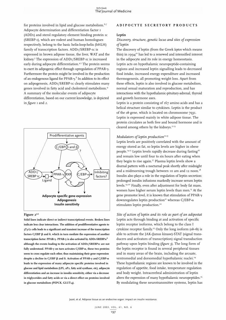
A summary of the molecular events of adipocyte
differentiation, based on our current knowledge, is depicted
in figure 1 and 2.
A D I P O C Y T E S E C R E T O R Y P R O D U C T S
LeptinDiscovery, structure, genetic locus and sites of expressionof leptinThe discovery of leptin (from the Greek leptos which means
thin) in 199410 has led to a renewed and intensified interest
in the adipocyte and its role in energy homeostasis.
Leptin acts on hypothalamic neuropeptide-containing
regions and increased leptin signalling leads to decreased
food intake, increased energy expenditure and increased
thermogenesis, all promoting weight loss. Apart from
these effects, leptin is also involved in glucose metabolism,
normal sexual maturation and reproduction, and has
interactions with the hypothalamic-pituitary-adrenal, thyroid
and growth hormone axes.
Leptin is a protein consisting of 167 amino acids and has a
helical structure similar to cytokines. Leptin is the product
of the ob gene, which is located on chromosome 7q31.
Leptin is expressed mainly in white adipose tissue. The
protein circulates as both free and bound hormone and is
cleared among others by the kidneys.11-13
Modulators of leptin production12,13
Leptin levels are positively correlated with the amount of
energy stored as fat, so leptin levels are higher in obese
people.14,15 Leptin levels rapidly decrease during fasting16
and remain low until four to six hours after eating when
they begin to rise again.17 Plasma leptin levels show a
diurnal pattern with a nocturnal peak shortly after midnight
and a midmorning trough between 10 am and 12 noon.18
Insulin also plays a role in the regulation of leptin secretion:
prolonged insulin infusions markedly increase serum leptin
levels.19,20 Finally, even after adjustment for body fat mass,
women have higher serum leptin levels than men.15 At the
gene promotor level, it is known that stimulation of PPAR-�
downregulates leptin production21 whereas C/EBP-�
stimulates leptin production.22
Site of action of leptin and its role as part of an adipostatLeptin acts through binding at and activation of specific
leptin receptor isoforms, which belong to the class I
cytokine receptor family.23 Only the long isoform (ob-rb) is
able to activate the JAK-(Janus kinase)-STAT (signal trans-
ducers and activators of transcription) signal transduction
pathway upon leptin binding (figure 3). The long form of
the leptin receptor is found in several peripheral tissues
and in many areas of the brain, including the arcuate,
ventromedial and dorsomedial hypothalamic nuclei.24
These hypothalamic regions are known to be involved in the
regulation of appetite, food intake, temperature regulation
and body weight. Intracerebral administration of leptin
alters the expression of many hypothalamic neuropeptides.25
By modulating these neurotransmitter systems, leptin has
Prodifferentiative agents
Adipocyte specific gene expressionAdipogenesis
Insulin sensitivity
Inhibitingfactors?
C/EBP � and �?
ADD1/SREBP 1c PPAR-� C/EBP-�RXR
Ligand
Figure 24,5
Solid lines indicate direct or indirect transcriptional events. Broken lines
indicate less clear interactions. The addition of prodifferentiative agents to
3T3-L1 cells leads to a significant and transient increase of the transcription
factors C/EBP � and �, which in turn mediate the expression of another
transcription factor: PPAR-�. PPAR-� is also activated by ADD1-SREBP1c8
although the events leading to the activation of ADD1/SREBP1c are not
fully understood. PPAR-� on turn activates C/EBP-�, these two proteins
seem to cross regulate each other, thus maintaining their gene expression
despite a decline in C/EBP � and �. Activation of PPAR-� and C/EBP-�
leads to the expression of many adipocyte specific proteins involved in
glucose and lipid metabolism (LPL, aP2, fatty acid synthase, etc), adipocyte
differentiation and an increase in insulin sensitivity, either via a decrease
in triglycerides and fatty acids or via a direct effect on proteins involved
in glucose metabolism (PEPCK, GLUT-4).

J U N E 2 0 0 3 , V O L . 6 1 , N O . 6
Jazet, et al. Adipose tissue as an endocrine organ: impact on insulin resistance.
198
a major role in maintaining energy balance and thus serves
as part of an adipostat. During fasting, serum insulin levels
fall and the uptake of glucose and lipids by the adipocyte
diminishes. This leads to a decreased expression of the
ob-gene, which is responsible for leptin formation and
hence the plasma leptin concentration falls. Reduced leptin
signalling leads to an increased expression of neuropeptide
Y (NPY) and agouti-related protein (AgRP) in the arcuate
nucleus of the hypothalamus. NPY and AgRP promote
body weight gain by stimulating food intake and decreasing
energy expenditure. Another neuronal cell type coproduces
cocaine-amphetamine related transcript (CART) and
pro-opiomelanocortin (POMC), from which �-melanocyte
stimulating hormone (�-MSH) is cleaved. CART and
�-MSH are both anorexigens and reduced leptin signalling
inhibits the synthesis of CART and POMC (figure 3).26,27
Finally, corticotropin-releasing hormone (CRH), which is
also produced in the hypothalamus, might be important
in mediating the effects of leptin, presumably via activation
of sympathetic outflow to BAT, WAT, liver and muscle.
Intracerebral injection of CRH stimulates thermogenesis
and oxygen consumption and reduces food intake and
body weight. CRH mRNA levels are increased by the
intraventricular administration of leptin.28
Role of leptin in obesityThe initial conception of leptin as an anti-obesity hormone,
whose primary role was to increase the metabolic rate and
decrease food intake and appetite through action in the
brain, was based on the following observations: 1) leptin
deficient ob/ob mice and leptin receptor deficient db/db
mice exert marked hyperphagia, decreased energy
expenditure, morbid obesity and insulin resistance;29,30 2)
administration of intravenous or intracerebroventricular
leptin decreases body weight and fat mass through
inhibition of food intake and increased energy expenditure
in ob/ob but not in db/db mice;31 3) there is a threshold
level of serum leptin (25-30 ng/ml) above which
increases in serum levels are not translated into propor-
tional increases in cerebrospinal or brain leptin levels, i.e.
the transport system must be saturable;32 4) the discovery
of leptin receptors in the hypothalamus, the region
Arcuate nucleus Paraventricular nucleus
NPYAgRP
POMC (�-MSH)CART
CRHMCH
Sympathetic nervous system
Food intake
ACTH Adrenal CortisolTSH Thyroid T4/T3
LH/FSH Gonads Sex steroidsGH Target
organsGrowth
Starvation Hypophysis
Leptin Insulin
Fat Pancreas
+
– –
–
Figure 3Starvation leads to a decrease in serum insulin levels and a decreased expression of the ob-gene leading to a decrease in serum leptin levels. This
subsequently leads to an increased expression of neuropeptide-Y and agouti-related protein in the hypothalamus and a decrease in POMC and CART
in the hypothalamus (see list of abbreviations for explanation). These hormones are involved in food intake and energy expenditure, leading to an increase
in food intake and a decrease in energy expenditure. Furthermore, the hypothalamic hormones have either a direct or an indirect (via CRH and MCH)
effect on various hormones secreted by the pituitary. Thus leptin has multiple effects, not only on food intake and energy metabolism but also on
the hypothalamic-pituitary-adrenal axis, thyroid function and sex steroids. (Dark grey is inhibition, light grey is stimulation.)

Jazet, et al. Adipose tissue as an endocrine organ: impact on insulin resistance.
J U N E 2 0 0 3 , V O L . 6 1 , N O . 6
199
involved in regulation of food intake and energy balance.27
However, in most obese humans the gene encoding leptin
is normal: up till now only two families with a mutation
in the leptin gene have been identified.33,34 In contrast,
most obese humans have increased serum leptin levels,14,15
indicating that obesity is a leptin-resistant state. Such a
resistance could theoretically occur at several levels of the
leptin signal transduction pathway, but this has not been
resolved yet.
Leptin and insulin resistance.Since obesity is associated with insulin resistance, it is
interesting to look at the role of leptin in the development
of insulin resistance and diabetes. A strong correlation
between serum leptin and insulin levels, independent of
body fatness, has been demonstrated in human studies.35,36
Hyperinsulinaemia induced by clamp techniques increases
serum leptin levels, though not acutely.19 Serum leptin levels
are increased by insulin therapy as well, both in type 1 and
type 2 diabetic patients.36,37 Vice versa, a fair amount of
evidence points to the fact that leptin has insulin- and
glucose-lowering properties, although some studies find just
the opposite. An extensive review on the association between
leptin and insulin resistance has recently been published.38
In both normal rodents39 and rodents with obesity and
insulin resistance40-42 leptin therapy improves hyper-
insulinaemia and hyperglycaemia. These effects are already
apparent before weight loss occurs and are not due to
energy restriction as was shown in pair-fed control studies.41,43
Most obese humans have increased serum leptin levels14,15
and thus far the overall effect of leptin therapy on weight
loss and metabolic parameters has been modest.44 It is
likely that very high plasma levels of the hormone are
needed to overcome the leptin-resistant state. A final point
pointing to an antidiabetogenic effect of leptin is that both
in lipodystrophic rodents45 and humans (who have an
extreme deficit of subcutaneous adipose tissue),46 a condition
associated with severe insulin resistance with hyperglycaemia,
hyperinsulinaemia and hypertriglyceridemia, leptin therapy
corrects all these metabolic abnormalities, independent
of the accompanying reduction in food intake.
Hypotheses with regard to the glucose and insulin-loweringeffect of leptinAs mentioned before, leptin seems to have an insulin-
sensitising effect on the whole body level but conflicting
results were reported when individual tissues were
examined. Most in vitro experiments suggest a diabetogenic
effect of leptin.38 Beside the differences between animals
and humans, sources of leptin and time of exposure to
this hormone might also play a causative role in the
differences found. Furthermore, the fact that leptin exerts
a glucose- and insulin-lowering effect and improves insulin
sensitivity in vivo, suggests involvement of centrally acting
mechanisms. This concept is further supported by the
observation that leptin fails to reverse insulin resistance
and lipid accumulation in mice with ventromedial hypo-
thalamic lesions.47 The peripheral mechanism by which
leptin exerts its glucose- and insulin-lowering effect might
be via promoting fatty acid oxidation and triglyceride
synthesis. Indeed, leptin administration activates 5’-AMP-
activated protein kinase (AMPK) in skeletal muscle, leading
to the inhibition of acetyl coenzyme A carboxylase and
subsequently stimulation of fatty acid oxidation. The
resulting intramyocellular lipid depletion will enhance
insulin sensitivity.48 Apart from insulin-sensitising effects,
leptin diminishes hyperinsulinaemia, probably via inhibition
of insulin secretion. Functional leptin receptors have been
demonstrated on insulin-secreting �-cells of the pancreas.49
Leptin inhibits glucose-stimulated insulin secretion both
in vitro50 and in vivo.51 The mechanism involved is activation
of the ATP-sensitive potassium channels in the �-cell.
Finally, leptin shares intracellular pathways with insulin,
both in peripheral tissues and in the central nervous system52
Many effects of both insulin and leptin are mediated via
activation of PI-3 (phospahtidylinositol-3-phosphate)
kinase, so a degree of crosstalk between insulin and leptin
may exist at the level of PI-3 kinase. Effects of leptin on
insulin signalling have been studied and support an
inhibitory effect of leptin on insulin signalling at the level
of tyrosine phosphorylation of IRS-1 (insulin receptor
substrate 1) and PI3-kinase binding to IRS-1.38 The effect
of hyperinsulinaemia on intracellular leptin signalling has
rarely been addressed but in one study supraphysiological
concentrations of insulin completely cancelled out the
leptin-induced insulin response.53
ConclusionThus, leptin is an adipocyte secretory product that is not
only involved in food intake and energy metabolism but
clearly also has a role in glucose metabolism. Since plasma
leptin levels are positively correlated with BMI, obesity
seems to reflect a leptin-resistant state. Resistance for the
action of leptin could promote obesity via decreased energy
expenditure and a failure to diminish food intake.
Furthermore, since leptin has a glucose- and insulin-lowering
effect on the whole body level in vivo, resistance for this
effect could induce insulin resistance. One explanation
for the insulin resistance seen in obesity might be that
the high leptin levels interfere with insulin signalling.
Another possibility is that there is a diminished activation
of AMPK in myocytes due to impaired leptin signalling.
The resultant decrease in fatty acid oxidation will lead to
an increase in intramyocellular lipids and thus to insulin
resistance. Finally, both peripheral and central leptin
resistance must be involved in insulin-resistant states
since leptin treatment fails to correct insulin resistance in
mice with ventromedial hypothalamic lesions.

J U N E 2 0 0 3 , V O L . 6 1 , N O . 6
Jazet, et al. Adipose tissue as an endocrine organ: impact on insulin resistance.
200
ResistinDiscovery, structure, genetic locus, sites and modulatorsof expression of resistinResistin is a unique protein with cysteine-rich residues,54
which belongs to a class of tissue-specific secreted proteins
termed the RELM (resistin-like molecule)/FIZZ (found in
inflammatory zone) family. Resistin/FIZZ 3 is specifically
expressed and secreted by adipocytes. The gene encoding
resistin in mice has been named Retn. The regulation of
resistin gene expression is controversial, see table 2.
Resistin in obesity and insulin resistanceThe initial report by Steppan et al.54 suggested that resistin
might constitute the link between obesity and insulin
resistance. Resistin serum levels were increased in obese
mice and resistin gene expression was induced during
adipocyte differentiation. In addition, administration of
resistin impaired glucose tolerance and insulin action in
wild-type mice and in vitro in 3T3-L1 adipocytes whereas
antiresistin antibody improved insulin sensitivity. The
fact that thiazolidinediones suppressed resistin secretion
led to the hypothesis that these insulin sensitisers exert
their effect via downregulation of resistin gene expression.
An increase in adipocyte gene expression during 3T3-L1
adipocyte differentiation61 and after the induction of
high-fat-diet induced obesity57 was found in two other
studies. Several other investigators, however, found a
decreased resistin gene expression in WAT in different
models of rodent obesity and insulin resistance,59,64,65 and
resistin did not seem to be involved in the aetiology of
insulin resistance in Fischer 344 rats, a good model for
the metabolic syndrome in humans.66 Studies in humans
are even more controversial. One study could not detect
any resistin mRNA in human fat cells at all in subjects
with varying degrees of insulin resistance and obesity.67
Another investigator found increased resistin mRNA in
adipose tissue of obese humans, compared with lean
controls, but decreased mRNA in freshly isolated human
adipocytes.60 In addition resistin mRNA was undetectable
in a severely insulin resistant subject. Janke et al. found
an increased resistin gene expression in cultured human
preadipocytes compared with mature adipocytes but again
no relationship between resistin gene expression and
either insulin resistance or body weight could be detected.68
Although the higher resistin mRNA levels found in
abdominal fat tissue compared with thigh could explain
the increased metabolic abnormalities in abdominal obesity,
the fact that resistin mRNA expression is very similar in
subcutaneous and omental adipose tissue suggests that it
is unlikely that resistin is the link between (visceral)
adiposity and insulin resistance.69
ConclusionThe conclusion must be that many questions still have to
be resolved. Conflicting results have been reported with
regard to the factors regulating resistin gene expression
(table 2). This is probably due to the difference between
3T3-L1 cell lines and in vivo models. Furthermore, the
observed relation between resistin mRNA, serum resistin
levels and insulin resistance in rodents cannot readily be
extrapolated to humans. Murine resistin is only about
56% identical to human resistin at the amino acid level.
Even in mouse models it is still unclear whether resistin
plays a causal role in insulin resistance. Experiments in
resistin knockout mice and in transgenic mice (which
overexpress resistin) will be needed to solve this problem,
but even then the relevance of resistin to human diabetes
remains unclear, especially because some groups have
found only minimal expression of the hormone in human
fat.69 Furthermore it would be interesting to know how
resistin exerts its presumed insulin-antagonising effects
and what its target organs are. For that purpose the
resistin receptor would have to be found and downstream
signalling pathways have to be unravelled.
AdiponectinDiscovery, sites of expression and stimuli leading toadiponectin productionAdiponectin is a recently identified70,71 adipocyte-specific
secretory protein of about 30 kD that appears to be involved
Table 2Regulators of resistin expression
FACTOR DECREASING RESISTIN INCREASING RESISTIN NO EFFECT
Thiazolidinediones [54-56,58] [59] [60]
Insulin [56,58] [59,61]
Glucose [58]
Dexamethasone [56,58]
�-adrenergic agonists [62] [56]
TNF-� [58,63]
Epinephrine [58]
Factors that have been reported to increase or decrease resistin expression with their references.

Jazet, et al. Adipose tissue as an endocrine organ: impact on insulin resistance.
J U N E 2 0 0 3 , V O L . 6 1 , N O . 6
201
in the regulation of energy balance and insulin action and
also seems to have anti-inflammatory and anti-atherogenic
properties. Adiponectin is the product of the adipose tissue
most abundant gene transcript-1 (apM1), which is exclusively
expressed in WAT and is located on chromosome 3q27.
Adiponectin is specifically expressed during adipocyte differ-
entiation and is not detectable in fibroblasts. The expression
of adiponectin is stimulated by insulin,70,72 IGF-172 and the
TZDs. Corticosteroids,72 TNF-�74 and �-adrenergic stimula-
tion75 inhibit adiponectin gene expression in 3T3-L1 adipocytes.
Serum and mRNA levels of adiponectin in obesity andinsulin resistanceSerum adiponectin levels are decreased in humans with
obesity76,77 and type 2 diabetes76,78 as well as in obese and
insulin-resistant rodents.79 In addition, adiponectin gene
transcription is decreased in adipocytes from obese71 and
diabetic80 humans and rodents.71,79 Plasma adiponectin
concentrations increase after weight reduction in obese
diabetic and nondiabetic patients.78 The degree of plasma
hypoadiponectinaemia was more closely related to the
degree of hyperinsulinaemia and insulin resistance than
to the degree of adiposity.76 Low plasma adiponectin
concentrations predicted a decrease in insulin sensitivity81
and an increase of type 2 diabetes82 in Pima Indians as
well as in a German population.83 In nondiabetics plasma
adiponectin levels are also positively correlated with insulin
sensitivity.84 A recent study confirmed that the relation
between low adiponectin levels and insulin resistance is
not determined by obesity since low plasma adiponectin
levels at baseline did not predict future obesity.85
Finally, the fact that the insulin-sensitising thiazolidinediones
strongly increase plasma adiponectin73,86 further supports
a role of adiponectin in insulin sensitivity.
Theory with regard to the possible mechanism of action ofadiponectinAdministration of recombinant adiponectin to normal,
obese and diabetic rodents led to acute normalisation of
serum glucose levels.79,87,88 Both decreased gluconeogenesis
of the liver87 and an increased fatty acid oxidation in
muscle79,88 have been proposed as underlying mechanisms.
Recently, Yamauchi underscored his previous hypothesis.89
Administration of adiponectin led to an increase in glucose
utilisation and fatty acid oxidation in cultured myocytes
and in soleus muscle of mice in vivo. In hepatocytes
AMPK was activated as well, leading to a reduction in
gluconeogenesis.
In addition, it has been shown that administering only
the globular domain of adiponectin instead of full-length
adiponectin is much more effective in improving insulin
sensitivity because this fragment augments insulin-induced
phosphorylation of insulin receptor substrate 1 (IRS-1) and
protein kinase B in skeletal muscle.79 Thus, adiponectin
might exert its insulin-sensitising effect via the following
mechanisms: 1) increased fatty acid oxidation leading to a
lower muscle triglyceride content and lower plasma
concentrations of free fatty acids which will both improve
insulin signalling; 2) direct improvement of insulin
signalling; 3) inhibition of gluconeogenesis, partly via
reduced substrate delivery and partly via reduction of
molecules involved in gluconeogenesis by activation of
AMPK.
Disappointingly, no positive correlation between plasma
adiponectin levels and 24-hour respiratory quotient (RQ)
measurement (pointing to an increase in carbohydrate
metabolism) could be demonstrated in healthy nondiabetic
Pima Indians.90 This does not rule out, however, that
administration of adiponectin to subjects with low levels
of this hormone will increase RQ and energy expenditure.
The acylation-stimulating protein (ASP) pathwayASP production and site of actionAcylation-stimulating protein (ASP) is a 76 amino acid
protein identical to C3adesArg, a cleavage product of
complement factor 3 (C3) formed via interaction of C3
with factor B and adipsin. C3, factor B and adipsin are all
components of the alternative complement pathway and
are produced by the adipocyte in a differentiation dependent
manner.91
The major site of action of ASP appears to be on the
adipocytes themselves, which have a specific saturable
receptor for ASP.92 In human adipocytes there are
differentiation and site-specific differences in ASP binding
which are proportional to the ASP response: differentiated
adipocytes bind more ASP and have a greater response to
ASP than undifferentiated adipocytes.93 Furthermore,
subcutaneous adipose tissue has greater affinity and greater
specific binding to ASP than undifferentiated adipocytes.94
ASP promotes triglyceride storageASP promotes triglyceride storage in adipocytes via three
mechanisms. First, ASP increases fatty acid esterification
in adipocytes by increasing the activity of diacylglycerol
acyltransferase, which is the final enzyme involved in trigly-
ceride synthesis.91 Second, ASP stimulates glucose transport
in human and murine adipocytes and preadipocytes.93
This effect on glucose transport is accomplished via
translocation of cell-specific glucose transporters to the cell
membrane. Third, ASP decreases lipolysis via inhibition
of hormone-sensitive lipase.95 The effects of ASP are
independent of and additional to the action of insulin.95
Stimuli leading to ASP productionIn vitro studies in cultured adipocytes indicate that insulin96
and even more so chylomicrons96,97 increase ASP production.
In vivo, plasma ASP concentrations seem to show little
change after an oral fat load.98 There is, however, post-

J U N E 2 0 0 3 , V O L . 6 1 , N O . 6
Jazet, et al. Adipose tissue as an endocrine organ: impact on insulin resistance.
202
prandially an increased venoarterial gradient of ASP across
a subcutaneous abdominal tissue bed with a maximum after
3 to 5 hours, indicating increased adipose tissue ASP produc-
tion.98 This increase in ASP postprandially is substantially
later than the increase in insulin but shows a close temporal
relationship with maximal plasma triacylglycerol clearance.98
Plasma ASP levels in obesityAn excellent review on the physiology of ASP in humans
and rodents has recently been published.99 Plasma levels
of ASP are 225-fold lower (weighted average 28.3 nM) than
its precursor C3. Studies measuring plasma ASP levels
should therefore be interpreted with caution while it might
very well be that ASP acts as a paracrine hormone.99
Plasma ASP levels are increased in obese humans100-103
and are reduced after fasting or weight loss.101,103 ASP has
also been shown to be significantly increased in type 2
diabetes102,104 but since type 2 diabetes is often associated
with obesity this might be a confounding factor. On the
other hand, plasma ASP levels were inversely correlated to
glucose disposal during a euglycaemic clamp in humans.102
Adipocytes from obese humans are as responsive to ASP
as adipocytes from lean people.105 Thus the increased
levels of ASP in human obesity in the face of a similar
responsiveness to ASP compared with lean subjects, may
promote energy storage, leading to adiposity.
Relation between ASP enhanced triglyceride clearanceand insulin resistanceASP production is increased in obese mice. Intraperitoneal
(i.p.) administration of ASP to normal mice resulted in
accelerated postprandial triglyceride (TG) and nonesterified
fatty acid (NEFA) clearance after an oral fat load.106 In
addition, plasma glucose levels returned faster to basal
levels. C3 knockout mice (KO), which are unable to produce
ASP, showed delayed plasma triglyceride clearance after
an oral fat load in the absence of any change in fasting
plasma TG levels. Administration of exogenous ASP
enhanced plasma TG clearance.107 Remarkably these C3 KO
mice were more insulin sensitive, had a reduced fat mass
and yet an increased food intake. It was later shown that
the hyperphagia/leanness was balanced by an increase in
energy expenditure.108
ConclusionIn summary, ASP promotes storage of energy as fat.
Decreased ASP production decreases lipid storage and
induces an obesity-resistant state and improved insulin
sensitivity. Plasma ASP levels are increased in obese
humans; whether this is the effect or cause of the increased
adipose tissue mass remains to be elucidated. Post or
propter, increased ASP levels together with a continuing
responsiveness of the ASP receptor will lead to further
triglyceride storage. Although enhanced fatty acid trapping
will decrease free fatty acid levels and hence diminish
hepatic gluconeogenesis, increased ASP functioning in
skeletal muscle will lead to an increase in skeletal muscle
triglyceride storage leading to insulin resistance.
Tumour necrosis factor-� (TNF-�)Structure of TNF-�, sites of production and receptorinteraction109
TNF-� is a cytokine produced mainly by activated
macrophages in response to invasive stimuli, but also by
nonimmune cells such as muscle and adipose tissue.
Furthermore, TNF-� has a variety of biological effects in
various tissues and cell types, and can thus be considered
a multifunctional cytokine.109
TNF-� is produced as a 26-kD membrane-bound precursor
that is proteolytically cleaved to a 17-kD soluble form.109
The cytokine interacts with two membrane-bound receptors,
a 60-kD and an 80-kD subtype also called type I and
type II receptor (TNFR-1 and TNFR-2). These receptors
have different cellular and tissue distribution patterns
and can bind other cytokines as well. TNF-� has a higher
affinity for TNFR-1 than for TNFR-2.109 Due to the high
affinity for its receptor TNF-� can act either as an autocrine
or paracrine cytokine at low concentrations or as an
endocrine cytokine at high concentrations.
In addition to the membrane-bound receptors, soluble
forms of the two receptors exist for which TNF-� has an
even higher affinity. When TNF-� is bound to these soluble
receptors no interaction can take place with the membrane-
bound forms and thus TNF-� action is inhibited.
Therefore, the physiological role of the soluble receptors
may be to regulate TNF-� action.
Modulators of TNF-� productionIn macrophages and monocytes, the expression and
production of TNF-� is stimulated by endotoxins such as
lipopolysaccharide (LPS). LPS resulted in a fivefold
stimulation of TNF-� in human adipose tissue and isolated
adipocytes in vitro, the latter indicating that it is unlikely
that the response is entirely due to macrophages and
monocytes in the stromal vascular fraction of adipose tissue.
Insulin and glucocorticoids did not have a significant
effect on TNF-� release from human adipose tissue or
isolated adipocytes in vitro.110 Thiazolidinediones reduced
adipocyte TNF-� release in obese rodents111 but no effect
was seen in human adipose tissue in vitro.110 Since high-fat
diets resulted in a significant increase in TNF-� mRNA
and protein in epidydimal and retroperitoneal fat pads in
rats, free fatty acids and/or triglycerides may play an
important role as inducers of TNF-� expression.112
Effect of TNF-� on glucose and lipid metabolismFirstly, TNF-� inhibits preadipocyte differentiation by
downregulating the expression of two important adipocyte

Jazet, et al. Adipose tissue as an endocrine organ: impact on insulin resistance.
J U N E 2 0 0 3 , V O L . 6 1 , N O . 6
203
transcription factors: PPAR-� and CEBP/�.113 Secondly,
TNF-� reduces the expression of GLUT-4, glycogen synthase
and fatty acid synthase, which are essential for insulin-
mediated glucose uptake and the subsequent conversion
of glucose to glycogen or fatty acids. Furthermore, genes
involved in the uptake of free fatty acids and the subsequent
conversion to triglycerides, such as lipoprotein lipase,
long-chain fatty acyl-CoA synthethase and diacylglycerol
acyltransferase, were also downregulated by TNF-�.113 The
above-mentioned changes in gene expression lead to a
diminished insulin-stimulated glucose uptake and an
altered lipid metabolism which can, via accumulation of
triglycerides in various organ systems, eventually lead to
insulin resistance of the muscle and liver. In addition,
insulin resistance can be induced via a direct toxic effect
of TNF-� on intracellular insulin signalling.114 TNF-�
reduces the insulin-stimulated autophosphorylation of
the insulin receptor in a variety of cell types. It does so by
phosphorylation of serine residues at the insulin receptor
substrate-1 (IRS-1); this modified IRS-1 subsequently
interferes with the insulin signalling capacity of the
insulin receptor.114
Relation between TNF-�, obesity and insulin resistanceA positive relationship between obesity, insulin resistance
and adipose tissue mRNA levels of TNF-� has clearly
been established in rodent models.115 Furthermore, mice
with no functional copy of the TNF-� gene (TNF-� -/-)
although developing marked obesity on a high-fat, high-
energy diet, remained highly insulin sensitive compared
with their control litter mates (TNF-� +/+).116
In contrast to rodents, the role of TNF-� in the induction
of insulin resistance in humans is less clear. Although
there seems to be a positive relationship between obesity
and TNF-� mRNA and protein levels in adipose tissue in
humans in vitro,117-119 TNF-� is expressed at much lower
levels in humans compared with rodents. In addition, no
difference in TNF-� concentration was found in a vein
draining subcutaneous adipose tissue compared with a
peripheral vein, suggesting no or very low TNF-� production
in vivo.120 Furthermore, circulating TNF-� concentrations in
obese diabetic and nondiabetic patients are not substantially
elevated.118,121 With regard to a direct relationship between
TNF-� and insulin sensitivity in vivo, two studies found a
strong and positive correlation between adipose tissue TNF-�
mRNA levels and hyperinsulinaemia.117,118 When the relation
between adipose tissue TNF-� secretion and insulin-
stimulated glucose transport was examined, a strong inverse
relationship was found that was independent of fat cell
volume, age and BMI.122
However, other studies121,123 showed no significant relation-
ship between adipose tissue mRNA for TNF-� and insulin
sensitivity. Furthermore, treatment of insulin-resistant
subjects with anti-TNF-� antibodies did not improve
insulin sensitivity.124 All these results implicate that TNF-�
might have an effect on insulin resistance but that it must
be a local factor. Interestingly, TNF-� is also produced by
muscle, and muscle TNF-� production is increased in
obesity.125 Since adipose tissue dispersed within muscle is
correlated with insulin resistance, the effect of fat cell
secretory products on insulin signalling in skeletal muscle
cells was recently studied in a model in which muscle
cells were co-cultured with adipocytes. A disturbance of
insulin signalling was found, but TNF-� did not seem to
be involved.126
ConclusionIn conclusion, TNF-� is a multifunctional cytokine produced
by adipocytes in proportion to the percentage body fat.
TNF-� has a variety of metabolic effects, including
increased lipolysis, decreased lipogenesis and decreased
insulin-stimulated glucose transport, contributing to
insulin resistance. These effects are induced by modulation
of genes involved in glucose and lipid metabolism.
Furthermore, TNF-� directly interferes with the early
steps of insulin signalling. However, the role of TNF-� in
obesity-induced insulin resistance in humans is not quite
clear yet, as might be obvious from the contradicting results
mentioned in the previous paragraph. The low plasma
levels of TNF-� in humans indicate that the hormone
most likely acts in a paracrine and or autocrine manner.
This might be the reason why treatment with anti-TNF-�
did not improve insulin sensitivity in humans in vivo.
Interleukin-6 (IL-6)Structure, genetic locus and site of production of IL-6IL-6 is a circulating, multifunctional cytokine that is
produced by a variety of cell types including fibroblasts,
endothelial cells, monocytes/macrophages, T-cell lines,
various tumour cell lines and adipocytes. The protein has
a molecular mass of 21 to 28 kD, depending on the cellular
source and preparation. The gene encoding IL-6 is localised
on chromosome 7p21 in humans.127
Although human adipocytes produce IL-6, adipocytes
accounted for only 10% of total adipose tissue IL-6
production when IL-6 production by isolated adipocytes
prepared from omental and subcutaneous fat depots was
examined.128 This means that cells in the stromal vascular
fraction of adipose tissue have a major contribution in
adipose tissue IL-6 release. The concentrations of IL-6 in
adipose tissue are up to 75 ng/ml, which is well within the
range to elicit biological effects.129 Furthermore, plasma
levels of IL-6 are markedly elevated in obesity and up to
30% of plasma levels could be derived from adipocytes.130
Modulators of IL-6 productionThe stimuli leading to IL-6 production differ with the cell
type; here only IL-6 production by adipocytes will be

J U N E 2 0 0 3 , V O L . 6 1 , N O . 6
Jazet, et al. Adipose tissue as an endocrine organ: impact on insulin resistance.
204
discussed. Both in rodent and human adipocytes, IL-6
production is stimulated by catecholamines and inhibited
by glucocorticoids, whereas insulin has no effect what-
soever.128,131,132 Finally, another stimulator of IL-6 release is
TNF-�, which has been reported to produce a 30-fold113
increase in IL-6 production in 3T3-L1 adipocytes.
Interestingly, IL-6 in turn inhibits the release of TNF-�!
IL-6 acts via receptor interactionIL-6 acts through binding at and activation of a specific
receptor, belonging to the class I cytokine receptors, which
act through JAK-STAT signalling (see figure 4 where leptin
signalling is explained).133 The IL-6 receptor consists of
two membrane glycoproteins, a 80-kD ligand binding
component and a 130-kD signal-transducing component
(gp130). The 80-kD component binds IL-6 with low
affinity; this complex subsequently binds with high affinity
to gp130 after which signal transduction can take place.127
Soluble forms of the IL-6 receptor have been found but
neither their functional significance nor the regulation of
their production is understood.
Effects of IL-6 on glucose and lipid metabolismIL-6 has pleiotropic effects on various cell types. Here we
will only focus on its role in glucose and lipid metabolism.
Infusion of rhIL-6 to humans increased whole body glucose
disposal and glucose oxidation but increased hepatic glucose
production134 and fasting blood glucose concentration in a
dose-dependent manner.135 With regard to lipid metabolism,
IL-6 decreases adipose tissue lipoprotein lipase (LPL)
activity129 and has been implicated in the fat depletion
taking place during wasting disorders, such as cancer,
perhaps via an increase in plasma norepinephrine, cortisol,
resting energy expenditure and fatty acid oxidation as was
assessed in eight renal cancer patients.134 In rats, IL-6
increased hepatic triglyceride secretion partly because the
increase of adipose tissue lipolysis resulted in an increased
delivery of free fatty acids to the liver.136 This increased
release of FFAs following rhIL-6 infusion was observed
in humans as well.134
IL-6 in obesity and insulin resistanceIn both mice132 and humans, IL-6 mRNA in adipose
tissue137,138 but even more so plasma levels of IL-6 are
positively correlated with BMI.132,137,138 Weight loss is
associated with a reduction in serum and IL-6 mRNA
levels. After one year of a multidisciplinary programme of
weight reduction, obese women lost at least 10% of their
original weight and this was associated with a reduction of
basal serum IL-6 levels from 3.18 to 1.7 pg/ml (p<0.01).139
In another study, both IL-6 mRNA in adipose tissue and
IL-6 serum levels were reduced with weight loss after
three weeks of a very low calorie diet in obese women.138
In this study, insulin sensitivity as assessed by the fasting
insulin resistance index (FIRI= fasting glucose x fasting
insulin/25) improved as well. The reduction in IL-6 levels
could play a role in this improvement, since several studies
found a significant correlation between circulating IL-6
levels and insulin sensitivity measured by either an intra-
venous glucose tolerance test137 or the fasting insulin
resistance index.138 Recently this correlation between
circulating IL-6 and insulin sensitivity was confirmed
using the gold standard for insulin sensitivity: the hyper-
insulinaemic euglycaemic clamp.140 In addition, a high
correlation between adipose tissue IL-6 content and
insulin sensitivity was found, both in vivo and in vitro.
Furthermore, for the first time IL-6 receptors were
demonstrated in 60% of the subcutaneous adipocytes
suggesting that IL-6 can alter adipocyte metabolism via
autocrine or paracrine mechanisms and have a local
STAT
STAT
STAT
STAT
STATSTAT
Leptin
LeptinCellmembrane
Cytoplasm
DNA with STAT-bindingregion
Nucleus
JAK JAKJAKJAKP
Y
P
PP
Y
Y
Y PP Y
Y
Figure 4The leptin receptor is a transmembrane receptor belonging to the class I
cytokine receptors. The receptor consists of two parts. The intracellular
domain is associated with the Janus kinase, a tyrosine kinase. Binding
of leptin to the receptor results in the fusion of the two receptor parts,
which results in trans-phosphorylation of the JAK molecules, which
subsequently phosphorylate the terminus of the leptin receptor. The
phosphorylated receptor then forms a docking site for a variety of Src
homology 2 (SH-2) domain containing proteins, including a novel family
of cytoplasmatic transcription factors termed STATs (signal transducers
and activators of transcription). STATs are then phosphorylated on a single
tyrosine residue by JAKs, after which the STATs dimerise, migrate into
the nucleus and regulate gene transcription.

Jazet, et al. Adipose tissue as an endocrine organ: impact on insulin resistance.
J U N E 2 0 0 3 , V O L . 6 1 , N O . 6
205
influence on insulin sensitivity.140 Further support for a
relationship between IL-6 and insulin sensitivity comes
from a genetic study. It appeared that subjects with an IL-6
gene polymorphism had lower IL-6 levels, a lower area
under the glucose curve after an oral glucose tolerance test,
lower glycosylated haemoglobin (HbA1C), lower fasting
insulin levels and an increased insulin sensitivity index
compared with carriers of the normal IL-6 allele, despite
similar age and BMI.141 Finally, basal serum IL-6 levels
are higher in type 2 diabetic patients.142
In contradiction with the above-mentioned positive
correlation of IL-6 with BMI and inverse relation with
insulin sensitivity is the observation that a lack of IL-6
also leads to obesity and a disturbed glucose tolerance, at
least in mice.
ConclusionVarious studies show a clear relationship between increased
IL-6 levels and obesity,132,137,138 and between IL-6 levels and
insulin resistance137,138,140 even when corrected for BMI.137
Furthermore, basal plasma IL-6 levels are higher in
patients with type 2 diabetes142 and subjects with an IL-6
gene polymorphism clearly have lower serum IL-6 levels
and this is correlated with improved insulin sensitivity
and postload glucose levels.141 IL-6 does have different
effects on the various end-organ tissues, however, with on
the one hand improved glucose uptake in adipocytes and
whole body glucose disposal, and on the other hand an
increased hepatic glucose output, decreased LPL activity
(leading to decreased triglyceride clearance) and increased
hepatic triglyceride synthesis. How then does IL-6 fit in
the insulin resistance syndrome? Is there a causal effect or
are the increased IL-6 levels found in obesity and insulin
resistance merely a reflection of the pathogenetic state or
the increased adipose tissue mass? Is IL-6 detrimental to
health or does it have a positive role in health. If we start
from the principle that IL-6 production is increased in
obesity and that it is involved in inducing insulin resistance,
what would the mechanisms be by which IL-6 causes
insulin resistance? Firstly, it has to be noted that omental
fat produces threefold more IL-6 than subcutaneous
adipose tissue.128 Because venous drainage of omental
tissue flows directly to the liver and IL-6 is known to
increase hepatic triglyceride secretion134,136 this might
explain the hypertriglyceridaemia associated with visceral
obesity. As mentioned before, increased triglyceride content
of muscle and liver leads to insulin resistance. Secondly,
IL-6 signal transduction is mediated via JAK-STAT
signalling; it is possible that feedback mechanisms
interfering with insulin signalling exist. Thirdly, IL-6 has
opposing effects to those of insulin on hepatic glycogen
metabolism143 and increases hepatic glucose production.135
On the contrary, despite an increase of IL-6 in obesity,
insulin resistance and type 2 diabetes, there is evidence
that IL-6 improves insulin sensitivity: 1) IL-6 increases
glucose uptake in 3T3-L1 adipocytes;144 2) infusion of
rhIL-6 to humans increased whole body glucose disposal
and glucose oxidation;134 3) IL-6 inhibits TNF-� production,
a cytokine with deleterious effects on insulin sensitivity;
and 4) physical exercise, which is related to an improvement
in insulin sensitivity, is coupled to an increased IL-6
secretion.145 It might be that muscle derived IL-6 down-
regulates TNF-�.145 So, in conclusion, it is still not clear
whether IL-6 has a positive or a negative metabolic role
in health. One of the reasons for the contradicting results
might be that there is a difference in the acute and chronic
exposure to IL-6 with regard to health implications.
Furthermore, there might be differences in local and
CNS-acting effects of IL-6. More transgenic mice studies
can help shed light on the role of IL-6 in insulin resistance.
Up until now, it is quite possible that the increased IL-6
levels observed in adiposity and type 2 diabetes are the
cause of an increased production by the enlarged adipose
tissue mass and/or an attempt to overcome either insulin
resistance or another metabolic defect, for example IL-6
resistance.
D I S C U S S I O N
Obesity, defined as a BMI >27, is the consequence of a
chronic imbalance between energy intake and energy
expenditure. This is partly due to the modern society with
excess (‘fast’) food intake and a sedentary lifestyle. The
role that should be ascribed to primary defects in energy
storage caused by adipocyte secretory products or impaired
hypothalamic functioning remains to be elucidated. At
the moment a combination of the two seems the most
likely. It is well known that obesity is associated with
insulin resistance and type 2 diabetes mellitus. An over-
whelming amount of evidence indicates that visceral fat
is associated with glucose intolerance and insulin
resistance,146-151 along with other facets of the metabolic
syndrome such as dyslipidaemia. Therefore, in the past,
the predominant theory used to explain the link between
obesity and insulin resistance was the portal/visceral
hypothesis,152 which states that increased visceral adiposity
leads to an increased free fatty acid flux into the portal
system and inhibition of insulin action via Randle’s
effect.153 However, several investigators have challenged
the singular importance of visceral adiposity in inducing
insulin resistance. They found an independent association
between total fat mass and subcutaneous truncal fat mass
and insulin resistance.154-156 Furthermore, the observations
that 1) triglyceride content within skeletal muscle cells is
increased in obesity157 and type 2 diabetes mellitus157,158
and is a strong predictor of insulin resistance;159 and 2)
lipodystrophy is associated with insulin resistance as

J U N E 2 0 0 3 , V O L . 6 1 , N O . 6
Jazet, et al. Adipose tissue as an endocrine organ: impact on insulin resistance.
206
well160,161 obviated the need to develop new theories to
explain the link between adipose tissue and insulin
resistance.162 A well-accepted theory is that of ectopic fat
storage.162,163 A limitation in the capacity of adipose tissue
to store triglycerides would divert triglycerides to be
deposited in liver cells and skeletal muscle cells.162,163 The
cause of the ectopic fat storage is unclear. It might be due
to impaired fat oxidation,162 since inhibition of fat oxidation
in rodents increased intracellular lipid content and
decreased insulin action.164 Furthermore, a mutation in
the AGPAT2 gene encoding 1-acylglycerol-3-phosphate
O-acyltransferase inhibits triacylglycerol synthesis and
storage in adipocytes but not in hepatocytes, thus leading
to hepatosteatosis, because the latter can accumulate
triacylglycerol via AGPAT-1.165 Another possibility is the
central and/or peripheral action of leptin, since leptin
therapy has been associated with the reversal of insulin
resistance and hepatic steatosis in patients with lipo-
dystrophy46 and also with improvement of intramyocellular
lipid content.163 Finally, a defect in the proliferation and/or
differentiation of adipocytes, whether or not due to alterations
in the expression of transcription factors,166 can lead either
to impaired adipocyte triglyceride storage and/or adipocyte
hypertrophy. This is where the third hypothesis emerges:
the adipocyte as an endocrine organ.162 Adipocytes secrete
a large number of cytokines and hormones that act in a
paracrine, autocrine and endocrine manner on adipocyte and
whole body metabolism. It is plausible that these enlarged
adipocytes are deregulated in their transcriptional setting and
secrete a different pattern of hormones or different amounts
of them compared with small adipocytes. On the other
hand, enlarged adipocytes might merely be a manifestation
of other, yet to be defined, pathogenethic factors.162
In obese humans and rodents there is, besides numerous
other proteins and cytokines that have not been discussed
here, overproduction of leptin,14,15 IL-6,132,137,138 TNF-�,115,117-119
ASP100,101 and resistin;54,60 and a decreased production of
adiponectin (see figure 5).71,77,78,80 Of leptin,23 TNF-�74 and
IL-6127 it is known that they act via receptors on the cell
surface and subsequent intracellular signalling cascades.
As can be seen in figure 5, all three cytokines decrease
food intake and increase energy expenditure and lipolysis
together with a decrease in lipogenesis. These are well-
adaptive mechanisms to prevent further weight gain.
Since all these cytokines are increased in adiposity it is
unlikely that they are the cause of adiposity unless there is
an impairment in cytokine signalling. Interestingly, leptin
and TNF-� have opposing effects with regard to insulin
sensitivity. TNF-� interferes with insulin signalling and
downregulates many genes encoding for proteins involved
in glucose and free fatty acid uptake.113 Leptin can act
through some components of the insulin-signalling cascade
as well.52 The relation between TNF-� and leptin in humans
is not clear. Infusion of TNF-� to patients has been reported
to acutely raise serum leptin levels,167 whereas chronic
exposure of cultured human adipocytes to TNF-� resulted in
a decrease in leptin production.168 If TNF-� increases leptin
production this might be an adaptive mechanism to com-
Figure 5Hyperplasia and hypertrophy of adipocytes as seen in adiposity leads to an increased (light grey arrow) production of leptin, TNF-�, IL-6, resistin,
ASP and many other proteins, and a decreased production (dark grey arrow) of adiponectin. The results of these increases, respectively decrease, are
mentioned below each protein.
+
+
–
+
+
+
+
ResistinContradicting reports, possiblyImprovement of insulin sensitivity
Many others
Leptin Food intake Energy expenditure Lipolysis Lipogenesis Insulin sensitivity
TNF-� Food intake Energy expenditure Lipolysis Lipogenesis Insulin sensitivity GLUT 4 LPL
IL-6 Food intake Energy expenditure Lipolysis Lipogenesis
AdiponectinAdiponectin decreases plasma glucose ->Mechanism? -> Gluconeogenesis FFA oxidationThus increased adiponectin leads to hyperglycaemia and insulin resistance
ASP Triglyceride synthesis via DAG GLUT 4 Lipolysis via HSL

Jazet, et al. Adipose tissue as an endocrine organ: impact on insulin resistance.
J U N E 2 0 0 3 , V O L . 6 1 , N O . 6
207
pensate for the TNF-� induced impaired insulin signalling.
When we take a further look at the mutual coherence of
the adipocyte secretory factors it is striking that both
insulin and TNF-� are, somehow, involved in the regulation
of all of the adipocyte secretory products. Insulin increases
the production of leptin,19,20,36,37 adiponectin70,72 and ASP,96
whereas no effect has been recorded with regard to TNF-�110
and a potentially positive effect on resistin levels.61 TNF-�
downregulates resistin58 and stimulates the production of
leptin,169 adiponectin74 and IL-6.113 The problem is that some
of these factors lead to an improvement of insulin sensitivity
whereas others have just the opposite effect. This makes
it extremely difficult to elucidate which factors are most
important in regulating insulin sensitivity. Furthermore,
the time of exposure to a stimulus seems to be important.
Thus it seems that leptin and insulin are long-term regulators
with regard to food intake and energy expenditure whereas
insulin has a direct effect on glucose uptake and lipolysis.
How do these adipocyte-derived factors mediate their effects?
What they all seem to have in common is a change in the
expression of genes encoding for proteins involved in
glucose and protein metabolism. Transcription of genes
can only take place if they are activated, which always occurs
via some kind of ligand-receptor interaction followed by
an intracellular signal transduction. Cytokine signalling
proceeds in part via the JAK-STAT pathway.170 The actions
of leptin, TNF-� and IL-6 may influence each other via
common signalling steps. Furthermore, it is known that
leptin can signal through some components of the insulin
signalling cascade such as IRS-1 and -2, PI3K and MAPK
and can modify insulin-induced changes in gene expression
in vitro and in vivo.171 TNF-� can interfere with the early
steps of insulin signalling as well.114 So, more and more
evidence exists that the adipocyte secretory cytokines leptin,
IL-6 and TNF-� not only interact with each other but also
with insulin on the level of intracellular signal transduction.
In the case of obesity and hyperinsulinaemia, there is an
increase in hormones and cytokines produced by the adipose
tissue. These hormones subsequently mediate a change
in the expression of genes encoding for proteins involved
in glucose and lipid metabolism. In case of ASP these
changes promote triglyceride uptake. However, in case of
IL-6, TNF-� and adiponectin there is a deleterious effect
on glucose uptake and fatty acid oxidation leading to
insulin resistance. The effect of increased serum resistin
levels remains to be elucidated. Everything seems to come
down to interference with intracellular signal transduction,
not only of insulin but also of the various adipocyte secretory
products, with a subsequent change in the expression of
genes involved in glucose and lipid metabolism leading to
a diminished glucose uptake and fatty acid oxidation. The
latter will, via accumulation of triglycerides in liver cells
and muscle cells, enhance insulin resistance, thus further
impairing glucose uptake.
Concluding remarksIt is now well established that adipose tissue not only has
an important function in the storage and release of
triglycerides but also has an important effect on whole body
metabolism and energy homeostasis via the production of
various hormones and cytokines.
Adipose tissue not only responds to insulin, glucagon, cortisol
and catecholamines but also to cytokines and products that
it produces itself, thereby regulating its own metabolism and
cell size. Some of the products produced by the adipocytes,
such as TNF-� and leptin, are clearly involved in the
induction of insulin resistance. The role of others (resistin,
IL-6) has yet to be defined. Their increase in obesity is at
least a manifestation of the increased adipose tissue mass
itself. Further research is needed to come to a better
understanding of the molecular pathways regulating the
production of these hormones, their individual actions
and target organs, and finally their mutual interaction
and role in insulin resistance. These new insights provide
the basis for the development of improved therapies for
obesity and insulin resistance related diseases as type 2
diabetes and cardiovascular complications.
A C K N O W L E D G E M E N T
The authors would like to acknowledge Professor J.A.
Maassen of the Department of Molecular Cell Biology of
Leiden University Medical Centre, the Netherlands, for
critically reading our manuscript.
R E F E R E N C E S
1. Matthaei S, Stumvoll M, Kellerer M, Haring HU. Pathophysiology and
pharmacological treatment of insulin resistance. Endocr Rev
2000;21(6):585-618.
2. King H, Aubert RE, Herman WH. Global burden of diabetes, 1995-2025:
prevalence, numerical estimates, and projections. Diabetes Care
1998;21(9):1414-31.
3. Bloomgarden ZT. American Diabetes Association Annual Meeting, 1999:
diabetes and obesity. Diabetes Care 2000;23(1):118-24.
4. Gregoire FM, Smas CM, Sul HS. Understanding adipocyte differentiation.
Physiol Rev 1998;78(3):783-809.
5. Rosen ED, Walkey CJ, Puigserver P, Spiegelman BM. Transcriptional
regulation of adipogenesis. Genes Dev 2000;14(11):1293-307.
6. Schoonjans K, Staels B, Auwerx J. The peroxisome proliferator activated
receptors (PPARS) and their effects on lipid metabolism and adipocyte
differentiation. Biochim Biophys Acta 1996;1302(2):93-109.
7. Desvergne B, Wahli W. Peroxisome proliferator-activated receptors:
nuclear control of metabolism. Endocr Rev 1999;20(5):649-88.
8. Kim JB, Wright HM, Wright M, Spiegelman BM. ADD1/SREBP1 activates
PPARgamma through the production of endogenous ligand. Proc Natl
Acad Sci USA 1998;95(8):4333-7.

Jazet, et al. Adipose tissue as an endocrine organ: impact on insulin resistance.
J U N E 2 0 0 3 , V O L . 6 1 , N O . 6
208
9. Brown MS, Goldstein JL. The SREBP pathway: regulation of cholesterol
metabolism by proteolysis of a membrane-bound transcription factor.
Cell 1997;89(3):331-40.
10. Zhang Y, Proenca R, Maffei M, Barone M, Leopold L, Friedman JM.
Positional cloning of the mouse obese gene and its human homologue.
Nature 1994;372(6505):425-32.
11. Ahima RS, Flier JS. Leptin. Annu Rev Physiol 2000;62:413-37.
12. Mantzoros CS. The role of leptin in human obesity and disease: a review
of current evidence. Ann Intern Med 1999;130(8):671-80.
13. Wauters M, Considine RV, Gaal LF van. Human leptin: from an
adipocyte hormone to an endocrine mediator. Eur J Endocrinol
2000;143(3):293-311.
14. Considine RV, Sinha MK, Heiman ML, et al. Serum immunoreactive-leptin
concentrations in normal-weight and obese humans. N Engl J Med
1996;334(5):292-5.
15. Lonnqvist F, Arner P, Nordfors L, Schalling M. Overexpression of the
obese (ob) gene in adipose tissue of human obese subjects. Nat Med
1995;1(9):950-3.
16. Boden G, Chen X, Mozzoli M, Ryan I. Effect of fasting on serum leptin in
normal human subjects. J Clin Endocrinol Metab 1996;81(9):3419-23.
17. Schoeller DA, Cella LK, Sinha MK, Caro JF. Entrainment of the diurnal
rhythm of plasma leptin to meal timing. J Clin Invest
1997;100(7):1882-7.
18. Sinha MK, Ohannesian JP, Heiman ML, et al. Nocturnal rise of leptin in
lean, obese, and non-insulin-dependent diabetes mellitus subjects. J Clin
Invest 1996;97(5):1344-7.
19. Kolaczynski JW, Nyce MR, Considine RV, et al. Acute and chronic effects
of insulin on leptin production in humans: Studies in vivo and in vitro.
Diabetes 1996;45(5):699-701.
20. Boden G, Chen X, Kolaczynski JW, Polansky M. Effects of prolonged
hyperinsulinemia on serum leptin in normal human subjects. J Clin
Invest 1997;100(5):1107-13.
21. Vos P de, Lefebvre AM, Miller SG, et al. Thiazolidinediones repress ob gene
expression in rodents via activation of peroxisome proliferator-activated
receptor gamma. J Clin Invest 1996;98(4):1004-9.
22. Miller SG, Vos P de, Guerre-Millo M, et al. The adipocyte specific
transcription factor C/EBPalpha modulates human ob gene expression.
Proc Natl Acad Sci USA 1996;93(11):5507-11.
23. Tartaglia LA, Dembski M, Weng X, et al. Identification and expression
cloning of a leptin receptor, OB-R. Cell 1995;83(7):1263-71.
24. Schwartz MW, Seeley RJ, Campfield LA, Burn P, Baskin DG. Identification
of targets of leptin action in rat hypothalamus. J Clin Invest
1996;98(5):1101-6.
25. Elmquist JK, Ahima RS, Elias CF, Flier JS, Saper CB. Leptin activates distinct
projections from the dorsomedial and ventromedial hypothalamic nuclei.
Proc Natl Acad Sci USA 1998;95(2):741-6.
26. Schwartz MW, Woods SC, Porte D Jr, Seeley RJ, Baskin DG. Central nervous
system control of food intake. Nature 2000;404(6778):661-71.
27. Elmquist JK, Elias CF, Saper CB. From lesions to leptin: hypothalamic
control of food intake and body weight. Neuron 1999;22(2):221-32.
28. Dijk G van. The role of leptin in the regulation of energy balance and
adiposity. J Neuroendocrinol 2001;13(10):913-21.
29. Campfield LA, Smith FJ, Guisez Y, Devos R, Burn P. Recombinant mouse
OB protein: evidence for a peripheral signal linking adiposity and central
neural networks. Science 1995;269(5223):546-9.
30. Halaas JL, Gajiwala KS, Maffei M, et al. Weight-reducing effects of the
plasma protein encoded by the obese gene. Science 1995;269(5223):543-6.
31. Friedman JM, Halaas JL. Leptin and the regulation of body weight in
mammals. Nature 1998;395(6704):763-70.
32. Caro JF, Kolaczynski JW, Nyce MR, et al. Decreased cerebrospinal-fluid/serum
leptin ratio in obesity: a possible mechanism for leptin resistance. Lancet
1996;348(9021):159-61.
33. Montague CT, Farooqi IS, Whitehead JP, et al. Congenital leptin deficiency
is associated with severe early-onset obesity in humans. Nature
1997;387(6636):903-8.
34. Strobel A, Issad T, Camoin L, Ozata M, Strosberg AD. A leptin missense
mutation associated with hypogonadism and morbid obesity. Nat Genet
1998;18(3):213-5.
35. Ryan AS, Elahi D. The effects of acute hyperglycemia and hyperinsulinemia
on plasma leptin levels: its relationships with body fat, visceral adiposity,
and age in women. J Clin Endocrinol Metab 1996;81(12):4433-8.
36. Widjaja A, Stratton IM, Horn R, Holman RR, Turner R, Brabant G.
UKPDS 20: plasma leptin, obesity, and plasma insulin in type 2 diabetic
subjects. J Clin Endocrinol Metab 1997;82(2):654-7.
37. Nagasaka S, Ishikawa S, Nakamura T, et al. Association of endogenous
insulin secretion and mode of therapy with body fat and serum leptin
levels in diabetic subjects. Metabolism 1998;47(11):1391-6.
38. Ceddia RB, Koistinen HA, Zierath JR, Sweeney G. Analysis of paradoxical
observations on the association between leptin and insulin resistance.
FASEB J 2002;16(10):1163-76.
39. Sivitz WI, Walsh SA, Morgan DA, Thomas MJ, Haynes WG. Effects of
leptin on insulin sensitivity in normal rats. Endocrinology
1997;138(8):3395-401.
40. Pelleymounter MA, Cullen MJ, Baker MB, et al. Effects of the obese gene
product on body weight regulation in ob/ob mice. Science
1995;269(5223):540-3.
41. Chinookoswong N, Wang JL, Shi ZQ. Leptin restores euglycemia and
normalizes glucose turnover in insulin- deficient diabetes in the rat.
Diabetes 1999;48(7):1487-92.
42. Yaspelkis BB III, Davis JR, Saberi M, et al. Leptin administration improves
skeletal muscle insulin responsiveness in diet-induced insulin-resistant
rats. Am J Physiol Endocrinol Metab 2001;280(1):E130-42.
43. Schwartz MW, Baskin DG, Bukowski TR, et al. Specificity of leptin action
on elevated blood glucose levels and hypothalamic neuropeptide Y gene
expression in ob/ob mice. Diabetes 1996;45(4):531-5.
44. Hukshorn CJ, Dielen FM van, Buurman WA, Westerterp-Plantenga MS,
Campfield LA, Saris WH. The effect of pegylated recombinant human leptin
(PEG-OB) on weight loss and inflammatory status in obese subjects. Int
J Obes Relat Metab Disord 2002;26(4):504-9.
45. Shimomura I, Hammer RE, Ikemoto S, Brown MS, Goldstein JL. Leptin
reverses insulin resistance and diabetes mellitus in mice with congenital
lipodystrophy. Nature 1999;401(6748):73-6.
46. Oral EA, Simha V, Ruiz E, et al. Leptin-replacement therapy for lipodystrophy.
N Engl J Med 2002;346(8):570-8.
47. Chlouverakis C, Bernardis LL, Hojnicki D. Ventromedial hypothalamic
lesions in obese-hyperglycaemic mice (obob). Diabetologia 1973;9(5):391-5.
48. Greco AV, Mingrone G, Giancaterini A, et al. Insulin resistance in morbid
obesity: reversal with intramyocellular fat depletion. Diabetes 2002;51(1):144-51.

49. Kieffer TJ, Heller RS, Habener JF. Leptin receptors expressed on pancreatic
beta-cells. Biochem Biophys Res Commun 1996;224(2):522-7.
50. Fehmann HC, Peiser C, Bode HP, et al. Leptin: a potent inhibitor of
insulin secretion. Peptides 1997;18(8):1267-73.
51. Cases JA, Gabriely I, Ma XH, et al. Physiological increase in plasma leptin
markedly inhibits insulin secretion in vivo. Diabetes 2001;50(2):348-52.
52. Sweeney G. Leptin signalling. Cell Signal 2002;14(8):655-63.
53. Kellerer M, Lammers R, Fritsche A, et al. Insulin inhibits leptin receptor
signalling in HEK293 cells at the level of janus kinase-2: a potential
mechanism for hyperinsulinaemia-associated leptin resistance.
Diabetologia 2001;44(9):1125-32.
54. Steppan CM, Bailey ST, Bhat S, et al. The hormone resistin links obesity
to diabetes. Nature 2001;409(6818):307-12.
55. Moore GB, Chapman H, Holder JC, et al. Differential regulation of
adipocytokine mRNAs by rosiglitazone in db/db mice. Biochem Biophys
Res Commun 2001;286(4):735-41.
56. Haugen F, Jorgensen A, Drevon CA, Trayhurn P. Inhibition by insulin
of resistin gene expression in 3T3-L1 adipocytes. FEBS Lett
2001;507(1):105-8.
57. Li J, Yu X, Pan W, Unger RH. Gene expression profile of rat adipose tissue
at the onset of high-fat-diet obesity. Am J Physiol Endocrinol Metab
2002;282(6):E1334-41.
58. Shojima N, Sakoda H, Ogihara T, et al. Humoral regulation of resistin
expression in 3T3-L1 and mouse adipose cells. Diabetes 2002;51(6):1737-44.
59. Way JM, Gorgun CZ, Tong Q, et al. Adipose tissue resistin expression is
severely suppressed in obesity and stimulated by peroxisome proliferator-
activated receptor gamma agonists. J Biol Chem 2001;276(28):25651-3.
60. Savage DB, Sewter CP, Klenk ES, et al. Resistin / Fizz3 expression in relation
to obesity and peroxisome proliferator-activated receptor-gamma action
in humans. Diabetes 2001;50(10):2199-202.
61. Kim KH, Lee K, Moon YS, Sul HS. A cysteine-rich adipose tissue-specific
secretory factor inhibits adipocyte differentiation. J Biol Chem
2001;276(14):11252-6.
62. Fasshauer M, Klein J, Neumann S, Eszlinger M, Paschke R. Isoproterenol
inhibits resistin gene expression through a G(S)-protein-coupled pathway
in 3T3-L1 adipocytes. FEBS Lett 2001;500(1-2):60-3.
63. Fasshauer M, Klein J, Neumann S, Eszlinger M, Paschke R. Tumor necrosis
factor alpha is a negative regulator of resistin gene expression and secretion
in 3T3-L1 adipocytes. Biochem Biophys Res Commun 2001;288(4):1027-31.
64. Le Lay S, Boucher J, Rey A, et al. Decreased resistin expression in mice
with different sensitivities to a high-fat diet. Biochem Biophys Res
Commun 2001;289(2):564-7.
65. Juan CC, Au LC, Fang VS, et al. Suppressed gene expression of adipocyte
resistin in an insulin-resistant rat model probably by elevated free fatty
acids. Biochem Biophys Res Commun 2001;289(5):1328-33.
66. Levy JR, Davenport B, Clore JN, Stevens W. Lipid metabolism and resistin
gene expression in insulin-resistant Fischer 344 rats. Am J Physiol
Endocrinol Metab 2002;282(3):E626-33.
67. Nagaev I, Smith U. Insulin resistance and type 2 diabetes are not related
to resistin expression in human fat cells or skeletal muscle. Biochem
Biophys Res Commun 2001;285(2):561-4.
68. Janke J, Engeli S, Gorzelniak K, Luft FC, Sharma AM. Resistin gene
expression in human adipocytes is not related to insulin resistance. Obes
Res 2002;10(1):1-5.
69. McTernan CL, McTernan PG, Harte AL, Levick PL, Barnett AH, Kumar S.
Resistin, central obesity, and type 2 diabetes. Lancet 2002;359(9300):46-7.
70. Scherer PE, Williams S, Fogliano M, Baldini G, Lodish HF. A novel serum
protein similar to C1q, produced exclusively in adipocytes. J Biol Chem
1995;270(45):26746-9.
71. Hu E, Liang P, Spiegelman BM. AdipoQ is a novel adipose-specific gene
dysregulated in obesity. J Biol Chem 1996;271(18):10697-703.
72. Halleux CM, Takahashi M, Delporte ML, et al. Secretion of adiponectin
and regulation of apM1 gene expression in human visceral adipose tissue.
Biochem Biophys Res Commun 2001;288(5):1102-7.
73. Yang WS, Jeng CY, Wu TJ, et al. Synthetic Peroxisome Proliferator-
Activated Receptor-gamma Agonist, Rosiglitazone, Increases Plasma
Levels of Adiponectin in Type 2 Diabetic Patients. Diabetes Care
2002;25(2):376-80.
74. Kappes A, Loffler G. Influences of ionomycin, dibutyryl-cycloAMP and
tumour necrosis factor-alpha on intracellular amount and secretion of
apM1 in differentiating primary human preadipocytes. Horm Metab Res
2000;32(11-12):548-54.
75. Fasshauer M, Klein J, Neumann S, Eszlinger M, Paschke R. Adiponectin
gene expression is inhibited by beta-adrenergic stimulation via protein
kinase A in 3T3-L1 adipocytes. FEBS Lett 2001;507(2):142-6.
76. Weyer C, Funahashi T, Tanaka S, et al. Hypoadiponectinemia in obesity
and type 2 diabetes: close association with insulin resistance and
hyperinsulinemia. J Clin Endocrinol Metab 2001;86(5):1930-5.
77. Arita Y, Kihara S, Ouchi N, et al. Paradoxical decrease of an adipose-specific
protein, adiponectin, in obesity. Biochem Biophys Res Commun
1999;257(1):79-83.
78. Hotta K, Funahashi T, Arita Y, et al. Plasma concentrations of a novel,
adipose-specific protein, adiponectin, in type 2 diabetic patients.
Arterioscler Thromb Vasc Biol 2000;20(6):1595-9.
79. Yamauchi T, Kamon J, Waki H, et al. The fat-derived hormone adiponectin
reverses insulin resistance associated with both lipoatrophy and obesity.
Nat Med 2001;7(8):941-6.
80. Statnick MA, Beavers LS, Conner LJ, et al. Decreased expression of apM1 in
omental and subcutaneous adipose tissue of humans with type 2 diabetes.
Int J Exp Diabetes Res 2000;1(2):81-8.
81. Stefan N, Vozarova B, Funahashi T, et al. Plasma adiponectin concentration
is associated with skeletal muscle insulin receptor tyrosine phosphorylation,
and low plasma concentration precedes a decrease in whole-body insulin
sensitivity in humans. Diabetes 2002;51(6):1884-8.
82. Lindsay RS, Funahashi T, Hanson RL, et al. Adiponectin and development
of type 2 diabetes in the Pima Indian population. Lancet
2002;360(9326):57-8.
83. Spranger J, Kroke A, Mohlig M, et al. Adiponectin and protection against
type 2 diabetes mellitus. Lancet 2003;361(9353):226-8.
84. Tschritter O, Fritsche A, Thamer C, et al. Plasma adiponectin concentrations
predict insulin sensitivity of both glucose and lipid metabolism. Diabetes
2003;52(2):239-43.
85. Vozarova B, Stefan N, Lindsay RS, et al. Low plasma adiponectin
concentrations do not predict weight gain in humans. Diabetes
2002;51(10):2964-7.
86. Maeda N, Takahashi M, Funahashi T, et al. PPARgamma ligands increase
expression and plasma concentrations of adiponectin, an adipose-derived
protein. Diabetes 2001;50(9):2094-9.
J U N E 2 0 0 3 , V O L . 6 1 , N O . 6
Jazet, et al. Adipose tissue as an endocrine organ: impact on insulin resistance.
209

Jazet, et al. Adipose tissue as an endocrine organ: impact on insulin resistance.
J U N E 2 0 0 3 , V O L . 6 1 , N O . 6
210
87. Berg AH, Combs TP, Du X, Brownlee M, Scherer PE. The adipocyte-
secreted protein Acrp30 enhances hepatic insulin action. Nat Med
2001;7(8):947-53.
88. Fruebis J, Tsao TS, Javorschi S, et al. Proteolytic cleavage product of 30-kDa
adipocyte complement-related protein increases fatty acid oxidation in
muscle and causes weight loss in mice. Proc Natl Acad Sci USA
2001;98(4):2005-10.
89. Yamauchi T, Kamon J, Minokoshi Y, et al. Adiponectin stimulates glucose
utilization and fatty-acid oxidation by activating AMP-activated protein
kinase. Nat Med 2002;8(11):1288-95.
90. Stefan N, Vozarova B, Funahashi T, et al. Plasma adiponectin levels
are not associated with fat oxidation in humans. Obes Res
2002;10(10):1016-20.
91. Cianflone K, Roncari DA, Maslowska M, Baldo A, Forden J, Sniderman
AD. Adipsin/acylation stimulating protein system in human adipocytes:
regulation of triacylglycerol synthesis. Biochemistry 1994;33(32):9489-95.
92. Saleh J, Christou N, Cianflone K. Regional specificity of ASP binding in
human adipose tissue. Am J Physiol 1999;276(5 Pt 1):E815-21.
93. Murray I, Parker RA, Kirchgessner TG, et al. Functional bioactive recombinant
acylation stimulating protein is distinct from C3a anaphylatoxin. J Lipid
Res 1997;38(12):2492-501.
94. Maslowska MH, Sniderman AD, MacLean LD, Cianflone K. Regional
differences in triacylglycerol synthesis in adipose tissue and in cultured
preadipocytes. J Lipid Res 1993;34(2):219-28.
95. Harmelen V van, Reynisdottir S, Cianflone K, et al. Mechanisms involved
in the regulation of free fatty acid release from isolated human fat cells by
acylation-stimulating protein and insulin. J Biol Chem 1999;274(26):18243-51.
96. Maslowska M, Scantlebury T, Germinario R, Cianflone K. Acute in vitro
production of acylation stimulating protein in differentiated human
adipocytes. J Lipid Res 1997;38(1):1-11.
97. Scantlebury T, Maslowska M, Cianflone K. Chylomicron-specific enhancement
of acylation stimulating protein and precursor protein C3 production in
differentiated human adipocytes. J Biol Chem 1998;273(33):20903-9.
98. Saleh J, Summers LK, Cianflone K, Fielding BA, Sniderman AD, Frayn KN.
Coordinated release of acylation stimulating protein (ASP) and triacylglycerol
clearance by human adipose tissue in vivo in the postprandial period.
J Lipid Res 1998;39(4):884-91.
99. Cianflone K, Xia Z, Chen LY. Critical review of acylation-stimulating protein
physiology in humans and rodents. Biochim Biophys Acta
2003;1609(2):127-43.
100.Weyer C, Pratley RE. Fasting and postprandial plasma concentrations of
acylation-stimulation protein (ASP) in lean and obese Pima Indians
compared with Caucasians. Obes Res 1999;7(5):444-52.
101. Sniderman AD, Cianflone KM, Eckel RH. Levels of acylation stimulating
protein in obese women before and after moderate weight loss. Int J
Obes 1991;15(5):333-6.
102.Koistinen HA, Vidal H, Karonen SL, et al. Plasma acylation stimulating
protein concentration and subcutaneous adipose tissue C3 mRNA
expression in nondiabetic and type 2 diabetic men. Arterioscler Thromb
Vasc Biol 2001;21(6):1034-9.
103. Cianflone K, Kalant D, Marliss EB, Gougeon R, Sniderman AD. Response
of plasma ASP to a prolonged fast. Int J Obes Relat Metab Disord
1995;19(9):604-9.
104.Ozata M, Gungor D, Turan M, et al. Improved glycemic control increases
fasting plasma acylation-stimulating protein and decreases leptin
concentrations in type II diabetic subjects. J Clin Endocrinol Metab
2001;86(8):3659-64.
105. Walsh MJ, Sniderman AD, Cianflone K, Vu H, Rodriguez MA, Forse RA.
The effect of ASP on the adipocyte of the morbidly obese. J Surg Res
1989;46(5):470-3.
106.Murray I, Sniderman AD, Cianflone K. Enhanced triglyceride clearance
with intraperitoneal human acylation stimulating protein in C57BL/6
mice. Am J Physiol 1999;277(3 Pt 1):E474-80.
107. Murray I, Sniderman AD, Cianflone K. Mice lacking acylation stimulating
protein (ASP) have delayed postprandial triglyceride clearance. J Lipid
Res 1999;40(9):1671-6.
108.Xia Z, Sniderman AD, Cianflone K. Acylation-stimulating protein (ASP)
deficiency induces obesity resistance and increased energy expenditure in
ob/ob mice. J Biol Chem 2002;277(48):45874-9.
109.Hube F, Hauner H. The role of TNF-alpha in human adipose tissue:
prevention of weight gain at the expense of insulin resistance? Horm
Metab Res 1999;31(12):626-31.
110. Sewter CP, Digby JE, Blows F, Prins J, O’Rahilly S. Regulation of tumour
necrosis factor-alpha release from human adipose tissue in vitro.
J Endocrinol 1999;163(1):33-8.
111. Peraldi P, Xu M, Spiegelman BM. Thiazolidinediones block tumor necrosis
factor-alpha-induced inhibition of insulin signaling. J Clin Invest
1997;100(7):1863-9.
112. Morin CL, Eckel RH, Marcel T, Pagliassotti MJ. High-fat diets elevate
adipose tissue-derived tumor necrosis factor- alpha activity. Endocrinology
1997;138(11):4665-71.
113. Ruan H, Hacohen N, Golub TR, Parijs L van, Lodish HF. Tumor necrosis
factor-alpha suppresses adipocyte-specific genes and activates expression
of preadipocyte genes in 3T3-L1 adipocytes: nuclear factor-kappaB activation
by TNF-alpha is obligatory. Diabetes 2002;51(5):1319-36.
114. Hotamisligil GS. Molecular mechanisms of insulin resistance and the role
of the adipocyte. Int J Obes Relat Metab Disord 2000;24(suppl 4):S23-7.
115. Hotamisligil GS, Shargill NS, Spiegelman BM. Adipose expression of
tumor necrosis factor-alpha: direct role in obesity-linked insulin resistance.
Science 1993;259(5091):87-91.
116. Uysal KT, Wiesbrock SM, Marino MW, Hotamisligil GS. Protection from
obesity-induced insulin resistance in mice lacking TNF-alpha function.
Nature 1997;389(6651):610-4.
117. Sethi JK, Hotamisligil GS. The role of TNF alpha in adipocyte metabolism.
Semin Cell Dev Biol 1999;10(1):19-29.
118. Hotamisligil GS, Arner P, Caro JF, Atkinson RL, Spiegelman BM.
Increased adipose tissue expression of tumor necrosis factor-alpha in
human obesity and insulin resistance. J Clin Invest 1995;95(5):2409-15.
119. Kern PA, Saghizadeh M, Ong JM, Bosch RJ, Deem R, Simsolo RB. The
expression of tumor necrosis factor in human adipose tissue. Regulation
by obesity, weight loss, and relationship to lipoprotein lipase. J Clin
Invest 1995;95(5):2111-9.
120.Mohamed-Ali V, Goodrick S, Rawesh A, et al. Subcutaneous adipose tissue
releases interleukin-6, but not tumor necrosis factor-alpha, in vivo. J Clin
Endocrinol Metab 1997;82(12):4196-200.
121. Pfeiffer A, Janott J, Mohlig M, et al. Circulating tumor necrosis factor
alpha is elevated in male but not in female patients with type II diabetes
mellitus. Horm Metab Res 1997;29(3):111-4.

122. Lofgren P, Harmelen V van, Reynisdottir S, et al. Secretion of tumor
necrosis factor-alpha shows a strong relationship to insulin-stimulated
glucose transport in human adipose tissue. Diabetes 2000;49(5):688-92.
123. Kellerer M, Rett K, Renn W, Groop L, Haring HU. Circulating TNF-alpha and
leptin levels in offspring of NIDDM patients do not correlate to individual
insulin sensitivity. Horm Metab Res 1996;28(12):737-43.
124. Ofei F, Hurel S, Newkirk J, Sopwith M, Taylor R. Effects of an engineered
human anti-TNF-alpha antibody (CDP571) on insulin sensitivity and
glycemic control in patients with NIDDM. Diabetes 1996;45(7):881-5.
125. Saghizadeh M, Ong JM, Garvey WT, Henry RR, Kern PA. The expression
of TNF alpha by human muscle. Relationship to insulin resistance. J Clin
Invest 1996;97(4):1111-6.
126. Dietze D, Koenen M, Rohrig K, Horikoshi H, Hauner H, Eckel J.
Impairment of insulin signaling in human skeletal muscle cells by co-culture
with human adipocytes. Diabetes 2002;51(8):2369-76.
127. Snick J van. Interleukin-6: an overview. Annu Rev Immunol 1990;8:253-78.
128. Fried SK, Bunkin DA, Greenberg AS. Omental and subcutaneous adipose
tissues of obese subjects release interleukin-6: depot difference and
regulation by glucocorticoid. J Clin Endocrinol Metab 1998;83(3):847-50.
129. Greenberg AS, Nordan RP, McIntosh J, Calvo JC, Scow RO, Jablons D.
Interleukin 6 reduces lipoprotein lipase activity in adipose tissue of mice
in vivo and in 3T3-L1 adipocytes: a possible role for interleukin 6 in cancer
cachexia. Cancer Res 1992;52(15):4113-6.
130. Mohamed-Ali V, Pinkney JH, Coppack SW. Adipose tissue as an endocrine
and paracrine organ. Int J Obes Relat Metab Disord 1998;22(12):1145-58.
131. Vicennati V, Vottero A, Friedman C, Papanicolaou DA. Hormonal regulation
of interleukin-6 production in human adipocytes. Int J Obes Relat Metab
Disord 2002;26(7):905-11.
132. Mohamed-Ali V, Flower L, Sethi J, et al. Beta-adrenergic regulation of IL-6
release from adipose tissue: in vivo and in vitro studies. J Clin Endocrinol
Metab 2001;86(12):5864-9.
133. Nakajima K, Matsuda T, Fujitani Y, et al. Signal transduction through IL-6
receptor: involvement of multiple protein kinases, stat factors, and a novel
H7-sensitive pathway. Ann N Y Acad Sci 1995;762:55-70.
134. Stouthard JM, Romijn JA, Pol T van der, et al. Endocrinologic and metabolic
effects of interleukin-6 in humans. Am J Physiol 1995;268(5 Pt 1):E813-9.
135. Tsigos C, Papanicolaou DA, Kyrou I, Defensor R, Mitsiadis CS, Chrousos
GP. Dose-dependent effects of recombinant human interleukin-6 on glucose
regulation. J Clin Endocrinol Metab 1997;82(12):4167-70.
136. Nonogaki K, Fuller GM, Fuentes NL, et al. Interleukin-6 stimulates hepatic
triglyceride secretion in rats. Endocrinology 1995;136(5):2143-9.
137. Kern PA, Ranganathan S, Li C, Wood L, Ranganathan G. Adipose tissue
tumor necrosis factor and interleukin-6 expression in human obesity and
insulin resistance. Am J Physiol Endocrinol Metab 2001;280(5):E745-51.
138. Bastard JP, Jardel C, Bruckert E, et al. Elevated levels of interleukin 6 are
reduced in serum and subcutaneous adipose tissue of obese women
after weight loss. J Clin Endocrinol Metab 2000;85(9):3338-42.
139. Ziccardi P, Nappo F, Giugliano G, et al. Reduction of inflammatory
cytokine concentrations and improvement of endothelial functions in
obese women after weight loss over one year. Circulation 2002;105(7):804-9.
140.Bastard JP, Maachi M, Nhieu JT van, et al. Adipose tissue IL-6 content
correlates with resistance to insulin activation of glucose uptake both in
vivo and in vitro. J Clin Endocrinol Metab 2002;87(5):2084-9.
141. Fernandez-Real JM, Broch M, Vendrell J, et al. Interleukin-6 gene
polymorphism and insulin sensitivity. Diabetes 2000;49(3):517-20.
142. Pickup JC, Chusney GD, Thomas SM, Burt D. Plasma interleukin-6,
tumour necrosis factor alpha and blood cytokine production in type 2
diabetes. Life Sci 2000;67(3):291-300.
143. Kanemaki T, Kitade H, Kaibori M, et al. Interleukin 1beta and interleukin 6,
but not tumor necrosis factor alpha, inhibit insulin-stimulated glycogen
synthesis in rat hepatocytes. Hepatology 1998;27(5):1296-303.
144. Stouthard JM, Oude Elferink RP, Sauerwein HP. Interleukin-6 enhances
glucose transport in 3T3-L1 adipocytes. Biochem Biophys Res Commun
1996;220(2):241-5.
145. Febbraio MA, Pedersen BK. Muscle-derived interleukin-6: mechanisms
for activation and possible biological roles. FASEB J 2002;16(11):1335-47.
146.Ross R, Aru J, Freeman J, Hudson R, Janssen I. Abdominal adiposity and
insulin resistance in obese men. Am J Physiol Endocrinol Metab
2002;282(3):E657-63.
147. Brochu M, Starling RD, Tchernof A, Matthews DE, Garcia-Rubi E,
Poehlman ET. Visceral adipose tissue is an independent correlate of glucose
disposal in older obese postmenopausal women. J Clin Endocrinol
Metab 2000;85(7):2378-84.
148. Fujioka S, Matsuzawa Y, Tokunaga K, Tarui S. Contribution of intra-abdominal
fat accumulation to the impairment of glucose and lipid metabolism in
human obesity. Metabolism 1987;36(1):54-9.
149.Park KS, Rhee BD, Lee KU, et al. Intra-abdominal fat is associated with
decreased insulin sensitivity in healthy young men. Metabolism
1991;40(6):600-3.
150. Gabriely I, Ma XH, Yang XM, et al. Removal of Visceral Fat Prevents Insulin
Resistance and Glucose Intolerance of Aging: An Adipokine-Mediated
Process? Diabetes 2002;51(10):2951-8.
151. Gautier JF, Mourier A, Kerviler E de, et al. Evaluation of abdominal fat
distribution in noninsulin-dependent diabetes mellitus: relationship to
insulin resistance. J Clin Endocrinol Metab 1998;83(4):1306-11.
152. Bjorntorp P. ‘Portal’ adipose tissue as a generator of risk factors for
cardiovascular disease and diabetes. Arteriosclerosis 1990;10(4):493-6.
153. Randle PJ, Garland PB, Hales CJ, et al. The glucose fatty acid cycle: its
role in insulin sensitivity and metabolic disturbances of diabetes mellitus.
Lancet 1963;1:7285-9.
154. Abate N, Garg A, Peshock RM, Stray-Gundersen J, Grundy SM.
Relationships of generalized and regional adiposity to insulin sensitivity
in men. J Clin Invest 1995;96(1):88-98.
155. Abate N, Garg A, Peshock RM, Stray-Gundersen J, Adams-Huet B,
Grundy SM. Relationship of generalized and regional adiposity to insulin
sensitivity in men with NIDDM. Diabetes 1996;45(12):1684-93.
156. Smith SR, Lovejoy JC, Greenway F, et al. Contributions of total body fat,
abdominal subcutaneous adipose tissue compartments, and visceral
adipose tissue to the metabolic complications of obesity. Metabolism
2001;50(4):425-35.
157. Goodpaster BH, Theriault R, Watkins SC, Kelley DE. Intramuscular lipid
content is increased in obesity and decreased by weight loss.
Metabolism 2000;49(4):467-72.
158. Kelley DE, Goodpaster BH, Storlien L. Muscle triglyceride and insulin
resistance. Annu Rev Nutr 2002;22:325-46.
159. Krssak M, Falk PK, Dresner A, et al. Intramyocellular lipid concentrations
are correlated with insulin sensitivity in humans: a 1H NMR spectroscopy
study. Diabetologia 1999;42(1):113-6.
J U N E 2 0 0 3 , V O L . 6 1 , N O . 6
Jazet, et al. Adipose tissue as an endocrine organ: impact on insulin resistance.
211

Jazet, et al. Adipose tissue as an endocrine organ: impact on insulin resistance.
J U N E 2 0 0 3 , V O L . 6 1 , N O . 6
212
160.Frayn KN. Adipose tissue and the insulin resistance syndrome. Proc Nutr
Soc 2001;60(3):375-80.
161. Garg A. Lipodystrophies. Am J Med 2000;108(2):143-52.
162. Ravussin E, Smith SR. Increased fat intake, impaired fat oxidation,
and failure of fat cell proliferation result in ectopic fat storage, insulin
resistance, and type 2 diabetes mellitus. Ann N Y Acad Sci
2002;967:363-78.
163. Garg A, Misra A. Hepatic steatosis, insulin resistance, and adipose tissue
disorders. J Clin Endocrinol Metab 2002;87(7):3019-22.
164.Dobbins RL, Szczepaniak LS, Bentley B, Esser V, Myhill J, McGarry JD.
Prolonged inhibition of muscle carnitine palmitoyltransferase-1 promotes
intramyocellular lipid accumulation and insulin resistance in rats.
Diabetes 2001;50(1):123-30.
165. Agarwal AK, Arioglu E, Almeida S de, et al. AGPAT2 is mutated in congenital
generalized lipodystrophy linked to chromosome 9q34. Nat Genet
2002;31(1):21-3.
166.Bastard JP, Caron M, Vidal H, et al. Association between altered expression
of adipogenic factor SREBP1 in lipoatrophic adipose tissue from
HIV-1-infected patients and abnormal adipocyte differentiation and
insulin resistance. Lancet 2002;359(9311):1026-31.
167. Zumbach MS, Boehme MW, Wahl P, Stremmel W, Ziegler R, Nawroth PP.
Tumor necrosis factor increases serum leptin levels in humans. J Clin
Endocrinol Metab 1997;82(12):4080-2.
168. Gottschling-Zeller H, Birgel M, Scriba D, Blum WF, Hauner H. Depot-specific
release of leptin from subcutaneous and omental adipocytes in suspension
culture: effect of tumor necrosis factor-alpha and transforming growth
factor-beta1. Eur J Endocrinol 1999;141(4):436-42.
169.Grunfeld C, Zhao C, Fuller J, et al. Endotoxin and cytokines induce
expression of leptin, the ob gene product, in hamsters. J Clin Invest
1996;97(9):2152-7.
170. Heim MH. The Jak-STAT pathway: cytokine signalling from the receptor
to the nucleus. J Recept Signal Transduct Res 1999;19(1-4):75-120.
171. Kim YB, Uotani S, Pierroz DD, Flier JS, Kahn BB. In vivo administration
of leptin activates signal transduction directly in insulin-sensitive tissues:
overlapping but distinct pathways from insulin. Endocrinology
2000;141(7):2328-39.
The combination of the patient’s history, the positive family history and the typical findings on physical examination led
to the diagnosis alkaptonuria. The diagnosis was confirmed by urine analysis. This test showed a normal secretion of
amino acids, but a greatly abnormal organic acid secretion. This was due to a greatly increased excretion of homogentisic
acid: 2.4 mmol per mmol creatinine.
Alkaptonuria is a rare autosomal recessively inherited disease, in which a deficiency of the enzyme homogentisate 1.2-
dioxygenase leads to the secretion of homogentisic acid in the urine and to accumulation of oxidised homogentisic acid
pigment in connective tissues (ochronosis). This ochronosis causes the typical grey-brown discolorations of the sclerae
and the concha, anthelix, and finally, the helix of the ear. Ultimately, ochronosis also causes degenerative arthritis in
middle age (achronotic arthropathy).
At the moment treatment is purely symptomatic: analgesia, NSAIDs, physiotherapy, orthopaedic treatment and intra-articular
corticosteroid injections.1 Treatment with NTBC (2-(2-nitro-4-trifluoromethylbenzoyl)-1,3-cyclohexanedione) combined
with some restriction in the intake of phenylalanine and tyrosine is still in an experimental stage, although the results
seem to be promising.2
D I A G N O S I S
Alkaptonuria
R E F E R E N C E S
1. Borman P, Bodur H, Ciliz D. Ochronotic arthropathy. Rheumatol Int 2002;21(5):205-9.
2. Phornphutkul C, Introne WJ, Perry MB, et al. Natural history of alkaptonuria. N Engl J Med 2002;347:2111-21.
A N S W E R T O P H O T O Q U I Z ( O N P A G E 1 9 3 )
A W O M A N W I T H B L U I S H - C O L O U R E D E A R S
Copyright © 2022 FDOKUMEN