Differential effects of JNK1 and JNK2 inhibition on murine steatohepatitis and insulin resistance
21
Differential Effects of JNK1 and JNK2 Inhibition on Murine Steatohepatitis and Insulin Resistance Rajat Singh 1,3 , Yongjun Wang 1,3 , Youqing Xiang 1,3 , Kathryn E. Tanaka 2 , William A. Gaarde 4 , and Mark J. Czaja 1,3 1 Department of Medicine, Albert Einstein College of Medicine Bronx, NY, 10461 2 Department of Pathology, Albert Einstein College of Medicine Bronx, NY, 10461 3 Marion Bessin Liver Research Center, Albert Einstein College of Medicine Bronx, NY, 10461 4 Isis Pharmaceuticals, Inc., Carlsbad, CA, 92008 Abstract Activation of c-Jun N-terminal kinase (JNK) has been implicated as a mechanism in the development of steatohepatitis. This finding together with the reported role of JNK signaling in the development of obesity and insulin resistance, two components of the metabolic syndrome and predisposing factors for fatty liver disease, suggest that JNK may be a central mediator of the metabolic syndrome and an important therapeutic target in steatohepatitis. To define the isoform specific functions of JNK in steatohepatitis associated with obesity and insulin resistance, the effects of JNK1 or JNK2 ablation were determined in developing and established steatohepatitis induced by a high fat diet. High fat diet-fed jnk1 null mice failed to develop excessive weight gain, insulin resistance or steatohepatitis. In contrast, jnk2 -/- mice fed a high fat diet were obese and insulin resistant similar to wild-type mice and had increased liver injury. In mice with established steatohepatitis, an antisense oligonucleotide knockdown of jnk1 decreased the amount of steatohepatitis in concert with a normalization of insulin sensitivity. Knockdown of jnk2 improved insulin sensitivity but had no effect on hepatic steatosis and markedly increased liver injury. A jnk2 knockdown increased hepatic expression of the pro-apoptotic Bcl-2 family members Bim and Bax and the increase in liver injury resulted in part from a Bim-dependent activation of the mitochondrial death pathway. Conclusion: JNK1 and JNK2 both mediate insulin resistance in high fat diet-fed mice, but the JNK isoforms have distinct effects on steatohepatitis with JNK1 promoting steatosis and hepatitis and JNK2 inhibiting hepatocyte cell death by blocking the mitochondrial death pathway. Keywords antisense oligonucleotides; Bim; high fat diet; metabolic syndrome; nonalcoholic fatty liver disease The hepatocellular mechanisms underlying the development of steatosis and the progression to steatohepatitis in nonalcoholic fatty liver disease (NAFLD) remain unclear.1 A critical factor in the pathogenesis of this disease is thought to be the existence of insulin resistance, and NAFLD can be considered a component of the metabolic syndrome whose Correspondence to: Mark J. Czaja. Address correspondence to: Mark J. Czaja, M.D. Marion Bessin Liver Research Center Albert Einstein College of Medicine 1300 Morris Park Avenue Bronx, NY 10461 Telephone: (718) 430-4255 Fax: (718) 430-8975 Email: [email protected]. Email addresses: [email protected], [email protected], [email protected], [email protected], [email protected] NIH Public Access Author Manuscript Hepatology. Author manuscript; available in PMC 2010 January 1. Published in final edited form as: Hepatology. 2009 January ; 49(1): 87–96. doi:10.1002/hep.22578. NIH-PA Author Manuscript NIH-PA Author Manuscript NIH-PA Author Manuscript
-
Upload
independent -
Category
Documents
-
view
4 -
download
0
Transcript of Differential effects of JNK1 and JNK2 inhibition on murine steatohepatitis and insulin resistance
Differential Effects of JNK1 and JNK2 Inhibition on MurineSteatohepatitis and Insulin Resistance
Rajat Singh1,3, Yongjun Wang1,3, Youqing Xiang1,3, Kathryn E. Tanaka2, William A.Gaarde4, and Mark J. Czaja1,3
1Department of Medicine, Albert Einstein College of Medicine Bronx, NY, 104612Department of Pathology, Albert Einstein College of Medicine Bronx, NY, 104613Marion Bessin Liver Research Center, Albert Einstein College of Medicine Bronx, NY, 104614Isis Pharmaceuticals, Inc., Carlsbad, CA, 92008
AbstractActivation of c-Jun N-terminal kinase (JNK) has been implicated as a mechanism in thedevelopment of steatohepatitis. This finding together with the reported role of JNK signaling inthe development of obesity and insulin resistance, two components of the metabolic syndrome andpredisposing factors for fatty liver disease, suggest that JNK may be a central mediator of themetabolic syndrome and an important therapeutic target in steatohepatitis. To define the isoformspecific functions of JNK in steatohepatitis associated with obesity and insulin resistance, theeffects of JNK1 or JNK2 ablation were determined in developing and established steatohepatitisinduced by a high fat diet. High fat diet-fed jnk1 null mice failed to develop excessive weight gain,insulin resistance or steatohepatitis. In contrast, jnk2-/- mice fed a high fat diet were obese andinsulin resistant similar to wild-type mice and had increased liver injury. In mice with establishedsteatohepatitis, an antisense oligonucleotide knockdown of jnk1 decreased the amount ofsteatohepatitis in concert with a normalization of insulin sensitivity. Knockdown of jnk2 improvedinsulin sensitivity but had no effect on hepatic steatosis and markedly increased liver injury. Ajnk2 knockdown increased hepatic expression of the pro-apoptotic Bcl-2 family members Bim andBax and the increase in liver injury resulted in part from a Bim-dependent activation of themitochondrial death pathway.
Conclusion: JNK1 and JNK2 both mediate insulin resistance in high fat diet-fed mice, but theJNK isoforms have distinct effects on steatohepatitis with JNK1 promoting steatosis and hepatitisand JNK2 inhibiting hepatocyte cell death by blocking the mitochondrial death pathway.
Keywordsantisense oligonucleotides; Bim; high fat diet; metabolic syndrome; nonalcoholic fatty liverdisease
The hepatocellular mechanisms underlying the development of steatosis and the progressionto steatohepatitis in nonalcoholic fatty liver disease (NAFLD) remain unclear.1 A criticalfactor in the pathogenesis of this disease is thought to be the existence of insulin resistance,and NAFLD can be considered a component of the metabolic syndrome whose
Correspondence to: Mark J. Czaja.Address correspondence to: Mark J. Czaja, M.D. Marion Bessin Liver Research Center Albert Einstein College of Medicine 1300Morris Park Avenue Bronx, NY 10461 Telephone: (718) 430-4255 Fax: (718) 430-8975 Email: [email protected] addresses: [email protected], [email protected], [email protected], [email protected],[email protected]
NIH Public AccessAuthor ManuscriptHepatology. Author manuscript; available in PMC 2010 January 1.
Published in final edited form as:Hepatology. 2009 January ; 49(1): 87–96. doi:10.1002/hep.22578.
NIH
-PA Author Manuscript
NIH
-PA Author Manuscript
NIH
-PA Author Manuscript
manifestations also include obesity and diabetes.2,3 Recent investigations have suggestedthat activation of the mitogen-activated protein kinase c-Jun N-terminal kinase (JNK) is acentral mechanism for the development of obesity and insulin resistance as well as forsteatohepatitis.4-7 JNK may therefore represent a unique common target for the therapy of avariety of diseases that comprise the metabolic syndrome, but whether an inhibition of JNKfunction will decrease or prevent further progression of established steatohepatitis iscurrently unknown.
Most cells including hepatocytes express two JNK genes, jnk1 and jnk2, which arealternatively spliced to yield α and β forms of both a p54 and p46 protein.8 Recent studieshave demonstrated that the JNK isoforms differ in function as exemplified by the ability ofJNK1 to phosphorylate and activate the transcription factor c-Jun, whereas JNK2 lacks thisactivity and may even oppose this action of JNK1.9 Our prior studies have implicated JNKsignaling in the development of steatohepatitis. First, it was demonstrated that hepatocyteoverexpression of the prooxidant enzyme cytochrome P450 2E1 as occurs in humanNAFLD induced sustained JNK activation and insulin resistance.10 Second, thedevelopment of murine steatohepatitis induced by a methionine- and choline-deficient dietwas associated with increased JNK activation.6 The development of steatosis and liverinjury was prevented in jnk1 but not jnk2 null mice, indicating that JNK1 specificallyfunctions in the development of this disease.6 JNK1 has also been demonstrated to mediatethe development of obesity and insulin resistance in both high fat diet (HFD)-fed andgenetically obese mouse models.4 JNK2 was reportedly not involved in these processes, butsubsequent studies in mice haploinsufficient for jnk1 and null for jnk2 suggested that JNK2may also promote the development of obesity and insulin resistance.7 These mice and jnk1-/-
mice also failed to develop steatosis from a HFD.7 Thus, JNK1 function is clearly linked tothe development of steatohepatitis, obesity and insulin resistance whereas the function ofJNK2 in these conditions is less clear.
To more clearly establish the function of JNK1 and JNK2 in steatohepatitis occurring in thesetting of obesity and insulin resistance, the effect of JNK1 or JNK2 ablation on thedevelopment of steatohepatitis was examined in HFD-fed mice. Although such studies areimportant to our understanding of the pathophysiology of NAFLD, findings derived frominvestigations of developing disease do not define JNK function in the progression orperpetuation of established steatohepatitis. Studies to examine the role of JNK in existingsteatohepatitis have not been performed and are critical to determine whether JNK is aviable therapeutic target in this disease. The development of antisense oligonucleotides(ASO) that are effective in inhibiting JNK1 or JNK2 expression in the liver,11 led us toexamine the effects of JNK inhibition in established as well as developing steatohepatitis inHFD-fed mice. The studies demonstrate that JNK1 mediates insulin resistance and both thedevelopment and progression of steatohepatitis. In contrast, although JNK2 promotedinsulin resistance, this JNK isoform did not mediate the development or progression ofsteatohepatitis. These findings indicate that the JNK isoforms have distinct functions thatdiffer among the various manifestations of the metabolic syndrome and have importantimplications for JNK as a therapeutic target in this disorder.
Materials and MethodsAnimal Models
Male wild-type C57BL/6, jnk1-/-,12 jnk2-/-,13 and bim-/-14 mice (Jackson Laboratory, BarHarbor, ME) were maintained under 12 h light/dark cycles with unlimited access to food andwater. Genotyping was performed by PCR with established primers.12-14 Knockout micehad been backcrossed onto a C57BL/6 background for greater than six generations. At 4weeks of age mice were continued on regular diet (Lab Diet, Richmond, IN, #5058) or
Singh et al. Page 2
Hepatology. Author manuscript; available in PMC 2010 January 1.
NIH
-PA Author Manuscript
NIH
-PA Author Manuscript
NIH
-PA Author Manuscript
begun on a HFD (60% kcal in fat; Research Diets, New Brunswick, NJ; #D12492). At theend of 16 weeks of HFD feeding, the mice were fasted for 6 h and then sacrificed. Allstudies were approved by the Animal Care and Use Committee of the Albert EinsteinCollege of Medicine and followed the National Institutes of Health guidelines on the careand use of animals.
An acute knockdown of JNK1 and JNK2 in mice with established steatohepatitis wasachieved by the administration of ASO to JNK1 and JNK2 as previously performed byGunawan et al.11 A control oligonucleotide (Isis 141923) and ASO targeting mouse JNK1(Isis 104492) and JNK2 (Isis 101759) were synthesized as 20 nucleotide, uniformphosphorothioate chimeric oligonucleotides and purified as previously described.15 Theseoligonucleotides are chimeric oligonucleotides containing 5 nuclease resistant 2′-O-methoxyethylribose-modified phosphorothioate residues on the 5′ and 3′ ends that flank a 2′-deoxyribonucleotide/phosphorothioate region that supports RNase H-based cleavage of thetargeted messenger RNA. The sequences of the ASO are: control - 5′-CCTTCCCTGAAGGTTCCTCC-3′; JNK1 - 5′-TGTTGTCACGTTTACTTCTG-3′; andJNK2 -5′-GCTCAGTGGACATGGATGAG-3′. Mice fed 12 weeks of HFD were begun ontwice weekly intraperitoneal injections of 25 mg/kg of ASO and received a total of 8injections before sacrifice after 16 weeks on HFD. Some mice were similarly injected withequal volumes of phosphate-buffered saline (PBS). Studies were performed on mice twodays after their last ASO injection.
Serum AssaysSerum alanine aminotransferase (ALT) was measured by commercial kit (TECODiagnostics, Anaheim, CA). Serum glucoses were determined with an Ascensia Contour™glucose meter (Bayer HealthCare, Mishawaka, IN). Serum insulin levels were measured byradioimmunoassay, as previously described.16 Levels of insulin resistance were determinedby the homeostasis model assessment of insulin resistance, or HOMA (insulin resistance =fasting glucose mmol/L × fasting insulin mU/ml).17
Protein Isolation and Western BlottingTotal protein isolation from mouse liver and Western blotting was performed, as previouslydescribed.6 The primary antibodies were: rabbit anti-JNK, rabbit anti-c-Jun, rabbit anti-phospho-c-Jun, mouse anti-Bax (Santa Cruz Biotechnology, Santa Cruz, CA), rabbit anti-phospho-JNK, rabbit anti-phospho-Bim, rabbit anti-Bad, rabbit anti-caspase 3, rabbit anti-caspase 7 (Cell Signaling, Beverly, MA), rabbit anti-Bim (EMD Biosciences, San Diego,CA), rabbit anti-Bid (kind gift of Xiao-Ming Yin, University of Pittsburgh, Pittsburgh, PA),rabbit anti-Mcl-1 (Rockland, Gilbertsville, PA) and rabbit protein disulfide isomerase (PDI;kind gift of Richard Stockert, Albert Einstein College of Medicine, Bronx, NY). Cytosolicand mitochondrial protein fractions were isolated from mouse liver, as previously described,18 and immunoblotting was performed with mouse anti-cytochrome c (PharMingen, SanDiego, CA), and mouse anti-cytochrome oxidase (MitoSciences, Eugene, Oregon)antibodies in addition to the previous antibodies.
Histologic AnalysisFormalin fixed, hematoxylin and eosin stained liver sections were examined in a blindedfashion by a single pathologist and graded for steatosis and inflammation. The degree ofsteatosis in each specimen was determined by assessing the overall percentage of liverparenchyma containing lipid vacuoles with 0 = none, 1 = mild (<30%), 2 = moderate(30-60%), and 3 = marked (>60%). Inflammation was graded by the presence or absence ofinflammatory cells with 0 = absent, 1 = minimal or focal occasional single clusters of
Singh et al. Page 3
Hepatology. Author manuscript; available in PMC 2010 January 1.
NIH
-PA Author Manuscript
NIH
-PA Author Manuscript
NIH
-PA Author Manuscript
inflammatory cells present in a few microscopic fields, 2 = mild inflammation, 3 = moderateinflammation, and 4 = marked inflammation.
Hepatic Triglyceride ContentHepatic triglyceride (TG) content was determined by the Trig/GB Kit™ (Roche DiagnosticsIndianapolis, IN), as previously described.6
Terminal Deoxynucleotide Transferase-mediated Deoxyuridine Triphosphate NickEndlabeling (TUNEL) Assay
The TUNEL assay was employed to determine the numbers of apoptotic cells in liversections. TUNEL was performed with a commercial kit (Promega, Madison, WI), aspreviously described.18 Numbers of TUNEL positive cells were counted per high poweredfield (400X magnification) under light microscopy in 20 random fields per liver.
Statistical analysisNumerical results are expressed as mean ± SE that represents data from a minimum of threeindependent experiments. Statistical significance was determined by the Student's t test anddefined as P<0.05. Calculations were made with Sigma Plot 2000 (SPSS Science, Chicago,IL).
ResultsAbsence of JNK1 Prevents Weight Gain and the Development of Insulin Resistance inHFD-fed Mice
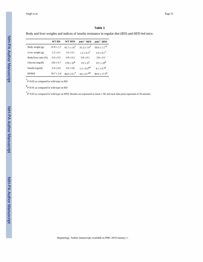
To define JNK isoform specific functions in the development of steatohepatitis occurring inthe setting of obesity and peripheral insulin resistance, wild-type, jnk1-/- and jnk2-/- micewere fed a HFD for 16 weeks. Wild-type mice fed the HFD had a 31% greater increase inbody weight at the end of the 16 week period than mice fed regular diet (Table 1). Inassociation with this weight gain, HFD-fed mice developed peripheral insulin resistance asdemonstrated by significantly increased HOMA values (Table 1). Thus, 16 weeks of HFDfeeding was sufficient to develop an overweight, insulin resistant mouse model.
JNK activation has been previously reported to mediate weight gain and the development ofperipheral insulin resistance,4 two predisposing factors for human NAFLD. Consistent withthis prior report, mice lacking JNK1 failed to develop the increases in total body and liverweight from the HFD feeding that occurred in wild-type mice (Table 1). HFD-inducedperipheral insulin resistance also failed to develop in jnk1-/- mice. Serum glucose and insulinlevels and HOMAs were significantly decreased in jnk1-/- mice as compared to wild-typemice with HFD feeding (Table 1). Insulin levels and HOMA values in HFD-fed jnk1-/- micewere even significantly lower than those of regular diet-fed wild-type mice (Table 1). Incontrast, HFD-fed jnk2-/- animals had significantly greater weight gain and higher HOMAvalues than wild-type mice, although the differences in HOMAs did not reach statisticalsignificance (Table 1). Thus, the excessive weight gain and insulin resistance that developedin this HFD-fed mouse model were JNK1 dependent whereas the absence of JNK2 led toincreased body weight.
JNK1 but not JNK2 Null Mice Develop Decreased Steatohepatitis from a HFDJNK activation in response to a HFD was assessed by immunoblot analysis of levels of theactive, phosphorylated forms of JNK and its downstream substrate c-Jun. Protein levels ofphospho-JNK and phospho-c-Jun were increased in the livers of mice fed 16 weeks of HFD(Fig. 1A). Levels of total JNK were unchanged whereas levels of total c-Jun were
Singh et al. Page 4
Hepatology. Author manuscript; available in PMC 2010 January 1.
NIH
-PA Author Manuscript
NIH
-PA Author Manuscript
NIH
-PA Author Manuscript

moderately increased (Fig. 1A). Thus, consistent with our previous findings in themethionine- and choline-deficient model of steatohepatitis,6 JNK/c-Jun activation occurredin association with the development of steatohepatitis. Jnk1-/- mice had a predominant lossof hepatic p46 JNK whereas jnk2-/- mice had decreased levels of p54 JNK (Fig. 1B and 1C),as previously reported.6 Phospho-JNK levels were decreased in both HFD-fed jnk knockoutmice although jnk1-/- mice had marked decreases in both p54 and p46 phospho-JNKwhereas in jnk2-/- mice the decrease was predominantly in p54 phospho-JNK (Fig. 1B and1C). Levels of phospho-c-Jun were decreased in HFD-fed jnk1-/- mice and increased injnk2-/- mice consistent with the known effects of the two JNK isoforms on c-Junphosphorylation (Fig. 1B and 1C).
By histology wild-type mice developed significant steatosis and hepatitis with a HFD (Fig.2A and 2B). Blinded histological grading of the degree of steatosis revealed increasedhepatic fat accumulation in wild-type mice fed the HFD (Fig. 3A). In contrast, steatosis didnot develop in jnk1-/- mice (Fig. 2C and 3A). Histological evidence of steatosis in jnk2-/-
mice was equivalent to that in wild-type mice (Fig. 2D and 3A). The protective effect ofJNK1 ablation on the development of steatosis was confirmed by measures of hepatic TGlevels. A significant increase in TG content occurred with HFD feeding in wild-type andjnk2-/- mice, but jnk1-/- mice had levels equivalent to regular diet-fed wild-type mice (Fig.3B). The development of hepatic steatosis in response to a HFD was therefore JNK1 but notJNK2 dependent.
Livers were also examined for the extent of injury and inflammation. In parallel with anabsence of steatosis, jnk1-/- mice had reduced liver injury as reflected in decreased serumALT levels and numbers of TUNEL positive cells (Fig. 3C and 3D). Histological evidenceof inflammation was also significantly decreased in HFD-fed jnk1-/- mice (Fig. 3E). Incontrast, HFD-fed jnk2-/- mice had significantly higher levels of serum ALT and TUNELstaining as compared to wild-type mice indicating that liver injury was increased in theabsence of JNK2 (Fig. 3C and 3D). Thus, JNK1 function was critical for the development ofsteatosis and hepatitis in response to a HFD. In contrast, although lipid accumulation wasunaffected by loss of JNK2, JNK2 functioned to protect against hepatocyte injury in thesetting of a fatty liver.
Knockdowns of JNK1 and JNK2 both Reverse Insulin Resistance but have OpposingEffects on Established Steatohepatitis in HFD-fed Animals
Although initial studies established that the development of HFD-induced murinesteatohepatitis was differentially regulated by JNK1 and JNK2, these investigations did notaddress the function of these factors in established liver disease. In an effort to definewhether JNK1 could be a therapeutic target in NAFLD, it was necessary to investigate therole of the JNK isoforms in promoting or perpetuating established steatohepatitis. To test theeffect of JNK inhibition on existing steatohepatitis, and to obtain further evidence of thedifferential effects of JNK1 and JNK2 in the pathophysiology of this disease, we employedASO to acutely inhibit JNK function. In addition to allowing the initiation of JNK inhibitionlate in the progression of the disease, this alternative approach to knocking down JNKfunction had the potential advantage of avoiding the confounding variable of compensatorychanges that may develop in knockout mice and affect their biological responses.
Within 12 weeks of being started on a HFD, wild-type mice developed significant insulinresistance (HOMA values of 41.9 ± 7.6 in HFD versus 16.5 ± 4.5 with regular diet),steatosis (triglyceride levels of 95.3 ± 16.8 and 22.1 ± 7.3 ug/mg protein in HFD- andregular diet-fed mice, respectively) and steatohepatitis (ALT levels of 95.3 ± 16.8 IU/ml inHFD-fed and 22.1 ± 7.3 IU/ml in regular diet-fed mice). Mice fed 12 weeks of the HFDwere randomly assigned to twice weekly injections of a control oligonucleotide or the JNK1
Singh et al. Page 5
Hepatology. Author manuscript; available in PMC 2010 January 1.
NIH
-PA Author Manuscript
NIH
-PA Author Manuscript
NIH
-PA Author Manuscript
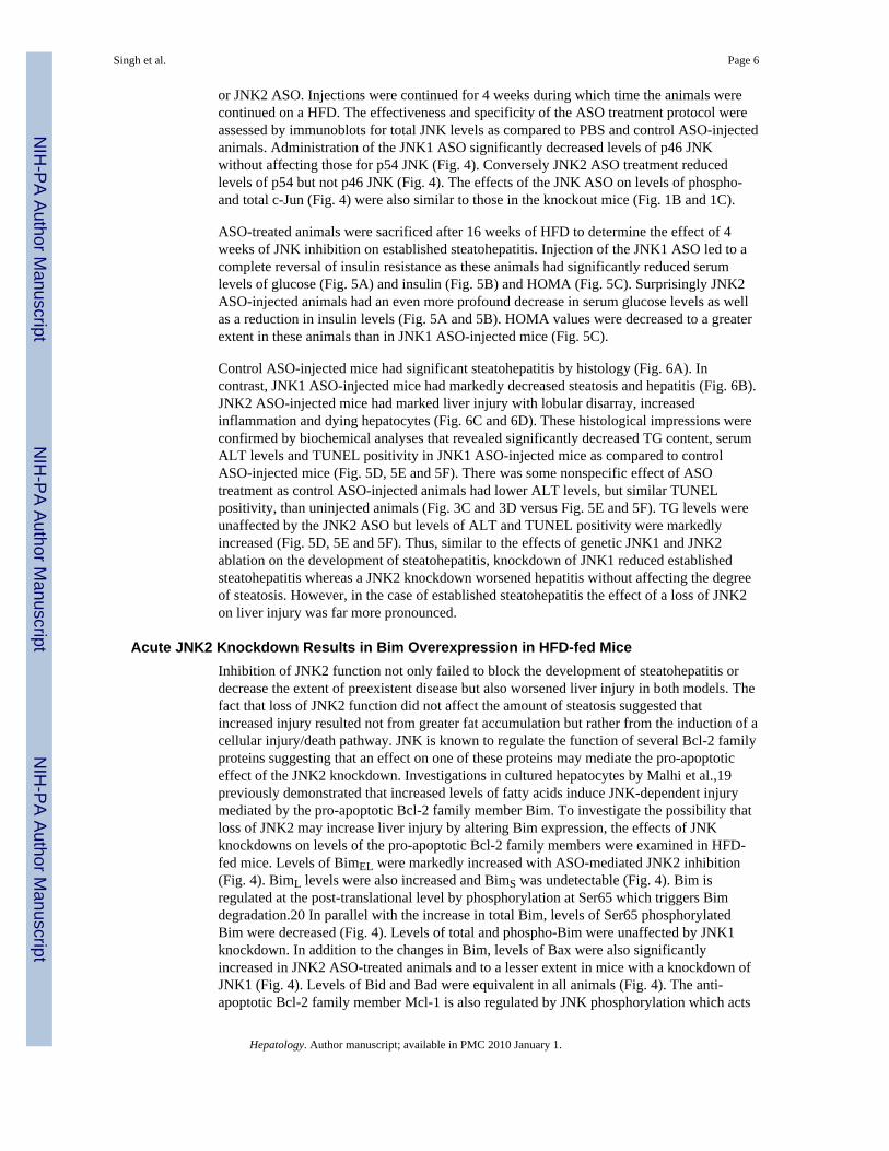
or JNK2 ASO. Injections were continued for 4 weeks during which time the animals werecontinued on a HFD. The effectiveness and specificity of the ASO treatment protocol wereassessed by immunoblots for total JNK levels as compared to PBS and control ASO-injectedanimals. Administration of the JNK1 ASO significantly decreased levels of p46 JNKwithout affecting those for p54 JNK (Fig. 4). Conversely JNK2 ASO treatment reducedlevels of p54 but not p46 JNK (Fig. 4). The effects of the JNK ASO on levels of phospho-and total c-Jun (Fig. 4) were also similar to those in the knockout mice (Fig. 1B and 1C).
ASO-treated animals were sacrificed after 16 weeks of HFD to determine the effect of 4weeks of JNK inhibition on established steatohepatitis. Injection of the JNK1 ASO led to acomplete reversal of insulin resistance as these animals had significantly reduced serumlevels of glucose (Fig. 5A) and insulin (Fig. 5B) and HOMA (Fig. 5C). Surprisingly JNK2ASO-injected animals had an even more profound decrease in serum glucose levels as wellas a reduction in insulin levels (Fig. 5A and 5B). HOMA values were decreased to a greaterextent in these animals than in JNK1 ASO-injected mice (Fig. 5C).
Control ASO-injected mice had significant steatohepatitis by histology (Fig. 6A). Incontrast, JNK1 ASO-injected mice had markedly decreased steatosis and hepatitis (Fig. 6B).JNK2 ASO-injected mice had marked liver injury with lobular disarray, increasedinflammation and dying hepatocytes (Fig. 6C and 6D). These histological impressions wereconfirmed by biochemical analyses that revealed significantly decreased TG content, serumALT levels and TUNEL positivity in JNK1 ASO-injected mice as compared to controlASO-injected mice (Fig. 5D, 5E and 5F). There was some nonspecific effect of ASOtreatment as control ASO-injected animals had lower ALT levels, but similar TUNELpositivity, than uninjected animals (Fig. 3C and 3D versus Fig. 5E and 5F). TG levels wereunaffected by the JNK2 ASO but levels of ALT and TUNEL positivity were markedlyincreased (Fig. 5D, 5E and 5F). Thus, similar to the effects of genetic JNK1 and JNK2ablation on the development of steatohepatitis, knockdown of JNK1 reduced establishedsteatohepatitis whereas a JNK2 knockdown worsened hepatitis without affecting the degreeof steatosis. However, in the case of established steatohepatitis the effect of a loss of JNK2on liver injury was far more pronounced.
Acute JNK2 Knockdown Results in Bim Overexpression in HFD-fed MiceInhibition of JNK2 function not only failed to block the development of steatohepatitis ordecrease the extent of preexistent disease but also worsened liver injury in both models. Thefact that loss of JNK2 function did not affect the amount of steatosis suggested thatincreased injury resulted not from greater fat accumulation but rather from the induction of acellular injury/death pathway. JNK is known to regulate the function of several Bcl-2 familyproteins suggesting that an effect on one of these proteins may mediate the pro-apoptoticeffect of the JNK2 knockdown. Investigations in cultured hepatocytes by Malhi et al.,19previously demonstrated that increased levels of fatty acids induce JNK-dependent injurymediated by the pro-apoptotic Bcl-2 family member Bim. To investigate the possibility thatloss of JNK2 may increase liver injury by altering Bim expression, the effects of JNKknockdowns on levels of the pro-apoptotic Bcl-2 family members were examined in HFD-fed mice. Levels of BimEL were markedly increased with ASO-mediated JNK2 inhibition(Fig. 4). BimL levels were also increased and BimS was undetectable (Fig. 4). Bim isregulated at the post-translational level by phosphorylation at Ser65 which triggers Bimdegradation.20 In parallel with the increase in total Bim, levels of Ser65 phosphorylatedBim were decreased (Fig. 4). Levels of total and phospho-Bim were unaffected by JNK1knockdown. In addition to the changes in Bim, levels of Bax were also significantlyincreased in JNK2 ASO-treated animals and to a lesser extent in mice with a knockdown ofJNK1 (Fig. 4). Levels of Bid and Bad were equivalent in all animals (Fig. 4). The anti-apoptotic Bcl-2 family member Mcl-1 is also regulated by JNK phosphorylation which acts
Singh et al. Page 6
Hepatology. Author manuscript; available in PMC 2010 January 1.
NIH
-PA Author Manuscript
NIH
-PA Author Manuscript
NIH
-PA Author Manuscript
to decrease Mcl-1's anti-apoptotic effect.21 JNK2 ASO treatment led to a slight increase inMcl-1 levels but no shift in mobility to indicate an alteration in Mcl-1 phosphorylation andtherefore Mcl-1 function.
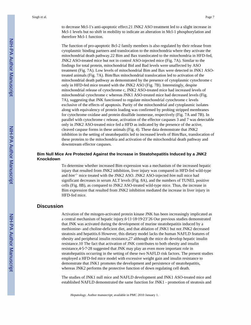
The function of pro-apoptotic Bcl-2 family members is also regulated by their release fromcytoplasmic binding partners and translocation to the mitochondria where they activate themitochondrial death pathway.22 Bim and Bax translocated to the mitochondria in HFD-fed,JNK2 ASO-treated mice but not in control ASO-injected mice (Fig. 7A). Similar to thefindings for total protein, mitochondrial Bid and Bad levels were unaffected by ASOtreatment (Fig. 7A). Low levels of mitochondrial Bim and Bax were detected in JNK1 ASO-treated animals (Fig. 7A). Bim/Bax mitochondrial translocation led to activation of themitochondrial death pathway as demonstrated by the presence of cytoplasmic cytochrome conly in HFD-fed mice treated with the JNK2 ASO (Fig. 7B). Interestingly, despitemitochondrial release of cytochrome c, JNK2 ASO-treated mice had increased levels ofmitochondrial cytochrome c whereas JNK1 ASO-treated mice had decreased levels (Fig.7A), suggesting that JNK functioned to regulate mitochondrial cytochrome c levelsexclusive of the effects of apoptosis. Purity of the mitochondrial and cytoplasmic isolatesalong with equivalency of protein loading was confirmed by probing stripped membranesfor cytochrome oxidase and protein disulfide isomerase, respectively (Fig. 7A and 7B). Inparallel with cytochrome c release, activation of the effector caspases 3 and 7 was detectableonly in JNK2 ASO-treated mice fed a HFD as indicated by the presence of the active,cleaved caspase forms in these animals (Fig. 4). These data demonstrate that JNK2inhibition in the setting of steatohepatitis led to increased levels of Bim/Bax, translocation ofthese proteins to the mitochondria and activation of the mitochondrial death pathway anddownstream effector caspases.
Bim Null Mice Are Protected Against the Increase in Steatohepatitis Induced by a JNK2Knockdown
To determine whether increased Bim expression was a mechanism of the increased hepaticinjury that resulted from JNK2 inhibition, liver injury was compared in HFD-fed wild-typeand bim-/- mice treated with the JNK2 ASO. JNK2 ASO-injected bim null mice hadsignificant decreases in serum ALT levels (Fig. 8A), and the numbers of TUNEL positivecells (Fig. 8B), as compared to JNK2 ASO-treated wild-type mice. Thus, the increase inBim expression that resulted from JNK2 inhibition mediated the increase in liver injury inHFD-fed mice.
DiscussionActivation of the mitogen-activated protein kinase JNK has been increasingly implicated asa central mechanism of hepatic injury.6,11,18,19,23-26 Our previous studies demonstratedthat JNK was activated during the development of murine steatohepatitis induced by amethionine- and choline-deficient diet, and that ablation of JNK1 but not JNK2 decreasedsteatosis and hepatitis.6 However, this dietary model lacks the human NAFLD features ofobesity and peripheral insulin resistance,27 although the mice do develop hepatic insulinresistance.10 The fact that activation of JNK contributes to both obesity and insulinresistance,4,5,7,28 suggested that JNK may play an even more important role insteatohepatitis occurring in the setting of these two NAFLD risk factors. The present studiesemployed a HFD-fed mice model with excessive weight gain and insulin resistance todemonstrate that JNK1 promotes the development and persistence of steatohepatitis,whereas JNK2 performs the protective function of down regulating cell death.
The studies of JNK1 null mice and NAFLD development and JNK1 ASO-treated mice andestablished NAFLD demonstrated the same function for JNK1 - promotion of steatosis and
Singh et al. Page 7
Hepatology. Author manuscript; available in PMC 2010 January 1.
NIH
-PA Author Manuscript
NIH
-PA Author Manuscript
NIH
-PA Author Manuscript
liver injury. JNK1 inhibition by either method dramatically reduced hepatic TG content inconcert with decreased liver injury and cell death. In particular, JNK1 was critical insteatosis development as HFD-fed jnk1-/- mice had hepatic TG levels equivalent to regulardiet-fed wild-type mice. The ability of JNK1 to mediate both steatosis and cellular injurymay reflect one or multiple functions of this kinase. JNK1 may simply promote hepatic lipidaccumulation, and the dramatic reduction in steatosis from JNK1 inhibition decreases liverinjury as well. Alternatively, JNK1 may promote steatosis and hepatitis by distinctmechanisms. The mechanism by which JNK1 promotes steatosis remains to be defined andmay involve hepatic and/or peripheral effects. The prevention or reversal of steatosis mayhave resulted simply from the dramatic effect of JNK inhibition on insulin sensitivity. JNK1is known to induce insulin resistance by phosphorylating inhibitory serine residues oninsulin receptor substrate 1 and mediating obesity-induced inflammation.29 Against thereduction in hepatic lipid being merely secondary to effects on insulin resistance is ourfinding that JNK2 ASO treatment restored insulin sensitivity but did not affect steatosis.Thus, direct effects of JNK1 on lipid metabolism may underlie the development andmaintenance of hepatic steatosis. Further studies will examine the regulation of componentsof lipid metabolism as JNK targets in these pathways have not yet been reported.30
The studies demonstrate a different function for JNK2 in HFD-induced steatohepatitis. Inboth developing and established steatohepatitis, JNK2 inhibition did not affect the amount ofsteatosis and worsened liver injury. As discussed above, JNK2 ASO administration had noeffect on steatosis despite a reversal of insulin resistance. This effect was different fromfindings in the HFD-fed JNK2 null animals which had no improvement in insulin sensitivityand even a trend to greater resistance. This difference may have been secondary tocompensatory changes in jnk2 null mice that did not occur in ASO-treated mice, or todisparate functions of JNK2 in developing versus established insulin resistance. A similarexplanation likely underlies the differences between our findings and those of Tuncman etal.7 who reported that JNK1 and JNK2 both contribute to steatosis based on studies of jnk2null mice haploinsufficient for jnk1. Alternatively, findings in the ASO-treated mice mayhave differed because ASO treatment resulted in a more liver specific effect of JNKinhibition as the liver has one of the highest concentrations of delivered ASO among tissues.31 In addition, the findings with JNK2 inhibition suggest that improved insulin sensitivityby itself is not sufficient to reverse established steatosis. Other recent studies have found thatsteatosis can persist despite reversal of insulin resistance,28 or suggested that insulinresistance may not mediate the development of this disease.32
The function of JNK2 in liver injury also differed from that of JNK1. In contrast to theimprovement in liver injury in mice lacking JNK1 function, knockout or knockdown ofJNK2 significantly worsened liver injury and apoptosis. The absence of an increase insteatosis with JNK2 inhibition suggested that JNK2 functioned specifically to block celldeath pathways. Based on the reported ability of JNK to regulate Bim expression,33-35 andfindings that Bim modulated hepatocyte toxicity from fatty acids in vitro,19 the effect of aknockdown of JNK2 on Bim was examined. Loss of JNK2 led to increased levels of Bimand to a lesser extent of Bax. Bim regulation is complex with steady-state protein levelsreflecting changes in transcription, phosphorylation-mediated degradation and inhibitoryprotein binding and translocation. A number of kinases including JNK, extracellular signal-regulated kinase 1/2 and Akt have been reported to phosphorylate Bim in different celltypes.20,33,36 In the livers of HFD-fed mice, JNK2 function was required for Bimphosphorylation at Ser65, a site whose phosphorylation triggers Bim degradation.20 Lack ofSer65 phosphorylation was associated with increased steady-state levels of total Bim inJNK2 knockdown mice. Our results are consistent with recent findings in a leukemia cellline in which JNK promoted chemoresistance by phosphorylating Bim at Ser65 and causingits degradation,33 and demonstrate that JNK can act to down regulate Bim-dependent
Singh et al. Page 8
Hepatology. Author manuscript; available in PMC 2010 January 1.
NIH
-PA Author Manuscript
NIH
-PA Author Manuscript
NIH
-PA Author Manuscript
apoptosis as well as to activate it. The effects of a JNK2 knockdown on Bim/Bax led toactivation of the mitochondrial death pathway and apoptosis as indicated by mitochondrialcytochrome c release, cleavage of the effector caspases 3 and 7 and increased TUNELstaining in JNK2 ASO-treated, HFD-fed mice. In contrast to findings with Bim and Bax, inresponse to a JNK2 knockdown levels of Bid and Bad were unchanged, Bid cleavage wasnot detected and neither protein translocated to the mitochondria, indicating that they wereunaffected by the loss of JNK2 and uninvolved in the increased cell death. Bim clearlymediated the increase in liver injury from JNK2 inhibition as bim null mice had asignificant, but partial, reduction in liver injury and cell death. The partial nature of theeffect may have been secondary to increased Bax translocation that promoted mitochondrialdeath pathway activation independent of Bim. JNK2-dependent promotion of liver injury inHFD-fed mice by activation of the mitochondrial death pathway is in direct opposition toour findings in a model of toxin-induced liver injury in which JNK2 functioned to inhibitthis pathway.18 These differences point to the complexity of JNK function in the liver andlikely reflect the effects of different types of injury, the time course of the injury, crosstalkwith other signaling pathways and possible extrahepatic effects of JNK.
The findings from the ASO-treated animals demonstrate that HFD-induced steatohepatitis inmice is quickly reversible. Despite the fact that approximately two weeks of injections wererequired to achieve a JNK knockdown, a dramatic decrease in steatohepatitis was achievedwith only four weeks of therapy. The rapidity of this effect is further evidence of the criticalnature of JNK1 activation in steatohepatitis and the potential efficacy of anti-JNK therapy inthe treatment of this disease.
The findings demonstrate that the JNK isoforms have differential functions in insulinresistance and steatohepatitis. Both JNK forms contributed to insulin resistance, however,JNK1 promoted both hepatic fat accumulation and injury whereas JNK2 was uninvolved inthe steatosis and inhibited liver injury. Significantly the findings demonstrate that anti-JNKtherapy can reverse chronic steatohepatitis in the absence of any reduction in the stimulusfor the disease, a high fat diet. Kinase inhibitors are already employed in the treatment ofhuman diseases,37 and JNK inhibitors have successfully reversed diabetes in animals.5 Ourfindings suggest that steatohepatitis can also be treated with this approach but that therapywill have to be targeted specifically to the JNK1 isoform. Inhibiting JNK2 may be effectivein increasing insulin sensitivity but have detrimental effects on the associated liver disease.
AcknowledgmentsWe thank Neil Kaplowitz for his helpful suggestions, Xiao-Ming Yin for the Bid antibody and Richard Stockert forthe PDI antibody.
Supported by NIH grants DK061498 (MJC) and DK020541 and an American Liver Foundation PostdoctoralResearch Fellowship Award (RS).
Abbreviations
ASO antisense oligonucleotide
HFD high fat diet
HOMA homeostasis model assessment of insulin resistance
JNK c-Jun N-terminal kinase
NAFLD nonalcoholic fatty liver disease
PBS phosphate-buffered saline
Singh et al. Page 9
Hepatology. Author manuscript; available in PMC 2010 January 1.
NIH
-PA Author Manuscript
NIH
-PA Author Manuscript
NIH
-PA Author Manuscript
PDI protein disulfide isomerase
TG triglyceride
References1. Adams LA, Lindor KD. Nonalcoholic fatty liver disease. Ann Epidemiol. 2007; 17:863–869.
[PubMed: 17728149]2. Marchesini G, Brizi M, Bianchi G, Tomassetti S, Bugianesi E, Lenzi M, et al. Nonalcoholic fatty
liver disease: a feature of the metabolic syndrome. Diabetes. 2001; 50:1844–1850. [PubMed:11473047]
3. Sanyal AJ, Campbell-Sargent C, Mirshahi F, Rizzo WB, Contos MJ, Sterling RK, et al.Nonalcoholic steatohepatitis: association of insulin resistance and mitochondrial abnormalities.Gastroenterology. 2001; 120:1183–1192. [PubMed: 11266382]
4. Hirosumi J, Tuncman G, Chang L, Gorgun CZ, Uysal KT, Maeda K, et al. A central role for JNK inobesity and insulin resistance. Nature. 2002; 420:333–336. [PubMed: 12447443]
5. Kaneto H, Nakatani Y, Miyatsuka T, Kawamori D, Matsuoka TA, Matsuhisa M, et al. Possiblenovel therapy for diabetes with cell-permeable JNK-inhibitory peptide. Nat Med. 2004; 10:1128–1132. [PubMed: 15448687]
6. Schattenberg JM, Singh R, Wang Y, Lefkowitch JH, Rigoli RM, Scherer PE, et al. JNK1 but notJNK2 promotes the development of steatohepatitis in mice. Hepatology. 2006; 43:163–172.[PubMed: 16374858]
7. Tuncman G, Hirosumi J, Solinas G, Chang L, Karin M, Hotamisligil GS. Functional in vivointeractions between JNK1 and JNK2 isoforms in obesity and insulin resistance. Proc Natl Acad SciUSA. 2006; 103:10741–10746. [PubMed: 16818881]
8. Davis RJ. Signal transduction by the JNK group of MAP kinases. Cell. 2000; 103:239–252.[PubMed: 11057897]
9. Sabapathy K, Hochedlinger K, Nam SY, Bauer A, Karin M, Wagner EF. Distinct roles for JNK1and JNK2 in regulating JNK activity and c-Jun-dependent cell proliferation. Mol Cell. 2004;15:713–725. [PubMed: 15350216]
10. Schattenberg JM, Wang Y, Singh R, Rigoli RM, Czaja MJ. Hepatocyte CYP2E1 overexpressionand steatohepatitis lead to impaired hepatic insulin signaling. J Biol Chem. 2005; 280:9887–9894.[PubMed: 15632182]
11. Gunawan BK, Liu ZX, Han D, Hanawa N, Gaarde WA, Kaplowitz N. c-Jun N-terminal kinaseplays a major role in murine acetaminophen hepatotoxicity. Gastroenterology. 2006; 131:165–178.[PubMed: 16831600]
12. Dong C, Yang DD, Wysk M, Whitmarsh AJ, Davis RJ, Flavell RA. Defective T cell differentiationin the absence of Jnk1. Science. 1998; 282:2092–2095. [PubMed: 9851932]
13. Yang DD, Conze D, Whitmarsh AJ, Barrett T, Davis RJ, Rincon M, et al. Differentiation of CD4+T cells to Th1 cells requires MAP kinase JNK2. Immunity. 1998; 9:575–585. [PubMed: 9806643]
14. Bouillet P, Metcalf D, Huang DC, Tarlinton DM, Kay TW, Kontgen F, et al. Proapoptotic Bcl-2relative Bim required for certain apoptotic responses, leukocyte homeostasis, and to precludeautoimmunity. Science. 1999; 286:1735–1738. [PubMed: 10576740]
15. Baker BF, Lot SS, Condon TP, Cheng-Flournoy S, Lesnik EA, Sasmor HM, et al. 2′-O-(2-Methoxy)ethyl-modified anti-intercellular adhesion molecule 1 (ICAM-1) oligonucleotidesselectively increase the ICAM-1 mRNA level and inhibit formation of the ICAM-1 translationinitiation complex in human umbilical vein endothelial cells. J Biol Chem. 1997; 272:11994–12000. [PubMed: 9115264]
16. D'Ambra R, Surana M, Efrat S, Starr RG, Fleischer N. Regulation of insulin secretion from beta-cell lines derived from transgenic mice insulinomas resembles that of normal beta-cells.Endocrinology. 1990; 126:2815–2822. [PubMed: 1693563]
Singh et al. Page 10
Hepatology. Author manuscript; available in PMC 2010 January 1.
NIH
-PA Author Manuscript
NIH
-PA Author Manuscript
NIH
-PA Author Manuscript
17. Matthews DR, Hosker JP, Rudenski AS, Naylor BA, Treacher DF, Turner RC. Homeostasis modelassessment: insulin resistance and beta-cell function from fasting plasma glucose and insulinconcentrations in man. Diabetologia. 1985; 28:412–419. [PubMed: 3899825]
18. Wang Y, Singh R, Lefkowitch JH, Rigoli RM, Czaja MJ. Tumor necrosis factor-induced toxicliver injury results from JNK2-dependent activation of caspase-8 and the mitochondrial deathpathway. J Biol Chem. 2006; 281:15258–15267. [PubMed: 16571730]
19. Malhi H, Bronk SF, Werneburg NW, Gores GJ. Free fatty acids induce JNK-dependent hepatocytelipoapoptosis. J Biol Chem. 2006; 281:12093–12101. [PubMed: 16505490]
20. Ley R, Ewings KE, Hadfield K, Howes E, Balmanno K, Cook SJ. Extracellular signal-regulatedkinases 1/2 are serum-stimulated “BimEL kinases” that bind to the BH3-only protein BimELcausing its phosphorylation and turnover. J Biol Chem. 2004; 279:8837–8847. [PubMed:14681225]
21. Inoshita S, Takeda K, Hatai T, Terada Y, Sano M, Hata J, et al. Phosphorylation and inactivationof myeloid cell leukemia 1 by JNK in response to oxidative stress. J Biol Chem. 2002; 277:43730–43734. [PubMed: 12223490]
22. Baskin-Bey ES, Gores GJ. Death by association: BH3 domain-only proteins and liver injury. Am JPhysiol Gastrointest Liver Physiol. 2005; 289:G987–G990. [PubMed: 16286505]
23. Corazza N, Jakob S, Schaer C, Frese S, Keogh A, Stroka D, et al. TRAIL receptor-mediated JNKactivation and Bim phosphorylation critically regulate Fas-mediated liver damage and lethality. JClin Invest. 2006; 116:2493–2499. [PubMed: 16955144]
24. Schwabe RF, Uchinami H, Qian T, Bennett BL, Lemasters JJ, Brenner DA. Differentialrequirement for c-Jun NH2-terminal kinase in TNFα- and Fas-mediated apoptosis in hepatocytes.FASEB J. 2004; 18:720–722. [PubMed: 14766793]
25. Uehara T, Bennett B, Sakata ST, Satoh Y, Bilter GK, Westwick JK, et al. JNK mediates hepaticischemia reperfusion injury. J Hepatol. 2005; 42:850–859. [PubMed: 15885356]
26. Qiao L, Han SI, Fang Y, Park JS, Gupta S, Gilfor D, et al. Bile acid regulation of C/EBPβ, CREB,and c-Jun function, via the extracellular signal-regulated kinase and c-Jun NH2-terminal kinasepathways, modulates the apoptotic response of hepatocytes. Mol Cell Biol. 2003; 23:3052–3066.[PubMed: 12697808]
27. Rinella ME, Green RM. The methionine-choline deficient dietary model of steatohepatitis does notexhibit insulin resistance. J Hepatol. 2004; 40:47–51. [PubMed: 14672613]
28. Solinas G, Naugler W, Galimi F, Lee MS, Karin M. Saturated fatty acids inhibit induction ofinsulin gene transcription by JNK-mediated phosphorylation of insulin-receptor substrates. ProcNatl Acad Sci USA. 2006; 103:16454–16459. [PubMed: 17050683]
29. Solinas G, Vilcu C, Neels JG, Bandyopadhyay GK, Luo JL, Naugler W, et al. JNK1 inhematopoietically derived cells contributes to diet-induced inflammation and insulin resistancewithout affecting obesity. Cell Metab. 2007; 6:386–397. [PubMed: 17983584]
30. Bogoyevitch MA, Kobe B. Uses for JNK: the many and varied substrates of the c-Jun N-terminalkinases. Microbiol Mol Biol Rev. 2006; 70:1061–1095. [PubMed: 17158707]
31. Geary RS, Yu RZ, Watanabe T, Henry SP, Hardee GE, Chappell A, et al. Pharmacokinetics of atumor necrosis factor-α phosphorothioate 2′-O-(2-methoxyethyl) modified antisenseoligonucleotide: comparison across species. Drug Metab Dispos. 2003; 31:1419–1428. [PubMed:14570775]
32. Fishman S, Muzumdar RH, Atzmon G, Ma X, Yang X, Einstein FH, et al. Resistance to leptinaction is the major determinant of hepatic triglyceride accumulation in vivo. FASEB J. 2007;21:53–60. [PubMed: 17099068]
33. Leung KT, Li KK, Sun SS, Chan PK, Ooi VE, Chiu LC. Activation of the JNK pathway promotesphosphorylation and degradation of BimEL-a novel mechanism of chemoresistance in T-cell acutelymphoblastic leukemia. Carcinogenesis. 2008; 29:544–551. [PubMed: 18174237]
34. Putcha GV, Moulder KL, Golden JP, Bouillet P, Adams JA, Strasser A, et al. Induction of BIM, aproapoptotic BH3-only BCL-2 family member, is critical for neuronal apoptosis. Neuron. 2001;29:615–628. [PubMed: 11301022]
Singh et al. Page 11
Hepatology. Author manuscript; available in PMC 2010 January 1.
NIH
-PA Author Manuscript
NIH
-PA Author Manuscript
NIH
-PA Author Manuscript
35. Whitfield J, Neame SJ, Paquet L, Bernard O, Ham J. Dominant-negative c-Jun promotes neuronalsurvival by reducing BIM expression and inhibiting mitochondrial cytochrome c release. Neuron.2001; 29:629–643. [PubMed: 11301023]
36. Qi XJ, Wildey GM, Howe PH. Evidence that Ser87 of BimEL is phosphorylated by Akt andregulates BimEL apoptotic function. J Biol Chem. 2006; 281:813–823. [PubMed: 16282323]
37. Demetri GD, von Mehren M, Blanke CD, Van den Abbeele AD, Eisenberg B, Roberts PJ, et al.Efficacy and safety of imatinib mesylate in advanced gastrointestinal stromal tumors. N Engl JMed. 2002; 347:472–480. [PubMed: 12181401]
Singh et al. Page 12
Hepatology. Author manuscript; available in PMC 2010 January 1.
NIH
-PA Author Manuscript
NIH
-PA Author Manuscript
NIH
-PA Author Manuscript
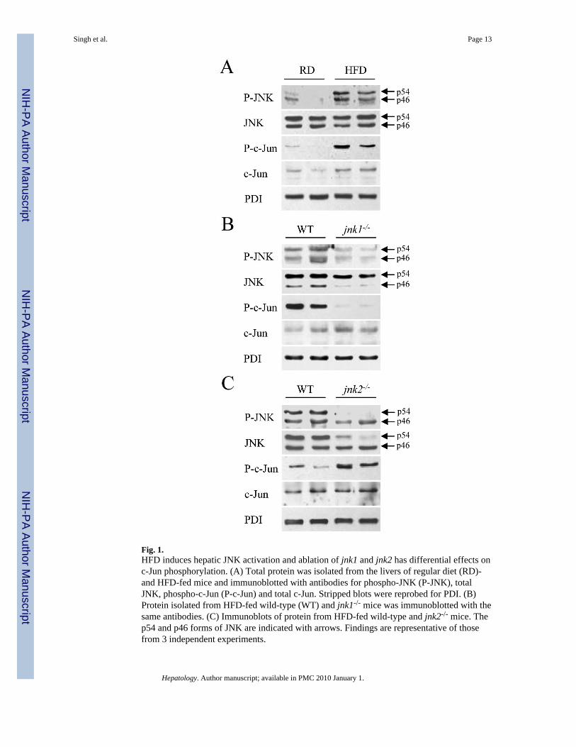
Fig. 1.HFD induces hepatic JNK activation and ablation of jnk1 and jnk2 has differential effects onc-Jun phosphorylation. (A) Total protein was isolated from the livers of regular diet (RD)-and HFD-fed mice and immunoblotted with antibodies for phospho-JNK (P-JNK), totalJNK, phospho-c-Jun (P-c-Jun) and total c-Jun. Stripped blots were reprobed for PDI. (B)Protein isolated from HFD-fed wild-type (WT) and jnk1-/- mice was immunoblotted with thesame antibodies. (C) Immunoblots of protein from HFD-fed wild-type and jnk2-/- mice. Thep54 and p46 forms of JNK are indicated with arrows. Findings are representative of thosefrom 3 independent experiments.
Singh et al. Page 13
Hepatology. Author manuscript; available in PMC 2010 January 1.
NIH
-PA Author Manuscript
NIH
-PA Author Manuscript
NIH
-PA Author Manuscript
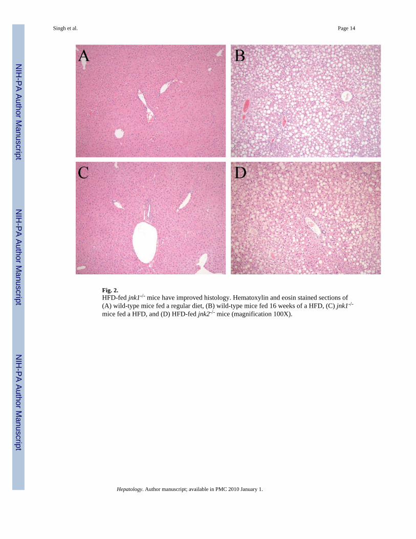
Fig. 2.HFD-fed jnk1-/- mice have improved histology. Hematoxylin and eosin stained sections of(A) wild-type mice fed a regular diet, (B) wild-type mice fed 16 weeks of a HFD, (C) jnk1-/-
mice fed a HFD, and (D) HFD-fed jnk2-/- mice (magnification 100X).
Singh et al. Page 14
Hepatology. Author manuscript; available in PMC 2010 January 1.
NIH
-PA Author Manuscript
NIH
-PA Author Manuscript
NIH
-PA Author Manuscript
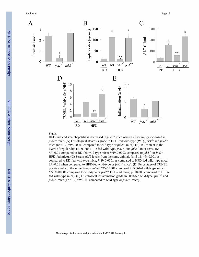
Fig. 3.HFD-induced steatohepatitis is decreased in jnk1-/- mice whereas liver injury increased injnk2-/- mice. (A) Histological steatosis grade in HFD-fed wild-type (WT), jnk1-/- and jnk2-/-
mice (n=7-12; *P<0.0001 compared to wild-type or jnk2-/- mice). (B) TG content in thelivers of regular diet (RD)- and HFD-fed wild-type, jnk1-/- and jnk2-/- mice (n=6-15;*P<0.01 compared to RD-fed wild-type mice; **P<0.0003 compared to jnk1-/- or jnk2-/-
HFD-fed mice). (C) Serum ALT levels from the same animals (n=5-13; *P<0.001 ascompared to RD-fed wild-type mice; **P<0.0001 as compared to HFD-fed wild-type mice;§P<0.01 when compared to HFD-fed wild-type or jnk1-/- mice). (D) Percentage of TUNELpositive cells in the same livers (n=5-9; *P<0.0001 compared to RD-fed wild-type mice;**P<0.00001 compared to wild-type or jnk2-/- HFD-fed mice; §P<0.005 compared to HFD-fed wild-type mice). (E) Histological inflammation grade in HFD-fed wild-type, jnk1-/- andjnk2-/- mice (n=7-12; *P<0.02 compared to wild-type or jnk2-/- mice).
Singh et al. Page 15
Hepatology. Author manuscript; available in PMC 2010 January 1.
NIH
-PA Author Manuscript
NIH
-PA Author Manuscript
NIH
-PA Author Manuscript
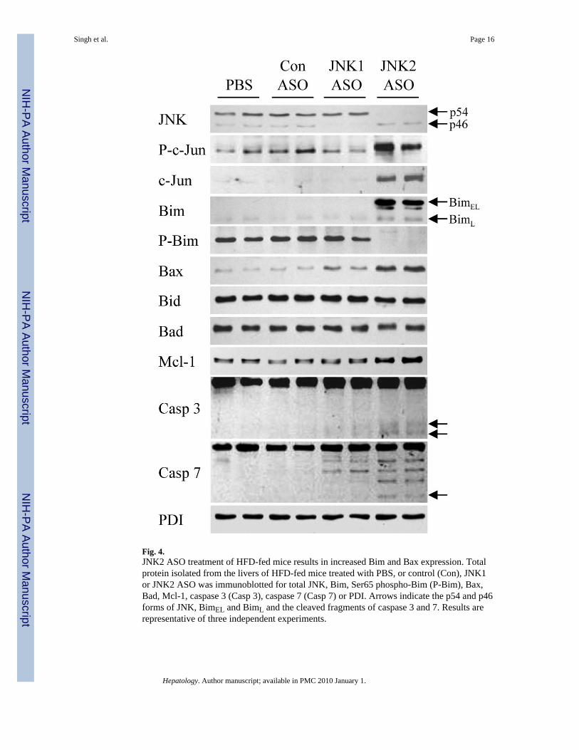
Fig. 4.JNK2 ASO treatment of HFD-fed mice results in increased Bim and Bax expression. Totalprotein isolated from the livers of HFD-fed mice treated with PBS, or control (Con), JNK1or JNK2 ASO was immunoblotted for total JNK, Bim, Ser65 phospho-Bim (P-Bim), Bax,Bad, Mcl-1, caspase 3 (Casp 3), caspase 7 (Casp 7) or PDI. Arrows indicate the p54 and p46forms of JNK, BimEL and BimL and the cleaved fragments of caspase 3 and 7. Results arerepresentative of three independent experiments.
Singh et al. Page 16
Hepatology. Author manuscript; available in PMC 2010 January 1.
NIH
-PA Author Manuscript
NIH
-PA Author Manuscript
NIH
-PA Author Manuscript
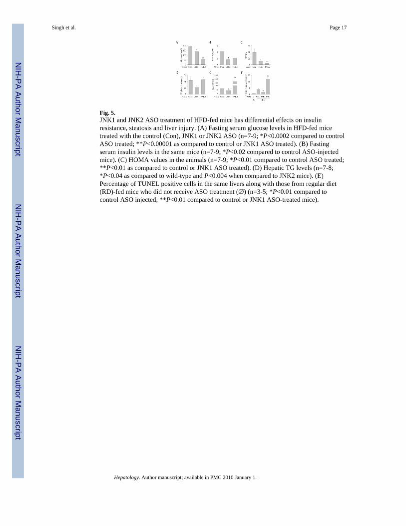
Fig. 5.JNK1 and JNK2 ASO treatment of HFD-fed mice has differential effects on insulinresistance, steatosis and liver injury. (A) Fasting serum glucose levels in HFD-fed micetreated with the control (Con), JNK1 or JNK2 ASO (n=7-9; *P<0.0002 compared to controlASO treated; **P<0.00001 as compared to control or JNK1 ASO treated). (B) Fastingserum insulin levels in the same mice (n=7-9; *P<0.02 compared to control ASO-injectedmice). (C) HOMA values in the animals (n=7-9; *P<0.01 compared to control ASO treated;**P<0.01 as compared to control or JNK1 ASO treated). (D) Hepatic TG levels (n=7-8;*P<0.04 as compared to wild-type and P<0.004 when compared to JNK2 mice). (E)Percentage of TUNEL positive cells in the same livers along with those from regular diet(RD)-fed mice who did not receive ASO treatment (∅) (n=3-5; *P<0.01 compared tocontrol ASO injected; **P<0.01 compared to control or JNK1 ASO-treated mice).
Singh et al. Page 17
Hepatology. Author manuscript; available in PMC 2010 January 1.
NIH
-PA Author Manuscript
NIH
-PA Author Manuscript
NIH
-PA Author Manuscript
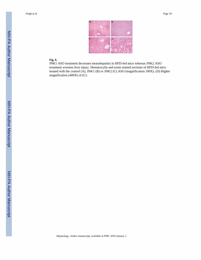
Fig. 6.JNK1 ASO treatment decreases steatohepatitis in HFD-fed mice whereas JNK2 ASOtreatment worsens liver injury. Hematoxylin and eosin stained sections of HFD-fed micetreated with the control (A), JNK1 (B) or JNK2 (C) ASO (magnification 100X). (D) Highermagnification (400X) of (C).
Singh et al. Page 18
Hepatology. Author manuscript; available in PMC 2010 January 1.
NIH
-PA Author Manuscript
NIH
-PA Author Manuscript
NIH
-PA Author Manuscript
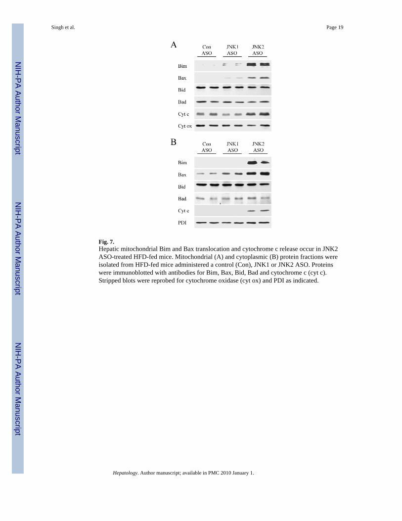
Fig. 7.Hepatic mitochondrial Bim and Bax translocation and cytochrome c release occur in JNK2ASO-treated HFD-fed mice. Mitochondrial (A) and cytoplasmic (B) protein fractions wereisolated from HFD-fed mice administered a control (Con), JNK1 or JNK2 ASO. Proteinswere immunoblotted with antibodies for Bim, Bax, Bid, Bad and cytochrome c (cyt c).Stripped blots were reprobed for cytochrome oxidase (cyt ox) and PDI as indicated.
Singh et al. Page 19
Hepatology. Author manuscript; available in PMC 2010 January 1.
NIH
-PA Author Manuscript
NIH
-PA Author Manuscript
NIH
-PA Author Manuscript
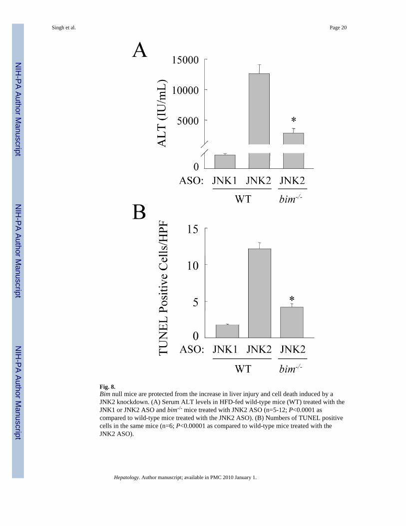
Fig. 8.Bim null mice are protected from the increase in liver injury and cell death induced by aJNK2 knockdown. (A) Serum ALT levels in HFD-fed wild-type mice (WT) treated with theJNK1 or JNK2 ASO and bim-/- mice treated with JNK2 ASO (n=5-12; P<0.0001 ascompared to wild-type mice treated with the JNK2 ASO). (B) Numbers of TUNEL positivecells in the same mice (n=6; P<0.00001 as compared to wild-type mice treated with theJNK2 ASO).
Singh et al. Page 20
Hepatology. Author manuscript; available in PMC 2010 January 1.
NIH
-PA Author Manuscript
NIH
-PA Author Manuscript
NIH
-PA Author Manuscript
NIH
-PA Author Manuscript
NIH
-PA Author Manuscript
NIH
-PA Author Manuscript
Singh et al. Page 21
Table 1
Body and liver weights and indices of insulin resistance in regular diet (RD) and HFD fed mice.
WT RD WT HFD jnk1-/- HFD jnk2-/- HFD
Body weight (g) 31.8 ± 1.7 41.7 ± 1.6* 31.2 ± 1.6† 50.6 ± 1.1*†
Liver weight (g) 1.2 ± 0.1 1.6 ± 0.1 1.2 ± 0.1† 1.9 ± 0.1*
Body/liver ratio (%) 3.9 ± 0.2 3.8 ± 0.2 3.8 ± 0.1 3.8 ± 0.1
Glucose (mg/dl) 120 ± 6.7 178 ± 10# 131 ± 6† 157 ± 10#
Insulin (ng/ml) 2.4 ± 0.4 3.6 ± 0.6 1.2 ± 0.2#† 4.1 ± 0.7#
HOMA 16.7 ± 2.4 46.0 ± 9.1* 4.6 ± 0.7#† 80.9 ± 17.5#
*P<0.05 as compared to wild-type on RD
#P<0.01 as compared to wild-type on RD
†P<0.02 as compared to wild-type on HFD. Results are expressed as mean ± SE and each data point represents 6-18 animals.
Hepatology. Author manuscript; available in PMC 2010 January 1.