Silica-Coated Liposomes for Insulin Delivery
9
Hindawi Publishing Corporation Journal of Nanomaterials Volume 2010, Article ID 652048, 8 pages doi:10.1155/2010/652048 Research Article Silica-Coated Liposomes for Insulin Delivery Neelam Dwivedi, 1, 2 M. A. Arunagirinathan, 1 Somesh Sharma, 2 and Jayesh Bellare 1 1 Department of Chemical Engineering, Indian Institute of Technology Bombay, Mumbai 400076, India 2 Piramal Life Sciences Limited, Research and Development Division, Mumbai 400063, India Correspondence should be addressed to Jayesh Bellare, [email protected] Received 4 November 2009; Revised 14 March 2010; Accepted 22 March 2010 Academic Editor: Maryam Tabrizian Copyright © 2010 Neelam Dwivedi et al. This is an open access article distributed under the Creative Commons Attribution License, which permits unrestricted use, distribution, and reproduction in any medium, provided the original work is properly cited. Liposomes coated with silica were explored as protein delivery vehicles for their enhanced stability and improved encapsulation efficiency. Insulin was encapsulated within the fluidic phosphatidylcholine lipid vesicles by thin film hydration at pH 2.5, and layer of silica was formed above lipid bilayer by acid catalysis. The presence of silica coating and encapsulated insulin was identified using confocal and electron microscopy. The native state of insulin present in the formulation was evident from Confocal Micro- Raman spectroscopy. Silica coat enhances the stability of insulin-loaded delivery vehicles. In vivo study shows that these silica coated formulations were biologically active in reducing glucose levels. 1. Introduction Over several decades pharmaceutical drug manufacturers are using different drug carriers like liposomes, microemulsion, polymeric nanoparticles, ceramic nanocomposites, silica, and titania [1]. Recent review by Porter et al. gives overview on lipid-based formulations of lipophilic drugs and their oral bioavailability [2]. Application of micelles, cochleate, sphingosomes, and vesicles for delivering various drugs and immunogens has been explained by Shapira et al. [3]. Silica- lipid hybrids have been used for enhanced oral absorption of hydrophobic drugs like celecoxib and indomethacin [4– 6]. Cationic lipid-coated silica nanoparticles were used for protein and vaccine delivery due to their controlled cellular immune response and improved pharmacokinetics [7, 8]. Silica-lipid hybrids as nonporous nanospheres and thin films has been prepared from dipalmitoylphosphatidylcholine and egg phosphatidylcholine with varying cholesterol content [4, 6, 9]. Encapsulation of insulin, gramicidin, bacteri- orhodopsin within phospholipid vesicles by varying choles- terol/lipid ratio and pH have been reported earlier, however, lipid bilayer stabilized by inert material like silica has not been studied for insulin delivery [10–16]. Silica nanospheres prepared from noncovalently bound organic templates (surfactant, polymers, supramolecular arrays), acoustic cavitation, electrospraying, spray drying, self assembly, freeze thawing, supercritical fluid technology, heterophase polymerization with sol-gel and surface living polymerization process have been reported [17–24]. Sol- gel derived silica nanospheres were used to encapsulate antibiotics, analgesic, pancreatic islets, dyes and perfumes. It is also used in chromatography, catalysis, waste removal and as fillers in coating [25, 26]. It is established that silica coating is chemically inert, biocompatible, hydrophilic and inexpensive. Here, we have prepared liposome having layer of silica to enhance the stability of the formulation. This formulation has improved encapsulation efficiency of macromolecule within the lipo- some by inhibiting leakage of insulin due to presence of outer silica coat. The present formulation can be used for encapsulation of peptides. 2. Experimental 2.1. Materials. Phosphatidylcholine (PC), Bovine insulin, and Tetra ethyl orthosilicate (TEOS) were obtained from Sigma-Aldrich. 2.2. Synthesis of Silica Coated PC Nanosphere. To obtain silica coated PC nanosphere, initially a thin film of phos- phatidylcholine (PC) was obtained on the walls of round
-
Upload
independent -
Category
Documents
-
view
0 -
download
0
Transcript of Silica-Coated Liposomes for Insulin Delivery

Hindawi Publishing CorporationJournal of NanomaterialsVolume 2010, Article ID 652048, 8 pagesdoi:10.1155/2010/652048
Research Article
Silica-Coated Liposomes for Insulin Delivery
Neelam Dwivedi,1, 2 M. A. Arunagirinathan,1 Somesh Sharma,2 and Jayesh Bellare1
1 Department of Chemical Engineering, Indian Institute of Technology Bombay, Mumbai 400076, India2 Piramal Life Sciences Limited, Research and Development Division, Mumbai 400063, India
Correspondence should be addressed to Jayesh Bellare, [email protected]
Received 4 November 2009; Revised 14 March 2010; Accepted 22 March 2010
Academic Editor: Maryam Tabrizian
Copyright © 2010 Neelam Dwivedi et al. This is an open access article distributed under the Creative Commons AttributionLicense, which permits unrestricted use, distribution, and reproduction in any medium, provided the original work is properlycited.
Liposomes coated with silica were explored as protein delivery vehicles for their enhanced stability and improved encapsulationefficiency. Insulin was encapsulated within the fluidic phosphatidylcholine lipid vesicles by thin film hydration at pH 2.5, and layerof silica was formed above lipid bilayer by acid catalysis. The presence of silica coating and encapsulated insulin was identifiedusing confocal and electron microscopy. The native state of insulin present in the formulation was evident from Confocal Micro-Raman spectroscopy. Silica coat enhances the stability of insulin-loaded delivery vehicles. In vivo study shows that these silicacoated formulations were biologically active in reducing glucose levels.
1. Introduction
Over several decades pharmaceutical drug manufacturers areusing different drug carriers like liposomes, microemulsion,polymeric nanoparticles, ceramic nanocomposites, silica,and titania [1]. Recent review by Porter et al. gives overviewon lipid-based formulations of lipophilic drugs and theiroral bioavailability [2]. Application of micelles, cochleate,sphingosomes, and vesicles for delivering various drugs andimmunogens has been explained by Shapira et al. [3]. Silica-lipid hybrids have been used for enhanced oral absorptionof hydrophobic drugs like celecoxib and indomethacin [4–6]. Cationic lipid-coated silica nanoparticles were used forprotein and vaccine delivery due to their controlled cellularimmune response and improved pharmacokinetics [7, 8].Silica-lipid hybrids as nonporous nanospheres and thin filmshas been prepared from dipalmitoylphosphatidylcholine andegg phosphatidylcholine with varying cholesterol content[4, 6, 9]. Encapsulation of insulin, gramicidin, bacteri-orhodopsin within phospholipid vesicles by varying choles-terol/lipid ratio and pH have been reported earlier, however,lipid bilayer stabilized by inert material like silica has notbeen studied for insulin delivery [10–16].
Silica nanospheres prepared from noncovalently boundorganic templates (surfactant, polymers, supramoleculararrays), acoustic cavitation, electrospraying, spray drying,
self assembly, freeze thawing, supercritical fluid technology,heterophase polymerization with sol-gel and surface livingpolymerization process have been reported [17–24]. Sol-gel derived silica nanospheres were used to encapsulateantibiotics, analgesic, pancreatic islets, dyes and perfumes. Itis also used in chromatography, catalysis, waste removal andas fillers in coating [25, 26].
It is established that silica coating is chemically inert,biocompatible, hydrophilic and inexpensive. Here, we haveprepared liposome having layer of silica to enhance thestability of the formulation. This formulation has improvedencapsulation efficiency of macromolecule within the lipo-some by inhibiting leakage of insulin due to presence ofouter silica coat. The present formulation can be used forencapsulation of peptides.
2. Experimental
2.1. Materials. Phosphatidylcholine (PC), Bovine insulin,and Tetra ethyl orthosilicate (TEOS) were obtained fromSigma-Aldrich.
2.2. Synthesis of Silica Coated PC Nanosphere. To obtainsilica coated PC nanosphere, initially a thin film of phos-phatidylcholine (PC) was obtained on the walls of round

2 Journal of Nanomaterials
bottomed flask by evaporating 10 ml of chloroform solutioncontaining 100 mg of PC under reduced pressure. The thinfilm of PC was hydrated by adding 100 ml of water andstirred for 15 minutes. The stirred mixture was furthersonicated for 10 minutes to obtain vesicles. To this vesiculardispersion, 5 ml of TEOS was added at a mole ratio 0.9 : 17 : 4(TEOS : PC : H2O ), and pH of the mixture was decreased to2.5 by adding 0.05 N HCl and then stirred for 2 hours. Thisreaction mixture was kept for aging at acidic pH of 2.5 for24 hours to increase silica network formation leading to PCvesicles coated with silica. This solution was stored at 4◦C forfurther studies.
2.3. Synthesis of Silica Coated PC Nanosphere EncapsulatingInsulin. Whereas to obtain insulin loaded silica coated PCnanosphere, 10 mg of insulin was dissolved in 1 ml of distilledwater to which 10 microlitre of 0.05 N HCl was added. Sim-ilarly for confocal microscopy studies FITC tagged insulinwas used. This insulin solution was further diluted withdistilled water to obtain total volume of 100 ml and lateradded to thin film of PC obtained from evaporating 100 mgof PC dissolved in 10 ml of chloroform in round bottomflask for hydration. This solution was stirred for 15 minutesfollowed by 10 minutes of sonication, and 5 ml of TEOSwas added which is then stirred for 2 hours and 24 hoursaging. The reaction mixture was centrifuged for 5 minutes,supernatant was discarded, and the bottom settled portionof material was resuspended in 50 ml of distilled waterthen again centrifuged for 5 minutes. The supernatant wasdiscarded and the paste like mass (silica coated insulin loadedvesicles) settled at the bottom was stored at 4◦C for furtherstudies.
2.4. Characterisation of Silica Coated PC Nanosphere
Formulation
2.4.1. Confocal Microscopy. Fluorophore tagged insulin(FITC-insulin) containing formulations was observed onglass cover slips under Confocal Laser Scanning Microscope(Fluoview FV500, Olympus) using Multi Argon Laser,488 nm.
2.4.2. TEM. To observe the microstructures of silica coatedinsulin loaded PC vesicles under TEM (Tecnai-12, FEI),simple drying, room temperature replica and Freeze fracturereplica were done. For simple drying, a drop of the samplewas allowed to dry on 300 mesh TEM grid having Formvarfilm stabilized with carbon coating and observed under TEM.Room temperature replica of the samples were prepared byplacing a drop of sample on a glass cover slip and allowedto dry at room temperature. After drying, replica was madeby coating 2 nm of Pt/C at 45 degree angle followed by40 nm carbon coating at 90 degree angle in Balzer BAF060freeze fracture unit. The cover slip containing the coatedsample was submerged in sodium hypochlorite solution for1 hour to facilitate detachment of replica from the sample.The detached replica was further gently washed with distilled
water and lifted over bare carbon TEM grid and allowed todry. The obtained replica was later observed under TEM.
To obtain freeze-fractured replica of the sample, 5–10 μlof the sample was placed between two copper planchetsobtained from Baltec and held by flat tip, rounded end,Dumont tweezer then plunged quickly into liquid nitrogen.The sandwiched copper planchets containing frozen samplewas inserted on to the slot of double replica specimen tablekept under liquid nitrogen and transferred through coldchain on to the precooled stage maintained at −150◦C inBalzer BAF060 freeze fracture unit. The sandwiched copperplanchets were made open inside the freeze fracture unit.The fractured frozen sample was coated with 2 nm of Pt/Cat 45-degree angle followed by 40 nm carbon coating at 90degree angle. The coated fractured specimen was brought toroom temperature and submerged in sodium hypochloritesolution for 1 hour to facilitate detachment of replica fromthe sample. The detached replica was further gently washedwith distilled water and lifted over bare carbon TEM grid andallowed to dry. The obtained replica was later observed underTEM.
2.4.3. HPLC. Insulin content of the samples was analyzedusing HPLC-UV at 277 nm (Waters, Photo diode arraydetector). Gradient elution was performed using 100% ace-tonitrile and 0.1% TFA (0.1%) at a flow rate of 1 ml/minutesand injection volume of 20 μl. Insulin was detected ata retention time of 5.7 minutes with detection limit of0.01 mg/ml. To check the total release of insulin, 100 μl of25 mM EDTA and 100 μl of 10% Triton-X 100 was added tothe 100 μl of formulation.
2.4.4. FTIR. Bovine insulin powder and silica-PC-insulinnanosphere mixed with KBr and compressed into discs by20 kN force at room temperature were studied by FTIR(Magna 550, Nicolet Instruments Corporation, USA).
2.4.5. Confocal Micro Raman Spectroscopy. Samples kept onglass cover slips were focused with Olympus optical micro-scope attached with Confocal Micro Raman spectrometer(Labram HR 800, Horiba Jobin Yvon) using 20X objectiveand spectra were recorded as an average of 3 scans with a timespan of 30 second each using Ar laser, 514.5 nm, 20 mW, 6 A.
2.4.6. SGF and SIF Study. A 50 mg formulation containing0.45 mg of insulin and control (1 mg of insulin alone) wereadded separately to 2 mL of simulated gastric fluid (SGF)and simulated intestinal fluid (SIF) in triplicate and studiedat 37◦C in shaking mode. A 100 μL of the sample waswithdrawn at definite interval and reaction was arrested byadding chilled 100 μL acetonitrile. The amount of insulinreleased over a period of 60 minutes in SGF and SIF wasdetermined using HPLC [27].
2.4.7. Biological Activity of Silica Coated PC Formulation inWistar Rats. The biological activity of insulin-encapsulatedin silica-PC nanospheres was tested in a rat model (Wistarrats, 200 ± 25 g, n = 3) by measuring decrease in blood

Journal of Nanomaterials 3
20 μm
(a)
20 μm
(b)
Figure 1: Confocal microscopy of FITC-insulin-encapsulated phosphatidylcholine vesicles. (a) Confocal mode (b) DIC mode. Bar =20 micron.
30 μm
(a)
30 μm
(b)
Figure 2: Silica coated phosphatidylcholine vesicles encapsulated with FITC-insulin. (a) Confocal mode (b) DIC mode. Bar = 30 micron.
glucose level. Animals fasted overnight (16 hours) wereanesthetized with 5% v/v isoflurane obtained from Isorane.Experimental animals considered as standard set receiveda subcutaneous injection of bovine insulin (4 IU/kg) whilecontrol animals were kept as they are without administeringinsulin or formulation and the test animals were subcu-taneously administered with formulation containing silicacoated liposomes encapsulated with 28 IU/kg of insulin. A0.3 ml of blood samples were withdrawn into eppendorftubes containing 1.5 microlitre heparin at 0, 30, 60, 120, 240minutes interval after subcutaneous injection. Blood sampleswere then centrifuged at 4 degree C, 7000 rpm to separateplasma from red blood cells. A 30 microlitre of plasma wasmixed with 150 microlitre of saline and vortexed for fewseconds and subjected to insitu glucose oxidase method inHitachi 902, glucose analyzer. The results were expressed asthe mean ± S.E.
3. Results and Discussion
Silica coated insulin-encapsulated phosphatidylcholine (PC)vesicle obtained by transcriptive templating mechanism was
studied for glucose reduction. Protective coating of soft drugdelivery vehicles are of immense importance to deliver thedrug in its active state from the deleterious action of gastricenzymes and pH effect. Hollow silica vesicles obtained athigher temperature by hydrothermal synthesis using zwitte-rionic, cationic and catanionic surfactant vesicle as templatehave been reported earlier for various applications [28–30].Here, silica coated insulin loaded liposomes were preparedat room temperature by acid catalyzed polymerisation ofsilica precursor. The acid catalysed polymerisation preventsthe extensive growth of polymeric silica framework therebyfavoring the formation of nanoparticles instead of largemicrometer sized-particles. Proteins find more stable envi-ronment upon encapsulation in a lipid-silica host, becauseof polymeric silica frame that grows around phospholipidsand protects it from gastric denaturation.
Silica coated insulin loaded PC nanosphere studied wereformed by supersaturation of anionic silanol species abovethe insulin-encapsulated PC nanosphere due to electrostaticinteraction. At reaction conditions of pH 2 the cationicnature of PC facilitates polyelectrolytic condensation ofanionic silica above it leading to extensive silica coat overinsulin-encapsulated PC nanosphere. Initiation of silica

4 Journal of Nanomaterials
500 nm
(a)
200 nm
(b)
500 nm
(c)
Figure 3: (a) TEM image of silica coated PC nanosphere. (b) Replica TEM image of silica coated PC nanosphere. (c) Freeze-fractured TEMimage of silica coated PC nanosphere encapsulated with insulin.
1000 2000 3000 4000
1
Insulin powder
SiO2-PC-insulin nanosphere
Wavenumber (cm−1)
T(%
)
Figure 4: FTIR spectra of bovine insulin powder and silica coatedPC nanosphere encapsulated with insulin.
growth above the surface of insulin loaded PC nanosphereoccurs through hydrolysis of silica precursor at lower pH(2.5) and promotes colloidal silica formation by releaseof alcohol followed by silanol condensation leading togelation. Presence of silica coating around insulin loaded PC
nanosphere prevents it from denaturation due to pH andenzymes [31–36].
Insulin encapsulation within PC vesicles and silicacoated PC nanosphere was studied by Confocal microscopicinvestigation using FITC-insulin during encapsulation. Flu-orescent nature of the FITC-insulin loaded PC vesicles(Figure 1(a)) and silica coated FITC-insulin loaded PCvesicles (Figure 2(a)) confirmed the presence of FITC-insulinin the formulations. Quasiaggregated state of silica coatedFITC-insulin loaded PC vesicles (Figures 2(a) and 2(b))indicate formation of silica coat and further silica growthby polyelectrolytic condensation of silica on FITC-insulinloaded PC vesicles. Due to polyelectrolytic condensation,the initially deposited silanol on freely dispersed PC vesiclesloaded with FITC-insulin acted as bridge for further interac-tion among themselves leading to aggregation. Encapsulatedinsulin in the formulations was found to be 80% (±6%) fromHPLC analysis. Transmission electron microscopic studyof room temperature dried, silica coated PC nanospheresamples revealed fusion of nanosphere (Figure 3(a)). Thenanosphere in the range of 200 nm implies its advantageas drug delivery vehicle. The fusion of silica coated PCnanosphere indicate strong interparticle interaction whichcould be due to silanol bonding between silica coatingpresent on the nanospheres as well as polyelectrolytic bridg-ing interaction between cationic PC through anionic silica.Similar inference can also be arrived from TEM observation

Journal of Nanomaterials 5
200 400 600 800 10000
1
Insulin powder
SiO2-PC-insulin nanosphere
Raman shift (cm−1)
Inte
nsi
ty(a
.u.)
(a)
1000 1200 1400 1600 18000
1
Insulin powder
SiO2-PC-insulin nanosphere
Raman shift (cm−1)
Inte
nsi
ty(a
.u.)
(b)
Figure 5: (a) Confocal micro Raman spectra of bovine insulinpowder and silica coated PC nanosphere encapsulated with insulin(200–1000 cm−1). (b) Confocal micro Raman spectra of bovineinsulin powder and silica coated PC nanosphere encapsulated withinsulin (1000–1800 cm−1).
of room temperature replica of silica coated PC nanosphere(Figure 3(b)). Freeze-fractured TEM observation of silicacoated insulin loaded PC nanosphere confirmed the fusionof smaller nanospheres below 200 nm and existence ofnanoparticles less than 500 nm (Figure 3(c)). FTIR andConfocal Micro Raman spectroscopy studies were carriedout to confirm the presence of insulin, silica coating, andbiologically active state of insulin in the formulations. Silicacoated insulin loaded PC nanosphere formulation havinginfrared absorption (Figure 4) around 3288 cm−1 (amideA band), 1644 cm−1 (amide I), 1514 cm−1 (amide II) and1236 cm−1 (amide III), in comparison with standard bovineinsulin confirmed the presence of insulin in the formulation.Silica coating present in the formulation was evident from
0 10 20 30 40 50 60
0
50
800
850
900
Time (min)
Insu
linco
nce
ntr
atio
n(μ
g/m
L)
Bare bovine insulin in SGFSilica-PC-insulin nanosphere in SGF
(a)
0 10 20 30 40 50 60
0
50
100
150
800
850
900
950
Time (min)
Insu
linco
nce
ntr
atio
n(μ
g/m
L)
Bovine insulin in SIFSilica-PC-insulin nanosphere in SIF
(b)
Figure 6: (a) Release of insulin from silica coated PC nanosphereformulation in simulated gastric fluid. (b) Release of insulin fromsilica coated PC nanosphere formulation in simulated intestinalfluid.
Si-O stretching at 1080 cm−1, Si-OH at 950 cm−1, Si-O-Si bending at 800 cm−1 and Si-O bending at 470 cm−1
[37]. Similarly Si-O tetrahedral vibration at 495 cm−1 asobserved in MicroRaman also indicates presence of silicacoating. The biologically active state of insulin in silica coatedinsulin loaded PC nanosphere formulation was confirmedfrom Raman shifts due to S-S stretching at 505, 517 cm−1,whereas C-S stretching at 668 cm−1 was masked by broadpeak due to Si-O-Si symmetric stretching 670–800 cm−1
in comparison with standard insulin by Confocal MicroRaman Spectroscopy (Figure 5(a)). Presence of disulphide
6 Journal of Nanomaterials
0 1 2 3 40
10
20
30
40
50
60
70
80
90
100
Time (hrs)
Pla
sma
glu
cose
(mg/
dl)n=
3
ControlInsulin 4U/kgSilica-PC-insulin nanosphere 28U/kg
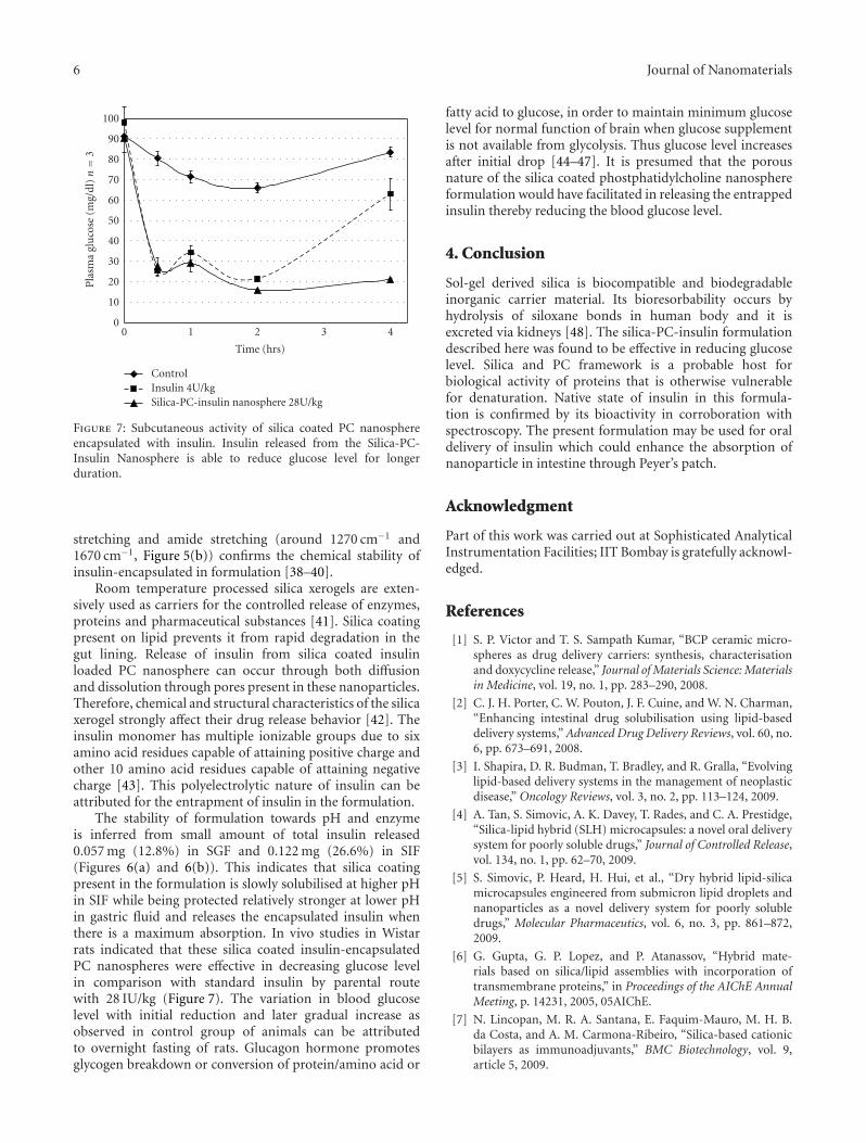
Figure 7: Subcutaneous activity of silica coated PC nanosphereencapsulated with insulin. Insulin released from the Silica-PC-Insulin Nanosphere is able to reduce glucose level for longerduration.
stretching and amide stretching (around 1270 cm−1 and1670 cm−1, Figure 5(b)) confirms the chemical stability ofinsulin-encapsulated in formulation [38–40].
Room temperature processed silica xerogels are exten-sively used as carriers for the controlled release of enzymes,proteins and pharmaceutical substances [41]. Silica coatingpresent on lipid prevents it from rapid degradation in thegut lining. Release of insulin from silica coated insulinloaded PC nanosphere can occur through both diffusionand dissolution through pores present in these nanoparticles.Therefore, chemical and structural characteristics of the silicaxerogel strongly affect their drug release behavior [42]. Theinsulin monomer has multiple ionizable groups due to sixamino acid residues capable of attaining positive charge andother 10 amino acid residues capable of attaining negativecharge [43]. This polyelectrolytic nature of insulin can beattributed for the entrapment of insulin in the formulation.
The stability of formulation towards pH and enzymeis inferred from small amount of total insulin released0.057 mg (12.8%) in SGF and 0.122 mg (26.6%) in SIF(Figures 6(a) and 6(b)). This indicates that silica coatingpresent in the formulation is slowly solubilised at higher pHin SIF while being protected relatively stronger at lower pHin gastric fluid and releases the encapsulated insulin whenthere is a maximum absorption. In vivo studies in Wistarrats indicated that these silica coated insulin-encapsulatedPC nanospheres were effective in decreasing glucose levelin comparison with standard insulin by parental routewith 28 IU/kg (Figure 7). The variation in blood glucoselevel with initial reduction and later gradual increase asobserved in control group of animals can be attributedto overnight fasting of rats. Glucagon hormone promotesglycogen breakdown or conversion of protein/amino acid or
fatty acid to glucose, in order to maintain minimum glucoselevel for normal function of brain when glucose supplementis not available from glycolysis. Thus glucose level increasesafter initial drop [44–47]. It is presumed that the porousnature of the silica coated phostphatidylcholine nanosphereformulation would have facilitated in releasing the entrappedinsulin thereby reducing the blood glucose level.
4. Conclusion
Sol-gel derived silica is biocompatible and biodegradableinorganic carrier material. Its bioresorbability occurs byhydrolysis of siloxane bonds in human body and it isexcreted via kidneys [48]. The silica-PC-insulin formulationdescribed here was found to be effective in reducing glucoselevel. Silica and PC framework is a probable host forbiological activity of proteins that is otherwise vulnerablefor denaturation. Native state of insulin in this formula-tion is confirmed by its bioactivity in corroboration withspectroscopy. The present formulation may be used for oraldelivery of insulin which could enhance the absorption ofnanoparticle in intestine through Peyer’s patch.
Acknowledgment
Part of this work was carried out at Sophisticated AnalyticalInstrumentation Facilities; IIT Bombay is gratefully acknowl-edged.
References
[1] S. P. Victor and T. S. Sampath Kumar, “BCP ceramic micro-spheres as drug delivery carriers: synthesis, characterisationand doxycycline release,” Journal of Materials Science: Materialsin Medicine, vol. 19, no. 1, pp. 283–290, 2008.
[2] C. J. H. Porter, C. W. Pouton, J. F. Cuine, and W. N. Charman,“Enhancing intestinal drug solubilisation using lipid-baseddelivery systems,” Advanced Drug Delivery Reviews, vol. 60, no.6, pp. 673–691, 2008.
[3] I. Shapira, D. R. Budman, T. Bradley, and R. Gralla, “Evolvinglipid-based delivery systems in the management of neoplasticdisease,” Oncology Reviews, vol. 3, no. 2, pp. 113–124, 2009.
[4] A. Tan, S. Simovic, A. K. Davey, T. Rades, and C. A. Prestidge,“Silica-lipid hybrid (SLH) microcapsules: a novel oral deliverysystem for poorly soluble drugs,” Journal of Controlled Release,vol. 134, no. 1, pp. 62–70, 2009.
[5] S. Simovic, P. Heard, H. Hui, et al., “Dry hybrid lipid-silicamicrocapsules engineered from submicron lipid droplets andnanoparticles as a novel delivery system for poorly solubledrugs,” Molecular Pharmaceutics, vol. 6, no. 3, pp. 861–872,2009.
[6] G. Gupta, G. P. Lopez, and P. Atanassov, “Hybrid mate-rials based on silica/lipid assemblies with incorporation oftransmembrane proteins,” in Proceedings of the AIChE AnnualMeeting, p. 14231, 2005, 05AIChE.
[7] N. Lincopan, M. R. A. Santana, E. Faquim-Mauro, M. H. B.da Costa, and A. M. Carmona-Ribeiro, “Silica-based cationicbilayers as immunoadjuvants,” BMC Biotechnology, vol. 9,article 5, 2009.

Journal of Nanomaterials 7
[8] J. Liu, A. Stace-Naughton, X. Jiang, and C. J. Brinker, “Porousnanoparticle supported lipid bilayers (protocells) as deliveryvehicles,” Journal of the American Chemical Society, vol. 131,no. 4, pp. 1354–1355, 2009.
[9] S. Begu, S. Girod, D. A. Lerner, N. Jardiller, C. Tourne-Peteilh,and J.-M. Devoisselle, “Characterization of a phospholipidbilayer entrapped into non-porous silica nanospheres,” Jour-nal of Materials Chemistry, vol. 14, no. 8, pp. 1316–1320, 2004.
[10] Y. Li and A. K. Mitra, “Effects of phospholipid chain length,concentration, charge, and vesicle size on pulmonary insulinabsorption,” Pharmaceutical Research, vol. 13, no. 1, pp. 76–79, 1996.
[11] R. N. Farias, A. L. Vinals, and R. D. Morero, “Fusion of neg-atively charged phospholipid vesicles by insulin. Relationshipwith lipid fluidity,” Journal of Biological Chemistry, vol. 261,no. 33, pp. 15508–15512, 1986.
[12] T.-Z. Yang, X.-T. Wang, X.-Y. Yan, and Q. Zhang, “Phos-pholipid deformable vesicles for buccal delivery of insulin,”Chemical and Pharmaceutical Bulletin, vol. 50, no. 6, pp. 749–753, 2002.
[13] Y.-Y. Huang and C.-H. Wang, “Pulmonary delivery of insulinby liposomal carriers,” Journal of Controlled Release, vol. 113,no. 1, pp. 9–14, 2006.
[14] F. Cui, K. Shi, L. Zhang, A. Tao, and Y. Kawashima,“Biodegradable nanoparticles loaded with insulin-phospholipid complex for oral delivery: preparation, invitro characterization and in vivo evaluation,” Journal ofControlled Release, vol. 114, no. 2, pp. 242–250, 2006.
[15] B.-Y. Kim, J. H. Jeong, K. Park, and J.-D. Kim, “Bioadhesiveinteraction and hypoglycemic effect of insulin-loaded lectin-microparticle conjugates in oral insulin delivery system,”Journal of Controlled Release, vol. 102, no. 3, pp. 525–538, 2005.
[16] H. Reithmeier, J. Herrmann, and A. Gopferich, “Lipidmicroparticles as a parenteral controlled release device forpeptides,” Journal of Controlled Release, vol. 73, no. 2-3, pp.339–350, 2001.
[17] N. K. Raman, M. T. Anderson, and C. J. Brinker,“Template-based approaches to the preparation of amor-phous, nanoporous silicas,” Chemistry of Materials, vol. 8, no.8, pp. 1682–1701, 1996.
[18] R. K. Rana, Y. Mastai, and A. Gedanken, “Acoustic cavitationleading to the morphosynthesis of mesoporous silica vesicles,”Advanced Materials, vol. 14, no. 19, pp. 1414–1418, 2002.
[19] M. R. Knecht and D. W. Wright, “Amine-terminated den-drimers as biomimetic templates for silica nanosphere forma-tion,” Langmuir, vol. 20, no. 11, pp. 4728–4732, 2004.
[20] M. Zhao, L. Sun, and R. M. Crooks, “Preparation of Cunanoclusters within dendrimer templates,” Journal of theAmerican Chemical Society, vol. 120, pp. 4877–4878, 1998.
[21] D. Dollimore and T. Shingles, “Preparation of microporoussilica by the technique of freeze-thawing,” Journal of ColloidAnd Interface Science, vol. 29, no. 4, pp. 601–604, 1969.
[22] J. Zhang, Z. Liu, B. Han, et al., “Preparation of silica and TiO2-SiO2 core-shell nanoparticles in water-in-oil microemulsionusing compressed CO2 as reactant and antisolvent,” Journal ofSupercritical Fluids, vol. 36, no. 3, pp. 194–201, 2006.
[23] A. Imhof, “Preparation and characterization of titania-coatedpolystyrene spheres and hollow titania shells,” Langmuir, vol.17, no. 12, pp. 3579–3585, 2002.
[24] T. Von Werne and T. E. Patten, “Atom transfer radicalpolymerization from nanoparticles: a tool for the preparationof well-defined hybrid nanostructures and for understandingthe chemistry of controlled/“living” radical polymerizations
from surfaces,” Journal of the American Chemical Society, vol.123, no. 31, pp. 7497–7505, 2001.
[25] S. Radin, T. Chen, and P. Ducheyne, “The controlled release ofdrugs from emulsified, sol gel processed silica microspheres,”Biomaterials, vol. 30, no. 5, pp. 850–858, 2009.
[26] J. Livage, F. Beteille, C. Roux, M. Chatry, and P. Davidson,“Sol-gel synthesis of oxide materials,” Acta Materialia, vol. 46,no. 3, pp. 743–750, 1998.
[27] A. Jintapattanakit, V. B. Junyaprasert, S. Mao, J. Sitterberg,U. Bakowsky, and T. Kissel, “Peroral delivery of insulin usingchitosan derivatives: a comparative study of polyelectrolytenanocomplexes and nanoparticles,” International Journal ofPharmaceutics, vol. 342, no. 1-2, pp. 240–249, 2007.
[28] S. Begu, S. Girod, D. A. Lerner, N. Jardiller, C. Tourne-Peteilh,and J.-M. Devoisselle, “Characterization of a phospholipidbilayer entrapped into non-porous silica nanospheres,” Jour-nal of Materials Chemistry, vol. 14, no. 8, pp. 1316–1320, 2004.
[29] H.-P. Hentze, S. R. Raghavan, C. A. McKelvey, and E. W. Kaler,“Silica hollow spheres by templating of catanionic vesicles,”Langmuir, vol. 19, no. 4, pp. 1069–1074, 2003.
[30] D. Lootens, C. Vautrin, H. Van Damme, and T. Zemb,“Facetted hollow silica vesicles made by templating catanionicsurfactant vesicles,” Journal of Materials Chemistry, vol. 13, no.9, pp. 2072–2074, 2003.
[31] W. Xu, Q. Gao, Y. Xu, D. Wu, and Y. Sun, “pH-Controlleddrug release from mesoporous silica tablets coated withhydroxypropyl methylcellulose phthalate,” Materials ResearchBulletin, vol. 44, no. 3, pp. 606–612, 2009.
[32] W. Xu, Q. Gao, Y. Xu, et al., “Controllable release of ibuprofenfrom size-adjustable and surface hydrophobic mesoporoussilica spheres,” Powder Technology, vol. 191, no. 1-2, pp. 13–20,2009.
[33] C. Charnay, S. Begu, C. Tourne-Peteilh, L. Nicole, D. A. Lerner,and J. M. Devoisselle, “Inclusion of ibuprofen in mesoporoustemplated silica: drug loading and release property,” EuropeanJournal of Pharmaceutics and Biopharmaceutics, vol. 57, no. 3,pp. 533–540, 2004.
[34] Q. Tang, Y. Xu, D. Wu, and Y. Sun, “A study of carboxylic-modified mesoporous silica in controlled delivery for drugfamotidine,” Journal of Solid State Chemistry, vol. 179, no. 5,pp. 1513–1520, 2006.
[35] C. A. Aerts, E. Verraedt, R. Mellaerts, et al., “Tunability of porediameter and particle size of amorphous microporous silicafor diffusive controlled release of drug compounds,” Journal ofPhysical Chemistry C, vol. 111, no. 36, pp. 13404–13409, 2007.
[36] Y. Li and W. T. Yip, “Liposomes as protective capsules for activesilica sol-gel biocomposite synthesis,” Journal of the AmericanChemical Society, vol. 127, no. 37, pp. 12756–12757, 2005.
[37] T. Coradin and J. Livage, “Effect of some amino acids andpeptides on silicic acid polymerization,” Colloids and SurfacesB, vol. 21, no. 4, pp. 329–336, 2001.
[38] N.-T. Yu, C. S. Liu, and D. C. O’Shea, “Laser Ramanspectroscopy and the conformation of insulin and proinsulin,”Journal of Molecular Biology, vol. 70, no. 1, pp. 117–132, 1972.
[39] N.-T. Yu, C. S. Liu, J. Culver, and D. C. O’Shea, “A preliminaryRaman spectroscopic study of native zinc-insulin crystals,”Biochimica et Biophysica Acta, vol. 263, no. 1, pp. 1–6, 1972.
[40] H. Fabian and P. Anzenbacher, “New developments in Ramanspectroscopy of biological systems,” Vibrational Spectroscopy,vol. 4, no. 2, pp. 125–148, 1993.
[41] H. Bottcher, P. Slowik, and W. Suß, “Sol-gel carrier systemsfor controlled drug delivery,” Journal of Sol-Gel Science andTechnology, vol. 13, no. 1–3, pp. 277–281, 1999.

8 Journal of Nanomaterials
[42] B. Dahlback, “Blood coagulation,” Lancet, vol. 355, no. 9215,pp. 1627–1632, 2000.
[43] B. Sarmento, D. Ferreira, F. Veiga, and A. Ribeiro, “Charac-terization of insulin-loaded alginate nanoparticles producedby ionotropic pre-gelation through DSC and FTIR studies,”Carbohydrate Polymers, vol. 66, no. 1, pp. 1–7, 2006.
[44] B. R. Landau, J. Wahren, V. Chandramouli, W. C. Schumann,K. Ekberg, and S. C. Kalhan, “Contributions of gluconeogene-sis to glucose production in the fasted state,” Journal of ClinicalInvestigation, vol. 98, no. 2, pp. 378–385, 1996.
[45] N. Barzilai, D. Massillon, and L. Rossetti, “Effects of fasting onhepatic and peripheral glucose metabolism in conscious ratswith near-total fat depletion,” Biochemical Journal, vol. 310,no. 3, pp. 819–826, 1995.
[46] P. J. Randle, P. B. Garland, C. N. Hales, and E. A. Newsholme,“The glucose fatty-acid cycle its role in insulin sensitivity andthe metabolic disturbances of diabetes mellitus,” The Lancet,vol. 281, no. 7285, pp. 785–789, 1963.
[47] L. K. Butler, “Regulation of blood glucose levels in normal anddiabetic rats,” in Proceedings of the 16th Workshop/Conferenceof the Association for Biology Laboratory Education (ABLE ’95),vol. 16, pp. 181–202, 1995.
[48] P. Kortesuo, M. Ahola, S. Karlsson, I. Kangasniemi, A. Yli-Urpo, and J. Kiesvaara, “Silica xerogel as an implantable carrierfor controlled drug delivery—evaluation of drug distributionand tissue effects after implantation,” Biomaterials, vol. 21, no.2, pp. 193–198, 2000.

Submit your manuscripts athttp://www.hindawi.com
ScientificaHindawi Publishing Corporationhttp://www.hindawi.com Volume 2014
CorrosionInternational Journal of
Hindawi Publishing Corporationhttp://www.hindawi.com Volume 2014
Polymer ScienceInternational Journal of
Hindawi Publishing Corporationhttp://www.hindawi.com Volume 2014
Hindawi Publishing Corporationhttp://www.hindawi.com Volume 2014
CeramicsJournal of
Hindawi Publishing Corporationhttp://www.hindawi.com Volume 2014
CompositesJournal of
NanoparticlesJournal of
Hindawi Publishing Corporationhttp://www.hindawi.com Volume 2014
Hindawi Publishing Corporationhttp://www.hindawi.com Volume 2014
International Journal of
Biomaterials
Hindawi Publishing Corporationhttp://www.hindawi.com Volume 2014
NanoscienceJournal of
TextilesHindawi Publishing Corporation http://www.hindawi.com Volume 2014
Journal of
NanotechnologyHindawi Publishing Corporationhttp://www.hindawi.com Volume 2014
Journal of
CrystallographyJournal of
Hindawi Publishing Corporationhttp://www.hindawi.com Volume 2014
The Scientific World JournalHindawi Publishing Corporation http://www.hindawi.com Volume 2014
Hindawi Publishing Corporationhttp://www.hindawi.com Volume 2014
CoatingsJournal of
Advances in
Materials Science and EngineeringHindawi Publishing Corporationhttp://www.hindawi.com Volume 2014
Smart Materials Research
Hindawi Publishing Corporationhttp://www.hindawi.com Volume 2014
Hindawi Publishing Corporationhttp://www.hindawi.com Volume 2014
MetallurgyJournal of
Hindawi Publishing Corporationhttp://www.hindawi.com Volume 2014
BioMed Research International
MaterialsJournal of
Hindawi Publishing Corporationhttp://www.hindawi.com Volume 2014
Nano
materials
Hindawi Publishing Corporationhttp://www.hindawi.com Volume 2014
Journal ofNanomaterials