Surface modification of silica-coated zirconia by chemical treatments
8
Applied Surface Science 257 (2010) 1228–1235 Contents lists available at ScienceDirect Applied Surface Science journal homepage: www.elsevier.com/locate/apsusc Surface modification of silica-coated zirconia by chemical treatments Christie Ying Kei Lung a , Edwin Kukk b , Toni Hägerth b , Jukka Pekka Matinlinna a,∗ a Dental Materials Science, Faculty of Dentistry, The University of Hong Kong, Hong Kong SAR, PR China b Department of Physics and Astronomy, Faculty of Mathematics and Natural Sciences, University of Turku, Finland article info Article history: Received 15 July 2010 Received in revised form 6 August 2010 Accepted 6 August 2010 Available online 12 August 2010 Keywords: Surface treatment Hydroxylation Binding energy Surface roughness abstract Zirconia surface modification by various chemical treatments after silica coating by sandblasting was investigated in this study. The surface of silica-coated dental zirconia was hydroxylated by treatment with different acids at room temperature for 4 h, rinsed with deionized water and air-dried. The modified sur- faces were characterized by X-ray photoelectron spectroscopy (XPS) and atomic force microscopy (AFM). Shifts in binding energies for Zr 3d 5/2 and Si 2p peaks were observed after treatment with acids, thereby showing a change in the chemical states of zirconium and silicon on the surface layer of silica-coated zirconia. The XPS analysis revealed that the silica-coated zirconia (SiO 2 –ZrO 2 ) surfaces had changed to hydrous silica-coated zirconia (SiO 2 –ZrO 2 ·nH 2 O). One-way ANOVA analysis revealed there was signifi- cant difference in both surface roughness parameters of silica-coated zirconia after chemical treatments and the surface topography varied depending on the acid treatment. © 2010 Elsevier B.V. All rights reserved. 1. Introduction In dentistry, it is desirable that strong bonding is achieved between materials and dental tissues to ensure that the material, for example, as a dental restoration, survives in the oral environ- ment. Zirconia has found wide applications in dental restorations such as crowns, bridges and implant abutments because of its high fracture toughness, semi-translucency, radiopacity and, most importantly, biocompatibility [1]. However, the bonding between resin cement and zirconia is relatively weak. The shear bond strength between yttria-stabilized zirconia and resin composite is reported to be only about 1.5 MPa, without any pretreatment or use of primer [2]. Many investigations have reported that the bond strength of resin cement to zirconia ceramic is greatly affected by surface mod- ifications of zirconia. Methods such as acid etching by hydrofluoric acid (HF), air-particle abrasion using alumina of various particle sizes, sandblasting with 3 M ESPE Rocatec Pre (110 m alumina), Plus (110 m silica-coated alumina) or Soft (30 m silica-coated alumina) and, more recently, laser irradiation are reported to improve adhesive strength [3–11]. Apart from HF acid etching, these methods mainly bring about physical changes of the sur- face topography of zirconia by different mechanisms to increase ∗ Corresponding author at: Dental Materials Science, Faculty of Dentistry, The University of Hong Kong, Hong, 4/F, Prince Philip Dental Hospital, 34 Hospital Road, Sai Ying Pun, Hong Kong SAR, PR China. Tel.: +852 2859 0380; fax: +852 2548 9464. E-mail addresses: [email protected] (C.Y.K. Lung), ekukk@utu.fi (E. Kukk), tjhage@utu.fi (T. Hägerth), [email protected] (J.P. Matinlinna). the surface roughness. Increased surface roughness in turn results in increased surface energy and better wetting for bonding. In principle, an alternative zirconia surface treatment is the chemi- cal treatment by reaction with concentrated strong acids and bases [12]. At ambient temperature and humidity, a certain quantity of hydroxyl groups is formed on the surface of yttria-stabilized zir- conia. The density of hydroxyl groups increases after treatment with concentrated acid [13]. Conceptual diagrams can illustrate this process for simplified 2D (Fig. 1) and a more realistic 3D surface mesh (Fig. 2). The zirconia surface must be silica-coated, because zirconia is inert and bond strength between resin composite and silanized zirconia is relatively weak [2]. When a silane solution after hydrolysis converts to the silanol ( Si–OR → Si–OH) and is then applied onto the silica-coated zirconia surface, the silanol reacts with surface hydroxyl groups by condensation to form siloxane bonds ( Si–O–Si ). Therefore, the more hydroxyl groups there are on the surface of silica-coated zirconia, the higher the degree of bonding there will be between the silane primer and the silica- coated zirconia surface. More energy is required to break through the interfacial layer. The result is thus a strengthening of the adhe- sion between the resin cement and the silicatized and silanized zirconia. In the present in vitro investigation, we studied surface treat- ments of yttria-stabilized zirconia with different mineral acids at room temperature. All surfaces were polished and sandblasted before acid treatment. These mineral acids are commonly used for etching on most metal surfaces in various industries. The quan- titative atomic composition, the change of chemical states of Si and Zr and surface topography after various surface treatments 0169-4332/$ – see front matter © 2010 Elsevier B.V. All rights reserved. doi:10.1016/j.apsusc.2010.08.029
Transcript of Surface modification of silica-coated zirconia by chemical treatments

S
Ca
b
a
ARRAA
KSHBS
1
bfmshirsro
riasPaitf
US
t
0d
Applied Surface Science 257 (2010) 1228–1235
Contents lists available at ScienceDirect
Applied Surface Science
journa l homepage: www.e lsev ier .com/ locate /apsusc
urface modification of silica-coated zirconia by chemical treatments
hristie Ying Kei Lunga, Edwin Kukkb, Toni Hägerthb, Jukka Pekka Matinlinnaa,∗
Dental Materials Science, Faculty of Dentistry, The University of Hong Kong, Hong Kong SAR, PR ChinaDepartment of Physics and Astronomy, Faculty of Mathematics and Natural Sciences, University of Turku, Finland
r t i c l e i n f o
rticle history:eceived 15 July 2010eceived in revised form 6 August 2010ccepted 6 August 2010
a b s t r a c t
Zirconia surface modification by various chemical treatments after silica coating by sandblasting wasinvestigated in this study. The surface of silica-coated dental zirconia was hydroxylated by treatment withdifferent acids at room temperature for 4 h, rinsed with deionized water and air-dried. The modified sur-
vailable online 12 August 2010
eywords:urface treatmentydroxylationinding energy
faces were characterized by X-ray photoelectron spectroscopy (XPS) and atomic force microscopy (AFM).Shifts in binding energies for Zr 3d5/2 and Si 2p peaks were observed after treatment with acids, therebyshowing a change in the chemical states of zirconium and silicon on the surface layer of silica-coatedzirconia. The XPS analysis revealed that the silica-coated zirconia (SiO2–ZrO2) surfaces had changed tohydrous silica-coated zirconia (SiO2–ZrO2·nH2O). One-way ANOVA analysis revealed there was signifi-cant difference in both surface roughness parameters of silica-coated zirconia after chemical treatments
hy va
urface roughness and the surface topograp. Introduction
In dentistry, it is desirable that strong bonding is achievedetween materials and dental tissues to ensure that the material,or example, as a dental restoration, survives in the oral environ-
ent. Zirconia has found wide applications in dental restorationsuch as crowns, bridges and implant abutments because of itsigh fracture toughness, semi-translucency, radiopacity and, most
mportantly, biocompatibility [1]. However, the bonding betweenesin cement and zirconia is relatively weak. The shear bondtrength between yttria-stabilized zirconia and resin composite iseported to be only about 1.5 MPa, without any pretreatment or usef primer [2].
Many investigations have reported that the bond strength ofesin cement to zirconia ceramic is greatly affected by surface mod-fications of zirconia. Methods such as acid etching by hydrofluoriccid (HF), air-particle abrasion using alumina of various particleizes, sandblasting with 3 M ESPE Rocatec Pre (110 �m alumina),lus (110 �m silica-coated alumina) or Soft (30 �m silica-coated
lumina) and, more recently, laser irradiation are reported tomprove adhesive strength [3–11]. Apart from HF acid etching,hese methods mainly bring about physical changes of the sur-ace topography of zirconia by different mechanisms to increase∗ Corresponding author at: Dental Materials Science, Faculty of Dentistry, Theniversity of Hong Kong, Hong, 4/F, Prince Philip Dental Hospital, 34 Hospital Road,ai Ying Pun, Hong Kong SAR, PR China. Tel.: +852 2859 0380; fax: +852 2548 9464.
E-mail addresses: [email protected] (C.Y.K. Lung), [email protected] (E. Kukk),[email protected] (T. Hägerth), [email protected] (J.P. Matinlinna).
169-4332/$ – see front matter © 2010 Elsevier B.V. All rights reserved.oi:10.1016/j.apsusc.2010.08.029
ried depending on the acid treatment.© 2010 Elsevier B.V. All rights reserved.
the surface roughness. Increased surface roughness in turn resultsin increased surface energy and better wetting for bonding. Inprinciple, an alternative zirconia surface treatment is the chemi-cal treatment by reaction with concentrated strong acids and bases[12].
At ambient temperature and humidity, a certain quantity ofhydroxyl groups is formed on the surface of yttria-stabilized zir-conia. The density of hydroxyl groups increases after treatmentwith concentrated acid [13]. Conceptual diagrams can illustrate thisprocess for simplified 2D (Fig. 1) and a more realistic 3D surfacemesh (Fig. 2). The zirconia surface must be silica-coated, becausezirconia is inert and bond strength between resin composite andsilanized zirconia is relatively weak [2]. When a silane solution afterhydrolysis converts to the silanol ( Si–OR → Si–OH) and is thenapplied onto the silica-coated zirconia surface, the silanol reactswith surface hydroxyl groups by condensation to form siloxanebonds ( Si–O–Si ). Therefore, the more hydroxyl groups there areon the surface of silica-coated zirconia, the higher the degree ofbonding there will be between the silane primer and the silica-coated zirconia surface. More energy is required to break throughthe interfacial layer. The result is thus a strengthening of the adhe-sion between the resin cement and the silicatized and silanizedzirconia.
In the present in vitro investigation, we studied surface treat-ments of yttria-stabilized zirconia with different mineral acids at
room temperature. All surfaces were polished and sandblastedbefore acid treatment. These mineral acids are commonly used foretching on most metal surfaces in various industries. The quan-titative atomic composition, the change of chemical states of Siand Zr and surface topography after various surface treatments
C.Y.K. Lung et al. / Applied Surface Science 257 (2010) 1228–1235 1229
surfac
wamr[tos
2
2
sbspe
Fig. 1. A conceptual schematic diagram of hydroxylation of silica-coated zirconia
ere examined by X-ray photoelectron spectroscopy (XPS) andtomic force microscopy (AFM). The study provides valuable infor-ation on the future study of the shear bond strength between
esin cement and silica-coated and silanized zirconia in dentistry14,15]. The hypotheses of this study were that acid treatment leadso (1) a surface chemical reaction, i.e. hydroxylation, on the surfacef silica-coated zirconia, and (2) changes of surface topography andurface roughness.
. Experimental
.1. Sample preparation
The materials used in this study are shown in Table 1. Ten
amples of yttria-stabilized dental zirconia (Nobel Biocare, Göte-org, Sweden) were used for this investigation. The surfaces of allamples were polished with 400-grit silicon carbide paper. Afterolishing, the samples were placed in a beaker filled with 70%thanol, cleaned in an ultrasonic bath (Decon Ultrasonics Ltd, Hovee by reaction of different acids with some dangling oxygen bonds on the surface.
Sussex, England) for 10 min, rinsed with 70% ethanol and then air-dried at room temperature. One sample was left untreated as acontrol. The surfaces of 8 zirconia samples were silica-coated bysandblasting using Rocatec Soft (3 M ESPE, Seefeld, Germany) ata pressure of 280 kPa for 30 s/cm2 at a perpendicular distance of10 mm. The 8 sandblasted samples were placed in a beaker of 70%ethanol, cleaned in the ultrasonic bath for 10 min and air-dried atroom temperature. Surfaces of 2 samples were etched by 5% HFgel (Ivoclar Vivadent AG, Liechtenstein) for 20 s according to theinstruction manual and rinsed with deionized water and air-driedat room temperature. When dry, 1 sample was further treated for4 h in 69–71% nitric acid (BDH, Poole, United Kingdom), whereasthe other was treated for 4 h with Piranha solution (4:1 (v/v) of98% sulphuric acid and 30% hydrogen peroxide (Merck, Darmstadt,
Germany)). The surfaces of the other 5 sandblasted zirconia sam-ples were treated for 4 h with 69–71% nitric acid; Piranha (4:1 (v/v)98% sulphuric acid and 30% hydrogen peroxide); 35.4% hydrochlo-ric acid (BDH, Poole, United Kingdom); 85% phosphoric acid (PekingChemical Works, Peking, China); and 5% HF gel. All samples were
1230 C.Y.K. Lung et al. / Applied Surface Science 257 (2010) 1228–1235
Fsa
r1t
2
2
ett(
Table 2Summary of various treatments on zirconia surfaces.
Sample Surface treatment conditions
1 Polishing the surface of zirconia with a 400-gritsilicon carbide paper
2 (i) Polishing, (ii) sandblasting (30 s/cm2) usingRocatec Soft sand (30 �m silica-coated alumina) ofthe surface of zirconia
3 (i) Polishing, (ii) sandblasting, (iii) 5% HF etchinggel (20 s), (iv) 69–71% HNO3 (4 h)
4 (i) Polishing, (ii) sandblasting, (iii) 5% HF etchinggel (20 s), (iv) 4:1 (v/v) 98% H2SO4 and 30% H2O2
(4 h)5 (i) Polishing, (ii) sandblasting, (iii) 69–71% HNO3
(4 h)6 (i) Polishing, (ii) sandblasting, (iii) 4:1 (v/v) 98%
H2SO4 and 30% H2O2 (4 h)7 (i) Polishing, (ii) sandblasting, (iii) 35.4% HCl (4 h)8 (i) Polishing, (ii) sandblasting, (iii) 85% H3PO4 (4 h)9 (i) Polishing, (ii) sandblasting, (iii) 5% HF etching
TM
ig. 2. Conceptual diagram of 3D zirconia surface mesh showing hydroxylation ofilica-coated zirconia surface after acid treatment, O-bridges linking Zr and Si atomsnd some O dangling bonds.
insed with deionized water and air-dried after treatment. Finally,sample was sandblasted for 60 s/cm2 only as a control. The surface
reatments are summarized in Table 2.
.2. Surface analysis
.2.1. XPS
The chemical composition of the surfaces of the samples wasxamined by XPS using a Perkin–Elmer PHI 5400 spectrometerhat had a mean radius of 140 mm and was equipped with a resis-ive anode detector. The ionization source was Mg K� radiationh� = 1253.6 eV) from a twin-anode X-ray tube. Broad-range sur-
able 1aterials used in the study.
Materials Chemical formula Manufacturer
Procera zirconia Y2O3–ZrO2 Nobel Biocare, GöteborgRocatec Soft SiO2–Al2O3 3M ESPE, Seefeld, GermaHydrofluoric acid HF Ivoclar Vivadent AG, LiecSulphuric acid H2SO4 BDH, Poole, United KingHydrogen peroxide H2O2 Merck, Darmstadt, GermNitric acid HNO3 BDH, Poole, United KingHydrochloric acid HCl BDH, Poole, United KingPhosphoric acid H3PO4 Peking Chemical Works,
gel (20 s)10 (i) Polishing, (ii) sandblasting (60 s/cm2) using
Rocatec Soft sand (30 �m silica-coated alumina) ofthe surface of zirconia
vey scans were performed to determine atomic concentration, ata pass energy of 89.45 eV and an entrance slit width of 4 mm.High-resolution narrow-range scans were also performed at a passenergy of 37.75 eV for selected specific photolines (Zr 3d, Si 2p, C 1s)to determine the chemical shifts. The base pressure in the cham-ber was maintained at about 6 × 10⁻¹⁰ Torr and the X-ray tube wasoperated at 200 W. The C 1s photopeaks were used to calibrate thebinding energy scale of the high-resolution spectra for chemicalshift measurements. The peak composition and energy positionswere then determined using the least-squares curve-fitting tech-nique in Igor Pro analysis environment with SPANCF macro package[16].
2.2.2. AFMThe surface topography of the zirconia samples was examined
by AFM using the Autoprobe CP (Park Scientific Instruments, CA,USA). All measurements were carried out at room temperaturein the non-contact mode by means of the Autoprobe CP’s NanoWorld NCSTR tip. Surface topography images were scanned overthree different areas of 50 �m × 50 �m for each sample. Numericalparameters of surface roughness were determined from each scan.
3. Results and discussion
3.1. XPS analysis
Zirconia surface modification by chemical reaction with various
acids after sandblasting was investigated in this study. The changeof state of zirconium and silicon on the surface of silica-coatedzirconia after acid treatment reflects chemical reactions involvingsilica and zirconia, i.e. hydroxylation. According to the XPS anal-ysis of the clean zirconia surface without any surface treatmentComposition Lot. number
, Sweden Yttria-stabilized zirconia N.A.ny 30 �m silica-coated alumina 353587htenstein 5% etching gel 671242
dom 98% 5748880Bany 30% 7247652
dom 69–71% 10168dom 35.4% K31897252Peking, China 85% 798414
C.Y.K. Lung et al. / Applied Surface Science 257 (2010) 1228–1235 1231
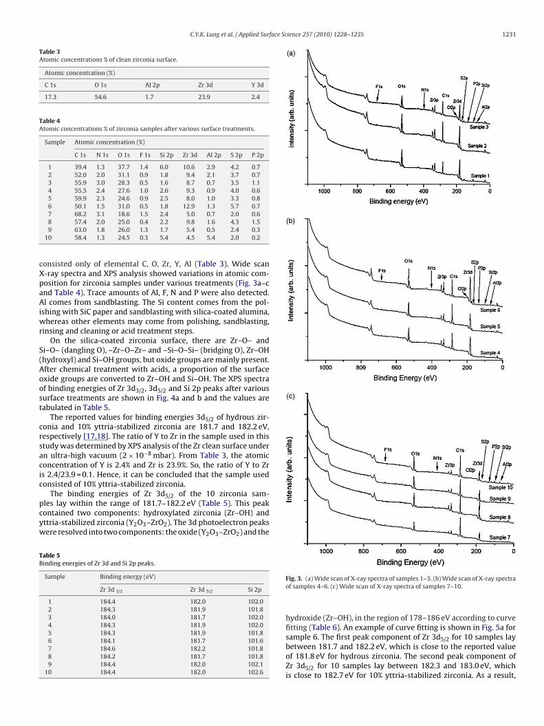
Table 3Atomic concentrations % of clean zirconia surface.
Atomic concentration (%)
C 1s O 1s Al 2p Zr 3d Y 3d
17.3 54.6 1.7 23.9 2.4
Table 4Atomic concentrations % of zirconia samples after various surface treatments.
Sample Atomic concentration (%)
C 1s N 1s O 1s F 1s Si 2p Zr 3d Al 2p S 2p P 2p
1 39.4 1.3 37.7 1.4 6.0 10.6 2.9 4.2 0.72 52.0 2.0 31.1 0.9 1.8 9.4 2.1 3.7 0.73 55.9 3.0 28.3 0.5 1.6 8.7 0.7 3.5 1.14 55.5 2.4 27.6 1.0 2.6 9.3 0.9 4.0 0.65 59.9 2.3 24.6 0.9 2.5 8.0 1.0 3.3 0.86 50.1 1.5 31.0 0.5 1.8 12.9 1.3 5.7 0.77 68.2 3.1 18.6 1.5 2.4 5.0 0.7 2.0 0.6
cXpaAiwr
S(Aoost
crsacic
pcyw
TB
8 57.4 2.0 25.0 0.4 2.2 9.8 1.6 4.3 1.59 63.0 1.8 26.0 1.3 1.7 5.4 0.5 2.4 0.3
10 58.4 1.3 24.5 0.3 5.4 4.5 5.4 2.0 0.2
onsisted only of elemental C, O, Zr, Y, Al (Table 3). Wide scan-ray spectra and XPS analysis showed variations in atomic com-osition for zirconia samples under various treatments (Fig. 3a–cnd Table 4). Trace amounts of Al, F, N and P were also detected.l comes from sandblasting. The Si content comes from the pol-
shing with SiC paper and sandblasting with silica-coated alumina,hereas other elements may come from polishing, sandblasting,
insing and cleaning or acid treatment steps.On the silica-coated zirconia surface, there are Zr–O– and
i–O– (dangling O), –Zr–O–Zr– and –Si–O–Si– (bridging O), Zr–OHhydroxyl) and Si–OH groups, but oxide groups are mainly present.fter chemical treatment with acids, a proportion of the surfacexide groups are converted to Zr–OH and Si–OH. The XPS spectraf binding energies of Zr 3d3/2, 3d5/2 and Si 2p peaks after variousurface treatments are shown in Fig. 4a and b and the values areabulated in Table 5.
The reported values for binding energies 3d5/2 of hydrous zir-onia and 10% yttria-stabilized zirconia are 181.7 and 182.2 eV,espectively [17,18]. The ratio of Y to Zr in the sample used in thistudy was determined by XPS analysis of the Zr clean surface undern ultra-high vacuum (2 × 10⁻⁸ mbar). From Table 3, the atomiconcentration of Y is 2.4% and Zr is 23.9%. So, the ratio of Y to Zrs 2.4/23.9 = 0.1. Hence, it can be concluded that the sample usedonsisted of 10% yttria-stabilized zirconia.
The binding energies of Zr 3d5/2 of the 10 zirconia sam-
les lay within the range of 181.7–182.2 eV (Table 5). This peakontained two components: hydroxylated zirconia (Zr–OH) andttria-stabilized zirconia (Y2O3–ZrO2). The 3d photoelectron peaksere resolved into two components: the oxide (Y2O3–ZrO2) and theable 5inding energies of Zr 3d and Si 2p peaks.
Sample Binding energy (eV)
Zr 3d 3/2 Zr 3d 5/2 Si 2p
1 184.4 182.0 102.02 184.3 181.9 101.83 184.0 181.7 102.04 184.3 181.9 102.05 184.3 181.9 101.86 184.1 181.7 101.67 184.6 182.2 101.88 184.2 181.7 101.89 184.4 182.0 102.1
10 184.4 182.0 102.6
Fig. 3. (a) Wide scan of X-ray spectra of samples 1–3. (b) Wide scan of X-ray spectraof samples 4–6. (c) Wide scan of X-ray spectra of samples 7–10.
hydroxide (Zr–OH), in the region of 178–186 eV according to curvefitting (Table 6). An example of curve fitting is shown in Fig. 5a forsample 6. The first peak component of Zr 3d for 10 samples lay
5/2between 181.7 and 182.2 eV, which is close to the reported valueof 181.8 eV for hydrous zirconia. The second peak component ofZr 3d5/2 for 10 samples lay between 182.3 and 183.0 eV, whichis close to 182.7 eV for 10% yttria-stabilized zirconia. As a result,
1232 C.Y.K. Lung et al. / Applied Surface Science 257 (2010) 1228–1235
Ftt
tz
atti
Table 6Curve fitting of Zr 3d peaks of 10 samples.
Sample Profile 1 Profile 2
3d5/2 3d3/2 3d5/2 3d3/2
1 181.9 184.3 182.5 184.92 182.0 184.4 182.6 185.03 181.7 184.1 182.3 184.74 181.9 184.3 182.4 184.85 181.9 184.3 182.7 185.06 181.7 184.1 182.3 184.77 182.2 184.6 183.0 185.48 181.8 184.2 182.4 184.89 181.9 184.3 182.6 184.9
10 182.1 184.4 182.7 185.1
mental silicon detected from polishing with SiC is negligibly small.For specimen 10, the atomic concentration of elemental Si wasbeyond the detection limit of the XPS spectrometer. The secondpeak binding energies for the 10 samples lay between 101.0 and
Table 7Curve fitting of Si 2p peaks of 10 samples.
Sample Profile 1 Profile 2 Profile 3
1 99.4 101.5 102.32 99.4 101.2 102.13 99.5 101.2 102.24 99.3 101.5 102.35 99.3 101.2 102.1
ig. 4. (a) Variation of binding energy peaks of Zr 3d peaks under various surfacereatments. (b) Variation of binding peaks of Si 2p peaks under various surfacereatments.
he chemical state of zirconium changed from zirconia to hydrousirconia.
XPS analysis of Si 2p peaks showed that elemental silicon waslso present. The Si 2p photoelectron peaks were resolved intohree components: the element, oxide and hydroxylated silica inhe region of 96–104 eV. An example of Si curve fitting is shownn Fig. 5b for sample 6. The binding energies of Si peak structures
Fig. 5. (a) Curve fitting of Zr 3d peaks structure for sample 6. (b) Curve fitting of Si2p peaks structure for sample 6.
resolved by curve fitting are tabulated in Table 7. The first peakbinding energies of samples 1–9 lay between 99.1 and 99.7 eV,which corresponds to elemental Si. However, the presence of ele-
6 99.1 101.0 102.17 99.7 101.4 102.38 99.2 101.3 102.29 99.5 101.5 102.2
10 – 101.6 102.5

face Science 257 (2010) 1228–1235 1233
1bw1ihos
fsuracu
hthtSptfisiczZ
Table 8Mean and standard deviation (SD) of root mean square (Rrms) and average surfaceroughness (Ra).
Sample Rrms ± SD (�m) Ra ± SD (�m)
1 0.18 ± 0.02 0.123 ± 0.0062 0.2 ± 0.2 0.17 ± 0.073 0.21 ± 0.01 0.16 ± 0.014 0.13 ± 0.02 0.09 ± 0.025 0.20 ± 0.03 0.13 ± 0.036 0.17 ± 0.05 0.13 ± 0.03
Fw
C.Y.K. Lung et al. / Applied Sur
01.6 eV owing to SiOx where x ranges from 1 to 2. The third peakinding energies for 10 samples lay between 102.1 and 102.5 eV,hich corresponds to hydrous silica. The reported binding energy is
02.6 for SiO, and 103.0 eV for SiO2 whereas for hydrous SiO2·nH2Ot is 103.5 eV [19]. The shift in binding energies for the SiO2 andydrous silica Si 2p peaks to lower values is due to the bondingf silica to zirconia (–SiO2–ZrO2–) on the surface layer after theandblasting process.
For samples 1, 2 and 10, which were not treated with acids, sur-ace hydroxylation also occurred. Probably, the silicatized zirconiaurface was hydroxylated during rinsing with 70% ethanol in theltra-sonic bath. The application of ultrasound would speed up theate of hydroxylation which varies with the vibration frequencypplied. However, we did not study the hydroxylation of the zir-onia surface in acid solution by the ultrasonic method using anltrasonicator. This would be a topic for future study.
Huang et al. [20] pointed out the difference between zirconiumydroxide (Zr(OH)4·nH2O) and hydrous zirconia (ZrO2·nH2O), inhat the binding energies of Zr 3d5/2 in zirconium hydroxide andydrous zirconia are 183.6 eV and 181.8 eV, respectively. In con-rast, the reported value for Zr 3d5/2 for zirconia is 182.2 eV [20].tructures of zirconium hydroxide and hydrous zirconia were pro-osed by Huang et al. [20] to underlie the difference between thewo compounds. Both structures contain hydroxyl groups on therst layer of the structure. The difference lies in the inner bulk
tructure because the Zr ions are linked by a pair of OH bridgesn zirconium hydroxide but by O bridges in hydrous zirconia. Basi-ally, the inner bulk structure of hydrous zirconia resembles that ofirconia. This resemblance may explain why the binding energy ofr 3d5/2 in hydrous zirconia is close to that of zirconia, but is higherig. 6. Formation of hydroxyl groups on the surface of silica-coated zirconia using sulphuhite circles represent lattice oxide ions, small dark grey circles represent silicon ions an
7 0.13 ± 0.04 0.09 ± 0.038 0.09 ± 0.03 0.06 ± 0.029 0.13 ± 0.02 0.10 ± 0.02
10 0.18 ± 0.02 0.14 ± 0.02
in zirconium hydroxide. Given these facts and the results, we mayconclude that the silica-coated zirconia surface after various acidtreatment, is changed from silica-coated zirconia (SiO2–ZrO2) tohydrous silica-coated zirconia (SiO2–ZrO2·nH2O).
On the surface of zirconia, the dangling bonds of surface oxy-gen atoms are the reactive site for surface chemical reactions.Tamura et al. [21] reported that the surface hydroxyl site density(mol/m2) of various metal oxides were similar. They suggested acommon mechanism of surface hydroxylation for different oxides,—namely, closely packed lattice oxide ions instead of lattice metalions are exposed to the surface because of their large anionic
size and low polarizability. These ions are not fully coordinatedto the lattice metal ions and act as strong Lewis bases whichreact with water and acid when the zirconia surface is treatedwith acid. Hydroxyl groups are formed on the surface oxide layer.A suggested mechanism for hydroxylation of silica-coated zirco-ric acid as an example for illustration (modified from Tamura et al. [21]). Key: larged small dark circles represent lattice zirconium ions.

1 face S
nhtpa
234 C.Y.K. Lung et al. / Applied Sur
ia surface by acid treatment is shown in Fig. 6. The extent of
ydroxylation depends on the type of acid and the duration andemperature of treatment. In this study, the duration and tem-erature for the surface treatments were kept at constant for 4 hnd at room temperature, respectively, for each acid. In general,Fig. 7. AFM images of samples 1–10 af
cience 257 (2010) 1228–1235
use of a stronger and more concentrated acid, higher tempera-
ture and longer treatment time would yield a greater extent ofhydroxylation. A high temperature (≥200 ◦C) for surface treatmentis not recommended because this would cause dehydroxylation[22].ter different surface treatments.

face Sc
3
eacs
R
R
al
fsaaHzwiTeoar
ta(
4
cCsszft
A
F
[
[
[
[
[
[
[
[
[
[
[
C.Y.K. Lung et al. / Applied Sur
.2. AFM analysis
The AFM images showing the detail of surface topography forach sample are shown in Fig. 7. The root mean square (Rrms)nd average surface roughness (Ra) coefficients (Table 8) were cal-ulated using the following equations, with z coordinate as thecanned area:
rms =
√√√√ N∑n=1
(zn − z̄)2
N − 1(1)
a =N∑
n=1
∣∣zn − z̄∣∣
N(2)
The mean surface roughness for both surface parameters wasnalyzed by one-way analysis of variance (ANOVA) at a significantevel of p < 0.05.
One-way ANOVA analysis revealed that there is significant dif-erence of surface roughness among the 10 samples with differenturface treatments (p < 0.002 for Rrms and p < 0.002 for Ra). There islso significant difference in surface roughness (p < 0.0001 for Rrms
nd p < 0.0001 for Ra) for different acid treatments (samples 3–9).owever, there is no significant difference in surface roughness ofirconia between samples 2 and 10 for sandblasting 30 s and 60 sithout acid treatment (p > 0.3 for Rrms and p > 0.4 for Ra), suggest-
ng there is no marked effect in doubling the sandblasting time.he principle of sandblasting in dentistry is bombardment of high-nergy particles (30 �m silica-coated alumina particles in this case)nto the zirconia ceramic surface to increase the surface roughnessnd thus surface area for better mechanical retention of the dentalestoration.
There were changes in topographical features of the surface forhe 10 samples after different surface treatments. The surfaces inll images were, in general, irregular with hillocks and deep poresFig. 7).
. Conclusion
In this study, the effect of various acid treatments on silica-oated zirconia surface has been assessed by XPS and AFM analysis.hemical reactions occurred between the silica-coated zirconiaurface and different acids. The chemical states of zirconium andilicon changed from silica-coated zirconia to hydrous silica-coatedirconia. Statistically significant difference was found for both sur-ace roughness parameters of silica-coated zirconia by chemicalreatments.
cknowledgments
This study was financially supported from a research grant (Seedunding Programme for Basic Research #200905159006) from The
[
[
ience 257 (2010) 1228–1235 1235
University of Hong Kong. The authors wish to thank Dr. TrevorLane, The University of Hong Kong, for proofreading a draft of themanuscript.
References
[1] R.M. Parker, Use of zirconia in restorative dentistry, Dent. Today March (2007)114–119.
[2] T. Derand, M. Molin, K. Kvam, Bond strength of composite luting cement tozirconia ceramic surfaces, Dent. Mater. 21 (2005) 1158–1162.
[3] M. Özcan, H. Nijhuis, L.F. Valandro, Effect of various surface conditioning meth-ods on the adhesion of dual-cure resin cement with MDP functional monomerto zirconia after thermal aging, Dent. Mater. J. 27 (2008) 99–104.
[4] J.P. Matinlinna, T. Heikkinen, M. Özcan, L.V.J. Lassila, P.K. Vallittu, Evaluation ofresin adhesion to zirconia ceramic using some organosilanes, Dent. Mater. 22(2006) 824–831.
[5] T.T. Heikkinen, J.P. Matinlinna, P.K. Vallittu, L.V.J. Lassila, Dental zirconia adhe-sion with silicon compounds using some experimental and conventionalsurface conditioning methods, Silicon 1 (2009) 199–202.
[6] S.S. Atsu, M.A. Kilicarslan, H.C. Kucukesmen, P.S. Aka, Effect of zirconium-oxideceramic surface treatments on the bond strength to adhesive resin, J. Prosthet.Dent. 95 (2006) 430–436.
[7] M. Kern, A. Barloi, B. Yang, Surface conditioning influences zirconia ceramicbonding, J. Dent. Res. 88 (2009) 817–822.
[8] A.N. Cavalcanti, R.M. Foxton, T.F. Watson, M.T. Oliveira, M. Giannini, G.M.Marchi, Bond strength of resin cements to a zirconia ceramic with differentsurface treatments, Oper. Dent. 34 (2009) 280–287.
[9] A.M. Spohr, G.A. Borges, L.H.B. Júnior, E.G. Mota, H.M.S. Oshima, Surface mod-ification of in-ceram zirconia ceramic by Nd:YAG laser, Rocatec system oraluminum oxide sandblasting and its bond strength to a resin cement, Pho-tomed. Laser Surg. 26 (2008) 203–208.
10] M. Özcan, P.K. Vallittu, Effect of surface conditioning methods on the bondstrength of luting cement to ceramics, Dent. Mater. 19 (2003) 725–731.
11] A. Casucci, E. Osorio, R. Osorio, F. Monticelli, M. Toledano, C. Mazzitelli, M. Fer-rari, Influence of different surface treatments on surface zirconia frameworks,J. Dent. 37 (2009) 891–897.
12] U. Lohbauer, M. Zipperle, K. Rischka, A. Petschelt, F.A. Müller, Hydroxylationof dental zirconia surfaces: characterization and bonding potential, J. Biomed.Mater. Res. Part B 87B (2008) 461–467.
13] M. Uchida, H.M. Kim, T. Kokubo, M. Nawa, T. Asano, K. Tanaka, T. Naka-mura, Apatite-forming ability of a zirconia/alumina nano-composite inducedby chemical treatment, J. Biomed. Mater. Res. Part A 60 (2002) 277–282.
14] C.Y.K. Lung, J.P. Matinlinna, Resin bonding to silicatized zirconia with two iso-cyanatosilanes and a cross-linking silane. Part I experimental, Silicon (2010),doi:10.1007/s12633-010-9044-9, in press.
15] C.Y.K. Lung, J.P. Matinlinna, Resin bonding to silicatized zirconia with two iso-cyanatosilanes and a cross-linking silane. Part II mechanistic approach, Silicon(2010), doi:10.1007/s12633-010-9043-x in press.
16] E. Kukk, Spectral Analysis by Curve Fitting (SPANCF), http://www.physics.utu.fi/en/research/material science/Fitting.html, 2009.
17] J.F. Liu, C. Nistorica, I. Gory, G. Skidmore, F.M. Mantiziba, B.E. Gnade, Layer-by-layer deposition of zirconium oxide films from aqueous solutions for frictionreduction in silicon-based microelectromechanical system devices, Thin solidfilms 492 (2005) 6–12.
18] Y.N. Makurin, V.N. Strekalovskii, É.G. Vovkotrub, Investigation of the Zr3d X-ray photoelectron spectra of zirconium compounds with different types ofbonding, J. Struct. Chem. 21 (1980) 147–150.
19] B.V. Crist, PDF Handbooks of Monochromatic XPS Spectra, Volume2–Commercially Pure Binary Oxides, XPS International, LLC, California,2005, p. 953.
20] C. Huang, Z. Tang, Z. Zhang, Differences between zirconium hydroxide
(Zr(OH)4·nH2O) and hydrous zirconia (ZrO2·nH2O), J. Am. Ceram. Soc. 84 (2001)1637–1638.21] H. Tamura, K. Mita, A. Tanaka, M. Ito, Mechanism of hydroxylation of metaloxide surfaces, J. Colloid Interface Sci. 243 (2001) 202–207.
22] J. Nawrocki, P.W. Carr, M.J. Annen, S. Froelicher, A TGA investigation of hydratedmonoclinic zirconia, Anal. Chim. Acta 327 (1996) 261–266.