Characterization of silica-coated Ag nanoparticles synthesized using a water-soluble nanoparticle...
7
Original paper Characterization of silica-coated Ag nanoparticles synthesized using a water-soluble nanoparticle micelle Soon-Gil Kim, Nobuhiro Hagura, Ferry Iskandar, Kikuo Okuyama * Department of Chemical Engineering, Graduate School of Engineering, Hiroshima University, 1-4-1 Kagamiyama, Higashi Hiroshima 739-8527, Japan article info Article history: Received 5 February 2008 Received in revised form 13 March 2008 Accepted 17 March 2008 Keywords: Nanoparticles Ag Silica coating HTAB Micelles abstract This article describes the synthesis of silica-coated Ag nanoparticles using a water-soluble nanoparticle micelle under basic conditions. Monodispersed Ag nanoparticles with a mean particle size of 7 nm were synthesized using AgNO 3 in the presence of ascorbic acid as a reducing agent. The Ag nanoparticles were easily re-dispersed into an aqueous solution by surface adsorption of surfactant molecules, indicating for- mation of water-soluble nanoparticle micelles. Silica-coated Ag nanoparticles ranging in size from 50 to 100 nm were obtained by controlling the surfactant, Ag nanoparticle and tetraethylortho silicate (TEOS) concentrations. Adsorbed surfactant monolayers on Ag nanoparticles were used as a template for the sil- ica shell because of the hydrophobicity of TEOS. In all cases, the size of the resulting particles increased linearly as these concentrations increased. Based on transmission electron microscopy, all the Ag nano- particles were completely covered with a silica shell. In most samples, however, Ag nanoparticle size increased from 7 to 50 nm due to evaporation of hexane by heating. Although mean particle size of sil- ica-coated Ag nanoparticles was drastically altered, characteristic absorption peaks were observed at approximately 410 nm. Ó 2008, The Society of Powder Technology Japan. Published by Elsevier BV and The Society of Powder Technology Japan. All rights reserved. 1. Introduction Recently, functional colloidal nanoparticles (e.g., metal, oxide, rare-earth fluoride and so on) have attracted considerable atten- tion due to their unique physical and chemical properties, as well as due to their potential use in electronic, photonic, catalytic and biological applications [1–7]. Such properties are directly corre- lated with particle size and shape, inter-particle distance and characteristics of the protecting organic or inorganic shell. For example, a superlattice of Au nanoparticles can be used in optical devices and also in single-electron tunneling devices, which manifest the coulomb blockade phenomenon at room tempera- ture [3]. Interest in metal nanoparticles (e.g., Au and Ag) stems from their characteristic surface plasmon resonance (SPR), which influences linear and nonlinear optical properties when metal nanoparticles are irradiated by a light source [8]. SPR characteristics are deter- mined by physical parameters of the metal fraction in a host matrix, such as silica [9,10]. Ag nanoparticles are some of the most well- developed materials because they are inexpensive relative to other materials and they possess good chemical and physical properties [11–15]. The optical properties of Ag nanoparticles can be con- trolled by coating them with a uniform shell of a dielectric material, such as silica [15]. Since Mulvaney and co-workers [16] synthesized various silica-coated metal nanoparticles using a silane coupling agent (e.g., aminopropyltriethoxy-silane, APS), various silica coat- ing processes have been extensively studied [16,17]. Although the process reported by Mulvaney is useful for industrial applications, it has the following disadvantages: (i) toxic coupling agents, such as APS, used for surface modification limit bio-applications, and (ii) a long reaction duration (more than 24 h) is required. Mulvaney et al. also reported the synthesis of silica-coated Ag nanoparticles with no coupling agent [18]. Although silica-coated Ag particles were synthesized using this method, in most cases excess silica par- ticles were observed and the process had to be performed using an alcoholic solution, such as 2-propanol. Brinker et al. successfully synthesized self-assembled Au nano- particles in a silica matrix using a water-soluble nanoparticle mi- celle [6]. They reported that Au nanoparticles dispersed in organic solvent could be readily transferred to an aqueous solution by surface modification of Au nanoparticles using a water-soluble surfactant, and as a result, ordered, robust, 3-D Au nanocrystal/sil- ica arrays formed. Adsorbed surfactant monolayers (or bilayers) on the nanoparticle surface have been used as templates for coating purposes [19]. The coating process has also been used as a nanoreactor for phase transformation of FePt nanoparticles with- out solid-state sintering [20]. Although metal nanoparticles were successfully covered with a silica shell and self-assembled into a silica matrix, the resulting particle size ranged from 3 to 10 lm. 0921-8831/$ - see front matter Ó 2008, The Society of Powder Technology Japan. Published by Elsevier BV and The Society of Powder Technology Japan. All rights reserved. doi:10.1016/j.apt.2008.10.006 * Corresponding author. Tel.: +81 82 4247716; fax: +81 82 4247850. E-mail address: [email protected] (K. Okuyama). Advanced Powder Technology 20 (2009) 94–100 Contents lists available at ScienceDirect Advanced Powder Technology journal homepage: www.elsevier.com/locate/apt
-
Upload
independent -
Category
Documents
-
view
3 -
download
0
Transcript of Characterization of silica-coated Ag nanoparticles synthesized using a water-soluble nanoparticle...

Advanced Powder Technology 20 (2009) 94–100
Contents lists available at ScienceDirect
Advanced Powder Technology
journal homepage: www.elsevier .com/locate /apt
Original paper
Characterization of silica-coated Ag nanoparticles synthesized usinga water-soluble nanoparticle micelle
Soon-Gil Kim, Nobuhiro Hagura, Ferry Iskandar, Kikuo Okuyama *
Department of Chemical Engineering, Graduate School of Engineering, Hiroshima University, 1-4-1 Kagamiyama, Higashi Hiroshima 739-8527, Japan
a r t i c l e i n f o a b s t r a c t
Article history:Received 5 February 2008Received in revised form 13 March 2008Accepted 17 March 2008
Keywords:NanoparticlesAgSilica coatingHTABMicelles
0921-8831/$ - see front matter � 2008, The Society odoi:10.1016/j.apt.2008.10.006
* Corresponding author. Tel.: +81 82 4247716; fax:E-mail address: [email protected] (K. O
This article describes the synthesis of silica-coated Ag nanoparticles using a water-soluble nanoparticlemicelle under basic conditions. Monodispersed Ag nanoparticles with a mean particle size of 7 nm weresynthesized using AgNO3 in the presence of ascorbic acid as a reducing agent. The Ag nanoparticles wereeasily re-dispersed into an aqueous solution by surface adsorption of surfactant molecules, indicating for-mation of water-soluble nanoparticle micelles. Silica-coated Ag nanoparticles ranging in size from 50 to100 nm were obtained by controlling the surfactant, Ag nanoparticle and tetraethylortho silicate (TEOS)concentrations. Adsorbed surfactant monolayers on Ag nanoparticles were used as a template for the sil-ica shell because of the hydrophobicity of TEOS. In all cases, the size of the resulting particles increasedlinearly as these concentrations increased. Based on transmission electron microscopy, all the Ag nano-particles were completely covered with a silica shell. In most samples, however, Ag nanoparticle sizeincreased from 7 to 50 nm due to evaporation of hexane by heating. Although mean particle size of sil-ica-coated Ag nanoparticles was drastically altered, characteristic absorption peaks were observed atapproximately 410 nm.
� 2008, The Society of Powder Technology Japan. Published by Elsevier BV and The Society of PowderTechnology Japan. All rights reserved.
1. Introduction
Recently, functional colloidal nanoparticles (e.g., metal, oxide,rare-earth fluoride and so on) have attracted considerable atten-tion due to their unique physical and chemical properties, as wellas due to their potential use in electronic, photonic, catalytic andbiological applications [1–7]. Such properties are directly corre-lated with particle size and shape, inter-particle distance andcharacteristics of the protecting organic or inorganic shell. Forexample, a superlattice of Au nanoparticles can be used in opticaldevices and also in single-electron tunneling devices, whichmanifest the coulomb blockade phenomenon at room tempera-ture [3].
Interest in metal nanoparticles (e.g., Au and Ag) stems from theircharacteristic surface plasmon resonance (SPR), which influenceslinear and nonlinear optical properties when metal nanoparticlesare irradiated by a light source [8]. SPR characteristics are deter-mined by physical parameters of the metal fraction in a host matrix,such as silica [9,10]. Ag nanoparticles are some of the most well-developed materials because they are inexpensive relative to othermaterials and they possess good chemical and physical properties[11–15]. The optical properties of Ag nanoparticles can be con-trolled by coating them with a uniform shell of a dielectric material,
f Powder Technology Japan. Publish
+81 82 4247850.kuyama).
such as silica [15]. Since Mulvaney and co-workers [16] synthesizedvarious silica-coated metal nanoparticles using a silane couplingagent (e.g., aminopropyltriethoxy-silane, APS), various silica coat-ing processes have been extensively studied [16,17]. Although theprocess reported by Mulvaney is useful for industrial applications,it has the following disadvantages: (i) toxic coupling agents, suchas APS, used for surface modification limit bio-applications, and(ii) a long reaction duration (more than 24 h) is required. Mulvaneyet al. also reported the synthesis of silica-coated Ag nanoparticleswith no coupling agent [18]. Although silica-coated Ag particleswere synthesized using this method, in most cases excess silica par-ticles were observed and the process had to be performed using analcoholic solution, such as 2-propanol.
Brinker et al. successfully synthesized self-assembled Au nano-particles in a silica matrix using a water-soluble nanoparticle mi-celle [6]. They reported that Au nanoparticles dispersed inorganic solvent could be readily transferred to an aqueous solutionby surface modification of Au nanoparticles using a water-solublesurfactant, and as a result, ordered, robust, 3-D Au nanocrystal/sil-ica arrays formed. Adsorbed surfactant monolayers (or bilayers) onthe nanoparticle surface have been used as templates for coatingpurposes [19]. The coating process has also been used as ananoreactor for phase transformation of FePt nanoparticles with-out solid-state sintering [20]. Although metal nanoparticles weresuccessfully covered with a silica shell and self-assembled into asilica matrix, the resulting particle size ranged from 3 to 10 lm.
ed by Elsevier BV and The Society of Powder Technology Japan. All rights reserved.

S.-G. Kim et al. / Advanced Powder Technology 20 (2009) 94–100 95
Moreover, numerous nanoparticles were contained in a singlesilica particle.
In this study, silica-coated Ag nanoparticles with controllableparticle size were synthesized using a water-soluble nanoparticlemicelle under basic conditions. We successfully prepared monodi-
Fig. 1. Illustration of the synthesis of (a) Ag nanop
Fig. 2. Characterization of as-synthesized Ag nanoparticles: (a) SEM image, (b) TEM imaimage of a Ag nanoparticle.
spersed Ag and silica-coated Ag nanoparticles. The Ag nanoparti-cles were easily dispersed into an aqueous solution using awater-soluble nanoparticle micelle. Size and morphology of the sil-ica-coated Ag nanoparticles were controlled by varying the con-centrations of Ag nanoparticles, silica precursor and surfactant.
articles and (b) silica-coated Ag nanoparticles.
ge, (c) XRD result and (d) UV–Vis spectra. The inset of (b) is a high-resolution TEM
96 S.-G. Kim et al. / Advanced Powder Technology 20 (2009) 94–100
Electron microscopy was used to analyze the morphology and sizeof silica-coated Ag nanoparticles. The dependence of optical prop-erties on the average particle size was also characterized. The coat-ing technique described in this paper can be directly applied to thepreparation of composite materials containing any of the func-tional colloidal nanoparticles with the ability to control particlesize.
2. Experimental
2.1. Materials
AgNO3 (99.8%, Kanto Chemical), ascorbic acid (99%, Triple Ben-zenes), oleylamine (96%, Tokyo Chemical Industry Co., Ltd.), n-hex-ane (96%, Wako Pure Chemical Industries) and toluene (99.8%,Aldrich) were used to prepare the colloidal Ag nanoparticles. Tetra-ethylortho silicate (TEOS, 99.9%, Kanto Chemicals), ammoniumhydroxide (NH4OH, 28–30%, Kanto Chemicals) and hexadecyltrim-ethyl ammonium bromide (HTAB, 99+%, Acros) were used for thesilica coating on the Ag nanoparticles. All chemicals were used aspurchased without any purification.
2.2. Preparation of Ag nanoparticles
In a typical synthesis of Ag nanoparticles, appropriate amountsof AgNO3 (1–6 mmol) were dissolved in 20 mL of a 1:1 (vol:vol)solution of oleylamine/toluene. The resulting solution was soni-cated for 10 min to completely dissolve the Ag salt. Ascorbic acid(3–18 mmol) was added to the solution. In the reaction, oleylamine
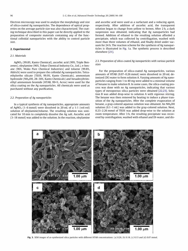
Fig. 3. SEM images of as-synthesized silica particles with different HT
and ascorbic acid were used as a surfactant and a reducing agent,respectively. After addition of ascorbic acid, the transparentsolution began to change from yellow to brown. After 2 h, a darksuspension was obtained, indicating that Ag nanoparticles hadformed. Addition of ethanol to the resulting solution afforded aprecipitate, which was collected by centrifugation, washed withmore than three volumes of ethanol, and finally dried under vac-uum for 24 h. The reaction scheme for the synthesis of Ag nanopar-ticles is illustrated in Fig. 1a. The synthetic process is describedelsewhere [21].
2.3. Preparation of silica-coated Ag nanoparticles with various particlesizes
For the preparation of silica-coated Ag nanoparticles, variousamounts of HTAB (0.07–0.26 mmol) were dissolved in 20 mL de-ionized (DI) water to form solution A. Varying amounts of Ag nano-particles ranging from 1 to 40 mg were added to a minimal volumeof hexane to make solution B. In some cases, the silica-coating pro-cess was done with no Ag nanoparticles, indicating that varioustypes of mesoporous silica particles were obtained [22,23]. Solu-tion B was added drop-wise to solution A with vigorous stirring.The hexane was then removed by heating to induce a phase tran-sition of the Ag nanoparticles. After the complete evaporation ofhexane, a gray-colored aqueous solution was obtained. An NH4OHsolution (0.1–1 mL) was added to the gray-colored solution. Next,0.23–2.26 mmol of TEOS was added drop-wise to the solution atroom temperature. After 3 h, the resulting precipitate was recov-ered by centrifugation, washed with ethanol and DI-water, and dis-
AB concentrations: (a) 0.26, (b) 0.18, (c) 0.13 and (d) 0.07 mmol.

S.-G. Kim et al. / Advanced Powder Technology 20 (2009) 94–100 97
solved in DI-water. The reaction scheme for the synthesis of silica-coated Ag nanoparticles is illustrated in Fig. 1b.
2.4. Characterization
The morphology and particle size of the Ag and silica-coated-Agnanoparticles were observed using a field emission scanning elec-tron microscope (FE-SEM, S-5000, Hitachi, Japan) operated at20 kV. The crystal structure of Ag nanoparticles was characterizedby X-ray diffraction (XRD, RINT 2200V, Rigaku Denki, Tokyo, Japan)using nickel-filtered Cu Ka radiation (k = 1.5408 Å) at 40 kV and30 mA. The morphology and size of the Ag and silica-coated-Agnanoparticles were analyzed using field emission transmissionelectronic microscopy (FE-TEM, JEM-3000F, JEOL, Japan) at300 kV. The optical properties were analyzed by measuring absor-bance spectra using a UV–Vis spectrophotometer (UV-3150, Shi-madzu, Japan).
3. Results and discussion
3.1. Characterization of as-synthesized Ag nanoparticles
Fig. 2 shows characteristics typical of as-synthesized Ag nano-particles. The nanoparticles were spherical in shape with an fccphase structure (Fig. 2a and c). Particle diameter, measured fromSEM images, was 7 nm with a standard deviation of 1.12 nm,whereas crystallite sizes calculated from Scherrer’s formula was4 nm. The calculated crystallite size of Ag nanoparticles was signif-icantly different from that measured in the SEM images because of
Fig. 4. SEM images of silica-coated Ag nanoparticles with different particle sizes. Samplesde-ionized water and 0.13 mmol HTAB. (d) Average particle size as a function of Ag con
polycrystallite formation [24]. TEM studies revealed that the Agnanoparticles with an average size of 6 nm were monocrystallineand polyhedral (Fig. 2b). Particle and crystallite size determinedfrom TEM images were in reasonable agreement with SEM andXRD results. The UV–Vis spectra of Ag nanoparticles dispersed inhexane showed a broad SPR peak at 410 nm (Fig. 2d). The increasedwidth of the SPR peak compared to that of pure Ag nanoparticlesmay be due to the surface-capping of oleylamine on the Ag nano-particles [13].
3.2. Characterization of silica particles with different HTABconcentrations
Due to the strong effect of surfactant concentration on theresulting particle size and shape [22,23], the optimal concentrationfor spherical silica-coating of Ag nanoparticles was determined bypreparation of the silica particles with different surfactant concen-trations, but with no Ag nanoparticles. Fig. 3 shows the SEMimages of the silica particles synthesized using HTAB concentra-tions ranging from 0.07 to 0.26 mmol in 20 mL DI-water. Particlesize and shape were greatly dependent on surfactant concentra-tion. For example, at 0.26 mmol HTAB, bar-type silica particleswith relatively uniform length and diameter (Fig. 3a) were ob-served owing to the formation of interconnected cylinders in water[23]. Silica particle size decreased with decreasing HTABconcentrations and spherical silica particles were observed at0.07 mmol HTAB (Fig. 3b–d). The measured particle sizes were130.26 ± 31.4, 115.97 ± 17.8, 120.97 ± 26.5 and 125.34 ± 27.5 nmwith aspect ratios of 5.3, 2.0, 1.6 and 1.0 for 0.26, 0.18, 0.13 and
were prepared by dispersing (a) 20, (b) 10 and (c) 5 mg of Ag nanoparticles in 20 mLcentration.

98 S.-G. Kim et al. / Advanced Powder Technology 20 (2009) 94–100
0.07 mmol HTAB, respectively. Based on these results, the optimalsurfactant concentration for silica coating was determined to bebetween 0.13 and 0.07 mmol HTAB. For a fixed HTAB concentra-tion, the size of silica-coated Ag nanoparticles could be varied bycontrolling the experimental conditions (e.g., concentrations ofAg, TEOS, water, or catalyst) [25].
3.3. Preparation of silica-coated Ag nanoparticles
The Ag nanoparticles were covered with a silica shell using awater-soluble nanoparticle micelle. Adsorbed surfactant layers onthe surface of Ag nanoparticles were used as templates for silicacoating due to the hydrophobicity of TEOS [19]. Ammoniumhydroxide was used to catalyze the hydrolysis and condensationof TEOS to silica. To create particles of different sizes, silica-coatedAg nanoparticles were synthesized by varying the Ag concentra-tion, ranging from 1 to 40 mg, in a 0.13 mmol HTAB solution in20 mL DI-water. Fig. 4 shows typical results for the synthesis of sil-ica-coated Ag nanoparticles using different Ag concentrations.Addition of 20 mg of Ag nanoparticles produced silica-coated Agnanoparticles with a mean diameter of 77 nm and a standard devi-ation of 20 nm (Fig. 4a). All coated particles were spherical; no bar-type particles were observed. This result indicates that 0.13 mmolHTAB was nearly optimal for the preparation of monolayeredstructures on the surface of Ag nanoparticles [19]. Particle size de-creased as Ag concentration decreased. When coated particleswere synthesized using 10 and 5 mg Ag, the diameters of theresulting particles were 63 and 52 nm, respectively (Fig. 4b and
Fig. 5. SEM images of silica-coated Ag nanoparticles with different particle sizes. SampleAg nanoparticles and 0.13 mmol HTAB. The SEM images in the inset are of a singlconcentration.
c). The dependence of particle size on Ag concentration revealedthat at high concentration particle–particle interactions betweenAg nanoparticles were easily induced in comparison with low con-centrations. Although particle size decreased, particle shape re-mained spherical. The relationship between particle size and Agconcentration is plotted in Fig. 4d. Mean particle size of silica-coated Ag nanoparticles increased with increasing Ag concentra-tion. Prior to silica-coating, Ag nanoparticles more readily aggre-gate at high concentration than at low concentration.
Fig. 5 shows the silica-coated Ag nanoparticles synthesized byadjusting the TEOS concentration. Although TEOS concentrationranged from 0.23 to 2.26 mmol, most particles were sphericaland bar-type structures were not observed. At 2.26 mmol TEOS,mean diameter of the silica-coated Ag nanoparticles was 98 nm(Fig. 5a). Particle size decreased with decreasing TEOS concentra-tions. When coated particles were synthesized using 0.9 and0.45 mmol TEOS, the diameters were 77 and 59 nm, respectively(Fig. 5b and c). Fig. 5d plots the size of silica-coated Ag nanoparticleas a function of TEOS concentration. Particle size increased linearlywith increasing TEOS concentrations. The result was reasonablyconsistent with that shown in Fig. 4d. As shown in the high-reso-lution SEM images in the inset, each particle had a different bright-ness (gray and white), indicating that the Ag nanoparticles werelocated inside the particles [26]. Although the experiments wereperformed using 0.13 mmol HTAB, surfactant concentration wasalso a dominant determinant of particle size.
Fig. 6 shows silica-coated Ag nanoparticles synthesized usingvarious concentrations of HTAB. Average particle size decreased
s were prepared by dispersing (a) 2.26, (b) 0.9 and (c) 0.45 mmol of TEOS in 20 mge silica-coated Ag nanoparticle. (d) Average particle size as a function of TEOS

Fig. 7. UV–Vis absorption spectra of an aqueous dispersion that contained silica-coated Ag nanoparticles synthesized at different TEOS concentrations: (a) 2.26, (b)0.9, (c) 0.45 and (d) 0.23 mmol.
S.-G. Kim et al. / Advanced Powder Technology 20 (2009) 94–100 99
with decreasing HTAB concentration, as the size of a micelle de-pends on surfactant concentration [23]. When silica-coated Agnanoparticles were synthesized using 0.07 and 0.13 mmol HTAB,the diameters were 77 and 56 nm, respectively (Fig. 6a and c). Asshown in Fig. 6b, silica-coated Ag nanoparticles synthesized using0.13 mmol HTAB were completely covered with a silica shell. Inaddition, because HTAB can act as a template, the resulting silicashell had many pores [27]. However, a broad size distribution ofAg nanoparticles was contained in a single silica particle. The resultmay be due to heating at 60 �C for the evaporation of hexane, sug-gesting that growth of Ag nanoparticles readily occurs at that tem-perature. In general, the melting point of nanoparticles is lowerthan that of the bulk material. As a result, solid-state sintering be-tween Ag nanoparticles may also readily occur at that temperature[28]. In the case of 0.07 mmol HTAB, Ag nanoparticles were iso-lated in the silica shell (Fig. 6d), indicating that both the HTAB con-centrations were optimal for the formation of monolayeredstructures on the Ag nanoparticle surface [19]. Interestingly,although both experiments were performed under the same condi-tions, except for the difference in the HTAB concentration, the silicashell morphologies were markedly different. Silica particles syn-thesized using 0.07 mmol HTAB formed spherical hollow struc-tures that contained movable cores of Ag nanoparticles [26].Note that most Ag cores (indicated by arrows in Fig. 6d) werenot located in the centers of the silica particles. Thus, it appearsthat coated Ag nanoparticles move freely within each silica shell.The shell of the hollow silica spheres was 3 nm thick. The mecha-nism by which spherical hollow silica forms is not fully under-stood, and additional studies investigating this phenomenon arecurrently underway.
Fig. 6. SEM and TEM images of silica-coated Ag nanoparticles synthesized at different HTEOS, and 20 mg Ag nanoparticles. The inset image in (d) is a high-resolution TEM imag
3.4. Optical properties of silica-coated Ag nanoparticles
Optical properties of these aqueous dispersions of silica-coatedAg colloids synthesized using various TEOS concentrations rangingfrom 0.23 to 2.26 mmol were characterized using a UV–Vis spec-trophotometer. Fig. 7 shows the absorption spectra of aqueous dis-persions of silica-coated Ag colloids with different particle sizes.Before coating, the Ag nanoparticles had a characteristic SPR peak
TAB concentrations: (a and b) 0.13 and (c and d) 0.07 mmol HTAB in 0.9 mmol ofe of the silica shell that contains Ag nanoparticle.

100 S.-G. Kim et al. / Advanced Powder Technology 20 (2009) 94–100
at a wavelength of 410 nm. Although average particle diameters ofsilica-coated Ag colloids were significantly increased from 50 to100 nm, characteristic SPR peaks remained at around 410 nm. Peakposition was slightly sensitive to changes in coating thickness, butpeak intensity increased as thicker silica shells formed. Because thereflective index of silica is similar to that of water, the plasmonpeaks remained at approximately 410 nm [15].
4. Conclusion
Silica-coated Ag nanoparticles were successfully synthesizedusing a water-soluble nanoparticle micelle. Mean particle diameterof as-synthesized Ag nanoparticles was 7 nm with a standard devi-ation of 1.12 nm. The Ag nanoparticles exhibited a characteristicabsorption peak at 410 nm. The Ag nanoparticles were readily dis-persed into an aqueous solution via water-soluble nanoparticle mi-celles. Adsorbed surfactant monolayers on the surface of Agnanoparticles were used as templates for silica coating due to thehydrophobicity of TEOS. By controlling experimental conditions(e.g., the concentrations of surfactant, Ag nanoparticles and TEOS),the average particle size was readily controlled over the range of50–100 nm. After evaporation by heating to remove hexane, sev-eral Ag nanoparticles had diameters >50 nm. Spherical hollow sil-ica structures that contained movable cores of Ag nanoparticleswere observed. Although average size of the silica-coated Ag nano-particles was dramatically altered, characteristic SPR peaks wereslightly changed by increasing particle size.
Acknowledgements
We acknowledge the Ministry of Education, Culture, Sports, Sci-ence and Technology (MEXT) of Japan for providing a doctoralscholarship (S.-G.K.). This work was supported by a HiroshimaPrefecture Institute of Industrial Science and Technology project.The authors thank Dr. Eishi Tanabe (Hiroshima Prefectural Inst.Industrial Sci. Tech.) for helping with the TEM and for productivediscussions.
References
[1] T. Hyeon, Chemical synthesis of magnetic nanoparticles, Chem. Commun. 9(2003) 927–934.
[2] S.U. Son, I.K. Park, J. Park, T. Hyeon, Synthesis of Cu2O coated Cu nanoparticlesand their successful applications to Ullmann-type amination couplingreactions of aryl chlorides, Chem. Commun. 10 (2004) 778–779.
[3] T. Shimizu, T. Teranishi, S. Hasegawa, M. Miyake, Size evolution of alkanethiol-protected gold nanoparticles by heat treatment in the solid state, J. Phys.Chem. B 107 (2003) 2719–2724.
[4] X. Wang, J. Zhuang, Q. Peng, Y.D. Li, A general strategy for nanocrystalsynthesis, Nature 437 (2005) 121–124.
[5] T. Fried, G. Shemer, G. Markovich, Ordered two-dimensional arrays of ferritenanoparticles, Adv. Mater. 13 (2001) 1158–1161.
[6] H.Y. Fan, K. Yang, D.M. Boye, T. Sigmon, K.J. Malloy, H.F. Xu, G.P. Lopez, C.J.Brinker, Self-assembly of ordered, robust, three-dimensional gold nanocrystal/silica arrays, Science 304 (2004) 567–571.
[7] K. Okuyama, M. Abdullah, I.W. Lenggoro, F. Iskandar, Preparation of functionalnanostructured particles by spray drying, Adv. Powder Technol. 17 (2006)587–611.
[8] A.A. Scalisi, G. Compagnini, L. D’Urso, O. Puglisi, Nonlinear optical activity inAg–SiO2 nanocomposite thin films with different silver concentration, Appl.Surf. Sci. 226 (2004) 237–241.
[9] R.K. Roy, S.K. Mandal, D. Bhattacharyya, A.K. Pal, An ellipsometric investigationof Ag/SiO2 nanocomposite thin films, Eur. Phys. J. B 34 (2003) 25–31.
[10] T.C. Wang, R.E. Cohen, M.F. Rubner, Metallodielectric photonic structuresbased on polyelectrolyte multilayers, Adv. Mater. 14 (2002) 1534–1537.
[11] X.Z. Lin, X.W. Teng, H. Yang, Direct synthesis of narrowly dispersed silvernanoparticles using a single-source precursor, Langmuir 19 (2003) 10081–10085.
[12] Y. Lu, G.L. Liu, L.P. Lee, High-density silver nanoparticle film with temperature-controllable interparticle spacing for a tunable surface enhanced Ramanscattering substrate, Nano Lett. 5 (2005) 5–9.
[13] M. Yamamoto, M. Nakamoto, Novel preparation of monodispersed silvernanoparticles via amine adducts derived from insoluble silver myristate intertiary alkylamine, J. Mater. Chem. 13 (2003) 2064–2065.
[14] B.J. Wiley, Y.C. Chen, J.M. McLellan, Y.J. Xiong, Z.Y. Li, D. Ginger, Y.N. Xia,Synthesis and optical properties of silver nanobars and nanorice, Nano Lett. 7(2007) 1032–1036.
[15] Y. Lu, Y. Yin, Z.Y. Li, Y. Xia, Synthesis and self-assembly of Au@SiO2 core-shellcolloids, Nano Lett. 2 (2002) 785–788.
[16] L.M. LizMarzan, M. Giersig, P. Mulvaney, Synthesis of nanosized gold–silicacore-shell particles, Langmuir 12 (1996) 4329–4335.
[17] S.H. Liu, M.Y. Han, Synthesis, functionalization, and bioconjugation ofmonodisperse, silica-coated gold nanoparticles: robust bioprobes, Adv.Funct. Mater. 15 (2005) 961–967.
[18] V.V. Hardikar, E. Matijevic, Coating of nanosize silver particles with silica, J.Colloid Interface Sci. 221 (2000) 133–136.
[19] G.J. Cho, B.M. Fung, D.T. Glatzhofer, J.S. Lee, Y.G. Shul, Preparation andcharacterization of polypyrrole-coated nanosized novel ceramics, Langmuir 17(2001) 456–461.
[20] Y. Tamada, S. Yamamoto, M. Takano, S. Nasu, T. Ono, Well-ordered L1(0)-FePtnanoparticles synthesized by improved SiO2-nanoreactor method, Appl. Phys.Lett. 90 (2007) 162509.
[21] Y. Kakihara, A production method in liquid phase of Ag ultrafine particles,Japan Patent. No. P2005-36309A, 2005.
[22] D. Myers, Surfactant Science and Technology, John Wiley & Sons Inc., NewJersey, 2006. pp. 107–116.
[23] M.P. Pileni, The role of soft colloidal templates in controlling the size and shapeof inorganic nanocrystals, Nat. Mater. 2 (2003) 145–150.
[24] S.G. Kim, W.N. Wang, T. Iwaki, A. Yabuki, K. Okuyama, Low-temperaturecrystallization of barium ferrite nanoparticles by a sodium citrate-aidedsynthetic process, J. Phys. Chem. C 111 (2007) 10175–10180.
[25] D.C. Lee, F.V. Mikulec, J.M. Pelaez, B. Koo, B.A. Korgel, Synthesis and magneticproperties of silica-coated FePt nanocrystals, J. Phys. Chem. B 110 (2006)11160–11166.
[26] K. Kamata, Y. Lu, Y.N. Xia, Synthesis and characterization of monodispersedcore-shell spherical colloids with movable cores, J. Am. Chem. Soc. 125 (2003)2384–2385.
[27] Y.F. Lu, H.Y. Fan, A. Stump, T.L. Ward, T. Rieker, C.J. Brinker, Aerosol-assistedself-assembly of mesostructured spherical nanoparticles, Nature 398 (1999)223–226.
[28] H.H. Lee, K.S. Chou, K.C. Huang, Inkjet printing of nanosized silver colloids,Nanotechnology 16 (2005) 2436–2441.