
Magnetic nanoparticle clusters as actuators of ssDNA release
9
This journal is © the Owner Societies 2014 Phys. Chem. Chem. Phys. Cite this: DOI: 10.1039/c3cp55470h Magnetic nanoparticle clusters as actuators of ssDNA release† M. Banchelli,‡ a S. Nappini,‡§ a C. Montis, a M. Bonini, a P. Canton, b D. Berti a and P. Baglioni* a One of the major areas of research in nanomedicine is the design of drug delivery systems with remotely controllable release of the drug. Despite the enormous progress in the field, this aspect still poses a challenge, especially in terms of selectivity and possible harmful interactions with biological components other than the target. We report an innovative approach for the controlled release of DNA, based on clusters of core–shell magnetic nanoparticles. The primary nanoparticles are functionalized with a single-stranded oligonucleotide, whose pairing with a half-complementary strand in solution induces clusterization. The application of a low frequency (6 KHz) alternating magnetic field induces DNA melting with the release of the single strand that induces clusterization. The possibility of steering and localizing the magnetic nanoparticles, and magnetically actuating the DNA release discloses new perspectives in the field of nucleic-acid based therapy. Introduction DNA is a versatile molecular building block for nanotechnology that, thanks to its pairing fidelity between complementary strands, 1 enables sub-nanometer control of the architecture of the constructs. For instance, gold nanoparticles (AuNPs) functionalized with single strands of DNA can be designed to assemble into ordered materials, taking advantage of base-pairing selectivity. 2,3 The DNA-directed formation of crystalline superlattices from isotropic 4 (i.e., spherical) and anisotropic 5 NPs is a remarkable example of this approach to bottom-up nanofabrication. In parallel, the biological function of nucleic acids makes polyvalent DNA AuNPs suitable for a range of applications in nanomedicine. The first proof of concept of their potential in this area was the diagnostic detection of DNA single strands through formation of AuNP clusters, revealed by a red shift of the localized surface plasmon resonance. 6 Recently, the discovery of the cellular regulatory properties of short DNAs and RNAs has prompted the use of individual AuNPs as drug delivery systems for oligonucleotides (ONs). 7 AuNPs have many advantages over other delivery methods for DNA, in terms of reduction of immune response and prompt cellular internalization. 8,9 However, the remote control of DNA release from these constructs still remains a challenge. DNA-conjugated NP clusters are thermally responsive, as their assembly–disassembly is determined by pairing of complementary strands: the unpairing of the strands is realized above the melting temperature, T m , which predictably depends on the length and the composition of the sequence. 10–12 This feature can be harnessed for DNA delivery purposes, inducing a temperature increase, which disassembles the cluster and releases the coupling strand where it is needed for therapeutic purposes. The body temperature variations could be in principle used to this aim: however, the very narrow variation range limits the feasibility of this approach. To overcome this restriction, a possible approach is offered by photosensitive nanostructures, such as gold-based nano- structures. 13–16 These nanostructures display intense absorption in the near-infrared (NIR) range, where the reduced absorption of biomolecules and tissues offers a relatively accessible spectral window for in vivo excitation, allowing successful photothermal treatments. Unfortunately, the use of gold nanostructures is restricted by the necessity of NIR excitation, which is not straightforward for in-depth treatments of organs. Here we propose an alternative principle, based on clusters of DNA–Au-coated magnetic NPs. The core introduces magnetic responsivity, which implies that a static magnetic field can steer the particles to the desired target site. The second advantage a Department of Chemistry ‘‘Ugo Schiff’’ and CSGI, University of Florence, 50019 Florence, Italy. E-mail: [email protected]; Fax: +39 055 4573032; Tel: +39 055 4573033 b Department of Molecular Sciences and Nanosystems, University Ca’ Foscari, 30170 Venezia, Italy † Electronic supplementary information (ESI) available: Procedures for the synthesis and functionalization of NPs, EDX of NPs, DLS size distributions of NPs, fluorescence of RhBITC in solution, plasmon absorbance spectra before and after thermal annealing of DNA-coated NPs, quantification of ssDNA and dsDNA, details about preparation of GUVs, circular dichroism results and description of the AMF setup. See DOI: 10.1039/c3cp55470h ‡ These authors contributed equally. § Present address: IOM-CNR. Laboratorio TASC, ss 14, Km 163,5 Basovizza, 34012 Trieste, Italy. Received 27th December 2013, Accepted 21st January 2014 DOI: 10.1039/c3cp55470h www.rsc.org/pccp PCCP PAPER Published on 21 January 2014. Downloaded on 31/01/2014 21:17:23. View Article Online View Journal
Transcript of Magnetic nanoparticle clusters as actuators of ssDNA release
This journal is© the Owner Societies 2014 Phys. Chem. Chem. Phys.
Cite this:DOI: 10.1039/c3cp55470h
Magnetic nanoparticle clusters as actuatorsof ssDNA release†
M. Banchelli,‡a S. Nappini,‡§a C. Montis,a M. Bonini,a P. Canton,b D. Bertia andP. Baglioni*a
One of the major areas of research in nanomedicine is the design of drug delivery systems with
remotely controllable release of the drug. Despite the enormous progress in the field, this aspect still
poses a challenge, especially in terms of selectivity and possible harmful interactions with biological
components other than the target. We report an innovative approach for the controlled release of DNA,
based on clusters of core–shell magnetic nanoparticles. The primary nanoparticles are functionalized
with a single-stranded oligonucleotide, whose pairing with a half-complementary strand in solution
induces clusterization. The application of a low frequency (6 KHz) alternating magnetic field induces
DNA melting with the release of the single strand that induces clusterization. The possibility of steering
and localizing the magnetic nanoparticles, and magnetically actuating the DNA release discloses new
perspectives in the field of nucleic-acid based therapy.
Introduction
DNA is a versatile molecular building block for nanotechnologythat, thanks to its pairing fidelity between complementarystrands,1 enables sub-nanometer control of the architectureof the constructs. For instance, gold nanoparticles (AuNPs)functionalized with single strands of DNA can be designed toassemble into ordered materials, taking advantage of base-pairingselectivity.2,3 The DNA-directed formation of crystalline superlatticesfrom isotropic4 (i.e., spherical) and anisotropic5 NPs is a remarkableexample of this approach to bottom-up nanofabrication.
In parallel, the biological function of nucleic acids makespolyvalent DNA AuNPs suitable for a range of applications innanomedicine. The first proof of concept of their potential inthis area was the diagnostic detection of DNA single strandsthrough formation of AuNP clusters, revealed by a red shift of thelocalized surface plasmon resonance.6 Recently, the discovery of
the cellular regulatory properties of short DNAs and RNAs hasprompted the use of individual AuNPs as drug delivery systemsfor oligonucleotides (ONs).7 AuNPs have many advantages overother delivery methods for DNA, in terms of reduction ofimmune response and prompt cellular internalization.8,9 However,the remote control of DNA release from these constructs stillremains a challenge.
DNA-conjugated NP clusters are thermally responsive, as theirassembly–disassembly is determined by pairing of complementarystrands: the unpairing of the strands is realized above the meltingtemperature, Tm, which predictably depends on the length and thecomposition of the sequence.10–12 This feature can be harnessed forDNA delivery purposes, inducing a temperature increase, whichdisassembles the cluster and releases the coupling strand whereit is needed for therapeutic purposes. The body temperaturevariations could be in principle used to this aim: however, the verynarrow variation range limits the feasibility of this approach.
To overcome this restriction, a possible approach is offeredby photosensitive nanostructures, such as gold-based nano-structures.13–16 These nanostructures display intense absorptionin the near-infrared (NIR) range, where the reduced absorptionof biomolecules and tissues offers a relatively accessible spectralwindow for in vivo excitation, allowing successful photothermaltreatments. Unfortunately, the use of gold nanostructures isrestricted by the necessity of NIR excitation, which is notstraightforward for in-depth treatments of organs.
Here we propose an alternative principle, based on clustersof DNA–Au-coated magnetic NPs. The core introduces magneticresponsivity, which implies that a static magnetic field can steerthe particles to the desired target site. The second advantage
a Department of Chemistry ‘‘Ugo Schiff’’ and CSGI, University of Florence,
50019 Florence, Italy. E-mail: [email protected]; Fax: +39 055 4573032;
Tel: +39 055 4573033b Department of Molecular Sciences and Nanosystems, University Ca’ Foscari,
30170 Venezia, Italy
† Electronic supplementary information (ESI) available: Procedures for thesynthesis and functionalization of NPs, EDX of NPs, DLS size distributions ofNPs, fluorescence of RhBITC in solution, plasmon absorbance spectra before andafter thermal annealing of DNA-coated NPs, quantification of ssDNA and dsDNA,details about preparation of GUVs, circular dichroism results and description ofthe AMF setup. See DOI: 10.1039/c3cp55470h‡ These authors contributed equally.§ Present address: IOM-CNR. Laboratorio TASC, ss 14, Km 163,5 Basovizza,34012 Trieste, Italy.
Received 27th December 2013,Accepted 21st January 2014
DOI: 10.1039/c3cp55470h
www.rsc.org/pccp
PCCP
PAPER
Publ
ishe
d on
21
Janu
ary
2014
. Dow
nloa
ded
on 3
1/01
/201
4 21
:17:
23.
View Article OnlineView Journal
Phys. Chem. Chem. Phys. This journal is© the Owner Societies 2014
relies on the local heating effects that can be induced by analternating magnetic field (AMF). Super-paramagnetic iron oxidenanoparticles (SPIONs) feature a single magnetic moment,whose orientation fluctuates because of Brownian and Neelrelaxations. When subjected to an AMF, these fluctuations areconverted into thermal energy. This principle is applied inMagnetic Fluid Hyperthermia, used for cancer therapy, wherethe application of a high frequency (in the hundreds of kHz toMHz range) AMF leads to the thermal ablation of cells in theproximity of SPIONs.17
The ability of magnetic NPs to heat tissues is measured interms of their specific absorption rate (SAR), which depends onthe intensity and frequency of the AMF and on the compositionand volume fraction of the particles.18 Very intense and highfrequency AMFs are often not desirable, due to harmful inter-actions with biological tissues, such as the intracellular ions.19
Moreover, if the final purpose is to trigger a thermally activatedprocess, like DNA release, the ablation effects on nearby cellsand tissues should be regarded as an unwanted side-effect andruled out as much as possible.
The use of magnetic clusters instead of individually dispersedNPs holds great promise towards this aim. Magnetic clustersdisplay a higher magnetic moment,20 resulting from the collectivecontributions of single SPIONs. This facilitates the magneticlocalization in a selected area. Therefore, compared to individualNPs, clusters can potentially achieve local heating effects withfrequencies as low as few kHz. Recently, we have shown that theapplication of low frequency (up to 6 Hz) alternating magneticfields (LF-AMF) is already effective in the release of drugs fromsupramolecular nanostructures, such as magnetoliposomes.21–23
Furthermore, it has been recently demonstrated that clustersof magnetic nanoparticles, when specifically targeted, areeffective in cancer therapy already under relatively mild magneticconditions.24
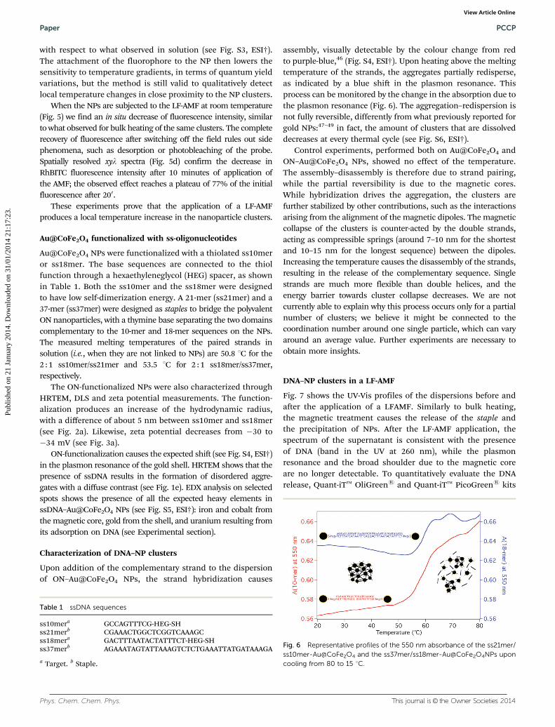
In this paper we explore this structural approach for thecontrolled delivery and release of ssDNAs, triggered by theapplication of a LF-AMF to clusters of DNA-functionalizedmagnetic NPs. The conceptual layout of the experiment isreported in Scheme 1. DNA hybridization takes place thanksto the target sequence:25 i.e., a ssDNA able to act as a linkerbetween two ssDNAs attached to the surface of two NPs. The ONdecoration of the pristine nanoparticles, and accordingly thetarget sequence, can be pre-programmed at will, to matchspecific therapeutic purposes, or else to release specificsequences in a spatially and temporally controllable fashion.
ExperimentalMaterials
Iron(III) chloride hexahydrate (97%), cobalt(II) nitrate hexahydrate(98%), gold(III) chloride trihydrate (>99.9%), sodium hydroxide(minimum 98%), tetramethylammonium hydroxide 25 wt%solution in water (TMAOH), sodium chloride (minimum 99.5%),sodium borohydride (98%), tri-sodium citrate dihydrate (>99%),citric acid monohydrate (>99.5%), Rhodamine B isothiocyanate,Rhodamine DHPE, and sucrose were purchased from Sigma-Aldrich. Concentrated nitric acid (90%) was purchased fromFluka. Quant-iTt OliGreens and Quant-iTt PicoGreens kitswere purchased from Invitrogen. POPC (1-palmitoyl-2-oleoyl-sn-glycero-3-phosphocholin)e was purchased from Avanti PolarLipids.
Synthesis of NPs
Magnetic nanoparticles (CoFe2O4 NPs) were synthesized throughthe co-precipitation method, introducing minor modifications tothe Massart procedure26,27 (see ESI† for details). The magneticcore was coated with a gold shell through a previously reportedprocedure28 that consists of the reduction of HAuCl4 with NaBH4
directly on the surface of CoFe2O4 NPs (see ESI† for details).
DNA sequences
The DNA sequences were custom-synthesized by ATD-BIO(Southampton, UK). Theoretical self-dimerization energieswere obtained through the OligoAnalyzer 3.1 software, providedby Integrated DNA Technologies. The calculated DG values are,respectively, �3.61 kcal mol�1 for the self-dimerization ofthe ss10mer with two base pairs (vs. �20.18 kcal mol�1 forthe hybridization with the complementary 10mer strand) and�4.85 kcal mol�1 for the self-dimerization of the ss18mer withfour base pairs (vs. �26.18 kcal mol�1 with the complementary18mer strand).
Preparation of thiolated oligonucleotide–Au@CoFe2O4 NPs
Thiolated ON–Au@CoFe2O4 NPs were prepared by mixingAu@CoFe2O4 NP solutions (final concentration 0.25 mM) withss18mer/ss10mer in a0.1 M aqueous solution of NaCl. Afterstanding for 48 h at room temperature under agitation, thesolution was centrifuged for 30 min at 10 000g to remove the ONexcess. Following removal of the supernatant, the dark-purpleprecipitate was re-dispersed in 1450 ml of 0.1 M NaCl solution.
High resolution transmission electron microscopy (HRTEM)
HRTEM analysis was carried out using a JEM 3010 (JEOL) electronmicroscope operating at 300 kV. Staining of ON–Au@CoFe2O4 NPswas carried out at room temperature using a 3% aqueous solutionof uranyl acetate.29 A drop of sample was placed on carbon-coatedcopper grids and then a drop of uranyl acetate solution was addedto each sample and left to dry. The chemical composition of thesamples was obtained using a EDX-TEM microanalysis technique.
Scheme 1 Conceptual sketch, showing the hybridization-driven formationof clusters and the release of ssDNA through magnetic actuation.
Paper PCCP
Publ
ishe
d on
21
Janu
ary
2014
. Dow
nloa
ded
on 3
1/01
/201
4 21
:17:
23.
View Article Online
This journal is© the Owner Societies 2014 Phys. Chem. Chem. Phys.
Quasi-Elastic Light Scattering (QELS)
QELS experiments were carried out using a 90Plus/BI-MASsystem (Brookhaven Instrument apparatus, New York, USA).The light source was a 15 mW solid state laser (l = 635 nm).Measurements were performed at 25 1C on 1.5 ml samples. Thetime autocorrelation functions of the intensity of the scatteredlight were measured at 901; their normalized version yields thefield normalized autocorrelation function through the Siegertrelationship:
g2(q,t) = 1 + b|g1(q,t)|2 (1)
where b is a spatial coherence factor dependent on the geometryof the detection system. The time autocorrelation functions ofthe scattered field were analyzed through a cumulant analysisstopped at the second order for monomodal distributions, andthrough Laplace Inversion with the CONTIN or Non-Negativelyconstrained Least Squares (NNLS) algorithms for bimodal orhighly polydisperse distributions.
Zeta potential experiments
Zeta potential measurements were performed using a Zeta PotentialAnalyzer (Brookhaven Instruments Corporation, Holtsville, NY). Zetapotentials were obtained from the electrophoretic mobility me,according to the Helmholtz–Smoluchowski equation:
z ¼ Zeme (2)
with Z being the viscosity of the medium and e the dielectricpermittivity of the dispersing medium. This equation is valid inthe limit:
k�Rh c 1 (3)
with k�1 the Debye length and Rh the hydrodynamic radius ofthe particles. The validity of these assumptions was tested aspreviously described,30 by determining Rh from DLS measurementsand by calculating k�1 from the relationship
k�1 = (eKBT)1/2�(2e2INA)�1/2 (4)
with KB the Boltzmann constant, e the electron charge, I theionic strength, NA the Avogadro constant.
The zeta potential values are reported as averages from tenmeasurements on each sample.
UV-Vis
UV-Vis absorption was measured on a Cary UV 100 spectro-photometer (Agilent Technologies, Italy) equipped with a 6 � 6multicell block Peltier temperature controller for melting analyses.Melting curves of the solutions were registered at 260 nm (foroligonucleotides) and at 550 nm (for functionalized NPs) in therange between 15 and 80 1C with a temperature ramping of0.51 min�1. The solutions were kept under continuous magneticstirring. The first derivative plot calculated from the meltinganalysis was used to determine the Tm values.
Alternating magnetic field
A sinusoidal magnetic field was generated in the gap of abroken toroidal magnet carrying a solenoid through which analternating electric current (AC) from a tone generator was led.Samples to be treated with LF-AMF (6 kHz) were placed in themiddle of the gap within 1 cm quartz cells. Details about theintensity of the magnetic field at different positions are given inthe ESI.†
Giant Unilamellar Vesicles (GUVs)
Giant Unilamellar Vesicles (GUVs) were prepared using theelectroformation method, originally developed by Angelovaand Dimitrov.31,32 A home-built chamber was set up sandwichingtwo Indium Tin Oxide (ITO)-coated microscope slides, separatedby an O-ring spacer.33 The electrical connection with both sides ofthe capacitor was obtained by attaching two Cu tapes on theconducting faces. The lipid (POPC, 3 mg ml�1) was dissolved inchloroform with 0.1% mol of Rhodamine DHPE (excitation andemission wavelengths 560 and 580 nm, respectively). 10 ml ofPOPC solution was spread on each conducting face of ITO-coatedmicroscope slides and dried under vacuum for at least 2 hours toremove the solvent. The O-ring was positioned around the film,and the two slides were sandwiched to form a chamber filled with350 ml of solution containing the Au@CoFe2O4–DNA clusters(Au final concentration E0.35 mM) and a PicoGreen solutiondiluted 30 times with sucrose 0.23 M. Finally, the chamber wasconnected to a function generator and a low-frequency AC electricfield (sinusoidal wave with a frequency of 10 Hz and an amplitudeof 2 V) was applied for 3 hours. GUV growth was monitored byoptical microscopy and, when complete, the solution was gentlyremoved from the electro-formation chamber. To reduce thefluorescence from PicoGreen-dsDNA not confined within theGUVs, the sample was diluted 1 : 50 with an iso-osmolar solutionof glucose 0.23 M. The diameter of the vesicles obtained with thismethod ranged from 5 to 50 mm.
Confocal Laser Scanning Microscopy (CLSM)
Confocal images were acquired using a DMIRE2 ConfocalMicroscope (Leica TCS SP2) with argon ion lasers and a waterimmersion objective 63�/1.2W (Zeiss). The same excitationwavelength (l = 488 nm, 30 mW at 35% of maximum power)was used for both the fluorescent probes (Rhodamine andPicoGreen), which were then detected in different spectralwindows (510–530 nm for PicoGreen; 570–620 nm for Rhodamine).The DNA release was quantitatively analyzed by measuring thefluorescence intensity decrease of PicoGreen in the aqueous poolinside the GUVs during the exposure to an AC-magnetic field. ForCLSM experiments on heating effects, RhBITC-labeled Au@CoFe2O4
NPs were prepared by incubating at room temperature Au@CoFe2O4
NPs with a 20 mM solution of Rhodamine B isothiocyanate (RhBITC)under stirring for 30 minutes. The dispersions were then added tothe sample chamber and incubated at room temperature for onehour to allow the aggregates to deposit onto the chamber bottom.CLSM and spatially resolved xyl spectroscopy were performed usinga 10� water immersion objective. The calibration of Au@CoFe2O4
PCCP Paper
Publ
ishe
d on
21
Janu
ary
2014
. Dow
nloa
ded
on 3
1/01
/201
4 21
:17:
23.
View Article Online

Phys. Chem. Chem. Phys. This journal is© the Owner Societies 2014
NPs-RhBITC fluorescence as a function of temperature wasperformed by means of a home-built thermostatic chamber,while a commercial (LAB-TEK, 8 Well) chamber was employedto monitor the response of RhBITC-labeled Au@CoFe2O4 NPsto LF-AMF.
Results and discussionCharacterization of CoFe2O4 and Au@CoFe2O4 NPs
Cobalt ferrite NPs have been chosen as the prototypical SPIONs,because of their high saturation magnetization and theirmagnetic anisotropy, which lead to a much larger coercitivitythan other iron oxides.34 Such properties make CoFe2O4 NPs theideal candidates to test the full potential of nanosized heaters.35
The in vivo application of cobalt-containing nanostructuresposes several toxicity concerns;36 nevertheless, recent studieson embryonic stem cells have shown that the coating with goldor silane shells makes these systems non embryotoxic.37 There-fore, the role of the gold shell is twofold: it endows the magneticcores with improved biocompatibility and, at the same time, itprovides a well-established route to chemically couple thiolatedssDNAs to the NP surface. It is worth noting that cobalt ferriteNPs are used here to validate our approach, but can be easilyreplaced by the classical SPION NPs.
Representative High Resolution Transmission ElectronMicroscopy (HRTEM) images of CoFe2O4 and Au@CoFe2O4 NPsare reported in Fig. 1. The size distributions of the NPs are shownin Fig. 2. Experimental results were fitted according to a log-normalsize distribution:
y = y0 + Ae�[ln(r/ravg)/w]2
(5)
The average radius (ravg) of CoFe2O4 NPs is slightly lowerthan 6 nm, while the width of the distribution (w) is about 0.45(see Fig. 2a), similarly to what previously observed.38,39 Fig. 1ahighlights the tendency of uncoated magnetic nanoparticles tospontaneously form linear aggregates because of the dipolarinteractions.40,41 The gold coating increases the average radiusup to 8 nm (see Fig. 2b), corresponding to an average gold shellthickness slightly larger than 2 nm. After the gold coating thepolydispersity is almost unchanged, suggesting a homogeneousprocess over all the NPs. The presence of a gold shell can beappreciated in Fig. 1d, where the crystalline pattern of the Aushell is superimposed to that of the magnetic core. EnergyDispersive X-ray analysis (EDX, see Fig. S1, ESI†) confirms thesuccessful coating, showing the characteristic signals of Co, Feand Au. Naked and Au-coated SPIONs were also characterizedby means of Dynamic Light Scattering (DLS) and zeta potential(z) measurements. The distribution of the hydrodynamic radii,Rh, resulting from the Laplace inversion of the autocorrelationfunctions according to a Non-Negatively constrained LeastSquares (NNLS) algorithm, is reported in Fig. S2, ESI.† Twosize distributions were found for both samples, in agreementwith the presence of single NPs together with aggregates of fewNPs. In Fig. 3a the Rh values of CoFe2O4 NPs and Au@CoFe2O4
NPs are reported. The values agree well with HRTEM results,
but are slightly larger due to the fact that the technique takesinto account also the counterion/solvent layer diffusing withthe NPs. Zeta potential results clearly confirm the formation ofthe gold shell as the charge reverses from positive to negativeupon metallic coating and stabilization of the core–shell nano-particles with citrate anions (see Fig. 3a). UV-Vis spectroscopyfurther supports the presence of the gold coating: the spectrumof the core–shell nanoparticle dispersion (Fig. 3b), which is
Fig. 1 TEM images at different magnifications of CoFe2O4 (a, b), Au@CoFe2O4
(c, d) and ssDNA–Au@CoFe2O4 (e, f) NPs. The inset in (d) represents theHanning Masked FFT calculated in domain 1, revealing the spots of Au {111} and{200} (2.355 Å and 2.039 Å, respectively).
Fig. 2 Size distributions (bars) and corresponding log-normal fittings(line) as obtained by the analysis of CoFe2O4 (a) and Au@CoFe2O4 (b)HRTEM images.
Paper PCCP
Publ
ishe
d on
21
Janu
ary
2014
. Dow
nloa
ded
on 3
1/01
/201
4 21
:17:
23.
View Article Online

This journal is© the Owner Societies 2014 Phys. Chem. Chem. Phys.
dominated by a broad shoulder centered below 500 nm forCoFe2O4 NPs,42 displays a surface plasmon resonance peakedat 540 nm (typical of Au bimetallic systems28,43) after coating.
Au@CoFe2O4 NPs in a LF-AMF
In order to verify the NP heating properties under a LF-AMF, aConfocal Laser Scanning Microscopy (CLSM) experiment wascarried out on Au@CoFe2O4 NPs, fluorescently labeled with thethermoresponsive probe Rhodamine B isothiocyanate (RhBITC).44
A linear decrease of the quantum yield of RhBITC withtemperature increase has been recently reported45 and verifiedby us between 25 1C and 55 1C for a 20 mM aqueous solution. Inthis range we observed a 40% decrease of quantum yield at583 nm. The same solution was employed to fluorescently label
the NPs. Fig. 4 and 5 display the CLSM images of RhBITC-labeledNP clusters, prepared as described in the experimental section.
The fluorescent NPs, not stabilized against aggregation inorder to simulate the DNA-driven assembly, formed largeclusters that slowly settled onto the bottom of the measurementchamber. The fluorescence intensity of RhBITC-labeled NPaggregates was monitored as a function of the bulk temperature.Fig. 4a and b display the confocal images acquired on the samespot at 25 1C and at 55 1C, respectively, where a decrease inemission is visually detectable. Spatially resolved xyl emissionspectra at increasing temperatures (Fig. 5c) reveal however anon-linear and generally lower decrease of fluorescence intensity,
Fig. 3 (a) Average radius (ravg) as obtained from the log-normal fitting ofHRTEM results of CoFe2O4 and Au@CoFe2O4 NPs, the hydrodynamicradius (Rh, as obtained by NNLS analysis of DLS results) and zeta potential(according to the Smoluchowski model) of CoFe2O4, Au@CoFe2O4, andssDNA–Au@CoFe2O4 NPs; (b) UV-Vis spectra of CoFe2O4, Au@CoFe2O4,and ssDNA–Au@CoFe2O4 NPs.
Fig. 4 RhBITC-labeled Au@CoFe2O4 NPs. (a, b) Representative ConfocalLaser Scanning Microscopy images acquired in the same spot at T = 25 1C(a) and T = 55 1C (b). (c) Spatially resolved xyl spectra acquired atincreasing temperature (25 1C, 35 1C, 45 1C, 55 1C) at lex = 561nm.
Fig. 5 RhBITC-labeled Au@CoFe2O4 NPs. (a–c) Representative CLSMimages acquired in the same region before (a), during (b) and after(c) the application of a 6 KHz AMF. (d) Spatially resolved xyl (lex =561 nm) spectra acquired before, during and after the application of theAMF (6 KHz).
PCCP Paper
Publ
ishe
d on
21
Janu
ary
2014
. Dow
nloa
ded
on 3
1/01
/201
4 21
:17:
23.
View Article Online
Phys. Chem. Chem. Phys. This journal is© the Owner Societies 2014
with respect to what observed in solution (see Fig. S3, ESI†).The attachment of the fluorophore to the NP then lowers thesensitivity to temperature gradients, in terms of quantum yieldvariations, but the method is still valid to qualitatively detectlocal temperature changes in close proximity to the NP clusters.
When the NPs are subjected to the LF-AMF at room temperature(Fig. 5) we find an in situ decrease of fluorescence intensity, similarto what observed for bulk heating of the same clusters. The completerecovery of fluorescence after switching off the field rules out sidephenomena, such as desorption or photobleaching of the probe.Spatially resolved xyl spectra (Fig. 5d) confirm the decrease inRhBITC fluorescence intensity after 10 minutes of application ofthe AMF; the observed effect reaches a plateau of 77% of the initialfluorescence after 200.
These experiments prove that the application of a LF-AMFproduces a local temperature increase in the nanoparticle clusters.
Au@CoFe2O4 functionalized with ss-oligonucleotides
Au@CoFe2O4 NPs were functionalized with a thiolated ss10meror ss18mer. The base sequences are connected to the thiolfunction through a hexaethyleneglycol (HEG) spacer, as shownin Table 1. Both the ss10mer and the ss18mer were designedto have low self-dimerization energy. A 21-mer (ss21mer) and a37-mer (ss37mer) were designed as staples to bridge the polyvalentON nanoparticles, with a thymine base separating the two domainscomplementary to the 10-mer and 18-mer sequences on the NPs.The measured melting temperatures of the paired strands insolution (i.e., when they are not linked to NPs) are 50.8 1C for the2 : 1 ss10mer/ss21mer and 53.5 1C for 2 : 1 ss18mer/ss37mer,respectively.
The ON-functionalized NPs were also characterized throughHRTEM, DLS and zeta potential measurements. The function-alization produces an increase of the hydrodynamic radius,with a difference of about 5 nm between ss10mer and ss18mer(see Fig. 2a). Likewise, zeta potential decreases from �30 to�34 mV (see Fig. 3a).
ON-functionalization causes the expected shift (see Fig. S4, ESI†)in the plasmon resonance of the gold shell. HRTEM shows that thepresence of ssDNA results in the formation of disordered aggre-gates with a diffuse contrast (see Fig. 1e). EDX analysis on selectedspots shows the presence of all the expected heavy elements inssDNA–Au@CoFe2O4 NPs (see Fig. S5, ESI†): iron and cobalt fromthe magnetic core, gold from the shell, and uranium resulting fromits adsorption on DNA (see Experimental section).
Characterization of DNA–NP clusters
Upon addition of the complementary strand to the dispersionof ON–Au@CoFe2O4 NPs, the strand hybridization causes
assembly, visually detectable by the colour change from redto purple-blue,46 (Fig. S4, ESI†). Upon heating above the meltingtemperature of the strands, the aggregates partially redisperse,as indicated by a blue shift in the plasmon resonance. Thisprocess can be monitored by the change in the absorption due tothe plasmon resonance (Fig. 6). The aggregation–redispersion isnot fully reversible, differently from what previously reported forgold NPs:47–49 in fact, the amount of clusters that are dissolveddecreases at every thermal cycle (see Fig. S6, ESI†).
Control experiments, performed both on Au@CoFe2O4 andON–Au@CoFe2O4 NPs, showed no effect of the temperature.The assembly–disassembly is therefore due to strand pairing,while the partial reversibility is due to the magnetic cores.While hybridization drives the aggregation, the clusters arefurther stabilized by other contributions, such as the interactionsarising from the alignment of the magnetic dipoles. The magneticcollapse of the clusters is counter-acted by the double strands,acting as compressible springs (around 7–10 nm for the shortestand 10–15 nm for the longest sequence) between the dipoles.Increasing the temperature causes the disassembly of the strands,resulting in the release of the complementary sequence. Singlestrands are much more flexible than double helices, and theenergy barrier towards cluster collapse decreases. We are notcurrently able to explain why this process occurs only for a partialnumber of clusters; we believe it might be connected to thecoordination number around one single particle, which can varyaround an average value. Further experiments are necessary toobtain more insights.
DNA–NP clusters in a LF-AMF
Fig. 7 shows the UV-Vis profiles of the dispersions before andafter the application of a LFAMF. Similarly to bulk heating,the magnetic treatment causes the release of the staple andthe precipitation of NPs. After the LF-AMF application, thespectrum of the supernatant is consistent with the presenceof DNA (band in the UV at 260 nm), while the plasmonresonance and the broad shoulder due to the magnetic coreare no longer detectable. To quantitatively evaluate the DNArelease, Quant-iTt OliGreens and Quant-iTt PicoGreens kits
Table 1 ssDNA sequences
ss10mera GCCAGTTTCG-HEG-SHss21merb CGAAACTGGCTCGGTCAAAGCss18mera GACTTTAATACTATTTCT-HEG-SHss37merb AGAAATAGTATTAAAGTCTCTGAAATTATGATAAAGA
a Target. b Staple.
Fig. 6 Representative profiles of the 550 nm absorbance of the ss21mer/ss10mer-Au@CoFe2O4 and the ss37mer/ss18mer-Au@CoFe2O4NPs uponcooling from 80 to 15 1C.
Paper PCCP
Publ
ishe
d on
21
Janu
ary
2014
. Dow
nloa
ded
on 3
1/01
/201
4 21
:17:
23.
View Article Online

This journal is© the Owner Societies 2014 Phys. Chem. Chem. Phys.
were used to determine the amounts of ssDNA and dsDNA (seeESI†). Table 2 reports the number of ssDNA molecules graftedonto NPs (obtained by difference with respect to the solutionused to functionalize the gold surface) and the amounts ofssDNA and dsDNA released after LF-AMF. The results show thatthe amount of single strand released after 15 minutes rangesbetween 23 and 31%, depending on the system, while theamount increases up to almost 40% after 35 minutes. Thepercentage of double strand released is about one order ofmagnitude smaller. The effective release of the single strand is afurther unmistakable proof of the heating effect produced by theLF-AMF and, most importantly, that a low frequency alternatingmagnetic field provides the thermal effect necessary for therelease of DNA from the cluster.
To visualize the magnetically triggered release behaviorinside a cellular-like compartment, freestanding giant unilamellarvesicles (GUVs) including DNA-magnetic clusters in their internallumen were prepared and investigated by means of CLSM underLF-AMF. Details about the preparation and experimental setup are
given in the ESI.† Rhodamine-labeled lipids were used to stain theGUV membrane (red color), while PicoGreens was used to tag theDNA double strands.
In this setup, the fluorescence disappearance means that thestrands are unparing and ss-DNA is released in the internalpool of the GUVs. The exposure to the beam causes a partialbleaching of the green probe (see Fig. S7, ESI†), which results ina linear decrease of fluorescence intensity upon irradiation.Nevertheless, the effect of a LF_AMF on the release of ssDNAs
Fig. 7 UV-Vis spectra of DNA–NP clusters before and after the applica-tion of LF-AMF (6 kHz). The disappearance of the absorption band in thevisible region accounts for the precipitation of clusters following theapplication of LF-AMF. Correspondingly, the absorption peak at 260 nmemerges, accounting for the presence in solution of the DNA releasedfrom the clusters.
Table 2 Quantification of ss10mer and ss18mer chemically grafted to NPs and staple and DNA double strands released from NP clusters as aconsequence of the application of the AMF
Amounts of ssDNA grafted on NPsss10mer ss18mer
ssDNA moleculesa 67 � 2 88 � 3
Amounts of staple and double strands released after 150 of LF-AMFss21mer + ss10mer-Au@CoFe2O4 ss37mer + ss18mer-Au@CoFe2O4
Staple 30.5 � 2% 23.6 � 3%Double strand 3.7 � 0.5% 2.1 � 0.4%
Amounts of staple and double strands released after 350 of LF-AMFss21mer + ss10mer-Au@CoFe2O4 ss37mer + ss18mer-Au@CoFe2O4
Staple 39.2 � 7% 37.1 � 2%Double strand 4.3 � 1% 3.0 � 0.8%
a Per single NP.
Fig. 8 Top: CLSM normalized fluorescence of PicoGreens under laserirradiation and both laser irradiation and the magnetic field. Right: CLSMimages of a GUV taken during the application of the AMF after 0, 2, 4, 6, 9and 12 minutes (going from (a) to (f)). Scale bar: 5 mm.
PCCP Paper
Publ
ishe
d on
21
Janu
ary
2014
. Dow
nloa
ded
on 3
1/01
/201
4 21
:17:
23.
View Article Online

Phys. Chem. Chem. Phys. This journal is© the Owner Societies 2014
results in a very fast decrease of PicoGreens fluorescenceintensity, as shown in Fig. 8. The fluorescence decreases almostto the background level after 12 minutes (the weak greenfluorescence from the background is due to the GUV preparationprocedure). Furthermore, the integrity of the membrane is notaffected by the presence of the NP clusters and by the LF-AMFand the heating effects are local and selective for the clusters.
Conclusions
This paper presents the design, preparation and characterization ofoligonucleotide functionalized Au@CoFe2O4 NPs and describestheir aggregation, driven by hybridization with a complementarystrand. The final aim is to engineer ssDNA releasing devices withmagnetic actuation. Thermal cycling of the clusters results indisassembly–assembly processes, with only partial reversibility.Remarkably, the same events occur upon application of a lowfrequency AMF, providing the proof of principle of the feasibility ofmagnetic actuation of oligonucleotide release.
In particular, using NP clusters resulted in the release of asignificant amount of ssDNA (together with a minor amount ofdsDNA) even under mild magnetic conditions.
The so-designed system holds great promise both in thebiomedical arena, where ssDNAs are considered the nextgeneration of therapeutic biomolecules, and in materialsscience, where DNA is used as a building block molecule withoutstanding selectivity and sub-nm addressing properties.
Some issues yet remain to be addressed, like the control ofoligonucleotide density and thus of the cluster size. However,the proof of principle of both magnetic nanoparticles locationcontrol and thermal oligonucleotide delivery triggered by anAMF discloses new perspectives for research in the field ofnanomedicine.
Acknowledgements
The authors acknowledge financial support from MIUR forfunding under the projects PRIN 2010-2011(2010BJ23MN) andFIRB RBPR05JH2P_007 ITALNANONET. M.B. acknowledges EU forfinancial support (Marie-Curie Reintegration Grant SUPRACRYST).
Notes and references
1 N. C. Seeman, Annu. Rev. Biochem., 2010, 79, 65–87.2 R. J. Macfarlane, B. Lee, M. R. Jones, N. Harris, G. C. Schatz
and C. A. Mirkin, Science, 2011, 334, 204–208.3 M. D. Massich, D. A. Giljohann, A. L. Schmucker, P. C. Patel
and C. A. Mirkin, ACS Nano, 2010, 4, 5641–5646.4 S. Y. Park, A. K. R. Lytton-Jean, B. Lee, S. Weigand,
G. C. Schatz and C. A. Mirkin, Nature, 2008, 451,553–556.
5 M. R. Jones, R. J. Macfarlane, B. Lee, J. Zhang, K. L. Young,A. J. Senesi and C. A. Mirkin, Nat. Mater., 2010, 9, 913–917.
6 N. L. Rosi and C. A. Mirkin, Chem. Rev., 2005, 105,1547–1562.
7 M. Bonini, D. Berti and P. Baglioni, Curr. Opin. ColloidInterface Sci., 2013, 18, 459–467.
8 N. L. Rosi, D. A. Giljohann, C. S. Thaxton, A. K. R. Lytton-Jean, M. S. Han and C. A. Mirkin, Science, 2006, 312,1027–1030.
9 D. A. Giljohann, D. S. Seferos, P. C. Patel, J. E. Millstone,N. L. Rosi and C. A. Mirkin, Nano Lett., 2007, 7, 3818–3821.
10 F. Baldelli Bombelli, F. Betti, F. Gambinossi, G. Caminati,T. Brown, P. Baglioni and D. Berti, Soft Matter, 2009, 5,1639–1645.
11 M. Banchelli, F. Betti, D. Berti, G. Caminati, F. BaldelliBombelli, T. Brown, L. M. Wilhelmsson, B. Norden andP. Baglioni, J. Phys. Chem. B, 2008, 112, 10942–10952.
12 F. Baldelli Bombelli, F. Gambinossi, M. Lagi, D. Berti,G. Caminati, T. Brown, F. Sciortino, B. Norden andP. Baglioni, J. Phys. Chem. B, 2008, 112, 15283–15294.
13 S. Lal, S. E. Clare and N. J. Halas, Acc. Chem. Res., 2008, 41,1842–1851.
14 L. Cheng, K. Yang, Y. Li, J. Chen, C. Wang, M. Shao, S.-T. Leeand Z. Liu, Angew. Chem., Int. Ed., 2011, 50, 7385–7390.
15 S. E. Skrabalak, J. Chen, Y. Sun, X. Lu, L. Au, C. M. Cobleyand Y. Xia, Acc. Chem. Res., 2008, 41, 1587–1595.
16 H. Liu, D. Chen, L. Li, T. Liu, L. Tan, X. Wu and F. Tang,Angew. Chem., Int. Ed., 2011, 50, 891–895.
17 A. Jordan, R. Scholz, P. Wust, H. Fahling and R. Felix,J. Magn. Magn. Mater., 1999, 201, 413–419.
18 R. E. Rosensweig, J. Magn. Magn. Mater., 2002, 252, 370–374.19 S. Laurent, D. Forge, M. Port, A. Roch, C. Robic,
L. Vander Elst and R. N. Muller, Chem. Rev., 2008, 108,2064–2110.
20 P. Qiu, C. Jensen, N. Charity, R. Towner and C. Mao, J. Am.Chem. Soc., 2010, 132, 17724–17732.
21 S. Nappini, F. Baldelli Bombelli, M. Bonini, B. Norden andP. Baglioni, Soft Matter, 2009, 6, 154–162.
22 S. Nappini, M. Bonini, F. Baldelli Bombelli, F. Pineider,C. Sangregorio, P. Baglioni and B. Norden, Soft Matter, 2011,7, 1025–1037.
23 S. Nappini, M. Bonini, F. Ridi and P. Baglioni, Soft Matter,2011, 7, 4801–4811.
24 M. Creixell, A. C. Bohorquez, M. Torres-Lugo and C. Rinaldi,ACS Nano, 2011, 5, 7124–7129.
25 P. W. K. Rothemund, Nature, 2006, 440, 297–302.26 C. R. Massart, IEEE Trans. Magn., 1981, 17, 1247–1248.27 M. Bonini, A. Wiedenmann and P. Baglioni, J. Phys. Chem. B,
2004, 108, 14901–14906.28 J. L. Lyon, D. A. Fleming, M. B. Stone, P. Schiffer and
M. E. Williams, Nano Lett., 2004, 4, 719–723.29 J. A. Terzakis, J. Ultrastruct. Res., 1968, 22, 168–184.30 C. Montis, S. Milani, P. Baglioni and D. Berti, J. Colloid
Interface Sci., 2012, 373, 57–68.31 M. I. Angelova and D. S. Dimitrov, Faraday Discuss. Chem.
Soc., 1986, 81, 303–311.32 M. I. Angelova, S. Soleau, P. Meleard, F. Faucon and
P. Bothorel, in Trends in Colloid and Interface Science VI,ed. C. Helm, M. Losche and H. Mohwald, Steinkopff, 1992,pp. 127–131.
Paper PCCP
Publ
ishe
d on
21
Janu
ary
2014
. Dow
nloa
ded
on 3
1/01
/201
4 21
:17:
23.
View Article Online

This journal is© the Owner Societies 2014 Phys. Chem. Chem. Phys.
33 S. Nappini, T. Al Kayal, D. Berti, B. Norden and P. Baglioni,J. Phys. Chem. Lett., 2011, 2, 713–718.
34 A. Tomitaka, M.-H. Jeun, S.-T. Bae and Y. Takemura,J. Magn., 2011, 16, 164–168.
35 T. E. Torres, A. G. Roca, M. P. Morales, A. Ibarra,C. Marquina, M. R. Ibarra and G. F. Goya, J. Phys.: Conf.Ser., 2010, 200, 072101.
36 V. Mariani, J. Ponti, G. Giudetti, F. Broggi, P. Marmorato,S. Gioria, F. Franchini, H. Rauscher and F. Rossi, Nanotox-icology, 2012, 6, 272–287.
37 C. Di Guglielmo, D. R. Lopez, J. De Lapuente,J. M. L. Mallafre and M. B. Suarez, Reprod. Toxicol., 2010,30, 271–276.
38 R. Massart, E. Dubois, V. Cabuil and E. Hasmonay, J. Magn.Magn. Mater., 1995, 149, 1–5.
39 A. Bee, R. Massart and S. Neveu, J. Magn. Magn. Mater., 1995,149, 6–9.
40 M. Bonini, A. Wiedenmann and P. Baglioni, J. Appl. Crystallogr.,2007, 40, s254–s258.
41 C. Neto, M. Bonini and P. Baglioni, Colloids Surf., A, 2005,269, 96–100.
42 M. Bonini, A. Wiedenmann and P. Baglioni, Phys. A, 2004,339, 86–91.
43 I. Robinson, L. D. Tung, S. Maenosono, C. Walti andN. T. K. Thanh, Nanoscale, 2010, 2, 2624–2630.
44 S. Freddi, L. Sironi, R. D’Antuono, D. Morone, A. Dona,E. Cabrini, L. D’Alfonso, M. Collini, P. Pallavicini, G. Baldi,D. Maggioni and G. Chirico, Nano Lett., 2013, 13, 2004–2010.
45 Y. Y. Chen and A. W. Wood, Bioelectromagnetics, 2009, 30,583–590.
46 J. Storhoff, R. Elghanian, R. C. Mucic, C. A. Mirkin andR. L. Letsinger, J. Am. Chem. Soc., 1998, 120, 1959–1964.
47 I.-I. S. Lim, L. Wang, U. Chandrachud, S. Gal andC.-J. Zhong, Res. Lett. Nanotechnol., 2008, 2008, 1–4.
48 Z. Li, R. Jin, C. A. Mirkin and R. L. Letsinger, Nucleic AcidsRes., 2002, 30, 1558–1562.
49 E. Dujardin, L.-B. Hsin, C. R. C. Wang and S. Mann, Chem.Commun., 2001, 1264–1265.
PCCP Paper
Publ
ishe
d on
21
Janu
ary
2014
. Dow
nloa
ded
on 3
1/01
/201
4 21
:17:
23.
View Article Online