The branching of insulin-like growth factor 1 and insulin: an immunohistochemical analysis during...
12
Regulatory Peptides, 48 (1993) 65-76 © 1993 Elsevier Science Publishers B.V. All rights reserved 0167-0115/93/$06.00 65 REGPEP 01568 The branching of insulin-like growth factor 1 and insulin: an immunohistochemical analysis during phylogeny Manfred Reinecke a, Caroline Maake a, Sture Falkmer b and Vicki R. Sara c alnstitute of Anatomy, Division of Neuroendocrinology, University of Zi2rich, Zarich (Switzerland), b Department of Tumor Pathology, Karolinska Institute and Hospital, Stockholm (Sweden) and c School of Life Science, Q UT Gardens Point Campus, Brisbane (Australia) (Received 26 April 1993; accepted 6 May 1993) Key words: IGF-1; Insulin; Co-existence; Pancreas; Bony fish; Cartilaginous fish; Cyclostomes Summary The co-existence of insulin-like growth factor 1 (IGF-1) with the classical islet hormones insulin (INS), glucagon (GLUC), somatostatin (SOM) and pancreatic polypeptide (PP) in the endocrine pancreas of repre- sentative species of cyclostomes (Myxine glutinosa), cartilaginous fish (Raja clavata, Squalus acanthias) and bony fish (Cottus scorpius, Carassius auratus, Cyprinus carpio, Anguilla anguilla) was studied by the use of monoclonal and polyclonal antisera and the double immunofluorescence technique. In all species investigated, IGF-l-like- immunoreactive cells were found in the endocrine pancreas, however, in varying localization. In Myxine glutinosa, all INS-immunoreactive cells and some of the SOM-immunoreactive cells contained IGF-l-like- immunoreactivity. In Raja and Squalus, only a minority of the INS-immunoreactive cells also displayed IGF- 1-1ike-immunoreactivity. The majority of the IGF-l-like-immunoreactivity was observed in SOM- and in GLUC-immunoreactive cells. Different results were obtained in bony fish. In Cottus, in the Brockmann bod- ies and the small islets IGF-l-like- and INS-immunoreactivities co-existed to 100%. In contrast, in the other bony fish studied IGF-l-like-immunoreactivity was not observed in INS-immunoreactive cells: in Cyprinus, IGF-1-1ike-immunoreactivity was found in GLUC-, PP- and SOM-immunoreactive cells and in Carassius and Anguilla, in SOM-immunoreactive cells only. Thus, in all bony fish species with the exception of Cottus, IGF-1 and insulin display a distinct cellular distribution, similar to that of mammals. The present results, thus, may indicate that the branching of IGF-1 and insulin has occurred at the phylogenetic level of bony fish. Correspondence to: M. Reinecke, Institute of Anatomy, Division of Neuroendocrinology, University of ZOrich-Irchel, Winterthurer Str. 190, CH-8057 ZOrich, Switzerland.
Transcript of The branching of insulin-like growth factor 1 and insulin: an immunohistochemical analysis during...

Regulatory Peptides, 48 (1993) 65-76 © 1993 Elsevier Science Publishers B.V. All rights reserved 0167-0115/93/$06.00
65
REGPEP 01568
The branching of insulin-like growth factor 1 and insulin: an immunohistochemical analysis during phylogeny
Manfred Reinecke a, Caroline Maake a, Sture Falkmer b and Vicki R. Sara c
a lnstitute of Anatomy, Division of Neuroendocrinology, University of Zi2rich, Zarich (Switzerland), b Department of Tumor Pathology, Karolinska Institute and Hospital, Stockholm (Sweden) and c School of Life Science, Q UT Gardens Point Campus, Brisbane (Australia)
(Received 26 April 1993; accepted 6 May 1993)
Key words: IGF-1; Insulin; Co-existence; Pancreas; Bony fish; Cartilaginous fish; Cyclostomes
Summary
The co-existence of insulin-like growth factor 1 (IGF-1) with the classical islet hormones insulin (INS), glucagon (GLUC), somatostatin (SOM) and pancreatic polypeptide (PP) in the endocrine pancreas of repre- sentative species of cyclostomes (Myxine glutinosa), cartilaginous fish (Raja clavata, Squalus acanthias) and bony fish (Cottus scorpius, Carassius auratus, Cyprinus carpio, Anguilla anguilla) was studied by the use of monoclonal and polyclonal antisera and the double immunofluorescence technique. In all species investigated, IGF-l-like- immunoreactive cells were found in the endocrine pancreas, however, in varying localization. In Myxine glutinosa, all INS-immunoreactive cells and some of the SOM-immunoreactive cells contained IGF-l-like- immunoreactivity. In Raja and Squalus, only a minority of the INS-immunoreactive cells also displayed IGF- 1-1ike-immunoreactivity. The majority of the IGF-l-like-immunoreactivity was observed in SOM- and in GLUC-immunoreactive cells. Different results were obtained in bony fish. In Cottus, in the Brockmann bod- ies and the small islets IGF-l-like- and INS-immunoreactivities co-existed to 100%. In contrast, in the other bony fish studied IGF-l-like-immunoreactivity was not observed in INS-immunoreactive cells: in Cyprinus, IGF-1-1ike-immunoreactivity was found in GLUC-, PP- and SOM-immunoreactive cells and in Carassius and Anguilla, in SOM-immunoreactive cells only. Thus, in all bony fish species with the exception of Cottus, IGF-1 and insulin display a distinct cellular distribution, similar to that of mammals. The present results, thus, may indicate that the branching of IGF-1 and insulin has occurred at the phylogenetic level of bony fish.
Correspondence to: M. Reinecke, Institute of Anatomy, Division of Neuroendocrinology, University of ZOrich-Irchel, Winterthurer Str. 190, CH-8057 ZOrich, Switzerland.
66
Introduction
The insulin-like growth factors ( IGFs) constitute
a family of polypeptides that consists of two major forms, IGF-1 and IGF-2, and some molecular vari-
ants [1-3] . They exert insulin-like and growth- promoting effects in their target cells. Because con-
siderable similarities exist in primary and tertiary structure [2], it has been presumed that in phylogeny
the I G F s and proinsulin have branched from a com-
mon ancestor [4-7] . This precursor molecule has
been suggested to be coded by a gene that underwent duplication at the time of the appearence of the first vertebrates [8,9].
Recently, this hypothesis has gained support from
studies on protochordates. F rom the cephalochor-
. ° .
date Branchiostoma californiens& a hybrid insulin/ insulin-like growth factor c D N A has been cloned
[6]. In Branchiostoma lanceolatum and in the uro-
chordate Ciona intestinalis, the co-existence of INS- and IGF-l-l ike-immunoreactivit ies in entero-endo-
crine cells and central neurons has been shown [7] suggesting these cells as the storage sites of the hy-
brid molecule found in Branchiostoma californiensis. Thus, a common insulin/IGF-1 ancestor molecule
seems to occur on the phylogenetic level of proto-
chordates. For mammals, the presence of IGF- l - immunore -
activity has been shown in rat [10-15] as well as in human and dog endocrine pancreas [ 15]. In the rat
islets, the distinct localization of IGF- l - immunore - activity is still under discussion [12,14]. While there
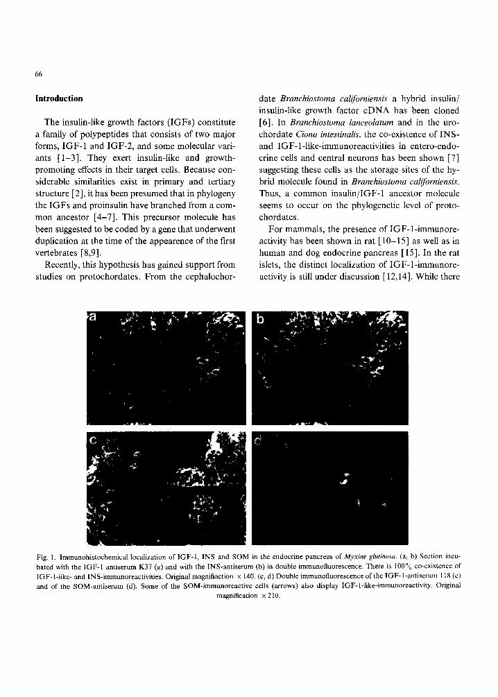
Fig. 1. lmmunohistochemical localization of IGF-1, INS and SOM in the endocrine pancreas of Myxine glut&osa. (a, b) Section incu- bated with the IGF-1 antiserum K37 (a) and with the INS-antiserum (b) in double immunofluorescence. There is 100Yo co-existence of IGF-l-like- and INS-immunoreactivities. Original magnifiaction x 140. (c, d) Double immunofluorescence of the IGF-l-antiserum 118 (c) and of the SOM-antiserum (d). Some of the SOM-immunoreactive cells (arrows) also display IGF-l-like-immunoreactivity. Original
magnification x 210.

&
. o ,
e
67
Fig. 2. Immunohistochemical localization of IGF-1 (a, c, e), INS (b), GLUC (d) and SOM (f) in the endocrine pancreas of Squalus acanthias. (a, b) Section incubated with the IGF-1 antiserum 116 (a) and with INS-antiserum (b) in double immunofluorescence. A mi- nority of the INS-immunoreactive cells also contains IGF-l-like-irnmunoreactivity (arrows). (c, d) Double immunofluorescence of the IGF-l-antiserum K37 (c) and of the GLUC-antiserum (d). Several of the GLUC-immunoreactive cells show also IGF-l-like- immunoreactivity. (e, f) Double immunofluorescence of the IGF-l-antiserum 118 (e) and of the SOM-antiserum (f). There is a high de-
gree of co-existence of IGF-l-like-and SOM-immunoreactivities. Original magnification x 210.
is c o n s e n t tha t no I G F - 1 - immunoreac t iv i ty occurs in
B-cells, bo th the D-cel ls [12] an d the A-cells [15]
have been descr ibed to r ep resen t the ma j o r store of
I G F - l - i m m u n o r e a c t i v i t y . A detai led analys is us ing
several specific an t i se ra in double immunof luo re s -
cence revealed the exclusive presence of I G F - 1 -
immunoreac t iv i t y in A-cells o f ra t a n d dog, bu t in
m a n singular D-cel ls also c o n t a i n e d I G F - l - i m m u -
noreact iv i ty [ 15 ].
In con t ras t to p ro tochorda te s [7] a n d m a m m a l s

68

69
[ 15], for lower vertebrates only the presence of IGF- l-like peptides in the islet parenchyma [ 14,16,17 ] has been shown, but no results have been presented yet on a possible co-existence of IGF-1 and insulin. However, information on the localization of insulin and IGF-1 may help to elucidate the phylogenetic level of the branching of these hormones from the likely precursor molecule. Thus, the present study using the double immunofluorescence technique aims: (1) to localize IGF-1 in the endocrine pancreas of representative bony fish, cartilaginous fish and cy- clostomes; (2) to compare the cellular storage sites of IGF-1 with those of insulin; and (3) in case of non- coexistence or minor partial co-existence of insulin and IGF-1 to localize IGF-1 and the classical islet hormones glucagon, somatostatin and pancreatic polypeptide.
Materials and Methods
Animals. The species studied were selected on the basis of the results of preceding studies on the phylogeny of IGF-1 [14,16]. However, in order to obtain more valid results other species were added to those investigated earlier. Thus, as representatives of the three lowest vertebrate classes the following spe- cies (number of individuals in parentheses) were used: Osteichthyes - Cottus scorpius (n = 8), Cyprinus carpio (n = 3), Carassius auratus (n = 5), Anguilla an- guilla (n = 5); Chondrichthyes - Raja clavata (n = 6), Squalus acanthias (n = 4); Cyclostomes - Myxine glu- tinosa (n = 8).
Fixation and embedding. Adult specimens of the species investigated were anesthetized by adding M S 222 R (Sandoz, Basel, Switzerland) to the surround-
ing water. Except for Carasshts and Anguilla, an iso- tonic saline solution was perfused via the heart for about 10-15 min to prevent blood clotting and to maintain colloid osmotic pressure, followed by per- fusion with acid-free Bouin's fixative for another 15 to 30 rain. After dissection, the pancreas were post- fixed by immersion in the fixative for 2 h. In Carassius and Anguilla, the pancreas were fixed by immersion in acid-free Bouin's solution for 4 h. All specimens were dehydrated in ascending series of ethanol and routinely embedded in paraplast. Sections were cut at 4/~m.
Antisera and hormones. For the immunohis- tochemical localization of IGF-1, three different an- tisera (codes: 116, 118, K37) raised in rabbit were used which have been shown to be specific for IGF-1 (116, 118, see Ref. 18; K37, see Refs. 12 and 14). For further information on the antisera against IGF-1 and the classical islet hormones insulin (INS), glu- cagon (GLUC), somatostatin (SOM) and pancreatic polypeptide (PP) see [15]. Recombinant human (h)IGF-1 was purchased from Bachem (Bubendorf, Switzerland), bovine (b)INS, b G L U C and SOM from Sigma (Buchs, Switzerland) and hPP from Peninsula (Heidelberg, Germany).
Immunohistochemical technique. At first, unspe- cific reactions were blocked by the use of phosphate- buffered saline (PBS) containing 2~o bovine serum albumine (BSA) and 2~o normal goat serum. For double immunofluorescence, single sections were in- cubated consecutively with the INS-antiserum 72C and with one of the IGF-1-antisera. Additional single sections were incubated consecutively: (1) with the mouse GLUC-ant ibody and an IGF-1-antiserum; (2) with the rat SOM-antiserum and an IGF-l-ant ise- rum. The possible co-existence of IGF-1 and PP was
Fig. 3. Double immunofluorescence of IGF-1 (a, c: antiserum 116, e: antiserum 118) and INS (b, d, O in the pyloric islet (a-d) and in a small islet (e, f) of Cottus scorpius. (a, b) Low power micrograph of the pyloric islet. IGF-l-like- and IN S-immunoreactivities display identical distribution patterns. Original magnification x 85. (c, d) Higher magnification of a portion of the pyloric islet shown in (a, b). There is a 100~o co-localisation of IGF-l-like-and INS-immunoreactivities. (e, f) All INS-immunoreactive (f) cells in the small islet contain also
IGF-l-like-immunoreactivity (e). Original magnification × 340.

70
71
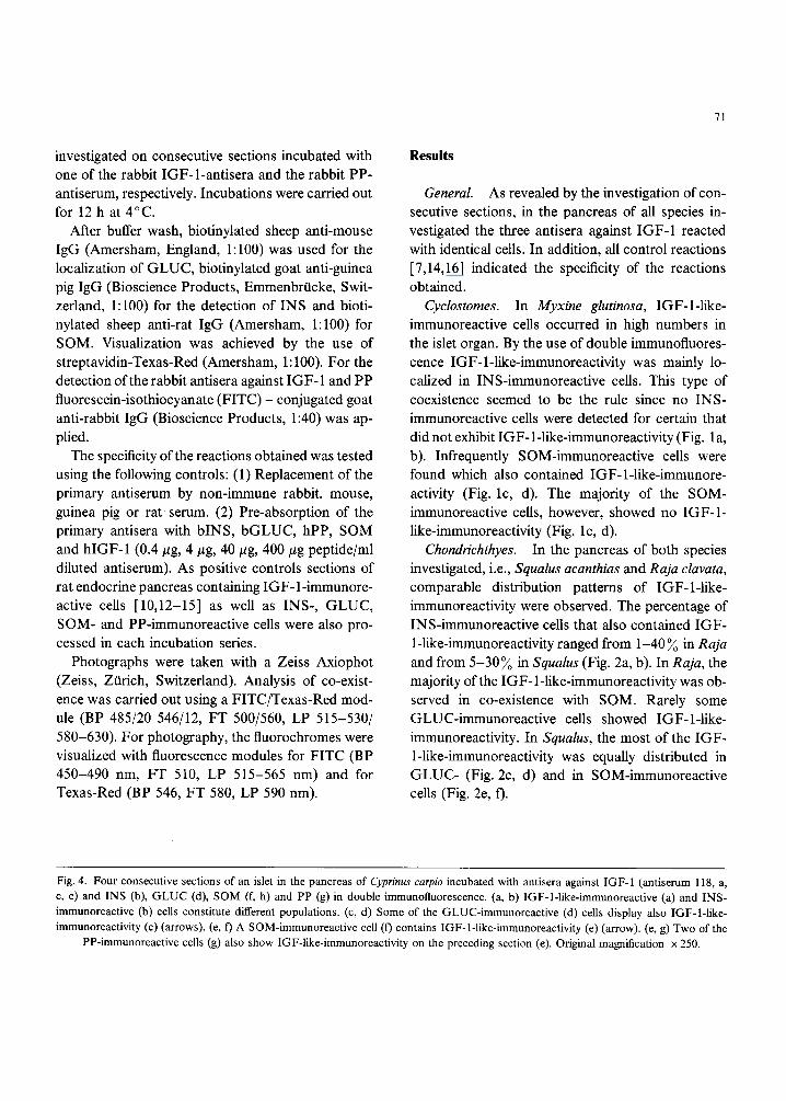
investigated on consecutive sections incubated with one of the rabbit IGF-1-antisera and the rabbit PP- antiserum, respectively. Incubations were carried out for 12 h at 4°C.
After buffer wash, biotinylated sheep anti-mouse IgG (Amersham, England, 1:100) was used for the localization of GLUC, biotinylated goat anti-guinea pig IgG (Bioscience Products, Emmenbrtlcke, Swit- zerland, 1:100) for the detection of INS and bioti- nylated sheep anti-rat IgG (Amersham, 1:100) for SOM. Visualization was achieved by the use of streptavidin-Texas-Red (Amersham, 1:100). For the detection of the rabbit antisera against IGF-1 and PP fluorescein-isothiocyanate (FITC) - conjugated goat anti-rabbit IgG (Bioscience Products, 1:40) was ap- plied.
The specificity of the reactions obtained was tested using the following controls: (1) Replacement of the primary antiserum by non-immune rabbit, mouse, guinea pig or rat serum. (2) Pre-absorption of the primary antisera with bINS, bGLUC, hPP, SOM and hlGF-1 (0.4/~g, 4/~g, 40 #g, 400/zg peptide/ml diluted antiserum). As positive controls sections of rat endocrine pancreas containing IGF-1-immunore- active cells [10,12-15] as well as INS-, GLUC, SOM- and PP-immunoreactive cells were also pro- cessed in each incubation series.
Photographs were taken with a Zeiss Axiophot (Zeiss, Ztirich, Switzerland). Analysis of co-exist- ence was carried out using a FITC/Texas-Red mod- ule (BP 485/20 546/12, FT 500/560, LP 515-530/ 580-630). For photography, the fluorochromes were visualized with fluorescence modules for FITC (BP 450-490 nm, FT 510, LP 515-565 nm) and for Texas-Red (BP 546, FT 580, LP 590 nm).
Results
General. As revealed by the investigation of con- secutive sections, in the pancreas of all species in- vestigated the three antisera against IGF-1 reacted with identical cells. In addition, all control reactions [7,14,16] indicated the specificity of the reactions obtained.
Cyclostomes. In Myxine glutinosa, IGF-l-like- immunoreactive cells occurred in high numbers in the islet organ. By the use of double immunofluores- cence IGF-l-like-immunoreactivity was mainly lo- calized in INS-immunoreactive cells. This type of coexistence seemed to be the rule since no INS- immunoreactive cells were detected for certain that did not exhibit IGF- 1-1ike-immunoreactivity (Fig. la, b). Infrequently SOM-immunoreactive cells were found which also contained IGF-l-like-immunore- activity (Fig. lc, d). The majority of the SOM- immunoreactive cells, however, showed no IGF-1- like-immunoreactivity (Fig. lc, d).
Chondrichthyes. In the pancreas of both species investigated, i.e., Squalus acanthias and Raja clavata, comparable distribution patterns of IGF-l-like- immunoreactivity were observed. The percentage of INS-immunoreactive cells that also contained IGF- 1-1ike-immunoreactivity ranged from 1-40 ~o in Raja and from 5-30~o in Squalus (Fig. 2a, b). In Raja, the majority of the IGF-1-1ike-immunoreactivity was ob- served in co-existence with SOM. Rarely some GLUC-immunoreactive cells showed IGF-l-like- immunoreactivity. In Squalus, the most of the IGF- 1-1ike-immunoreactivity was equally distributed in GLUC- (Fig. 2c, d) and in SOM-immunoreactive cells (Fig. 2e, f).
Fig. 4. Four consecutive sections of an islet in the pancreas of Cyprinus carpio incubated with antisera against IGF-I (antiserum 118, a, c, e) and INS (b), G L U C (d), SOM (f, h) and PP (g) in double immunofluorescence. (a, b) IGF-l-like-immunoreactive (a) and INS- immunoreactive (b) cells constitute different populations. (c, d) Some of the GLUC-immunoreactive (d) cells display also IGF-l-like- immunoreactivity (c) (arrows). (e, ti) A SOM-immunoreactive cell (13 contains IGF-1-1ike-immunoreactivity (e) (arrow). (e, g) Two of the
PP-immunoreactive cells (g) also show IGF-like-immunoreactivity on the preceding section (e). Original magnification × 250.

72
Fig. 5. Three consecutive sections of an islet in the pancreas ofAnguilla anguilla incubated with antisera against IGF-1 (a, c, antiserum 118, e, antiserum K37) and INS (b), G L U C (d) and SOM (f) in double immunofluorescence. While no IGF-l-like-immunoreactivity is found in INS-immunoreactive (b) or in GLUC-immunoreactive (d) cells, all of the IGF-l-like-immunoreactivity is present in SOM-
immunoreactive cells (some are marked by arrows). Original magnification × 230.

73
Osteichthyes. Different results were obtained for the bony fish species studied. In all endocrine islets of Cottus, i.e., the large pyloric (Fig. 3a-d) and splenic islets (Brockmann bodies) and the small islets (Fig. 3e, f'), dispersed throughout the exocrine paren- chyma, IGF-l-like- and INS-immunoreactivities co- existed to 100~o. In contrast, in Cyprinus, Anguilla
a
(3
and Carassius, IGF- 1-1ike-immunoreactivity was not observed in INS-immunoreactive cells (Figs. 4a, b; 5a, b; 6a, b). Among the three latter species some differences were found as regards to the localization of IGF- 1-1ike-immunoreactivity. In Cyprinus, the ma- jority of IGF-l-like-immunoreactivity was found in GLUC-immunoreactive cells (Fig. 4c, d), the remain-
Fig. 6. Three consecutive sections of an islet in the pancreas of Carassius auratus incubated with antiserum 116 against IGF-1 (a, c, e) and with antisera against INS (b), GLUC (d) and SOM (f) in double immunofluorescence. No IGF-l-like-immunoreactivity is found in INS-immunoreactive (b) or in GLUC-immunoreactive (d) cells, but all of the IGF-l-like-immunoreactivity is present in SOM-
immunoreactive cells (f). Original magnification × 210.

74
ing portion occurred in SOM- (Fig. 4e, f) and in PP-immunoreactive cells (Fig. 4e, g). In Anguilla and Carassius, IGF-l-like-immunoreactivity was de- tected in SOM-immunoreactive cells (Figs. 5e, f; 6e, f) while it seemed to be absent from the GLUC- immunoreactive cells (Figs. 5c, d; 6c, d).
Discussion
The present immunohistochemical study using three different IGF-1 antisera on 4 bony fish, 2 car- tilaginous fish and one cyclostomian species sup- ports and expands results obtained before for one representative of each of the three lowest vertebrate classes by the use of antiserum K37 [14,16].
In the lowest vertebrate class, the cyclostomes, insulin-like immunoreactive materials detected in the brain of Eptatretus stouti and Myxine glutinosa have been speculated to contain an IGF-like factor [19]. In addition, in Myxine liver the sequence of a prepro- IGF cDNA has been determined exhibiting 70 ~o ho- mology in the A and B domain to both human IGF- 1 and IGF-2 and conservation of all residues neces- sary for the tertiary structure [ 17]. Furthermore, the presence of Myxine IGF mRNA in a variety of or- gans, including islet and brain, has been determined by Northern blot analysis [17]. These results are in accordance with the detection of IGF- 1-related pep- tides in gastro-entero-pancreatic (GEP) system and brain ofMyxine [ 16]. For chondrichthyes, apart from the results presented here, the existence of IGF-1- like peptides has been shown only for Raja elavata by the use of chromatographic and immunohistochemi- cal methods [14]. For bony fish, there is increasing evidence for the presence of IGF- l-like peptides. Re- cently, the cDNA sequence encoding preproIGF-1 has been determined from Oncorhynchus kisuteh, the coho salmon [20], and from Oncorhynchus mykiss, the rainbow trout [21]. The deduced amino acid se- quences of the bony fish peptides showed about 80~o homology to mammalian (human) IGF-1 [20,21], and rainbow trout IGF-1 displayed 98 ~o homology to coho salmon IGF-1 [21]. In agreement, the im-
munohistochemical localization of IGF-l-related peptides in the GEP-system has been described for a representative bony fish [ 14]. Thus, the presence of IGF-l-like peptides may be common to lower ver- tebrates.
These findings raise questions as to the functional significance of the IGF-1 in lower vertebrates. Re- cently, IGF-1 receptors have been identified in vari- ous tissues of cyclostomes, cartilaginous and bony fish [22], supporting their biological role. Injections of bovine growth hormone increased the level of preproIGF-1 mRNA in the coho salmon liver [20]. Studies on fish have shown that similarly to mam- mals, IGF-1 is a major regulator of growth and de- velopment. An additional role in osmoregulation and saline tolerance has been demonstrated. In the trout for example, IGF-1 is a potent modulator of the action of growth hormone in seawater adaptation [23] as well as stimulating 35SO 4 incorporation into cartilage of several teleosts [24,25].
Based upon cellular localization by immunohis- tochemistry, the present findings suggest a branching of IGF- 1 and insulin at the phylogenetic level of the bony fish. There appears to be a gradual change from co-localization in lower vertebrates such as Myxine through to appearance in separate and discrete cell populations in bony fish. As is found in mammalian species [ 15], at the time of appearance of the bony fish, no IGF-1 and insulin co-localization is ob- served. In accordance with such a continuum of evo- lution, the cartilaginous fish investigated in the present study display an intermediate amount of IGF-1/insulin co-localization. The exception to this scheme seems to be Cottus which shows 100~o co- localization. Since Cottus is looked upon as a highly developed bony fish, analogous studies in amphibia, reptiles and birds will be carried out to investigate the possible co-localization of IGF-1 and insulin in higher vertebrates. The hypothesis of the IGF-1/ insulin branching at the phylogenetic level of the bony fish needs to be explored further at the gene level to identify the cellular sites of synthesis as well as to characterize the gene products during evolution.

Acknowledgements
The study was supported by the Swiss National Foundation (Grant No. 32-33349.92), by the Swed- ish Medical Research Council (Project Nos 718, 8637, 6825 and 9033), the Cancer Society of Stock- holm, the Nordic Insulin Foundation and the Fac- ulty of Medicine at the Karolinska Institute. For gen- erous supply of the IGF-1 antisera 116 and 118 we thank Dr. J. Zapf (University Hospital ZOrich, Swit- zerland) and for K37 Dr. M. Lake (Kabi Pharmacia, Uppsala, Sweden). Special thanks are due to Ms A. Burlet for excellent technical assistance. We are also grateful to the Director and the staff of the Kristineberg Marine Biological Station (Fiskeb~ick- skil, Sweden), where part of this work was carried out.
References
1 Daughaday, W.H. and Rotwein, P., Insulin-like growth factor I and II. Peptide, messenger ribonucleic acid and gene struc- tures, serum and tissue concentrations, Endocr. Rev., 10 (1989) 68-90.
2 Humbel, R.E., Insulin-like growth factors I and II, Eur. J. Biochem., 190 (1990) 445-462.
3 Sara, V.R. and Hall, K., The insulin-like growth factors and their binding proteins, Physiol. Rev., 70 (1990) 591-614.
4 Steiner, D.F., Chan, S.J., Docherty, K., Emdin, S.O., Dod- son, G.G. and Falkmer S., Evolution ofpolypeptide hormones and their precursor processing mechanisms. In S. Falkmer, R. Hakanson and F. Sundler (Eds.), Evolution and Tumour Pa- thology of the Neuroendocrine System, Elsevier, Amsterdam, 1985, pp. 203-223.
5 Plisetskaya, E.M., Pancreatic peptides. In S. Holmgren (Ed), The Comparative Physiology of Regulatory Peptides, Chap- man and Hall, London, 1989, pp. 174-202.
6 Chan, SJ., Cao, Q.-P. and Steiner, D.F., Evolution of the insulin superfamily: cloning of a hybrid insulin/insulin-like growth factor cDNA from amphioxus, Proc. Natl. Acad. Sci. USA, 87 (1990) 9319-9323.
7 Reinecke, M., Betzler, D., Drakenberg, K., Falkmer, S. and Sara, V.R., Occurrence of members of the insulin superfam- ily in central nervous system and digestive tract of protochor- dates, Histochemistry, 99 (1993) 277-285.
8 Blundell, T.L. and Humbel, R.E., Hormone families: pancre-
75
atic hormones and homologous growth factors, Nature (Lon- don), 287 (1980) 781-787.
9 Froesch, E.R. and Zapf, J., Insulin-like growth factors and insulin: comparative aspects, Diabetologica, 28 (1985) 485- 493.
10 Andersson, I., Billig, H., Fryklund, L., Hansson, H.-A., Isaks- son, O., Isgaard, J., Nilsson, A., Rozell, B., Skottner, A. and Stemme, S., Localisation of IGF-I in adult rats, Immunohis- tochemical studies, Acta Physiol Scand., 126 (1986) 311-312.
11 Han, V.K.M,, Hill, D.J., Strain, A.J., Towle, A.C., Lauder, J.M., Underwood, L.E. and D'Ercole, A.J., Identification of somatomedin/insulin-like growth factor immunoreactive cells in the human fetus, Pediat. Res., 22 (1987) 245-249.
12 Hansson, H.-A., Edwall, D., LOwenadler, B., Norstedt, G., Paleus, S. and Skottner, A., Insulin-like growth factor I in the pancreas of normal and diabetic adult rats, Acta Physiol. Scand., 132 (1988a) 569-576.
13 Hansson, H.-A., Nilsson, A., Isgaard, J., Billig, H., Isaksson, O., Skottner, A., Andersson, I.K. and Rozell, B., Immuno- histochemical localization of insulin-like growth factor I in the adult rat, Histochemistry, 89 (1988b) 403-410.
14 Reinecke, M., Drakenberg, K., Falkmer, S. and Sara, V.R., Peptides related to insulin-like growth factor 1 in the gastro- entero-pancreatic system of bony fish and cartilaginous fish, Regul. Pept., 37 (1992) 155-165.
15 Maake, C. and Reinecke, M., Immunohistochemical localiza- tion of insulin-like growth factor 1 and 2 in the endocrine pancreas of rat, dog and man and their coexistence with clas- sical islet hormones, Cell Tissue Res., 273 (1993) 249-259.
16 Reinecke, M., Drakenberg, K., Falkmer, S. and Sara, V.R., Presence of IGF-l-like peptides in the neuroendocrine system of the Atlantic Hagfish, Myxine glutinosa (Cyclostomata): evi- dence derived by chromatography, radioimmunoassay and im- munohistochemistry, Histochemistry, 96 (1991) 191-196.
17 Nagamatsu, S., Chan, S.J., Falkmer, S. and Steiner, D.F., Evolution of the insulin gene super family. Sequence of a preproinsulin-like growth factor cDNA from the Atlantic hag- fish, J. Biol. Chem., 266 (1991) 2397-2402.
18 Zapf, J., Walter, H. and Froesch, E.R., Radioimmunological determination of insulin-like growth factors I and II in normal subjects and in patients with growth disorders and extrapan- creatic tumor hypoglycemia, J. Clin. Invest., 68 (1981) 1321- 1330.
19 Thorndyke, M.C,, Purvis, D. and Plitsetskaya, E.M., Insulin- like immunoreactivity in the brain of two Hagfishes, Eptatretus stouti and Myxine glutinosa, Gen. Comp. Endocrinol., 76 (1989) 371-381.
20 Cao, Q.P., Duguay, S.J., Plisetskaya, E., Steiner, D.F. and Chan, S.J., Nucleotide sequence and growth hormone- regulated expression of salmon insulin-like growth factor 1 mRNA, Molec. Endocrinol., 3 (1989) 2005-2010.

76
21 Shamblott, M.J. and Chen, T.T., Identification of a second insulin-like growth factor in a fish species, Proc. Natl. Acad. Sci. USA, 89 (1992) 8913-8917.
22 Drakenberg, K., Sara, V.R., Falkmer, S., Gammeltoft, S, Maake, C. and Reinecke, M., Identification of IGF-1 recep- tors in primitive vertebrates, Regul. Pept., 43 (1993) 73-82.
23 McCormick, S.D., Sakamoto, T., Hasegawa, S. and Hirano, T., Osmoregulatory functions of insulin-like growth factor-I in rainbow trout (Oncorhynchus mykiss), J. Endocrinol. 130 ( 1991 ) 87-92.
24 Duan, C. and Hirano, T., Stimulation of [35S]sulphate uptake by mammalian insulin-like growth factors I and II in cultured cartilages of the japanese eel, Anguillajaponica, J. Exp. Zool., 256 (1990) 347-350.
25 Bern, H.A., McCormick, S.D,, Kelley, K.M., Gray, E.S., Nishioka, R.S., Madsen, S.S. and Tsai, P.I., Insulin-like growth factors 'under water': role in growth and function of fish and other poikilotermic vertebrates. In E.M. Spencer (Ed.), Modern Concepts of Insulin-like Growth Factors, Elsevier, Amsterdam, 1991, pp. 85-96.