Insulin resistance in penile arteries from a rat model of metabolic syndrome
15
RESEARCH PAPER Insulin resistance in penile arteries from a rat model of metabolic syndrome Cristina Contreras 1 , Ana Sánchez 1 , Pilar Martínez 2 , Rafaela Raposo 1 , Belén Climent 1 , Albino García-Sacristán 1 , Sara Benedito 1 and Dolores Prieto 1 1 Departamento de Fisiología, Facultad de Farmacia, Universidad Complutense de Madrid, Madrid, Spain, and 2 Departamento de Anatomía y Anatomía Patológica Comparadas, Facultad de Veterinaria, Universidad Complutense de Madrid, Madrid, Spain Correspondence Dra. Dolores Prieto, Dpto. de Fisiología, Facultad de Farmacia, Plaza de Ramón y Cajal s/n, Universidad Complutense de Madrid, 28040-Madrid, Spain. E-mail: [email protected] ---------------------------------------------------------------- Keywords: penile arteries; insulin resistance; endothelial dysfunction; b-adrenoceptors; obese Zucker rat ---------------------------------------------------------------- Received 19 January 2010 Revised 15 March 2010 Accepted 11 April 2010 BACKGROUND AND PURPOSE Metabolic and cardiovascular abnormalities accompanying metabolic syndrome, such as obesity, insulin resistance and hypertension, are all associated with endothelial dysfunction and are independent risk factors for erectile dysfunction. The purpose of the present study was to investigate the vascular effects of insulin in penile arteries and whether these effects are impaired in a rat model of insulin resistance and metabolic syndrome. EXPERIMENTAL APPROACH Penile arteries from obese Zucker rats (OZR) and their counterpart, lean Zucker rats (LZR), were mounted on microvascular myographs and the effects of insulin were assessed in the absence and presence of endothelium and of specific inhibitors of nitric oxide (NO) synthesis, phosphatidylinositol 3-kinase (PI3K) and mitogen-activated protein kinase (MAPK). Insulin-induced changes in intracellular Ca 2+ concentration [Ca 2+ ]i were also examined. KEY RESULTS OZR exhibited mild hyperglycaemia, hypercholesterolemia, hypertryglyceridemia and hyperinsulinemia. Insulin induced endothelium- and NO-dependent relaxations in LZR that were impaired in OZR. Inhibition of PI3K reduced relaxation induced by insulin and by the b-adrenoceptor agonist isoprenaline, mainly in arteries from LZR. Antagonism of endothelin 1 (ET-1) receptors did not alter insulin-induced relaxation in either LZR or OZR, but MAPK blockade increased the responses in OZR. Insulin decreased [Ca 2+ ]i, a response impaired in OZR. CONCLUSIONS AND IMPLICATIONS Insulin-induced relaxation was impaired in penile arteries of OZR due to altered NO release through the PI3K pathway and unmasking of a MAPK-mediated vasoconstriction. This vascular insulin resistance is likely to contribute to the endothelial dysfunction and erectile dysfunction associated with insulin resistant states. Abbreviations ED, erectile dysfunction; ET-1, endothelin 1; KPSS, high K + -physiological saline solution; L-NNA, N w -nitro-L-arginine; LZR, lean Zucker rat; MAPK, mitogen-activated protein kinase; NOS, nitric oxide synthase; OZR, obese Zucker rat; PI3K, phosphatidylinositol 3-kinase; PSS, physiological saline solution; VSMC, vascular smooth muscle cells Introduction Erectile dysfunction (ED) is currently considered as an early sign of subclinical vascular disease and is a highly prevalent condition in patients with cardio- vascular risk factors such as hypertension, dyslipi- demia, central obesity and insulin resistance (Montorsi et al., 2003). The cluster of these meta- bolic and cardiovascular abnormalities, jointly referred to as metabolic syndrome, further increases the risk for the development of ED (Fonseca and Jawa, 2005; Jackson, 2006). BJP British Journal of Pharmacology DOI:10.1111/j.1476-5381.2010.00825.x www.brjpharmacol.org 350 British Journal of Pharmacology (2010) 161 350–364 © 2010 The Authors British Journal of Pharmacology © 2010 The British Pharmacological Society
-
Upload
independent -
Category
Documents
-
view
1 -
download
0
Transcript of Insulin resistance in penile arteries from a rat model of metabolic syndrome
RESEARCH PAPER
Insulin resistance in penilearteries from a rat model ofmetabolic syndromeCristina Contreras1, Ana Sánchez1, Pilar Martínez2, Rafaela Raposo1,
Belén Climent1, Albino García-Sacristán1, Sara Benedito1 and
Dolores Prieto1
1Departamento de Fisiología, Facultad de Farmacia, Universidad Complutense de Madrid,
Madrid, Spain, and 2Departamento de Anatomía y Anatomía Patológica Comparadas, Facultad
de Veterinaria, Universidad Complutense de Madrid, Madrid, Spain
CorrespondenceDra. Dolores Prieto, Dpto. deFisiología, Facultad de Farmacia,Plaza de Ramón y Cajal s/n,Universidad Complutense deMadrid, 28040-Madrid, Spain.E-mail: dprieto@farm.ucm.es----------------------------------------------------------------
Keywords:penile arteries; insulin resistance;endothelial dysfunction;b-adrenoceptors; obese Zucker rat----------------------------------------------------------------
Received19 January 2010Revised15 March 2010Accepted11 April 2010
BACKGROUND AND PURPOSEMetabolic and cardiovascular abnormalities accompanying metabolic syndrome, such as obesity, insulin resistance andhypertension, are all associated with endothelial dysfunction and are independent risk factors for erectile dysfunction. Thepurpose of the present study was to investigate the vascular effects of insulin in penile arteries and whether these effects areimpaired in a rat model of insulin resistance and metabolic syndrome.
EXPERIMENTAL APPROACHPenile arteries from obese Zucker rats (OZR) and their counterpart, lean Zucker rats (LZR), were mounted on microvascularmyographs and the effects of insulin were assessed in the absence and presence of endothelium and of specific inhibitors ofnitric oxide (NO) synthesis, phosphatidylinositol 3-kinase (PI3K) and mitogen-activated protein kinase (MAPK). Insulin-inducedchanges in intracellular Ca2+ concentration [Ca2+]i were also examined.
KEY RESULTSOZR exhibited mild hyperglycaemia, hypercholesterolemia, hypertryglyceridemia and hyperinsulinemia. Insulin inducedendothelium- and NO-dependent relaxations in LZR that were impaired in OZR. Inhibition of PI3K reduced relaxation inducedby insulin and by the b-adrenoceptor agonist isoprenaline, mainly in arteries from LZR. Antagonism of endothelin 1 (ET-1)receptors did not alter insulin-induced relaxation in either LZR or OZR, but MAPK blockade increased the responses in OZR.Insulin decreased [Ca2+]i, a response impaired in OZR.
CONCLUSIONS AND IMPLICATIONSInsulin-induced relaxation was impaired in penile arteries of OZR due to altered NO release through the PI3K pathway andunmasking of a MAPK-mediated vasoconstriction. This vascular insulin resistance is likely to contribute to the endothelialdysfunction and erectile dysfunction associated with insulin resistant states.
AbbreviationsED, erectile dysfunction; ET-1, endothelin 1; KPSS, high K+-physiological saline solution; L-NNA, Nw-nitro-L-arginine;LZR, lean Zucker rat; MAPK, mitogen-activated protein kinase; NOS, nitric oxide synthase; OZR, obese Zucker rat; PI3K,phosphatidylinositol 3-kinase; PSS, physiological saline solution; VSMC, vascular smooth muscle cells
Introduction
Erectile dysfunction (ED) is currently considered asan early sign of subclinical vascular disease and is ahighly prevalent condition in patients with cardio-vascular risk factors such as hypertension, dyslipi-
demia, central obesity and insulin resistance(Montorsi et al., 2003). The cluster of these meta-bolic and cardiovascular abnormalities, jointlyreferred to as metabolic syndrome, further increasesthe risk for the development of ED (Fonseca andJawa, 2005; Jackson, 2006).
BJP British Journal ofPharmacology
DOI:10.1111/j.1476-5381.2010.00825.xwww.brjpharmacol.org
350 British Journal of Pharmacology (2010) 161 350–364 © 2010 The AuthorsBritish Journal of Pharmacology © 2010 The British Pharmacological Society
Insulin resistance and hyperinsulinemia play amajor role in the pathogenesis of type 2 diabetesand have been associated with the development ofcardiovascular disease and hypertension (De Fronzoand Ferrannini, 1991). Insulin resistance is typicallydefined as a decreased sensitivity to the metabolicactions of insulin such as insulin-mediated glucosedisposal. However, in addition to its classical physi-ological actions, insulin has vascular actions thatcouple the metabolic and haemodynamic homeo-stasis under healthy conditions (Kim et al., 2006).The association between insulin resistance and car-diovascular disease was initially ascribed to theactions of insulin on vascular smooth musclegrowth and extracellular matrix production (Tama-roglio and Lo, 1994). However, experimental evi-dence shows that endothelial function is impairedin insulin resistance and diabetic patients anddiminished sensitivity or resistance to the actions ofinsulin in the vascular endothelium also contributeto the phenotype of the insulin-resistant states(Baron et al., 1991; Steinberg et al., 1996; Kim et al.,2006).
Insulin binding to its receptor at the endothelialcells activates the phosphatidylinositol 3-kinase(PI3K)/Akt pathway that in turns phosphorylatesendothelial nitric oxide synthase (eNOS) at Ser 1177and induces nitric oxide (NO) release and vasodila-tation (Zeng et al., 2000). Stimulation of endothelialinsulin receptors can also lead to the activation ofthe Ras-Raf-mitogen-activated protein kinase(MAPK) pathway that is associated with gene expres-sion, cell growth and the production of vasocon-strictor endothelin 1 (ET-1) (Cardillo et al., 1999). Inobese insulin-resistant men and patients with type 2diabetes, the insulin-mediated activation of thePI3K/Akt pathway underlying both the metabolicand the vascular actions of the hormone isimpaired, and endothelial dysfunction and bluntedvasodilatation have been reported (Steinberg et al.,1996; Cusi et al., 2000).
The metabolic and cardiovascular abnormalitiesaccompanying metabolic syndrome, that is, glucoseintolerance, dyslipidemia, insulin resistance andhypertension, act as independent risk factors for EDand are all associated to some extent with endothe-lial dysfunction (Fonseca and Jawa, 2005), althoughthe mechanisms for this dysfunction in the penilevasculature are not completely understood. Novelstudies in an animal model of type 1 diabetes-associated ED have demonstrated an impairment ofthe PI3K/Akt/eNOS pathway in the erectile tissueassociated with a decreased erectile capability(Musicki et al., 2005). However, the effects of insulinon the penile vasculature of both healthy individu-als and of type 2 diabetes and insulin resistance-
associated ED models are unknown. In the presentstudy, we investigated the mechanisms underlyingthe vasoactive actions of insulin in penile arteriesfrom the obese Zucker rat (OZR), a well-establishedgenetic model of insulin resistance and metabolicsyndrome caused by a dysfunctional gene of theleptin receptor. These animals progressively developobesity, type 2 diabetes and hypertension (Guerre-Millo 1997). We have recently demonstrated pro-found alterations in vascular structure andendothelial dysfunction in dorsal penile arteriesfrom the OZR (Villalba et al., 2009), data consistentwith the ED reported in these animals (Wingardet al., 2007). Here, we aimed to further investigatewhether the vascular effects of insulin are impairedin penile arteries under conditions of insulin resis-tance in the OZR.
Methods
Animal modelAll animal care and experimental protocols conformto European Union guidelines for the Care and Useof Laboratory Animals and were approved by theInstitutional Animal Care and Use Committee ofthe Complutense University of Madrid. Male OZR (n= 47) and lean Zucker rats (LZR, n = 46) wereobtained from Charles River Laboratories (Barce-lona, Spain) and used for study at 17–18 weeks ofage.
Experimental procedures for the functionalexperimentsOn the day of the experiment, blood samples wereobtained from the tail vein before death and plasmawas frozen for determination of non-fasting glucose,cholesterol, triglycerides and insulin plasma levels.Glucose, total triglyceride and total cholesterollevels were determined by using commercially avail-able kits. Plasma insulin levels were measured byspecific enzyme-linked immunosorbent assay.
Immediately after killing of the animal (cervicaldislocation and exsanguination), the penis wasremoved and placed in cold physiological salinesolution (PSS) of the following composition (mM):NaCl 119, NaHCO3 25, KCl 4.7, K2H2PO4 1.17,MgSO4 1.18, CaCl2 1.5, EDTA 0.027 and glucose 11.The penile arteries, first- or second-order branches ofthe dorsal penile artery from LZR and OZR werecarefully dissected by removing the connective andfat tissue, as described previously (Villalba et al.,2009; Sánchez et al., 2010). These arteries were usedin order to assess erectile tissue function as they cansupply blood to the cavernous tissue and are of a sizein the rat that allows handling without damaging
BJPPenile vascular insulin resistance in metabolic syndrome
British Journal of Pharmacology (2010) 161 350–364 351
the vascular endothelium (Villalba et al., 2009). Onearterial segment from a LZR and another from anOZR were mounted in parallel on double microvas-cular myographs (DMT, Denmark) and stretched totheir optimal internal lumen diameter l1 corre-sponding to 90% of the diameter the arteries wouldhave in situ under an internal transmural pressure of100 mmHg (Villalba et al., 2009). Aortic rings fromLZR and OZR were also used in order to compare thevascular effects of insulin with those on penile arter-ies. Thoracic aortas were isolated and cleaned fromthe fatty and connective tissue, and arterial ringswere mounted in vascular myographs and a passiveinitial tension of 10 mN applied.
At the beginning of each experiment, vasocon-strictor responses to 120 mM K+ (highK+-physiological saline solution, KPSS) confirmedvessel viability. The vasoactive effects of insulinwere evaluated by adding cumulative concentra-tions of the hormone to arteries precontracted withphenylephrine (1 mM), in the absence or presence ofspecific inhibitors of NOS (Nw-nitro-L-arginine,L-NNA, 100 mM), PI3K (LY-294002, 3 mM), cyclooxy-genase (indomethacin, 1 mM), MAPK (PD-98059,5 mM) and the non-selective antagonist of ET-1receptors (bosentan, 10 mM). Because insulin effectswere not reproducible in a second stimulation, twoconsecutive arterial segments of the same animalwere used, one as a control and in the second thetreatment was applied. The role of the vascularendothelium was assessed in arteries where theendothelium was mechanically removed by passinga human hair through the vessel lumen. Theabsence of functional endothelium was confirmedby the lack of relaxation to acetylcholine (ACh,10 mM). The effects of the b-adrenoceptor agonistisoprenaline were also evaluated in the absence andpresence of LY-294002. In order to assess the cAMP-dependent relaxations in penile arteries,concentration–response curves for the adenylatecyclase activator forskolin were also performed.
Measurements of intracellular Ca2+ ([Ca2+]i)Simultaneous measurements of [Ca2+]i and forcewere performed in intact penile arteries as previ-ously described (Villalba et al., 2007). The myographwas mounted on an inverted microscope (AxiovertS100 TV) equipped for dual excitation wavelengthmicrofluorimetry. Penile arteries were incubated inthe dark in PSS with 8 mM Fura 2-acetoxymethylester for 3 h at 37°C, changing the solution after1.5 h. Then arteries were washed for 45 min toremove remaining Fura 2-acetoxymethyl ester. Afterloading, the arteries were illuminated with alternat-ing 340 and 380 nm light using a monochromator-based system (Deltascan, PTI). The fluorescence
emission was detected at a wavelength of 510 nm.At the end of each experiment, Ca2+-insensitivesignals were determined after quenching with25 mM Mn2+, and the values obtained were sub-stracted from those obtained during the experi-ment. The ratio (R) F340/F380 was taken as a measureof [Ca2+]i.
Arteries were initially stimulated with KPSS inorder to test vessel viability. Then, the effect of asingle dose of insulin (0. 1 mM) was evaluated inarteries pre-contracted with phenylephrine (1 mM).Finally, the effect of ACh (10 mM) was also tested.
Data analysisValues are expressed as means � SEM of n (numberof arteries, 1–2 per animal). Relaxants responses toinsulin are given as a percentage of precontractioninduced by phenylephrine. The sensitivity of thearteries to the vasoactive agonists is expressed interms of pEC50 values, where pEC50 is -logEC50. EC50
is the concentration of the agonist required toproduce 50% of the response and was calculated byusing standard computer software (Prism 5.0,Graphpad, San Diego, CA, USA). The statistical dif-ferences between means were analysed by eitherunpaired Student’s t-test or by one-way ANOVA.Probability levels lower than 5% were consideredsignificant.
MaterialsThe sources of the compounds used in this MS wereas follows: L-NNA, indomethacin, insulin, PD98059,isoprenaline, acetylcholine, forskolin and phenyle-phrine were from Sigma Chemicals (Madrid, Spain).LY 294002 and endothelin1 (ET-1) were from Tocris,Cookson, (Bristol, UK); Fura 2-acetoxymethyl ester(Fura-2-AM) was from Molecular Probes (OR, USA).Bosentan was a generous gift of F. Hoffman La RocheLaboratories (Basel, Switzerland). All drug andmolecular target nomenclature follows Alexanderet al., (2009).
Results
General parametersAt the time of the experiment, the OZR were signifi-cantly heavier than the LZR and exhibited mildhyperglycaemia, hyperinsulinemia and dyslipi-demia with elevated total cholesterol and triglycer-ide levels (Table 1). In penile arteries from the OZRgroup, contractions to KPSS were reduced (2.11 �0.14 Nm-1 in LZR, n = 46; and 1.62 � 0.10 Nm-1 inOZR, n = 47, P < 0.01), and the ACh-induced relax-ations were impaired (58 � 4% in LZR, n = 27; and
BJP C Contreras et al.
352 British Journal of Pharmacology (2010) 161 350–364
43 � 5% in OZR, n = 38, P < 0.05), indicatingreduced contractility and endothelial dysfunctionrespectively.
Structure of penile arteriesNormalized internal lumen diameters (l1) of penilearteries were significantly smaller in OZR (129 �4 mm, n = 36) than in LZR (155 � 6 mm, n = 38, P <0.001), indicating inward vascular remodelling ininsulin-resistant animals. Penile weights were sig-nificantly lower in OZR than LZR (0.5 � 0.1 g and0.7 � 0.1 g, n = 7, P < 0.01, respectively), also sug-gesting structural remodelling of penile erectiletissue. Masson’s trichrome staining confirmed wallthickening and smaller internal diameters in arteriesfrom OZR (not shown).
Effect of insulin on penile arteries from LZRand OZRExogenously applied insulin (0.1–100 nM) inducedconcentration-dependent relaxations in penilearteries of LZR pre-contracted with phenylephrine(Figure 1). The pEC50 and response to the highestconcentration of insulin used (100 nM) was 8.02 �0.12 and 24.3 � 3.6% (n = 27) of the phenylephrine-induced contraction respectively (Figure 1A,C). Therelaxant responses to insulin were impaired inpenile arteries from OZR (insulin-induced (100 nM)relaxation of 7.5 � 2.5%, n = 31, P < 0.01 vs. LZR)(Figure 1B,C). In aortas from OZR, responses toinsulin were also blunted and, in some samples,turned into contractions, the response to 100 nMinsulin being -4.1 � 10.9% (n = 10) of thephenylephrine-induced contraction. In LZR aortas,the corresponding response to insulin was greater(24.0 � 7.2%, n = 11) of the phenylephrine-inducedcontraction (P < 0.01).
Effects of endothelial cell removal on insulinrelaxant responsesMechanical removal of the endothelium impairedinsulin-induced relaxations in penile arteries from
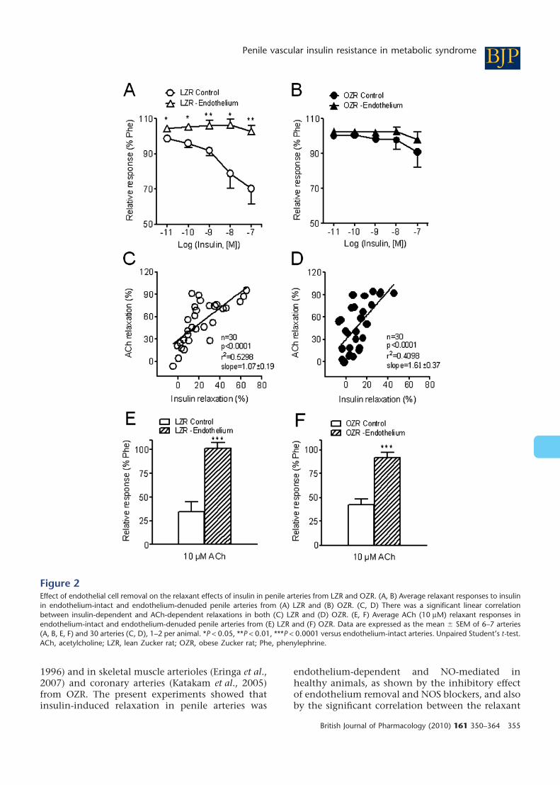
LZR but did not significantly alter responses in OZR(Figure 2A,B, Table 2). There was a significant linearcorrelation between the 100 nM insulin-inducedrelaxation and that elicited by the endothelial cellagonist ACh (10 mM) in both LZR and OZR(Figure 2C,D). The absence of functional endothe-lium was verified by the blunted vasodilator effect ofACh after endothelial cell removal (Figure 2E,F,Table 2).
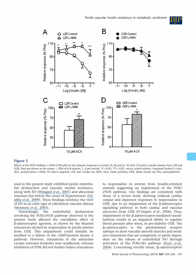
Effects of eNOS and PI3K inhibitors oninsulin relaxant responsesTreatment with the NOS blocker L-NNA (100 mM)produced a significant inhibition of the insulin-induced relaxation in penile arteries of LZR but notof OZR (Figure 3A,B, Table 2). The maximal AChrelaxant responses were significantly diminished inboth LZR and OZR after NOS blockade (Figure 3C,D,Table 2).
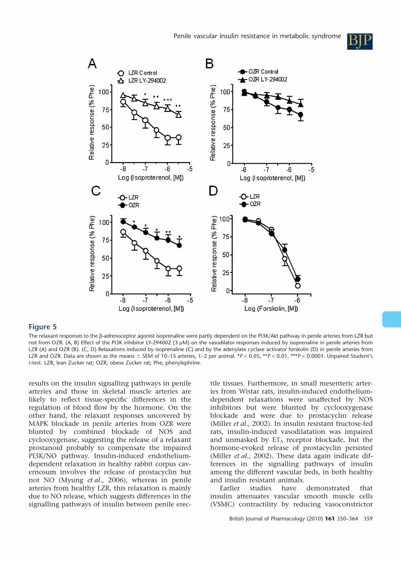
Inhibition of PI3K with LY-294002 (1 mM) mark-edly reduced the relaxant effect of insulin in arteriesof LZR, and to a lesser extent in OZR (Figure 4A,B,Table 2), without significantly altering the relaxanteffects of ACh in either LZR or OZR (Figure 4C,D,Table 2). Interestingly, LY-294002 also inhibited therelaxant responses induced by the b-adrenoceptoragonist isoprenaline in LZR but not in OZR(Figure 5A,B). The maximal relaxant effect of isopre-naline was significantly impaired in OZR comparedwith controls (Figure 5C). However, the relaxanteffect of the adenylate cyclase activator forskolinwas not different in penile arteries from OZR com-pared with LZR (Figure 5D), pEC50 being 6.18 � 0.21and 6.30 � 0.21 (n = 15, not significantly different)respectively.
Effect of ET-1 receptor antagonist and MAPKinhibitors on arterial responses to insulinThe antagonist of ET-1 receptors, bosentan, did notmodify the relaxant responses to insulin
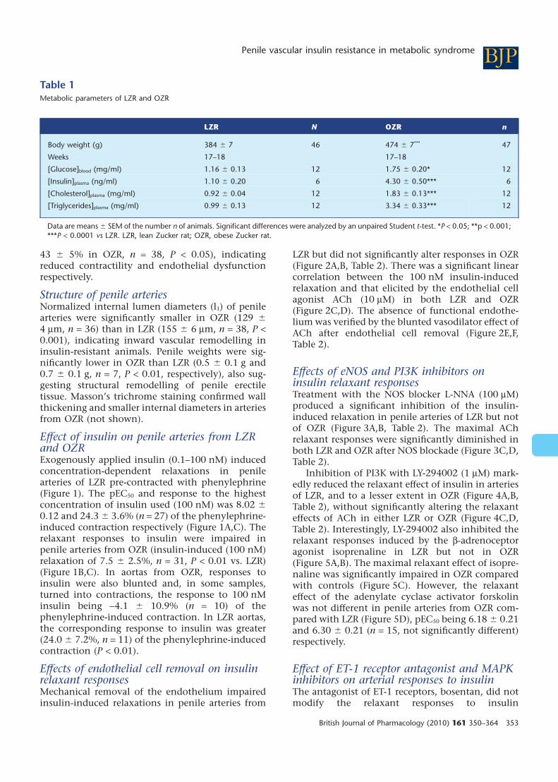
Table 1Metabolic parameters of LZR and OZR
LZR N OZR n
Body weight (g) 384 � 7 46 474 � 7*** 47
Weeks 17–18 17–18
[Glucose]blood (mg/ml) 1.16 � 0.13 12 1.75 � 0.20* 12
[Insulin]plasma (ng/ml) 1.10 � 0.20 6 4.30 � 0.50*** 6
[Cholesterol]plasma (mg/ml) 0.92 � 0.04 12 1.83 � 0.13*** 12
[Triglycerides]plasma (mg/ml) 0.99 � 0.13 12 3.34 � 0.33*** 12
Data are means � SEM of the number n of animals. Significant differences were analyzed by an unpaired Student t-test. *P < 0.05; **p < 0.001;***P < 0.0001 vs LZR. LZR, lean Zucker rat; OZR, obese Zucker rat.
BJPPenile vascular insulin resistance in metabolic syndrome
British Journal of Pharmacology (2010) 161 350–364 353

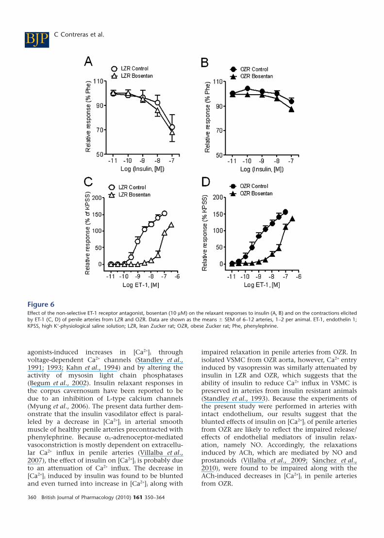
(Figure 6A,B) in penile arteries from either LZR orOZR. However, bosentan inhibited the contractionsinduced by ET-1 in both LZR (Figure 6C,D) (pEC50
values: 8.94 � 0.13, n = 8, vs. 7.23 � 0.12, n = 10, P< 0.0001, in control and treated LZR arteries, respec-tively) and OZR (pEC50 9.20 � 0.34, n = 7, vs. 7.23 �0.11, n = 12, P < 0.01, in control and treated arteries,respectively).
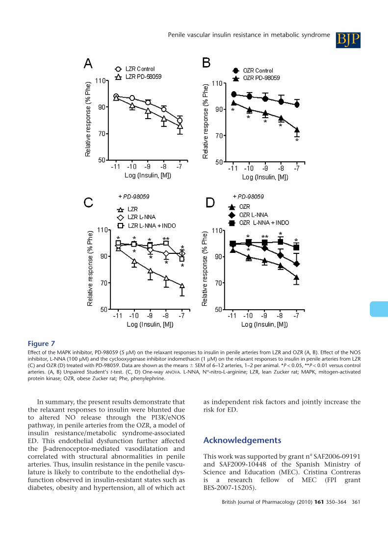
The MAPK inhibitor, PD-98059, did not alter therelaxant responses to insulin in LZR (Figure 7A), butsignificantly enhanced those in OZR (Figure 7B).The relaxant responses to insulin in the presence ofPD-98059 were significantly inhibited by NOSblockade in LZR (Figure 7C), but blunted only aftercombined blockade of NOS and cyclooxygenase inOZR (Figure 7D).
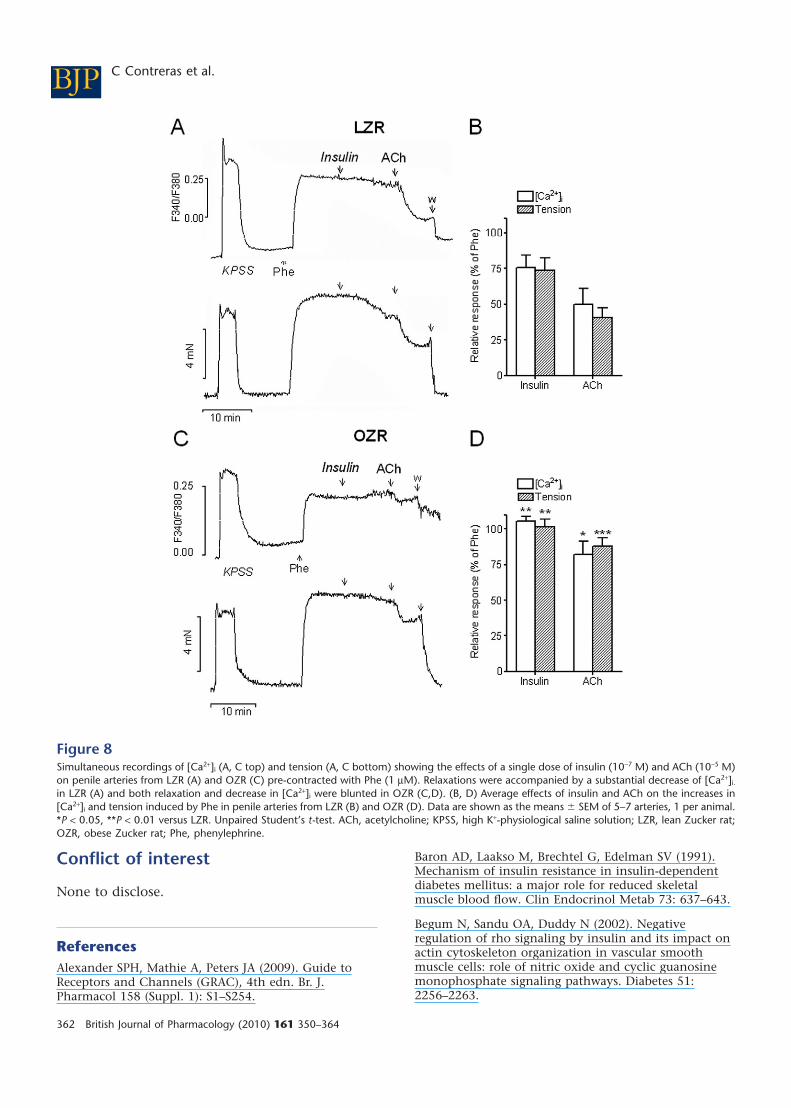
Effect of insulin on [Ca2+]i
Stimulation of penile arteries from LZR and OZRwith a high K+ solution (KPSS) induced simulta-neous increases in both [Ca2+]i [D(F340/F380) = 0.51� 0.05, n = 7, in LZR, and 0.35 � 0.04, n = 7, in OZR,P < 0.05] and tension (1.17 � 0.16 Nm-1, n = 7 in LZRand 0.95 � 0.18 Nm-1, n = 7, in OZR, P < 0.01)(Figure 8A,C). The a1-adrenoceptor agonist phenyle-phrine (1 mM) also evoked sustained increases in[Ca2+]i [D(F340/F380) = 0.38 � 0.06, n = 7, in LZR and0.26 � 0.04, n = 7, in OZR, P < 0.05] and tension(2.49 � 0.26 Nm-1, n = 7 in LZR and 1.31 �0.24 Nm-1, n = 7, in OZR, P < 0.01) (Figure 8A,C).
The relaxations induced by a single dose of bothinsulin (100 nM) and ACh (10 mM) were accompa-nied by simultaneous decreases in [Ca2+]i in penilearteries from LZR pre-contracted with phenyleph-rine (Figure 8A,B). These decreases in [Ca2+]i wereimpaired, along with relaxation, in arteries fromOZR (Figure 8C,D). In some arteries from OZR,stimulation with insulin was eventually accompa-nied by increases in [Ca2+]i (Figure 8C).
Discussion
The present study demonstrates that insulininduced endothelium-dependent relaxations inpenile arteries through the release of NO via thePI3K/eNOS pathway. This pathway was impaired inarteries from insulin-resistant OZR thus unmaskingMAPK-dependent vasoconstrictor effects of insulin.Penile endothelial dysfunction in insulin resistantanimals, measured here as a decreased insulin-stimulated, NO-mediated, relaxation of the penilearterial rings, correlates with abnormal structureand vascular remodelling (Villalba et al., 2009), andprobably contributes to the decreased erectile capa-bility reported for this animal model (Wingard et al.,2007) and to the ED associated to insulin-resistantstates, such as diabetes, obesity and hypertension(Fonseca and Jawa, 2005).
The impaired relaxant effect of insulin in penilearteries of OZR was also found in the aorta of thesame animals and is in agreement with the reportedimpairment of the insulin-mediated vasodilatationin obese subjects (Baron et al., 1991; Steinberg et al.,
Figure 1Relaxant responses to insulin were significantly impaired in penilearteries from OZR compared with LZR. (A, B) Representative tracesshowing the insulin-induced relaxations in penile arteries from (A)LZR and (B) OZR. (C) Average concentration–response curves for therelaxant effect of insulin in arteries from LZR and OZR. Data areshown as the mean � SEM of 26 arteries (24 animals, LZR) and 28arteries (25 animals, OZR). *P < 0.05, **P < 0.01 versus LZR. Verticalbars show force in mN and horizontal bars show time in min. ACh,acetylcholine; KPSS, high K+-physiological saline solution; LZR, leanZucker rat; OZR, obese Zucker rat; Phe, phenylephrine.
BJP C Contreras et al.
354 British Journal of Pharmacology (2010) 161 350–364
1996) and in skeletal muscle arterioles (Eringa et al.,2007) and coronary arteries (Katakam et al., 2005)from OZR. The present experiments showed thatinsulin-induced relaxation in penile arteries was
endothelium-dependent and NO-mediated inhealthy animals, as shown by the inhibitory effectof endothelium removal and NOS blockers, and alsoby the significant correlation between the relaxant
Figure 2Effect of endothelial cell removal on the relaxant effects of insulin in penile arteries from LZR and OZR. (A, B) Average relaxant responses to insulinin endothelium-intact and endothelium-denuded penile arteries from (A) LZR and (B) OZR. (C, D) There was a significant linear correlationbetween insulin-dependent and ACh-dependent relaxations in both (C) LZR and (D) OZR. (E, F) Average ACh (10 mM) relaxant responses inendothelium-intact and endothelium-denuded penile arteries from (E) LZR and (F) OZR. Data are expressed as the mean � SEM of 6–7 arteries(A, B, E, F) and 30 arteries (C, D), 1–2 per animal. *P < 0.05, **P < 0.01, ***P < 0.0001 versus endothelium-intact arteries. Unpaired Student’s t-test.ACh, acetylcholine; LZR, lean Zucker rat; OZR, obese Zucker rat; Phe, phenylephrine.
BJPPenile vascular insulin resistance in metabolic syndrome
British Journal of Pharmacology (2010) 161 350–364 355

effect of insulin and that of the endothelial agonistACh. Furthermore, our data provide pharmacologi-cal evidence for the involvement of the PI3K/Aktpathway in the vasoactive effects of insulin in penilearteries. This pathway plays a key role in the physi-ology of erection, as it is responsible for the sus-tained production of NO and full erection uponstimulation of eNOS by shear stress, after theincreased blood flow to the corpus cavernosumcaused by nerve-derived NO released by sexualstimuli (Hurt et al., 2002; Prieto, 2008).
The functional significance of the NO-mediatedvascular effects of insulin via the PI3K/Akt/eNOSpathway has earlier been reported in studiesshowing vascular protection by the hormone, in thecoronary circulation. Thus, in vivo administration ofinsulin improved endothelium-dependent vasodila-tation and reduced post-ischaemic myocardial apo-ptotic death (Gao et al., 2002; Ma et al., 2006). Onthe other hand, the endothelial dysfunctionreported for insulin-resistant aging animals wasassociated with impaired arterial insulin-inducedvasorelaxation and eNOS phosphorylation (Li et al.,2009). The activity of the PI3K/Akt pathway andphosphorylation of eNOS have been reported to bealso impaired in models of type 1 diabetes-associated ED due to the O-linked glycosylation andinactivation of eNOS by hyperglycaemia (Federiciet al., 2002; Musicki et al., 2005). In the presentstudy, we further demonstrated that the PI3K/Aktpathway is impaired in penile tissue from an animalmodel of insulin resistance/metabolic syndrome, asshown by the blunted vasodilator effects elicited notonly by insulin but also by the b-adrenoceptoragonist isoprenaline, and by the reduced effect ofthe selective PI3K inhibitor on these relaxations in
penile arteries from OZR. In contrast to thatreported for type 1 diabetes models, impairment ofthe PI3K/Akt pathway is unlikely to be due to hyper-glycaemia, because the insulin-resistant OZRanimals used in our study were hyperinsulinemicbut they only exhibited mild hyperglycaemia. Onthe other hand, decreased insulin-stimulated pro-duction of NO and impaired vasodilatation havebeen ascribed to decreased eNOS expression in skel-etal muscle arterioles from OZR (Eringa et al., 2007),but in penile arteries we have recently shown thateNOS protein content is unaltered (Villalba et al.,2009). The functional data of the present studyconfirm earlier biochemical studies demonstratingthat insulin-stimulated PI3K activity and serinephosphorylation of eNOS were markedly reduced inmicrovessels from OZR (Jiang et al., 1999). More-over, reduced insulin-stimulated vasodilatation(Kobayashi et al., 2004) coupled to reduced phos-phorylation and expression of Akt also occurs inarteries from type 2 diabetic patients (Okon et al.,2005) and rodent models (Kobayashi et al., 2004).The present data therefore suggest that penileendothelial dysfunction and impaired NO-mediatedvasodilatation was in part due to the impaired PI3K/eNOS pathway in penile arteries from insulin-resistant OZR. In a recent study in which thecontribution from vascular insulin signalling toeNOS in regulating endothelial function and bloodpressure was assessed, high levels of free fatty acidsrather that defective upstream signalling via Aktwere demonstrated to be responsible for theimpaired insulin-stimulated eNOS phosphorylation,abnormal endothelial function and hypertension inmice with diet-induced obesity (Symons et al.,2009). The 17–18-week-old hyperinsulinemic OZR
Table 2Effects of endothelium removal (-Endo) and of the inhibitors of NOS, L-NNA (100 mM), and PI3K, LY-294002 (1 mM) on the relaxant responsesto insulin and to acetylcholine in penile arteries from LZR and OZR
Insulin (100 nM) Acetylcholine (10 mM)LZR N OZR n LZR n OZR n
Control 29.7 � 8.9 7 9.1 � 9.0 7 65.1 � 9.9 7 57.4 � 6.1 7
-Endo -2.7 � 3.3b 7 2.0 � 4.2 6 4.5 � 4.1c 7 7.8 � 5.2c 6
Control 34.9 � 6.8 8 16.0 � 8.8 6 53.8 � 9.9 8 43.1 � 15.3 6
L-NNA 7.4 � 8.4a 7 8.0 � 5.3 7 29.3 �8.5a 7 16.8 � 6.1a 7
Control 20.2 � 4.8 6 12.6 � 3.4 7 58.9 � 6.9 10 57.3 � 10.9 10
LY-294002 2.0 � 1.5c 7 3.7 � 0.9b 8 44.7 � 12.1 10 45.6 � 12.1 11
Values represent mean � SEM of the number n of individual arteries, 1–2 per animal. The relaxant responses to insulin and acetylcholine areexpressed as percentage of the contraction induced by phenylephrine (1 mM), prior to the addition of the agonist in each artery. Significantdifferences from controls were assessed by an unpaired Student’s t-test.aP < 0.05; bP < 0.01; cP < 0.001.L-NNA, Nw-nitro-L-arginine; LZR, lean Zucker rat; NOS, nitric oxide synthase; OZR, obese Zucker rat; PI3K, phosphatidylinositol 3-kinase.
BJP C Contreras et al.
356 British Journal of Pharmacology (2010) 161 350–364
used in the present study exhibited penile endothe-lial dysfunction and vascular insulin resistance,along with ED (Wingard et al., 2007) and abnormalstructure but before the onset of hypertension (Vil-lalba et al., 2009). These findings reinforce the viewof ED as an early sign of subclinical vascular disease(Montorsi et al., 2003).
Interestingly, the endothelial dysfunctioninvolving the PI3K/eNOS pathway observed in thepresent study affected the vasodilator effect ofb-adrenoceptor agonists, as shown by the bluntedrelaxations elicited by isoprenaline in penile arteriesfrom OZR. This impairment could initially beascribed to a failure of the cAMP/protein kinase Apathway. However, relaxations to the adenylatecyclase activator forskolin were unaffected, whereasinhibition of PI3K did not further reduce relaxations
to isoprenaline in arteries from insulin-resistantanimals suggesting an impairment of the PI3K/eNOS pathway. Our findings are consistent withthose of a recent study showing reduced cardiacoutput and depressor responses to isoprenaline inOZR, due to an impairment of the b-adrenoceptorsignalling pathway in both cardiac and vascularmyocytes from OZR (D’Angelo et al., 2006). Thus,impairment of the b-adrenoceptor-mediated vasodi-latation results in an impaired ability to regulateblood pressure after stress, in pre-diabetic OZR. Theb2-adrenoceptor is the predominant receptorsubtype in most vascular smooth muscles and medi-ates relaxations that are completely or partly depen-dent on the release of endothelial NO throughactivation of the PI3K/Akt pathway (Ferro et al.,2004). Concerning erectile tissue, b2-adrenoceptors
Figure 3Effects of the NOS inhibitor, L-NNA (100 mM) on the relaxant responses to insulin (A, B) and (C, D) ACh (10 mM) in penile arteries from LZR andOZR. Data are shown as the means � SEM of 6–8 arteries, 1–2 per animal. *P < 0.05, **P < 0.01, versus control arteries. Unpaired Student’s t-test.ACh, acetylcholine; L-NNA, Nw-nitro-L-arginine; LZR, lean Zucker rat; NOS, nitric oxide synthase; OZR, obese Zucker rat; Phe, phenylephrine.
BJPPenile vascular insulin resistance in metabolic syndrome
British Journal of Pharmacology (2010) 161 350–364 357

are involved in the relaxation of penile arteries(Simonsen et al., 1997) and b-adrenoceptor-mediated vasodilatation has been proposed to play arole in the erectile response to the adrenaline releaseassociated with sexual activity (Simonsen et al.,1997; Prieto, 2008). Therefore, the bluntedresponses to b-adrenoceptor agonists found for thefirst time in penile arteries from OZR would furthercontribute to the endothelial and ED associatedwith insulin-resistant states.
The MAPK-dependent branch of the insulin sig-nalling pathway promotes pro-hypertensive andpro-atherogenic actions in part mediated throughthe production of ET-1, but usually opposed by thePI3K/Akt pathway and NO in healthy individuals(Kim et al., 2006). In insulin-resistant states, thereis a selective impairment of the PI3K/Akt branch(Jiang et al., 1999) and the compensatory hyperin-
sulinemia activates the MAPK pathway andenhances pro-hypertensive and pro-atherogenicactions of insulin (Kim et al., 2006). Accordingly,in the present study, inhibition of MAPK signifi-cantly increased the relaxant effect of insulin inpenile arteries from hyperinsulinemic OZR but notLZR, thus suggesting that insulin could exert con-strictor effects through the MAPK branch, probablyunmasked by the impairment of the PI3K/eNOSpathway. However, antagonism of ET-1 receptorsdid not change insulin relaxant responses in penilearteries of either LZR or OZR. This is in contrast tothe findings in skeletal muscle arterioles, whereinsulin lacked a net vasoactive effect in arteriesfrom LZR, but ET-1 antagonism uncovered insulin-induced vasodilatation and inhibits insulin-induced vasoconstriction in arteries from OZR(Eringa et al., 2007). The discrepancies between our
Figure 4Effect of the PI3K inhibitor LY-294002 (3 mM) on the vasodilator responses induced by insulin (A, B) and ACh (C, D) in penile arteries from LZRand OZR. Data are shown as the means � SEM of 7–8 arteries, 1–2 per animal. *P < 0.05, **P < 0.01, versus control arteries. Unpaired Student’st-test. ACh, acetylcholine; LZR, lean Zucker rat; OZR, obese Zucker rat; Phe, phenylephrine.
BJP C Contreras et al.
358 British Journal of Pharmacology (2010) 161 350–364
results on the insulin signalling pathways in penilearteries and those in skeletal muscle arteries arelikely to reflect tissue-specific differences in theregulation of blood flow by the hormone. On theother hand, the relaxant responses uncovered byMAPK blockade in penile arteries from OZR wereblunted by combined blockade of NOS andcyclooxygenase, suggesting the release of a relaxantprostanoid probably to compensate the impairedPI3K/NO pathway. Insulin-induced endothelium-dependent relaxation in healthy rabbit corpus cav-ernosum involves the release of prostacyclin butnot NO (Myung et al., 2006), whereas in penilearteries from healthy LZR, this relaxation is mainlydue to NO release, which suggests differences in thesignalling pathways of insulin between penile erec-
tile tissues. Furthermore, in small mesenteric arter-ies from Wistar rats, insulin-induced endothelium-dependent relaxations were unaffected by NOSinhibitors but were blunted by cyclooxygenaseblockade and were due to prostacyclin release(Miller et al., 2002). In insulin resistant fructose-fedrats, insulin-induced vasodilatation was impairedand unmasked by ETA receptor blockade, but thehormone-evoked release of prostacyclin persisted(Miller et al., 2002). These data again indicate dif-ferences in the signalling pathways of insulinamong the different vascular beds, in both healthyand insulin resistant animals.
Earlier studies have demonstrated thatinsulin attenuates vascular smooth muscle cells(VSMC) contractility by reducing vasoconstrictor
Figure 5The relaxant responses to the b-adrenoceptor agonist isoprenaline were partly dependent on the PI3K/Akt pathway in penile arteries from LZR butnot from OZR. (A, B) Effect of the PI3K inhibitor LY-294002 (3 mM) on the vasodilator responses induced by isoprenaline in penile arteries fromLZR (A) and OZR (B). (C, D) Relaxations induced by isoprenaline (C) and by the adenylate cyclase activator forskolin (D) in penile arteries fromLZR and OZR. Data are shown as the means � SEM of 10–15 arteries, 1–2 per animal. *P < 0.05, **P < 0.01, ***P < 0.0001. Unpaired Student’st-test. LZR, lean Zucker rat; OZR, obese Zucker rat; Phe, phenylephrine.
BJPPenile vascular insulin resistance in metabolic syndrome
British Journal of Pharmacology (2010) 161 350–364 359
agonists-induced increases in [Ca2+]i throughvoltage-dependent Ca2+ channels (Standley et al.,1991; 1993; Kahn et al., 1994) and by altering theactivity of mysosin light chain phosphatases(Begum et al., 2002). Insulin relaxant responses inthe corpus cavernosum have been reported to bedue to an inhibition of L-type calcium channels(Myung et al., 2006). The present data further dem-onstrate that the insulin vasodilator effect is paral-leled by a decrease in [Ca2+]i in arterial smoothmuscle of healthy penile arteries precontracted withphenylephrine. Because a1-adrenoceptor-mediatedvasoconstriction is mostly dependent on extracellu-lar Ca2+ influx in penile arteries (Villalba et al.,2007), the effect of insulin on [Ca2+]i is probably dueto an attenuation of Ca2+ influx. The decrease in[Ca2+]i induced by insulin was found to be bluntedand even turned into increase in [Ca2+]i along with
impaired relaxation in penile arteries from OZR. Inisolated VSMC from OZR aorta, however, Ca2+ entryinduced by vasopressin was similarly attenuated byinsulin in LZR and OZR, which suggests that theability of insulin to reduce Ca2+ influx in VSMC ispreserved in arteries from insulin resistant animals(Standley et al., 1993). Because the experiments ofthe present study were performed in arteries withintact endothelium, our results suggest that theblunted effects of insulin on [Ca2+]i of penile arteriesfrom OZR are likely to reflect the impaired release/effects of endothelial mediators of insulin relax-ation, namely NO. Accordingly, the relaxationsinduced by ACh, which are mediated by NO andprostanoids (Villalba et al., 2009; Sánchez et al.,2010), were found to be impaired along with theACh-induced decreases in [Ca2+]i in penile arteriesfrom OZR.
Figure 6Effect of the non-selective ET-1 receptor antagonist, bosentan (10 mM) on the relaxant responses to insulin (A, B) and on the contractions elicitedby ET-1 (C, D) of penile arteries from LZR and OZR. Data are shown as the means � SEM of 6–12 arteries, 1–2 per animal. ET-1, endothelin 1;KPSS, high K+-physiological saline solution; LZR, lean Zucker rat; OZR, obese Zucker rat; Phe, phenylephrine.
BJP C Contreras et al.
360 British Journal of Pharmacology (2010) 161 350–364
In summary, the present results demonstrate thatthe relaxant responses to insulin were blunted dueto altered NO release through the PI3K/eNOSpathway, in penile arteries from the OZR, a model ofinsulin resistance/metabolic syndrome-associatedED. This endothelial dysfunction further affectedthe b-adrenoceptor-mediated vasodilatation andcorrelated with structural abnormalities in penilearteries. Thus, insulin resistance in the penile vascu-lature is likely to contribute to the endothelial dys-function observed in insulin-resistant states such asdiabetes, obesity and hypertension, all of which act
as independent risk factors and jointly increase therisk for ED.
Acknowledgements
This work was supported by grant n° SAF2006-09191and SAF2009-10448 of the Spanish Ministry ofScience and Education (MEC). Cristina Contrerasis a research fellow of MEC (FPI grantBES-2007-15205).
Figure 7Effect of the MAPK inhibitor, PD-98059 (5 mM) on the relaxant responses to insulin in penile arteries from LZR and OZR (A, B). Effect of the NOSinhibitor, L-NNA (100 mM) and the cyclooxygenase inhibitor indomethacin (1 mM) on the relaxant responses to insulin in penile arteries from LZR(C) and OZR (D) treated with PD-98059. Data are shown as the means � SEM of 6–12 arteries, 1–2 per animal. *P < 0.05, **P < 0.01 versus controlarteries. (A, B) Unpaired Student’s t-test. (C, D) One-way ANOVA. L-NNA, Nw-nitro-L-arginine; LZR, lean Zucker rat; MAPK, mitogen-activatedprotein kinase; OZR, obese Zucker rat; Phe, phenylephrine.
BJPPenile vascular insulin resistance in metabolic syndrome
British Journal of Pharmacology (2010) 161 350–364 361
Conflict of interest
None to disclose.
ReferencesAlexander SPH, Mathie A, Peters JA (2009). Guide toReceptors and Channels (GRAC), 4th edn. Br. J.Pharmacol 158 (Suppl. 1): S1–S254.
Baron AD, Laakso M, Brechtel G, Edelman SV (1991).Mechanism of insulin resistance in insulin-dependentdiabetes mellitus: a major role for reduced skeletalmuscle blood flow. Clin Endocrinol Metab 73: 637–643.
Begum N, Sandu OA, Duddy N (2002). Negativeregulation of rho signaling by insulin and its impact onactin cytoskeleton organization in vascular smoothmuscle cells: role of nitric oxide and cyclic guanosinemonophosphate signaling pathways. Diabetes 51:2256–2263.
Figure 8Simultaneous recordings of [Ca2+]i (A, C top) and tension (A, C bottom) showing the effects of a single dose of insulin (10-7 M) and ACh (10-5 M)on penile arteries from LZR (A) and OZR (C) pre-contracted with Phe (1 mM). Relaxations were accompanied by a substantial decrease of [Ca2+]i.
in LZR (A) and both relaxation and decrease in [Ca2+]i were blunted in OZR (C,D). (B, D) Average effects of insulin and ACh on the increases in[Ca2+]i and tension induced by Phe in penile arteries from LZR (B) and OZR (D). Data are shown as the means � SEM of 5–7 arteries, 1 per animal.*P < 0.05, **P < 0.01 versus LZR. Unpaired Student’s t-test. ACh, acetylcholine; KPSS, high K+-physiological saline solution; LZR, lean Zucker rat;OZR, obese Zucker rat; Phe, phenylephrine.
BJP C Contreras et al.
362 British Journal of Pharmacology (2010) 161 350–364
Cardillo C, Nambi SS, Kilcoyne CM, Choucair WK,Katz A, Quon MJ et al. (1999). Insulin stimulates bothendothelin and nitric oxide activity in the humanforearm. Circulation 100: 820–825.
Cusi K, Maezono K, Osman A, Pendergrass M, Patti ME,Pratipanawatr T et al. (2000). Insulin resistancedifferentially affects the PI3-kinase- and MAPkinase-mediated signaling in human muscle. J ClinInvest 105: 311–320.
D’Angelo G, Mintz JD, Tidwell JE, Schreihofer AM,Pollock DM, Stepp DW (2006). Exaggeratedcardiovascular stress responses and impairedbeta-adrenergic-mediated pressor recovery in obeseZucker rats. Hypertension 48: 1109–1115.
De Fronzo RA, Ferrannini E (1991). Insulin resistance. Amultifaceted syndrome responsible for NIDDM, obesity,hypertension, dyslipidemia, and atheroscleroticcardiovascular disease. Diabetes Care 14: 173–194.
Eringa EC, Stehouwer CD, Roos MH, Westerhof N,Sipkema P (2007). Selective resistance to vasoactiveeffects of insulin in muscle resistance arteries of obeseZucker (fa/fa) rats. Am J Physiol Endocrinol Metab 293:E1134–E1139.
Federici M, Menghini R, Mauriello A, Hribal ML,Ferrelli F, Lauro D et al. (2002). Insulin-dependentactivation of endothelial nitric oxide synthase isimpaired by O-linked glycosylation modification ofsignaling proteins in human coronary endothelial cells.Circulation 106: 466–472.
Ferro A, Coash M, Yamamoto T, Rob J, Ji Y, Queen L(2004). Nitric oxide-dependent beta2-adrenergicdilatation of rat aorta is mediated through activation ofboth protein kinase A and Akt. Br J Pharmacol 143:397–403.
Fonseca VA, Jawa A (2005). Endothelial and erectiledysfunction, diabetes mellitus, and the metabolicsyndrome: common pathways and treatments? Am JCardiol 96: 13M–18M.
Gao F, Gao E, Yue TL, Ohlstein EH, Lopez BL,Christopher TA et al. (2002). Nitric oxide mediates theantiapoptotic effect of insulin in myocardialischemia-reperfusion: the roles of PI3-kinase, Akt, andendothelial nitric oxide synthase phosphorylation.Circulation 105: 1497–1502.
Guerre-Millo M (1997). Regulation of ob geneoverexpression in obesity. Biomed Pharmacother 51:318–323.
Hurt KJ, Musicki B, Palese MA, Crone JK, Becker RE,Moriarity JL et al. (2002). Akt-dependentphosphorylation of endothelial nitric-oxide synthasemediates penile erection. Proc Natl Acad Sci USA 99:4061–4066.
Jackson G (2006). The metabolic syndrome and erectiledysfunction: multiple vascular risk factors andhypogonadism. Eur Urol 50: 426–427.
Jiang ZY, Lin YW, Clemont A, Feener EP, Hein KD,Igarashi M et al. (1999). Characterization of selectiveresistance to insulin signaling in the vasculature ofobese Zucker (fa/fa) rats. J Clin Invest 104: 447–457.
Kahn AM, Allen JC, Seidel CL, Song T (1994). Insulininhibits serotonin-induced Ca2+ influx in vascularsmooth muscle. Circulation 90: 384–390.
Katakam PV, Tulbert CD, Snipes JA, Erdös B, Miller AW,Busija DW (2005). Impaired insulin-inducedvasodilation in small coronary arteries of Zucker obeserats is mediated by reactive oxygen species. Am JPhysiol Heart Circ Physiol 288: H854–H860.
Kim JA, Montagnani M, Koh KK, Quon MJ (2006).Reciprocal relationships between insulin resistance andendothelial dysfunction: molecular andpathophysiological mechanisms. Circulation 113:1888–1904.
Kobayashi T, Taguchi K, Yasuhiro T, Matsumoto T,Kamata K (2004). Impairment of PI3-K/Akt pathwayunderlies attenuated endothelial function in aorta oftype 2 diabetic mouse model. Hypertension 44:956–962.
Li QX, Xiong ZY, Hu BP, Tian ZJ, Zhang HF, Gou WYet al. (2009). Aging-associated insulin resistancepredisposes to hypertension and its reversal by exercise:the role of vascular vasorelaxation to insulin. Basic ResCardiol 104: 269–284.
Ma H, Zhang HF, Yu L, Zhang QJ, Li J, Huo JH et al.(2006). Vasculoprotective effect of insulin in theischemic/reperfused canine heart: role of Akt-stimulatedNO production. Cardiovasc Res 69: 4–6.
Miller AW, Tulbert C, Puskar M, Busija DW (2002).Enhanced endothelin activity prevents vasodilation toinsulin in insulin resistance. Hypertension 40: 78–82.
Montorsi P, Montorsi F, Schulman CC (2003). Is erectiledysfunction the ‘tip of the iceberg’ of a systemicvascular disorder? Eur Urol 44: 352–354.
Musicki B, Kramer MF, Becker RE, Burnett AL (2005).Inactivation of phosphorylated endothelial nitric oxidesynthase (Ser-1177) by O-GlcNAc in diabetes-associatederectile dysfunction. Proc Natl Acad Sci USA 102:11870–11875.
Myung SC, Keum EM, Park SY, Lee MY, Kim SC (2006).Vasomotor action of insulin in rabbit normal corpumcavernosum. Eur J Pharmacol 536: 142–147.
Okon EB, Chung AW, Rauniyar P, Padilla E, Tejerina T,McManus BM et al. (2005). Compromised arterialfunction in human type 2 diabetic patients. Diabetes 54:2415–2423.
Prieto D (2008). Physiological regulation of penilearteries and veins. Int J Impot Res 20: 17–29.
Sánchez A, Contreras C, Villalba N, Martínez P,Martínez AC, Briones A et al. (2010). Altered arachidonicacid metabolism via COX-1 and COX-2 contributes tothe endothelial dysfunction of penile arteries fromobese Zucker rats. Br J Pharmacol 159: 604–616.
Simonsen U, Prieto D, Hernández M, Sáenz de Tejada I,García-Sacristán A (1997). Adrenoceptor-mediatedregulation of the contractility in horse penile resistancearteries. J Vasc Res 34: 90–102.
BJPPenile vascular insulin resistance in metabolic syndrome
British Journal of Pharmacology (2010) 161 350–364 363
Standley PR, Zhang F, Ram JL, Zemel MB, Sowers JR(1991). Insulin attenuates vasopressin-induced calciumtransients and a voltage-dependent calcium response inrat vascular smooth muscle cells. J Clin Invest 88:1230–1236.
Standley PR, Ram JL, Sowers JR (1993). Insulinattenuation of vasopressin-induced calcium responses inarterial smooth muscle from Zucker rats. Endocrinology133: 1693–1699.
Steinberg HO, Chaker H, Leaming R, Johnson A,Brechtel G, Baron AD (1996). Obesity/insulin resistanceis associated with endothelial dysfunction. Implicationsfor the syndrome of insulin resistance. J Clin Invest 97:2601–2610.
Symons JD, McMillin SL, Riehle C, Tanner J,Palionyte M, Hillas E et al. (2009). Contribution ofinsulin and Akt1 signaling to endothelial nitric oxidesynthase in the regulation of endothelial function andblood pressure. Circ Res 104: 1085–1094.
Tamaroglio TA, Lo CS (1994). Regulation of fibronectinby insulin-like growth factor-I in cultured rat thoracicaortic smooth muscle cells and glomerular mesangialcells. Exp Cell Res 215: 338–346.
Villalba N, Stankevicius E, García-Sacristán A,Simonsen U, Prieto D et al. (2007). Contribution of bothCa2+ entry and Ca2+ sensitization to thealpha1-adrenergic vasoconstriction of rat penile smallarteries. Am J Physiol Heart Circ Physiol 292:H1157–H1169.
Villalba N, Martinez MP, Briones AM, Sánchez A,Salaíces M, García-Sacristán A et al. (2009). Differentialstructural and functional changes of penile andcoronary arteries from obese Zucker rats. Am J PhysiolHeart Circ Physiol 297: H696–H707.
Wingard C, Fulton D, Husain S (2007). Altered penilevascular reactivity and erection in the Zuckerobese-diabetic rat. J Sex Med 4: 348–462.
Zeng G, Nystrom FH, Ravichandran LV, Cong LN,Kirby M, Mostowski H et al. (2000). Roles for insulinreceptor, PI3-kinase, and Akt in insulin-signalingpathways related to production of nitric oxide inhuman vascular endothelial cells. Circulation 101:1539–1545.
BJP C Contreras et al.
364 British Journal of Pharmacology (2010) 161 350–364