Manipulating insulin signaling to enhance mosquito reproduction
11
BioMed Central Page 1 of 11 (page number not for citation purposes) BMC Physiology Open Access Research article Manipulating insulin signaling to enhance mosquito reproduction Anam J Arik 1 , Jason L Rasgon 2,3 , Kendra M Quicke 1 and Michael A Riehle* 1 Address: 1 Department of Entomology – University of Arizona, Tucson, AZ, USA, 2 The W. Harry Feinstone Department of Molecular Microbiology and Immunology – Bloomberg School of Public Health, Johns Hopkins University, Baltimore, MD, USA and 3 The Johns Hopkins Malaria Research Institute – Johns Hopkins University, Baltimore, MD, USA Email: Anam J Arik - [email protected]; Jason L Rasgon - [email protected]; Kendra M Quicke - [email protected]; Michael A Riehle* - [email protected] * Corresponding author Abstract Backgrond: In the mosquito Aedes aegypti the insulin/insulin growth factor I signaling (IIS) cascade is a key regulator of many physiological processes, including reproduction. Two important reproductive events, steroidogenesis in the ovary and yolk synthesis in the fat body, are regulated by the IIS cascade in mosquitoes. The signaling molecule phosphatase and tensin homolog (PTEN) is a key inhibitor of the IIS cascade that helps modulate the activity of the IIS cascade. In Ae. aegypti, six unique splice variants of AaegPTEN were previously identified, but the role of these splice variants, particularly AaegPTEN3 and 6, were unknown. Results: Knockdown of AaegPTEN or its specific splice variant AaegPTEN6 (the splice variant thought to regulate reproduction in the ovary and fat body) using RNAi led to a 15–63% increase in egg production with no adverse effects on egg viability during the first reproductive cycle. Knockdown of AaegPTEN3, expressed predominantly in the head, had no effect on reproduction. We also characterized the protein expression patterns of these two splice variants during development and in various tissues during a reproductive cycle. Conclusion: Previous studies in a range of organisms, including Drosophila melanogaster and Caenorhabditis elegans, have demonstrated that disruption of the IIS cascade leads to decreased reproduction or sterility. In this study we demonstrate that knockdown of the IIS inhibitor PTEN can actually increase reproduction in the mosquito, at least during the first reproductive cycle. Background Mosquito-borne diseases such as dengue, malaria and lymphatic filariasis are an increasing global health prob- lem. Dengue, along with dengue hemorrhagic fever (DHF), is transmitted by the mosquito Aedes aegypti and is becoming an increasing threat in more than one hundred countries [1]. A better understanding of the mosquito's reproductive physiology could lead to novel control strat- egies that could complement or replace current methods of control. It has been theorized that increased reproduc- tive effort results in a trade-off with lifespan. Such a trade- off is supported by studies conducted on a wide range of organisms, including birds, mammals, fruit flies and roundworms [2-4]. However, recent studies also indicate that lifespan and reproduction can be uncoupled [5]. The insulin/insulin growth factor I signaling (IIS) cascade lies at the heart of this interplay between reproduction and lifespan in eukaryotic organisms [6-8]. Studies in Caenorhabditis elegans and Drosophila melanogaster show that gene mutations in signaling molecules within the IIS Published: 20 August 2009 BMC Physiology 2009, 9:15 doi:10.1186/1472-6793-9-15 Received: 15 July 2009 Accepted: 20 August 2009 This article is available from: http://www.biomedcentral.com/1472-6793/9/15 © 2009 Arik et al; licensee BioMed Central Ltd. This is an Open Access article distributed under the terms of the Creative Commons Attribution License (http://creativecommons.org/licenses/by/2.0 ), which permits unrestricted use, distribution, and reproduction in any medium, provided the original work is properly cited.
Transcript of Manipulating insulin signaling to enhance mosquito reproduction

BioMed CentralBMC Physiology
ss
Open AcceResearch articleManipulating insulin signaling to enhance mosquito reproductionAnam J Arik1, Jason L Rasgon2,3, Kendra M Quicke1 and Michael A Riehle*1Address: 1Department of Entomology – University of Arizona, Tucson, AZ, USA, 2The W. Harry Feinstone Department of Molecular Microbiology and Immunology – Bloomberg School of Public Health, Johns Hopkins University, Baltimore, MD, USA and 3The Johns Hopkins Malaria Research Institute – Johns Hopkins University, Baltimore, MD, USA
Email: Anam J Arik - [email protected]; Jason L Rasgon - [email protected]; Kendra M Quicke - [email protected]; Michael A Riehle* - [email protected]
* Corresponding author
AbstractBackgrond: In the mosquito Aedes aegypti the insulin/insulin growth factor I signaling (IIS) cascadeis a key regulator of many physiological processes, including reproduction. Two importantreproductive events, steroidogenesis in the ovary and yolk synthesis in the fat body, are regulatedby the IIS cascade in mosquitoes. The signaling molecule phosphatase and tensin homolog (PTEN)is a key inhibitor of the IIS cascade that helps modulate the activity of the IIS cascade. In Ae. aegypti,six unique splice variants of AaegPTEN were previously identified, but the role of these splicevariants, particularly AaegPTEN3 and 6, were unknown.
Results: Knockdown of AaegPTEN or its specific splice variant AaegPTEN6 (the splice variantthought to regulate reproduction in the ovary and fat body) using RNAi led to a 15–63% increasein egg production with no adverse effects on egg viability during the first reproductive cycle.Knockdown of AaegPTEN3, expressed predominantly in the head, had no effect on reproduction.We also characterized the protein expression patterns of these two splice variants duringdevelopment and in various tissues during a reproductive cycle.
Conclusion: Previous studies in a range of organisms, including Drosophila melanogaster andCaenorhabditis elegans, have demonstrated that disruption of the IIS cascade leads to decreasedreproduction or sterility. In this study we demonstrate that knockdown of the IIS inhibitor PTENcan actually increase reproduction in the mosquito, at least during the first reproductive cycle.
BackgroundMosquito-borne diseases such as dengue, malaria andlymphatic filariasis are an increasing global health prob-lem. Dengue, along with dengue hemorrhagic fever(DHF), is transmitted by the mosquito Aedes aegypti and isbecoming an increasing threat in more than one hundredcountries [1]. A better understanding of the mosquito'sreproductive physiology could lead to novel control strat-egies that could complement or replace current methodsof control. It has been theorized that increased reproduc-
tive effort results in a trade-off with lifespan. Such a trade-off is supported by studies conducted on a wide range oforganisms, including birds, mammals, fruit flies androundworms [2-4]. However, recent studies also indicatethat lifespan and reproduction can be uncoupled [5]. Theinsulin/insulin growth factor I signaling (IIS) cascade liesat the heart of this interplay between reproduction andlifespan in eukaryotic organisms [6-8]. Studies inCaenorhabditis elegans and Drosophila melanogaster showthat gene mutations in signaling molecules within the IIS
Published: 20 August 2009
BMC Physiology 2009, 9:15 doi:10.1186/1472-6793-9-15
Received: 15 July 2009Accepted: 20 August 2009
This article is available from: http://www.biomedcentral.com/1472-6793/9/15
© 2009 Arik et al; licensee BioMed Central Ltd. This is an Open Access article distributed under the terms of the Creative Commons Attribution License (http://creativecommons.org/licenses/by/2.0), which permits unrestricted use, distribution, and reproduction in any medium, provided the original work is properly cited.
Page 1 of 11(page number not for citation purposes)
BMC Physiology 2009, 9:15 http://www.biomedcentral.com/1472-6793/9/15
cascade affect lifespan and reproduction [9-12]. A wellconserved IIS cascade has also been identified in mosqui-toes and, as in other organisms, it plays an important rolein regulating lifespan and reproduction [8,13,14]. In theyellow fever mosquito, Aedes aegypti, the IIS cascade con-trols the production of ecdysteroids in the follicle cellssurrounding the ovary [8]. These ovarian ecdysteroids reg-ulate yolk production in the fat body and egg develop-ment. The IIS cascade has also been implicated in theprocess of vitellogenesis itself, where insulin signaling isone of the components regulating the gene expression ofyolk protein precursor (YPP) [15].
A key negative regulator of IIS cascade is phosphatase andtensin homolog (PTEN) that resets the cascade to its rest-ing state [16]. Being an inhibitor of the IIS cascade, PTENis an important candidate for controlling reproductionand lifespan at the molecular level. Originally identifiedas a tumor suppressor, PTEN was later found to down reg-ulate the IIS by acting as a direct antagonist of phosphoi-nositide 3-kinase (PI3K) [17]. PTEN antagonizes PI3K byhydrolyzing the PI 3-phosphates at the membrane, nega-tively regulating the insulin signaling pathway and its var-ious downstream events. In mammals, PTEN has beenshown to control ovarian follicle activation and hencereproduction [18]. In mice, PTEN is expressed in a cellspecific manner in the ovary. Mutant mice with sup-pressed PTEN are fertile, ovulate more oocytes and pro-duce moderately more pups than control mice [19]. TheDrosophila orthologue of PTEN, dPTEN, reduces the sizesof follicle cells following an increase in cell size due tohigh dAktmyr expression [20].
Multiple splice variants of PTEN have been identified invarious organisms. In humans, differential expression ofthree PTEN splice variants has been predicted to influencedifferent phenotypes [21]. Three splice variants of the Dro-sophila orthologue of PTEN (dPTEN) have been identifiedand all three are expressed throughout development asactive phosphatases [22]. In Ae. aegypti, six splice variantsof AaegPTEN were identified [23]. These splice variantsappear to be unique to mosquitoes and were shown to bedifferentially expressed during mosquito developmentand in adult tissues [23]. Three of these splice variants,AaegPTEN1, 4 and 5, arise through intron retention,resulting in the early termination of translation. Thesesplice variants are expressed at low levels in all tissues andare likely to be quickly degraded since they lack the C-ter-minal regulatory sites. The other three splice variants,AaegPTEN2, 3 and 6 arise from alternative splicing of theterminal exon, which results in the expression of fulllength, bioactive proteins. AaegPTEN6 is the only splicevariant with a PDZ binding motif. AaegPTEN2, 3 and 6splice variants have unique expression patterns.AaegPTEN2 transcript is only expressed in the ovaries at
very low levels. AaegPTEN3 transcript is expressed pre-dominantly in the head and to a lesser extent the midgut,two key tissues involved in lifespan regulation in D. mela-nogaster and C. elegans [24-26]. AaegPTEN6 transcript isexpressed predominantly in the fat body and ovary, keyreproductive tissues in insects [15,23]. The possibility thata single splice variant (PTEN6) is involved in regulatingboth ovarian ecdysteroids production and yolk produc-tion in the fat body has been proposed [23].
The aim of this study was to determine if we could manip-ulate mosquito reproduction by manipulating the IIS cas-cade. Towards this goal we examined two splice variantsof AaegPTEN (3 and 6) in the mosquito Aedes aegypti andthe role they play in mosquito reproduction. These twovariants were selected because they encode full lengthphosphatases that are highly expressed in a range of tis-sues. We determined the protein expression patterns ofAaegPTEN3 and 6 at different stages of development andin different adult tissues. We explored the dynamics ofAaegPTEN3 protein expression in the head andAaegPTEN6 in fat body, midgut and ovaries during areproductive cycle and post oviposition. Most impor-tantly, we knocked down expression of the AaegPTENsplice variants and demonstrated a significant increase inegg production with the AaegPTEN6 splice variant knock-down.
ResultsProtein expression pattern of AaegPTEN3 and 6 in distinct tissuesThe transcript expression patterns of all six AaegPTENsplice variants have been previously characterized [23]. Inthis study it was shown that AaegPTEN6 was predomi-nantly expressed in reproductive tissue (ie the ovary andfat body) and to a lesser extent in the thorax and midgut.In contrast, AaegPTEN3 was abundantly expressed in thehead, and to a lesser extent the midgut. To examine pro-tein expression patterns of AaegPTEN3 and 6 and com-pare them with the transcript expression data, wegenerated peptide-based polyclonal antibodies againstthe C-terminus of AaegPTEN3 and 6. The specificity ofAaegPTEN3 and 6 specific antibodies was confirmedusing AaegPTEN3 and 6 recombinant proteins. Both anti-bodies recognized the appropriate AaegPTEN recom-binant protein with no cross reactivity (Figure 1a). Thiswas particularly important for the AaegPTEN6 antibodywhich was generated against a peptide that shared fouramino acids with all six splice variants. Next, we examinedthe protein expression of the two AaegPTEN splice vari-ants on various mosquito tissues including heads, thorax,fat bodies, midguts and ovaries. AaegPTEN3 was stronglyexpressed in the head, but either absent or very weaklyexpressed in the other tissues (Figure 1b). This is consist-ent with transcript expression patterns where AaegPTEN3
Page 2 of 11(page number not for citation purposes)
BMC Physiology 2009, 9:15 http://www.biomedcentral.com/1472-6793/9/15
was abundantly expressed in the head [23]. TheAaegPTEN6 protein was expressed in all tissues examined,including the midgut (Figure 1b) in fairly equal amounts.This is in contrast with the transcript expression pattern ofAaegPTEN6 where expression in the midgut is low andother tissues, such as the ovary and fat body, have moder-ate to high levels of expression [23].
During development, AaegPTEN6 had distinct proteinexpression patterns (Figure 1c). It was expressed in earlyembryos, pupae and adults. However, it was found at verylow levels in developmentally arrested eggs (>48 h post-oviposition) and not found at all during larval stages. Thehigh level of expression in early embryos is consistentwith its strong transcript expression. Interestingly,AaegPTEN6 transcript is found at low levels in all the
developmental stages even during the larval stages whenwe couldn't detect the protein.
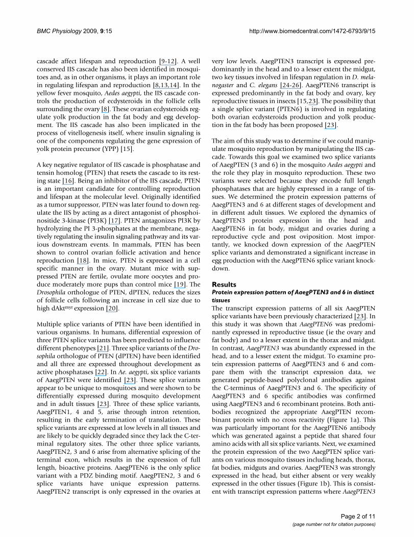
Protein expression pattern of AaegPTEN 6 during a reproductive cycleThe possibility of AaegPTEN6 being a key regulator ofreproduction was indicated by the prominent expressionof AaegPTEN6 transcript in two key reproductive tissues,the fat body and ovary. Before exploring this possibilitywe characterized the AaegPTEN6 protein expressiondynamics throughout the mosquito's reproductive cyclein four key tissues: ovaries, fat bodies, heads and midguts.In ovaries, AaegPTEN6 protein was abundantly expressedduring the first 12 h of the reproductive cycle and wasabsent in the ovaries from 24 hr post-bloodfeeding untiloviposition (Figure 2a). After oviposition (72 h) theAaegPTEN6 protein reappeared in the ovaries. This pat-tern is similar to the expression dynamics of other insulinsignaling components in the ovary e.g. Aaegp110 [27] andthe mosquito insulin receptor [13]. In the fat body, headand midgut, AaegPTEN6 was expressed evenly throughoutthe reproductive cycle (Figure 2b). AaegPTEN3 protein inthe head was also evenly expressed throughout the repro-ductive cycle, the very low expression at 6 and 48 h is dueto a loading error (Figure 2b).
Effects of RNAi-mediated knockdown on egg production and egg viabilityThe effects of AaegPTEN and its splice variants on thereproductive success of female mosquitoes were examinedusing RNA interference. Double stranded RNA (dsRNA)was generated that specifically targeted the AaegPTEN3and 6 splice variants. In addition, a "universal" AaegPTENdsRNA was generated that targeted all six splice variants(Figure 3). Adult female mosquitoes (12–24 h posteclosion) were injected with 2 μg of dsRNA againstAaegPTEN, its splice variants or a non-specific controldsRNA against the red fluorescent protein from Discosoma(DsRed). Survivorship of mosquitoes injected withdsRNA was >95% in all treatment groups and comparableto uninjected mosquitoes (data not shown).
In the abdomen/fat body, we achieved 90% and 98%knockdown of AaegPTEN6 transcript with AaegPTEN andAaegPTEN6 dsRNA injections respectively (Figure 4A).This was significantly less than the DsRed (AaegPTEN vs.DsRed p = 0.02; AaegPTEN6 vs. DsRed p = 0.002) orAaegPTEN3 (AaegPTEN vs. AaegPTEN3 p = 0.014;AaegPTEN6 vs. AaegPTEN3 p = 0.002) controls. In theovaries, we achieved a 51% and 99% knockdown ofAaegPTEN6 transcript after injections with AaegPTEN andAaegPTEN6 dsRNA respectively (Figure 4B). Due to oursmall sample size (three qRT-PCR experiments; tenpooled ovaries/experiment; samples run in duplicate)these results were not significant (p = 0.081), however the
Protein expression of AaegPTEN3 & 6 in female tissuesFigure 1Protein expression of AaegPTEN3 & 6 in female tis-sues: AaegPTEN3 and 6 expression in various female tissues was detected using immunoblot analysis. Tissues from 3–5 day old female mosquitoes were dissected in 10× protease inhibitor (PI) buffer, size fractioned on 12% SDS-PAGE gels, transferred to PVDF membrane and probed with anti-AaegPTEN3 or 6 antibodies. A. AaegPTEN3 and 6 recom-binant proteins were immunoblotted and each were probed with the AaegPTEN3 or 6 antibodies. Both the AaegPTEN3 and 6 antibodies were specific to the appropriate recom-binant protein and did not recognize the other splice variant. B. AaegPTEN6 was detected in all tissues from adult females examined, including non-bloodfed heads (HD), thoraxes (TH), fat bodies (FB), midguts (MG) and ovaries (OV). AaegPTEN3 could only be detected in the head. C. AaegPTEN6 was expressed in early embryos, pupae and adults. However, it was found at very low levels in diapausing eggs and not at all during larval stages.
Page 3 of 11(page number not for citation purposes)
BMC Physiology 2009, 9:15 http://www.biomedcentral.com/1472-6793/9/15
trend was consistent with the fat body results. At both 24and 48 h post bloodmeal, the ovaries of females injectedwith universal AaegPTEN dsRNA had the same number of,but more developed follicles (Figure 5) compared to theDsRed controls (p = 0.0028).
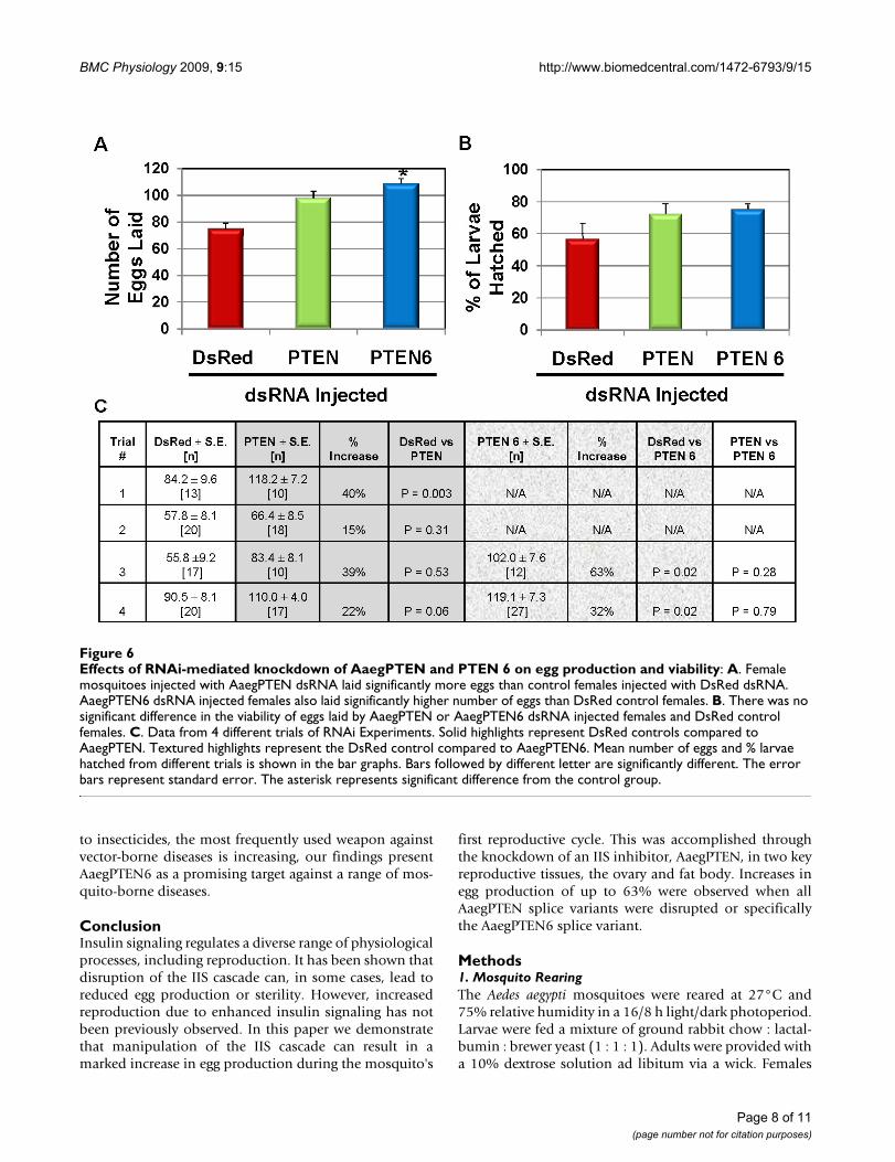
Female mosquitoes injected with dsRNA specifically tar-geting the AaegPTEN6 splice variant laid an average of51% more eggs than the DsRed control females (p =0.045). Female mosquitoes injected with the universalAaegPTEN dsRNA also laid an average of 31% more eggsthan control mosquitoes injected with DsRed dsRNA (Fig-ure 6a; p = 0.06). The number of eggs laid by the mosqui-
toes injected with AaegPTEN and AaegPTEN6 did notdiffer significantly (p = 0.79). Egg viability was measuredby hatching the eggs oviposited by each individual dsRNAinjected female, counting the 2nd instar larvae, and com-paring that to the total number of eggs laid. The percent-age of hatched larvae from the eggs laid by the AaegPTENand AaegPTEN6 injected females was greater than DsReddsRNA injected females, but did not differ significantly (p= 0.09). Importantly, this demonstrates that there was nodecrease in egg fitness from mosquitoes producing artifi-cially high numbers of eggs. The data from replicate trialsare detailed in Figure 6c. Thus, knockdown of bothAaegPTEN and AaegPTEN6 impacted the reproductivesuccess of females by increasing egg production, while theviability of these eggs was unchanged or even increasedslightly.
Since AaegPTEN3 protein was predominantly expressedin the head, we hypothesized that knockdown ofAaegPTEN3 would have little or no effect on egg produc-tion or viability. To examine this hypothesis, dsRNAagainst the unique 3' terminus of AaegPTEN3 was injectedin newly eclosed females under the same conditions asdescribed above. In heads, we achieved a 24 and 40%knockdown of AaegPTEN3 transcript and in the midgut,we achieved a 61 and 67% knockdown after injectionswith AaegPTEN and AaegPTEN3 dsRNA injections. How-ever, due to small sample sizes this decrease did not differsignificantly (data not shown). We found no significantdifference in the number or viability of eggs laid byfemales injected with AaegPTEN3 dsRNA compared toeggs laid by DsRed control females. However, this may bedue to insufficient knockdown of the AaegPTEN3 tran-script.
DiscussionEgg production in the mosquito is a carefully orchestratedseries of hormonal and cell signaling events and is regu-lated by two major physiological processes; steroidogene-sis in the ovaries and vitellogenesis in the fat body. One ofthe key signaling cascades regulating both steroidogenesisand vitellogenesis is the IIS cascade. PTEN, being aninhibitor of this cascade, is thus an important regulator ofegg production at the molecular level. RNAi-mediatedknockdown of AaegPTEN and more specifically theAaegPTEN6 splice variant significantly increased thereproductive success of female mosquitoes during theirfirst reproductive cycle. There are at least two mechanismsby which knockdown of PTEN expression could increasereproductive output. The most likely method is thatincreased IIS signaling results in increased ecdysteroidproduction in the ovary, increased yolk protein produc-tion in the fat body, or both. It has been demonstratedthat insulin stimulates ecdysteroid production by the ova-ries of Ae. aegypti that is regulated by the PI-3K/Akt path-
Protein expression of AaegPTEN6 in female tissues during a reproductive cycleFigure 2Protein expression of AaegPTEN6 in female tissues during a reproductive cycle: To assess the protein expression pattern during reproduction, tissue samples from fully engorged females were collected at various time points (0 (non-fed), 3, 6, 12, 24, 36, 48, and 72 hr (post oviposi-tion)). Tissues from 10–15 females were dissected into 10× PI buffer, size fractioned on 12% SDS-PAGE gels, transferred to PVDF membrane and probed with anti-AaegPTEN 6 anti-body. A. AaegPTEN6 was expressed at constant levels throughout the reproductive cycle in heads (HD), midguts (MG) and fat bodies (FB). However, in the ovaries (OV), AaegPTEN6 was strongly expressed prior to and during the first 12 h post-bloodmeal. It could not be detected 24 to 36 h after the bloodmeal, but was again detected at 72 h post-bloodmeal, following oviposition. B. AaegPTEN3 protein in the head was evenly expressed throughout the reproductive cycle. Low expression at 6 and 48 h is due to loading errors as determined by GAPDH expression (not shown).
Page 4 of 11(page number not for citation purposes)

BMC Physiology 2009, 9:15 http://www.biomedcentral.com/1472-6793/9/15
way [8]. Increased IIS signaling in the ovary could lead tothe secretion of additional 20-hydroxyecdysone, which inturn signals vitellogenesis in the fat body [28]. Alterna-tively, increased insulin signaling in the fat body maydirectly impact vitellogenesis. It was demonstrated thatinsulin, in addition to 20-HE, was required for full expres-sion of vitellogenin in the fat body [15]. Since weobserved efficient knockdown of AaegPTEN6 in both thefat body (98%) and the ovary (99%) it is possible thatboth of these physiological processes played a role in theincreased egg production and viability that we observed.
A second possibility is that increased insulin signalingimpairs the mosquitoes' ability to resorb eggs that are par-tially developed. Between 20 – 25 h post bloodmeal, amajority of follicles have some yolk deposits. Follicles thatare insufficiently developed at that time get degraded andresorbed. In our study, ovaries dissected 24 h post blood-meal from mosquitoes previously injected withAaegPTEN dsRNA had more developed follicles, whichcould be a result of over stimulation of vitellogenesis inthe fat body. Increased production of vitellogenin mightcause faster deposition of yolk in the follicles and hence,prevent them from degrading. A considerable percentageof vitellogenic follicles also get resorbed between 25–30 hpost bloodmeal. However, ovary dissections at 48 h postbloodmeal showed a greater number of more developedfollicles suggesting that fewer of the vitellogenic follicleswere being resorbed.
A third possibility is that knockdown of AaegPTEN andincreased insulin signaling impact the number of poten-tial follicles in each ovary. In Ae. aegypti there is an average
of 50 – 60 follicles in each ovary, although this numbervaries greatly [29]. This means that on average a maxi-mum of 100 – 120 eggs can be produced in a single repro-ductive cycle. Our dsRNA injections were performedshortly after adult emergence when the ovaries are stillbeing developed and prepared for reproduction. Insulinsignaling has been shown to play an important role ingrowth, including body size, cell size and cell number[30]. Thus, it is possible that mosquitoes with AaegPTENknockdown may have unchecked ovary growth and anincrease in the number of ovarioles. We are currentlyexploring this possibility.
Egg viability after dsRNA injections was examined bymeasuring the percentage of larvae hatched. Since femaleshave limited resources, we suspected that we mightobserve a decrease in egg viability due to a compromise inresource allocation towards individual egg provisions.Surprisingly, eggs laid by AaegPTEN and AaegPTEN6dsRNA injected females were not only higher in number,but also had similar or even slightly increased rates of lar-val hatching compared to DsRed controls. The increasedegg production and viability could be a factor of resourceallocation favored towards reproduction by the femalemosquitoes. After a bloodmeal, mosquitoes can accumu-late energy reserves (lipids and glycogen) for survival andflight [31,32]. In mosquitoes with increased insulin sign-aling more of the bloodmeal resources may be devoted toegg production instead of energy reserves. Mosquitoesalso partition and allocate the blood meal proteinsbetween reproduction and energy storage, which can beimpacted by factors such as larval nutrition [33], adultnutrition [34] and gonotrophic cycles [35]. For our exper-
Generation of dsRNA constructs against AaegPTEN and two key splice variantsFigure 3Generation of dsRNA constructs against AaegPTEN and two key splice variants: Three dsRNA constructs were synthesized against AaegPTEN. Universal AaegPTEN dsRNA was generated against the identical PTEN region and was capable of knocking down all AaegPTEN splice variants. The AaegPTEN3 dsRNA was generated against ~300 bp of its unique 3' termi-nus and should only silence the AaegPTEN3 splice variant. AaegPTEN6 dsRNA was generated against the unique 18 bp coding sequence and ~300 bp of the 3' UTR and should only silence the AaegPTEN6 splice variant. dsRNA was also generated against ~300 bp of DsRed as a control (not shown).
Page 5 of 11(page number not for citation purposes)

BMC Physiology 2009, 9:15 http://www.biomedcentral.com/1472-6793/9/15
iments, the larval and adult nutrition of mosquitoes wascontrolled by rearing all mosquitoes under identical con-ditions. However, we examined the effects of dsRNA injec-tions only for the first gonotrophic cycle. It would beinteresting to analyze the effects of increased insulin sign-aling on subsequent gonotrophic cycles and adult survi-vorship. However, the transient nature of transcriptknockdown with dsRNA injections would make lifelongstudies impractical and necessitates the use of transgenicmosquitoes which we are actively pursuing.
Both splice variant AaegPTEN3 and 6 had different pro-tein expression patterns in various tissues of adult femalemosquitoes. AaegPTEN3 transcript is predominant in thehead and midgut, whereas, the protein was stronglyexpressed in the head only. The abundance of AaegPTEN3protein in the head is consistent with the transcriptexpression but the protein was only expressed at low lev-els in the midgut. AaegPTEN6 transcript is predominantin the ovary and fat body. However, the protein wasexpressed in all tissues examined. Invariable expression of
AaegPTEN6 could be due to its higher stability as a proteinsince AaegPTEN6 transcript is expressed at low levels in allthe tissues. During developmental stages, AaegPTEN6transcript is strongly expressed in early embryos and alsofound at lower levels in all other immature stages. Wefound AaegPTEN6 protein in early embryos but not in lar-val stages. This supports the notion of Riehle and Brown[23] that AaegPTEN6 may play a role in early embryogen-esis. During larval stages, there is a possibility that otherAaegPTEN splice variants play a role in development.
The protein expression pattern of AaegPTEN6 in ovariesduring a reproductive cycle closely resembles the expres-sion patterns for other components of the IIS cascade suchas the mosquito insulin receptor (MIR) [8] and AaegP110[27]. The physiological events taking place in the ovaryduring a reproductive cycle correlate with the expressiondynamics of the proteins involved in the IIS cascade. Dur-ing the first 12 – 24 h post-bloodmeal, follicle cells sur-rounding the developing oocytes produce ecdysteroidsthat stimulates yolk protein synthesis in the fat body
Knockdown of AaegPTEN6 in the abdomen/fat body and ovaryFigure 4Knockdown of AaegPTEN6 in the abdomen/fat body and ovary: Female mosquitoes (48 h post eclosion) were injected with 2 μg of dsRNA (0.5 μl of a 4 μg/μl solution). Injected mosquitoes were allowed to recover for 48 h before being mated with male mosquitoes and provided with a bloodmeal. At 48 h post-bloodmeal (96 h post-injection) we generated cDNA from the total RNA of abdomens/fat body and ovaries to assess the transcript knockdown after specific dsRNA injec-tions. qRT-PCR was performed using primers specific to AaegPTEN6 and actin as a control. Abdomens/fat body injected with universal AaegPTEN dsRNA had a 90% reduction in AaegPTEN6 transcript, while those injected with AaegPTEN6 specific dsRNA had a 98% reduction in AaegPTEN6 transcript. Similarily, ovaries injected with universal AaegPTEN dsRNA had a 51% reduction in AaegPTEN6 transcript, while those injected with AaegPTEN6 specific dsRNA had a 99% reduction in AaegPTEN6 transcript relative to control mosquitoes injected with DsRed dsRNA. Different letters indicate a significant difference between samples.
Page 6 of 11(page number not for citation purposes)

BMC Physiology 2009, 9:15 http://www.biomedcentral.com/1472-6793/9/15
(vitellogenesis). After ~24 h the follicle cells switch rolesfrom steroid production to eggshell synthesis. Insulin sig-naling is not involved in the eggshell formation and thuscomponents of the IIS cascade, including AaegPTEN6, arenot expressed during this time. However, after oviposition(~72 h post bloodmeal) the expression of AaegPTEN6 andother components of the IIS cascade is restored in the newprimary follicle in preparation for next reproductive cycle[8,27].
The differential expression pattern of AaegPTEN3 and 6raises the possibility of each splice variant affecting partic-ular phenotypes. Although AaegPTEN6 protein isexpressed in all tissues, its distinctive expression in theovaries and fat body during a reproductive cycle suggestsa significant effect on mosquito fecundity. Conversely, the
AaegPTEN3 protein is predominantly present in the headand expressed uniformly during a reproductive cycle, sug-gesting a possible role in the IIS cascade's other physiolog-ical roles such as aging. The location of AaegPTEN3 insidethe head is unknown. Inefficient knockdown ofAaegPTEN3 in the head suggest that it is localized in thenervous tissue and not the pericerebral fatbody, sincenervous tissue is more resistant to RNAi [36].
Identification of AaegPTEN6 as a direct regulator of eggproduction may enable us to directly target the reproduc-tive success of mosquitoes. Reproduction is one consider-ation when determining the efficiency of vector-bornedisease transmission [37]. Therefore, a direct target ofmosquito reproduction could be an advantageous newtool to augment present control strategies. Since resistance
The effect of AaegPTEN knockdown on egg developmentFigure 5The effect of AaegPTEN knockdown on egg development: Newly eclosed Ae. aegypti were injected with 1.4 μg of dsRNA and allowed to recover for 48 h prior to blood feeding. A. Representative ovaries (24 h post bloodmeal) dissected from mosquitoes injected with dsRNA against AaegPTEN or DsRed (Control). B. Mosquitoes inoculated with AaegPTEN dsRNA produced significantly (* p = 0.0028) more mature follicles (106/ovary pair; n = 17) than the DsRed controls (74/ovary pair; n = 9). There was no significant difference in average follicle size (p = 0.31).
Page 7 of 11(page number not for citation purposes)
BMC Physiology 2009, 9:15 http://www.biomedcentral.com/1472-6793/9/15
to insecticides, the most frequently used weapon againstvector-borne diseases is increasing, our findings presentAaegPTEN6 as a promising target against a range of mos-quito-borne diseases.
ConclusionInsulin signaling regulates a diverse range of physiologicalprocesses, including reproduction. It has been shown thatdisruption of the IIS cascade can, in some cases, lead toreduced egg production or sterility. However, increasedreproduction due to enhanced insulin signaling has notbeen previously observed. In this paper we demonstratethat manipulation of the IIS cascade can result in amarked increase in egg production during the mosquito's
first reproductive cycle. This was accomplished throughthe knockdown of an IIS inhibitor, AaegPTEN, in two keyreproductive tissues, the ovary and fat body. Increases inegg production of up to 63% were observed when allAaegPTEN splice variants were disrupted or specificallythe AaegPTEN6 splice variant.
Methods1. Mosquito RearingThe Aedes aegypti mosquitoes were reared at 27°C and75% relative humidity in a 16/8 h light/dark photoperiod.Larvae were fed a mixture of ground rabbit chow : lactal-bumin : brewer yeast (1 : 1 : 1). Adults were provided witha 10% dextrose solution ad libitum via a wick. Females
Effects of RNAi-mediated knockdown of AaegPTEN and PTEN 6 on egg production and viabilityFigure 6Effects of RNAi-mediated knockdown of AaegPTEN and PTEN 6 on egg production and viability: A. Female mosquitoes injected with AaegPTEN dsRNA laid significantly more eggs than control females injected with DsRed dsRNA. AaegPTEN6 dsRNA injected females also laid significantly higher number of eggs than DsRed control females. B. There was no significant difference in the viability of eggs laid by AaegPTEN or AaegPTEN6 dsRNA injected females and DsRed control females. C. Data from 4 different trials of RNAi Experiments. Solid highlights represent DsRed controls compared to AaegPTEN. Textured highlights represent the DsRed control compared to AaegPTEN6. Mean number of eggs and % larvae hatched from different trials is shown in the bar graphs. Bars followed by different letter are significantly different. The error bars represent standard error. The asterisk represents significant difference from the control group.
Page 8 of 11(page number not for citation purposes)

BMC Physiology 2009, 9:15 http://www.biomedcentral.com/1472-6793/9/15
were fed on porcine blood with 0.038% sodium citrateadded as a preservative. Blood feeding was performedusing artificial membranes with membrane feedersattached to a circulating water bath maintained at 37°C.
2. Splice variant specific antibodies generationWe generated peptide-based polyclonal antibodiesagainst the C-terminus of the AaegPTEN3 and 6 splice var-iants. Since only the C-terminal amino acids are differentfor each variant we had limited options for antibody selec-tion. AaegPTEN3 has a 101 amino acid tail providing uswith some flexibility. We selected an 18 amino acid pep-tide (VNSPAESLVRYRLLSEPE) predicted to be highly anti-genic on the unique C-terminal tail of AaegPTEN3. Incontrast, AaegPTEN6 has only six unique amino acids.Thus, we generated a ten amino acid peptide (GWDSGE-STYL), including four amino acids (GWDS) common toall three variants. The peptides were conjugated to KLHand injected into two rabbits by Proteintech Group, Inc.Neat serum from both rabbits injected with AaegPTEN6worked extremely well on mosquito tissue samples. TheAaegPTEN3 serum recognized several non-specific bandson mosquito samples and was therefore affinity purifiedwith nickel-chelating resin (Aminolink Plus, Pierce) andthe peptide used to generate the AaegPTEN3 antibody.Affinity purification resulted in a marked increase in spe-cificity.
3. Protein extraction and immunoblot analysisImmunoblot analysis was performed to assess the proteinexpression pattern of AaegPTEN3 and 6. Protein extractswere prepared from early eggs (0–6 h post oviposition),late eggs (> 48 h post oviposition), 2nd instar larvae, 4th
instar larvae, early pupae (0–4 h post pupation), latepupae (48 h post pupation) and 3–5 day old adult malesand adult females. Adult females were further separatedby tissue type into head, midgut, fat body/abdominalbody wall and ovary samples. To assess protein expressionduring a reproductive cycle, female mosquitoes werebloodfed and tissue samples were collected at varioustime points (0 (non-fed), 3, 6, 12, 24, 36, 48, and 72 hr(post oviposition)) from fully engorged females. Tissuesfrom 5–10 mosquitoes were collected into 50 ul of 10×Complete protease Inhibitor (PI; Roche Diagnostics) inAedes saline (125 mM NaCl, 5 mM KCl, 1.85 mM CaCl2,pH adjusted to 6.5 using NaOH). Protein extraction wasfacilitated by homogenizing the samples with a pestle in4× loading buffer, (200 mMTris (adjusted to pH 6.8),10% SDS, 50% glycerol, 400 mM DTT, 0.1% CoomassieBlue, in deionized water). After tissue disruption, the sam-ples were boiled for 10 minutes at 95°C and then chilledon ice for 5 minutes. One to two tissues equivalents wereloaded on to a 12% SDS-Page gel (Pierce or ISC BioEx-press) and size fractionated. The proteins were transferredto a PVDF membrane (Whatman) and blocked with 5%
low-fat dried milk in TBS and 0.1% Tween20 (TBST) over-night at 4°C with gentle rocking. Primary antibodies werediluted in fresh blocking solution (AaegPTEN3 1:1000dilution; AaegPTEN6 1:25,000 dilution) and incubated atroom temperature (RT) for 2 h. The blot was subsequentlywashed 3× for 30 minute at RT with TBST followed byincubation with an anti-mouse-HRP secondary antibody(400 μg/ml dilution in TBST; Pierce, Rockford, IL) for 2 hat RT. The blot was again washed 3× with TBST for 30minute at RT followed by chemiluminescent detection(Pierce Super-Signal dura) on HyBlot CL autoradiographyfilm (Denville Scientific). As a loading control, primaryantibody against glyceraldehyde 3-phosphate dehydroge-nase (GAPDH) was used. GAPDH was diluted in 5% BSAin TBST (1:10,000 dilution) and incubated at RT for 2 hfollowed by subsequent steps as described above.
4. RNAi-mediated knockdown ExperimentsDouble stranded RNA (dsRNA) was generated againstboth AaegPTEN3 and 6 splice variants, as well as a "uni-versal" AaegPTEN dsRNA that recognized all six splice var-iants (Figure 3). The AaegPTEN3 dsRNA was generatedagainst ~300 bp of its unique 3' terminus of the codingregion. AaegPTEN6 has only six unique amino acids,therefore, AaegPTEN6 dsRNA was generated against ~300bp of the unique 18 bp coding sequence and the 3' UTR.The pGEM-T Easy vectors (Promega) containingAaegPTEN, AaegPTEN3 and 6 sequences were used astemplates for dsRNA production. To facilitate in vitro tran-scription, PCR amplification was performed to attach a T7promoter sequence (TAATACGACTCACTATAGGG) to the5' and 3' end of the target sequences. The PCR product waspurified (Invitrogen PCR cleanup kit) and used in combi-nation with the RiboMAX Large Scale RNA ProductionSystem (Promega) to generate sense and anti-sense RNAstrands that were subsequently annealed together to gen-erate the final dsRNA product. As a control, a similarlysized dsRNA fragment was generated against the red fluo-rescent protein, DsRed. The dsRNAs were concentrated to4 μg/μl using a speedVac.
Virgin female mosquitoes (48 h post eclosion) wereinjected with 2 μg of dsRNA (0.5 μl of a 4 μg/μl solution).The injected mosquitoes recovered for 48 h in a humidchamber, were mated with male mosquitoes and pro-vided with a bloodmeal. At 24 h post bloodmeal, ovarieswere dissected to examine egg development. At 48 h postbloodmeal we dissected ovaries, counted the number ofdeveloped follicles and measured the size of individualfollicles with an ocular micrometer on a stereo micro-scope set at 40×. Also, at 48 h post bloodmeal we dis-sected tissues from a subset of the females to look attranscript knockdown relative to DsRed controls. Toassess the transcript knockdown, total RNA (Qiagen) wasextracted from the dissected tissues (head, abdomen, mid-
Page 9 of 11(page number not for citation purposes)
BMC Physiology 2009, 9:15 http://www.biomedcentral.com/1472-6793/9/15
gut and ovary) of ten dsRNA injected females. Total RNA(1 μg) was used to make cDNA (Applied Biosystems) thatthen served as template for qRT-PCR (ABI Real-Time PCR)using primers specific to AaegPTEN3, AaegPTEN6 andactin as a control (Riehle and Brown, 2007). The qRT-PCRexperiments were replicated three times and each sampletested in duplicate. Differences in normalized geneexpression levels were compared by Kruskal-Wallace test.Due to small sample sizes, multiple pairwise comparisonswere conducted using the Conover-Inman method. Afterother sample collections at 48 hr post bloodmeal, 15–20females were individually provided with an ovipositionsubstrate to oviposit eggs. After a 3 day ovipositionperiod, the eggs were counted and compared to the DsRedcontrols. To look at the affects of dsRNA injections on eggviability, the eggs were dried on paper for ~24 h, hatchedand the total number of 2nd instar larvae was counted.Because data did not conform to parametric assumptions,they were analyzed using non-parametric methods. For 2-group experiments (DsRed vs. PTEN3), data were ana-lyzed by Mann-Whitney U test. For 3-group experiments(DsRed vs. PTEN vs. PTEN6), data were analyzed byKruskal-Wallace test, using the Dwass-Steel-Critchlow-Fligner method for multiple pairwise comparisons. Vari-ances among trials were not significant (squared rankstest, p > 0.05) and data were pooled for analysis.
Authors' contributionsMAR and AJA designed and planned the experiments. AJAand KMQ performed the experiments. JLR performed thestatistical analysis. AJA and MAR wrote the manuscript.All authors read and approved the final manuscript.
AcknowledgementsWe would like to thank Teresa Joy and Dr. Frank Ramberg for helping with mosquito rearing, Ben Pri-Tal for providing samples used in preliminary studies and Drs. Roger Miesfeld, Kathleen Walker and Yevgeniya Antonova for their support and critical review of this manuscript.
References1. Kyle JL, Harris E: Global spread and persistence of dengue.
Annu Rev Microbiol 2008, 62:71-92.2. Holmes DJ, Fluckiger R, Austad SN: Comparative biology of aging
in birds: an update. Exp Gerontol 2001, 36(4–6):869-883.3. Sgro CM, Partridge L: A delayed wave of death from reproduc-
tion in Drosophila. Science 1999, 286(5449):2521-2524.4. Jenkins NL, McColl G, Lithgow GJ: Fitness cost of extended
lifespan in Caenorhabditis elegans. Proc Biol Sci 2004,271(1556):2523-2526.
5. Partridge L, Gems D, Withers DJ: Sex and death: what is the con-nection? Cell 2005, 120(4):461-472.
6. Furnari FB, Lin H, Huang HS, Cavenee WK: Growth suppressionof glioma cells by PTEN requires a functional phosphatasecatalytic domain. Proc Natl Acad Sci USA 1997,94(23):12479-12484.
7. Myers MG Jr, Mendez R, Shi P, Pierce JH, Rhoads R, White MF: TheCOOH-terminal tyrosine phosphorylation sites on IRS-1bind SHP-2 and negatively regulate insulin signaling. J BiolChem 1998, 273(41):26908-26914.
8. Riehle MA, Brown MR: Insulin stimulates ecdysteroid produc-tion through a conserved signaling cascade in the mosquitoAedes aegypti. Insect Biochem Mol Biol 1999, 29(10):855-860.
9. Hsin H, Kenyon C: Signals from the reproductive system reg-ulate the lifespan of C. elegans. Nature 1999,399(6734):362-366.
10. Kenyon C: A conserved regulatory system for aging. Cell 2001,105(2):165-168.
11. Clancy DJ, Gems D, Harshman LG, Oldham S, Stocker H, Hafen E,Leevers SJ, Partridge L: Extension of life-span by loss of CHICO,a Drosophila insulin receptor substrate protein. Science 2001,292(5514):104-106.
12. Flatt T, Min KJ, D'Alterio C, Villa-Cuesta E, Cumbers J, Lehmann R,Jones DL, Tatar M: Drosophila germ-line modulation of insulinsignaling and lifespan. Proc Natl Acad Sci USA 2008,105(17):6368-6373.
13. Riehle MA, Brown MR: Insulin receptor expression duringdevelopment and a reproductive cycle in the ovary of themosquito Aedes aegypti. Cell Tissue Res 2002, 308(3):409-420.
14. Kang MA, Mott TM, Tapley EC, Lewis EE, Luckhart S: Insulin regu-lates aging and oxidative stress in Anopheles stephensi. J ExpBiol 2008, 211:741-748.
15. Roy SG, Hansen IA, Raikhel AS: Effect of insulin and 20-hydroxy-ecdysone in the fat body of the yellow fever mosquito, Aedesaegypti. Insect Biochem Mol Biol 2007, 37(12):1317-1326.
16. Vazquez F, Grossman SR, Takahashi Y, Rokas MV, Nakamura N, Sell-ers WR: Phosphorylation of the PTEN tail acts as an inhibi-tory switch by preventing its recruitment into a proteincomplex. J Biol Chem 2001, 276(52):48627-48630.
17. Tang X, Powelka AM, Soriano NA, Czech MP, Guilherme A: PTEN,but not SHIP2, suppresses insulin signaling through thephosphatidylinositol 3-kinase/Akt pathway in 3T3-L1 adi-pocytes. J Biol Chem 2005, 280(23):22523-22529.
18. Reddy P, Liu L, Adhikari D, Jagarlamudi K, Rajareddy S, Shen Y, Du C,Tang W, Hamalainen T, Peng SL, Lan ZJ, Cooney AJ, Huhtaniemi I, LiuK: Oocyte-specific deletion of Pten causes premature activa-tion of the primordial follicle pool. Science 2008,319(5863):611-613.
19. Fan HY, Liu Z, Cahill N, Richards JS: Targeted disruption of Ptenin ovarian granulosa cells enhances ovulation and extendsthe life span of luteal cells. Mol Endocrinol 2008, 22(9):2128-2140.
20. Cavaliere V, Donati A, Hsouna A, Hsu T, Gargiulo G: dAkt kinasecontrols follicle cell size during Drosophila oogenesis. DevDyn 2005, 232(3):845-854.
21. Sarquis MS, Agrawal S, Shen L, Pilarski R, Zhou XP, Eng C: Distinctexpression profiles for PTEN transcript and its splice vari-ants in Cowden syndrome and Bannayan-Riley-Ruvalcabasyndrome. Am J Hum Genet 2006, 79(1):23-30.
22. Maehama T, Kosaka N, Okahara F, Takeuchi K, Umeda M, Dixon JE,Kanaho Y: Suppression of a phosphatidylinositol 3-kinase sig-nal by a specific spliced variant of Drosophila PTEN. FEBS Lett2004, 565:1-3.
23. Riehle MA, Brown JM: Characterization of Phosphatase andTensin Homolog expression in the mosquito Aedes aegypti:Six splice variants with developmental and tissue specificity.Insect Mol Biol 2007, 16(3):277-286.
24. Hwangbo DS, Gershman B, Tu MP, Palmer M, Tatar M: DrosophiladFOXO controls lifespan and regulates insulin signalling inbrain and fat body. Nature 2004, 429(6991):562-566.
25. Wolkow CA, Kimura KD, Lee MS, Ruvkun G: Regulation of C. ele-gans life-span by insulinlike signaling in the nervous system.Science 2000, 290(5489):147-150.
26. Murphy CT, Lee SJ, Kenyon C: Tissue entrainment by feedbackregulation of insulin gene expression in the endoderm ofCaenorhabditis elegans. Proc Natl Acad Sci USA 2007,104(48):19046-19050.
27. Pri-Tal BM, Brown JM, Riehle MA: Identification and characteri-zation of the catalytic subunit of phosphatidylinositol 3-kinase in the yellow fever mosquito Aedes aegypti. Insect Bio-chem Mol Biol 2008, 38(10):932-939.
28. Raikhel AS, Kokoza VA, Zhu J, Martin D, Wang SF, Li C, Sun G,Ahmed A, Dittmer N, Attardo G: Molecular biology of mosquitovitellogenesis: from basic studies to genetic engineering ofantipathogen immunity. Insect Biochem Mol Biol 2002,32(10):1275-1286.
29. Clements AN, Boocock MR: Ovarian development in mosqui-toes: stages of growth and arrest, and follicular resorption.Physiological Entomology 1984, 9(1):1-8.
Page 10 of 11(page number not for citation purposes)
http://www.ncbi.nlm.nih.gov/entrez/query.fcgi?cmd=Retrieve&db=PubMed&dopt=Abstract&list_uids=9356475
http://www.ncbi.nlm.nih.gov/entrez/query.fcgi?cmd=Retrieve&db=PubMed&dopt=Abstract&list_uids=9356475
http://www.ncbi.nlm.nih.gov/entrez/query.fcgi?cmd=Retrieve&db=PubMed&dopt=Abstract&list_uids=9356475
http://www.ncbi.nlm.nih.gov/entrez/query.fcgi?cmd=Retrieve&db=PubMed&dopt=Abstract&list_uids=9756938
http://www.ncbi.nlm.nih.gov/entrez/query.fcgi?cmd=Retrieve&db=PubMed&dopt=Abstract&list_uids=9756938
BMC Physiology 2009, 9:15 http://www.biomedcentral.com/1472-6793/9/15
Publish with BioMed Central and every scientist can read your work free of charge
"BioMed Central will be the most significant development for disseminating the results of biomedical research in our lifetime."
Sir Paul Nurse, Cancer Research UK
Your research papers will be:
available free of charge to the entire biomedical community
peer reviewed and published immediately upon acceptance
cited in PubMed and archived on PubMed Central
yours — you keep the copyright
Submit your manuscript here:http://www.biomedcentral.com/info/publishing_adv.asp
BioMedcentral
30. Garofalo RS: Genetic analysis of insulin signaling in Drosophila.Trends Endocrinol Metab 2002, 13(4):156-62.
31. Nayar JK, Sauerman DM Jr: The effects of nutrition on survivaland fecundity in Florida mosquitoes. Part 1. Utilization ofsugar for survival. J Med Entomol 1975, 12(1):92-98.
32. Clements AN, Clements AN: The biology of mosquitoes 1st edition.London; New York: Chapman & Hall; 1992.
33. Brazil DP, Hemmings BA: Ten years of protein kinase B signal-ling: a hard Akt to follow. Trends Biochem Sci 2001,26(11):657-664.
34. Gary RE Jr, Foster WA: Effects of available sugar on the repro-ductive fitness and vectorial capacity of the malaria vectorAnopheles gambiae (Diptera: Culicidae). J Med Entomol 2001,38(1):22-28.
35. Briegel H, Hefti M, DiMarco E: Lipid metabolism during sequen-tial gonotrophic cycles in large and small female Aedesaegypti. J Insect Physiol 2002, 48(5):547-554.
36. Buckingham SD, Esmaeili B, Wood M, Sattelle DB: RNA interfer-ence: from model organisms towards therapy for neural andneuromuscular disorders. Hum Mol Genet 2004,13(2):R275-R288.
37. Macdonald G: The Epidemiology and Control of Malaria London: OxfordUniversity Press; 1957.
Page 11 of 11(page number not for citation purposes)