Reproduction and Development in Chondrichthyan Fishes
32
AMER. ZOOL., 17:379-410 (1977). Reproduction and Development in Chondrichthyan Fishes JOHN P. WOURMS Department of Zoology, Clemson University, Clemson, South Carolina 29631 SYNOPSIS Patterns of chondrichthyan reproduction and development are diverse. Species either are reproductively active throughout the year, or have a poorly defined annual cycle with one or two peaks of activity, or have a well defined annual or biennial cycle. Based on embryological origin and adult morphology, their reproductive system is more similar to tetrapods than to teleosts. Primordial germ cells are of endodermal origin. The Wolffian ducts in males and Mullerian ducts in females become the functional urogenital ducts. Differentiation is under hormonal control. Unusual features of the reproductive system include an epigonal organ in males and females. It contains lymphoid and hemopoietic tissue. Leydig's gland, a modified region of the kidney, produces seminal fluid. In some species, sperm passing through the vas deferens, is enclosed in spermatophores. Rotating about their long axis, helical spermatozoa can move forward or reverse direction. Spermatogenesis often occurs in bicellular units, spermatocysts. These consist of a spermatogonium enclosed in a Sertoh cell. Fertilization is internal. Claspers, modified portions of the pelvic fins act as intromittent organs. In many viviparous sharks and rays, the female reproductive system is asymmetrical. Eggs of some sharks are the largest known cells. Yolk platelets contain lipovitellin. Oocytes have lampbrush chromosomes. Eggs released from the ovary into the body cavity are transported by ciliary action to the ostium of the oviduct. There they are fertilized. Physiological polyspermy is normal. The shell gland, a specialized region of the anterior oviduct, functions both in long term sperm storage and in egg case production. Egg cases of sharks and skates consist of unique collagenous protein with a 400 A period, organized as a cholesteric liquid crystal. Chimaeroid egg cases contain 550 A pseudotubules in orthogonal lattices. In small sharks, males copulate by coiling around the female. A parallel position is assumed by large sharks. Skates and rays copulate with ventral surfaces apposed or by a dorsal approach. Biting is a pre-copulatory release mechanism. Parental care, except for selective oviposition, is lacking. Heavily yolked eggs undergo meroblastic, discoidal cleavage. Development is lengthy, shortest (2-4 months) in rays, longer in skates (3-8 months) and longest (9-22 months) in sharks and chimaeras. Most sharks and all rays are viviparous. Chimaeras, skates, and some sharks are oviparous. Viviparity either involves a yolk sac placenta or is aplacental. If aplacental, the embryo derives nutrients either from yolk reserves, or by intra-uterine embryonic cannibalism, or from placental analogues which secrete "uterine milk." Phylogenetic position, geographical distribution, benthic vs. pelagic habitat, adult size, egg-embryo size, feeding ecology, and embryonic osmoregulation are factors in the retention of oviparity or the evolution of viviparity. INTRODUCTION the two groups have in common than by the ways in which they differ. This is The chondrichthyan or cartilaginous especially true with respect to patterns of fishes include the sharks, skates, rays, and reproduction and development in which chimeras. They are one of the oldest living structural and functional similarity that is groups of jawed vertebrates. They are manifest at the anatomical level becomes often considered to be primitive or ar- e ven more striking at the level of tissue, chaic. Yet, when one considers that the ce ll, and molecule. These fishes almost chondrichthyan fishes and the amniotes see m to constitute an experimental system represent two extremes of vertebrate in which many of the novel structures and evolution, one is more impressed by what processes characteristic of the vertebrates were first developed. I am grateful to Dr. James W. Atz of the American . Chondrichtyan fishes are of particular Museum of Natural History for reading portions of interest to reproductive and developmen- the manuscript and for many helpful suggestions. tal biologists since, for the first time in the 379 Downloaded from https://academic.oup.com/icb/article/17/2/379/163593 by guest on 19 January 2022
-
Upload
khangminh22 -
Category
Documents
-
view
0 -
download
0
Transcript of Reproduction and Development in Chondrichthyan Fishes

AMER. ZOOL., 17:379-410 (1977).
Reproduction and Development in Chondrichthyan Fishes
JOHN P. WOURMS
Department of Zoology, Clemson University, Clemson, South Carolina 29631
SYNOPSIS Patterns of chondrichthyan reproduction and development are diverse. Specieseither are reproductively active throughout the year, or have a poorly defined annual cyclewith one or two peaks of activity, or have a well defined annual or biennial cycle. Based onembryological origin and adult morphology, their reproductive system is more similar totetrapods than to teleosts. Primordial germ cells are of endodermal origin. The Wolffianducts in males and Mullerian ducts in females become the functional urogenital ducts.Differentiation is under hormonal control. Unusual features of the reproductive systeminclude an epigonal organ in males and females. It contains lymphoid and hemopoietictissue. Leydig's gland, a modified region of the kidney, produces seminal fluid. In somespecies, sperm passing through the vas deferens, is enclosed in spermatophores. Rotatingabout their long axis, helical spermatozoa can move forward or reverse direction.Spermatogenesis often occurs in bicellular units, spermatocysts. These consist of aspermatogonium enclosed in a Sertoh cell. Fertilization is internal. Claspers, modifiedportions of the pelvic fins act as intromittent organs. In many viviparous sharks and rays,the female reproductive system is asymmetrical. Eggs of some sharks are the largest knowncells. Yolk platelets contain lipovitellin. Oocytes have lampbrush chromosomes. Eggsreleased from the ovary into the body cavity are transported by ciliary action to the ostiumof the oviduct. There they are fertilized. Physiological polyspermy is normal. The shellgland, a specialized region of the anterior oviduct, functions both in long term spermstorage and in egg case production. Egg cases of sharks and skates consist of uniquecollagenous protein with a 400 A period, organized as a cholesteric liquid crystal.Chimaeroid egg cases contain 550 A pseudotubules in orthogonal lattices. In small sharks,males copulate by coiling around the female. A parallel position is assumed by large sharks.Skates and rays copulate with ventral surfaces apposed or by a dorsal approach. Biting is apre-copulatory release mechanism. Parental care, except for selective oviposition, islacking. Heavily yolked eggs undergo meroblastic, discoidal cleavage. Development islengthy, shortest (2-4 months) in rays, longer in skates (3-8 months) and longest (9-22months) in sharks and chimaeras. Most sharks and all rays are viviparous. Chimaeras,skates, and some sharks are oviparous. Viviparity either involves a yolk sac placenta or isaplacental. If aplacental, the embryo derives nutrients either from yolk reserves, or byintra-uterine embryonic cannibalism, or from placental analogues which secrete "uterinemilk." Phylogenetic position, geographical distribution, benthic vs. pelagic habitat, adultsize, egg-embryo size, feeding ecology, and embryonic osmoregulation are factors in theretention of oviparity or the evolution of viviparity.
INTRODUCTION the two groups have in common than bythe ways in which they differ. This is
The chondrichthyan or cartilaginous especially true with respect to patterns offishes include the sharks, skates, rays, and reproduction and development in whichchimeras. They are one of the oldest living structural and functional similarity that isgroups of jawed vertebrates. They are manifest at the anatomical level becomesoften considered to be primitive or ar- even more striking at the level of tissue,chaic. Yet, when one considers that the cell, and molecule. These fishes almostchondrichthyan fishes and the amniotes s e em to constitute an experimental systemrepresent two extremes of vertebrate in which many of the novel structures andevolution, one is more impressed by what processes characteristic of the vertebrates
were first developed.I am grateful to Dr. James W. Atz of the American . Chondrichtyan fishes are of particular
Museum of Natural History for reading portions of interest to reproductive and developmen-the manuscript and for many helpful suggestions. tal biologists since, for the first time in the
379
Dow
nloaded from https://academ
ic.oup.com/icb/article/17/2/379/163593 by guest on 19 January 2022

380 JOHN P. WOURMS
vertebrate line, the following processeseither make their appearance or else be-come well established: 1) internal fertiliza-tion; 2) viviparity; 3) placental mechanismsfor fetal maintenance; 4) patterns of geni-tal tract development and sex differen-tiation, closely resembling the ones in am-niotes; 5) vertebrate type of reproductiveendocrinology. The first four topics will bereviewed as well as related areas of interestsuch as: patterns of reproduction;gametogenesis; fertilization and early de-velopment; egg case ultrastructure; therole of the shell gland in egg case forma-tion; and the significance of viviparity.Reproduction and development of holo-cephalans (chimaeras) will be discussed interms of available information. The endo-crinology of reproduction will not betreated in detail since it has been exten-sively reviewed in recent years (Chieffi,1967; Dodd, 1972, 1975).
HISTORICAL REVIEW
The earliest recorded observations on thereproductive biology of chondrichthyanfishes are those of Aristotle. He distin-guished between oviparous and viviparousspecies and described the egg cases of theformer. He discovered the yolk sacplacenta in Mustelus laevis Risso [= M. canis(Mitchill)] and was aware that it differedfrom the mammalian placenta (Aristotle,Peck translation 1965, 1970). AlthoughAristotle's observations were neglected formany centuries, it is unlikely that knowl-edge of elasmobranch reproduction wasever lost entirely. Since skates and dogfishhave continued to be staple items of dietsince ancient time, both fishermen andcooks would undoubtedly have distin-guished between oviparous and viviparousspecies and would have been familiar withtheir anatomy. A revival of formal interestin elasmobranch reproduction coincidedwith a revival of interest in natural historyduring the Renaissance. Rondelet (1554),one of the early zoological encyclopedistsillustrated part of the ovary and the eggcase of a skate. He also depicts a femaleshark (probably M. laevis) which is con-nected to a fetus by an elongated yolk stalk
which passes from the fetus through thecloaca of the female. A placenta is notillustrated, however. By 1673, the anato-mist Steno had rediscovered and publishedan illustrated account of the placenta in -Mustelus laevis.
What happens next constitutes a curiouschapter in the history of biology. It hasbeen generally claimed that the works ofAristotle, Rondelet, and Steno were over-looked until the time of Johannes Muller(1842). This does not seem to have beenthe case. Review works such as that ofBohadsch (1776, also 1761) cite all three ofthese authorities. Moreover, Muller (1842)discusses in detail the work of his im-mediate and distant predecessors. It ismore accurate to say that Muller, an out-standing anatomist and one of the found-ers of physiology, was better able to ap-preciate the significance of his findingsand to communicate them to a receptiveaudience. His classic paper marks the be-ginning of modern studies of chondrich-thyan reproduction and development. Notonly did he review all previous studies, buthe also greatly expanded on them with hisown original observations. The structureand probable function of the placenta inthe dogfish M. laevis and the blue sharkPrionace glauca (Linnaeus) were treated indetail. A comparative approach was usedto study reproduction in non-placentalviviparous species, ovoviviparous species,and oviparous species. In the latter, notonly were shark and skate egg casesstudied but also those of the chimaera,Callorhynchus.
Following Muller, research has tendedto progress through areas of current in-terest to the developing science of biology.From Miiller's time to the present, certainpapers can be recognized as landmarks. Afew are listed here. Embryological studieswere firmly established by Balfour (1885).Ruckert (1899) and Ziegler and Ziegler(1892) worked out the early stages of de-velopment. Dean and his associates pro-duced monographs on: the reproductionand development of chimaeras (Dean,1906); the frilled shark Chlamydoselachus(Smith, 1937; Gudger, 1940); and theheterodontid sharks (Smith 1942). Van-
Dow
nloaded from https://academ
ic.oup.com/icb/article/17/2/379/163593 by guest on 19 January 2022

CHONDRICHTHYAN REPRODUCTION 381
debroek (1936) carried out the principalstudy of morphogenetic movements as-sociated with gastrulation. Needham(1942) reviewed the physiology of de-
^ velopment. A number of studies have dealtwith the anatomy of the reproductive sys-tem (c/., Dean 1916-1922 for early refer-ences). In this respect, Borcea's (1905)study is outstanding. He paid particularattention to the role of the shell gland inegg case production. Chemical studies ofthe egg case and its cellular origin origi-nate with Faure-Fremiet and Baudoy(1938) and Filhol and Garrault (1938).Beginning in 1922, Leigh-Sharpe reportedon a series of investigations of secondarysex characteristics. Viviparity has been anarea of special interest (Gudger 1912,1951; Te Winkel, 1943, 1950). In a uniqueseries of papers Ranzi (1932, 1934) re-ported on comparative studies of adapta-tions for viviparity. In addition to placentalspecies, he also considered non-placentalspecies that displayed special embryonicand maternal adaptations, viz., "placentalanalogues." Intra-uterine oophagy was re-ported by Shann (1923) and Springer(1948). The physiology of gestation hasattracted the attention of Daiber and hisassociates (Price and Daiber, 1967;Graham, 1967). Gilbert and his co-workershave contributed not only to the study ofplacentation (Gilbert and Schlernitzauer,1966; Schlernitzauer and Gilbert, 1966) butalso to an understanding of clasper-siphonsac function (Gilbert and Heath, 1972).Reproduction and reproductive seasonshave been treated by Matthews (1950) inthe basking shark Cetorhinus, the blueshark Prionace glauca (Tucker and Newn-ham, 1957), the spiny dogfish Squalusacanthias Linnaeus (Hishaw and Albert,1947; Jensen, 1966), and the skate Rajaerinacea Mitchill (Fitz and Daiber, 1963;Richards et al., 1963).
REPRODUCTIVE CYCLES
On the basis of Breder and Rosen's(1966) massive review offish reproductivepatterns and cycles, it is apparent thatchondrichthyan fishes are either oviparousor viviparous. Information on reproduc-
tive cycles tends to be incomplete or frag-mentary. The present state of knowledgeis unsatisfactory especially when comparedto rigorous studies of invertebrate repro-ductive cycles (Giese, 1959; Giese andPearse, 1974). Sampling is a major prob-lem. Large samples need to be taken atappropriate intervals over a period of sev-eral years. This has been done only for afew small, inshore species or for largerspecies which have been the subject ofcommercial fisheries. Based on a limitedsurvey of three inshore forms, Squalus,Mustelus, and Scyliorhinus, Dodd (1972)postulated the existence of sophisticatedannual reproductive cycles. The situationis more complex. Three basic types ofcycles are encountered: 1) reproductionthroughout the year; 2) a partially dennedannual cycle with one or two peaks; and 3)a well defined annual or biennial cycle.
The first category consists of thosespecies which are either reproductively ac-tive throughout the year or for the majorpart of the year, e.g., Scyliorhinus,Chlamydoselachus, and Heterodontus (inpart). Ford (1921) and Metten (1939) re-port that a population of Scyliorhinuscanicula (L.) from the English Channel inthe vicinity of Plymouth breeds through-out the year. Although somewhat moreprolific in spring and summer, they haveno definite breeding season. Based on2000 specimens collected in the II-fracombe region, Harris (1952) concludedthat the spawning season lasts about 8-9months starting in November and continu-ing until July. Dodd's (1972) observationsthat ovaries of female dogfish from areasof the Irish Sea become quiescent duringthe summer support Harris' view. Arethese differences real or due to samplingbias? Harris (1952) raised the question ofsampling error since Ilfracombe probablyrepresents a spawning ground into whichfemales migrate at the spawning season.Gudger (1940) reported that the frilledshark, Chlamydoselachus anguineus Garman,a viviparous species, breeds throughoutthe year. He attributes its continuous re-productive activity to the relative con-stancy of a deep sea habitat. Finally, avariety of cycles are encountered within
Dow
nloaded from https://academ
ic.oup.com/icb/article/17/2/379/163593 by guest on 19 January 2022
382 JOHN P. WOURMS
the genus Heterodontus. Barnhart (1932)states that the California species H. fran-cisci (Girard) spawns throughout the year.Although the population of H. japonicusMacleay in the vicinity of Misaki, Japan,does deposit eggs throughout the year,there is an apparent peak in activity inMarch-April (Smith, 1942). Recent fieldstudies of McLaughlin and O'Gower (1971)reveal a well defined August-Septemberspawning season for Australian popula-tions of//, portusjacksoni (Meyer).
The second category includes thosespecies with a partially defined annual cy-cle. Although reproductively activethroughout the year, they tend to exhibitone or two peaks in activity. Raja erinaceaMitchill and Hydrolagus colliei (Lay and Ben-nett) exemplify this pattern. Richards et al.(1963) reported that R. erinacea is repro-ductively active throughout the year.(Their study lasted almost 10 years andincluded detailed examination of 15,000specimens per year). Once having attainedmaturity, adult males produce sperm con-tinuously. Mating, fertilization, and theproduction of egg cases take placethroughout the year. The study comparedtwo distinct, but geographically closepopulations, one in Block Island Soundand the other in Long Island Sound. Thepresence of egg cases within a female wasconsidered evidence of its reproductiveactivity. The Block Island Sound popula-tions displayed two peaks. DuringNovember-January, 30-60% of the adultfemales were "gravid" and during Juneand July, 15-30% of the females were"gravid." During the remainder of theyear, the per cent of "gravid" females wasunder 25% and tended to average about10%. The Long Island Sound populationhad a similar pattern with peaks of 5-15%during the November-January and June-July periods as well as a low level of"gravid" females during the rest of theyear. Similar patterns were found in aDelaware Bay population although thesummer peak was greater than the fall(Fitz and Daiber, 1963). In a relatedspecies, R. eglanteria Bosc they found thategg maturation and spawning occurredonly during the spring. Differences in re-
productive cycles between two populationsof the same species as well as differencesbetween species, suggest a need for carefulstudy and cautious generalization. Amongthe chimaeroids, only Hydrolagus colliei(Lay and Bennett) of the Pacific Coast ofNorth America is readily accessible forstudy. Dean (1906) reported that althoughit is reproductively active throughout theyear, a peak in activity probably occursduring late summer and early fall. Morerecently, Stanley (1961, cited in Johnsonand Horton, 1972) confirmed that thesummer was a period of peak reproductiveactivity although 33% of the females andall the males showed evidence of activitythroughout the year.
The third category includes thosespecies with a well defined annual or bien-nial cycle, viz.,Squalus acanthias L.,Musteluscanis (Mitchell) and Urolophus halleriCooper. It may also include other migra-tory forms such as Eulamia milberti (Miillerand Henle) and Cetorhinus maximus (Gun-ner). The spiny dogfish, Squalus acanthias,widely distributed throughout the north-ern regions of the Atlantic and Pacificoceans, shows a striking periodicity. Malesdisplay an annual cycle. Females display abiennial cycle which can be attributed to agestation period of 22 months. Parturitionusually occurs in the autumn. Shortlythereafter copulation ensues (Hisaw andAlbert, 1947; Holden and Meadows, 1964;Jensen, 1966). Simpson and Wardle (1967)discovered an annual cycle of activity inthe male testes. Maximum sperm accumu-lation coincided with the January-February breeding period of the biennialfemale cycle. The smooth dogfish, Musteluscanis, has also been reported to have anannual cycle (Hisaw and Abramowitz,1939). The details of the cycle were estab-lished by Graham (1967) for a migratorypopulation in Delaware Bay. This speciesis also viviparous. The gestation period,however, is 11 months long (Te Winkel,1950). M. canis winters off North Carolina.Northward migration begins in earlyspring. In Delaware Bay, males first ap-pear during the last week of April and thefirst week of May. Females first appearduring the next two weeks. Parturition
Dow
nloaded from https://academ
ic.oup.com/icb/article/17/2/379/163593 by guest on 19 January 2022

CHONDRICHTHYAN REPRODUCTION 383
occurs at this time. Ovulation occurs dur-ing the first three weeks of June. Mating isrestricted to the time after parturition andbefore ovulation. Ovulation is said to bedependent upon copulation (Hisaw andAbramowitz, 1939). The population seemsremarkably well synchronized since allgravid females carry embryos at the samestage of development. In the round stingray of California, Urolophus halleri Cooper,the major reproductive season occurs inlate May, June, and early July (Babel,1967). At this time, most males are inmating condition, and the majority offemales ovulate. Shortly thereafter smallembryos are to be found in their uteri.Quantitative studies demonstrated an an-nual cycle both of spermatogenesis andoogenesis. A small number of females areout of synchrony with the rest of thepopulation and ovulate in December.They are successfully fertilized with spermwhich has been stored for several months.Annual cycles have also been reported insome of the larger migratory species ofsharks, both inshore and pelagic. Availableinformation although fragmentary is ofinterest. The primary winter range of thesandbar shark, Eulamia (=Carcharhinus)milberti is off the southeastern coast of theUnited States. It is extended as far north asLong Island and Cape Cod to form aprimary nursery range where from lateMarch to early August the young are born.Although the period of gestation is ninemonths, females only seem to reproduceevery other year. Sexes tend to be segre-gated, except during courtship and mat-ing. In southeastern Florida, June is thetime of maximum mating activity(Springer, 1960). The basking shark,Cetorhinus maximus (Gunner), illustrates thecomplications which seasonal migrations,both vertical and horizontal, introduceinto the study of reproductive cycles. In hisdefinitive study, Matthews (1950) presentscircumstantial evidence that this shark isviviparous. Direct evidence in the form ofgravid females is lacking. This is remark-able considering that this species, the sec-ond largest shark, is common and has beenthe subject of commercial fisheries. Mat-thews (1950) is of the opinion that the re-
productive cycle of the basking shark iscorrelated with its seasonal migration. Offthe west coast of Scotland where his studieswere conducted, these sharks begin to ap-pear in April and become most numerousin May and June. Similar spring inshoremovements have been reported in Norwayand British Columbia. All of the adult fishwhich were examined were in breedingcondition and showed signs of recentcopulation. Based on these observations,there appears to be a single yearly repro-ductive cycle which is at its peak during thesecond half of May. There remains, how-ever, the problem of the nature and dura-tion of the female cycle. Since gravidfemales have not been reported in moderntimes, Matthews (1950) suggests that theydesist from basking at the surface some-time before the embryo reaches a recog-nizable size. One assumption is that theymigrate to deep, off shore water duringpregnancy. The period of gestation obvi-ously is not known, hence the duration ofthe female cycle is not known. Older re-ports, cited by Matthews, suggest thatmales may be sexually active during theentire year.
DEVELOPMENT OF THE CONADS AND GENITALDUCTS
On the basis of its embryological originand morphology, the reproductive systemof chondrichthyan fishes is more similar toamphibia and amniotes than to teleosts. Inmost vertebrates, including chondrich-thyan fishes, the somatic portion of thegonad has a dual origin, the cortex andmedulla. These two tissues, although inclose proximity, are distinct and have dif-ferent developmental histories. This pat-tern is in contrast to cyclostomes and tele-osts where the somatic tissue of the gonadhas a single origin, the peritonealepithelium. Hence the somatic portion ofthe gonad is comprised entirely of thecortex (Chieffi, 1967; Hoar, 1969). Chieffi(1967) has provided a modern account ofgonad development and differentiation inTorpedo ocellata and Scyliorhinus caniculus.Gonads develop in the dorsolateral liningof the peritoneal cavity in the posterior
Dow
nloaded from https://academ
ic.oup.com/icb/article/17/2/379/163593 by guest on 19 January 2022

384 JOHN P. WOURMS
half of the body. Usually there is onegonad on each side of the dorsal mesen-tery. The undifferentiated gonad is de-rived from two sources, the cortex and themedulla. The cortex which is more later-ally located develops first. It appears as anelongated strip of the mesodermalepithelium which forms the peritonealwall. This thickening becomes a mul-tilayered convex mass of cells which pro-trudes into the coelomic cavity to form thegerminal ridge. On its dorsal side, theridge develops a hollow which is filled bymigrating mesoderm cells. These cellscomprise the medulla. In Scyliorhinus, themedulla is derived from the nephrogeniccord (=interrenal blastema) while in Tor-pedo, it is derived from the same center ofproliferation which forms the nephrogeniccord.
Under normal conditions, genetic fac-tors determine whether the embryonicgonad will differentiate into an ovary ortestes. The first step involves the migrationof primordial germ cells to the gonad.Hoar (1969) states that there is considera-ble evidence for a widespread origin of theprimordial germ cells. In the elasmo-branchs, however, it is well established thatthe primordial germ cells segregate fromthe primitive entoderm quite early in de-velopment (prior to embryo formation?)and migrate via the mesoderm into the siteof the developing gonad (Beard, 1903-04;Woods, 1902; Hardisty, 1967). There theysettle in the cortical region of the gonad.At this stage, the gonad is considered in-different. In genetic females, the primor-dial germ cells once having settled in thecortex retain their cortical location. In Tor-pedo, a few germ cells do migrate from thecortex to the medulla in 24-32 mm femaleembryos. Transient connections are estab-lished with the mesonephric tubules, butthese soon degenerate. Formation of theprimary ovarian follicles occurs by the 75mm stage. In genetic males, primordialgerm cells migrate from the cortex to themedulla (at 22 mm in Torpedo; at 30-32 mmin Scyliorhinus). Formation of the sex cordsand the seminiferous ampullae occur dur-ing the 40-60 mm stages of Torpedo(Chieffi, 1967). Once germ cell migration
has been completed, that region, eithercortex or medulla, which will form thedefinitive gonad grows rapidly while theremaining region fails to develop. Thus,the sex of the individual is determined 4quite early.
Secondary sex characters also make anearly appearance. According to Chieffi(1967), their appearance coincides withsexual differentiation in the genital ridges.The reproductive ducts provide the meansfor conveying gametes from the gonads tothe exterior. In common with most verte-brate embryos, elasmobranchs develop twosets of urogenital ducts, only one of whichwill function as a reproductive duct. Inmales, the functional duct is the mesoneph-ric or Wolffian duct and in females, it isthe Miillerian duct. The mesonephric ductis derived, by direct transition, from thepronephric duct. In males, the excretoryand genital functions of the Wolffian(mesonephric) duct become segregated. InSqualus acanthias, mesonephric tubules es-tablish connections between the seminifer-ous tubules of the testes and the Wolffianduct. These tubules become the vasa ef-ferentia or efferent ducts. The remainderof the duct differentiates into anepididymis, vas deferens, and seminal sac.The urinary or opisthonephric duct is in-dependent of the mesonephric duct. Infemale embryos, however, the upper partof the Wolffian duct atrophies while thelower part serves as urinary duct (Balfour,1885; Kerr, 1919; Goodrich, 1930; Nelsen,1953). In female elasmobranchs a secondset of ducts, the Miillerian ducts, functionin reproduction. They become theoviducts. Although the embryonic de-velopment of the Miillerian ducts in elas-mobranchs differs from that of other ver-tebrates, their function and anatomical re-lationships in the adults are remarkablysimilar. The Miillerian ducts of elasmo-branchs are well developed and appearearly during the course of ontogeny. Theanterior end of each duct opens into thecoelom by a funnel, the ostium tubae. Notinfrequently, the funnels of the right andleft ducts combine to form a single medianostium. The duct, proper, is of large size,regionally specialized, and opens inde-
Dow
nloaded from https://academ
ic.oup.com/icb/article/17/2/379/163593 by guest on 19 January 2022

CHONDRICHTHYAN REPRODUCTION 385
pendently into the cloaca. Miillerian ductsdevelop from the pronephros and thepronephric duct. The funnel region isderived directly from one or more pro-
k nephric nephrostomes. The main portionof the Miillerian duct develops from thepronephric duct. The pronephric ductundergoes a gradual longitudinal splittinginto an anterior-posterior direction toproduce a dorsal and ventral tube. Theventral tube is continuous with the pro-nephric funnel and becomes the Miillerianduct. The dorsal tube receives the kidneytubules. It is a true Wolffian (mesonephric)duct which persists as the functional uri-nary duct of the opisthonephros. Bothducts open separately into the cloaca(Kerr, 1919; Goodrich, 1930). Miillerianducts also develop in males. In immaturemales of some species, e.g., Notorynchusmaculatus Ayers, they may persist asrudimentary right and left oviducts(Daniel, 1928). Normally, they atrophy orsurvive only as vestigal funnels (Goodrich,1930). Chieffi (1967) reviewing the reac-tion of the duct systems to steriod hor-mone treatment presents evidence forhormonal control of differentiation. Otherimportant secondary sex characters areexternal and are found in males, viz., thepelvic claspers or copulatory organ, foundin almost all Chondrichthyes; the frontal(cephalic) clasper of the chimaeras; andalar spines on the pectoral fins of skates.Of these, only the pelvic claspers havereceived any significant attention. Theclaspers are derived from the medial mar-gin of the pelvic fins and develop simul-taneously with the sexual differentiation ofthe testes. In Torpedo, growth of the clasperis not affected by treatment with steriodhormones. Chieffi (1967) suggests thatthey are a somato-sexual character.
MALE REPRODUCTIVE SYSTEM
Functional Organization
The male reproductive system consistsof the testes, accessory glands, genitalducts, and secondary sex organs. Its or-ganization has been the subject of numer-ous studies {cf., Borcea, 1905; Dean, 1906;
Daniel, 1928; Matthews, 1950; Stanley,1963) so it will not be treated in detail here.The testes are paired organs attached tothe body wall along either side of thevertebral column by a mesorchium. Testesvary in size and shape and are often en-larged during the breeding season. Closelyassociated or even sometimes combinedwith the elasmobranch testes are the epig-onal organs. These consist of lymphoid orhemopoietic tissue (Matthews, 1950). Ac-cording to Stanley (1963), the epigonalorgan and hemopoietic tissue are not pres-sent in the reproductive system ofholocephalans. Spermatogenesis occurswithin the testis, in units termed ampullae.Hoar (1969) has pointed out inter-specificvariation in the basic organization of thetestis. The testis of the basking shark con-sists of many lobules, separated by connec-tive tissue trabeculae (Matthews, 1950).Each lobule is equivalent to the entire testisof the dogfish, Scyliorhinus (Mellinger,1965). In the dogfish testis, the sper-matogenic ampullae are arranged in sixzones which correspond to stages in am-pullae formation and spermatogenesis(Mellinger, 1965). Mature sperm are dis-charged from the testis through the effe-rent ducts (vasa efferentia). In sharks, thenumber of efferent ducts range from twoto six. In skates and rays, there is a singleefferent duct (Daniel, 1928; Babel, 1967).The efferent duct(s) joins the epididymis.The epididymis usually assumes the formof a coiled tubule but in some species, e.g.,Cetorhinus, it may form a compact mass ofhighly convoluted tubules (Matthews,1950). The epididymis passes into the vasdeferens. There is usually a well definedregion of demarcation between the twostructures. The anterior portion of the vasdeferens tends to be coiled while the pos-terior portion extends as a straight tube tothe urogenital sinus. In many elasmo-branchs, "sperm sacs" arise as diverticulafrom the posterior region of the vas defer-ens. Their size varies according to species.Sperm passes from the vas deferens intothe urogenital sinus and from these via theurogenital papilla into the cloaca.
Leydig's gland empties into the vas de-ferens. Leydig's gland is the anterior part
Dow
nloaded from https://academ
ic.oup.com/icb/article/17/2/379/163593 by guest on 19 January 2022

386 JOHN P. WOURMS
of the kidney which has lost its excretoryfunction in males and has acquired a secre-tory function. It consists of a mass ofconvoluted tubules embedded in connec-tive tissue. According to Matthews (1950),it is responsible for secreting the greaterpart of the seminal fluid.
The size of the vas deferens varies con-siderably among the elasmobranchs. It isoften expanded to form an ampulla whichis used for sperm storage, e.g., Urolophus(Babel, 1967). This tendency becomes ex-treme in the basking shark where the ex-panded ampulla may contain 20-25 litersof spermatophores. In addition to size, theampulla of the vas deferens in the baskingshark has been structurally modified. Theinterior consists of a series of transversefolds each of which forms a circular dia-phragm with an eccentric aperture. Theampulla is lined with an epithelium that ismade up of two cell types, viz. tall ciliatedcells and short cells. The latter occupy abasal position between the ciliated cells.The short cells secrete the material whichforms the cortex of the spermatophores(Matthews, 1950). The vas deferens servesone of two functions. In most species,sperm diluted with seminal fluid is storedin it. In other species, e.g., the baskingshark, sperm is packaged into sper-matophores. Spermatophores of Cetorhinusmay attain a size of 30 mm. They consist ofa cortex of hyaline material which sur-rounds a central mass of sperm. The pack-aging of sperm into spermatophores variesin different species from simple spermaggregation to the complex structures ofCetorhinus (Hoar, 1969). Spermatophoresare not present in the chimaera, Hydrolaguscolliei but may occur in Chimaera monstrosa(Stanley, 1963). The basic organization ofthe male reproductive system in thechimaera, H. colliei closely resembles otherchondrichthyans. Important differencesdo exist, e.g., the complex chambered am-pulla of the vas deferens whose epitheliallining is regionally differentiated into sev-eral secretory regions (Stanley, 1963).
Spermatogenesis
The organization of chondrichthyan
spermatozoa has been reviewed byGinzburg (1972). Sperm are very large,exceeding 100 /A in total length. The headportion is also long, 30-40 \x, which is10-20 times the length of the sperm head *in most teleosts. The sperm is charac-terized by a long, pointed, spirally twistedhead which gives it a corkscrew appear-ance (Retzius, 1902). The helical twistingalso includes the midpiece and tail. Thehelical shape of the sperm is correlatedwith its locomotion. Squalus sperm moveprimarily by rotation about their long axisrather than by a lateral lashing motion.Rotation of the gyres of the helix appar-ently can be reversed, allowing the spermto reverse direction without turning (Stan-ley, 19716). Both at the microscopic andultrastructural levels, chondrichthyansperm are conservative in structure and donot differ significantly from other verte-brate sperm (Boisson, et al., 1968; Stanley,1971<z,i). Attempts to determine the ul-trastructural basis of spiralization have notbeen successful (Stanley, 19716).
A number of investigations have dealtwith spermatogenesis in elasmobranchs(cf., Mellinger, 1965; Stanley, 1966,I97la,b; Boisson et al., 1968 for earlierreferences). Stanley's 1966 study ofScyliorhinus will be reviewed here. In theelasmobranch testis, spermatogenesis takesplace in spherical seminiferous follicles orampullae located at the termini of a highlybranched system of collecting ductules.Follicles originate at fixed sites on thedorsal or dorsal-lateral margins of the tes-tis. New seminiferous follicles are formedwhen one or two spermatogonia are sur-rounded by several epithelial cells. Theepithelial or follicle cells are considered tobe homologous with mammialian Sertolicells (Stanley, 1966). Both the sper-matogonia and follicle cells undergo aninital period of mitotic activity. At the endof this period there are about 500 Sertolicells in a Scyliorhinus follicle and about 250in a Torpedo follicle. Spermatogonia arepresent in equal number. Within a follicle,the two cell types segregate into two con-centric single layers. Sertoli cells surroundthe central lumen, while spermatogoniaare adjacent to the limiting membrane.
Dow
nloaded from https://academ
ic.oup.com/icb/article/17/2/379/163593 by guest on 19 January 2022

CHONDRICHTHYAN REPRODUCTION 387
Each spermatogonium is engulfed by asingle follicle cell. The result is a bicellularunit, the spermatocyst. After this, the Ser-toli cell undergoes no further division.Within a seminiferous follicle, there aremany spermatocysts all of which differen-tiate synchronously. Each sperma-togonium undergoes four successivedivisions to produce 16 spermatogonia.These transform into 16 primary sper-matocytes which undergo meiosis to pro-duce 32 secondary spermatocytes and then64 spermatids. Spermatids differentiateinto mature sperm. Differentiating spermcells are connected by intercellular cyto-plasmic bridges. Spermiogenesis has beendescribed at the ultrastructural level byBoisson et al. (1968) and Stanley (\91\a,b).Except for details previously noted, it dif-fers little from spermiogenesis in othervertebrates. Spermiation takes place in ma-ture follicles, i.e., those with fully differen-tiated spermatozoa, when an openingforms in the follicle wall and continuity isestablished with the attached terminalbranch of the collecting ductule system.The bundles of mature sperm pull awayfrom each of the Sertoli cells and flow intothe ductules. Following this, the folliclecontracts until the Sertoli cells form a solidmass in the interior. Then they degenerateand are resorbed. In the chimaera, H.colliei, the general features of sper-matogenesis are similar to those inScyliorhinus (Stanley, 1963).
Secondary sex characters
Highly developed male secondary sexcharacters are found in the Chon-drichthyes. These include the claspers orcopulatory organs and the siphon sac. Inskates and rays, the siphon sac is replacedby a clasper gland (La Marca, 1964; Babel,1967). Fertilization is internal in theChondrichthyes. The claspers function asintromittent organs during copulation(Gilbert and Heath, 1972; Hoar, 1969).They represent modifications of the malepelvic fin. Their structure varies in differ-ent species (Leigh-Sharpe, 1920-1926, citedin Hoar, 1969). The clasper is formed inpart by cartilaginous elements which sup-
port the medial margin of the pelvic finand extend beyond the posterior marginas a rod. The clasper can be looked uponas a part of the fin rolled up to form a tubewhose edges overlap. The proximal open-ing of the clasper tube is the apopyle andits distal opening is the hypopyle (Leigh-Sharpe, 1920). The mechanism of theclasper-siphon sac or clasper gland func-tion has been subject to experimentalstudy in rays, Urolophus (La Marca, 1964;Babel, 1967) and in the sharks, Squalus andMustelus (Gilbert and Heath, 1972). Dur-ing erection, the clasper in Urolophus,bends forward to lie along the ventralsurface of the animal (Babel, 1967). LaMarca (1964) reports a medial flexure of85°. At the same time, the clasper rotatesso the dorsally located apopyle widens andcontacts the cloaca. Sperm then passesfrom the urogenital papilla through thecloaca and into the clasper tube. Contrac-tion of the striated muscles which sheaththe clasper gland expels a secretion intothe clasper tube. The secretion moves thesperm through the tube and out of itsdistal end (Babel, 1967; La Marca, 1964).Although some details differ, clasper func-tion is similar during copulation in Squalusand Mustelus (Gilbert and Heath, 1972).One clasper is flexed medially about 90°and inserted into the oviduct where it isanchored to the wall by a cartilaginouscomplex at its tip. During copulation,sperm is passed from the urogenitalpapilla into the clasper groove (=tube)where it is then washed into the oviduct bysea water expelled from the siphon sac.The siphon sac had previously been filledwith water by repeated flexing of theclasper (Gilbert and Heath, 1972). Insharks, the siphon sacs are paired, sub-dermal structures located in the pelvicregion on either side of the mid line be-tween the skin and the abdominal wall.The sacs are blind pockets, closed at theiranterior end and opening into the claspergroove at their posterior end. Sacs aresheathed with muscle and lined with asecretory epithelium. Upon electricalstimulation of the muscular wall, the sacscontract to 85 per cent of their originallength. The relative sizes of the sacs vary. In
Dow
nloaded from https://academ
ic.oup.com/icb/article/17/2/379/163593 by guest on 19 January 2022
388 JOHN P. WOURMS
S. acanthias they measure 12 per cent of thetotal body length and 30 per cent of thebody length inM. canis (Gilbert and Heath,1972).
Secretory cells occur throughout the en-tire epithelium of the siphon sac of sharksand in localized regions of the claspergland in skates and rays (La Marca, 1964;Babel, 1967; Gilbert and Heath, 1972).Mann (1960) has shown that the undilutedsiphon sac secretion of sexually maturespiny dogfish, S. acanthias, contains a highconcentration of 5-hydroxytryptamine (5-HT), 5.7-7.7% or about 16 mg/animal.The siphons of immature males contain200 times less 5-HT. Mann and Prosser(1963) demonstrated that application ofeither 5-HT or siphon sac fluid to spinydogfish uterus in vitro initiated a brief butpowerful contraction which was succeededby periodic contractions. They suggestedthat 5-HT, introduced during copulation,caused uterine contractions which wouldaid in sperm transport. This generalizationmay be premature since 5-HT could not befound in the clasper glands of Torpedo andRaja and was present only in traces inMustelus canis. The apparent discrepancycould be explained by a possible diversityof function. Clasper glands of skates andrays produce a white, viscous, slightly acidfluid which coagulates on contact with seawater. La Marca (1964) reported that thesecretion of the stingray, Urolophus, con-tained a muco or glycoprotein and a phos-pholipid. He proposed that some of thesecretion coagulates, sealing the marginsof the clasper groove and thus convertingit into a closed tube. The remainder servesas a vehicle for sperm suspension andtransport. One also wonders whether thesesecretions participate in the formation of"sperm plugs" found in the uteri of Rajaerinacea (Richards, et al., 1963).
FEMALE REPRODUCTIVE SYSTEM
Functional organization
The female reproductive system consistsof the ovaries and oviducts. These are inclose association but are not morphologi-cally continuous. Oviducts display a con-
siderable degree of regional differentia-tion. The morphology of the reproductivesystem has been well described (Borcea,1905; Dean, 1906; Daniels, 1928; Gudger,1940; Metten, 1939; Matthews, 1950; Stan- Aley, 1963; Hoar, 1969). Organization ofthe ovary and oviduct is highly variabledue to species diversity and a wide rangeof reproductive patterns. Adaptation forviviparity have a profound effect on theorganization of the oviduct.
Although the ovaries and oviducts begindevelopment as paired structures, theyoften become asymmetrical in adults. Inthe sharks, Scyliorhinus, Pristiophorus, Car-charhinus, Galeus, Mustelus, and Sphyrna,the right ovary is functional and the leftovary atrophies. Both oviducts are present(Daniel, 1928). With the exception ofScyliorhinus, these sharks are viviparous.Among the viviparous rays, the right ovaryand oviduct undergo varying degrees ofreduction or loss. In Urolophus, the rightovary is non-functional but both oviductsare functional. In contrast, both the rightovary and oviduct are absent in Dasyatisbleekeri (Babel, 1967). In skates which areoviparous, both ovaries and oviducts arepresent and functional. The oviducts oftenfunction in synchrony (Wourms, unpub-lished). This is also true for the chimera,Hydrolagus colliei (Dean, 1906; Stanley,1963).
The ovaries are paired structures, ex-cept as noted above. They are attached oneither side of the vertebral column to theanterior-dorsal wall of the body cavity by amesentery, the mesovarium. Structure andshape are variable. The ovary is usuallyelongate in sharks and some rays (Metten,1939; Babel, 1967) but may be nearlyspherical in skates (Wourms, unpublish-ed). In most Chondrichthyes, the ovariesare naked (gymnovarium condition). Thegerminal epithelium covers the outer sur-face of the ovary. Ovarian follicles developfrom the germinal epithelium. Ripe folli-cles burst through the surface to dischargeripe ova into the abdominal cavity. In mostinstances the ovary is solid. When theovary is hollow, lymph spaces developwithin the ovarian stroma. Development ofthe ovarian follicle has been reviewed
Dow
nloaded from https://academ
ic.oup.com/icb/article/17/2/379/163593 by guest on 19 January 2022

CHONDRICHTHYAN REPRODUCTION 389
(Hoar, 1969; Dodd, 1972). Except for sev-eral points to be considered elsewhere, it isnot noticably different from other verte-brates. Although the ovary of the baskingshark, Cetorhinus, differs considerablyfrom that of other elasmobranchs(Matthew, 1950), it is often erroneouslyused to illustrate the viviparous condition.In Cetorhinus, the surface of the ovary isinvested by a fibrous coat. The germinalepithelium invaginates from the surface toform a network of tubules. The ovary is, ineffect, hollow. The tubules open to thesurface in a pocket on the right side. Ovaare discharged into the pocket and passfrom there, via the peritoneum, to theoviduct. In Cetorhinus and some otherelasmobranchs, epigonal organs are as-sociated with the ovary (Matthews, 1950).Corpora atretica and corpora lutea havebeen described for a number of species.Their probable role in hormone produc-tion has been considered (Hoar, 1969;Dodd, 1972).
The organization of the oviduct variesaccording to function and reproductivepattern. Hoar (1969) summarizes the func-tions as: 1) egg collection; 2) transport ofeggs to exterior; 3) egg case formation; 4)site for development of young in vivipa-rous forms; 5) site of sperm reception andstorage; and 6) dissolution of the sper-matophore cortex. The oviduct ( = Muller-ian duct) originates as a simple tube whichthen undergoes regional differentiation.Four regions can be distinguished: an an-terior ostium tubae or funnel; the shellgland or nidamental gland; a connectingisthmus; and an expanded posterioruterus. The ostium is a funnel at theanterior end of the oviduct which serves tocollect ovulated eggs. It is formed either bythe fusion of the anterior end of theoviducts, e.g., Cetorhinus, or by asymmetri-cal development of one primitive funnel,e.g., that of the right side in Scyliorhinus(Metten, 1939). From the ostium, a tubularportion of the oviduct leads to the shell ornidamental gland. This gland is best de-veloped in oviparous species. In viviparousspecies, it tends to be reduced or vestigial.In its fully developed state, the shell glandis a compound tubular gland. It synthe-
sizes and secretes albumin and mucus in allspecies (Threadgold, 1957). In oviparousspecies and those viviparous species whichproduce egg cases, it also secretes egg caseproteins (Borcea, 1905; Filhol and Gar-rault, 1938). In some species, e.g.,Scyliorhinus, it is involved in sperm storage(Metten, 1939). An isthmus leads from theshell gland to the posterior region of theoviduct. The latter region can be ex-panded, especially in viviparous species, toform a uterus. In oviparous species, theuterus normally serves only as a passage-way for the eggs. An apparent exception isthe chimaera, H. colliei where the uterineepithelium participates in the mor-phogenesis of the egg case (Dean, 1906).The uterus of viviparous species is highlydeveloped and displays various modifica-tions for viviparity. These will be discussedin the section on viviparity. Oviducts some-times merge at their extreme posterior endto form a common vagina (Daniels, 1928;Matthews, 1950). In most species, theoviducts either singly or as a commonvagina open into the cloaca usually dorsalto the rectal opening. A hymen or tissuemembrane may be present near the poster-ior end of the oviduct (Daniels, 1928). Inthe chimaera, H. colliei, both oviductsopen directly to the exterior (Dean, 1906).
Oocytes, oogenesis, egg transport
Chondrichthyan fishes produce smallquantities of large eggs. Mature eggs rangein size from one mm in Scoliodon sorrakowah(Prasad, 1951, cited in Ginzburg, 1972) to100 mm or more in Ginglymostoma andChlamydoselachus (Gudger, 1940). The eggsof the latter two sharks are probably thelargest known cells of any living animal.Egg size generally reflects the reproduc-tive strategy of the species. In Scoliodon andGymnura (= Pteroplatea) where the egg ismuch reduced in size, the developing em-bryo receives almost all of its nutrientsfrom the mother via a placenta ortrophonemata (Ranzi, 1934). Massive ac-cumulation of yolk occurs in oviparousspecies and in viviparous species such asGinglymostoma and Chlamydoselachus inwhich the developing embryo is solely de-
Dow
nloaded from https://academ
ic.oup.com/icb/article/17/2/379/163593 by guest on 19 January 2022

390 JOHN P. WOURMS
pendent on its yolk reserves (Gudger,1940). Mature ovarian and ovulated eggshave a regular spherical shape. The eggmay assume an ellipsoidal shape whenenclosed in an egg case.
Yolk reserves dominate the structure ofthe mature egg. They consist of granulesand platelets associated with smallamounts of cytoplasm. In section, the eggsof some fishes, e.g., Torpedo, display con-centric layers of bright and dark yolk simi-lar to what is seen in the avian egg (Ruc-kert, 1899). Yolk color tends to be charac-teristic of a species. It is usually yellow ororange, but may be pink or light green. Alens shaped blastodisc is located at onepole. The blastodisc also may have a dis-tinctive color which differs from that of theyolk. The egg nucleus is located within andnear the surface of the blastodisc. Accord-ing to Ginzburg (1972) the nucleus is ar-rested at metaphase of the second matura-tion division. The egg is surrounded by anextracellular egg envelope, closely ap-posed to the egg surface. Some confusionexists as to the number of egg envelopesand their origin (Ginzburg, 1972). Most ofthe material is produced by the oocyte,hence is a primary egg envelope (Balfour,1885). Follicle cells may contribute addi-tional material. The older literature makesmention of a zona radiata. In teleosts,ultrastructural studies have shown that itconsists of the egg envelope matrix andclosely spaced microvilli of the oocyte sur-face. The latter may interdigitate with fol-licle cell microvilli (Wourms, 1976). Thechondrichthyan zona radiata is probably ofsimilar structure.
Mature eggs contain a relatively smallamount of cytoplasm located in the blas-todisc and at the peripheral. The cyto-plasm contains the standard cell organelles(Jollie and Jollie, 1967a). Most of the egg,however, is made up of yolk inclusions.Yolk platelets predominate together with adiverse spectrum of inclusion bodies (Jollieand Jollie, 1967a). The yolk platelets weredescribed by Riickert (1899) who pointedout that their size and shape differed ac-cording to species. An important chemicaland morphological study of yolk plateletsin Raja batis was done by Faure-Fremiet
(1933). Re-examined in terms of currentknowledge, his results indicate that theplatelets contain livetin, lipovitellin andprobably phosvitin. Fujii (1960) used mod-ern analytical techniques to confirm thepresence of lipovitellin. He also demon-strated that the physical and chemicalproperties of the lipoproteins of thedogfish Scyliorhinus stellaris were very muchsimilar to those of a frog and the hen. Withregard to structure, platelets occur in theform of strongly birefringent, rectangularor pyramidal polygons enclosed in afluid-filled vacuole. Faure-Fremiet's exper-iments (1933) on the platelets indicate thatyolk proteins of skates are assembled in aparacrystalline lattice which is probablysimilar to that of amphibian yolk (Wallace,1963; Karasaki, 1963). The yolk plateletsof Mustelus canis closely resemble those ofR. batis (Grodzinski, 1958). Electron mi-crographs of Squalus acanthias eggs whileconfirming the general structure of yolkinclusions were not of sufficiently highresolution to demonstrate the presence orabsence of periodicities in yolk platelets(Jollie and Jollie, 1967a). Of the early workon the chemical composition of eggs, re-viewed by Needham (1942), that part deal-ing with total composition and lipid frac-tions is still valid. Eggs contain a significantamount (5.9% wet weight) of urea as dothe tissues of adult fishes. Protein and fatcontent varies according to species. Rajaeggs contain 28% protein and 7% fatwhereas eggs of the shark Heptranchus con-tain 25% protein and 23% fat. The fatfraction contained 88% neutral fats and12% unsaponifiable material. Oleic,linolenic, and iwaskic acids accounted for79% of the neutral fats. The remaining21% was made up of isopalmitic, palmitic,and stearic acids. The unsaponifiable frac-tion contained octadecyl, cetyl, selachyl al-cohols, and cholesterol. Subsequentstudies have confirmed these figures,shown squalene to be present in quantity,and added considerably to the list of fattyacids. Studies on the eggs of deep seasharks have shown an increase in the un-saponifiable fat fraction (21-44%) (Higashietal., 1953; Zama et al., 1955; Shimma andShimma, 1968).
Dow
nloaded from https://academ
ic.oup.com/icb/article/17/2/379/163593 by guest on 19 January 2022
CHONDRICHTHYAN REPRODUCTION 391
Although a challenging and perhapsideal system for the study of oogenesis,there have been few modern investigationsof chondrichthyan eggs. Certainly nothing
^ approaches the combined chemical, struc-tural, and endocrinological studies of am-phibian eggs (Wallace, 1972). A major ex-ception to this are those studies which havebeen concerned with the endocrine func-tion of the ovary. The general pattern ofoogenesis does not differ appreciably fromthat of other lower vertebrates with heavilyyolked eggs. Details and further refer-ences can be found in Balfour (1885),Wallace (1903), Marechal (1906), Mat-thews (1950), Bertin's review (1958), andBabel (1967). Oogenesis in chimaeras wasdescribed by Dean (1906) and Stanley(1963).
Only two topics will be considered here,lampbrush chromosomes and specializedstructures associated with yolk formation.Riickert (1892, cited in Callan, 1957) de-scribed lampbrush chromosomes in theoocytes of the shark Pristiurus only tenyears after they had been discovered byFlemming. He appears to have been thefirst to use the term, lampbrush. In hisdefinitive study of lampbrush chromo-somes in Pristiurus and Scyllium, Marechal(1906) described them as filaments, mostlyin the form of loops projecting laterallyfrom the chromosome axis. In 1957 Callanre-examined the lampbrush chromosomesin the oocytes of Scyllium. He was able todemonstrate, for the first time, the pres-ence of axial chromomeres and to showthat they closely resembled the classicalamphibian lampbrush chromosome.
Due to yolk accumulation duringoogenesis, there is a massive increase inegg size. Sometimes yolk accumulation hasinvolved the modification of pre-existingstructures. Babel (1967) reported an un-usual proliferation and infolding of thefollicular epithelium in the oocytes of theray Urolophus halleri. Extensive folding car-ries the follicular epithelium far into theegg's center. This infolding amplifies thesurface area of the follicular epithelium. Italso alters its spatial distribution so that thetransport of metabolites or yolk precursorswould occur not only at the surface but
also in the interior of the oocyte. A similarfollicular infolding has been observed inthe heavily yolked eggs of cephalopods(Cowden, 1968). In addition to the infold-ing, cells of the follicular epithelium ap-pear to function as nutrient cells (Wallace,1903). Babel (1967) has convincingly dem-onstrated that enlarged follicle cells con-tain inclusions which are structurally iden-tical to the yolk granules of the oocyte. Itwould appear that these follicle cellssynthesize and transport yolk as do follicleand nurse cells of some invertebrates.
Since a direct connection between theovary and the oviduct is lacking in mostchondrichthyans, mature eggs are dis-charged from the ovary directly into thebody cavity. They are then transported toand enter the oviduct via the ostium orfunnel at its anterior end. In some species,e.g., Cetorhinus, the ostium is in close prox-imity to a specialized ovarian pocket fromwhich the ova are discharged. Ova passdirectly into the oviduct (Matthew, 1950).In most species, ovulation occurs at anypoint along the surface of the ovary. Ovareleased into the body cavity are thentransported in the oviduct. In ScyliorhinusMetten (1939) has experimentally demon-strated that this is effected by continuouslybeating cilia arranged in tracts within theabdominal cavity. Selective orientation ofthe cilia with respect to the direction oftheir power strokes enables these tracts toact as unidirectional pathways. Cilia occuron: the peritoneal wall; outer surface ofthe oviducts; and portions of the liver, bileduct, and hepatic portal vein. The absenceof cilia in males and immature females(Metten, 1939) suggests that ciliation isunder hormonal control.
Sperm storage
Sperm storage occurs in females of sev-eral species of sharks and skates. Informa-tion on chimaeroids is insufficient to drawconclusions (Stanley, 1963). In addition todemonstrating sperm storage in sharksand skates, Metten (1939) and Richards etal. (1963) reviewed the previous evidence.A series of observations have establishedthat female elasmobranchs maintained in
Dow
nloaded from https://academ
ic.oup.com/icb/article/17/2/379/163593 by guest on 19 January 2022
392 JOHN P. WOURMS
isolation are able to repeatedly depositfertilized eggs after an initial mating. Forexample, Clark (1922) reported that afemale skate, in the absence of males,deposited fertilized eggs during a periodof five-six weeks. Metten (1939) concludedthat the shell gland is the site of spermstorage since he found sperm there but notin other regions of the reproductive tract.In freshly excised glands, clusters of activesperm were found within the lumen ofthose tubules which secrete the egg shell.Sperm were not found in the albumin ormucous secreting regions of the gland.
Egg cases and egg case formation
The eggs of all oviparous species andmany viviparous species are enclosed inleathery egg cases. Chondrichthyans pro-duce two types of egg cases. Oviparousspecies produce permanent egg caseswhich are usually deposited on or near thebottom. Embryonic development is com-pleted within the egg case. Both egg caseand embryo are subject to physical andbiological hazards. In viviparous species,diverse reproductive strategies govern thefate of egg cases. In some instances, e.g.,the stingray Urolophus, an egg case is notformed (Babel, 1967). In many species, atemporary egg case is formed from whichthe embryo emerges to complete de-velopment in utero. Finally the egg casemay be retained during the entire periodof intra-uterine development and even in-corporated into the placenta, e.g., M. cams(Te Winkel, 1963). Size and shape of eggcases vary according to species. The eggcase of the whale shark Rhincodon is 150 x300 mm (Baughman, 1955) whereas thatof Scoliodon sorrakowah is 3 x 5 mm (Prasad,1951, cited in Ginzburg, 1972). Four basicshapes are encountered (cf. Cox, 1963, forillustrations). Egg cases of skates and somesharks, e.g., Rhincodon, are quandrangularwith horn-like processes at each corner.The dorsal surface is usually arched whilethe ventral surface is flattened. Speciesdifferences which may be associated withenvironmental adaptations have been de-scribed in skates (Ishiyama, 1958). Eggcases of sharks tend to be more rounded
and resemble ellipsoids. There are anumber of exceptions. Eggs of theheterodontid sharks resemble a woodauger (Smith, 1942). The ellipsoidal bodyof the egg case is surmounted by two Aflanges which spiral around the case. Theegg cases of chimaeras are intricate andmorphologically complex {cf., Dean, 1906for detailed descriptions). They are spin-dle or tadpole-shaped and are exception-ally large (150-450 mm) both in absoluteand relative terms. Their shape conformsnot only to the shape of the egg but to theshape of the fully developed embryo(Dean, 1906). They possess a series ofrespiratory pores and elaborately sculp-tured lateral flanges. Newly formed eggcases are light colored, often white. Theyrapidly darken after oviposition (Dean,1906).
Modern work on the structure andcomposition of egg cases begins withFaure-Fremiet. Working with Scyliorhinusand two species of Raja, he showed that theegg case was made up of many layers.When examined with polarizing optics,alternating layers were strongly birefrin-gent. Protein was found to be the principalstructural component. On the basis of itsrelative insolubility and sulfur content, thestructural protein was considered to be atype of keratin, ovokeratin (Faure-Fremiet, 1938; Faure-Fremiet andBaudouy, 1938). Subsequent chemical andphysical studies, however, indicated thatthe structural protein(s) was not keratinbut collagen (Gross et al., 1958). Earlyelectron microscope studies (Gross et al.,1958) failed to find axial periodicities.Wourms and Sheldon (1971, 1972) re-ported that shark and skate egg cases con-tain an imperfectly ordered orthogonalarray of structural components. One com-ponent displays a 400 A periodicity whosebanding pattern remains in lateral registerover long distances. The repeat units con-sists of: 1) a 90 A dense band; 2) a 125 Alight band traversed by fine filaments inladder-like fashion; 3) a second 90 Adense band; and 4) a 90 A light band nottraversed by filaments. Each dense bandcan be resolved into two dense sub-bandsseparated by a light band. Equidistant
Dow
nloaded from https://academ
ic.oup.com/icb/article/17/2/379/163593 by guest on 19 January 2022
CHONDRICHTHYAN REPRODUCTION 393
50-75 A point densities observed in trans-verse sections probably are sectioned fila-ments. The periodicity and banding pat-tern differs from that of most vertebrate
fe collagens. It closely resembles the elas-toidin form of collagen found in sharks byMcGavin and Pyper (1964). The organiza-tion of egg case collagen does not appearto be fibrillar. A cholesteric liquid crystalmodel of egg case collagen best explainsboth the completed structure and its modeof assembly (Wourms, unpublished). Thesame periodicities and banding patternshave now been found in Heterodontus, Ap-risturus, Ginglymostoma, Galeorhinus,Halaelurus, CephaloscyIlium, and Raja(Wourms and Sheldon, unpublished). Re-cently, Knight and Hunt (1974, 1976) haveconfirmed and extended these observa-tions in the egg cases of Scyliorhinuscaniculus using biochemical, X-ray diffrac-tion, and ultrastructural methods. Theirresults were corroborated by Rusaoven etal. (1976). While collagen is the majorstructural component of shark and skateegg cases, the question remains whetherthere are other components present. Eggcase collagen may have unique physicaland chemical properties. Chemical studieshave shown that the tyrosine content isunusually high for collagen. Tyrosine res-idues may provide the basis for phenoliccrosslinking. This is consistent with thedemonstration of a quinone-tanned pro-tein in the egg case (Brown, 1955) andphenol oxidases in the shell gland(Threadgold, 1957; Krishnan, 1959). Thepresence of sulfur in egg cases, originallyreported by Faure-Fremiet, is not incon-sistent with collagen since Blanquet andLenhoff (1966) have found disulfide-linked collagenous proteins in nemato-cysts.
Egg cases of chimeras display a consid-erably different structural organizationwhich probably reflects a basic chemicaldifference. Egg cases of Hydrolagus, Cal-lorhynchus, and Harriotta, representingmembers of the three families of livingchimeras, have an identical structural or-ganization. The egg case is made up ofthree structural components arranged indiscrete, birefringent layers. The number
of layers and their relative positions vary.Ultrastructural examination revealed: 1) agranular layer smilar to the outer layer ofthe dogfish egg case; 2) layers of fibrilswithout banding patterns or periodicity;and 3) a population of tubular compo-nents. Granules are of different sizes andthey seem to be aggregates of smallerunits. The tubular component forms theegg case surface, associated sculpturing,and the filamentous projections. The tubu-lar component is a lattice of 550 Apseudotubules. Lattices are arranged inlayers in which all of the pseudotubules areoriented in one direction. Adjacent layersare aligned at 90° angles to form an imper-fect orthogonal array. The 550 A unit is apseudotubule since the walls of adjacenttubules are shared in common. The wallscontain 10-15 subunits about 90 A indiameter. The subunits appear to be hol-low cylinders (Wourms and Sheldon,1971; unpublished). Illustrations of fossilchimaeroid egg cases (Dean, 1906) differlittle from modern egg cases. The remark-able difference in structure and the proba-ble difference in chemical composition ofelasmobranch and holocephalan egg caseproteins is consistent with Patterson's(1965) view that a direct relationship be-tween the two groups cannot be demon-strated.
The chondrichthyan egg case is a ter-tiary egg envelope (Wilson, 1925, follow-ing Ludwig, 1875) since its structural pro-teins are synthesized and secreted by theoviduct. At the time of spawning, an eggenters the oviduct, is fertilized and is en-closed in an egg case. The shell gland, aspecialized region of the anterior oviductsynthesizes and secretes egg case structuralproteins (Borcea, 1905; Dean, 1906; Filholand Garrault, 1938). It also appears tocontrol the deposition and morphogenesisof the egg case in sharks and skates(Borcea, 1905; Fitz and Daiber, 1963). Inthe chimaera, H. colliei, the posterior re-gion of the oviduct appears to participatein the moulding of the lateral flanges andrespiratory pores (Dean, 1906). The se-quence of egg case formation in the skateR. eglanteria proceeds according to thefollowing sequence: 1) formation of the
Dow
nloaded from https://academ
ic.oup.com/icb/article/17/2/379/163593 by guest on 19 January 2022

394 JOHN P. WOURMS
anterior horns; 2) formation of the an-terior two-thirds of the egg case; 3) fer-tilized egg flows into case; 4) posteriorthird forms; and 5) posterior horns arecompleted (Fitz and Daiber, 1963). In thechimaera H. colliei the sequence is similar.The egg containing capsule is formed firstand then the spindle shaped tail portion(Dean, 1906).
The shell gland is a compound tubulargland (Borcea, 1905; Filhol and Garrault,1938). The epithelial cells forming thetubules are secretory cells. Zones of specif-ic secretory activity can be distinguished.Although there is variation, especially inviviparous species, three regions have beendistinguished using simple stainingtechniques, viz., an anterior zone of oval-bumin synthesis, a zone of mucous synthe-sis, and a zone of shell protein synthesis(Metten, 1939). Histochemical studies ofthe same species of Scyliorhinus (Thread-gold, 1957) showed five distinct regions."These zones secrete respectively a car-bohydrate, a carbohydrate substancewhich is metachromatic, a polyphenoloxidase, a protein, a phenol, and perhaps aphenolic protein and a basic protein."Similar results were obtained by Krishnan(1959). Combined histochemical and in-corporation studies with radioactive pro-line suggest that two classes of cells arepresent within the same shell secretingtubules and occupy alternate positions(Rusaouen et al., 1976). One cell secretescollagen and the other secretes a proteincontaining large amounts of tyrosine andsulfhydryl groups. These findings wouldbe consistent with a collagen cross linkedby phenolic tanning and disulfide linkages.Wourms and Sheldon (1972; unpublished)reported on the ultrastructure of the shellgland in Raja inornata Jordan and Gilbert.They found that within the tubule of theshell secreting region all of the cells in agiven region were identical. Shell secretingcells have an abundant and well developedrough endoplasmic reticulum. They alsocontain many granules which consist of anouter ring of light material and an innercore of dark material. Granules areformed within the Golgi complex from
. material derived from the rough endo-
plasmic reticulum. Granules are secretedinto the lumen where they coalesce intofibrils. Light material within the fibrils hasa 400 A periodicity and the banding pat-tern of egg case collagen. It is possible that mboth egg case collagen and the phenolictanning enzymes are packaged in the samegranule. The terminal stages of secretionagree with Borcea's (1905) scheme. Shellsecreting tubules empty into commonducts at the base of ciliated lamellae. Paral-lel rows of lamellae extend across the innersurface of the gland. It is suggested thatnascent fibrils leaving the tubules fuse intosheets which are sequentially extrudedfrom the structurally polarized lamellae.Each lamella would control the depositionof a single layer in the egg case. At thispoint the advantages of a liquid crystalmodel of collagen organization are appar-ent. It provides the plasticity necessary tomake rearrangements at the molecularand supramolecular level.
MATING AND PARENTAL CARE
Mating
In chondrichthyan fishes, fertilization isinternal and obviously requires close con-tact between the sexes. Copulation, how-ever, has been observed only on infrequentoccasion. Most accounts deal with small,inshore fishes, e.g., Scyliorhinus, Heterodon-tus, and Raja. In spite of the paucity ofinformation, mating appears to follow oneof several patterns. These seem to be de-termined by the size and shape of thefishes.
In small sharks and probably alsochimaeras, the male coils himself aroundthe female. This was first observed inScyliorhinus and probably also occurs inSqualus and Mustelus (Gilbert and Heath,1972). A more complete description ofmating behavior is available for Heterodon-tusfrancisci (Dempster and Herald, 1961).Courtship commenced with the male bit-ing the female on almost any part of thebody. Seizing the left pectoral fin of thefemale in his mouth, the male manoeuvredhis tail over the back of the female im-mediately in front of the second dorsal fin.
Dow
nloaded from https://academ
ic.oup.com/icb/article/17/2/379/163593 by guest on 19 January 2022

CHONDRICHTHYAN REPRODUCTION 395
With the purchase thus afforded, he in-serted the right clasper into the cloaca.Copulation lasted about a half hour duringwhich the female was passive. Althoughcopulation has not been observed inchimaeras, there is circumstantial evidencethat the series of actions involved is similarto that of small sharks. Male holocephalanspossess a second pair of claspers, theantero-pelvic claspers. They also possess acephalic clasper. Neither organ is found inelasmobranchs (Dean, 1906). The clasperis a cartilagenous rod equipped with denti-cles. Movement is effected through at-tachments to the musculature of the lowerjaw and labial cartilage (Raikow andSwierczewski, 1975). Dean (1906) suggeststhat the cephalic clasper is used to graspthe female since the pattern of scars foundnear the dorsal fin of egg-laying femalesmatches the arrangement of denticles onthe clasper. Dean (1906) concludes that inHydrolagus colliei, the male wraps his bodyaround the female and gains purchase byattaching the cephalic clasper near thefemale's dorsal fin. The antero-pelvic clas-pers which are erectile probably serve asadditional means of attachment.
Relatively little is known about copula-tory behavior in large sharks. Clark (1963)reports observations made by Brown onthe lemon shark Negaprion brevirostris Poey.Copulation took place at night. The pairassumed a parallel position with the headsslightly apart and the posterior half of thebodies in very close contact. The pairmoved in tandem with closely syn-chronized swimming motions. It is reason-able to assume that this parallel position istypical of copulation in large sharks. Theirless flexible bodies preclude them fromassuming the coiled position of smallerspecies.
There are two main patterns of copula-tion in skates and rays. Smaller species ofskates mate with the ventral surfaces ap-posed while males of larger species makeeither a dorsal or ventral approach(Richards et al., 1963). Libby and Gilbert(I960) observed mating in Raja eglantaria.It lasted for more than two hours. Themale bit the caudal margin of the female'spectoral fin. Copulation took place with
the pair resting ventral side down on thebottom. The male bent his tail 75° beneaththe female's, flexed one clasper medially90° and inserted it into the cloaca andoviduct. Spines (=alar spines?) on theupper anterior surface of the male's pec-toral fin assisted in holding the female.Small species of skates mate with the ven-tral surfaces apposed. Richards et al.(1963) state that "after the male mountsthe female, she rolls her pectorals insidehis allowing him to maintain a firm hold onher back with his alar spines." Alar spinesare retractile, claw-like spines found inadult male skates where they occupy one tofive rows at the outer margin of the dorsalsurface of each pectoral fin (Bigelow andSchroeder, 1953). Although definitive evi-dence is lacking, rays seem to copulate asdo the small skates. La Marca (1964) statesthat the length and manner of flexion ofthe claspers in the stingray Urolophus rulesout any method of copulation except ven-tral apposition.
Female sharks of a variety of specieshave been reported to bear the scars oftooth cuts on their bodies (Springer, 1960,1967; Stevens, 1974). In the blue shark, P.glauca, tooth cuts which are found only onfemale sharks longer than 180 cm areconsidered to be courtship scars (Stevens,1974). Somewhat earlier Springer (1967)had reached a similar conclusion based onthe examination of a number of species oflarge sharks. Drawing on Clark's accountof copulation in the lemon shark, Stevens(1974) concluded that the bites were notmade to aid the male in hanging on duringcopulation. Instead, biting serves as anecessary pre-copulatory release mecha-nism in females. Biting behavior observedduring the courtship of'Heterodontns (Demp-ster and Herald, 1961) and Raja eglan-taria (Libby and Gilbert, 1960) supportsthis view. Tooth sexual dimorphism re-ported in some sharks (Springer, 1967)seems to be related to courtship behavior.
Parental care
In the strict sense of the term, parentalcare of the young is unknown. Early re-ports of parental care based on the obser-
Dow
nloaded from https://academ
ic.oup.com/icb/article/17/2/379/163593 by guest on 19 January 2022

396 JOHN P. WOURMS
vation of family groups composed ofmother and young are misinterpretations(Breder and Rosen, 1966). It was not pa-rental care but rather the tendency ofsmall sharks to follow any large movingobject. The closest approach to parentalcare appears to involve the selection ofincubation sites and the specific orienta-tion of egg capsules within these sites.McLaughlin and O'Gower (1971) presentcircumstantial evidence for this in PortJackson sharks,//, portusjacksoni. Spawningprobably occurs in open areas. Theysuggest that the female takes the newlyextruded egg in her mouth, swims to aprotected site, and places the egg in acrevice. After hardening, the egg due to itscorkscrew shape is effectively anchored inplace. If confirmed, this would account forthe "nest" of H. japonicus eggs described bySmith (1942).
EARLY DEVELOPMENT
Fertilization and polyspermy
Fertilization is internal in all chon-drichthyan fishes. Gametic encountertakes place in the upper part of theoviduct, anterior to the shell gland whenthis gland is present. Riickert (1899) dem-onstrated that eggs which were at the entr-ance of the shell gland had been fertilizedand contained spermatozoa which weretransforming into pronuclei. In somespecies, e. g., Scyliorhinus where spermstorage occurs, fertilization is believed tooccur in the shell gland (Metten, 1939).Further study is needed. In either case, itfollows that fertilization occurs before theegg case is fully formed.
Physiological polyspermy, as a normalaspect of fertilization has been recognizedsince the latter part of the 19th century(Ginzburg, 1972). Chondrichthyan eggsare heavily yolked and their developmentsuperficially resembles that of avian andreptilian eggs. Fertilization and early de-velopment occur within the blastodisc.Riickert (1899), Dean (1906) and othershave shown that the blastodisc is pene-trated by a number of spermatozoa (over100 in some sharks). Monospermy does
not seem to occur. Riickert (1899) reportsthat all of the spermatozoa entering theblastodisc are transformed into pronucleiof similar size and structure. The femalepronucleus fuses with the closest malepronucleus. At first, supernumerary malepronuclei are evenly distributed in theblastodisc, but are subsequently displacedby derivatives of the zygote nucleus. Thesupernumerary pronuclei undergo mito-sis, display typical metaphase figures, andremain viable throughout the blastulastage. Although they do not participate inembryogeneses, their exact fate is notknown. According to Riickert (1899), theybecome the periblast nuclei, a theorywhich is the basis of an historical con-troversy (Nelsen, 1953). Although theymay persist for long periods, it is likely thatthe supernumerary pronuclei degenerate.Periblast cells probably arise from periph-eral blastoderm cells.
Development
An overview of chondrichthyan de-velopment is given by Balfour (1885);Ziegler (1902); Nelsen (1953); and Pasteels(1957). The chondrichthyan egg is mark-edly telolecithal. Cleavage is meroblasticand is usually confined to a small cap ofcytoplasm, at one pole, the blastodisc. Eggsof the horned shark, Heterodontus, and thechimera, Hydrolagus, also undergo a seriesof cleavages within the yolk mass (Smith,1942; Dean, 1906). Prior to the the firstcleavage, the zygote nucleus divides sev-eral times producing four or more nucleiand establishing a temporary syncytium.The first four cleavages are meridional orvertical. The resulting blastoderm consistsof a central group of three or four cellssurrounded by 10-12 marginal cells. Thefifth cleavage plane is horizontal and pro-duces an upper and lower layer of blasto-meres in the central region. A cavity sub-sequently appears between the two layers.Successive cleavages establish three majorcell populations: surface blastomeres, deepblastomeres, and periblast. The surfaceblastomeres, consisting of a single layer ofcells, encloses the segmentation cavity.Deep blastomeres are numerous and lie
Dow
nloaded from https://academ
ic.oup.com/icb/article/17/2/379/163593 by guest on 19 January 2022

CHONDRICHTHYAN REPRODUCTION 397
within the segmentation cavity. Only thesetwo populations participate in embryoformation. The third population, the syn-cytial periblast, occupies a peripheral posi-
m. tion and also extends centrally as the floorof the segmentation cavity. The blas-toderm acquires a flattened disc shape ascleavage continues. Its upper surface ismade up of a single layer of flattened cellsbeneath which is a tightly packed mass ofdeep blastomeres. The segmentation cav-ity is present only in the caudal portion ofthe blastoderm. Vandebroek (1936) hasprovided a fate map of the presumptiveorgan forming areas in the blastoderm ofthe shark, Scyllium. Most of the futureendoderm lies on the surface in a smallsector at the caudal margin of the blas-toderm. Some endodermal cells also liedeep within the blastoderm. The epiderm-al ectoderm occupies almost the entirecephalic portion of the blastoderm. Im-mediately behind it is a large semi-circularregion of presumptive neuroectoderm.The prechordal plate forms a small me-dian region on the surface immediatelycephalic to the endoderm. A thin crescentof notochord lies between the pre-chordalplate and the neural ectoderm. The pre-sumptive mesoderm is found in two cres-cent shaped regions along the lateral-caudal margins of the blastoderm (Van-debroek, 1936; Nelsen, 1953).
In addition to the classical accounts,Vandebroek (1936) has provided ananalysis of the complex morphogeneticmovements which culminate in em-bryogenesis. During gastrulation, the pre-sumptive epidermal and neural ectodermretain their surface location and expandgreatly. Notochordal, pre-chordal plate,mesodermal and endodermal cells migratefrom the surface, over the caudal edge ofthe blastodisc, into the segmentation cavi-ty. The cells of the notochord and pre-chordal plate move into a median positionbeneath the neural plate. As the embryobegins to take shape, it rises above thesurface of the blastoderm. A head fold ispresent at the anterior end while anotochord has formed in the medianplane. The mesoderm and endoderm lieon either side of it. Subsequently, the en-
doderm establishes a complete floor bygrowing medially beneath the meso-dermal plates and fusing below thenotochord (Vanderbroek, 1936; Nelsen,1953). Vandebroek's (1936) fate map andaccount of morphogenetic movementsshould be approached with considerablecaution. The recent drastic revision of theclassical account of morphogenetic move-ments and the fate map of the trout (Bal-lard, I966a,b, 1968) calls into questionother studies of the same vintage andemploying similar methods. In point offact, preliminary studies by Ballard (per-sonal communication) have shown thatVanderbroek's account is in error andmust be redone. In this respect, it is worthnoting that there are very few experimen-tal studies of the early development ofelasmobranchs. Vivien (1955) reportedthat the pregastrula blastoderm ofScyliorhinus could be divided into two orfour segments without affecting develop-ment. After formation of the subgerminalcavity, sagittal section of the blastodermcaused twin formation. Isolated marginalregions did not form embryos. Two em-bryos with normal orientation were ob-tained by transverse section of the caudalregion of the blastoderm, i.e., over thesubgerminal cavity. Loss of regulatory abil-ity coincided with the onset of active mor-phogenetic movement.
The later phases of development havebeen dealt with on numerous occasionsand except for the following topics will notbe considered here (cf., Balfour, 1885;Ziegler, 1903; Dean, 1906; Nelsen, 1953;Pasteels, 1958). Dean (1906) in his study ofthe chimaeroid fish, H. colliei, states thattheir development is shark-like. Severalpoints of difference exist and should bere-examined. The behaviour of the yolkduring development is remarkable. By day32, and early embryo, the yolk mass hasbeen partitioned into fragments of varyingsize. Prior to that, it underwent a modifiedtotal cleavage. Only a small portion of theyolk is enclosed within the yolk sac. Therest, an estimated 80-90%, forms a creamymass. Dean (1906) suggests that this yolkeither is absorbed via the external gills or isswallowed. Dean (1906) also describes and
Dow
nloaded from https://academ
ic.oup.com/icb/article/17/2/379/163593 by guest on 19 January 2022

398 JOHN P. WOURMS
figures an open blastopore which com-municates with an archenteron. The ar-chenteron is lined with several layers ofcells and is distinct from the segmentationcavity. Since Dean worked with a limitedamount of preserved material, the needfor critical re-examination is obvious.
Embryonic development is a lengthyprocess. It is longest in sharks andchimaeras, of intermediate length inskates, and shortest in the rays. Develop-mental rate is temperature dependent(Harris, 1952) and also species specific.Development is relatively rapid in the vivip-arous rays. The gestation period is 4months in Myliobatis bovina, 3 monthsin Urolophus halleri, and 2 months forDasyatis (=Trygon) violacea (Ranzi, 1934;Babel 1967). Development in the ovipa-rous skates is of intermediate duration. Insix species of skates within the genus Raja,Clark (1922) observed incubation timeswhich ranged from 4.5-5.5 months in R.clavata Linnaeus to 8 months in R. naevusMiiller and Henle. An apparent exceptionis R. marginata Lacepede with a duration ofalmost 15 months. In R. eglantaria, Libbyand Gilbert (1960) found an incubationtime of 9 weeks in a Florida population.Whereas Fitz and Daiber (1963) reported12 weeks for northern (Delaware-NewYork) populations. Among sharks andchimaeras, especially those from temper-ate waters, the longest incubation or gesta-tion periods occur. Dean (1906) reportsincubation times of 9-12 months for thechimaera, H. colliei. Similar times havebeen reported by McLaughlin andO'Gower (1971) for three species withinthe oviparous shark genus Heterodontus. InScyliorhinus the incubation time is 6-8months (Ranzi, 1934; Harris, 1952). Gesta-tion periods of 11 months and 8-12months respectively have been reportedfor the carcharhinid sharks, Mustelus canis(Te Winkel, 1950) and C. milberti (Spring-er, 1960). The 22-24 month gestationperiod of the spiny dogfish, S. acanthias isthe longest period thus far documented.At the other extreme, an Indian shark,Chiloscylium grispem Miiller and Henle layseggs that take 2.5-3.0 months to completedevelopment (Aiyar and Nalini, 1938 cited
in Breder and Rosen, 1966). One wouldlike to know whether the rate of develop-ment is intrinsically more rapid in warmwater species or is brevity due to a highertemperature.
REPRODUCTIVE PATTERNS—VIVIPARITY
Reproductive patterns in chondrichthy-an fishes have been reviewed by Brederand Rosen (1966) and others (Ranzi, 1932,1934; Needham, 1942; Budker 1958;Amoroso, 1960; Hoar, 1969). An overallpicture emerges. All recent chondrichthy-an fishes employ internal fertilization.With few exceptions, e.g., Cetorhinus(Matthews, 1960), they produce a rela-tively small number of large, heavilyyolked eggs. These fishes are eitheroviparous or viviparous (Tables 1, 2). Ifthey are oviparous (chimaeras, skates,some sharks) their eggs are enclosed in anegg case and deposited in the externalenvironment. Development is completedoutside of the body of the mother. Eggsare retained in viviparous species and em-bryonic development is completed in utero.Traditionally, these fishes have beencategorized as oviparous, ovoviviparous,or viviparous species. Budker (1958) andHoar (1969) pointed out that the distinc-tion between ovoviviparity and viviparity isartificial. Ranzi (1934) has shown that inlive bearing species fetal/maternal nutri-tional dependency ranges from nil to al-most complete. Placental species only oc-cupy an intermediate position in thisseries. Moreover, the placenta developsonly after a period of yolk reserve de-pendency. Budker (1958) subdivided theviviparous fishes into placental and apla-
TABLE 1. Reproductive patterns in chondnchthyan fishes.
I. OviparityII. Viviparity
A. Aplacental viviparity1. Dependent solely on yolk reserves2. Oophagy: Intrauterine embryonic can-
nibalism3. Placental analogues — uterine milk secre-
tion and/or uterine trophonemataB. Placental viviparity
1. Yolk sac placenta: Ontogenetic transitionfrom yolk reserve dependency
Dow
nloaded from https://academ
ic.oup.com/icb/article/17/2/379/163593 by guest on 19 January 2022
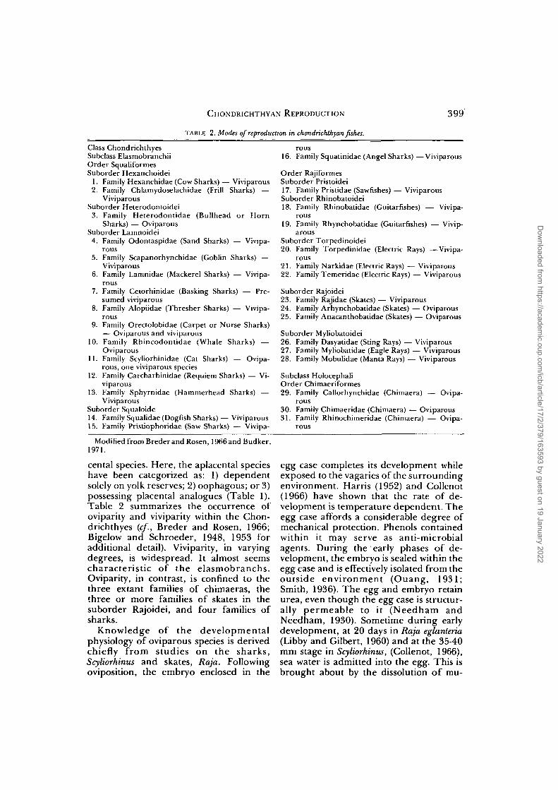
CHONDRICHTHYAN REPRODUCTION 399
TABLE 2. Modes of reproduction in chondrichthyan fishes.
Class ChondrichthyesSubclass ElasmobranchiiOrder SqualiformesSuborder Hexanchoidei
1. Family Hexanchidae (Cow Sharks)— Viviparous2. Family Chlamydoselachidae (Frill Sharks) —
ViviparousSuborder Heterodontoidei
3. Family Heterodontidae (Bullhead or HornSharks) — Oviparous
Suborder Lamnoidei4. Family Odontaspidae (Sand Sharks) — Vivipa-
rous5. Family Scapanorhynchidae (Goblin Sharks) —
Viviparous6. Family Lamnidae (Mackerel Sharks) — Vivipa-
rous7. Family Cetorhinidae (Basking Sharks) — Pre-
sumed viviparous8. Family Alopiidae (Thresher Sharks) — Vivipa-
rous9. Family Orectolobidae (Carpet or Nurse Sharks)
— Oviparous and viviparous10. Family Rhincodontidae (Whale Sharks) —
Oviparous11. Family Scyliorhinidae (Cat Sharks) — Ovipa-
rous, one viviparous species12. Family Carcharhinidae (Requiem Sharks) — Vi-
viparous13. Family Sphyrnidae (Hammerhead Sharks) —
ViviparousSuborder Squaloide14. Family Squalidae (Dogfish Sharks) — Viviparous15. Family Pristiophoridae (Saw Sharks) — Vivipa-
16. Family Squatinidae (Angel Sharks) —Viviparous
Order RajiformesSuborder Pristoidei17. Family Pristidae (Sawfishes) — ViviparousSuborder Rhinobatoidei18. Family Rhinobatidae (Guitarfishes) — Vivipa-
rous19. Family Rhynchobatidae (Guitarfishes) — Vivip-
arousSuborder Torpedinoidei20. Family Torpedinidae (Electric Rays) —Vivipa-
rous21. Family Narkidae (Electric Rays) — Viviparous22. Family Temeridae (Electric Rays) — Viviparous
Suborder Rajoidei23. Family Rajidae (Skates) — Viviparous24. Family Arhynchobatidae (Skates) — Oviparous25. Family Anacanthobatidae (Skates) — Oviparous
Suborder Myliobatoidei26. Family Dasyatidae (Sting Rays) — Viviparous27. Family Myliobatidae (Eagle Rays) — Viviparous28. Family Mobulidae (Manta Rays) — Viviparous
Subclass HolocephaliOrder Chimaeriformes29. Family Callorhynchidae (Chimaera) — Ovipa-
rous30. Family Chimaeridae (Chimaera) — Oviparous31. Family Rhinochimeridae (Chimaera) — Ovipa-
rous
Modified from Breder and Rosen, 1966and Budker,1971.
cental species. Here, the aplacental specieshave been categorized as: 1) dependentsolely on yolk reserves; 2) oophagous; or 3)possessing placental analogues (Table 1).Table 2 summarizes the occurrence ofoviparity and viviparity within the Chon-drichthyes (cf., Breder and Rosen, 1966;Bigelow and Schroeder, 1948, 1953 foradditional detail). Viviparity, in varyingdegrees, is widespread. It almost seemscharacteristic of the elasmobranchs.Oviparity, in contrast, is confined to thethree extant families of chimaeras, thethree or more families of skates in thesuborder Rajoidei, and four families ofsharks.
Knowledge of the developmentalphysiology of oviparous species is derivedchiefly from studies on the sharks,Scyliorhinus and skates, Raja. Followingoviposition, the embryo enclosed in the
egg case completes its development whileexposed to the vagaries of the surroundingenvironment. Harris (1952) and Collenot(1966) have shown that the rate of de-velopment is temperature dependent. Theegg case affords a considerable degree ofmechanical protection. Phenols containedwithin it may serve as anti-microbialagents. During the early phases of de-velopment, the embryo is sealed within theegg case and is effectively isolated from theoutside environment (Ouang, 1931;Smith, 1936). The egg and embryo retainurea, even though the egg case is structur-ally permeable to it (Needham andNeedham, 1930). Sometime during earlydevelopment, at 20 days in Raja eglanteria(Libby and Gilbert, 1960) and at the 35-40mm stage in Scyliorhinus, (Collenot, 1966),sea water is admitted into the egg. This isbrought about by the dissolution of mu-
Dow
nloaded from https://academ
ic.oup.com/icb/article/17/2/379/163593 by guest on 19 January 2022

400 JOHN P. WOURMS
cous plugs in preformed passages (Col-lenot, 1966) or the opening of respiratorypores (Dean, 1906). Rhythmic movementsof the embryo bring about a continuousflow of sea water through the egg case.The egg contains all of the organic mate-rial required for development. Althoughmostly used in forming embryonic struc-tures, some serves as an energy source.This accounts for the 21% loss of organicmaterial during the development ofScyliorhinus (Ranzi, 1932; Amoroso, 1960).Water and inorganic material are derivedfrom the environment (Amoroso, 1960).
Organic material, stored in the yolk, istransferred to the developing embryo byseveral different processes (Beard, 1896;Te Winkel, 1943; Jollie and Jollie, 1967a).In the earliest phase, yolk is incorporatedinto blastoderm cells, probably byphagocytosis, and digested intracellularly.In the next phase, yolk was once believedto be digested extracellularly by merocytes,i.e., extra-embryonic yolk cells. Jollie andJollie (1967a) have shown that in Squalusthe merocytes are part of the peripheralsyncytial cytoplasm of the yolk sac. Theproducts of digestion pass into the vitellinecirculation. Extracellular digestion by theendodermal epithelium of the yolk sac andsubsequent absorption of digested yolkinto the vitelline circulation may also oc-cur. In the Chondrichthyes, all three germlayers take part in the epiboly of the blas-todisc to form a three-layered yolk sac(Ruckert, 1922). In the later phases ofdevelopment, yolk is mainly digestedwithin the intestine of the embryos (Beard,1896; Te Winkel, 1943). Yolk platelets aremoved directly from the external yolk sacinto the gut by ciliated epithelia. In someinstances, e.g., Scyliorhinus (Collenot, 1966),the yolk duct or umbilical canal is enlargedat its terminal end to form an internal yolksac which empties directly into the intes-tine.
The least specialized of the viviparousfishes are those aplacental species whichdepend solely on yolk reserves. Thiscategory corresponds in part to Types laand Ib of Ranzi (1934). Fishes exhibitingthis pattern, e.g., Squalus acanthias, Scymnus,Centrophorus, display a considerable (15-
55%) loss of organic material during de-velopment (Ranzi, 1934; Hisaw and Al-bert, 1947; Amoroso, 1960). S. acanthias istypical. One to four fertilized eggs areenclosed in an elongated egg case. The eggcase remains intact for about six months,after which the embryos hatch and com-plete the remaining 12-16 months gesta-tion in the uterus. The processes of yolkabsorption and utilization are similar tothose of oviparous species, especiallyScyliorhinus (Te Winkel, 1943). The syncy-tial yolk sac cytoplasm digests and absorbsyolk during early stages (Jollie and Jollie,1967a). During later stages, yolk plateletsare moved from the external sac up theyolk stalk into the internal sac and fromthere into the intestine. Movement is ef-fected by ciliated epithelia. The intestinebecomes functional when the embryo isapproximately 65-70 mm long. During de-velopment, the internal yolk sac increasesin size while the external yolk sac is di-minished. Absorption is almost complete atparturition (Te Winkel, 1943). Jollie andJollie (19676) describe ultrastructuralchanges in the mucosa of the pregnantuterus. It is transformed into a respiratorymembrane which may also regulate thetransport of water and electrolytes.Changes involve extensive vascularizationand a reduction in the quantity of tissuebetween the maternal blood and theuterine lumen.
Oophagy, a curious adaptation for vi-viparity, has evolved in several families ofshark. It is a form of intra-uterine em-bryonic cannibalism and can be said torepresent a final solution to sibling rivalry.The phenomenon was first recognized anddescribed by Lohberger (1910) and sub-sequently confirmed by Shann (1923) inembryos of porbeagle sharks, Lamna spp.Porbeagle embryos are quite large andmassive, over 55 cm in length. They ap-pear to have an enormous yolk sac. Theyolk sac, however, is absorbed at an early(6 cm) stage (Shann, 1923). What appearsto be a yolk sac is really the cardiacstomach, enormously distended by eggswhich have been ingested by the develop-ing embryo. In Lamna cornubica, eachoviduct may contain two embryos. Some
Dow
nloaded from https://academ
ic.oup.com/icb/article/17/2/379/163593 by guest on 19 January 2022

CHONDRICHTHYAN REPRODUCTION 401
authors (Gudger, 1940) have overlookedthe phenomenon of oophagy. As a resultthey have concluded that the enormoussize of the porbeagle embryos is due toeggs which are of enormous size. Morerecently, Springer (1948) has reported onoophagy in the sand shark Odontaspistaurus (Rafinesque) (=Carcharius taurus).Living embryos were exceedingly active inutero. They dashed about, open mouthed,inside the oviduct snapping at whateverthey encountered, including the inves-tigator's hand. In this case, there was onlyone embryo in each oviduct. Normally,several fertilized ova are enclosed withinone egg capsule, but only a single activeembryo hatches. This strategy is strikinglysimilar to that of some gastropods, e.g., theBuccinidae and Muricidae. Once hatched,the developing embryo feeds on successivecrops of ovulated eggs. Springer (1948)suggests that oophagy offers two advan-tages. It produces very large embryos (upto 105 cm) which carry an extra reserve ofnutrients. Also, as a result of its intra-uterine behaviour, the fetal shark emergesat parturition as an experienced predator.The frequency with which oophagy occursis not known. Springer (1948) and Budker(1971) suggest that additional species ofmackerel sharks (Lamnidae) and somethresher sharks (Alopiidae) are oopha-gous. Even without chemical analysis, it isobvious that oophagous embryos undergoa great increase in organic content. Addedmaterial is of maternal origin.
The final category of aplacental vivip-arity includes species which have placentalanalogues. It corresponds to Group III ofRanzi (1934). Placental analogues are mod-ified regions of the uterine epitheliumwhich secrete an embryotrophe or"uterine milk" which is absorbed or in-gested by the embryo (Amoroso, 1960).Leucocytes are sometimes found in thesecretion. Their origin is unknown. In theray, Dasayatis violacea, the "milk" has anorganic content of 13% and a total fatcontent of 8% (Ranzi, 1934). The secretionprocess in the epithelial cells of Dasayatisand Myliobatis commences with the en-trance of a leucocyte into the cell. Theleucocyte disintegrates. Shortly thereafter
lipid and protein containing granules ap-pear, mature, and are secreted. The mosthighly developed placental analoguesoccur in the form of villous tufts of uterineepithelium, e.g., Dasyatis violacea andMyliobatis bovina or trophonemata, e.g.,Gymnura micrura (Ranzi, 1934). Troph-onemata are long, flattened glandularappendages. They enter the embryothrough the spiracles and pass into theesophagus where they release secretoryproduct into the gut. Wood-Mason andAlcock (1891) introduced the term,trophonemata, to distinguish them fromintestinal villi. The efficiency of placentalanalogues surpasses that of the yolk sacplacenta. They account for a 1700-5000%increase in organic material during de-velopment compared to the 840% and1050% increase in two placental species,the blue shark, P. glauca, and the dogfish,M. laevis (Ranzi, 1934; Needham, 1942).
The placental analogues found in thesethree species of rays represent the culmi-nation of a series of adaptations whichoriginate with yolk dependent species.Stages in the evolution of placentalanalogues become apparent when the elec-tric rays, Torpedo ocellata and T. mormorataare considered. Both show a net loss oforganic material during development,22% and 34% respectively (Ranzi, 1934;Amoroso, 1960). While the uterine lininghas elaborated prominent folds and lamel-lae, relatively few secretory cells are pres-ent (Ranzi, 1934). The uterine milk con-tains little (1.2%) organic material and lessfat (0.1%). Weight loss occurs since thequality and quantity of secreted material isinadequate for the energy requirements ofthe embryo. The comparative study ofRanzi (1934) reveals a progressive shift inthe energy budget of development from anegative to positive balance. This changeapparently is accomplished by an increasein efficiency of the placental analogues.There is an increase in the total number ofsecretory cells in the uterine epithelium aswell as an increase in the organic content(5-9%) of their secretory product, e.g.,Mustelis vulgaris, M. antarcticus (Ranzi,1934). Parallel evolution of efficient pla-cental analogues in sharks and rays proba-
Dow
nloaded from https://academ
ic.oup.com/icb/article/17/2/379/163593 by guest on 19 January 2022

402 JOHN P. WOURMS
bly reflects a basic selection for increasedefficiency in maternal-fetal maintenance.In some instances, evolution of efficientplacental analogues and placentae haveresulted in reduction of egg size, e.g., theray, Gymnura, and the shark, Scoliodon.
The placental form of viviparity isconfined to two families of sharks, theCarcharhinidae (requiem sharks) and theSphyrnidae (hammerhead sharks). Withinthese groups, placentae are of frequentoccurrence and appear to have evolvedindependently. In the Carcharhinidae,species within the genera Carcharhinus,Prionace, Scoliodon, Carcharias, HemigaleusParagaleus and Mustelus have placentae.Within a genus some species, e.g., Musteluscanis may have a placenta while others, e.g.,M. antarcticus do not. In the Sphyrnidae,Sphyrna is the principal placental genus(Schlernitzauer and Gilbert, 1966).
Placental structure has been described indetail for Mustelus canis (Miiller, 1840;Ranzi, 1934) Prionace glauca (Miiller, 1840;Calzoni, 1936), Carcharhinus falciformis(Gilbert and Schlernitzauer, 1966),Scoliodon surrakowah and S. palasorrah(Mahadevan, 1940), and Sphyrna tiburo(Schlernitzauer and Gilbert, 1966). Theplacenta in all instances is a yolk sacplacenta. Variation exists with respect tothe intimacy of fetal-maternal contact andthe thickness and number of interveningtissue layers. In most species, e.g., M. canis,P. glauca, Sphyrna tiburo, there is an inti-mate interdigitation of the maternal andfetal tissues. In contrast, the placentae ofCarcharhinus dussumerieri (Mahadevan,1940) and C. falciformis (Gilbert andSchlernitzauer, 1966) rest on a thickened,vascularizied region of the uterine wall.
Schlernitzauer and Gilbert (1966) statethat all exchange between the fetal andmaternal blood systems involves up to fivetissue layers. These are: 1) maternal en-dothelium; 2) maternal epithelium; 3) eggcase; 4) fetal epithelium; and 5) fetal en-dothelium. Reduction or loss of one ormore of these layers may occur. Only a fewsharks retain the egg envelope and incor-porate it into the placenta, e.g., M. canisand S. tiburo. Schlernitzauer and Gilbert(1966) report that the fetal epithelium in
C. falciformis is greatly reduced while thematernal epithelium remains relativelyunchanged. Both maternal and fetalepithelia are greatly reduced in the placen-tae of M. canis and S. tiburo. For details ofthe histological organization of placentae,reference should be made to the studieslisted above.
Several peculiarities are associated withplacentae in the sharp-nosed shark,Scoliodon. In S. palasorrah, the yolk sacestablished a specialized connection with atrophonematous cup (Mahadevan, 1940).In S. sorrakowah, due perhaps to small eggsize and the consequent need for establish-ing a placental connection, the yolk sac isnot filled with granules but with a networkof cellular strands and blood vessels. Inaddition, a yolk stalk does not form. Insteadof an umbilical stalk, a placental cord car-rying two blood vessels connects the em-bryo to the placenta. In Scoliodon as well asParagaleus and Sphyrna, the umbilical stalkor placental cord is festooned with finger-like processes called appendiculae. An ab-sorptive function is attributed to them(Budker, 1958).
The details of placenta formation arebest known in M. canis (Te Winkel, 1950,1963; Graham, 1967). Information onSphyrna and Scoliodon can be found inSchlernitzauer and Gilbert (1966) andMahadevan (1940). In Mustelus canis, thegestation period is 10-11 months. Duringthe first three months, the developing em-bryo is yolk dependent. It undergoes anontogenetic transition during the thirdand fourth months and establishes a pla-cental connection which is retained untilbirth. This pattern is common to all pla-cental sharks. The process begins with thefertilization of three to ten eggs. Each eggis enclosed in a thin membranous capsuleand released into the uterus at 16-24 hourintervals. Development proceeds for thenext three months. During this period,transverse folds grow from the endo-metrium and form compartments aroundeach embryo (Graham, 1967). Com-partmentalization occurs in most placentaispecies. The endometrium undergoesother changes. At first, it is slippery due tosecreted fluids. As placentation begins, the
Dow
nloaded from https://academ
ic.oup.com/icb/article/17/2/379/163593 by guest on 19 January 2022

CHONDRICHTHYAN REPRODUCTION 403
endometrium becomes "sticky." Changesoccur in the yolk sac and egg capsule.During the second month of development,the yolk sac begins to expand. Expansioncontinues until the embryo is completelysurrounded by the sac. The yolk sac thencontracts and becomes localized in a smallarea where the placenta is forming.Changes in adhesivity are associated withplacentation. Prior to placentation, yolksac, egg capsule, and endometrium areeasily separated. As placentation com-mences, they adhere but can be separated.They can no longer be separated after theplacenta forms. The egg capsule is incor-porated into the placenta, and on the basisof histological evidence seems to undergochemical changes. Placentation also in-volves folding of the yolk sac and uterineepithelia and the subsequent interdigita-tion of the two tissues.
Structural differences between the distaland proximal portions of the yolk sac oftenoccur (Gilbert and Schlernitzauer, 1966).In most carcharhinids, connection be-tween the yolk sac, placenta and the em-bryo is by a modified yolk stalk or umbilicalstalk. This contains the yolk duct (vitello-intestinal duct) and the umbilical vein andartery (Budker, 1958).
In the 40 years that have elapsed sinceRanzi's classic work, a surfeit of researchproblems have been available yet havegone untouched. Regretably, there is stilllittle to report on the physiology of vivip-arous species, especially placental ones.An exception to this is Graham's (1967)study of placentation in Mustelus canis.Using 3H-glucose, he demonstrated thatlow molecular weight organic substances"filter" across the placental tissues into theyolk sac cavities and from there are passedup the yolk duct into the intestine. Heconcluded that nutrient transfer throughthe placenta of late term fetuses is non-hemotrophic. Obvious questions of em-bryonic nutrition and placental functionare raised. The 800-1000% increase inorganic material in the embryos of placen-tal species indicates an efficient mode ofnutrient transfer (Ranzi, 1934). What isinvolved? The uterine milk of M. canis hasa high (9%) organic content. What role
does it play? Alternate pathways of absorp-tion do exist. After the establishment ofthe placenta in M. canis, there is a dramat-ic increase in the uptake of labelled trac-ers from the uterine cavity (Graham,1967). The state of knowledge is evenmore unsatisfactory in other species. Theformation of a trophonematal cup at thesite of placental attachment in Scoliodon aswell as the elaboration of "absorptive" ap-pendiculae on the placental cords ofScoliodon, Sphyrna, and Paragaleus (Budker,1958) suggest that intra-uterine secretionsare being absorbed. One would like toknow the balance between absorption andplacental transfer. In addition the func-tional difference between the occluded,vascular placental cord of Scoliodon and thepatent yolk stalk of the carcharhinidplacenta should be pursued.
Viviparity
The reproductive strategies of chon-drichthyan fishes are diverse and success-ful. Oviparity and placental vivparity arethe two extremes in a continuum of repro-ductive adaptations. Current interest cen-ters about the retention of oviparity andthe evolution of viviparity. Both processesare subject to the forces of selection. Sinceselection operates primarily at the level ofthe individual and population, the advant-ages or disadvantates of each patternmust be considered in terms of the indi-vidual and the species (cf., Wourms andEvans, 1974a,b; Wourms and Cohen,1975, for examples in teleosts). A studyinvolving the 600 or more chondrichthyanspecies is premature. What follows is aprolegomena to a more detailed study ofreproductive strategies. Basic trends inchondrichthyan reproduction and somefactors which may effect the evolution ofviviparity will be explored.
Eight factors seem to be important in theevolution of viviparity and the retention ofoviparity (Table 3). The first of these is thephylogenetic position of a species. Ovipa-rity is the least specialized and primitivepattern of chondrichthyan reproduction.From it, viviparity has independentlyevolved in several different groups.
Dow
nloaded from https://academ
ic.oup.com/icb/article/17/2/379/163593 by guest on 19 January 2022
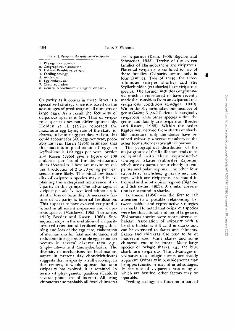
404 JOHN P. WOURMS
TABLE 3. Factors in the evolution of viviparity.
1. Phylogenetic position2. Geographical distribution3. Habitat: Benthic vs. pelagic4. Feeding ecology5. Adult size6. Egg/embryo size7. Osmoregulation8. General reproductive strategy of viviparity
Oviparity as it occurs in these fishes is aspecialized strategy since it is based on theadvantages of producing small numbers oflarge eggs. As a result the fecundity ofoviparous species is low. That of vivipa-rous species does not differ appreciably.Holden et al. (1971) reported themaximum egg laying rate of the skate, R.clavata, to be one egg per day. At best, thiscould account for 360 eggs per year, prob-ably far less. Harris (1950) estimated thatthe maximum production of eggs inScyliorhinus is 120 eggs per year. Brederand Rosen (1966) give a figure of 108embryos per brood for the viviparousshark Hexanchus. These are maximum val-ues. Production of 2 to 50 young per yearseems more likely. The initial low fecun-dity of oviparous species may aid in ex-plaining the widespread occurrence of vi-viparity in this group. The advantages ofviviparity could be acquired without sub-stantial loss of fecundity. A necessary fea-ture of viviparity is internal fertilization.This appears to have evolved early and isfound in all extant oviparous and vivipa-rous species (Matthews, 1955; Tortonese,1950; Breder and Rosen, 1966). Sub-sequent steps in the evolution of viviparityinvolved retention of fertilized eggs, thin-ning and loss of the egg case, elaborationof mechanisms for fetal maintenance, andreduction in egg size. Simple egg retentionoccurs in several diverse taxa, e.g.,Ginglymostoma and Chlamydoeslachus. Thediversity of mechanisms for fetal mainte-nance in present day chondrichthyanssuggests that viviparity is still evolving. Inthis respect, it would appear that onceviviparity has evolved, it is retained. Interms of phylogenetic position (Table 2)several points are of interest. All livingchimaeras and probably all fossil chimaeras
are oviparous (Dean, 1906; Bigelow andSchroeder, 1953). Twelve of the sixteenfamilies of elasmobranchs are viviparous.Placental viviparity is confined to two ofthese families. Oviparity occurs only in dfour families. Two of these, the Orec-tolobidae (carpet sharks) and theScyliorhinidae (cat sharks) have viviparousspecies. The former includes Ginglymosto-ma which is considered to have recentlymade the transition from an oviparous to aviviparous condition (Gudger, 1940).Within the Scyliorhinidae, one member ofgenus Galeus, G. polli Cadenat is marginallyviviparous while other species within thegenus and family are oviparous (Brederand Rosen, 1966). Within the orderRajiformes, derived from sharks or shark-like ancestors, only the skates have re-tained oviparity whereas members of theother four suborders are all viviparous.
The geographical distribution of themajor groups of the Rajiformes tends to becorrelated with their reproductivestrategies. Skates (suborder Rajoidei)which are oviparous occur chiefly in tem-perate and polar regions. The other foursuborders, sawfishes, guitarfishes, andrays, which are viviparous, are found intropical and sub-tropical regions (Bigelowand Schroeder, 1953). A similar correla-tion is not found in sharks.
Tortonese (1950) was the first to callattention to a possible relationship be-tween habitat and reproductive strategiesin sharks. He noted that oviparous specieswere benthic, littoral, and not of large size.Viviparous species were more diverse inhabitat. Association of oviparity with abenthic habitat is still valid in sharks andcan be extended to skates and chimeras.Skates and chimeras also tend to be ofmoderate size. Many skates and somechimeras tend to be littoral. Many largespecies of pelagic sharks, e.g., the blueshark, are viviparous. The advantages ofviviparity to a pelagic species are readilyapparent. Oviparity in benthic species maybe opportunistic or may offer advantages.In the case of viviparous rays many ofwhich are benthic, other factors may beoperable.
Feeding ecology is a function in part of
Dow
nloaded from https://academ
ic.oup.com/icb/article/17/2/379/163593 by guest on 19 January 2022
CHONDRICHTHYAN REPRODUCTION 405
size and also habitat. Large sharks andsawfishes, active predators of fishes, tendto be viviparous. Smaller sharks e.g.,Heterodontidae and Scyliorhinidae, andthe skates, both of which feed on benthicinvertebrates and small fishes, are ovipa-rous. Torpedos, sting rays, and eagle rayswhich have a similar feeding ecology areviviparous. Why? One family and twospecies of chondrichthyan fishes are con-sidered macro-plankton feeders: the whaleshark, Rhincodon; the basking shark,Cetorhinns; and the devil rays, Mobulidae(Bigelow and Schroeder, 1948, 1953).They are large, pelagic fishes. The first isoviparous (Baughman, 1955). Cetorhinus ispresumed viviparous (Matthews, 1950)and the Mobulidae are viviparous. If anysignificance can be attached to the differ-ence, one is tempted to suggest that ovipa-rity is less advantageous in view of the ap-parent rarity of the whale shark. Sincegigantism in the marine environment isoften associated with plankton feeding, sizerather than feeding ecology may be morerelevant to reproductive strategies.
Tortonese (1950) stated that viviparitywas a function of large adult size in sharks.This generalization can be extended to theskates, rays, and chimaeras. Viviparousspecies tend to produce larger offspring.The largest oviparous embryo is that of thewhale shark, circa 35 cm (Baughman,1955). Most oviparous embryos tend to beconsiderably smaller, under 15 cm. In con-trast, viviparous embryos of 45-60 cm arenot uncommon (P. glauca, Wourms, un-published). Embryos of about 100 cm havebeen reported in Odontaspis (Springer,1948). Absolute size is an advantage to thelarge offspring of viviparous species. Em-bryos of 45-60 cm length are considerablyabove the median size of 15-30 cm foradult fishes (Marshall, 1971). In terms ofHutchison and MacArthur's (1959) modelof size distribution, several advantages ac-crue from increased size: 1) reduction innumber of potential predators; 2) reduc-tion in number of competitors; and 3) agreater number of potential food or-ganisms. Increased embryo size also resultsin greater efficiency, since swimmingspeed is a function of absolute size (Mar-
shall, 1971). Growth phenomena may beanother reason why large species tend tobe viviparous. As a generalization, earlyphases of postembryonic growth tend toalmost be exponential. Embryos whose ini-tial size is large tend to grow to a largeadult size (Marshall, 1953).
The factor of egg/embryo size is relatedin part to adult size. As a rule, large adultsdevelop from large embryos. Post-partumembryos of viviparous species are largerthan newly hatched oviparous embryos.For this reason large species tend to beviviparous. Size differences can be ac-counted for in terms of embryonic andmaternal energetics and upper limits toegg size. Oviparous embryos consume 25%or more of their organic content duringdevelopment (Ranzi, 1934). Viviparousspecies suffer the same loss but in manyinstances have evolved efficient means ofcontinuous fetal nutrition. Wheremechanisms for continuous fetal nutritionhave evolved, the fetus attains a large size.The maternal energy budget is moreefficient since nutrients are delivered ondemand rather than sequestered in theegg prior to demand (Wourms and Cohen,1975). In oviparous species, the size of theembryo is limited by the size of the egg.The eggs of some sharks are gigantic cells.The egg of the whale shark may representan upper limit in absolute size. In spite ofthis, the embryo is only half the size of theaverage viviparous shark embryo.
Price and Daiber (1967) suggested thatthe inability of elasmobranch embryos toregulate urea content and osmotic pres-sure during early development constituteda selective disadvantage which led to theevolution of viviparity. Read's (1968) dem-onstration of ornithine-urea cycle enzymesin all stages of development appears tocontradict this. Pang, et al. (1967) reviewedthe subject but did not resolve the issue. Itwould seem that even if early stage ovipa-rous embryos can control their urea con-tent, it can be done more efficiently andwith less expenditure of embryonic energyin the uterine environment.
Finally, the evolution of viviparity in thechondrichthyans cannot be divorced fromthe general strategy of viviparity. A bal-
Dow
nloaded from https://academ
ic.oup.com/icb/article/17/2/379/163593 by guest on 19 January 2022

406 JOHN P. WOURMS
ance sheet of evolutionary advantages anddisadvantages shows the following. Thematernal environment offers the advant-ages of protection from predators andother hazards. It also provides physiologi-cal regulation of the environment. In someinstances, supplemental mechanisms offetal nutrition have evolved. Viviparityleads to increased size of full term em-bryos. Size increase is advantageous. Anapparent reduction in fecundity, consider-ed a negative aspect of viviparity, does notseem important in the Chondrichthyes.
In an overview of chondrichthyan re-productive strategies, several themesemerge. The primitive reproductive pat-tern is oviparity. In the Chondrichthyes, itis a specialized strategy since the produc-tion of a small number of large eggs hasbeen selected for. Reproductive success(Richards et al, 1963) and diversity ofspecies in the skates attest to the advant-ages of this strategy. Viviparity seems tohave evolved independently, almost on agroup-specific or species-specific basis.The repeated evolution of viviparity indifferent taxa, its prevalence in the elas-mobranchs, and the reproductive successof viviparous species attest to its advant-ages. Eight factors are considered to affectthe retention of oviparity or the evolutionof viviparity. They probably act in concert.The relative influence of each factor prob-ably varies according to species.
REFERENCES
Aristotle. Historia animalium. A. L. Peck (trans.)Vol. 1 (1965), pp. 119-121; Vol. 2 (1970), pp.255-265. Harvard University Press, Cambridge.
Amoroso, E. C. 1960. Viviparity in fishes. Symp. Zool.Soc. (London), 1:153-181.
Babel, T. S. 1967. Reproduction, life history, andecology of the round stringray, Urolophus halleriCooper. Calif. Fish and Game Bull. 137:1-104.
Balfour, F. M. 1885. The development of elasmo-branch fishes. In M. Foster and A. Sedgwick (eds.),The works of Francis Maitland Balfour, Vol. 1, pp.60-520; Vol. 4, pp. 3-23. MacMillan, London.
Ballard, W. W. 1966a. Origin of the hypoblast inSalmo. Does the blastodisc edge turn inward? J.Exp. Zool. 161:201-210.
Ballard, W. W. 19666. Origin of the hypoblast inSalmo. II. Outward movement of deep central cells.J. Exp. Zool. 161:211-220.
Ballard, W. W. and L. M. Dodes. 1968. The mor-
phogenetic movements of the lower surface of theblastodisc in salmonid embryos. J. Exp. Zool.168:67-84.
Barnhart, P. S. 1932. Notes on the habits, eggs andyoung of some fishes of Southern California. Bull.Scripps Inst. Oceanogr. Tech. Ser. 3:87-99.
Baughman, J. L. 1955. The oviparity of the whaleshark Rhineodon lypus with records of this and otherfishes in Texas waters. Copeia 1955:54-55.
Beard, J. 1896. The yolk-sac, yolk and merocytes inScyllium and Lepidosteus. Anat. Anz. 12:334-347.
Beard, J. 1903-04. The germ cells. J. Anat. Physiol.(London) 38:82-102; 205-232; 341-359.
Benin, L. 1958. Sexualite et fecondation. In P. P.Grasse (ed.), Traite de Zoologie, Vol. 13, Part 2, pp.1584-1652. Masson, Paris.
Bigelow, H. B. and W. C. Schroeder. 1948. Fishes ofthe western North Atlantic. Part 1. Lancelets, cyclo-stomes, and sharks. Sears Foundation for MarineResearch, New Haven.
Bigelow, H. B. and W. C. Schroeder, 1953. Fishes ofthe western North Atlantic. Part 2. Sawfishes,guitarfishes, skates and rays. Sears Foundationfor Marine Research, New Haven.
Blanquet, R. and H. M. Lenhoff. 1966. A disulfide-linked collagenous protein of nematocyst capsules.Science 154:152-153.
Bohadsch, J. B. 1776. Beschretbung einiger minder-bekannten Seethiere und ihren Eigenschaften. N. G.Leske (trans.). Waltherischen Hofbuchhandlung,Dresden.
Boisson, C, X. Mattel, and C. Mattei. 1968. Laspermiogenese de Rhinobatus cemiculus Geof. St.-Hilaire (Selacien Rhinobatidae). Etude au micro-scope electronique. (A): Bull. Inst. Fond. AfriqueNoire 30:659-673 pis. 1-8.
Borcea, I. 1905. Recherches sur le systeme uro-genital des elasmobranches. Arch. Zool. Exptl.Gen. 4:199-484.
Breder, C. M. and D. E. Rosen. 1966. Modes ofreproduction in fishes. The Natural History Press,Garden City, New York.
Brown, C. H. 1955. Egg capsule proteins of selach-ians and trout. Quart. J. Micr. Sci. 96:483-488.
Budker, P. 1958. La viviparite chez les selaciens. In P.P. Grasse (ed.), Traite de Zoologte, Vol. 13, Part 2, pp.1755-1790. Masson, Paris.
Budker, P. 1971. The life of sharks. Columbia Univ.Press, New York.
Callan, H. G. 1957. The lampbrush chromosomes ofSepia officinalis L., Anilocra physodes L. and Scyiliumcatulus Cuv. and their structural relationship to thelampbrush chromosomes of amphibia. Publ. Staz.Zool. Napoli. 29:329-346.
Calzoni, M. 1936. Ricerche sulla placenta del Car-chanasglaucus. Publ. Staz. Zool. Napoli 15:169-174.
Chieffi, G. 1967. The reproductive system of elasmo-branchs: Development and endocrinological as-pects. In P. W. Gilbert, R. F. Mathewson, and D. P.Rail (eds.), Sharks, rays and skates, pp. 553-580.Johns Hopkins Press, Baltimore.
Clark, E. 1963. The maintenance of sharks in captiv-ity, with a report on their instrumental condition-ing. In P. W. Gilbert (ed.), Sharks and survival, pp.145-146. D. C. Heath & Co., Boston.
Dow
nloaded from https://academ
ic.oup.com/icb/article/17/2/379/163593 by guest on 19 January 2022
CHONDRICHTHYAN REPRODUCTION 407
Clark, R. S. 1922. Rays and skates (Raiae). No. 1. Eggcapsules and young. J. Mar. Biol. Ass. U. K.12:577-643.
Collenot, G. 1966. Observations relatives au de-veloppment au laboratoire d'embryons et d'indi-vidus juveniles de Scyliorhinus canicula (L). CahiersBiol. Mar. 7:319-330.
Cowden, R. R. 1968. Cytological and cytochemicalstudies of oocyte development and development ofthe follicular epithelium in the squid, Loligo brevts.Acta Embryol. Morphol. Exp. 10:160-173.
Cox, K. W. 1964. Egg-cases of some elasmobranchsand a cyclostome from California waters. Calif. Fishand Game 49:271-289.
Daniel, J. F. 1928. The elasmobranch fishes. Univ. Calif.Press, Berkeley.
Dean, B. 1906. Chimaeroid fishes and their development.Carnegie Inst. Wash. Publ. 32, Washington.
Dean, B. 1916-1923./I bibliography of fishes. Vols. 1-3.American Museum of Natural History, New York.
Dempster, R. P. and E. S. Herald. 1961. Notes on thehorn shark Heterodontus francisis with observationson mating activities. Occ. Papers. Cal. Acad. Sci.33:1-7.
Dodd, T. M. 1972. Ovarian control in cyclostomesand elasmobranchs. Amer. Zool. 12:325-339.
Dodd, T. M. 1975. The hormones of sex and repro-duction and their effects in fish and lower chor-dates: Twenty years on. Amer. Zool. 15 (Suppl.1):137-171.
Faure-Fremiet. E. 1933. Les constituants cytoplas-miques de loeuf de raie. Protoplasma 19:63-77.
Faure-Fremiet. E. 1938. Structure de la capsuleovulaire chez quelques selachiens. Arch. Anat. Mi-croscop. 34:23-51.
Faure-Fremiet. M. E. and C. Baudouy. 1938. Surl'ovokeratine des selachiens. Bull. Soc. Chim. Biol.20:14-23.
Filhol, J. and H. Garrault. 1938. La secretion de laprokeratine et la formation de la capsule ovulairechez la selachiens. Arch. Anat. Microscop. 34:105-145.
Fitz, E. S. and F. C. Daiber. 1963. An introduction tothe biology of Raja eglantena Bosc 1802 and Rajaerinacea Mitchill 1825 as they occur in DelawareBay. Bull. Bingham Oceanogr. Coll. 18:69-97.
Ford, E. 1921. A contribution to our knowledge ofthe life histories of dogfishes landed at Plymouth.Jour. Mar. Biol. Ass. U. K. 12:468-505.
Fujii, T. 1960. Comparative biochemical studies ofthe egg-yolk proteins of various animal species.Acta Embryol. Morphol. Exp. 3:260-285.
Giese, A. C. 1959. Annual reproductive cycles ofmarine invertebrates. Ann. Rev. Physiol. 21:547-576.
Giese, A. C. and J. S. Pearse. 1974. Introduction:General principles. In A. C. Giese and J. S. Pearse(eds.), Reproduction of marine invertebrates, pp. 2-49.Academic Press, New York.
Gilbert, P. W. and G. W. Heath. 1972. The clasper-siphon sac mechanism in Squalus acanthias andMustelus canis. Comp. Biochem. Physiol 42A:97-119.
Gilbert, P. W. and D. A. Schlernitzauer. 1966. Theplacenta and gravid uterus of Carcharhinusfalcifor-
mis. Copeia 1966: 461-457.Ginzburg, A. S. 1972. Fertilization in fishes and the
problem of polyspermy. Israel Program for ScientificTranslation, Jerusalem.
Goodrich, E. S. 1930. Studies on the structure anddevelopment of the vertebrates. Macmillan, New York.
Graham, C. R. 1967. Nutrient transfer from motherto fetus and placental formation. Ph.D. Diss., Univ.of Delaware.
Grodzinski, Z. 1958. The yolk of the dogfish. Acta.Biol. Cracoviensia, Ser. Zool. 1:55-68.
Gross, J., B. Dumsha, and N. Glazer. 1958. Compara-tive biochemistry of collagen Some ami no acidsand carbohydrates. Biochem. Biophys. Acta.30:294-297.
Gudger, E. W. 1912. Natural history notes on someBeaufort, N. C. fishes, 1910-11. No. 1. Elasmo-branchii — with special reference to utero-gestation. Proc. Biol. Soc. Wash. 25:141-156.
Gudger, E. W. 1940. The breeding habits, reproduc-tive organs, and external embryonic developmentof Chlamydoselachus based on notes and drawingsleft by Bashford Dean. In E. W. Gudger (ed.),Bashford Dean memorial volume—Archaic fishes, Art.7, pp. 521-646. American Museum of NaturalHistory, New York.
Gudger, E. W. 1951. How difficult parturition incertain viviparous sharks and rays is overcome. J.Elisha Mitchell Scientific Soc. 67:56-86.
Hardisty, M. W. 1967. The numbers of primordialgerm cells. Biol Rev. 42:265-287.
Harris, J. E. 1952. A note on the breeding season, sexratio and embryonic development of the dogfishScyliorhinus canicula (L.) J. Mar. Biol Ass. U. K.31:269-275.
Hogashi, H., T. Kaneko, and K. Sugii. 1953. Studieson utilization of the liver oil of deep sea sharks. IV,V, VI. Bull. Jap. Soc. Sci. Fish. 19:836-870.
Hisaw, F. L. and A. Albert. 1947. Observations on thereproduction of the spiny dogfish, Squalus acan-thias. Biol. Bull. 92:187-199.
Hisaw, F. L. and A. A. Abramowitz. 1939. Physiologyof reproduction in the dogfishes, Mustelus cams andSqualus acanthias. Rept. Woods Hole Oceanog. Inst.1938, p. 22.
Hoar, W. S. 1969. Reproduction. In W. S. Hoar andD. J. Randall (eds.), Fish physiology, Vol. 3, pp. 1-72.Academic Press, New York.
Holden, M. J. and P. S. Meadows. 1964. The fecun-dity of the spurdog (Squalus acanthias L ). J. Cons.Perm. Int. Explor. Mer. 28:418-424.
Holden, M. J., D. W. Rout, and C. N. Humphreys.1971. The rate of egg laying by three species of ray.J. Cons. Perm. Int. Explor. Mer. 33:335-339.
Hutchinson, G. E., and R. B. MacArthur. 1959. Atheoretical ecological model of size distributionamong species of animals. Amer. Nat. 93:1 17-126.
Ishiyama, R. 1958. Observations on the egg capsulesof skates of the family Rajidae found in Japan andits adjacent waters. Bull. Mus. Comp. Zool. 118(No. l):l-24.
Jensen, A. C. 1966. Life history of the spiny dogfish.U.S. Fish and Wildlife Serv., Fish. Bull. 65:527-554.
Johnson, A. G. and H. F. Horton. 1972. Length-weight relationship, food habits, parasites, and sex
Dow
nloaded from https://academ
ic.oup.com/icb/article/17/2/379/163593 by guest on 19 January 2022

408 JOHN P. WOURMS
and age determination of the ratfish, Hydrolaguscolliei (Lay and Bennett). Fish. Bull. 70:421-429.
Jollie, W. P. and L. G. Jollie, 1967a Electron micro-scopic observations on the yolk sac of the spinydogfish, Squalns acanthias. J. Ultrastruct. Res.18:102-126.
Jollie, W. P. and L. G. Jollie. 19676. Electronmicro-scopic observations on accommodations to preg-nancy in the uterus of the spiny dogfish, Squalusacanthias.]. Ultrastruct. Res. 20:161-178.
Karasaki, S. 1963. Studies on amphibian yolk. I. Theultrastructure of the yolk platelet. J. Cell. Biol.18:135-151.
Kerr, J. G. 1919. Textbook of embryology, Vol. 2, Ver-tebrata. MacMillan, London.
Knight, D. P. and S. Hunt, 1974. Fibril structure ofcollagen in egg capsule of dogfish. Nature249:379-380.
Knight, D. P. and S. Hunt. 1976. Fine structure of thedogfish egg case: A unique collagenous material.Tiss. Cell 8:183-193.
Krishman, G. 1959. Histochemical studies on thenature and formation of egg capsules of the sharkChiloscylhum griseum. Biol. Bull. 117:298-307.
La Marca, M. J. 1964. The functional anatomy of theclasper and clasper gland of the yellow stingray,Urolophus jamaicensis (Cuvier). J. Morph. 114:303-324.
Leigh-Sharpe, W. H. 1920. The comparative mor-phology of the secondary sexual characters ofelasmobranch fishes. Mem. 1. J. Morph. 34:245-265.
Leigh-Sharpe, W. H. 1922. The comparative mor-phology of the secondary sexual characters ofelasmobranch fishes. Mem. 3, 4, and 5. J. Morph.36:191-243.
Libby, E. L. and P. W. Gilbert. 1960. Reproduction inthe clear-nosed skate, Raja eglantana. Anat. Rec.138:365. (Abstr.)
Lohberger, J. 1910. Ueber Zwei Riesige Embryonenvon Lamna. (Beitrage zur Naturgeschichte Osta-siens) Abh. Bayer Akad. Wiss. 4 (Suppl. No. 2):1-45.
McGavin, S. and A. S. Pyper. 1964. An electronmicroscope study of elastoidin. Biochim. Biophys.Acta. 79:600-605.
McLaughlin, R. H. and A. K. O'Gower. 1971. Lifehistory and underwater studies of heterodontsharks. Ecol. Monographs. 41:271-289.
Mahadevan, G. 1940. Preliminary observations on thestructure of the uterus and the placenta of a fewIndian elasmobranchs. Proc Ind. Aca. Sci. B, 11:2-38.
Mann, T. 1960. Serotonin (5-hydroxytryptamine) inthe male reproductive tract of the spiny dogfish.Nature. 188:941-942.
Mann, T. and C. L. Prosser. 1963. Uterine responseto 5-hydroxytryptamine in the clasper-siphon se-cretion of the spiny dogfish Squalus acanthias. Biol.Bull. 125:384-385.
Marechal, T. 1906. Sur 1'ovogenese des selaciens et dequelques autres chordates. La Cellule 24:7-239.
Marshall, N. B. 1953. Egg size in Arctic, Antarctic,and deep sea fishes. Evolution 7:328-341.
Marshall, N. B. 1971. Explorations in the life of fishes.
Harvard Univ. Press, Cambridge.Matthews, L. H. 1950. Reproduction in the basking
shark, Cetorhinus maximus (Gunner). Phil. Trans.Roy. Soc. London. 234 B:247-316, pis. 10-20.
Matthews, L. H. 1955. The evolution of viviparity invertebrates. Mem. Soc. Endocrinol. 4:129-148.
Mellinger, J. 1965. Stades de la spermatogenese chezScyhorhinus camculus (L.): Description, donnes his-tochimiques, variations normales et experimen-tales. Z. Zellforsch. 67:247-316.
Metten, H. 1939. Studies on the reproduction of thedogfish. Phil. Trans. Roy. Soc. London, 230:217-238.
Mtiller, J. 1842. Uber den glatten Hai des Aristotlesund {iber die Verschiedenheiten unter denHaifischen und Rochen in der Entwickelung desEies. Abhandl. Aiad. Wiss., Berlin, (1840) 27:187-257.
Needham, J. 1942. Biochemistry and morphogenesis.Cambridge Univ. Press, Cambridge.
Needham, T. and D. M. Needham. 1930. Nitrogenexcretion in selachian ontogeny. J. Exp. Biol. 7:7-18.
Nelsen, O. E. 1953. Comparative embryology of thevertebrates. Blakiston Co., New York.
Ouang, T. Y. 1931. La glande de l'eclosion chez lesPlagiostomes. Ann. Inst. Ocean. 10:283-370.
Pang, P. K. T., J. W. Atz, and R. W. Griffiths. 1977.Osmoregulation in elasmobranchs. Amer. Zool.17:365-377.
Pasteels.J. 1958. Development embryonnaire./n P. P.Grasse (ed.), Traite de Zoologie, Vol. 13, Part 2, pp.1685-1754. Masson, Paris.
Patterson, C. 1965. The phylogeny of chimaeroids.Phil. Trans. Roy. Soc. (London). 249B: 101-219.
Price, K. S. and F. C. Daiber. 1967. Osmotic environ-ments during fetal development of dogfish, Mus-telus canis (Mitchill) and Squalus acanthias Linnaeus,and some comparison with skates and rays. Physiol.Zool. 40:248-260.
Raikow, R. J. and E. V. Swierczewski. 1975. Func-tional anatomy and sexual dimorphism of thecephalic clasper in the Pacific Ratfish (Chimaeracolhei).]. Morph. 145:435-440.
Ranzi, S. 1932. Le basi fisio-morfologische dellosviluppo embrionale dei Selaci—Parti I. Pubb.Staz. Zool. Napoli- 13:209-290.
Ranzi, S. 1934. Le basi fisio-morfologische dellosviluppo embrionale dei Selaci — Parti II e III.Pubb Staz. Zool. Napoli. 13:331-437.
Read, L. J. 1968. Ornithine-urea cycle enzymes inearly embryos of the dogfish Squalus suckleyi and theskate Raja brinoculata. Comp. Biochem. Physiol.24:669-674.
Retzius, G. 1902. Uber einen Spiralfaserapparat amKopfe der Spermien der Selachier. Biol. Unter-such. N. F. 10:61-64.
Richards, S. W., D. Merriman, and L. H. Calhoun.1963. Studies on the marine resources of southernNew England, IX. The biology of the little skate,Raja ennacea Mitchill. Bull. Bingham Oceanogr.Coll. 18:5-67.
Rondelet, G. 1554. De Piscibus Marinis. MattiasBonhomme, Lyons.
Ruckert, J. 1899. Die erste Entwicklung des Eies der
Dow
nloaded from https://academ
ic.oup.com/icb/article/17/2/379/163593 by guest on 19 January 2022
CHONDRICHTHYAN REPRODUCTION 409
Elasmobranchier. In Festschrift zum siebenzigsten Ge-burtstag von Carl von Kupffer, pp. 581-704. GustavFisher, Jena.
Riickert, J. 1921. Ober die Entwicklung der Dotter-sackgefasse des Selachiereies. Archiv. f. Mikr. Anat.95:203-241, pis. 9-10.
Rusaouen, M., J. P. Pujol, J. Bocquet, A. Veillard, andJ. P. Borel. 1976. Evidence of collagen in the eggcapsule of the dogfish, Scyliorhinus canicula. Comp.Biochem. Physiol. 53B:539-543.
Schlernitzauer, D. A. and P. W. Gilbert. 1966. Placen-tation and associated aspects of gestation in thebonnethead shark, Sphyrna tiburo. J. Morph.120:219-231.
Shann, E. W. 1923. The embryonic development ofthe porbeagle shark, Lamna cornubica. Proc. Zool.Soc. (Lond.) 11:161-171.
Shimma, H. and Y. Shimma. 1968. Studies on egg oilof deep-sea sharks of Suruga Bay. Bull. Jap. Soc.Sci. Fish. 34:1015-1021.
Simpson, T. H. and C. S. Wardle. 1967. A seasonalcycle of the testis of the spurdog, Squalus acanthms,and the sites of 3B-hydroxysteroid dehydrogenaseactivity. J. Marine Biol. Assoc. U.K. 47:699-708.
Smith, B. G. 1937. The anatomy of the frilled shark,Chlamydoselachus anguineus Carman. In E. W.Gudger (ed.), Bashford Dean memorial volume-Ar-chaic fishes, Art. 6, pp. 331-520. American Museumof Natural History, New York.
Smith, B. G. 1942. The heterodontid sharks: Theirnatural history and the external development ofHeterodontus (Cestracion) japonicus based on notesand drawings by Bashford Dean. In E. W. Gudger(Ed.), Bashford Dean memorial volume-Archaic fishes,Art. 8, pp. 651-770. American Museum of NaturalHistory, New York.
Smith, H. W. 1936. The retention and physiologicalrole of urea in the Elasmobranchii. Biol. Rev.11:49-82.
Springer, S. 1948. Oviphagous embryos of the sandshark, Carchanas taurus. Copeia 1948:153-157.
Springer, S. 1960. Natural history of the sandbarshark, Eulamia milberti. U. S. Fish Wildl. Serv. Fish.Bull. 61:1-38.
Springer, S. 1967. Social organization of shark popu-lations. In P. W. Gilbert, R. F. Mathewson, and D. P.Rail (eds.), Sharks, skates, and rays, pp. 149-174.Johns Hopkins Press, Baltimore.
Stanley, H. P. 1961. Studies on the genital systemsand reproduction in the chimaeroid fish Hydrolaguscolliet (Lay and Bennett). Ph.D. Diss., Oregon StateUniv.
Stanley, H. P. 1963. Urogenital morphology in thechimseroid fish Hydrolagus colliei (Lay and Bennett).J. Morph. 112:99-127.
Stanley, H. P. 1966. The structure and developmentof the seminiferous follicle in Scyliorhinus caniculusand Torpedo marmorata (Elasmobranchii). Z.Zellforsch. 75:453-468.
Stanley, H. P. 1971a. Fine structure of sper-miogenesis in the elasmobranch fish Squalussuckleyi. I. Acrosome formation, nuclear elongationand differentiation of the midpiece axis. J. Ultra-struct. Res. 36:86-102.
Stanley, H. P. 19716. Fine structure of sper-
miogenesis in the elasmobranch fish Squalussuckleyi. II. Late stages of differentiation and struc-ture of the mature spermatozoon. J. Ultrastruct.Res. 36:103-118.
Steno, N. 1673. Observationes anatomicae spectantesova viviparorum. Acta Med. Hafniensia, 1673, No.2.
Stevens, J. D. 1974. The occurrence and significanceof tooth cuts on the blue shark (Prionace glauca L.)from British waters. J. Mar. Biol. Assoc. U.K.54:373-378.
Te Winkel, L. E. 1943. Observations on later phasesof embryonic nutrition Squalus acanthias. J. Morph.73:177-205.
Te Winkel, L. E. 1950. Notes on ovulation, ova, andearly development in the smooth dogfish, Musteluscams. Biol. Bull. 99:474-486.
Te Winkel, L. E. 1963. Notes on the smooth dogfish,Mustelus canis, during the first three months ofgestation. II. Structural modifications of yolk-sacsand yolk-stalks correlated with increasing absorp-tive function. J. Exp. Zool. 152:123-137.
Threadgold, L. D. 1957. A histochemical study ofthe shell gland of Scylhorhinus caniculus. J. His-tochem. Cytochem. 5:159-166.
Tortonese, E. 1950. Studi sui Plagiostomi. III. Laviviparity: Un fondamentale carattere biologicodegli squali. Arch. Zool. Ital. Torino. 35:101-155.
Tucker, D. W. and C. T. Newnham. 1957. The blueshark Prionace glauca (L.) breeds in British seas.Ann. Mag. Nat. Hist. Ser. 12.10:673-688, pis. 22-23.
Vandebroek, G. 1936. Les mouvements mor-phogenetiques au cours de la gastrulation chezScyllium canicula Cuv. Arch. Biol. (Paris) 47:499-582.
Vivien, J. H. 1955. Quelques exemples de l'utilisationdu germ et de l'embryon de selacien dans lesrecherches experimentales concernant la regula-tion, les parageneses l'organogenese et laphysiologie embryonnaire. Arch. Anat. Histol.Embryol. 37:163-174.
Wallace, W. 1903. Observations on ovarian ova andfollicles in certain teleostean and elasmobranchfishes. Quart. J. Micr. Sci. 47:161-213.
Wallace, R. A. 1963. Studies on amphibian yolk. IV.An analysis of the main-body component of yolkplatelets. Biochim. Biophys. Acta. 86:286-294.
Wallace, R. A. 1972. The role of protein uptake invertebrate oocyte growth and yolk formation. In J.D. Biggers and A. W. Schuetz (eds.), Oogenesis, pp.339-359. University Park Press, Baltimore.
Wilson, E. B. 1925. The cell in development and heredity.MacMillan, New York.
Wood-Mason, J. and A. Alcock. 1891. On the uterinevilliform papillae of Pteroplatea micrura, and theirrelation to the embryo. Proc. Roy. Soc. London.49:359-367.
Woods, F. A. 1902. Origin and migration of the germcells in Acanthias. Am. J. Anat. 1:307-320.
Wourms, J. P. 1976. Annual fish oogenesis. 1) Dif-ferentiation of the mature oocyte and formation ofthe primary egg envelope. Develop. Biol. 50:338-354.
Wourms, J. P. and D. Cohen. 1975. Trophotaeniae,embryonic adaptations in the viviparous ophidioid
Dow
nloaded from https://academ
ic.oup.com/icb/article/17/2/379/163593 by guest on 19 January 2022

410 JOHN P. WOURMS
fish, Oligoptis longhursli: A study of museum speci-mens. J. Morph. 147:385-401.
Wourms, J. P. and D. Evans. 1974a. The annualreproductive cycle of the black prickleback, Xiphis-ter atropurpureus, a Pacific coast blennioid fish. Can.J. Zool. 52:795-802.
Wourms, J. P. and D. Evans. 19744. The embryonicdevelopment of the black prickleback, Xiphister at-ropurpureus, a Pacific coast blennioid fish. Can. J.Zool. 52:879-887.
Wourms, J. P. and H. Sheldon. 1971. Orthogonalordering and collagen-like periodicity in elasmo-branch egg cases. J. Cell. Biol. 51 (Suppl.):332.
(Abstr.)Wourms, J. P. and H. Sheldon. 1972. Elasmobranch
egg case formation: Epithelial cell origin of colla-gen. J. Cell Biol. 55 (No. 2, Pt. 2):290 A. (Abstr.)
Zama, K., T. Katada, and H. Igarashi. 1955. Studieson the egg fat of the shark (Squalus suckleyi Girard).Bull. Jap. Soc. Sci. Fish. 21:244-247.
Ziegler, H. E. 1902. Lehrbuch der vergleichenden Entwic-kelungsgeschichte der Niederen Wirbeltiere. GustavFisher, Jena.
Ziegler, H. E. and F. Ziegler. 1892. Beitrage zurEntwickelungsgeschichte von Torpedo. Arch. Mikr.Anat. 39:56-102, pis. 3,4.
Dow
nloaded from https://academ
ic.oup.com/icb/article/17/2/379/163593 by guest on 19 January 2022