An evolutionarily conserved function of the Drosophila insulin receptor and insulin-like peptides in...
9
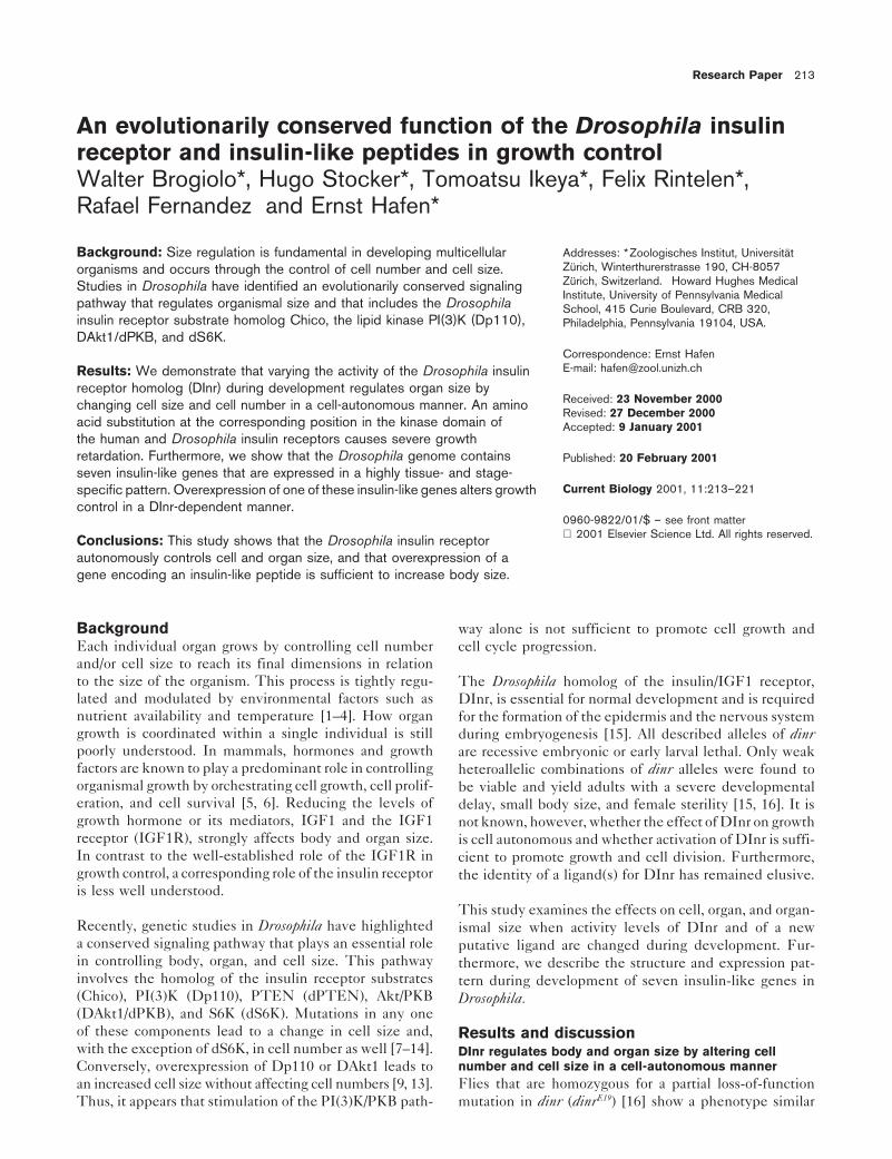
Research Paper 213 An evolutionarily conserved function of the Drosophila insulin receptor and insulin-like peptides in growth control Walter Brogiolo*, Hugo Stocker*, Tomoatsu Ikeya*, Felix Rintelen*, Rafael Fernandez and Ernst Hafen* Background: Size regulation is fundamental in developing multicellular Addresses: * Zoologisches Institut, Universita ¨t Zu ¨ rich, Winterthurerstrasse 190, CH-8057 organisms and occurs through the control of cell number and cell size. Zu ¨ rich, Switzerland. Howard Hughes Medical Studies in Drosophila have identified an evolutionarily conserved signaling Institute, University of Pennsylvania Medical pathway that regulates organismal size and that includes the Drosophila School, 415 Curie Boulevard, CRB 320, insulin receptor substrate homolog Chico, the lipid kinase PI(3)K (Dp110), Philadelphia, Pennsylvania 19104, USA. DAkt1/dPKB, and dS6K. Correspondence: Ernst Hafen E-mail: [email protected] Results: We demonstrate that varying the activity of the Drosophila insulin receptor homolog (DInr) during development regulates organ size by Received: 23 November 2000 changing cell size and cell number in a cell-autonomous manner. An amino Revised: 27 December 2000 acid substitution at the corresponding position in the kinase domain of Accepted: 9 January 2001 the human and Drosophila insulin receptors causes severe growth retardation. Furthermore, we show that the Drosophila genome contains Published: 20 February 2001 seven insulin-like genes that are expressed in a highly tissue- and stage- Current Biology 2001, 11:213–221 specific pattern. Overexpression of one of these insulin-like genes alters growth control in a DInr-dependent manner. 0960-9822/01/$ – see front matter 2001 Elsevier Science Ltd. All rights reserved. Conclusions: This study shows that the Drosophila insulin receptor autonomously controls cell and organ size, and that overexpression of a gene encoding an insulin-like peptide is sufficient to increase body size. Background way alone is not sufficient to promote cell growth and Each individual organ grows by controlling cell number cell cycle progression. and/or cell size to reach its final dimensions in relation to the size of the organism. This process is tightly regu- The Drosophila homolog of the insulin/IGF1 receptor, lated and modulated by environmental factors such as DInr, is essential for normal development and is required nutrient availability and temperature [1–4]. How organ for the formation of the epidermis and the nervous system growth is coordinated within a single individual is still during embryogenesis [15]. All described alleles of dinr poorly understood. In mammals, hormones and growth are recessive embryonic or early larval lethal. Only weak factors are known to play a predominant role in controlling heteroallelic combinations of dinr alleles were found to organismal growth by orchestrating cell growth, cell prolif- be viable and yield adults with a severe developmental eration, and cell survival [5, 6]. Reducing the levels of delay, small body size, and female sterility [15, 16]. It is growth hormone or its mediators, IGF1 and the IGF1 not known, however, whether the effect of DInr on growth receptor (IGF1R), strongly affects body and organ size. is cell autonomous and whether activation of DInr is suffi- In contrast to the well-established role of the IGF1R in cient to promote growth and cell division. Furthermore, growth control, a corresponding role of the insulin receptor the identity of a ligand(s) for DInr has remained elusive. is less well understood. This study examines the effects on cell, organ, and organ- Recently, genetic studies in Drosophila have highlighted ismal size when activity levels of DInr and of a new a conserved signaling pathway that plays an essential role putative ligand are changed during development. Fur- in controlling body, organ, and cell size. This pathway thermore, we describe the structure and expression pat- involves the homolog of the insulin receptor substrates tern during development of seven insulin-like genes in (Chico), PI(3)K (Dp110), PTEN (dPTEN), Akt/PKB Drosophila. (DAkt1/dPKB), and S6K (dS6K). Mutations in any one of these components lead to a change in cell size and, Results and discussion with the exception of dS6K, in cell number as well [7–14]. DInr regulates body and organ size by altering cell number and cell size in a cell-autonomous manner Conversely, overexpression of Dp110 or DAkt1 leads to Flies that are homozygous for a partial loss-of-function an increased cell size without affecting cell numbers [9, 13]. Thus, it appears that stimulation of the PI(3)K/PKB path- mutation in dinr (dinr E19 ) [16] show a phenotype similar
-
Upload
independent -
Category
Documents
-
view
1 -
download
0
Transcript of An evolutionarily conserved function of the Drosophila insulin receptor and insulin-like peptides in...
Research Paper 213
An evolutionarily conserved function of the Drosophila insulinreceptor and insulin-like peptides in growth controlWalter Brogiolo*, Hugo Stocker*, Tomoatsu Ikeya*, Felix Rintelen*,Rafael Fernandez† and Ernst Hafen*
Background: Size regulation is fundamental in developing multicellular Addresses: *Zoologisches Institut, UniversitatZurich, Winterthurerstrasse 190, CH-8057organisms and occurs through the control of cell number and cell size.Zurich, Switzerland. † Howard Hughes MedicalStudies in Drosophila have identified an evolutionarily conserved signalingInstitute, University of Pennsylvania Medical
pathway that regulates organismal size and that includes the Drosophila School, 415 Curie Boulevard, CRB 320,insulin receptor substrate homolog Chico, the lipid kinase PI(3)K (Dp110), Philadelphia, Pennsylvania 19104, USA.DAkt1/dPKB, and dS6K.
Correspondence: Ernst HafenE-mail: [email protected]: We demonstrate that varying the activity of the Drosophila insulin
receptor homolog (DInr) during development regulates organ size byReceived: 23 November 2000changing cell size and cell number in a cell-autonomous manner. An aminoRevised: 27 December 2000
acid substitution at the corresponding position in the kinase domain of Accepted: 9 January 2001the human and Drosophila insulin receptors causes severe growthretardation. Furthermore, we show that the Drosophila genome contains Published: 20 February 2001seven insulin-like genes that are expressed in a highly tissue- and stage-
Current Biology 2001, 11:213–221specific pattern. Overexpression of one of these insulin-like genes alters growthcontrol in a DInr-dependent manner.
0960-9822/01/$ – see front matter 2001 Elsevier Science Ltd. All rights reserved.Conclusions: This study shows that the Drosophila insulin receptor
autonomously controls cell and organ size, and that overexpression of agene encoding an insulin-like peptide is sufficient to increase body size.
Background way alone is not sufficient to promote cell growth andEach individual organ grows by controlling cell number cell cycle progression.and/or cell size to reach its final dimensions in relationto the size of the organism. This process is tightly regu- The Drosophila homolog of the insulin/IGF1 receptor,lated and modulated by environmental factors such as DInr, is essential for normal development and is requirednutrient availability and temperature [1–4]. How organ for the formation of the epidermis and the nervous systemgrowth is coordinated within a single individual is still during embryogenesis [15]. All described alleles of dinrpoorly understood. In mammals, hormones and growth are recessive embryonic or early larval lethal. Only weakfactors are known to play a predominant role in controlling heteroallelic combinations of dinr alleles were found toorganismal growth by orchestrating cell growth, cell prolif- be viable and yield adults with a severe developmentaleration, and cell survival [5, 6]. Reducing the levels of delay, small body size, and female sterility [15, 16]. It isgrowth hormone or its mediators, IGF1 and the IGF1 not known, however, whether the effect of DInr on growthreceptor (IGF1R), strongly affects body and organ size. is cell autonomous and whether activation of DInr is suffi-In contrast to the well-established role of the IGF1R in cient to promote growth and cell division. Furthermore,growth control, a corresponding role of the insulin receptor the identity of a ligand(s) for DInr has remained elusive.is less well understood.
This study examines the effects on cell, organ, and organ-Recently, genetic studies in Drosophila have highlighted ismal size when activity levels of DInr and of a newa conserved signaling pathway that plays an essential role putative ligand are changed during development. Fur-in controlling body, organ, and cell size. This pathway thermore, we describe the structure and expression pat-involves the homolog of the insulin receptor substrates tern during development of seven insulin-like genes in(Chico), PI(3)K (Dp110), PTEN (dPTEN), Akt/PKB Drosophila.(DAkt1/dPKB), and S6K (dS6K). Mutations in any oneof these components lead to a change in cell size and, Results and discussionwith the exception of dS6K, in cell number as well [7–14]. DInr regulates body and organ size by altering cell
number and cell size in a cell-autonomous mannerConversely, overexpression of Dp110 or DAkt1 leads toFlies that are homozygous for a partial loss-of-functionan increased cell size without affecting cell numbers [9, 13].
Thus, it appears that stimulation of the PI(3)K/PKB path- mutation in dinr (dinrE19) [16] show a phenotype similar

214 Current Biology Vol 11 No 4
Figure 1
DInr regulates body and organ size by alteringcell number and cell size in a cell-autonomousmanner. (a) dinrE19/dinrE19 flies (right) show aproportionate body size reduction comparedto dinrE19/1 control flies (left). (b,e) Selectiveremoval of DInr function in eye progenitorcells generates flies with strongly reducedeyes and head capsule while all other bodyparts are of wild-type size. Note the relativelysmaller head size of a null allele (e) than ofa hypomorphic allele (b) compared to controls.(c,f) Tangential sections through a mosaiceye containing small homozygous dinr mutantommatidia (lacking pigment) and normal-sizedheterozygous ommatidia (containing pigment).Homozygous dinrE19 (c) and dinr31 (f) mutantphotoreceptors are reduced in size byapproximately one third and more than half,respectively. Arrowheads point torhabdomeres of small homozygous mutantphotoreceptors within genotypically mixedommatidial units coexisting with normalheterozygous photoreceptors, demonstratinga cell-autonomous requirement for DInr. (d)Confocal microscope section of a third instareye imaginal disc containing mitotic clonesof dinr304 mutant cells (absence of greenstaining), which are significantly smallerthan their wild-type (bright green staining)sister clones; anterior is to the top. (g)Quantitation of body and organ size in dinrE19
homozygous mutant male flies and inheterozygous siblings. In homozygous flies,the body weight and number of ommatidia isreduced by 52% and 45%, respectively. Thewing area is decreased by 36%, due to asignificant decrease in cell size and cellnumber by 23% and 17%, respectively. Valuesare means 6 standard deviation; all valuesof homozygous flies are significantly reducedrelative to their heterozygous siblings (t-test,p , 0.0001). Similar results were obtainedwith female flies. (h) Five dinr alleles carrypoint mutations in conserved amino acidresidues of the kinase domain. An alignmentof the kinase domain of the human insulinreceptor (HInRKin) and the Drosophilahomolog of insulin receptor (DInRKin) is predicted active site residues conferring Flies were of the following genotypes: (a) y w;shown with the localization and molecular substrate specificity [21]. The shading marks dinrE19/TM3, Sb Ser (left) and y w; dinrE19/description of the point mutations. dinr31 and amino acid identity (black) or similarity (gray). dinrE19 (right), (b,e) y w ey-Flp; FRT82B dinrE19,
dinr304 are nonsense mutations in the same We used the amino acid numbering of 31/TM6B, y1 (left) and y w ey-Flp; FRT82Bcodon at different nucleotide positions Fernandez et al. [15] for DInRKin and of Ebina dinrE19, 31/FRT82B P(w1) 3R3.7 (right), (c,f) y(Trp1439Stop). The mutations leading to an et al. [45] for HInRKin. The sequences were w ey-Flp; FRT82B dinrE19, 31/FRT82B P(w1)amino acid substitution are dinr353 aligned with the GCG program package. The 3R3.7, (d) y w hsFlp/y w; FRT82B dinr304/(Arg1419Cys), dinr339 (Gly1491Glu), and abbreviations are single amino acid letter FRT82B P(arm-lacZ, w1), and (g) y w; dinrE19/dinr211 (Gly1551Arg). Asterisks denote code, with “al” indicating an activation loop. 1 and y w; dinrE19/dinrE19.
to that previously described for weak heteroallelic combi- dinrE19 flies have an almost 2-fold increase in lipid content(data not shown). The small body size is attributable tonations. The developmental time is extended from 10
to 20 days, and body size is severely but proportionally a reduction in cell size and cell number by 23% and 17%,respectively (Figure 1g) as revealed by measuring cellreduced. The mutant flies are approximately half the
weight of their heterozygous siblings (Figure 1a,g), and density in the wing. Similarly, the average number ofommatidia in the compound eye of mutant male flies isfemales are sterile. Furthermore, like chico mutant flies,

Research Paper Drosophila insulin genes in growth control Brogiolo et al. 215
Figure 2
Overexpression of DInr in the eye leads to ahyperproliferative outgrowth and an increasein cell size. (a) Scanning electron micrograph(SEM) and (d) tangential section of controleyes. (b) SEM of an eye displaying a dramaticoutgrowth due to an increase in ommatidianumber. This phenotype is caused by theoverexpression of UAS-dinrwt with ey-Gal4 inproliferating eye precursor cells. A tangentialsection of the corresponding eye (e) showsthat normal ommatidial arrangement andarchitecture are retained. (c,f) Eyesoverexpressing UAS-dinrwt in clones under thecontrol of the GMR enhancer, which is mainlyactivated in differentiating cells. Externally,such clones display enlarged ommatidialunits compared with the surrounding wild-typeommatidia (c). In tangential sections (f), theseclones (less pigment) contain enlargedommatidia with enlarged photoreceptors andincreased interommatidial distance. Themagnification in (d–f) is the same. Thegenotypes are (a,d) y w; ey-Gal4/1 (b,e) y w;ey-Gal4/UAS-dinrwt and (c,f) y w hsFlp;GMR.w1; .Gal4/UAS-dinrwt.
378 6 8 compared to 683 6 8 in heterozygous control zygous mutant tissue and heterozygous tissue; within thesame ommatidial unit, small homozygous cells (arrowheadflies (Figure 1g). We did not observe any dominant sizein Figure 1c,f) coexist with normal-sized heterozygousreduction with various dinr alleles.cells. Although cells lacking DInr function survive anddifferentiate normally, they have a growth disadvantageThe reduced overall size could be due to DInr acting
in the humoral regulation of growth or to it functioning compared to heterozygous cells. When homozygous mu-tant cell clones are induced during early larval life andautonomously in a cell- and tissue-specific manner. To
test whether DInr affects body parts autonomously, we analyzed in the imaginal discs in the third instar, clonesize is greatly reduced compared to the wild-type sisterselectively removed DInr function in the eye imaginal
disc using the ey-FLP technique [17]. The eye imaginal clone (Figure 1d). The phenotypes of dinr mutant cellsare strikingly similar to those of mutants in the PI(3)K/disc gives rise to the adult eye and the head capsule.
Mosaic flies with heads largely homozygous for various PKB pathway. Therefore, it is likely that DInr directlyregulates cell growth at least in part through the PI(3)K/dinr alleles displayed a dramatic reduction in eye tissue
and in the head capsule, whereas the other body parts PKB pathway.were of wild-type size (Figure 1b,e). Notably, the headsize was dependent on the allele. This allowed us to Identification of mutations affecting conserved aminoarrange the alleles according to their phenotypic strength acid residues of the kinase domain of DInr(compare Figure 1b with 1e). The strongest reduction in The structure of DInr is similar to the mammalian insulinhead size was observed with dinr339, a putative null allele receptor (Inr) and the IGF1 receptor (IGF1R) [15]. It is(see below), followed by dinr31, dinr211, dinrE19, and dinr353. a tetramer composed of two a subunits containing theThus, DInr regulates head size autonomously. putative ligand binding domains and two transmembrane
b subunits containing the cytoplasmic tyrosine kinase do-Comparison of homozygous mutant tissue with heterozy- mains. In contrast to human receptors, DInr possessesgous tissue in tangential sections of mosaic eyes (Figure extensions at the amino and carboxy termini. The C-ter-1c,f) revealed an estimated reduction in ommatidial size minal extension contains binding sites for downstreamof one third for dinrE19 homozygous mutant tissue (Figure components similar to those found in insulin receptor1c) and of more than half for a candidate null allele (Figure substrates (IRS), and has been shown to be able to signal1f). Importantly, this growth defect does not impede in the absence of IRS proteins [18]. Furthermore, geneticproper cell fate determination, given that the normal ar- evidence in Drosophila suggests that DInr can signal inrangement of the photoreceptor rhabdomeres is retained the absence of Chico, the IRS1-4 homolog [7]. In order(Figure 1c,f). Furthermore, the cell size reduction is cell to understand the molecular basis for differences in
strength of DInr phenotypes, we sequenced the cyto-autonomous, as can be seen at the border between homo-

216 Current Biology Vol 11 No 4
Figure 3
A new family of genes encoding putativeinsulin-related peptides. (a) Genomicorganization of the dilp gene cluster containingdilp1–5 mapping to 67C1-2 on the thirdchromosome. Genomic DNA (GenBankaccession number AE003550.1) isrepresented by a line (top) with distances inkb and an arrow pointing to the centromere.Black boxes indicate exons of dilps for whichmRNA expression has been detected (Table 1).A gray box indicates the predicted exon ofdilp1, which did not show mRNA expressionand therefore may be a pseudogene.Exon–intron boundaries were determined bycomparing genomic DNA and expressedsequence tags (EST; bottom lines), or werepredicted by the Genscan program [38]. dilp5is separated by one intervening gene fromdilp4. Interestingly, each intron splits thereading frame between position 1 and 2 ofa codon; this is typical for insulin-related genes[46]. (b) Schematic representation of thepredicted structure of Drosophila insulin-likepeptides (DILP1–7) and comparison withhuman IGF and insulin. Domains are denotedby letters. The spaces between domainsrepresent predicted proteolytic cleavage [26]during maturation of the propeptide (see alsosupplementary figure). The active peptides(dark boxes) consist of one polypeptide chain(IGF) or two chains (DILP1–7 and humaninsulin). Disulfide bonds between conservedcysteines are indicated. (c) Proportionateincrease in body size of male fliesoverexpressing DILP2 (top). To achieveubiquitous expression of DILP2, we used anhs-Gal4 driver line (Bloomington stockcenter). Heat shocks at 378C were appliedfor 1 hr every 12 hr during development,starting at 24 hr AED. Genotypes are y w;hs-Gal4/UAS-dilp2 (top) and y w; hs-Gal4/1(bottom). (d) DILP2 overexpressionincreases organismal size by increasing thecell size and cell number of individual organs. increase in cell size and cell number by 9% 0.001). Male flies that were 2–3 days old wereCompared to control flies, body weight and and 11%, respectively. Values are means 6 weighed and analyzed. Similar results werenumber of ommatidia are increased by 39% standard deviation; all values of hs-Gal4/UAS- obtained with female flies. Five independentand 5%, respectively. The wing area is dilp2 males are significantly increased UAS-dilp2 transgenic lines yielded similarincreased by 21% due to a significant relative to hs-Gal4/1 control flies (t-test, p , results (n 5 16–78).
plasmic region of several dinr alleles [15, 16, 19]. In the [22]. It is the only reported homozygous viable mutationin the kinase domain of the human Inr. The patient’scytoplasmic portion, 5 out of 22 alleles carry a point muta-
tion. All of them map to conserved amino acid residues parents were heterozygous for this substitution and hadsevere insulin resistance, but no growth anomalies. Simi-within the kinase domain. Two of these point mutations
lead to premature stop codons and three are missense larly, heterozygosity for dinr alleles does not lead to growthphenotypes. These results suggest a role for the insulinmutations (Figure 1h). In humans, most of the mutations
that occur within the tyrosine kinase domain of the Inr receptor in growth control that has been conserved frominsects to humans.have been shown to impair insulin-stimulated tyrosine
kinase activity [20]. dinr353 (Arg1419Cys) affects an activesite residue, which mediates insulin receptor kinase sub- Overexpression of DInr in the eye leadsstrate specificity [21]. Remarkably, a human patient with to hyperproliferation and an increase
in cell sizesevere growth retardation associated with insulin resis-It has been proposed that a bona fide growth controltance, a syndrome called leprechaunism, carries an amino
acid exchange at the corresponding position (Arg1092Glu) gene should meet two criteria [6], namely that elimination
Research Paper Drosophila insulin genes in growth control Brogiolo et al. 217
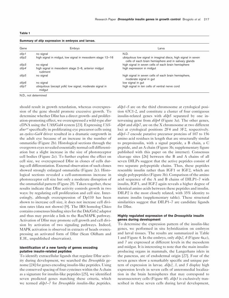
Table 1
Summary of dilp expression in embryos and larvae.
Gene Embryo Larva
dilp1 no signal N.D.dilp2 high signal in midgut, low signal in mesoderm stage 12–16 ubiquitous low signal in imaginal discs, high signal in seven
cells of each brain hemisphere and in salivary glandsdilp3 no signal high signal in seven cells of each brain hemispheredilp4 high signal in mesoderm stage 2–6, anterior midgut high expression in midgut
rudimentdilp5 no signal high signal in seven cells of each brain hemisphere,
moderate signal in gutdilp6 no signal low signal in gutdilp7 ubiquitous (except yolk) low signal, moderate signal in high signal in ten cells of ventral nerve cord
midgut
N.D., not determined
should result in growth retardation, whereas overexpres- dilp1–5 are on the third chromosome at cytological posi-tion 67C1-2, and constitute a cluster of four contiguoussion of the gene should promote excessive growth. To
determine whether DInr has a direct growth- and prolifer- insulin-related genes with dilp5 separated by one in-tervening gene from dilp4 (Figure 3a). The other genes,ation-promoting effect, we overexpressed a wild-type dinr
cDNA using the UAS/Gal4 system [23]. Expressing UAS- dilp6 and dilp7, are on the X chromosome at two differentloci at cytological positions 2F4 and 3F2, respectively.dinr wt specifically in proliferating eye precursor cells using
an eyeless-Gal4 driver resulted in a dramatic outgrowth in dilp1–7 encode putative precursor proteins of 107 to 156amino acid residues in length that are structurally similarthe adult eye because of an increase in the number of
ommatidia (Figure 2b). Histological sections through the to preproinsulin, with a signal peptide, a B chain, a Cpeptide, and an A chain (Figure 3b; supplementary figureovergrown eyes revealed essentially normal cell differenti-
ation but a slight increase in the size of photoreceptor published with this paper on the internet). Consensuscleavage sites [26] between the B and A chains of allcell bodies (Figure 2e). To further explore the effect on
cell size, we overexpressed DInr in clones of cells dur- seven DILPs suggest that the active peptides consist oftwo separate polypeptide chains. Thus, these peptidesing cell differentiation. External observation of such clones
showed strongly enlarged ommatidia (Figure 2c). Histo- resemble insulin rather than IGF1 or IGF2, which aresingle polypeptides (Figure 3b). Comparison of the aminological sections revealed a cell-autonomous increase in
photoreceptor cell size but only a moderate disruption of acid sequence of the A and B chains of DILP1–7 withinsulin, IGF1, and IGF2 again reveals a higher degree ofthe ommatidial pattern (Figure 2f). Taken together, these
results indicate that DInr activity controls growth in two identical amino acids between these peptides and insulin.DILP2 is the most closely related, with 35% identity toways: by regulating cell proliferation and cell size. Inter-
estingly, although overexpression of Dp110 has been mature insulin (supplementary table). These structuralsimilarities suggest that DILP1–7 are candidate ligandsshown to increase cell size, it does not increase cell divi-
sion rates (data not shown) [9]. The IRS homolog Chico for DInr.contains consensus binding sites for the Drk/Grb2 adaptor
Highly regulated expression of the Drosophila insulinand thus may provide a link to the Ras/MAPK pathway.genes during developmentActivation of DInr may promote cell growth and cell divi-To determine the expression pattern of the insulin-likesion by activation of two signaling pathways. Indeed,genes, we performed in situ hybridization on embryosMAPK activation is observed in extracts of heads overex-and larval tissues. The results are summarized in Tablepressing an activated form of DInr (Sean Oldham and1 and Figure 4. In the embryo, only dilp2, 4 (Figure 4a,c),E.H., unpublished observation).and 7 are expressed at different levels in the mesodermand midgut. It is interesting to note that the main insulin-Identification of a new family of genes encoding
putative insulin-related peptides producing organs in mammals, the Langerhans islets inthe pancreas, are of endodermal origin [27]. Four of theTo identify extracellular ligands that regulate DInr activ-
ity during development, we searched the Drosophila ge- seven genes show a remarkably specific and unique pat-tern of expression in larvae. dilp2, 3, and 5 display highnome [24] for genes encoding insulin-like peptides. Using
the conserved spacing of four cysteines within the A chain expression levels in seven cells of anteromedial localiza-tion in the brain hemispheres that may correspond toas a signature for insulin-like peptides [25], we identified
seven predicted genes matching these criteria, which neurosecretory cells (Figure 4f). dilp3 is exclusively tran-scribed in these seven cells during larval development,we termed dilp1–7 for Drosophila insulin-like peptides.

218 Current Biology Vol 11 No 4
Figure 4 whereas dilp2 (Figure 4c,d,e) and dilp5 show additionalexpression domains. dilp7 mRNA detection is restrictedto the ventral nerve cord in a segmental fashion, in fourpairs of ventrally located cells in the most posterior ab-dominal segments and in one pair of dorsally located cellsin A1 or A2 (Figure 4g,h). Interestingly, neither of thedilps shows detectable levels of expression in the larvalfat body.
Expression of insulin-related genes in neurosecretory cellshas been identified in other invertebrates, such as the in-sects Bombyx mori and Locusta migratoria and in the molluscLymnaea stagnalis [28–30]. In Bombyx mori, the neurosecre-tory cells in the brain are connected to the corpora cardi-aca, a secretory gland from which release of insulin-likehormones is triggered by nutrient levels (possibly carbo-hydrate levels) [31]. We speculate that DILP-expressingneurosecretory cells are connected to the ring gland (thecompound endocrine gland of Drosophila), which includesthe cells of the corpora cardiaca. Release of DILPs fromthe ring gland may also be under nutritional control. Thecomplex expression pattern of the DILPs, however, sug-gests a combination of neurosecretory and autocrine/para-crine control mechanisms of cell growth and division dur-ing larval development. Mutations in individual dilp genesor targeted ablation of specific DILP-expressing cells mayhelp resolve the functions of the Drosophila insulins.
DILP2 overexpression increases organismalsize by increasing cell size and cell numberof individual organsTo gain insight into the function of the DILPs, we over-expressed one insulin-like peptide. For this purpose, wechose DILP2 because it is the closest homolog of humaninsulin (see supplementary table) and because it is theonly DILP with broad expression in imaginal discs (Figure
Spatial and temporal regulation of the expression of Drosophila insulin- 4d). If DILP2 is a limiting ligand of DInr, we expect thatlike peptides. Wild-type embryos (a,c) and third instar larval tissues overexpression of DILP2 should promote growth. Indeed,(b,d–h) after in situ hybridization with dilp antisense probes. (a) dilp4
repeated induction of ubiquitous expression of DILP2mRNA is detected at the blastoderm stage in the presumptiveduring development by means of the UAS/Gal4 systemmesoderm and in the anterior midgut rudiment. Shown is a ventral
view. Expression in the mesoderm is still detected after gastrulation, gives rise to bigger flies (39% increase in body weight;but is reduced from stage 12 onward. (b) dilp4 is reexpressed in the Figure 3c,d). Analysis of the eyes of such flies revealedlarval midgut. (c) dilp2 shows a broad expression in the embryonic an increase in the number of ommatidia (from 733 6 10mesoderm starting at stage 12 and an especially intense expression
to 767 6 25 in male flies). Furthermore, quantitative analy-in the midgut, which diminishes at late stage 16. A lateral view isshown. (d) dilp2 is ubiquitously expressed in third larval imaginal discs sis of the wing blade showed an increase in both cell sizeand in the salivary glands (e). In addition, dilp2 mRNA is specifically (by 9%) and cell number (by 11%). These results suggesttranscribed in seven cells of each brain hemisphere (f). The right panel a role for DILP2 in controlling organismal size by aug-is a high magnification image. Expression in these seven cells is
menting both cell number and cell size of different organs.also detected for dilp3 and 5. (g) dilp7 is expressed in a segmentalfashion in the ventral nerve cord in four pairs of ventrally locatedcells in the posterior-most segments and (h) in one pair of dorsally In humans, the in vivo role of insulin as a growth factorlocated cells in the abdominal segment A1 or A2. Anterior is to the is inferred from clinical syndromes, in which excessiveleft in (a,c,f,g,h). Note that sense probes of dilps did not detect anyspecific signals in all experiments except for the dilp2 sense probe,which was uniquely detected in the same seven cells of each brainhemisphere as for the dilp2, 3, and 5 antisense probes. This may
to the dilp2 region of the dilp3 transcript. Consistently, dilp3 mRNAbe explained by the fact that dilp3 is located adjacent to and in theis also uniquely detected in those seven cells of the brain duringopposite orientation of dilp2 in the genome. Therefore, it is possibledevelopment.that during dilp3 transcription, the polymerase reads through into
the dilp2 region and, thus, the dilp2 sense probe could hybridize

Research Paper Drosophila insulin genes in growth control Brogiolo et al. 219
Figure 5
Dominant suppression of a DInr-mediated bigeye phenotype by a deficiency uncoveringdilp1–5. (a) Overexpression of UAS-dinrwt
with the GMR-Gal4 driver line in differentiatingeye cells leads to bulging and rougher eyes.(b) Df(3L)AC1 dominantly suppresses thebig eye phenotype caused by theoverexpression of DInr. (c) Introducing onecopy of UAS-dilp2 efficiently counteracts thesuppressive effect of Df(3L)AC1. The crosseswere performed at 188C. Flies are of thefollowing genotypes: (a) y w; GMR-Gal4, UAS-dinrwt/1, (b) y w; GMR-Gal4, UAS-dinrwt/1;Df(3L)AC1/1, and (c) y w; GMR-Gal4,UAS-dinrwt/UAS-dilp2; Df(3L)AC1/1.
insulin secretion results in excessive growth and where a expression phenotype, we lowered DInr activity in asevere deficiency of insulin secretion is associated with DILP2-overexpressing background. Indeed, introducingpoor intrauterine and postnatal growth [32]. For instance, one mutant copy of dinr (dinr304) dominantly reduces theneonates born to women with diabetes in pregnancy or increased body weight, cell size, and cell number causedborn with Beckwith-Wiedemann syndrome or Nesidio- by ubiquitous DILP2 overexpression, indicating a strongblastosis are macrosomic. In all cases, the growth anomaly genetic interaction between dinr and dilp2 (data notis associated with hyperinsulinemia during embryonic de- shown). Persistent expression of DILP2 under the controlvelopment [32–35]. Our demonstration in transgenic flies of an actin promoter (Act5C-Gal4) caused embryonic lethal-that overexpression of an insulin-like peptide during de- ity. This lethality is dependent on normal levels of DInr,velopment can increase animal size provides further evi- as expression of DILP2 in the presence of strongly re-dence for an evolutionarily conserved role of the insulin duced levels of DInr (see Materials and methods for geno-pathway in growth control. types) generated viable adults that were small and devel-
opmentally delayed. These results are consistent withDInr mediating the effects of DILP2. Furthermore, givenDILP2 genetically interacts with DInrthat a viable heteroallelic combination of dPKB alleles isThe complementarity between the loss-of-function phe-also able to suppress the embryonic lethal phenotype ofnotype of dinr and the DILP2 overexpression phenotypeDILP2 overexpression, we postulate that the action of(increase in size) suggests that DILP2 may be one of theDILP2 by DInr is transduced at least in part through theligands for DInr. We found that a deficiency (Df(3L)AC1)Chico/PI(3)K/dPKB pathway.uncovering dilp1–5 dominantly suppressed the big and
rough eye phenotype caused by targeted overexpressionof DInr in differentiating eye cells (Figure 5a,b). To test Conclusionswhether the observed dominant suppression was caused In humans, syndromes with mutations in the insulin re-by hemizygosity for dilp2, we selectively increased the ceptor or with excessive insulin secretion lead to growthdilp2 gene dosage by crossing in the UAS-dilp2 transgene. abnormalities. This study shows in vivo that altering ex-A single copy of UAS-dilp2 was sufficient to revert the pression levels of a Drosophila insulin-like gene and vary-suppression by Df(3L)AC1 (Figure 5c), strongly suggesting ing the activity of the Drosophila insulin receptor changesthat dilp2 is rate limiting for the DInr overexpression the size and number of cells in organs, thereby regulatingphenotype. An analysis of individually mutated dilp genes organismal size. It seems, therefore, that the insulin recep-will be required to determine the contribution of the other tor pathway has been conserved during evolution for adilps of the cluster (dilp1 and 3–5) to the suppressive role in growth control from insects to humans. Given theeffect of Df(3L)AC1. highly tissue-specific expression of the dilps in the central
nervous system and a broad expression in precursor tissuesof adult organs, we propose a nutritionally regulatedTo examine whether DInr is limiting for the DILP2 over-

220 Current Biology Vol 11 No 4
mechanism whereby Drosophila insulin-like peptides co- Genome search for insulin-like genes, gene structure,and protein structure analysisordinate growth in a neurosecretory and local fashion.We searched for insulin-like genes in the Drosophila nucleotide databasewith the human A chain, which yielded a full-length dilp2 cDNA clone,GH11579. We then used the predicted Drosophila A chains to identifyMaterials and methodsa total of seven insulin-like genes. Searches with A chains were performed
Generation of mosaic flies by mitotic recombination with the TBLASTN program (National Center for Biotechnology Informa-The dinr alleles (E19, 31, 304, 211, 339, and 353) were individually tion). Gene prediction and conceptual translation was performed withrecombined onto the FRT 82B chromosome [36]. Mosaic heads were Genscan [38], and signal peptides were predicted using the SignalPgenerated with the ey-FLP/FRT recombination system [17], which drives program [39]. Cleavage sites were predicted at either specific single orthe FLP recombinase under the control of the eyeless enhancer, thereby pairs of basic residues of the general formula (R/K)-Xn-(R/K), whereinducing mitotic recombination in eye progenitor cells of embryos that cleavage preference decreases with n 5 0, 2, 4, or 6 [26].are heterozygous for different dinr alleles. A recessive cell lethal mutationon the homologous chromosome prevents the growth of the sister clone. The Berkeley Drosophila Genome Project (BDGP) [24] has identifiedThus, the dinr homozygous mutant clone contributes the majority of cells four genes as being insulin-like (corresponding to dilp1–4). dilp5 hasin the eye and the head capsule. Such flies have heads that are largely not been predicted by BDGP. dilp5 has its predicted translational starthomozygous mutant while the rest of the body is heterozygous. To site at nucleotide position 237,598 and stop codon at 237,990 withinestablish an allelic series, we determined the growth defect of each the contig (nucleotide positions refer to GenBank accession numbermosaic head (n 5 10) by measuring the ratio between head and thorax AE003550.1). We found that the dilp3 gene consists of two exons andwidths and by analyzing the cell size defect in histological sections. The has its translational start at 259,432 and stop at 259,854. Correspondinghead to thorax ratio of mosaic heads (and control flies) are, in the order gene numbers in the genome annotation database of Drosophila (Gad-of severity, dinr339: 0.68 (1.23), dinr31: 0.74 (1.20), dinr211: 0.79 (1.22), Fly) for dilp1–4 and dilp6 and 7 are: CG14173, CG8167, CG14167,dinrE19: 0.81 (1.16), and dinr353: 0.82 (1.19). The differences in head CG6736, CG14049, and CG13317.size compared to controls and between the alleles analyzed were signifi-cant (p , 0.0001 and p , 0.005).
In situ hybridizationIn situ hybridization to larval tissues and embryos was performed essen-tially as described [40, 41]. The genomic region including dilp genesTo generate clones of homozygous dinr mutant cells in imaginal discs,was PCR amplified and cloned into the pCRII-Topo vector (Invitrogen)y w hsFLP/y w; FRT82B dinr304/FRT82B P(arm-lacZ, w1) larvae wereexcept for dilp2 and dilp4, which we obtained as an EST clone (LD06542heat shocked 24–48 hr AED for 0.5 hr at 348C to induce expressionand GH11579) from Research Genetics. In vitro transcription was doneof the FLP recombinase. Larvae were dissected at the late third instarusing the DIG RNA labeling kit (Roche). Digoxigenin (DIG)–labeledstage. Discs were fixed, permeabilized, and stained with mouse anti-RNA sense probes were transcribed by SP6 polymerase, and antisenseb-gal (1/1000) and FITC-conjugated secondary antibodies (1/200).probes by T7 polymerase. Hydrolysis time was shortened to 5 min toprevent cross-hybridization.
Measurements of cell number, cell size, and weightEach cell in the wing blade gives rise to a single wing hair. Thus, the Genetic interaction analysiscell density was assessed by counting the number of wing hairs on the Df(3L)AC1 (breakpoints 67A2; 67D13, Flybase) removes dilp1–5, asdorsal wing surface in a 10,000 mm2 area just posterior to the posterior dshc (67B3) [42], which lies distal to the dilp cluster (67C1-2), andcross vein. The reciprocal value of the cell density is the cell area. Gap1 (67C2-3) [43], which lies proximal to the dilp cluster, are bothThe approximate number of cells in the whole wing was calculated by uncovered by this deficiency. Targeted overexpression of UAS-dinrwt inmultiplying the cell density by the wing area excluding the alula and the differentiating eye cells by means of GMR-Gal4 [44] leads to bulgingcostal cell. NIH Image 1.60 was used to measure the wing area. Individual rough eyes (Figure 5a), a phenotype that is dominantly suppressed bymale and female flies were weighed with a precision scale (Mettler ME30, Df(3L)AC1 (Figure 5b). Introducing one copy of UAS-dilp2 restores therange 0.001–10 mg). Flies homozygous for dinrE19 were obtained after big eye phenotype (Figure 5c). This cannot simply be an additive effectremoving linked lethal mutations by recombination. because in a wild-type background, overexpression of USA-dilp2 under
the control of GMR-Gal4 is phenotypically neutral.
Molecular analysis of dinr alleles The lethality resulting from overexpression of UAS-dilp2 with Act5C-The following dinr alleles were sequenced: E16, E19, E21, 31, EC34, Gal4 (Bloomington stock center) was rescued in the following genotypic35, 76, 87, 117, 211, 242, 262, 273, 277, 304, 306, 310, 313, 322, backgrounds, in order of decreasing viability: (1) y w; Act5C-Gal4/UAS-327, 339, and 353 [15, 16, 19]. Genomic DNA was extracted from dilp2; dinr05545/dinr211, (2) y w; Act5C-Gal4/UAS-dilp2; dinr05545/dinrE19,heterozygous flies balanced over TM3 Sb. The region coding for the (3) y w; Act5C-Gal4/UAS-dilp2; dPKB1/ dPKB3, (4) y w; Act5C-Gal4/cytoplasmic portion was amplified by PCR (primer sequences are avail- UAS-dilp2; dinrE19/dinr211, (5) y w; Act5C-Gal4/UAS-dilp2; dinrE19/able on request), sequenced, and analyzed with SequencherTM software dinrE19, and (6) y w; Act5C-Gal4/UAS-dilp2; dinr304/1.and compared to the published sequence [15].
Supplementary materialSupplementary material including a figure showing an alignment of theGeneration of transgenic fliespredicted amino acid sequences of Drosophila insulin-like peptides with
The dinr cDNA was subcloned as an EcoRI fragment into the pUASThuman preproinsulin, and a table comparing the predicted mature
vector to generate transgenic flies by means of P element–mediatedDILP1–7 with human insulin and IGFs are available at http://www.
germline transformation. To achieve expression of UAS-dinrwt in proliferat-current-biology.com/supmat/supmatin.htm.
ing eye precursor cells, the ey-Gal4 driver line was used [37]. To achieveexpression of UAS-dinrwt in clones of eye precursor cells undergoing a
Acknowledgementsfinal round of division and subsequent differentiation, we induced Gal4We thank Peter Gallant and Lindsay MacDougall for critical reading of theexpression under the control of the GMR enhancer by heat shockingmanuscript, Thomas Gutjahr for outstanding technical support with imagewandering larvae for 15 min at 348C containing a heat shock–inducible processing and scanning electron microscopy, Peder Zipperlen and Jurg
FLP recombinase and a flp-out transgene (GMR.w1,STOP.Gal4). A Berger for DNA sequencing, Eva Niederer for assistance in wing and weightfull-length cDNA clone, EST GH11579 (obtained from Research Genet- measurements, and Christof Hugentobler for fly work. We greatly appreciateics), was used to subclone dilp2 as a 0.65 kb EcoRI/XhoI fragment into the organizational help of Manolo Bellotto and the helpful discussions with
Hafen laboratory members. We thank Robert S. Garofalo and the Blooming-pUAST.
Research Paper Drosophila insulin genes in growth control Brogiolo et al. 221
ton stock center for fly stocks. W.B. is a fellow of the postgraduate course of altering cell fates and generating dominant phenotypes.Development 1993, 118:401-415.in experimental medicine and biology that is supported by the Swiss National
24. Adams MD, et al.: The genome sequence of DrosophilaScience Foundation.melanogaster. Science 2000, 287:2185-2195.
25. Blundell TL, Humbel RE: Hormone families: pancreaticReferences hormones and homologous growth factors. Nature 1980,1. Stern DL, Emlen DJ: The developmental basis for allometry in 287:781-787.
insects. Development 1999, 126:1091-1101. 26. Seidah NG, Chretien M: Eukaryotic protein processing:2. Britton JS, Edgar BA: Environmental control of the cell cycle in endoproteolysis of precursor proteins. Curr Opin Biotechnol
Drosophila: nutrition activates mitotic and endoreplicative 1997, 8:602-607.cells by distinct mechanisms. Development 1998, 125:2149- 27. Madsen OD, Jensen J, Blume N, Petersen HV, Lund K, Karlsen C,2158. et al.: Pancreatic development and maturation of the islet
3. Partridge L, Barrie B, Fowler K, French V: Evolution and B cell: studies of pluripotent islet cultures. Eur J Biochem 1996,development of body size and cell size in Drosophila 242:435-445.melanogaster in response to temperature. Evolution 1994, 28. Kawakami A, Iwami M, Nagasawa H, Suzuki A, Ishizaki H: Structure48:1269-1276. and organization of four clustered genes that encode
4. Robertson FW: Studies in quantitative inheritance. XII. Cell size bombyxin, an insulin-related brain secretory peptide of theand number in relation to genetic and environmental variation silkmoth Bombyx mori. Proc Natl Acad Sci USA 1989,of body size in Drosophila. Genetics 1959, 44:869-896. 86:6843-6847.
5. Conlon I, Raff M: Size control in animal development. Cell 1999, 29. Hetru C, Li KW, Bulet P, Lagueux M, Hoffmann JA: Isolation and96:235-244. structural characterization of an insulin-related molecule,
a predominant neuropeptide from Locusta migratoria. Eur J6. Efstratiadis A: Genetics of mouse growth. Int J Dev Biol 1998,Biochem 1991, 201:495-499.42:955-976.
30. Smit AB, Vreugdenhil E, Ebberink RH, Geraerts WP, Klootwijk J,7. Bohni R, Riesgo-Escovar J, Oldham S, Brogiolo W, Stocker H,Joosse J: Growth-controlling molluscan neurons produce theAndruss BF, et al.: Autonomous control of cell and organ sizeprecursor of an insulin-related peptide. Nature 1988, 331:by CHICO, a Drosophila homolog of vertebrate IRS1-4. Cell535-538.1999, 97:865-875.
31. Masumura M, Satake S, Saegusa H, Mizoguchi A: Glucose8. Leevers SJ, Weinkove D, MacDougall LK, Hafen E, Waterfield MD:stimulates the release of bombyxin, an insulin-relatedThe Drosophila phosphoinositide 3-kinase Dp110peptide of the silkworm Bombyx mori. Gen Comp Endocrinolpromotes cell growth. EMBO J 1996, 15:6584-6594.2000, 118:393-399.9. Weinkove D, Neufeld TP, Twardzik T, Waterfield MD, Leevers SJ:
32. Menon RK, Sperling MA: Insulin as a growth factor. EndocrinolRegulation of imaginal disc cell size, cell number and organMetab Clin North Am 1996, 25:633-647.size by Drosophila class I(A) phosphoinositide 3-kinase and
33. Breschi MC, Seghieri G, Bartolomei G, Gironi A, Baldi S, Ferranniniits adaptor. Curr Biol 1999, 9:1019-1029.E: Relation of birthweight to maternal plasma glucose and10. Goberdhan DC, Paricio N, Goodman EC, Mlodzik M, Wilson C:insulin concentrations during normal pregnancy. DiabetologiaDrosophila tumor suppressor PTEN controls cell size and 1993, 36:1315-1321.number by antagonizing the Chico/PI3-kinase signaling 34. Elliott M, Bayly R, Cole T, Temple IK, Maher ER: Clinical featurespathway. Genes Dev 1999, 13:3244-3258. and natural history of Beckwith-Wiedemann syndrome:
11. Huang H, Potter CJ, Tao W, Li DM, Brogiolo W, Hafen E, et al.: presentation of 74 new cases. Clin Genet 1994, 46:168-174.PTEN affects cell size, cell proliferation and apoptosis 35. Milner RD: Nesidioblastosis unravelled. Arch Dis Child 1996,during Drosophila eye development. Development 1999, 74:369-372.126:5365-5372. 36. Xu T, Rubin GM: Analysis of genetic mosaics in developing
12. Gao X, Neufeld TP, Pan D: Drosophila PTEN regulates cell and adult Drosophila tissues. Development 1993, 117:1223-growth and proliferation through PI3K- dependent and 1237.-independent pathways. Dev Biol 2000, 221:404-418. 37. Halder G, Callaerts P, Flister S, Walldorf U, Kloter U, Gehring WJ:
13. Verdu J, Buratovich MA, Wilder EL, Birnbaum MJ: Cell-autonomous Eyeless initiates the expression of both sine oculis and eyesregulation of cell and organ growth in Drosophila by Akt/ absent during Drosophila compound eye development.PKB. Nat Cell Biol 1999, 1:500-506. Development 1998, 125:2181-2191.
14. Montagne J, Stewart MJ, Stocker H, Hafen E, Kozma SC, Thomas 38. Burge C, Karlin S: Prediction of complete gene structures inG: Drosophila S6 kinase: a regulator of cell size. Science human genomic DNA. J Mol Biol 1997, 268:78-94.
39. Nielsen H, Engelbrecht J, Brunak S, von Heijne G: Identification1999, 285:2126-2129.of prokaryotic and eukaryotic signal peptides and15. Fernandez R, Tabarini D, Azpiazu N, Frasch M, Schlessinger J: Theprediction of their cleavage sites. Protein Eng 1997, 10:1-6.Drosophila insulin receptor homolog: a gene essential for
40. O’Neill JW, Bier E: Double-label in situ hybridization usingembryonic development encodes two receptor isoforms withbiotin and digoxigenin-tagged RNA probes. Biotechniquesdifferent signaling potential. EMBO J 1995, 14:3373-3384.1994, 17:870.16. Chen C, Jack J, Garofalo RS: The Drosophila insulin receptor
41. Lehmann R, Tautz D: In situ hybridization to RNA. In Drosophilais required for normal growth. Endocrinology 1996, 137:melanogaster. Practical Use in Cell and Molecular Biology. Edited846-856.by Goldstein LSB and Fyrberg EA. San Diego: Academic Press;17. Newsome TP, Asling B, Dickson BJ: Analysis of Drosophila1994:575-598.photoreceptor axon guidance in eye-specific mosaics.
42. Luschnig S, Krauss J, Bohmann K, Desjeux I, Nusslein-Volhard C:Development 2000, 127:851-860.The Drosophila SHC adaptor protein is required for18. Yenush L, Fernandez R, Myers MG Jr, Grammer TC, Sun XJ, Blenissignaling by a subset of receptor tyrosine kinases. Mol CellJ, et al.: The Drosophila insulin receptor activates multiple 2000, 5:231-241.signaling pathways but requires insulin receptor substrate 43. Gaul U, Mardon G, Rubin GM: A putative Ras GTPase activating
proteins for DNA synthesis. Mol Cell Biol 1996, 16:2509-2517. protein acts as a negative regulator of signaling by the19. Azpiazu N, Frasch M: Tinman and bagpipe: two homeo box Sevenless receptor tyrosine kinase. Cell 1992, 68:1007-1019.
genes that determine cell fates in the dorsal mesoderm of 44. Hay BA, Wolff T, Rubin GM: Expression of baculovirus P35Drosophila. Genes Dev 1993, 7:1324-1340. prevents cell death in Drosophila. Development 1994, 120:
20. Baynes KC, Whitehead J, Krook A, O’Rahilly S: Molecular 2121-2129.mechanisms of inherited insulin resistance. QJM 1997, 45. Ebina Y, Ellis L, Jarnagin K, Edery M, Graf L, Clauser E, et al.: The90:557-562. human insulin receptor cDNA: the structural basis for
21. Hubbard SR, Wei L, Ellis L, Hendrickson WA: Crystal structure of hormone-activated transmembrane signalling. Cell 1985,the tyrosine kinase domain of the human insulin receptor. 40:747-758.
46. Duret L, Guex N, Peitsch MC, Bairoch A: New insulin-like proteinsNature 1994, 372:746-754.with atypical disulfide bond pattern characterized in22. Takahashi Y, Kadowaki H, Momomura K, Fukushima Y, Orban T, OkaiCaenorhabditis elegans by comparative sequence analysisT, et al.: A homozygous kinase-defective mutation in theand homology modeling. Genome Res 1998, 8:348-353.insulin receptor gene in a patient with leprechaunism.
Diabetologia 1997, 40:412-420.23. Brand AH, Perrimon N: Targeted gene expression as a means