Crystal Structure of N-Glycosylated Human Glypican-1 Core Protein: STRUCTURE OF TWO LOOPS...
16
Crystal Structure of N-Glycosylated Human Glypican-1 Core Protein STRUCTURE OF TWO LOOPS EVOLUTIONARILY CONSERVED IN VERTEBRATE GLYPICAN-1 * □ S Received for publication, November 10, 2011, and in revised form, January 25, 2012 Published, JBC Papers in Press, February 20, 2012, DOI 10.1074/jbc.M111.322487 Gabriel Svensson ‡ , Wael Awad §¶ , Maria Håkansson , Katrin Mani ‡ , and Derek T. Logan §1 From the ‡ Department of Experimental Medical Science, Division of Neuroscience, Glycobiology Group, Lund University, Biomedical Center A13, SE-221 84, Lund, Sweden, the § Department of Biochemistry and Structural Biology, Center for Molecular Protein Science, Lund University, Box 124, SE-221 00 Lund, Sweden, the ¶ Biophysics Department, Faculty of Science, Cairo University, 12613 Giza, Egypt, and SARomics Biostructures AB, Medicon Village, SE-223 81 Lund, Sweden Background: The glypican family of cell-surface proteoglycans regulates growth factor signaling during development through their core proteins and heparan sulfate chains. Results: The crystal structure of N-glycosylated human glypican-1 is described. Conclusion: The structure reveals the complete disulfide bond arrangement for the conserved Cys residues in glypicans. Significance: Increased structural knowledge of glypicans will help elucidate their important functions in shaping animal development. Glypicans are a family of cell-surface proteoglycans that reg- ulate Wnt, hedgehog, bone morphogenetic protein, and fibro- blast growth factor signaling. Loss-of-function mutations in glypican core proteins and in glycosaminoglycan-synthesizing enzymes have revealed that glypican core proteins and their gly- cosaminoglycan chains are important in shaping animal devel- opment. Glypican core proteins consist of a stable -helical domain containing 14 conserved Cys residues followed by a gly- cosaminoglycan attachment domain that becomes exclusively substituted with heparan sulfate (HS) and presumably adopts a random coil conformation. Removal of the -helical domain results in almost exclusive addition of the glycosaminoglycan chondroitin sulfate, suggesting that factors in the -helical domain promote assembly of HS. Glypican-1 is involved in brain development and is one of six members of the vertebrate family of glypicans. We expressed and crystallized N-glycosylated human glypican-1 lacking HS and N-glycosylated glypican-1 lacking the HS attachment domain. The crystal structure of glypican-1 was solved using crystals of selenomethionine-la- beled glypican-1 core protein lacking the HS domain. No addi- tional electron density was observed for crystals of glypican-1 containing the HS attachment domain, and CD spectra of the two protein species were highly similar. The crystal structure of N-glycosylated human glypican-1 core protein at 2.5 Å, the first crystal structure of a vertebrate glypican, reveals the complete disulfide bond arrangement of the conserved Cys residues, and it also extends the structural knowledge of glypicans for one -helix and two long loops. Importantly, the loops are evolu- tionarily conserved in vertebrate glypican-1, and one of them is involved in glycosaminoglycan class determination. Proteoglycans (PGs) 2 consist of a core protein covalently substituted with glycosaminoglycans (GAGs), long unbranched polysaccharide chains. Several genetic diseases in humans and numerous genetic experiments in fruit flies, nematodes, and mice have revealed that GAGs and their core proteins are important in shaping animal development (1, 2). Glypicans are a family of cell-surface PGs substituted with the GAG heparan sulfate (HS). Glypicans regulate growth factor signaling by binding of growth factors to their HS chains or, as has been shown recently, by binding of growth factors to the core pro- teins. Based on sequence comparisons, glypicans fall into two subfamilies: glypican-1, -2, -4, and -6 and glypican-3 and -5, with 25% amino acid identity between the groups. Glypicans are around 550 amino acids in length and all share an N-termi- nal secretory signal peptide, 14 evolutionarily conserved Cys residues, an HS attachment domain near the C terminus, and a hydrophobic domain for addition of the glycosylphosphatidyli- nositol (GPI) anchor at the C terminus. Aside from the six members of the glypican family in vertebrates, there are also two glypicans in Drosophila melanogaster (Dally and Dally-like protein) and two in Caenorhabditis elegans (Gpn-1 and Lon-2). Dally-like protein (DLP) is divergent from the other glypicans, in particular by the insertion of 100 amino acids N-terminal to the last two conserved Cys residues (supplemental Fig. S1). * This work was supported by Swedish Research Council Grants 2006-4387 (to D. L.) and 2005-6749 and 2010-3914 (to K. M.), the Swedish Cancer Foundation, the Medical Faculty of Lund University, the Royal Physio- graphic Society in Lund, and the Crafoord, Wiberg, Jeanson, Segerfalk, and Kock Foundations. □ S This article contains supplemental Figs. 1 and 2. The atomic coordinates and structure factors (codes 4ACR and 4AD7) have been deposited in the Protein Data Bank, Research Collaboratory for Structural Bioinformatics, Rutgers University, New Brunswick, NJ (http://www.rcsb.org/). 1 To whom correspondence should be addressed. Tel.: 4646-2221443; Fax: 4646-2224116; E-mail: [email protected]. 2 The abbreviations used are: PG, proteoglycan; HS, heparan sulfate; GAG, glycosaminoglycan; BisTris, 2-[bis(2-hydroxyethyl)amino]-2-(hydroxy- methyl)propane-1,3-diol; SeMet, selenomethionine; CS, chondroitin sulfate; GPI, glycosylphosphatidylinositol; rh, recombinant human; mdeg, millidegrees, EXTL, exostosin-like. THE JOURNAL OF BIOLOGICAL CHEMISTRY VOL. 287, NO. 17, pp. 14040 –14051, April 20, 2012 © 2012 by The American Society for Biochemistry and Molecular Biology, Inc. Published in the U.S.A. 14040 JOURNAL OF BIOLOGICAL CHEMISTRY VOLUME 287 • NUMBER 17 • APRIL 20, 2012 at Lund University Libraries, Head Office, on April 9, 2013 www.jbc.org Downloaded from http://www.jbc.org/content/suppl/2012/02/20/M111.322487.DC1.html Supplemental Material can be found at:
Transcript of Crystal Structure of N-Glycosylated Human Glypican-1 Core Protein: STRUCTURE OF TWO LOOPS...
Crystal Structure of N-Glycosylated Human Glypican-1 CoreProteinSTRUCTURE OF TWO LOOPS EVOLUTIONARILY CONSERVED IN VERTEBRATEGLYPICAN-1*!S
Received for publication, November 10, 2011, and in revised form, January 25, 2012 Published, JBC Papers in Press, February 20, 2012, DOI 10.1074/jbc.M111.322487
Gabriel Svensson‡, Wael Awad§¶, Maria Håkansson!, Katrin Mani‡, and Derek T. Logan§1
From the ‡Department of Experimental Medical Science, Division of Neuroscience, Glycobiology Group, Lund University,Biomedical Center A13, SE-221 84, Lund, Sweden, the §Department of Biochemistry and Structural Biology, Center for MolecularProtein Science, Lund University, Box 124, SE-221 00 Lund, Sweden, the ¶Biophysics Department, Faculty of Science, CairoUniversity, 12613 Giza, Egypt, and !SARomics Biostructures AB, Medicon Village, SE-223 81 Lund, Sweden
Background: The glypican family of cell-surface proteoglycans regulates growth factor signaling during developmentthrough their core proteins and heparan sulfate chains.Results: The crystal structure of N-glycosylated human glypican-1 is described.Conclusion: The structure reveals the complete disulfide bond arrangement for the conserved Cys residues in glypicans.Significance: Increased structural knowledge of glypicans will help elucidate their important functions in shaping animaldevelopment.
Glypicans are a family of cell-surface proteoglycans that reg-ulate Wnt, hedgehog, bone morphogenetic protein, and fibro-blast growth factor signaling. Loss-of-function mutations inglypican core proteins and in glycosaminoglycan-synthesizingenzymes have revealed that glypican core proteins and their gly-cosaminoglycan chains are important in shaping animal devel-opment. Glypican core proteins consist of a stable !-helicaldomain containing 14 conserved Cys residues followed by a gly-cosaminoglycan attachment domain that becomes exclusivelysubstituted with heparan sulfate (HS) and presumably adopts arandom coil conformation. Removal of the !-helical domainresults in almost exclusive addition of the glycosaminoglycanchondroitin sulfate, suggesting that factors in the !-helicaldomainpromote assembly ofHS.Glypican-1 is involved in braindevelopment and is one of six members of the vertebrate familyof glypicans. We expressed and crystallized N-glycosylatedhuman glypican-1 lacking HS and N-glycosylated glypican-1lacking the HS attachment domain. The crystal structure ofglypican-1 was solved using crystals of selenomethionine-la-beled glypican-1 core protein lacking the HS domain. No addi-tional electron density was observed for crystals of glypican-1containing the HS attachment domain, and CD spectra of thetwo protein species were highly similar. The crystal structure ofN-glycosylated human glypican-1 core protein at 2.5 Å, the firstcrystal structure of a vertebrate glypican, reveals the completedisulfide bond arrangement of the conserved Cys residues, and
it also extends the structural knowledge of glypicans for one!-helix and two long loops. Importantly, the loops are evolu-tionarily conserved in vertebrate glypican-1, and one of them isinvolved in glycosaminoglycan class determination.
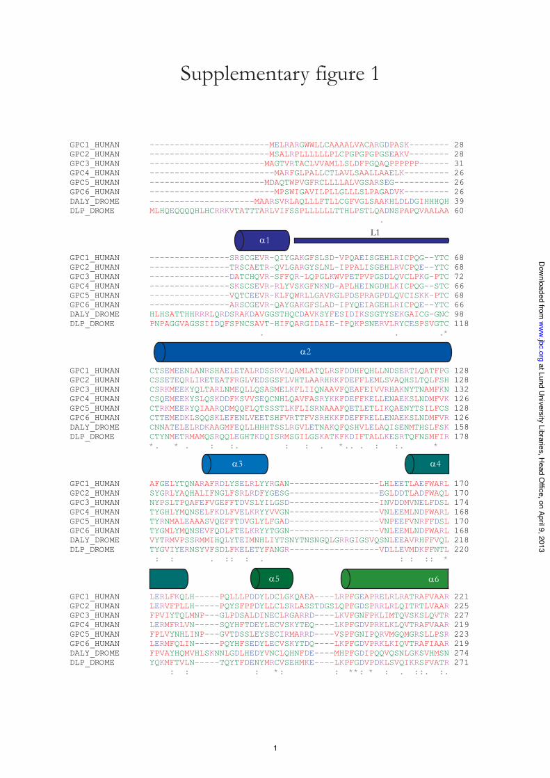
Proteoglycans (PGs)2 consist of a core protein covalentlysubstitutedwith glycosaminoglycans (GAGs), long unbranchedpolysaccharide chains. Several genetic diseases in humans andnumerous genetic experiments in fruit flies, nematodes, andmice have revealed that GAGs and their core proteins areimportant in shaping animal development (1, 2). Glypicans area family of cell-surface PGs substituted with the GAG heparansulfate (HS). Glypicans regulate growth factor signaling bybinding of growth factors to their HS chains or, as has beenshown recently, by binding of growth factors to the core pro-teins. Based on sequence comparisons, glypicans fall into twosubfamilies: glypican-1, -2, -4, and -6 and glypican-3 and -5,with !25% amino acid identity between the groups. Glypicansare around 550 amino acids in length and all share an N-termi-nal secretory signal peptide, 14 evolutionarily conserved Cysresidues, an HS attachment domain near the C terminus, and ahydrophobic domain for addition of the glycosylphosphatidyli-nositol (GPI) anchor at the C terminus. Aside from the sixmembers of the glypican family in vertebrates, there are alsotwo glypicans inDrosophila melanogaster (Dally and Dally-likeprotein) and two inCaenorhabditis elegans (Gpn-1 and Lon-2).Dally-like protein (DLP) is divergent from the other glypicans,in particular by the insertion of 100 amino acids N-terminal tothe last two conserved Cys residues (supplemental Fig. S1).
* This work was supported by Swedish Research Council Grants 2006-4387(to D. L.) and 2005-6749 and 2010-3914 (to K. M.), the Swedish CancerFoundation, the Medical Faculty of Lund University, the Royal Physio-graphic Society in Lund, and the Crafoord, Wiberg, Jeanson, Segerfalk, andKock Foundations.
!S This article contains supplemental Figs. 1 and 2.The atomic coordinates and structure factors (codes 4ACR and 4AD7) have been
deposited in the Protein Data Bank, Research Collaboratory for StructuralBioinformatics, Rutgers University, New Brunswick, NJ (http://www.rcsb.org/).
1 To whom correspondence should be addressed. Tel.: 4646-2221443; Fax:4646-2224116; E-mail: [email protected].
2 The abbreviations used are: PG, proteoglycan; HS, heparan sulfate; GAG,glycosaminoglycan; BisTris, 2-[bis(2-hydroxyethyl)amino]-2-(hydroxy-methyl)propane-1,3-diol; SeMet, selenomethionine; CS, chondroitinsulfate; GPI, glycosylphosphatidylinositol; rh, recombinant human; mdeg,millidegrees, EXTL, exostosin-like.
THE JOURNAL OF BIOLOGICAL CHEMISTRY VOL. 287, NO. 17, pp. 14040 –14051, April 20, 2012© 2012 by The American Society for Biochemistry and Molecular Biology, Inc. Published in the U.S.A.
14040 JOURNAL OF BIOLOGICAL CHEMISTRY VOLUME 287 • NUMBER 17 • APRIL 20, 2012
at Lund University Libraries, H
ead Office, on April 9, 2013
ww
w.jbc.org
Dow
nloaded from
http://www.jbc.org/content/suppl/2012/02/20/M111.322487.DC1.html Supplemental Material can be found at:

Loss-of-function mutations in GPC3 cause Simpson-Golabi-Behmel syndrome, an X-linked condition characterized by pre-natal and postnatal overgrowth (3). Mutations in GPC6 causeomodysplasia-1, a congenital disease affecting the skeletal sys-tem (4). Clinical features of omodysplasia-1 involve short stat-ure and facial dysmorphism. Also, common variation in theGPC5 gene is associated with acquired nephrotic syndrome (5).GPC1 is expressed in the developing and adult brain, andGPC1knock-out mice have reduced brain size at birth (6).The structurally related but functionally distinct GAGs HS
and chondroitin sulfate (CS) are added to core proteins thatcontain a GAG attachment motif consisting of the dipeptideSer-Gly. Synthesis is initiated by the addition of a xylose residueto the hydroxyl group of the Ser residue in theGAGattachmentdomain, followed by sequential addition of two galactose (Gal)and one glucuronic acid (GlcA) residue. This forms the linkerregion common for HS and CS, GlcA!1–3Gal!1–3Gal!1–4Xyl!1-O-Ser. Synthesis of HS is initiated by the addition of anN-acetylglucosamine (GlcNAc) residue, whereas CS synthesisbegins with the addition of an N-acetylgalactosamine (Gal-NAc). After the addition of theGlcNAcorGalNAc residues,HSand CS chains are polymerized, forming their respective disac-charide backbones. Exactlywhat determineswhether aGlcNAcresidue or a GalNAc is added to the common linker region isnot known. It has been suggested thatHS assembly is promotedby the repetition of the Ser-Gly dipeptide and by flanking acidicresidues (7, 8). Glypicans are substituted with HS and containGAG attachment domains similar to those suggested to pro-mote HS assembly. However, expression of the rat GPC1 GAGattachment domain alone results in substitution with!90%CS(9), strongly suggesting that factors in the glypican core proteinfurther away from the GAG attachment sites promote theassembly of HS.Recently, it has been shown that functional aspects of glypi-
cans also reside in the protein cores. For example, Hedgehoggrowth factor signaling is deficient in D. melanogaster animalswhen the D. melanogaster glypican Dlp has been knockeddown, and the Hedgehog response can be rescued by expres-sion of DLP without the HS chains (10, 11). Moreover, otherreports have shown that glypican core proteins bind directly togrowth factors such as Hedgehog (12), bone morphogeneticprotein 4 (13), decapentaplegic, Wnt (14, 15), and insulin-likegrowth factor II (16) with significant biological function inde-pendent of the presence of HS chains.We have previously shown that anchorless recombinant
human GPC1 (rhGPC1) is a stable "-helical protein that resistshigh concentrations of the chemical denaturants urea and gua-nidine-HCl (17). Unfolding occurs in a single step, and unfold-ing data can be closely fitted by a two-state model, indicatingthat GPC1 is a densely packed globular protein. Recently, thecore protein structure of DLP was published, revealing anextended "-helical fold for glypican core proteins (18). Here,we present the independently solved structure of GPC1 coreprotein using multiple-wavelength anomalous dispersionphasing on selenomethionine (SeMet)-labeled rhGPC1, thefirst crystal structure of a vertebrate glypican. Structuralalignment of GPC1 andDLP reveals a common fold for glypi-can core proteins. The GPC1 structure presented here also
expands our knowledge of glypican core proteins, in partic-ular for the disulfide bond arrangement of the conserved Cysresidues, one "-helix and two long loops. One of the loopshas been shown to be involved in GAG class determination,but lack of structural data has previously hampered furthermutagenic investigations.
EXPERIMENTAL PROCEDURES
Materials—Human GPC1 cDNA clone IMAGE ID 6275649was obtained from Geneservice (UK). The expression vectorpCEP-BM40-HisTEV and 293 human embryonic kidney(HEK293) cells were obtained as described previously (17). Allrestriction enzymes used were from Fermentas (Canada).Nickel-nitrilotriacetic acid columns were from GE Healthcare.Protein-free medium for CHO cells and Coomassie Blue werefrom Sigma. Lipofectamine, 4–12% BisTris SDS-polyacryl-amide gels, L-methionine-free Dulbecco’s modified Eagle’smedium, dialyzed fetal bovine serum (FBS), and hygromycin Bwere from Invitrogen. L-Selenomethionine was from AcrosOrganics (Belgium). Crystallization plates and polyethyleneglycol Mr 6000 (PEG 6000) were obtained from HamptonResearch. Other materials related to crystallography were pur-chased fromMolecular Dimensions (UK).Cloning and Purification of Human GPC1—The cDNA
sequence coding for the mature form of human GPC1, aminoacids 24–529, i.e. without the sequences for the N- and C-ter-minal signal peptides, was amplified and introduced into thevector pCEP4-BM40-HisTEV as described previously (17).This vector contains the sequence for an N-terminal BM40secretion peptide followedby aHis6 tag and a tobacco etch virusprotease cleavage site. Furthermore, the HS attachment siteswere disrupted by site-directed mutagenesis to create a vectorexpressing GPC1 "HS (17). A vector coding for GPC1 "C wascreated by amplifying the sequence for amino acids 24–479using the forward primer 5#-TTAAGCTTGACCCGGCCAG-CAAGAG-3# and the reverse primer 5#-TTGGATCCT-TAGAAGTCCACGTCGTTGCCGTT-3# and by introducingthe PCR product into the pCEP4-BM40-HisTEV plasmid.Restriction enzyme sites are shown in boldface. All constructswere verified by sequencing at Eurofins MWG Operon (Ger-many). Stable HEK 293 cell clones expressing GPC1 "HS andGPC1 "C were generated by limiting dilution after severalweeks of growth in selective antibiotics. Stable cell clonesexpressing large amounts of rhGPC1were grown to confluencein minimal essential medium with Earle’s salts supplementedwith 10% (v/v) FBS, 2mM L-glutamine, penicillin (100 units/ml),streptomycin (100 #g/ml), and hygromycin B (200 #g/ml).After extensive washing with PBS, the rhGPC1-expressing cellsweremaintained for 3 or 4 days in protein-freemedium supple-mentedwith 2mM L-glutamine, penicillin (100 units/ml), strep-tomycin (100 #g/ml), and hygromycin B (100 #g/ml). Condi-tioned medium was stored frozen, and rhGPC1 was purifiedfrom conditioned medium of !200-ml batches using nickelaffinity chromatography. Protein purification was analyzed bySDS-PAGE, and purified rhGPC1 was dialyzed to 20 mM Tris-HCl, pH 8.0, concentrated by ultrafiltration, and stored frozen.SeMet labeling of GPC1 "C was performed in L-methionine-free Dulbecco’s modified Eagle’s medium supplemented with
Crystal Structure of Human Glypican-1 Core Protein
APRIL 20, 2012 • VOLUME 287 • NUMBER 17 JOURNAL OF BIOLOGICAL CHEMISTRY 14041
at Lund University Libraries, H
ead Office, on April 9, 2013
ww
w.jbc.org
Dow
nloaded from

2% dialyzed FBS. After a 6-h preincubation with L-methionine-free medium, the medium was replaced with medium contain-ing 30 mg/liter L-selenomethionine. Cells suffered from SeMettoxicity, which made it difficult to grow the cells in mediumsupplemented with SeMet for more than 24 h. SeMet-labeledGPC1 "C was purified in the presence of 0.1 mM tris(2-car-boxyethyl)phosphine hydrochloride to avoid oxidation ofSeMet residues (19).CD Spectroscopy—CD spectra were recorded using a Jasco
J-810 spectropolarimeter equipped with a Peltier thermostatedcell holder. The cuvette was thermostated at 20 °C. For far-UVCD, a 1-mm quartz cuvette was used, and the measurementrange was 190–250 nm. The following parameters were used:sensitivity, 100millidegrees (mdeg); data pitch, 1 nm; scan rate,100 nm/min; response time, 4 s; bandwidth, 1 nm; and accumu-lation, 2. Near-UV CD was measured in a 10-mm quartzcuvette, and the measurement range was 250–340 nm. Thefollowing parameters were used: sensitivity, 100 mdeg; datapitch, 0.2 nm; scan rate, 50 nm/min; response time, 4 s; band-width, 0.5 nm; and accumulation, 4. Base-line spectra wererecorded for pure buffer and subtracted from the correspond-ing protein spectra. Stock solutions of rhGPC1were clarified bycentrifugation at 15,000$ g for 15min, and protein concentra-tion was determined by measuring the absorbance at 280 nmusing amicro-volume spectrophotometer (GEHealthcare) andthe molar extinction coefficient for bovine serum albumin.Samples were prepared by diluting the rhGPC1 stock solutionto 0.1 mg/ml for far-UV CD and to 1 mg/ml for near-UV CD.Protein wasmaintained in 20mM sodium phosphate buffer, pH7.4. Mean residue ellipticity was calculated using 110.5 and112.0 as themean residuemolecular weights of GPC1"HS andGPC1 "C, respectively.Crystallization—Initial high throughput crystallization
screens performed with GPC1 "HS at a protein concentrationof 40mg/ml were set up inMRC plates using aMosquito nano-liter pipetting robot (TTP Labtech, UK) at the MAX-lab Crys-tallization Facility, Lund University. The initial screens, PACTpremier (Molecular Dimensions), Crystal Screen 1 and 2(Hampton Research), and JCSG% (Qiagen GmbH, Germany),were set up using the vapor diffusion technique in sitting dropswhere 100 nl of protein solution were mixed with 100 nl ofreservoir solution and equilibrated against 80 #l of reservoirsolution. After 1 day, needle clusters began to appear underconditions containing polyethylene glycol Mr & 3350 (PEG3350) with anions such as Br', F', and NO3
', pH 6.5. After 1week, crystal plates appeared in dropswith 20%PEG6000, 0.2MCaCl2 in 0.1 M Tris-HCl, pH 8.0. After 2 weeks, the sameappeared in drops with 20% PEG 6000, 0.2 M NH4Cl in 0.1 MTris-HCl, pH 8.0. Crystal-like material also appeared in 20%PEG 8000, 0.2 M magnesium acetate, 0.1 M sodium cacodylatetrihydrate, pH 6.5, after 2 weeks. Optimization screens wereproduced from stock solutions using a liquid handling pipettingrobot (Tecan Group Ltd., Switzerland) and set up in MRCplates using the nanoliter pipetting robot as for the initialscreens. The best results were obtained with the crystals grownin CaCl2 with PEG 6000 as precipitating agent. The optimizedconditions were scaled up to microliter drops in 24-well platesusing the sitting drop vapor diffusion method at 20 °C with 500
#l of reservoir solution. Typically, crystals of rhGPC1 weregrown by mixing 2 #l of protein solution with 2 #l of reservoirsolution containing 12–14% PEG 6000, 0.1 M Tris-HCl, 0.2 MCaCl2, pH 8.0. GPC1 "C was crystallized at 20 mg/ml. GPC1"HSwas crystallized at a higher protein concentration, up to 40mg/ml, and benefited from low concentrations of dithiothreitolin the protein solution. In general, crystallization of GPC1 "Cwas more easily reproduced in comparison to crystallization ofGPC1"HS.Crystallization of SeMet-labeledGPC1"Cwas alsoperformed using the same conditions but with lower proteinconcentration, and the protein was maintained in 20 mM Tris-HCl, pH 8.0, supplemented with 0.1 mM tris(2-carboxyethyl-)phosphine hydrochloride. Large but thin plate-like crystalswith dimensions around 0.6 $ 0.3 mm formed within 1 weekfor GPC1 "HS and GPC1 "C. Crystals of SeMet-labeled GPC1"Cwere typically smaller, around 0.2$ 0.2mm.Crystallizationof rhGPC1 was not affected by the presence of the His6 tag.DataCollection andProcessing—Crystals were soaked in 15%
ethylene glycol, 18% PEG 6000, 0.1 M Tris-HCl, 0.2 MCaCl2, pH8.0, and flash-frozen under a stream of liquid nitrogen. X-raydiffraction data were collected on stations I911-2 and I911-3 ofthe MAX-II synchrotron radiation source, Lund, Sweden, on165- and 225-mm CCD detectors from MarResearch (Ger-many). All data sets were integrated and scaled using the XDSsuite (20) and further processed using programs from theCCP4(21) and Phenix (22) suites. Data collection statistics are givenin Table 1. Diffraction patterns of GPC1 "HS crystals could beindexed in space group P21 and had unit cell dimensions a &47.2 Å, b & 169.0 Å, c & 151.6 Å, ! & 95.0°. Variation in celldimensions was seen between different crystals, in particularfor the c dimension, with lengths ranging from 148 to 154 Å.The crystals contain four GPC1 molecules in the asymmetricunit, giving aMatthews volume of 2.7 Å3/Da and a solvent con-tent of 54%. Crystals of GPC1 "C and SeMet GPC1 "C alsobelong to space group P21 with similar unit cell dimensions.The diffraction anisotropy of the data from GPC1 "C crystalswas analyzed using phenix.xtriage (22) and found to be signifi-cant. The B factor was twice as large in the c* direction as in thea* and b* directions. This effectively limited the resolution toaround 2.9 Å in the c* direction. However, refinement againstdata truncated using the UCLA anisotropy server (23) did notresult in improvedmaps, so the original datawere retained. Thedata also show a significant translational noncrystallographicsymmetry, with a peak at (0.5, 0.021, 0.5) in the native Pattersonof 35% of the height of the origin peak. This translation relateschains A and B to C and D in the asymmetric unit, respectively.Structure Determination and Refinement—X-ray data were
collected to 3.3 Å resolution from SeMet GPC1 "C crystals atselenium peak and inflection wavelengths at station I911-3 ofMAX-II. The processed data sets were sent to the AutoRick-shaw server at the European Molecular Biology Laboratory inHamburg, Germany (24, 25), for structure determination usingthe two-wavelengthmultiple anomalous dispersionmethod. InAutoRickshaw, the data sets were prepared using the CCP4programs CAD and SCALEIT (21, 26). Furthermore, heavyatoms were found using SHELXD (27), and heavy atom refine-ment and phase calculation were performed using SHARP (28)and MLPHARE (21). Initial density modification and phase
Crystal Structure of Human Glypican-1 Core Protein
14042 JOURNAL OF BIOLOGICAL CHEMISTRY VOLUME 287 • NUMBER 17 • APRIL 20, 2012
at Lund University Libraries, H
ead Office, on April 9, 2013
ww
w.jbc.org
Dow
nloaded from

extension were performed using DM (29) and further exploredusing RESOLVE (30). Finally, the electron density map wasinterpreted using HELICAP (31), and model building was per-formed by BUCCANEER (32). This resulted in amore than 90%complete model built at 3.8 Å that was partially refined by CNS(33) and Refmac5 (34) at 3.3 Å (Rmodel & 23% and Rfree & 32%).The selenium positions were confirmed by calculating ananomalous difference map at peak wavelength using phasesfrom a partially refined model. The initial structure wasimproved by extensivemanual rebuilding in Coot (35) followedbymany rounds of refinement using the diffraction data to 2.55Å from a native GPC1 "C crystal. Phenix.refine (22) with Ram-achandran restraints was used for the final model refinement,and Molprobity (36) was used for validation. Structural align-ments weremade using the secondary structurematching algo-rithm as implemented in Coot. All graphical representations ofthe GPC1 structure were generated using the PyMOLMolecu-lar Graphics System, Version 1.2r3pre, Schrödinger.Structural Conservation Analysis—Alignments of glypican
amino acid sequences were performed using ClustalW2 (37).The amino acid sequences used are given in Fig. 5 figure legend.DNA sequences were translated using the ExPASy TranslateTool. Structural conservation analysis was performed using theConSurf server (38).
RESULTS
Expression and Characterization of Anchorless GPC1—Weexpressed His6-tagged full-length rhGPC1 without the GPI
anchor signal peptide in 293 human embryonic kidney cells.This secreted rhGPC1 (protein referred to as GPC1 WT) waspurified under native conditions, and analysis on SDS-PAGE ofthe purified protein showed a core protein band of !60 kDaand a PG smear of !60–120 kDa (Fig. 1A, lane 1). Degradationwith HS lyase and peptide:N-glycosidase F showed that thisprotein was substituted exclusively with HS and also containedN-linked glycans (17, 39). To produce rhGPC1 for conforma-tional and structural studies, the HS attachment sites were dis-rupted by site-directed mutagenesis, resulting in expression ofcore protein containingN-linked glycanswith amolecularmassof !60 kDa (protein referred to as GPC1 "HS, Fig. 1A, lane 2).Also, theHS attachment site in GPC1 is regarded, by secondarystructure and disorder predictions, as adopting a random coilconformation despite the fact that it contains two Cys residues.Therefore, rhGPC1 was truncated C-terminally before the HSattachment domain, resulting in a core protein band (GPC1"C) of smaller size (!55 kDa) on SDS-PAGE (Fig. 1A, lane 3).We have previously shown that the PG form of GPC1WT andGPC1 "HS is identical in folding, as judged by far-UV CD andfluorescence emission spectroscopy (17). To ensure that GPC1"HS and GPC1 "C are also structurally similar, we subjectedthe two forms of rhGPC1 to far- and near-UVCDspectroscopy.Far-UVCD reports on the peptide bond conformation (i.e. sec-ondary structure content) of the investigated protein andnear-UV CD reports on the immediate environment of Trp,Tyr, and Phe residues (i.e. the tertiary structure). Both protein
FIGURE 1. SDS-PAGE and spectroscopic analysis of rhGPC1. A, SDS-PAGE and Coomassie Blue staining of nickel-nitrilotriacetic acid-purified full-lengthrhGPC1 expressed without the GPI anchor signal peptide (lane 1). Expression of full-length rhGPC1 with disrupted HS attachment site (GPC1 "HS) resulted inexpression of core protein only (lane 2). GPC1 with the C-terminal HS attachment site removed by truncation resulted in expression of core protein of smallersize (lane 3). Three #g of purified protein were added to the well of lane 1, and 1 #g was loaded in the wells of lanes 2 and 3. B and C, far- and near-UV-CD spectra,respectively, of GPC1 "HS (black, solid line) and GPC1 "C (gray, solid line) in 20 mM sodium phosphate, pH 7.4, were recorded at 20 °C.
Crystal Structure of Human Glypican-1 Core Protein
APRIL 20, 2012 • VOLUME 287 • NUMBER 17 JOURNAL OF BIOLOGICAL CHEMISTRY 14043
at Lund University Libraries, H
ead Office, on April 9, 2013
ww
w.jbc.org
Dow
nloaded from
forms exhibited very similar far- and near-UV CD spectra withpeaks and troughs located at identical wavelengths, indicatingthat removal of the HS attachment domain affects neither thesecondary nor the tertiary structure of GPC1 (Fig. 1, B and C).Also, expression levels were similar for GPC1 "HS and GPC1"C. Together, this suggests that the C-terminal HS attachmentsite in GPC1 does not contribute to the folding and that the twoCys residues in the HS attachment domain either form a disul-fide bond together or are free.Overall Three-dimensional Structure of GPC1 "C—GPC1
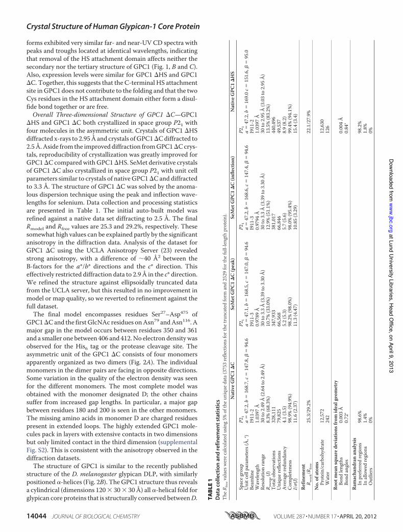
"HS and GPC1 "C both crystallized in space group P21 withfour molecules in the asymmetric unit. Crystals of GPC1 "HSdiffracted x-rays to 2.95Å and crystals ofGPC1"Cdiffracted to2.5Å.Aside from the improved diffraction fromGPC1"Ccrys-tals, reproducibility of crystallization was greatly improved forGPC1"C comparedwithGPC1"HS. SeMet derivative crystalsof GPC1 "C also crystallized in space group P21 with unit cellparameters similar to crystals of nativeGPC1"C and diffractedto 3.3 Å. The structure of GPC1 "C was solved by the anoma-lous dispersion technique using the peak and inflection wave-lengths for selenium. Data collection and processing statisticsare presented in Table 1. The initial auto-built model wasrefined against a native data set diffracting to 2.5 Å. The finalRmodel and Rfree values are 25.3 and 29.2%, respectively. Thesesomewhat high values can be explained partly by the significantanisotropy in the diffraction data. Analysis of the dataset forGPC1 "C using the UCLA Anisotropy Server (23) revealedstrong anisotropy, with a difference of !40 Å2 between theB-factors for the a*/b* directions and the c* direction. Thiseffectively restricted diffraction data to 2.9 Å in the c* direction.We refined the structure against ellipsoidally truncated datafrom the UCLA server, but this resulted in no improvement inmodel or map quality, so we reverted to refinement against thefull dataset.The final model encompasses residues Ser27–Asp475 of
GPC1"Cand the first GlcNAc residues onAsn79 andAsn116. Amajor gap in the model occurs between residues 350 and 361and a smaller one between 406 and 412.No electron densitywasobserved for the His6 tag or the protease cleavage site. Theasymmetric unit of the GPC1 "C consists of four monomersapparently organized as two dimers (Fig. 2A). The individualmonomers in the dimer pairs are facing in opposite directions.Some variation in the quality of the electron density was seenfor the different monomers. The most complete model wasobtained with the monomer designated D; the other chainssuffer from increased gap lengths. In particular, a major gapbetween residues 180 and 200 is seen in the other monomers.The missing amino acids in monomer D are charged residuespresent in extended loops. The highly extended GPC1 mole-cules pack in layers with extensive contacts in two dimensionsbut only limited contact in the third dimension (supplementalFig. S2). This is consistent with the anisotropy observed in thediffraction datasets.The structure of GPC1 is similar to the recently published
structure of the D. melanogaster glypican DLP, with similarlypositioned "-helices (Fig. 2B). The GPC1 structure thus revealsa cylindrical (dimensions 120$ 30$ 30 Å) all "-helical fold forglypican core proteins that is structurally conserved betweenD. TA
BLE
1D
ata
colle
ctio
nan
dre
finem
ents
tati
stic
sTh
eRfreevalues
were
calcu
lated
using5
%of
theu
niqu
edata(
3753
reflections
forthe
truncated
form
and2529
forthe
full-len
gthprotein
).Na
tiveG
PC1"
CSe
Met
GPC1
"C(peak)
SeMet
GPC1
"C(in
flecti
on)
Nativ
eGPC
1"HS
Spaceg
roup
P21
P21
P21
P21
Unitcellp
aram
eters(Å,
°)a&
47.2,
b&16
8.7,c
&14
7.8,!
&94
.6a&
47.1,
b&16
8.5,c
&14
7.0,!
&94
.6a&
47.2,
b&16
8.6,c
&14
7.4,!
&94
.6a&
47.2,
b&16
9.0c&
151.6
,!&
95.0
Beam
line
I911
-2I911
-3I911
-3I911
-2W
avele
ngth
1.039
7Å0.9
789Å
0.979
4Å1.0
397Å
Resolutio
nrang
e30
to2.4
9Å(2.64
to2.4
9Å)
30to
3.3Å(3.39
to3.3
0Å)
30to
3.3Å(3.39
to3.3
0Å)
30to
2.95Å
(3.03
to2.9
5Å)
R merge(I)
8.3%(68.3
%)10
.7%(33.0
%)12
.9%(51.1
%)13
.5%(83.2
%)To
talo
bservatio
ns32
8,111
347,9
3338
1,017
440,4
96Un
ique
refle
ctions
79,82
566
,568
66,84
649
,537
Averager
edun
danc
y4.1
(3.5)
5.2(5.3)
5.7(5.6)
8.9(8.2)
Completen
ess
98.9%
(94.9
%)98
.2%(98.0
%)98
.0%(95.4
%)99
.4%(94.1
%)I/$
(I)11
.6(2.37
)11
.3(4.47
)10
.85(3.29
)15
.4(3.4)
Refin
emen
tR w
ork/R
free
25.3/
29.2%
22.1/
27.9%
No.o
fatoms
Protein
/carbo
hydrate
12,57
212
,630
Water
242
126
Root
means
quared
eviat
ions
from
idealg
eometry
Bond
lengths
0.003
Å0.0
04Å
Bond
angle
s0.7
2°0.8
4°Ra
macha
ndrana
nalys
isIn
prefe
rredregio
ns98
.6%98
.2%In
allow
edregio
ns1.4
%1.8
%Outliers
0%0%
Crystal Structure of Human Glypican-1 Core Protein
14044 JOURNAL OF BIOLOGICAL CHEMISTRY VOLUME 287 • NUMBER 17 • APRIL 20, 2012
at Lund University Libraries, H
ead Office, on April 9, 2013
ww
w.jbc.org
Dow
nloaded from

melanogaster and humans, although GPC1 and DLP only share25% sequence identity (as determined by FASTA (40)). Theoverlap between GPC1 and DLP structures is shown as "-car-bon traces in Fig. 2B. The root mean square deviation in "-car-bon positions betweenmonomer D of GPC1"C and DLP is 2.6Å for 331 positionswhen superimposed by the secondary struc-ture matching algorithm in Coot (35). The "-helical fold is alsoconsistentwith the far-UVCD spectra in Fig. 1B, displaying onepeak at 195 nm and two troughs at 209 and 222 nm, which istypical for"-helical proteins. TheGPC1 structure consists of 14
"-helices and three major loops, termed "1–"14 and L1–L3(Fig. 2C). In comparison with the DLP structure, the GPC1structure extends structural knowledge of glypicans, especiallyfor the N-terminal loops (L1 and L3) and helix "1. The 60-res-idue, 83-Å-long helix "2 (71–130) spans the whole protein,with N-linked glycans near either end (on Asn79 and Asn116).Out of 14 evolutionarily conservedCys residues inGPC1, 12 arelocated in a Cys-rich region near the N terminus. This regionwas termed the N-lobe (18) based on the observation that itcontains the N terminus of the "-helix ("2 in GPC1) that spans
FIGURE 2. Overview of GPC1 "C structure and comparison with DLP. A, overview of the asymmetric unit of GPC1 "C with two dimer pairs. B, ribbonrepresentation of "-carbon alignment of GPC1 "C monomer (blue) and DLP (green, Protein Data Bank code 3ODN). Alignment was performed in PyMOL,version 1.2r3pre, Schrödinger, LLC. C, overview of the GPC1 "C structure represented as schematic in rainbow colors (N terminus in blue, C terminus in red) withloops 1–3 (L1–L3) and "-helices ("1–"14). Also indicated are the disulfide bonds (yellow solid lines), the first residues of the N-glycans (GlcNAc), and theassignment of different lobes in the GPC1 "C structure.
Crystal Structure of Human Glypican-1 Core Protein
APRIL 20, 2012 • VOLUME 287 • NUMBER 17 JOURNAL OF BIOLOGICAL CHEMISTRY 14045
at Lund University Libraries, H
ead Office, on April 9, 2013
ww
w.jbc.org
Dow
nloaded from

the entire protein. We propose that this region should insteadbe termed the Cys-rich lobe (Fig. 2C), as the lobe is not entirelycontainedwithin theN-terminal part of the sequence. TheCys-rich lobe is followed by a middle segment containing five heli-ces,"2,"6,"9,"12, and"14, forming the heart of the structure.This region is stabilized by two evolutionarily conserved hydro-phobic centers that connect the "-helices and also contain theC terminus of GPC1 "C. This lobe was termed the M-lobe inRef. 18. Based on the conserved hydrophobic centers in thislobe, we propose that this part of GPC1 should be called thecentral lobe. The last part of theGPC1 structure contains a longloop, L2, stretching from Cys343 to Glu376. This loop is pro-cessed by furin proteases in many of the glypican core proteins(41, 42); however, onlyminor processing of GPC1 occurs in ourhands. Because of the disorder of the loop, residues 350–361are not modeled. This lobe was termed the C-lobe in Ref. 18; analternative name could be the protease site lobe.Analysis of Crystal Contacts—The interface between pairs of
GPC1molecules in the crystal is located in the central lobe andinvolves "9 and "12 in both interacting monomers. Analysis ofthe crystal packing reveals at least two additional crystal con-tacts, one involving the end of "2 interacting with "14 inanothermonomer and the other involving theN-terminal partsof different monomers (supplemental Fig. 2). The interfaceinvolving"2 and"14 is the largest, with a buried surface area of!500 Å2. However, calculations of the stabilities of the inter-faces using PISA software (43) indicate that the interactions aretooweak to formoligomeric states of GPC1 in solution. Prelim-inary dynamic light scattering experiments confirm these cal-culations, with profiles exhibiting single peaks for both GPC1"HS and GPC1 "C (data not shown).Evolutionarily Conserved Cysteine Residues in Anchorless
GPC1—All of the 14 Cys residues evolutionarily conserved inthe glypican family are clearly defined in the electron densityand all are clearly involved in disulfide bonds. This gives a highdegree of confidence to the presented model. The disulfidebonds and their connections are presented in Table 2, and adetailed view of the model and electron densities for the disul-fide bonds is shown in Fig. 3. Six disulfide bonds connect thestructure in the Cys-rich lobe. Disulfides Cys268–Cys415 andCys272–Cys401 connect the structure between the furin-likeconvertase site, consistent with the two fragments that appearwhen running reducing SDS-PAGE on glypicans processed byfurin-like convertases (41, 42). Together with Cys246–Cys279,this forms three disulfide bonds in or closely situated to "8 inthe Cys-rich lobe (Fig. 3A). Cys32–Cys68, Cys62–Cys256, andCys69–Cys259 form three longitudinally placed disulfide bondsin theN-terminal part of theCys-rich lobe (Fig. 3B). All detailedpictures of the GPC1 structure are presented with the Cys-rich
lobe facing upwards. The remaining disulfide bond is located inthe protease lobe, being formed by Cys191–Cys343. The struc-ture of DLP (18) lacked three out of seven disulfide bonds,because the electron density was not clear in the Cys-richregion. However, the four disulfide bonds present in the DLPstructure are consistent with those presented here.GPC1 also contains two additional Cys residues in the HS
attachment region, shared with GPC4 and GPC6. These Cysresidues are not part of the presented GPC1 model and caneither be free or form a disulfide bond. Crystallization of GPC1"HS is facilitated by the addition of low concentrations ofreducing agent, suggesting that the Cys residues are not boundtogether.
TABLE 2Disulfide bonds in GPC1
Cysteine residues Connected structural elementsCys32–Cys68 "1-L1Cys62–Cys256 L1-"7Cys69–Cys259 L1-"7Cys191–Cys343 "5-"11Cys246–Cys279 "7-"8Cys268–Cys415 Turn between "7 and "8-L3Cys272–Cys401 "8-"13
FIGURE 3. Structure model and electron densities of the six disulfidebonds in the N-terminal Cys-rich lobe of GPC1. Red, oxygen; blue, nitrogen;green, carbon; yellow, sulfur. Model of the three disulfide bonds (Cys415–Cys268; Cys272–Cys401; Cys246–Cys279) in "8 in the Cys-rich region (A) and of thethree longitudinally placed disulfide bonds in the L1 loop (Cys32–Cys68;Cys69–Cys259, and Cys62–Cys256) (B). Electron densities, contoured at 2.0 $ in Aand 1.0 $ in B, were calculated as 2"Fo"'"Fc" maps using the program FFT inCCP4 (21, 55). Figures were generated using PyMOL.
Crystal Structure of Human Glypican-1 Core Protein
14046 JOURNAL OF BIOLOGICAL CHEMISTRY VOLUME 287 • NUMBER 17 • APRIL 20, 2012
at Lund University Libraries, H
ead Office, on April 9, 2013
ww
w.jbc.org
Dow
nloaded from
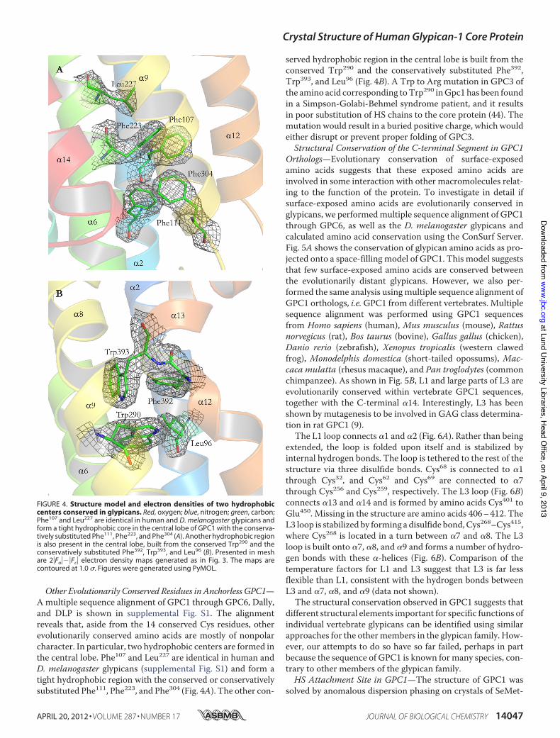
Other Evolutionarily Conserved Residues in Anchorless GPC1—Amultiple sequence alignment of GPC1 through GPC6, Dally,and DLP is shown in supplemental Fig. S1. The alignmentreveals that, aside from the 14 conserved Cys residues, otherevolutionarily conserved amino acids are mostly of nonpolarcharacter. In particular, two hydrophobic centers are formed inthe central lobe. Phe107 and Leu227 are identical in human andD. melanogaster glypicans (supplemental Fig. S1) and form atight hydrophobic region with the conserved or conservativelysubstituted Phe111, Phe223, and Phe304 (Fig. 4A). The other con-
served hydrophobic region in the central lobe is built from theconserved Trp290 and the conservatively substituted Phe392,Trp393, and Leu96 (Fig. 4B). A Trp to Arg mutation in GPC3 ofthe amino acid corresponding toTrp290 inGpc1 has been foundin a Simpson-Golabi-Behmel syndrome patient, and it resultsin poor substitution of HS chains to the core protein (44). Themutation would result in a buried positive charge, which wouldeither disrupt or prevent proper folding of GPC3.Structural Conservation of the C-terminal Segment in GPC1
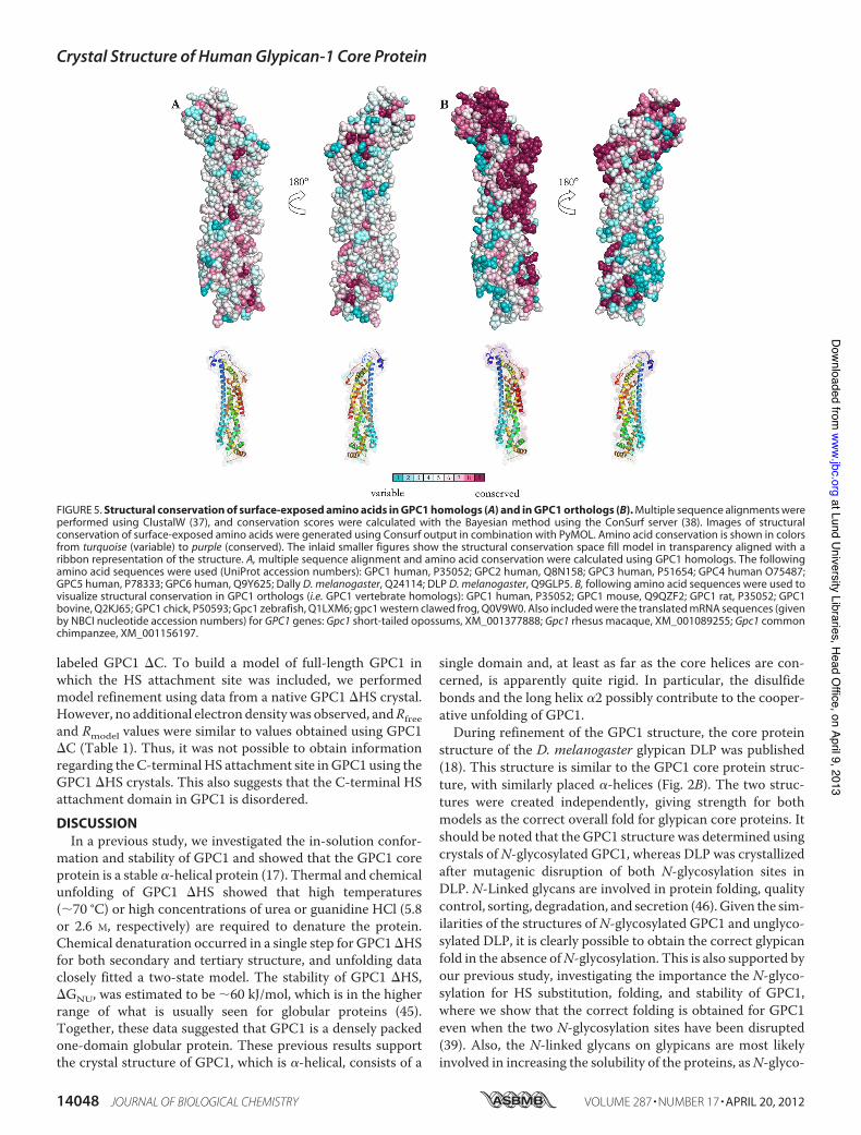
Orthologs—Evolutionary conservation of surface-exposedamino acids suggests that these exposed amino acids areinvolved in some interaction with other macromolecules relat-ing to the function of the protein. To investigate in detail ifsurface-exposed amino acids are evolutionarily conserved inglypicans, we performedmultiple sequence alignment of GPC1through GPC6, as well as the D. melanogaster glypicans andcalculated amino acid conservation using the ConSurf Server.Fig. 5A shows the conservation of glypican amino acids as pro-jected onto a space-filling model of GPC1. This model suggeststhat few surface-exposed amino acids are conserved betweenthe evolutionarily distant glypicans. However, we also per-formed the same analysis usingmultiple sequence alignment ofGPC1 orthologs, i.e.GPC1 from different vertebrates. Multiplesequence alignment was performed using GPC1 sequencesfrom Homo sapiens (human), Mus musculus (mouse), Rattusnorvegicus (rat), Bos taurus (bovine), Gallus gallus (chicken),Danio rerio (zebrafish), Xenopus tropicalis (western clawedfrog), Monodelphis domestica (short-tailed opossums), Mac-caca mulatta (rhesus macaque), and Pan troglodytes (commonchimpanzee). As shown in Fig. 5B, L1 and large parts of L3 areevolutionarily conserved within vertebrate GPC1 sequences,together with the C-terminal "14. Interestingly, L3 has beenshown by mutagenesis to be involved in GAG class determina-tion in rat GPC1 (9).The L1 loop connects "1 and "2 (Fig. 6A). Rather than being
extended, the loop is folded upon itself and is stabilized byinternal hydrogen bonds. The loop is tethered to the rest of thestructure via three disulfide bonds. Cys68 is connected to "1through Cys32, and Cys62 and Cys69 are connected to "7through Cys256 and Cys259, respectively. The L3 loop (Fig. 6B)connects "13 and "14 and is formed by amino acids Cys401 toGlu450. Missing in the structure are amino acids 406–412. TheL3 loop is stabilized by forming a disulfide bond,Cys268–Cys415,where Cys268 is located in a turn between "7 and "8. The L3loop is built onto "7, "8, and "9 and forms a number of hydro-gen bonds with these "-helices (Fig. 6B). Comparison of thetemperature factors for L1 and L3 suggest that L3 is far lessflexible than L1, consistent with the hydrogen bonds betweenL3 and "7, "8, and "9 (data not shown).
The structural conservation observed in GPC1 suggests thatdifferent structural elements important for specific functions ofindividual vertebrate glypicans can be identified using similarapproaches for the othermembers in the glypican family. How-ever, our attempts to do so have so far failed, perhaps in partbecause the sequence of GPC1 is known for many species, con-trary to other members of the glypican family.HS Attachment Site in GPC1—The structure of GPC1 was
solved by anomalous dispersion phasing on crystals of SeMet-
FIGURE 4. Structure model and electron densities of two hydrophobiccenters conserved in glypicans. Red, oxygen; blue, nitrogen; green, carbon;Phe107 and Leu227 are identical in human and D. melanogaster glypicans andform a tight hydrophobic core in the central lobe of GPC1 with the conserva-tively substituted Phe111, Phe223, and Phe304 (A). Another hydrophobic regionis also present in the central lobe, built from the conserved Trp290 and theconservatively substituted Phe392, Trp393, and Leu96 (B). Presented in meshare 2"Fo"'"Fc" electron density maps generated as in Fig. 3. The maps arecontoured at 1.0 $. Figures were generated using PyMOL.
Crystal Structure of Human Glypican-1 Core Protein
APRIL 20, 2012 • VOLUME 287 • NUMBER 17 JOURNAL OF BIOLOGICAL CHEMISTRY 14047
at Lund University Libraries, H
ead Office, on April 9, 2013
ww
w.jbc.org
Dow
nloaded from
labeled GPC1 "C. To build a model of full-length GPC1 inwhich the HS attachment site was included, we performedmodel refinement using data from a native GPC1 "HS crystal.However, no additional electron densitywas observed, andRfreeand Rmodel values were similar to values obtained using GPC1"C (Table 1). Thus, it was not possible to obtain informationregarding theC-terminalHS attachment site inGPC1 using theGPC1 "HS crystals. This also suggests that the C-terminal HSattachment domain in GPC1 is disordered.DISCUSSIONIn a previous study, we investigated the in-solution confor-
mation and stability of GPC1 and showed that the GPC1 coreprotein is a stable "-helical protein (17). Thermal and chemicalunfolding of GPC1 "HS showed that high temperatures(!70 °C) or high concentrations of urea or guanidine HCl (5.8or 2.6 M, respectively) are required to denature the protein.Chemical denaturation occurred in a single step for GPC1"HSfor both secondary and tertiary structure, and unfolding dataclosely fitted a two-state model. The stability of GPC1 "HS,"GNU, was estimated to be !60 kJ/mol, which is in the higherrange of what is usually seen for globular proteins (45).Together, these data suggested that GPC1 is a densely packedone-domain globular protein. These previous results supportthe crystal structure of GPC1, which is "-helical, consists of a
single domain and, at least as far as the core helices are con-cerned, is apparently quite rigid. In particular, the disulfidebonds and the long helix "2 possibly contribute to the cooper-ative unfolding of GPC1.During refinement of the GPC1 structure, the core protein
structure of the D. melanogaster glypican DLP was published(18). This structure is similar to the GPC1 core protein struc-ture, with similarly placed "-helices (Fig. 2B). The two struc-tures were created independently, giving strength for bothmodels as the correct overall fold for glypican core proteins. Itshould be noted that the GPC1 structure was determined usingcrystals ofN-glycosylated GPC1, whereas DLP was crystallizedafter mutagenic disruption of both N-glycosylation sites inDLP. N-Linked glycans are involved in protein folding, qualitycontrol, sorting, degradation, and secretion (46). Given the sim-ilarities of the structures ofN-glycosylated GPC1 and unglyco-sylated DLP, it is clearly possible to obtain the correct glypicanfold in the absence ofN-glycosylation. This is also supported byour previous study, investigating the importance the N-glyco-sylation for HS substitution, folding, and stability of GPC1,where we show that the correct folding is obtained for GPC1even when the two N-glycosylation sites have been disrupted(39). Also, the N-linked glycans on glypicans are most likelyinvolved in increasing the solubility of the proteins, asN-glyco-
FIGURE 5. Structural conservation of surface-exposed amino acids in GPC1 homologs (A) and in GPC1 orthologs (B). Multiple sequence alignments wereperformed using ClustalW (37), and conservation scores were calculated with the Bayesian method using the ConSurf server (38). Images of structuralconservation of surface-exposed amino acids were generated using Consurf output in combination with PyMOL. Amino acid conservation is shown in colorsfrom turquoise (variable) to purple (conserved). The inlaid smaller figures show the structural conservation space fill model in transparency aligned with aribbon representation of the structure. A, multiple sequence alignment and amino acid conservation were calculated using GPC1 homologs. The followingamino acid sequences were used (UniProt accession numbers): GPC1 human, P35052; GPC2 human, Q8N158; GPC3 human, P51654; GPC4 human O75487;GPC5 human, P78333; GPC6 human, Q9Y625; Dally D. melanogaster, Q24114; DLP D. melanogaster, Q9GLP5. B, following amino acid sequences were used tovisualize structural conservation in GPC1 orthologs (i.e. GPC1 vertebrate homologs): GPC1 human, P35052; GPC1 mouse, Q9QZF2; GPC1 rat, P35052; GPC1bovine, Q2KJ65; GPC1 chick, P50593; Gpc1 zebrafish, Q1LXM6; gpc1 western clawed frog, Q0V9W0. Also included were the translated mRNA sequences (givenby NBCI nucleotide accession numbers) for GPC1 genes: Gpc1 short-tailed opossums, XM_001377888; Gpc1 rhesus macaque, XM_001089255; Gpc1 commonchimpanzee, XM_001156197.
Crystal Structure of Human Glypican-1 Core Protein
14048 JOURNAL OF BIOLOGICAL CHEMISTRY VOLUME 287 • NUMBER 17 • APRIL 20, 2012
at Lund University Libraries, H
ead Office, on April 9, 2013
ww
w.jbc.org
Dow
nloaded from

sylated GPC1 was crystallized at a protein concentration of 20mg/ml in comparison with nonglycosylated DLP at 3.5 mg/ml.The core proteins of proteoglycans have been regarded as
inert, with all function of the PGs related to the GAG chains.This view arose from a wealth of genetic experiments implicat-ing GAGs as regulators of morphogenesis and because GAGsare fairly easy to label and purify. The PG core proteins, how-ever, are often large, heavily glycosylated, and difficult toexpress in bacteria and have therefore been more difficult tostudy. Moreover, although GPC1 is a fairly large protein withlongest dimension around 120 Å, this is rather small in com-parison with the HS chains that become attached to glypicans.An HS chain can consist of up to 50–100 repeating disaccha-
ride units of GlcNAc and GlcA. From the solution structure ofHS, the length of an HS chain consisting of eight disaccharideunits has been estimated to be around 70 Å (47). Thus, a full HSchain can be !500 Å long, as compared with the 100 Å for thefolded glypican core protein. However, in recent years manyinteractions with PG core proteins have been found. For exam-ple, the ectodomains of syndecan core proteins have beenshown to interact directly with integrins (48, 49). Glypican coreproteins have been suggested to have functional roles, by inter-acting with a number of growth factors, including hedgehog,bone morphogenetic protein 4, decapentaplegic, insulin-likegrowth factor II, and Wnt. Recently, a region was identified inDLP as being important for hedgehog signaling, althoughhedgehog itself does not seem to bind and the interaction part-ner for this region remains elusive (18). This region is localizedto "4 and the first part of "5 (in our numbering "5 and "6,because the first "-helix is missing in the DLP structure). Thisregion contains a number of amino acids conserved in GPC1through GPC6 as well as Dally and Dlp that are involved in thefolding of the protein. Among the amino acids in this region istheGPC1Cys191–Cys343 disulfide bond. The structural similar-ity in this region is confirmed by our GPC1 "C structure, withtwo "-helices connected by a turn. The possibility arises thatthe interaction is governed by charged residues in this regionand thatmutagenesis of GPC1 tomimic the DLP region impor-tant for hedgehog signaling may confer hedgehog signalingeffects on GPC1 core protein.Amino acids in protein loops display higher sequence varia-
tion than residues that form the core of a structure, unless theloops are involved in the function of the protein. Also, proteinloops can adopt a more unique folding than the common pro-tein secondary structure elements "-helix and !-sheet. This isperhaps best exemplified by the hypervariable loops on anti-bodies, which are responsible for the unique binding of anti-bodies to their antigens. By our analysis of GPC1 vertebrateorthologs, the L1 and L3 loops of GPC1 are evolutionarily con-served, suggesting that they are involved in the function ofGPC1. No interaction partner has been found for GPC1 coreprotein, contrary to some of the other glypican core proteins.GPC1 knock-out mice are viable and fertile but have reducedbrain size at birth (6). This has been suggested to be an effect ofa transient reduction in HS-dependent fibroblast growth factorsignaling during embryogenesis.Moreover, heparan sulfate andchondroitin sulfate proteoglycans (HSPGs and CSPGs, respec-tively) serve different functions during brain development, withCSPGs inhibiting and HSPGs promoting axon extension (50).GPC1 is exclusively substituted with HS, whereas GPC5, forexample, can exist as an HS CS/DS (where DS is dermatansulfate) hybrid. However, expression of the GPC1 GAG attach-ment domain alone results in addition of!90%CS (9), stronglysuggesting that factors in the GPC1 core protein further awayfrom the GAG attachment sites promote the assembly of HS.Mutagenesis of amino acids in the L3 loop resulted in increasedproportion of CS substituted on GPC1, but mutagenesis of theentire loop resulted in no protein expression (9). The structureof the L3 loop presented here should facilitate further muta-genic studies. It should also be noted that the informationregarding HS assembly enzymes has increased during recent
FIGURE 6. Structure of evolutionarily conserved L1 and L3. A, L1 (blue) withstart and end marked by arrows is folded upon itself with hydrogen bonds(dotted yellow lines) and is tethered to the rest of the structure by severaldisulfide bonds (yellow solid lines). B, L3 (orange and red) with start and endmarked by arrows is connected to "7, "8, and "9 with several hydrogenbonds (dotted yellow lines).
Crystal Structure of Human Glypican-1 Core Protein
APRIL 20, 2012 • VOLUME 287 • NUMBER 17 JOURNAL OF BIOLOGICAL CHEMISTRY 14049
at Lund University Libraries, H
ead Office, on April 9, 2013
ww
w.jbc.org
Dow
nloaded from
years. HS polymerization and initiation are performed by theexostosin and exostosin-like (EXTL) families, respectively.EXT1 and EXT2 function as an HS co-polymerase and werefirst described as tumor suppressor genes, as theywere found tobe the cause of hereditary multiple exostoses, a disease charac-terized by the formation of multiple cartilaginous tumors (51,52). The EXTL family consists of three members, EXTL1–3,where EXTL2 and EXTL3 harbor HS initiating "1,4-GlcNActransferase activity and are ubiquitously expressed in thehuman body (53). Only EXTL3 has aD. melanogaster ortholog,suggesting that this is the functional HS initiation enzyme,whereas structural data are available for EXTL2, the smallestmember in the EXTL family. Given the requirement for theglypican core protein to ensure exclusive HS attachment onGPC1, it is reasonable to suggest that EXTL2 or EXTL3 interactnot only with the HS attachment site but also with the coreprotein. Mutagenesis of the L3 loop suggests that this is theinteracting surface for EXTL and glypicans. It is also possiblethat disruption of the L3 loop results in displacement of the HSattachment site. An alternative explanation could also be thatthe glypican core protein provides steric hindrance for the CSinitiation enzyme, chondroitin N-acetylgalactosaminyltrans-ferase (54), to ensure exclusive HS attachment. Unfortunately,no structural information is available for the CS initiatingenzymes. The L1 loop is also evolutionarily conserved as shownby our analysis of structural conservation of GPC1 vertebrateorthologs, but thus far, no function has been suggested for thisloop.The glypican core proteins have long been assumed to adopt
a globular fold, but the structures of GPC1 and DLP are cylin-drical in shape. TheC-terminal"14 is placed on the long side ofthe protein, making it difficult to predict how the core proteinsare situated in the membranes. The first Ser residue in the HSattachment site is located 10 amino acids from the C terminusof the GPC1 "C structure, placing the HS attachment close tothe folded core protein. We have previously shown that the HSchains onGPC1 do not affect its secondary or tertiary structure(17). In fact, results obtained by differential scanning calorim-etry suggest that the HS chains cause the core protein mono-mers to repel one another, possibly by electrostatic repulsion ofthe negatively charged HS chains. Others have reported thatglypican core protein bindsweakly to heparin-agarose (18). TheC-terminal Ser residue responsible for GPI attachment islocated 50 amino acids downstream of the visible ends of themodels of GPC1 and DLP, which is long enough that the Cterminus of the protein could be placed in a wide variety oforientations with respect to the core protein. Thus, when theGPI anchor is anchored in the membrane, it may allow greatfreedom of orientation of GPC1 and DLP with respect to themembrane surface. Our attempts to locate the GPC1 HSattachment domain and C terminus using crystallography havefailed, which together with the presented in-solution data onthe GPC1 conformation indicate that the HS attachment site isa disordered tail in the protein. Experimental approaches otherthan x-ray crystallography may be needed to finally answer theimportant question of the placement of the C-terminal part inglypicans, how the HS chains are extended from the core pro-tein, and how glypicans are situated in the membranes. More-
over, the presented GPC1 structure provides new informationthat will be important in determining the mechanism behindexclusive HS attachment on glypicans.
Acknowledgments—We thank the beamline staff atMAX-lab for helpduring data collection and Dr. Mattias Hansson at the MAX-labCrystallization Facility for help with setting up high throughput crys-tallization plates. The technical assistance of Sol Da Rocha is greatlyappreciated. We are also grateful to Axel Hyrenius Wittsten for helpwith CD.
REFERENCES1. Bülow, H. E., and Hobert, O. (2006) Themolecular diversity of glycosami-
noglycans shapes animal development. Annu. Rev. Cell Dev. Biol. 22,375–407
2. Häcker, U., Nybakken, K., and Perrimon, N. (2005) Heparan sulfate pro-teoglycans. The sweet side of development. Nat. Rev. Mol. Cell Biol. 6,530–541
3. Pilia, G., Hughes-Benzie, R. M., MacKenzie, A., Baybayan, P., Chen, E. Y.,Huber, R., Neri, G., Cao, A., Forabosco, A., and Schlessinger, D. (1996)Mutations in GPC3, a glypican gene, cause the Simpson-Golabi-Behmelovergrowth syndrome. Nat. Genet. 12, 241–247
4. Campos-Xavier, A. B.,Martinet, D., Bateman, J., Belluoccio, D., Rowley, L.,Tan, T. Y., Baxová, A., Gustavson, K. H., Borochowitz, Z. U., Innes, A. M.,Unger, S., Beckmann, J. S., Mittaz, L., Ballhausen, D., Superti-Furga, A.,Savarirayan, R., and Bonafé, L. (2009) Mutations in the heparan-sulfateproteoglycan glypican 6 (GPC6) impair endochondral ossification andcause recessive omodysplasia. Am. J. Hum. Genet. 84, 760–770
5. Okamoto, K., Tokunaga, K., Doi, K., Fujita, T., Suzuki, H., Katoh, T., Wa-tanabe, T., Nishida, N., Mabuchi, A., Takahashi, A., Kubo, M., Maeda, S.,Nakamura, Y., and Noiri, E. (2011) Common variation in GPC5 is associ-ated with acquired nephrotic syndrome. Nat. Genet. 43, 459–463
6. Jen, Y.H.,Musacchio,M., and Lander, A. (2009)Glypican-1 controls brainsize through regulation of fibroblast growth factor signaling in early neu-rogenesis. Neural Dev. 4, 33
7. Zhang, L., and Esko, J. D. (1994) Amino acid determinants that driveheparan sulfate assembly in a proteoglycan. J. Biol. Chem. 269,19295–19299
8. Zhang, L., David, G., and Esko, J. D. (1995) Repetitive Ser-Gly sequencesenhance heparan sulfate assembly in proteoglycans. J. Biol. Chem. 270,27127–27135
9. Chen, R. L., and Lander, A. D. (2001)Mechanisms underlying preferentialassembly of heparan sulfate on glypican-1. J. Biol. Chem. 276, 7507–7517
10. Williams, E. H., Pappano,W. N., Saunders, A. M., Kim, M. S., Leahy, D. J.,and Beachy, P. A. (2010) Dally-like core protein and its mammalian ho-mologs mediate stimulatory and inhibitory effects on Hedgehog signalresponse. Proc. Natl. Acad. Sci. U.S.A. 107, 5869–5874
11. Yan,D.,Wu, Y., Yang, Y., Belenkaya, T. Y., Tang, X., and Lin, X. (2010) Thecell-surface proteins Dally-like and Ihog differentially regulate Hedgehogsignaling strength and range during development. Development 137,2033–2044
12. Capurro, M. I., Xu, P., Shi, W., Li, F., Jia, A., and Filmus, J. (2008) Glypi-can-3 inhibits Hedgehog signaling during development by competingwith patched for Hedgehog binding. Dev. Cell 14, 700–711
13. Kirkpatrick, C. A., Knox, S. M., Staatz, W. D., Fox, B., Lercher, D. M., andSelleck, S. B. (2006)The function of aDrosophila glypican does not dependentirely on heparan sulfate modification. Dev. Biol. 300, 570–582
14. Capurro, M. I., Xiang, Y. Y., Lobe, C., and Filmus, J. (2005) Glypican-3promotes the growth of hepatocellular carcinoma by stimulating canoni-cal Wnt signaling. Cancer Res. 65, 6245–6254
15. Ohkawara, B., Yamamoto, T. S., Tada, M., and Ueno, N. (2003) Role ofglypican 4 in the regulation of convergent extension movements duringgastrulation in Xenopus laevis. Development 130, 2129–2138
16. Cheng, W., Tseng, C. J., Lin, T. T., Cheng, I., Pan, H. W., Hsu, H. C., andLee, Y. M. (2008) Glypican-3-mediated oncogenesis involves the insulin-
Crystal Structure of Human Glypican-1 Core Protein
14050 JOURNAL OF BIOLOGICAL CHEMISTRY VOLUME 287 • NUMBER 17 • APRIL 20, 2012
at Lund University Libraries, H
ead Office, on April 9, 2013
ww
w.jbc.org
Dow
nloaded from

like growth factor-signaling pathway. Carcinogenesis 29, 1319–132617. Svensson, G., Linse, S., andMani, K. (2009) Chemical and thermal unfold-
ing of glypican-1. Protective effect of heparan sulfate against heat-inducedirreversible aggregation. Biochemistry 48, 9994–10004
18. Kim,M. S., Saunders, A.M., Hamaoka, B. Y., Beachy, P. A., and Leahy, D. J.(2011) Structure of the protein core of the glypican Dally-like and local-ization of a region important for hedgehog signaling. Proc. Natl. Acad. Sci.U.S.A. 108, 13112–13117
19. Wu, H., Lustbader, J. W., Liu, Y., Canfield, R. E., and Hendrickson, W. A.(1994) Structure of human chorionic gonadotropin at 2.6 Å resolutionfromMAD analysis of the selenomethionyl protein. Structure 2, 545–558
20. Kabsch, W. (2010) Integration, scaling, space-group assignment, andpost-refinement. Acta Crystallogr. D Biol. Crystallogr. 66, 133–144
21. Collaborative Computational Project No. 4 (1994) The CCP4 suite. Pro-grams for protein crystallography.Acta Crystallogr D Biol. Crystallogr. 50,760–763
22. Adams, P.D., Afonine, P. V., Bunkóczi, G., Chen,V. B., Davis, I.W., Echols,N., Headd, J. J., Hung, L. W., Kapral, G. J., Grosse-Kunstleve, R. W., Mc-Coy, A. J., Moriarty, N. W., Oeffner, R., Read, R. J., Richardson, D. C.,Richardson, J. S., Terwilliger, T. C., and Zwart, P. H. (2010) PHENIX. Acomprehensive Python-based system for macromolecular structure solu-tion. Acta Crystallogr. D Biol. Crystallogr. 66, 213–221
23. Strong, M., Sawaya, M. R., Wang, S., Phillips, M., Cascio, D., and Eisen-berg, D. (2006) Toward the structural genomics of complexes. Crystalstructure of a PE-PPE protein complex fromMycobacterium tuberculosis.Proc. Natl. Acad. Sci. U.S.A. 103, 8060–8065
24. Panjikar, S., Parthasarathy, V., Lamzin, V. S., Weiss, M. S., and Tucker,P. A. (2005) Auto-rickshaw. An automated crystal structure determina-tion platform as an efficient tool for the validation of an x-ray diffractionexperiment. Acta Crystallogr. D Biol. Crystallogr. 61, 449–457
25. Panjikar, S., Parthasarathy, V., Lamzin, V. S., Weiss, M. S., and Tucker,P. A. (2009) On the combination of molecular replacement and single-wavelength anomalous diffraction phasing for automated structure deter-mination. Acta Crystallogr. D Biol. Crystallogr. 65, 1089–1097
26. Howell, P. L., and Smith, G. D. (1992) Identification of heavy atom deriv-atives by normal probability methods. J. Appl. Crystallogr. 25, 81–86
27. Schneider, T. R., and Sheldrick, G. M. (2002) Substructure solution withSHELXD. Acta Crystallogr. D Biol. Crystallogr. 58, 1772–1779
28. de La Fortelle, E., Bricogne, G., and Carter, C. W., Jr. (1997) Maximum-likelihood heavy-atom parameter refinement for multiple isomorphousreplacement andmultiwavelength anomalous diffractionmethods.Meth-ods Enzymol. 276, 472–494
29. Cowtan, K. (1999) Error estimation and bias correction in phase-improve-ment calculations. Acta Crystallogr. D Biol. Crystallogr. 55, 1555–1567
30. Terwilliger, T. C. (2000) Maximum-likelihood density modification. ActaCrystallogr. D Biol. Crystallogr. 56, 965–972
31. Morris, R. J., Zwart, P. H., Cohen, S., Fernandez, F. J., Kakaris,M., Kirillova,O., Vonrhein, C., Perrakis, A., and Lamzin, V. S. (2004) Breaking goodresolutions with ARP/wARP. J. Synchrotron Radiat. 11, 56–59
32. Cowtan, K. (2006) The Buccaneer software for automatedmodel building.1. Tracing protein chains. Acta Crystallogr. D Biol. Crystallogr. 62,1002–1011
33. Brunger, A. T. (2007) Version 1.2 of theCrystallography andNMR system.Nat. Protoc. 2, 2728–2733
34. Murshudov, G. N., Vagin, A. A., and Dodson, E. J. (1997) Refinement ofmacromolecular structures by the maximum-likelihood method. ActaCrystallogr. D Biol. Crystallogr. 53, 240–255
35. Emsley, P., and Cowtan, K. (2004) Coot. Model-building tools for molec-ular graphics. Acta Crystallogr. D Biol. Crystallogr. 60, 2126–2132
36. Chen, V. B., Arendall, W. B., 3rd, Headd, J. J., Keedy, D. A., Immormino,R.M., Kapral, G. J., Murray, L.W., Richardson, J. S., and Richardson, D. C.(2010) MolProbity. All-atom structure validation for macromolecularcrystallography. Acta Crystallogr. D Biol. Crystallogr. 66, 12–21
37. Thompson, J. D., Higgins, D. G., and Gibson, T. J. (1994) ClustalW. Im-
proving the sensitivity of progressive multiple sequence alignmentthrough sequence weighting, position-specific gap penalties, and weightmatrix choice. Nucleic Acids Res. 22, 4673–4680
38. Ashkenazy, H., Erez, E., Martz, E., Pupko, T., and Ben-Tal, N. (2010) Con-Surf 2010. Calculating evolutionary conservation in sequence and struc-ture of proteins and nucleic acids. Nucleic Acids Res. 38,W529–W533
39. Svensson, G., Hyrenius Wittsten, A., Linse, S., and Mani, K. (2011) Thestructural role ofN-linked glycans on human glypican-1. Biochemistry 50,9377–9387
40. Pearson, W. R., and Lipman, D. J. (1988) Improved tools for biologicalsequence comparison. Proc. Natl. Acad. Sci. U.S.A. 85, 2444–2448
41. De Cat, B., Muyldermans, S. Y., Coomans, C., Degeest, G., Vander-schueren, B., Creemers, J., Biemar, F., Peers, B., and David, G. (2003) Proc-essing by proprotein convertases is required for glypican-3 modulation ofcell survival, Wnt signaling, and gastrulationmovements. J. Cell Biol. 163,625–635
42. Capurro, M. I., Shi, W., Sandal, S., and Filmus, J. (2005) Processing byconvertases is not required for glypican-3-induced stimulation of hepato-cellular carcinoma growth. J. Biol. Chem. 280, 41201–41206
43. Krissinel, E., and Henrick, K. (2007) Inference of macromolecular assem-blies from crystalline state. J. Mol. Biol. 372, 774–797
44. Veugelers, M., Cat, B. D., Muyldermans, S. Y., Reekmans, G., Delande, N.,Frints, S., Legius, E., Fryns, J. P., Schrander-Stumpel, C., Weidle, B.,Magdalena, N., and David, G. (2000) Mutational analysis of the GPC3/GPC4 glypican gene cluster on Xq26 in patients with Simpson-Golabi-Behmel syndrome. Identification of loss-of-function mutations in theGPC3 gene. Hum. Mol. Genet. 9, 1321–1328
45. Makhatadze, G. I., and Privalov, P. L. (1995) Energetics of protein struc-ture. Adv. Protein Chem. 47, 307–425
46. Helenius, A., and Aebi, M. (2004) Roles of N-linked glycans in the endo-plasmic reticulum. Annu. Rev. Biochem. 73, 1019–1049
47. Khan, S., Rodriguez, E., Patel, R., Gor, J.,Mulloy, B., and Perkins, S. J. (2011)The solution structure of heparan sulfate differs from that of heparin:implications for function. J. Biol. Chem. 286, 24842–24854
48. Whiteford, J. R., and Couchman, J. R. (2006) A conserved NXIP motif isrequired for cell adhesion properties of the syndecan-4 ectodomain.J. Biol. Chem. 281, 32156–32163
49. Beauvais, D. M., Burbach, B. J., and Rapraeger, A. C. (2004) The synde-can-1 ectodomain regulates "v!3 integrin activity in human mammarycarcinoma cells. J. Cell Biol. 167, 171–181
50. Matsumoto, Y., Irie, F., Inatani, M., Tessier-Lavigne, M., and Yamaguchi,Y. (2007) Netrin-1/DCC signaling in commissural axon guidance requirescell-autonomous expression of heparan sulfate. J. Neurosci. 27,4342–4350
51. Stickens, D., Clines, G., Burbee, D., Ramos, P., Thomas, S., Hogue, D.,Hecht, J. T., Lovett, M., and Evans, G. A. (1996) The EXT2 multiple exo-stoses gene defines a family of putative tumor suppressor genes. Nat.Genet. 14, 25–32
52. Lind, T., Tufaro, F., McCormick, C., Lindahl, U., and Lidholt, K. (1998)The putative tumor suppressors EXT1 and EXT2 are glycosyltransferasesrequired for the biosynthesis of heparan sulfate. J. Biol. Chem. 273,26265–26268
53. Kitagawa,H., Shimakawa,H., and Sugahara, K. (1999) The tumor suppres-sor EXT-like gene EXTL2 encodes an "1,4-N-acetylhexosaminyltrans-ferase that transfers N-acetylgalactosamine and N-acetylglucosamine tothe common glycosaminoglycan-protein linkage region. The key enzymefor the chain initiation of heparan sulfate. J. Biol. Chem. 274, 13933–13937
54. Uyama, T., Kitagawa, H., Tamura, Ji. J., and Sugahara, K. (2002)Molecularcloning and expression of human chondroitin N-acetylgalactosaminyl-transferase. The key enzyme for chain initiation and elongation of chon-droitin/dermatan sulfate on the protein linkage region tetrasaccharideshared by heparin/heparan sulfate. J. Biol. Chem. 277, 8841–8846
55. Read, R. J., and Schierbeek, A. J. (1988) A phased translation function.J. Appl. Crystallogr. 21, 490–495
Crystal Structure of Human Glypican-1 Core Protein
APRIL 20, 2012 • VOLUME 287 • NUMBER 17 JOURNAL OF BIOLOGICAL CHEMISTRY 14051
at Lund University Libraries, H
ead Office, on April 9, 2013
ww
w.jbc.org
Dow
nloaded from
GPC1_HUMAN SFVQGLGVASDVVRKVAQVP---LGPECSRAVMKLVYCAHCLGVPG-----ARPCPDYCR 273 GPC2_HUMAN AFVQGLETGRNVVSEALKVP---VSEGCSQALMRLIGCPLCRGVPS-----LMPCQGFCL 277 GPC3_HUMAN IFLQALNLGIEVINTTDHLK---FSKDCGRMLTRMWYCSYCQGLMM-----VKPCGGYCN 279 GPC4_HUMAN TFAQGLAVAGDVVSKVSVVN---PTAQCTHALLKMIYCSHCRGLVT-----VKPCYNYCS 271 GPC5_HUMAN TFLQALNLGIEVINTTDYLH---FSKECSRALLKMQYCPHCQGLAL-----TKPCMGYCL 275 GPC6_HUMAN TFVQGLTVGREVANRVSKVS---PTPGCIRALMKMLYCPYCRGLPT-----VRPCNNYCL 271 DALY_DROME VFMNALLQAAEVLSEADALYGEQLTDTCKLHLLKMHYCPNCNGHHSSSRSETKLCYGYCK 334 DLP_DROME TYGQALTTASEVAKKVLNVR---LNADCTGALTKMQHCGACKGYT------EKPCTNYCV 322 : :.* . :* . : * : :: * * * * .:* GPC1_HUMAN NVLKGC-LANQADLDAEWRNLLDSMV-LITDKFWGTSGVESVIGSVHTWLAEAINALQDN 331 GPC2_HUMAN NVVRGC-LSSRG-LEPDWGNYLDGLL-ILADKLQGPFSFELTAESIGVKISEGLMYLQEN 334 GPC3_HUMAN VVMQGC-MAGVVEIDKYWREYILSLE-ELVNGMYRIYDMENVLLGLFSTIHDSIQYVQKN 337 GPC4_HUMAN NIMRGC-LANQGDLDFEWNNFIDAML-MVAERLEGPFNIESVMDPIDVKISDAIMNMQDN 329 GPC5_HUMAN NVMRGC-LAHMAELNPHWHAYIRSLE-ELSDAMHGTYDIGHVLLNFHLLVNDAVLQAHLN 333 GPC6_HUMAN NVMKGC-LANQADLDTEWNLFIDAML-LVAERLEGPFNIESVMDPIDVKISEAIMNMQEN 329 DALY_DROME NVMRGCSAEYAGLLDSPWSGVVDSLNNLVTTHILSDTGIINVIKHLQTYFSEAIMAAMHN 394 DLP_DROME NVIKGC-LHYQHEFDSEWENFAMAMD-KVAERLLGSFNIVMVVEPLNIKISEAIMNFQDS 380 :::** :: * .: : : .. . . . :.: . GPC1_HUMAN RDTLTAKVIQGCGNPK----VNPQGPGP-------------------------------- 355 GPC2_HUMAN SAKVSAQVFQECGPPDPVPARNRRAPPPR------------------------------- 363 GPC3_HUMAN AGKLTTTIGKLCAHSQQRQYRSAYYPEDL------------------------------- 366 GPC4_HUMAN SVQVSQKVFQGCGPPKPLPAGRISRSISE------------------------------- 358 GPC5_HUMAN GQKLLEQVNRICGRPVRTPTQSPRCSFDQ------------------------------- 362 GPC6_HUMAN SMQVSAKVFQGCGQPKPAPALRSARSAPE------------------------------- 358 DALY_DROME GPELEKKVKKTCGTPSLTPYSSGEPDARP------------------------------- 423 DLP_DROME GQDITNRVFQGCGRPKLKKMKRSISPKLQGVQILNARSPVEADTLDIDETLDEAIVLRER 440 : : : *. . GPC1_HUMAN -------------------------------------------------EEKRRRGKLAP 366 GPC2_HUMAN ------------------------------------------------EEAGRLWSMVTE 375 GPC3_HUMAN ------------------------------------------------------FIDKKV 372 GPC4_HUMAN ------------------------------------------------SAFSARFRPHHP 370 GPC5_HUMAN ------------------------------------------------------SKEKHG 368 GPC6_HUMAN ------------------------------------------------N-FNTRFRPYNP 369 DALY_DROME -----------------------------------------------------------P 424 DLP_DROME RAAEPGSQETSSQQSQEQGVGKSGNGGGGGGGNNRRQQQRRKQQQQRRKQQNNRDDNDDD 500 GPC1_HUMAN RERPPSGT---LEKLVSEAKAQLRDVQDFWISLPGTLCS-EKMALSTASDDR-CWNGMAR 421 GPC2_HUMAN EERPTTAAGTNLHRLVWELRERLARMRGFWARLSLTVCGDSRMAADASLEAAPCWTGAGR 435 GPC3_HUMAN LKVAHVEHEETLSSRRRELIQKLKSFISFYSALPGYICSHSPVAE----NDTLCWNGQEL 428 GPC4_HUMAN EERPTTAAGTSLDRLVTDVKEKLKQAKKFWSSLPSNVCNDERMAAGNGNEDD-CWNGKGK 429 GPC5_HUMAN MKTTTRNSEETLANRRKEFINSLRLYRSFYGGLADQLCANELAAA----DGLPCWNGEDI 424 GPC6_HUMAN EERPTTAAGTSLDRLVTDIKEKLKLSKKVWSALPYTICKDESVTAGTSNEEE-CWNGHSK 428 DALY_DROME PHKNNVKWATDPDPGMVLFLSTIDKSKEFYTTIVDNFCDEQQHSR----DDHSCWSGDRF 480 DLP_DROME DNESGGGREPILDRIVRDIRQRVKDYKKFWSNLPHSVCSNEDIAS-SSDVDGMCWNGHTI 559 . : .: : .* . : **.*
GPC1_HUMAN ------------------------MELRARGWWLLCAAAALVACARGDPASK-------- 28 GPC2_HUMAN ------------------------MSALRPLLLLLLPLCPGPGPGPGSEAKV-------- 28 GPC3_HUMAN -----------------------MAGTVRTACLVVAMLLSLDFPGQAQPPPPPP------ 31 GPC4_HUMAN -------------------------MARFGLPALLCTLAVLSAALLAAELK--------- 26 GPC5_HUMAN -----------------------MDAQTWPVGFRCLLLLALVGSARSEG----------- 26 GPC6_HUMAN -------------------------MPSWIGAVILPLLGLLLSLPAGADVK--------- 26 DALY_DROME ---------------------MAARSVRLAQLLLFTLLCGFVGLSAAKHLDLDGIHHHQH 39 DLP_DROME MLHQEQQQQHLHCRRKVTATTTARLVIFSSPLLLLLLTTHLPSTLQADNSPAPQVAALAA 60 . GPC1_HUMAN ----------------SRSCGEVR-QIYGAKGFSLSD-VPQAEISGEHLRICPQG--YTC 68 GPC2_HUMAN ----------------TRSCAETR-QVLGARGYSLNL-IPPALISGEHLRVCPQE--YTC 68 GPC3_HUMAN ----------------DATCHQVR-SFFQR-LQPGLKWVPETPVPGSDLQVCLPKG-PTC 72 GPC4_HUMAN ----------------SKSCSEVR-RLYVSKGFNKND-APLHEINGDHLKICPQG--STC 66 GPC5_HUMAN ----------------VQTCEEVR-KLFQWRLLGAVRGLPDSPRAGPDLQVCISKK-PTC 68 GPC6_HUMAN ----------------ARSCGEVR-QAYGAKGFSLAD-IPYQEIAGEHLRICPQE--YTC 66 DALY_DROME HLHSATTHHRRRLQRDSRAKDAVGGSTHQCDAVKSYFESIDIKSSGTYSEKGAICG-GNC 98 DLP_DROME PNPAGGVAGSSIIDQFSPNCSAVT-HIFQARGIDAIE-IPQKPSNERVLRYCESPSVGTC 118 . . .* GPC1_HUMAN CTSEMEENLANRSHAELETALRDSSRVLQAMLATQLRSFDDHFQHLLNDSERTLQATFPG 128 GPC2_HUMAN CSSETEQRLIRETEATFRGLVEDSGSFLVHTLAARHRKFDEFFLEMLSVAQHSLTQLFSH 128 GPC3_HUMAN CSRKMEEKYQLTARLNMEQLLQSASMELKFLIIQNAAVFQEAFEIVVRHAKNYTNAMFKN 132 GPC4_HUMAN CSQEMEEKYSLQSKDDFKSVVSEQCNHLQAVFASRYKKFDEFFKELLENAEKSLNDMFVK 126 GPC5_HUMAN CTRKMEERYQIAARQDMQQFLQTSSSTLKFLISRNAAAFQETLETLIKQAENYTSILFCS 128 GPC6_HUMAN CTTEMEDKLSQQSKLEFENLVEETSHFVRTTFVSRHKKFDEFFRELLENAEKSLNDMFVR 126 DALY_DROME CNNATELELRDKAAGMFEQLLHHHTSSLRGVLETNAKQFQSHVLELAQISENMTHSLFSK 158 DLP_DROME CTYNMETRMAMQSRQQLEGHTKDQISRMSGILGSKATKFKDIFTALLKESRTQFNSMFIR 178 *. * . : :. : : . *.. . : :. * GPC1_HUMAN AFGELYTQNARAFRDLYSELRLYYRGAN------------------LHLEETLAEFWARL 170 GPC2_HUMAN SYGRLYAQHALIFNGLFSRLRDFYGESG------------------EGLDDTLADFWAQL 170 GPC3_HUMAN NYPSLTPQAFEFVGEFFTDVSLYILGSD------------------INVDDMVNELFDSL 174 GPC4_HUMAN TYGHLYMQNSELFKDLFVELKRYYVVGN------------------VNLEEMLNDFWARL 168 GPC5_HUMAN TYRNMALEAAASVQEFFTDVGLYLFGAD------------------VNPEEFVNRFFDSL 170 GPC6_HUMAN TYGMLYMQNSEVFQDLFTELKRYYTGGN------------------VNLEEMLNDFWARL 168 DALY_DROME VYTRMVPSSRMMIHQLYTEIMNHLIYTSNYTNSNGQLGRRGIGSVQSNLEEAVRHFFVQL 218 DLP_DROME TYGVIYERNSYVFSDLFKELETYFANGR------------------VDLLEVMDKFFNTL 220 : : . :: : . : : :: * GPC1_HUMAN LERLFKQLH-----PQLLLPDDYLDCLGKQAEA----LRPFGEAPRELRLRATRAFVAAR 221 GPC2_HUMAN LERVFPLLH-----PQYSFPPDYLLCLSRLASSTDGSLQPFGDSPRRLRLQITRTLVAAR 225 GPC3_HUMAN FPVIYTQLMNP---GLPDSALDINECLRGARRD----LKVFGNFPKLIMTQVSKSLQVTR 227 GPC4_HUMAN LERMFRLVN-----SQYHFTDEYLECVSKYTEQ----LKPFGDVPRKLKLQVTRAFVAAR 219 GPC5_HUMAN FPLVYNHLINP---GVTDSSLEYSECIRMARRD----VSPFGNIPQRVMGQMGRSLLPSR 223 GPC6_HUMAN LERMFQLIN-----PQYHFSEDYLECVSKYTDQ----LKPFGDVPRKLKIQVTRAFIAAR 219 DALY_DROME FPVAYHQMVHLSKNNLGDLHEDYVNCLQHNFDE----MHPFGDIPQQVQSNLGKSVHMSN 274 DLP_DROME YQKMFTVLN-----TQYTFDENYMRCVSEHMKE----LKPFGDVPDKLSVQIKRSFVATR 271 : : : *: : **: * : . ::. :.
Supplementary figure 1
1
2
3 4
5 6
7 8
9 10
11
12 13
14
L1
L3
L3
GPC1_HUMAN GR-YLPEVMGDGLANQINNPEVEVDITKPDMTIRQQIMQLKIMTNRLRSAYNGNDVDFQD 480 GPC2_HUMAN GR-YLPPVVGGSPAEQVNNPELKVDASGPDVPTRRRRLQLRAATARMKTAALGHDLDGQD 494 GPC3_HUMAN VERYSQKAARNGMKNQFNLHELKMKGPEPVVSQIIDKLKHINQLLRTMSMPKGRVLDKNL 488 GPC4_HUMAN SR-YLFAVTGNGLANQGNNPEVQVDTSKPDILILRQIMALRVMTSKMKNAYNGNDVDFFD 488 GPC5_HUMAN VKSYTQRVVGNGIKAQSGNPEVKVKGIDPVINQIIDKLKHVVQLLQGRSPKPDKWELLQL 484 GPC6_HUMAN AR-YLPEIMNDGLTNQINNPEVDVDITRPDTFIRQQIMALRVMTNKLKNAYNGNDVNFQD 487 DALY_DROME GD-YTQLLINPGTDSQRYNPEVPFNAKAQTGKLNELVDKLFKIRKSIGAAAPSNSIQATH 539 DLP_DROME DR-YMHSITTEHGSNPEFTG--NPASTKQTAQMASQLSHLKNAIVHLRNAYNGQDVEWSE 616 * .. GPC1_HUMAN ASDD---GSGSGSGDGCLDDLCSR--------------KVSRKSSSSRTPLTHALPGLSE 523 GPC2_HUMAN ADED---ASGSGGGQQYADDWMAG--------------AVAPPARPPRPPYPPRRDGSGG 537 GPC3_HUMAN DEEG----FESGDCGDDEDECIGGSGDGM----IKVKNQLRFLAELAYDLDVDDAPGNSQ 540 GPC4_HUMAN ISDE---SSGEGSGSGCEYQQCPS--------------EFDYNATDHAGKSANE------ 525 GPC5_HUMAN GSGGGMVEQVSGDCDDEDG--CGGSGSG------EVKRTLKITDWMPDDMNFSDVKQIHQ 536 GPC6_HUMAN TSDE---SSGSGSGSGCMDDVCPT--------------EFEFVTTEAPAVDPDR------ 524 DALY_DROME DIQN-DMGEGSGGGEGQIGDDEEEYGG---------AHGSGDGSGDGPHTPIEESEGTTT 589 DLP_DROME QEELPYAGSGAGSGSGSEDDEDDDEGSGLGPFEPSHKPDVERPSVDADNDDDEDAGGRGH 676 *. GPC1_HUMAN Q-----------------------------------------------EGQKTSAASCPQ 536 GPC2_HUMAN KGGGGSARYN--------------------------------------QGRSRSGGASIG 559 GPC3_HUMAN QATPKDNE----------------------------------------ISTFHNLGN-VH 559 GPC4_HUMAN --------------------------------------------------KADSAGVRPG 535 GPC5_HUMAN TDTG---------------------------------------------STLDTTGAGCA 551 GPC6_HUMAN --------------------------------------------------REVDSSAAQR 534 DALY_DROME NEVE---------------------------------------------SRDSGKTSGSN 604 DLP_DROME MPTHTSRPTSGVDDKNPLIHTTHFDQDHNDLDEDHRQLDEDEDTDAGHDGANDNRSSDAP 736 GPC1_HUMAN PPTFLLPLLLFLALTVARPRWR------- 558 GPC2_HUMAN FHTQTILILSLSALALLGPR--------- 579 GPC3_HUMAN SPLKLLTSMAISVVCFFFLVH-------- 580 GPC4_HUMAN AQAYLLTVFCILFLVMQREWR-------- 556 GPC5_HUMAN VATESMTFTLISVVMLLPGIW-------- 572 GPC6_HUMAN GHSLLSWSLTCIVLALQRLCR-------- 555 DALY_DROME PLEGTATWMLLTLVTMLFSSCS------- 626 DLP_DROME EKMTLRRALVVYLLPLYMAWFGGVCADLL 765
1
at Lund University Libraries, H
ead Office, on April 9, 2013
ww
w.jbc.org
Dow
nloaded from
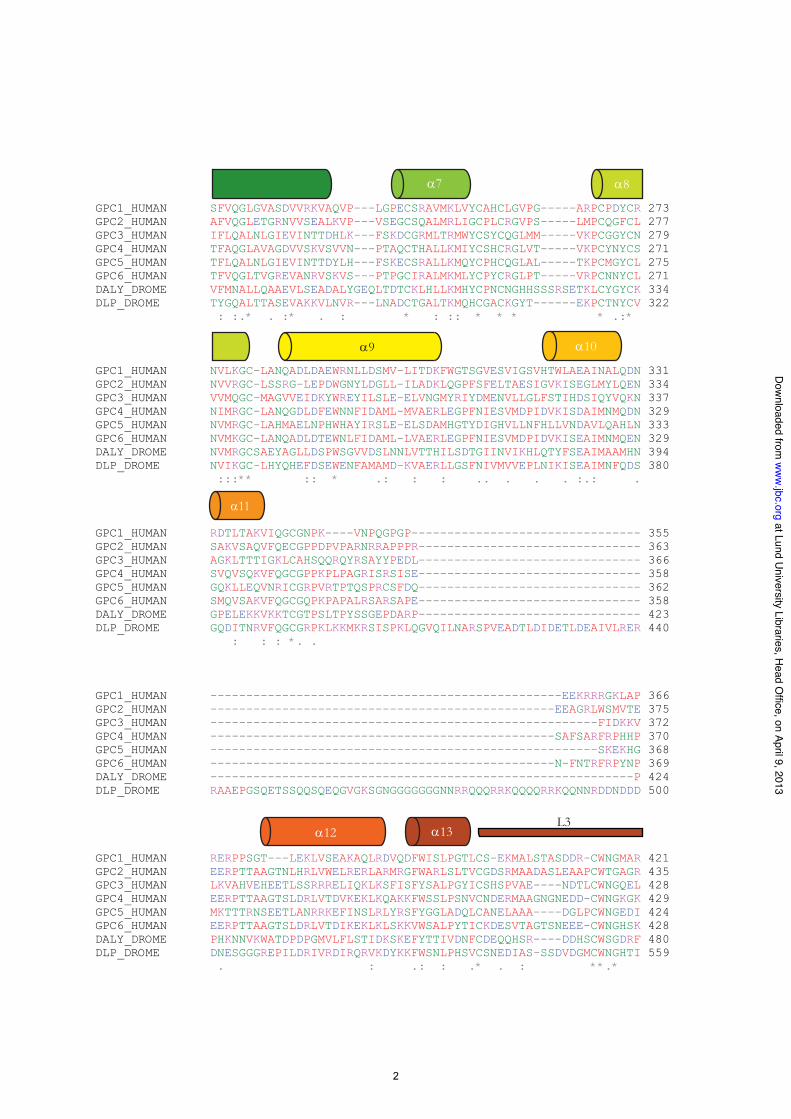
GPC1_HUMAN SFVQGLGVASDVVRKVAQVP---LGPECSRAVMKLVYCAHCLGVPG-----ARPCPDYCR 273 GPC2_HUMAN AFVQGLETGRNVVSEALKVP---VSEGCSQALMRLIGCPLCRGVPS-----LMPCQGFCL 277 GPC3_HUMAN IFLQALNLGIEVINTTDHLK---FSKDCGRMLTRMWYCSYCQGLMM-----VKPCGGYCN 279 GPC4_HUMAN TFAQGLAVAGDVVSKVSVVN---PTAQCTHALLKMIYCSHCRGLVT-----VKPCYNYCS 271 GPC5_HUMAN TFLQALNLGIEVINTTDYLH---FSKECSRALLKMQYCPHCQGLAL-----TKPCMGYCL 275 GPC6_HUMAN TFVQGLTVGREVANRVSKVS---PTPGCIRALMKMLYCPYCRGLPT-----VRPCNNYCL 271 DALY_DROME VFMNALLQAAEVLSEADALYGEQLTDTCKLHLLKMHYCPNCNGHHSSSRSETKLCYGYCK 334 DLP_DROME TYGQALTTASEVAKKVLNVR---LNADCTGALTKMQHCGACKGYT------EKPCTNYCV 322 : :.* . :* . : * : :: * * * * .:* GPC1_HUMAN NVLKGC-LANQADLDAEWRNLLDSMV-LITDKFWGTSGVESVIGSVHTWLAEAINALQDN 331 GPC2_HUMAN NVVRGC-LSSRG-LEPDWGNYLDGLL-ILADKLQGPFSFELTAESIGVKISEGLMYLQEN 334 GPC3_HUMAN VVMQGC-MAGVVEIDKYWREYILSLE-ELVNGMYRIYDMENVLLGLFSTIHDSIQYVQKN 337 GPC4_HUMAN NIMRGC-LANQGDLDFEWNNFIDAML-MVAERLEGPFNIESVMDPIDVKISDAIMNMQDN 329 GPC5_HUMAN NVMRGC-LAHMAELNPHWHAYIRSLE-ELSDAMHGTYDIGHVLLNFHLLVNDAVLQAHLN 333 GPC6_HUMAN NVMKGC-LANQADLDTEWNLFIDAML-LVAERLEGPFNIESVMDPIDVKISEAIMNMQEN 329 DALY_DROME NVMRGCSAEYAGLLDSPWSGVVDSLNNLVTTHILSDTGIINVIKHLQTYFSEAIMAAMHN 394 DLP_DROME NVIKGC-LHYQHEFDSEWENFAMAMD-KVAERLLGSFNIVMVVEPLNIKISEAIMNFQDS 380 :::** :: * .: : : .. . . . :.: . GPC1_HUMAN RDTLTAKVIQGCGNPK----VNPQGPGP-------------------------------- 355 GPC2_HUMAN SAKVSAQVFQECGPPDPVPARNRRAPPPR------------------------------- 363 GPC3_HUMAN AGKLTTTIGKLCAHSQQRQYRSAYYPEDL------------------------------- 366 GPC4_HUMAN SVQVSQKVFQGCGPPKPLPAGRISRSISE------------------------------- 358 GPC5_HUMAN GQKLLEQVNRICGRPVRTPTQSPRCSFDQ------------------------------- 362 GPC6_HUMAN SMQVSAKVFQGCGQPKPAPALRSARSAPE------------------------------- 358 DALY_DROME GPELEKKVKKTCGTPSLTPYSSGEPDARP------------------------------- 423 DLP_DROME GQDITNRVFQGCGRPKLKKMKRSISPKLQGVQILNARSPVEADTLDIDETLDEAIVLRER 440 : : : *. . GPC1_HUMAN -------------------------------------------------EEKRRRGKLAP 366 GPC2_HUMAN ------------------------------------------------EEAGRLWSMVTE 375 GPC3_HUMAN ------------------------------------------------------FIDKKV 372 GPC4_HUMAN ------------------------------------------------SAFSARFRPHHP 370 GPC5_HUMAN ------------------------------------------------------SKEKHG 368 GPC6_HUMAN ------------------------------------------------N-FNTRFRPYNP 369 DALY_DROME -----------------------------------------------------------P 424 DLP_DROME RAAEPGSQETSSQQSQEQGVGKSGNGGGGGGGNNRRQQQRRKQQQQRRKQQNNRDDNDDD 500 GPC1_HUMAN RERPPSGT---LEKLVSEAKAQLRDVQDFWISLPGTLCS-EKMALSTASDDR-CWNGMAR 421 GPC2_HUMAN EERPTTAAGTNLHRLVWELRERLARMRGFWARLSLTVCGDSRMAADASLEAAPCWTGAGR 435 GPC3_HUMAN LKVAHVEHEETLSSRRRELIQKLKSFISFYSALPGYICSHSPVAE----NDTLCWNGQEL 428 GPC4_HUMAN EERPTTAAGTSLDRLVTDVKEKLKQAKKFWSSLPSNVCNDERMAAGNGNEDD-CWNGKGK 429 GPC5_HUMAN MKTTTRNSEETLANRRKEFINSLRLYRSFYGGLADQLCANELAAA----DGLPCWNGEDI 424 GPC6_HUMAN EERPTTAAGTSLDRLVTDIKEKLKLSKKVWSALPYTICKDESVTAGTSNEEE-CWNGHSK 428 DALY_DROME PHKNNVKWATDPDPGMVLFLSTIDKSKEFYTTIVDNFCDEQQHSR----DDHSCWSGDRF 480 DLP_DROME DNESGGGREPILDRIVRDIRQRVKDYKKFWSNLPHSVCSNEDIAS-SSDVDGMCWNGHTI 559 . : .: : .* . : **.*
GPC1_HUMAN ------------------------MELRARGWWLLCAAAALVACARGDPASK-------- 28 GPC2_HUMAN ------------------------MSALRPLLLLLLPLCPGPGPGPGSEAKV-------- 28 GPC3_HUMAN -----------------------MAGTVRTACLVVAMLLSLDFPGQAQPPPPPP------ 31 GPC4_HUMAN -------------------------MARFGLPALLCTLAVLSAALLAAELK--------- 26 GPC5_HUMAN -----------------------MDAQTWPVGFRCLLLLALVGSARSEG----------- 26 GPC6_HUMAN -------------------------MPSWIGAVILPLLGLLLSLPAGADVK--------- 26 DALY_DROME ---------------------MAARSVRLAQLLLFTLLCGFVGLSAAKHLDLDGIHHHQH 39 DLP_DROME MLHQEQQQQHLHCRRKVTATTTARLVIFSSPLLLLLLTTHLPSTLQADNSPAPQVAALAA 60 . GPC1_HUMAN ----------------SRSCGEVR-QIYGAKGFSLSD-VPQAEISGEHLRICPQG--YTC 68 GPC2_HUMAN ----------------TRSCAETR-QVLGARGYSLNL-IPPALISGEHLRVCPQE--YTC 68 GPC3_HUMAN ----------------DATCHQVR-SFFQR-LQPGLKWVPETPVPGSDLQVCLPKG-PTC 72 GPC4_HUMAN ----------------SKSCSEVR-RLYVSKGFNKND-APLHEINGDHLKICPQG--STC 66 GPC5_HUMAN ----------------VQTCEEVR-KLFQWRLLGAVRGLPDSPRAGPDLQVCISKK-PTC 68 GPC6_HUMAN ----------------ARSCGEVR-QAYGAKGFSLAD-IPYQEIAGEHLRICPQE--YTC 66 DALY_DROME HLHSATTHHRRRLQRDSRAKDAVGGSTHQCDAVKSYFESIDIKSSGTYSEKGAICG-GNC 98 DLP_DROME PNPAGGVAGSSIIDQFSPNCSAVT-HIFQARGIDAIE-IPQKPSNERVLRYCESPSVGTC 118 . . .* GPC1_HUMAN CTSEMEENLANRSHAELETALRDSSRVLQAMLATQLRSFDDHFQHLLNDSERTLQATFPG 128 GPC2_HUMAN CSSETEQRLIRETEATFRGLVEDSGSFLVHTLAARHRKFDEFFLEMLSVAQHSLTQLFSH 128 GPC3_HUMAN CSRKMEEKYQLTARLNMEQLLQSASMELKFLIIQNAAVFQEAFEIVVRHAKNYTNAMFKN 132 GPC4_HUMAN CSQEMEEKYSLQSKDDFKSVVSEQCNHLQAVFASRYKKFDEFFKELLENAEKSLNDMFVK 126 GPC5_HUMAN CTRKMEERYQIAARQDMQQFLQTSSSTLKFLISRNAAAFQETLETLIKQAENYTSILFCS 128 GPC6_HUMAN CTTEMEDKLSQQSKLEFENLVEETSHFVRTTFVSRHKKFDEFFRELLENAEKSLNDMFVR 126 DALY_DROME CNNATELELRDKAAGMFEQLLHHHTSSLRGVLETNAKQFQSHVLELAQISENMTHSLFSK 158 DLP_DROME CTYNMETRMAMQSRQQLEGHTKDQISRMSGILGSKATKFKDIFTALLKESRTQFNSMFIR 178 *. * . : :. : : . *.. . : :. * GPC1_HUMAN AFGELYTQNARAFRDLYSELRLYYRGAN------------------LHLEETLAEFWARL 170 GPC2_HUMAN SYGRLYAQHALIFNGLFSRLRDFYGESG------------------EGLDDTLADFWAQL 170 GPC3_HUMAN NYPSLTPQAFEFVGEFFTDVSLYILGSD------------------INVDDMVNELFDSL 174 GPC4_HUMAN TYGHLYMQNSELFKDLFVELKRYYVVGN------------------VNLEEMLNDFWARL 168 GPC5_HUMAN TYRNMALEAAASVQEFFTDVGLYLFGAD------------------VNPEEFVNRFFDSL 170 GPC6_HUMAN TYGMLYMQNSEVFQDLFTELKRYYTGGN------------------VNLEEMLNDFWARL 168 DALY_DROME VYTRMVPSSRMMIHQLYTEIMNHLIYTSNYTNSNGQLGRRGIGSVQSNLEEAVRHFFVQL 218 DLP_DROME TYGVIYERNSYVFSDLFKELETYFANGR------------------VDLLEVMDKFFNTL 220 : : . :: : . : : :: * GPC1_HUMAN LERLFKQLH-----PQLLLPDDYLDCLGKQAEA----LRPFGEAPRELRLRATRAFVAAR 221 GPC2_HUMAN LERVFPLLH-----PQYSFPPDYLLCLSRLASSTDGSLQPFGDSPRRLRLQITRTLVAAR 225 GPC3_HUMAN FPVIYTQLMNP---GLPDSALDINECLRGARRD----LKVFGNFPKLIMTQVSKSLQVTR 227 GPC4_HUMAN LERMFRLVN-----SQYHFTDEYLECVSKYTEQ----LKPFGDVPRKLKLQVTRAFVAAR 219 GPC5_HUMAN FPLVYNHLINP---GVTDSSLEYSECIRMARRD----VSPFGNIPQRVMGQMGRSLLPSR 223 GPC6_HUMAN LERMFQLIN-----PQYHFSEDYLECVSKYTDQ----LKPFGDVPRKLKIQVTRAFIAAR 219 DALY_DROME FPVAYHQMVHLSKNNLGDLHEDYVNCLQHNFDE----MHPFGDIPQQVQSNLGKSVHMSN 274 DLP_DROME YQKMFTVLN-----TQYTFDENYMRCVSEHMKE----LKPFGDVPDKLSVQIKRSFVATR 271 : : : *: : **: * : . ::. :.
Supplementary figure 1
1
2
3 4
5 6
7 8
9 10
11
12 13
14
L1
L3
L3
GPC1_HUMAN GR-YLPEVMGDGLANQINNPEVEVDITKPDMTIRQQIMQLKIMTNRLRSAYNGNDVDFQD 480 GPC2_HUMAN GR-YLPPVVGGSPAEQVNNPELKVDASGPDVPTRRRRLQLRAATARMKTAALGHDLDGQD 494 GPC3_HUMAN VERYSQKAARNGMKNQFNLHELKMKGPEPVVSQIIDKLKHINQLLRTMSMPKGRVLDKNL 488 GPC4_HUMAN SR-YLFAVTGNGLANQGNNPEVQVDTSKPDILILRQIMALRVMTSKMKNAYNGNDVDFFD 488 GPC5_HUMAN VKSYTQRVVGNGIKAQSGNPEVKVKGIDPVINQIIDKLKHVVQLLQGRSPKPDKWELLQL 484 GPC6_HUMAN AR-YLPEIMNDGLTNQINNPEVDVDITRPDTFIRQQIMALRVMTNKLKNAYNGNDVNFQD 487 DALY_DROME GD-YTQLLINPGTDSQRYNPEVPFNAKAQTGKLNELVDKLFKIRKSIGAAAPSNSIQATH 539 DLP_DROME DR-YMHSITTEHGSNPEFTG--NPASTKQTAQMASQLSHLKNAIVHLRNAYNGQDVEWSE 616 * .. GPC1_HUMAN ASDD---GSGSGSGDGCLDDLCSR--------------KVSRKSSSSRTPLTHALPGLSE 523 GPC2_HUMAN ADED---ASGSGGGQQYADDWMAG--------------AVAPPARPPRPPYPPRRDGSGG 537 GPC3_HUMAN DEEG----FESGDCGDDEDECIGGSGDGM----IKVKNQLRFLAELAYDLDVDDAPGNSQ 540 GPC4_HUMAN ISDE---SSGEGSGSGCEYQQCPS--------------EFDYNATDHAGKSANE------ 525 GPC5_HUMAN GSGGGMVEQVSGDCDDEDG--CGGSGSG------EVKRTLKITDWMPDDMNFSDVKQIHQ 536 GPC6_HUMAN TSDE---SSGSGSGSGCMDDVCPT--------------EFEFVTTEAPAVDPDR------ 524 DALY_DROME DIQN-DMGEGSGGGEGQIGDDEEEYGG---------AHGSGDGSGDGPHTPIEESEGTTT 589 DLP_DROME QEELPYAGSGAGSGSGSEDDEDDDEGSGLGPFEPSHKPDVERPSVDADNDDDEDAGGRGH 676 *. GPC1_HUMAN Q-----------------------------------------------EGQKTSAASCPQ 536 GPC2_HUMAN KGGGGSARYN--------------------------------------QGRSRSGGASIG 559 GPC3_HUMAN QATPKDNE----------------------------------------ISTFHNLGN-VH 559 GPC4_HUMAN --------------------------------------------------KADSAGVRPG 535 GPC5_HUMAN TDTG---------------------------------------------STLDTTGAGCA 551 GPC6_HUMAN --------------------------------------------------REVDSSAAQR 534 DALY_DROME NEVE---------------------------------------------SRDSGKTSGSN 604 DLP_DROME MPTHTSRPTSGVDDKNPLIHTTHFDQDHNDLDEDHRQLDEDEDTDAGHDGANDNRSSDAP 736 GPC1_HUMAN PPTFLLPLLLFLALTVARPRWR------- 558 GPC2_HUMAN FHTQTILILSLSALALLGPR--------- 579 GPC3_HUMAN SPLKLLTSMAISVVCFFFLVH-------- 580 GPC4_HUMAN AQAYLLTVFCILFLVMQREWR-------- 556 GPC5_HUMAN VATESMTFTLISVVMLLPGIW-------- 572 GPC6_HUMAN GHSLLSWSLTCIVLALQRLCR-------- 555 DALY_DROME PLEGTATWMLLTLVTMLFSSCS------- 626 DLP_DROME EKMTLRRALVVYLLPLYMAWFGGVCADLL 765
2
at Lund University Libraries, H
ead Office, on April 9, 2013
ww
w.jbc.org
Dow
nloaded from

14L3
GPC1_HUMAN GR-YLPEVMGDGLANQINNPEVEVDITKPDMTIRQQIMQLKIMTNRLRSAYNGNDVDFQD 480GPC2_HUMAN GR-YLPPVVGGSPAEQVNNPELKVDASGPDVPTRRRRLQLRAATARMKTAALGHDLDGQD 494GPC3_HUMAN VERYSQKAARNGMKNQFNLHELKMKGPEPVVSQIIDKLKHINQLLRTMSMPKGRVLDKNL 488GPC4_HUMAN SR-YLFAVTGNGLANQGNNPEVQVDTSKPDILILRQIMALRVMTSKMKNAYNGNDVDFFD 488GPC5_HUMAN VKSYTQRVVGNGIKAQSGNPEVKVKGIDPVINQIIDKLKHVVQLLQGRSPKPDKWELLQL 484GPC6_HUMAN AR-YLPEIMNDGLTNQINNPEVDVDITRPDTFIRQQIMALRVMTNKLKNAYNGNDVNFQD 487DALY_DROME GD-YTQLLINPGTDSQRYNPEVPFNAKAQTGKLNELVDKLFKIRKSIGAAAPSNSIQATH 539DLP_DROME DR-YMHSITTEHGSNPEFTG--NPASTKQTAQMASQLSHLKNAIVHLRNAYNGQDVEWSE 616
* ..
GPC1_HUMAN ASDD---GSGSGSGDGCLDDLCSR--------------KVSRKSSSSRTPLTHALPGLSE 523GPC2_HUMAN ADED---ASGSGGGQQYADDWMAG--------------AVAPPARPPRPPYPPRRDGSGG 537GPC3_HUMAN DEEG----FESGDCGDDEDECIGGSGDGM----IKVKNQLRFLAELAYDLDVDDAPGNSQ 540GPC4_HUMAN ISDE---SSGEGSGSGCEYQQCPS--------------EFDYNATDHAGKSANE------ 525GPC5_HUMAN GSGGGMVEQVSGDCDDEDG--CGGSGSG------EVKRTLKITDWMPDDMNFSDVKQIHQ 536GPC6_HUMAN TSDE---SSGSGSGSGCMDDVCPT--------------EFEFVTTEAPAVDPDR------ 524DALY_DROME DIQN-DMGEGSGGGEGQIGDDEEEYGG---------AHGSGDGSGDGPHTPIEESEGTTT 589DLP_DROME QEELPYAGSGAGSGSGSEDDEDDDEGSGLGPFEPSHKPDVERPSVDADNDDDEDAGGRGH 676
*.
GPC1_HUMAN Q-----------------------------------------------EGQKTSAASCPQ 536GPC2_HUMAN KGGGGSARYN--------------------------------------QGRSRSGGASIG 559GPC3_HUMAN QATPKDNE----------------------------------------ISTFHNLGN-VH 559GPC4_HUMAN --------------------------------------------------KADSAGVRPG 535GPC5_HUMAN TDTG---------------------------------------------STLDTTGAGCA 551GPC6_HUMAN --------------------------------------------------REVDSSAAQR 534DALY_DROME NEVE---------------------------------------------SRDSGKTSGSN 604DLP_DROME MPTHTSRPTSGVDDKNPLIHTTHFDQDHNDLDEDHRQLDEDEDTDAGHDGANDNRSSDAP 736
GPC1_HUMAN PPTFLLPLLLFLALTVARPRWR------- 558GPC2_HUMAN FHTQTILILSLSALALLGPR--------- 579GPC3_HUMAN SPLKLLTSMAISVVCFFFLVH-------- 580GPC4_HUMAN AQAYLLTVFCILFLVMQREWR-------- 556GPC5_HUMAN VATESMTFTLISVVMLLPGIW-------- 572GPC6_HUMAN GHSLLSWSLTCIVLALQRLCR-------- 555DALY_DROME PLEGTATWMLLTLVTMLFSSCS------- 626DLP_DROME EKMTLRRALVVYLLPLYMAWFGGVCADLL 765
Supplementary figure 1. Alignment of glypican primary structures. An alignment of human Gpc-1
through Gpc-6 together with the D. melanogaster glypicans, Dally and Dlp is shown together with a
graphical representation of the helices and major loops in Gpc-1. The alignment was performed
using the glypican pro-protein amino acids sequences, i.e. the N- and C-terminal signal peptides are
included. Asterisks denote identical amino acid residues in the glypicans while two dots indicate
conserved amino acids substitutions (i.e. amino acids belonging to the same class of amino acids) and
one dot indicates semiconserved substitutions.
3
at Lund University Libraries, H
ead Office, on April 9, 2013
ww
w.jbc.org
Dow
nloaded from
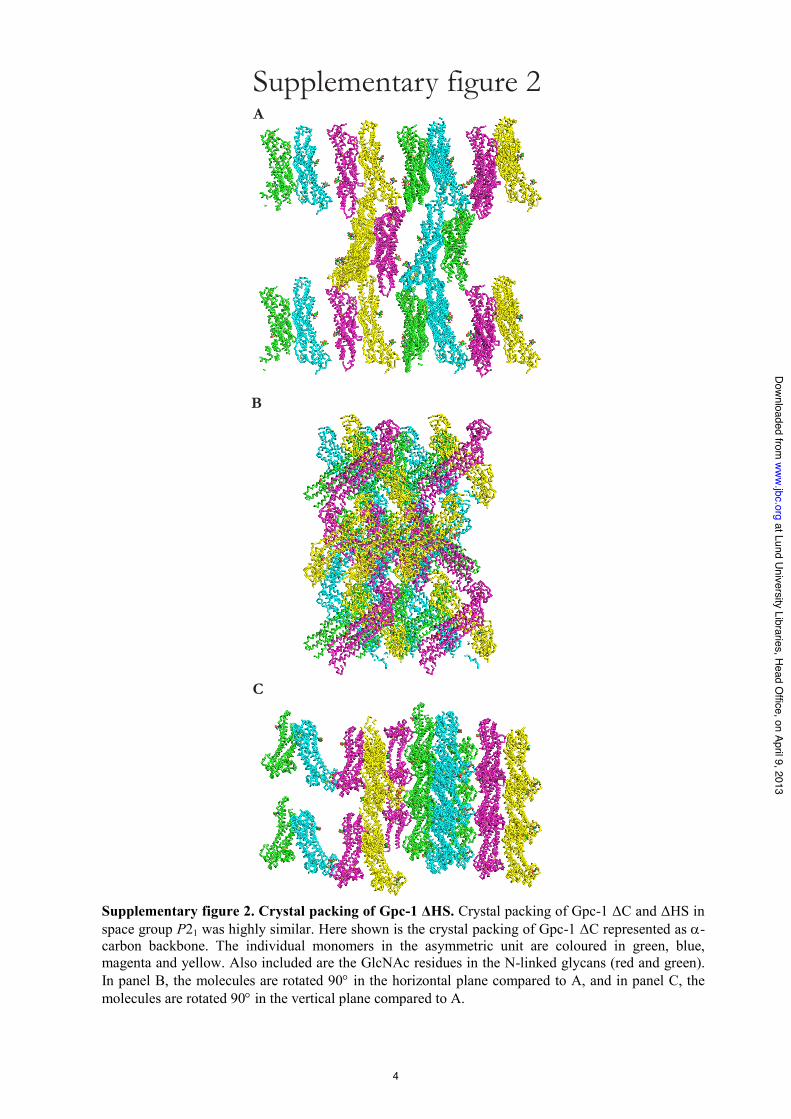
Supplementary figure 2A
B
C
Supplementary figure 2. Crystal packing of Gpc-1 HS. Crystal packing of Gpc-1 C and HS in
space group P21 was highly similar. Here shown is the crystal packing of Gpc-1 C represented as -
carbon backbone. The individual monomers in the asymmetric unit are coloured in green, blue,
magenta and yellow. Also included are the GlcNAc residues in the N-linked glycans (red and green).
In panel B, the molecules are rotated 90 in the horizontal plane compared to A, and in panel C, the
molecules are rotated 90 in the vertical plane compared to A.
4
at Lund University Libraries, H
ead Office, on April 9, 2013
ww
w.jbc.org
Dow
nloaded from