Copper-Catalyzed One-Pot Synthesis of Glycosylated Iminocoumarins and 3-Triazolyl-2-Iminocoumarins

Available online at www.sciencedirect.com
www.elsevier.com/locate/ejpb
European Journal of Pharmaceutics and Biopharmaceutics 69 (2008) 844–851
Research paper
Sustained release of bioactive glycosylated glial cell-line derivedneurotrophic factor from biodegradable polymeric microspheres
E. Garbayo a, E. Ansorena a, J.L. Lanciego b, M.S. Aymerich b, M.J. Blanco-Prieto a,*
a Department of Pharmacy and Pharmaceutical Technology, School of Pharmacy, University of Navarra, Pamplona, Spainb Neuromorphology Tracers Laboratory, CIMA, University of Navarra, Pamplona, Spain
Received 28 November 2007; accepted in revised form 18 February 2008Available online 23 February 2008
Abstract
Glial cell-line derived neurotrophic factor (GDNF), a potent neurotrophic factor for dopaminergic neurons, appeared as a promisingcandidate for treating Parkinson’s disease. GDNF microencapsulation could ensure protection against degradation due to the fragilenature of the protein. Poly(lactide-co-glycolide) (PLGA) microparticles loaded with recombinant glycosylated GDNF obtained in amammalian cell line were prepared by TROMS, a semi-industrial technique capable of encapsulating fragile molecules maintaining theirnative properties. The effects of several parameters as PLGA copolymer type, PEG 400 quantity co-encapsulated with GDNF or drugloading, on the properties of the particles were investigated. Microparticles showed a mean diameter between 8 and 30 lm, compatiblewith their stereotaxic implantation. The drug entrapment efficiency ranged from 50.6% to 100% depending on the microsphere compo-sition. GDNF was better encapsulated using hydrophilic polymers with high molecular weight such as RG 503H. In vitro drug releasewas influenced by the polymer type as well as by the amount of PEG 400 co-encapsulated with GDNF. Microparticles prepared usingPLGA RG 503H released 67% of the total protein content within 40 days. Moreover, very low concentrations of poly(vinyl alcohol) weredetected after microparticles washing and freeze-drying. Finally, a PC-12 bioassay demonstrated that the in vitro GDNF released wasbioactive.� 2008 Elsevier B.V. All rights reserved.
Keywords: Rat recombinant GDNF; Biodegradable microparticles; PLGA; TROMS; PEG 400
1. Introduction
Therapeutic proteins are a new type of drug that repre-sents a growing sector in the drug market [1]. Numeroustherapeutic proteins have been approved or are inadvanced clinical testing [2]. In particular, a total of 16new therapeutic proteins were approved by the FDA dur-ing 2004 and 2005 [3]. In contrast to other therapeuticagents that are normally used, these macromolecules havehigh specificity and activity at relatively low level concen-trations. However, protein administration presents other
0939-6411/$ - see front matter � 2008 Elsevier B.V. All rights reserved.
doi:10.1016/j.ejpb.2008.02.015
* Corresponding author. Department of Pharmacy and PharmaceuticalTechnology, School of Pharmacy, University of Navarra, C/Irunlarrea 1,E-31080 Pamplona, Spain. Tel.: +34 948 425 600x6519; fax: +34 948 425649.
E-mail address: [email protected] (M.J. Blanco-Prieto).
serious limitations due to their short in vivo half-life, phys-ical and chemical instability and low oral bioavailability[4]. Several years ago, the use of microspheres as carriersof these compounds was proposed as an attractive way toovercome these problems. Microspheres would offer sev-eral advantages since they could provide protein protectionfrom degradation and a prolonged delivery. The encapsula-tion of a wide range of proteins has been studied, althoughthe development of delivery systems for protein as drugs isstill a major challenge [5]. Among the methods described toprepare microparticles, multiple emulsion solvent evapora-tion technique (W/O/W) is widely accepted as the mostsuitable for encapsulation of labile hydrophilic compoundssuch as peptides and proteins [6]. However, there are twocritical steps in this process that could affect the proteinactivity; shear stress and the water/organic interface.Microsphere preparation by Total Recirculation One-

E. Garbayo et al. / European Journal of Pharmaceutics and Biopharmaceutics 69 (2008) 844–851 845
Machine System (TROMS) could provide an appropriatealternative to avoid the first aspect. This system, basedon the injection of the phases under a turbulent regime,does not need vigorous agitation to prepare the micro-spheres [7]. Thus, shear stress is avoided and proteins couldremain active. Moreover, the use of protein stabilizers suchas sugars, proteins, polyols or metals, prevents the denatu-ralization of the protein in the w/o interface [6].
Among the biodegradable polymers, the copolymers oflactic and glycolic acids (PLGAs) have become widely usedfor therapeutic protein delivery due to their excellent biode-gradability and biocompatibility [8]. Numerous proteinshave successfully been encapsulated into PLGA micro-spheres such as erythropoietin [9], growth hormone [10],interferon gamma [10], calcitonin [11], chorionic gonado-trophin [12], nerve growth factor [13], brain derived neuro-trophic factor [14] or glial cell-line derived neurotrophicfactor (GDNF) [15] among others.
GDNF, the protein used in this study, has beendescribed as one of the most potent neurotrophic factorsfor dopaminergic neurons with both neuroprotective andneurorestorative properties [16]. In addition, it stimulatesregenerative growth and axonal sprouting in animal mod-els of Parkinson’s disease (PD) [17]. Two open-label trialsinvolving continuous recombinant non-glycosylatedGDNF infusion into the putamen of PD patients showedthat the growth factor significantly improved motor scoresas measured with the Unified Parkinson’s disease RatingScale (UPDRS) [18–20]. However, a double-blind placebocontrolled study presented by AMGEN did not demon-strate beneficial effects in patients [21,22]. Differences indoses, catheter design and delivery protocols may beresponsible for the discrepancy between phase I and IIstudies. Clearly, alternative methods of GDNF deliveryto the brain must be developed. In this sense, polymer-based drug delivery systems could be a valuable strategy.
In the present work, the preparation and characteriza-tion of PLGA microspheres loaded with highly purerecombinant glycosylated GDNF obtained in a mamma-lian cell line are described. Particles were prepared by sol-vent evaporation technique using TROMS technology.The effects of several formulation parameters such asPLGA copolymer type, PEG 400 quantity co-encapsulatedwith GNDF and drug loading on particle size, encapsula-tion efficiency and in vitro release kinetics were analyzed.Attention was also focused on the bioactivity of theprotein.
2. Materials and methods
2.1. Materials
Rat recombinant GDNF was expressed and purified aspreviously described [23]. GDNF enzyme linked immuno-sorbant assay kit (ELISA) was purchased from Promega(Madison, USA). Poly(lactic-co-glycolic) acid (PLGA)with a lactic:glycolic ratio of 50:50 Resomer RG 503
(MW 34 kDa), Resomer RG 502H (MW 13.7 kDa) andResomer RG 503H (MW 34 kDa) were provided by Boeh-ringer-Ingelheim (Ingelheim, Germany). Poly(ethylene gly-col) 400 (PEG 400) and human serum albumin (HSA) wereprovided by Sigma–Aldrich (Barcelona, Spain). Dichloro-methane and acetone were obtained from Panreac QuimicaS.A. (Barcelona, Spain). Poly(vinyl alcohol) (PVA) 88%hydrolyzed (MW: �125,000) was obtained from Polyscienc-es, Inc. (WA, USA). The adrenal rat PC-12 cell line waspurchased from American Type Culture Collection(ATCC) (Rockville, MD, USA). Silver Stain Plus wasobtained from BioRad (CA, USA). General laboratoryreagents were purchased from Sigma–Aldrich (Barcelona,Spain) unless specified in the text.
2.2. Protein purity assay
SDS–PAGE and silver staining were performed to assessGDNF purity. Purified rat recombinant GDNF wasloaded onto 12.5% polyacrylamide gels under reducingconditions. After the electrophoresis, gels were stained withSilver Stain Plus according to manufacturer’s instructions.
2.3. Microparticles preparation
Microparticles containing purified rat recombinantGDNF were prepared by solvent extraction/evaporationmethod using Total Recirculation One-Machine System(TROMS) [7]. Briefly, the organic solution composed of2 ml of dichlorometane:acetone (3:1) containing the poly-mer was injected through a needle with an inner diameterof 0.17 mm at 30 ml/min into the inner aqueous phase.The inner water phase contained rat recombinant GDNFin 10 mM phosphate, 50 mM sodium chloride (PBS), pH7.9, 5% of HSA and different quantities of PEG 400. Next,the previously formed inner emulsion (W1/O) was recircu-lated through the system for 3 min under a turbulentregime at a flow rate of 30 ml/min. After this step, the firstemulsion was injected into 30 ml of an aqueous phase (W2)composed of 1.5% PVA. The turbulent injection throughthe needle with an inner diameter of 0.50 mm resulted inthe formation of a multiple emulsion (W1/O/W2), whichwas further homogenized by circulation through the systemfor 4 min. The resulting W1/O/W2 emulsion was stirred forat least 3 h at room temperature. Microparticles werewashed 3 times with ultrapure water by consecutive centri-fugation at 4 �C (20,000g, 10 min). Finally, the particleswere resuspended in 1 ml of ultrapure water, frozen at�80 �C, lyophilized (Genesis 12EL, Virtis) and stored at4 �C.
2.4. Microparticle characterization
2.4.1. Particle size
Size and size distribution of the microspheres weredetermined by laser diffractometry using a Mastersizer-S�
(Malvern Instruments, Malvern, UK). Microspheres were

846 E. Garbayo et al. / European Journal of Pharmaceutics and Biopharmaceutics 69 (2008) 844–851
dispersed in distilled water and analyzed under continuousstirring. The results were expressed in volumetric meandiameter, which is the diameter that divides the volume dis-tribution curve of the sampled microparticles in two equalparts. Samples were measured in triplicate.
2.4.2. MorphologyMorphology of the microspheres with highest drug
loadings was analyzed by scanning electron microscopy(SEM). The lyophilized particles were directly depositedonto a carbon conductive tape on aluminium stubs andcoated with gold to a 16-mm thickness (Emitek K550equipment). The samples were observed in a Zeiss DSM940A microscope with a digital imaging capture system(DISS of Point Electronic GmbH, Halle, Germany).
2.4.3. Determination of residual PVA
The amount of PVA associated with microparticles wasdetermined by a colorimetric method based on the forma-tion of a coloured complex between two adjacent hydroxylgroups of PVA and an iodine molecule [24,25]. Briefly,2 mg of lyophilized microparticles samples were treatedwith 2 ml of 0.5 M NaOH for 15 min at 60 �C. Each samplewas neutralized with 900 ll of 1 N HCl and the volume wasadjusted to 5 ml with distilled water. To each sample, 3 mlof a 0.65 M solution of boric acid, 0.5 ml of a solution ofI2/KI (0.05 M/0.15 M) and 1.5 ml of distilled water wereadded. Finally, the absorbance of the samples was mea-sured at 690 nm using an Agilent 8453 UV–visible spectro-photometer (Agilent technologies, Palo Alto, CA, USA)after 15 min of incubation. A standard plot of PVA wasprepared under identical conditions.
2.4.4. Determination of GDNF content in the microparticles
To quantify the GDNF content in the microparticles,1 ml of dimethyl sulfoxide (DMSO) was added to 5 mgof freeze-dried loaded particles. Previously, it was verifiedthat DMSO did not affect GDNF stability. The mixturewas vortexed vigorously for 5 min and the amount ofGDNF was measured by ELISA. Briefly, 96-well micro-plates (Greiner Bio-One, Germany) were coated withmonoclonal anti-GDNF antibody diluted in 0.025 M car-bonate buffer, pH 8.2, and incubated overnight at 4 �C.Plates were blocked for 1 h with blocking solution providedby the manufacturers. The samples were added to thecoated wells (100 ll each) and incubated for 6 h at roomtemperature. Plates were washed 5 times and boundedGDNF was incubated with anti-GDNF polyclonal anti-body overnight at 4 �C. After additional washed, plateswere incubated for 2 h at room temperature with anti-chicken IgY, HRP conjugate. Then, plates were washed 5times and incubated with TMB/peroxidase substrate solu-tion for 15 min. The enzyme reaction was stopped by add-ing an acidic solution. Optical density was determined in aplate reader set at 450 nm. Sample values were determinedfrom the regression standard line for the purified ratGDNF (ranging from 15 to 1000 pg/ml) prepared for each
assay. Points of the regression standard line were treatedwith the same amount of DMSO as the samples. Each sam-ple was assayed in triplicate. Drug entrapment efficacy wascalculated as the ratio of the final drug content in themicrospheres to the initial drug content, expressed as apercentage.
2.5. In vitro release of GDNF
GDNF-loaded microparticles, accurately weighed(1 mg, n = 3), were incubated in 0.5 ml of 10 mM phos-phate, 150 mM sodium chloride, pH 7.4, containing 0.1%BSA and 0.02% w/w sodium azide. Incubation took placein rotating vials at 37 �C for 1 week or 40 days dependingon the experiments. Due to the instability of the proteinin the release medium, the amount of drug released wasdetermined indirectly by measuring the amount of GDNFremaining in the microspheres. At defined time intervals,sample tubes were centrifuged (25,000g, for 15 min). Afterremoval of the supernatant, microspheres were dissolvedwith DMSO and the protein content was determined byELISA as described above. Release profiles were expressedin terms of cumulative release, and plotted versus time.
2.6. In vitro bioactivity assay
The differentiation of PC-12 cells was used to evaluatethe bioactivity of GDNF released from microparticles.These cells differentiate to a neuronal phenotype extendingneurites in response to neurotrophic factors such as NGFor GDNF [23,26]. PC-12 cells were cultured in DMEMsupplemented with 5% horse serum, 10% foetal bovineserum and 1% penicillin/streptomycin. For studies of neu-rite outgrowth, PC-12 cells were plated onto 12-well cultureplate at a low density, 2 � 103 cells/cm2 in 1 ml of culturemedia. The culture medium was supplemented 24 h laterwith 50 ng of GDNF released from microspheres over24 h, which had previously been quantified by ELISA.Neurite outgrowth was visualized after 7 days in cultureunder phase contrast illumination with a Leika DM IRBinverted microscope connected to a Hamamatsu ORCA-ER digital camera. PC-12 cells incubated with 50 ng/mlof purified rat recombinant GDNF were used as a positivecontrol of the technique. The released medium from non-loaded microspheres was used as negative control for theexperiment.
3. Results and discussion
3.1. Protein purity assay
A GDNF expression and purification procedure hadpreviously been developed in order to obtain bioactive,highly pure and glycosylated protein [23]. Since GDNFwas intended to be used for in vivo studies, testing the pur-ity of the protein was considered crucial. Rat recombinantGDNF was analyzed by SDS–PAGE and posterior silver

E. Garbayo et al. / European Journal of Pharmaceutics and Biopharmaceutics 69 (2008) 844–851 847
staining. Silver staining showed a highly pure protein(Fig. 1). GDNF migrated as a 26 kDa band that corre-sponds to the most abundant glycosylated form of the pro-tein. Highly pure protein was obtained, in sufficientamounts to be microencapsulated. This was an importantaspect, since impurities could affect not only the encapsula-tion efficiency and the release profile of the neurotrophicfactor but also the in vivo efficacy. Thus, recombinant N-glycosylated GDNF, obtained in a mammalian cell lineand similar to the endogenous protein, was used to formu-late the microspheres.
3.2. Microsphere preparation and characterization
Several attempts were made to overcome the criticalsteps of microsphere preparation process: shear stress andwater/organic interface. Attention was focused on the pres-
Fig. 1. SDS–PAGE and silver staining of GDNF. (Lane A) Molecularweight markers, (Lane B) purified recombinant rat GDNF with amolecular weight of 26 kDa indicating that the protein is glycosylated.
Table 1Composition, particle size, encapsulation efficiency and drug content of biode
Batch#
TheoreticalGDNFloading (lg)
PEG 400concentrationin W1 (% w/w)
W1
(ll)Polymer typ(Resomer�100 mg
1 0.5 1 125 502H2 0.5 1 125 5033 0.5 1 125 503H4 0.5 1 125 502H+503H5 20 1 200 503H6 20 5 200 503H7 20 10 200 503H8 135 5 200 503H
The processing solvent was dichlorometane:acetone (ratio 3:1).
ervation of the protein biological activity. In this study,several formulations containing rat recombinant glycosyl-ated GDNF were prepared by emulsion solvent evapora-tion technique using TROMS (Table 1). This procedureavoids shear stress produced by sonication and ultraturraxthat normally would affect protein integrity and conse-quently, their biological activity [7]. Garcia del Barrio com-pared the activity of a labile compound such as adenovirusafter its microencapsulation by conventional multipleemulsion solvent evaporation technique and by TROMS.Microparticles prepared by TROMS presented 4.5 and 3times more activity at 8 and 32 h than microparticles pre-pared by the conventional methods [27]. Another advan-tage of TROMS over microparticle production throughemulsion methods is the ability to produce homogeneousbatches on a semi-industrial scale. Consequently, thiswould be of great interest considering scaling-up andindustrial issues.
HSA and PEG 400 were co-encapsulated with GDNF inorder to stabilize the primary emulsion and to reduce pro-tein–polymer interactions. Previous studies showed thatPEG 400 dissolved in the inner aqueous phase was a goodcandidate to protect NGF against denaturing by contactwith an organic phase during emulsification without mod-ifying the microparticle structure [28]. In this case, PEG400 limited protein penetration in the interfilm of the pri-mary emulsion and reduced the contact between the pro-tein and the organic phase. Furthermore PEG 400promoted sufficient release of entrapped NGF. Subse-quently, this compound was co-encapsulated with insulinlike growth factor I [29], L-asparaginase enzyme [5] ornon-glycosylated GDNF [30] to prepare particles by con-ventional multiple emulsion solvent evaporation method.
3.2.1. Particle size and morphological analysis
Particle size was measured by laser diffractometry. Allthe microparticles exhibited a monomodal size distributionwith a mean diameter between 8.4 and 30.9 lm, compatiblewith a stereotaxic injection (Table 1). The mean particlesize decreases when the nominal drug loading increases.Thus, at nominal GDNF content of 0.5 lg, the mean par-ticle size was around 29 lm independently of the polymer
gradable PLGA microparticles containing rat recombinant GDNF
e)
Particle size(lm)
Encapsulationefficiency (% w/w)
Drug content(ng GDNF/mgpolymer)
28.01 ± 1.03 78.2 3.9130.9 ± 0.97 79 3.95
28.57 ± 0.82 100 529.29 ± 1.11 69.7 3.4827.74 ± 1.66 57 11421.71 ± 2.93 68 13620.49 ± 2.08 65 1308.42 ± 0.77 50.6 680
848 E. Garbayo et al. / European Journal of Pharmaceutics and Biopharmaceutics 69 (2008) 844–851
used. Surprisingly, when 135 lg of GDNF were included inthe inner water phase, the mean particle size decreased to8.4 lm. Non-loaded microspheres prepared under the sameconditions showed a mean particle size of 30 lm. Thedecrease in microparticle size could be ascribed to tensioac-tive properties of the protein.
The mean particle size was slightly influenced by theaddition of PEG. As shown in Table 1, when the concen-tration of PEG in the inner water phase was increased upto 10%, a decrease in the particle size from 27.7 to20.4 lm was observed.
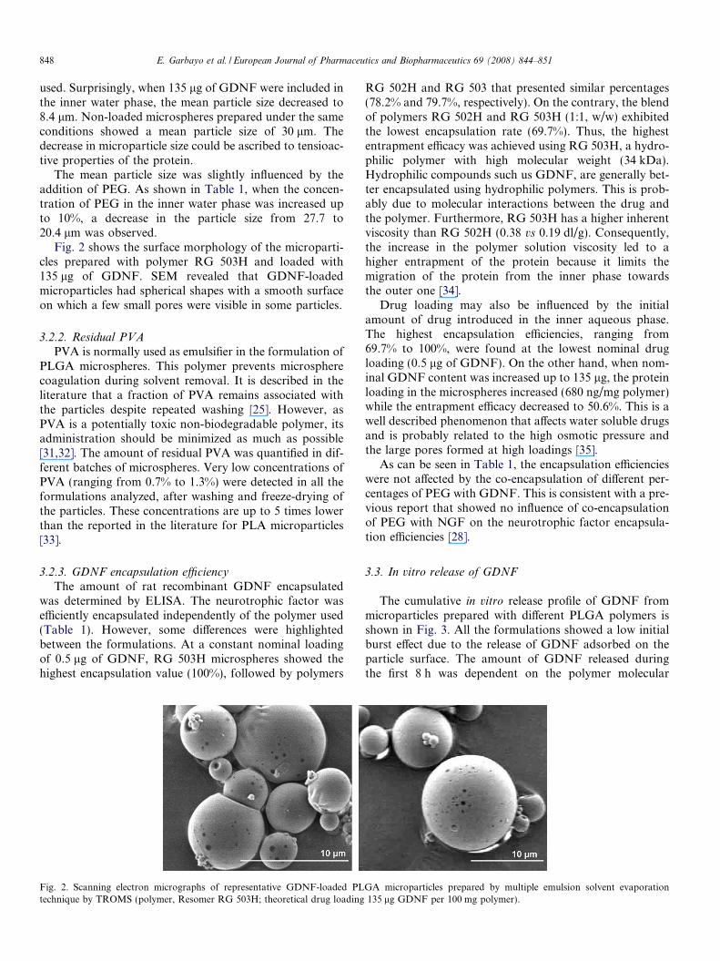
Fig. 2 shows the surface morphology of the microparti-cles prepared with polymer RG 503H and loaded with135 lg of GDNF. SEM revealed that GDNF-loadedmicroparticles had spherical shapes with a smooth surfaceon which a few small pores were visible in some particles.
3.2.2. Residual PVA
PVA is normally used as emulsifier in the formulation ofPLGA microspheres. This polymer prevents microspherecoagulation during solvent removal. It is described in theliterature that a fraction of PVA remains associated withthe particles despite repeated washing [25]. However, asPVA is a potentially toxic non-biodegradable polymer, itsadministration should be minimized as much as possible[31,32]. The amount of residual PVA was quantified in dif-ferent batches of microspheres. Very low concentrations ofPVA (ranging from 0.7% to 1.3%) were detected in all theformulations analyzed, after washing and freeze-drying ofthe particles. These concentrations are up to 5 times lowerthan the reported in the literature for PLA microparticles[33].
3.2.3. GDNF encapsulation efficiency
The amount of rat recombinant GDNF encapsulatedwas determined by ELISA. The neurotrophic factor wasefficiently encapsulated independently of the polymer used(Table 1). However, some differences were highlightedbetween the formulations. At a constant nominal loadingof 0.5 lg of GDNF, RG 503H microspheres showed thehighest encapsulation value (100%), followed by polymers
Fig. 2. Scanning electron micrographs of representative GDNF-loaded PLtechnique by TROMS (polymer, Resomer RG 503H; theoretical drug loading
RG 502H and RG 503 that presented similar percentages(78.2% and 79.7%, respectively). On the contrary, the blendof polymers RG 502H and RG 503H (1:1, w/w) exhibitedthe lowest encapsulation rate (69.7%). Thus, the highestentrapment efficacy was achieved using RG 503H, a hydro-philic polymer with high molecular weight (34 kDa).Hydrophilic compounds such us GDNF, are generally bet-ter encapsulated using hydrophilic polymers. This is prob-ably due to molecular interactions between the drug andthe polymer. Furthermore, RG 503H has a higher inherentviscosity than RG 502H (0.38 vs 0.19 dl/g). Consequently,the increase in the polymer solution viscosity led to ahigher entrapment of the protein because it limits themigration of the protein from the inner phase towardsthe outer one [34].
Drug loading may also be influenced by the initialamount of drug introduced in the inner aqueous phase.The highest encapsulation efficiencies, ranging from69.7% to 100%, were found at the lowest nominal drugloading (0.5 lg of GDNF). On the other hand, when nom-inal GDNF content was increased up to 135 lg, the proteinloading in the microspheres increased (680 ng/mg polymer)while the entrapment efficacy decreased to 50.6%. This is awell described phenomenon that affects water soluble drugsand is probably related to the high osmotic pressure andthe large pores formed at high loadings [35].
As can be seen in Table 1, the encapsulation efficiencieswere not affected by the co-encapsulation of different per-centages of PEG with GDNF. This is consistent with a pre-vious report that showed no influence of co-encapsulationof PEG with NGF on the neurotrophic factor encapsula-tion efficiencies [28].
3.3. In vitro release of GDNF
The cumulative in vitro release profile of GDNF frommicroparticles prepared with different PLGA polymers isshown in Fig. 3. All the formulations showed a low initialburst effect due to the release of GDNF adsorbed on theparticle surface. The amount of GDNF released duringthe first 8 h was dependent on the polymer molecular
GA microparticles prepared by multiple emulsion solvent evaporation135 lg GDNF per 100 mg polymer).

02468
10121416
0 1 2 3 4 5 6 7Time (Days)
Cum
ulat
ive
rele
ase
of G
DN
F (%
)
503502H: 503H (1:1)502H503H
Fig. 3. Influence of PLGA type [RG 503, RG 502H, RG 503H and RG502H: RG 503H (1:1)] on GDNF in vitro release (n = 3). Theoretical drugloading: 0.5 lg GDNF/100 mg polymer.
0
10
20
30
40
50
60
0 1 2 3 4 5 6 7Time (Days)
Cum
ulat
ive
rele
ase
of G
DN
F (%
)
10% PEG 4005% PEG 4001% PEG 400
Fig. 4. Influence of PEG 400 added to the inner water phase on GDNFin vitro release profile from PLGA microspheres (n = 3). Theoretical drugloading: 20 lg GDNF/100 mg polymer.
E. Garbayo et al. / European Journal of Pharmaceutics and Biopharmaceutics 69 (2008) 844–851 849
weight. Thus, the highest molecular weight polymers (RG503H and RG 503) exhibited highest initial burst (8.9%and 8.7%, respectively). On the other hand, the blend ofResomer RG 502H and RG 503H released 6.3% andResomer RG 502H released 5.7% of the total dose. Posi-tively charged molecules could potentially interact withPLGA negatively charged carboxylic end groups [36]. Thiscould be the case for GDNF, since it has a pI of 9.44 and itis positively charged at pH 7.9, the inner water phase pH.Such interactions could explain the faster drug releaseobserved at higher polymer molecular weights [36]. After1 week, GDNF released from PLGA microspheres werenot strongly affected by polymer molecular weight. Micro-particles prepared with RG 503, RG 502H and the blend ofRG 503H and RG 502H showed a similar cumulative drugrelease (11.1%, 11.6% and 11.4%, respectively). However,RG 503H microspheres released GDNF slightly fasterand 13.5% of the drug was released during the first week.Thus, taking into account the highest encapsulation effi-ciencies values and the GDNF in vitro release kinetics,the copolymer RG 503H was selected for further studies.
The incorporation of additives to the formulation cansubstantially modify the drug release profile. Since PEG400 was co-encapsulated with GDNF to protect the proteinbiological activity, the effect of PEG on GDNF release pro-file from RG 503H microparticles was investigated (Fig. 4).All the formulations studied showed a biphasic release pro-file characterized by an initial burst release phase followedby a slower drug release phase. The co-encapsulation ofPEG with GDNF influenced the initial release and theamount of released protein over a 7-day period but didnot change the biphasic drug release pattern (Fig. 4). Asshown in Fig. 4, during the first 8 h microspheres contain-ing 1% of PEG yielded an initial release of 20% as com-pared to 34.3% and 41.8% observed for the formulationscontaining 5% and 10% of PEG, respectively. A similareffect was observed during the release of NGF from PLGAmicrospheres [28]. This effect could be due to the inhibitionof protein–polymer adsorption and to the improvement ofpolymer solubility [28]. Following the initial burst, up to27.5% (1% PEG), 45.8% (5% PEG) or 52.4% (10% PEG)
of total GDNF was released throughout 1 week (Fig. 4).Among the different percentages of PEG analyzed, micro-spheres prepared with 5% and 10% provided the bestrelease characteristics over 1 week, for the tested conditions(Fig. 4). Taking into consideration that the final aim of thisproject will be to administer the microparticles into thebrain of animal models, microparticles prepared with alower amount of PEG were selected and the release ofGDNF was studied over 40 days. The release of the proteinwas biphasic. After an initial burst caused by the release ofGDNF adsorbed on the particle surface, a sustainedrelease was observed from day 1 to day 14, in which drugdiffuses through the polymer. Finally, an increase in therate of release was observed from day 14 to 40 due to poly-mer degradation and, 67% of the total GDNF was releasedwithin the first 40 days. Other authors have previouslynoticed a correlation between increased protein releaseand polymer degradation [26].
Besides the polymer type, and the incorporation of addi-tives, the initial release could also be affected by the proteinloading. Since the initial release is normally attributed tothe surface-associated drug, higher protein loading led toa higher amount of drug located close to the particle sur-face and, in consequence, to an increase in the initial burst.In this sense, it was observed that the initial GDNFreleased during the first 8 hours increased from 8.9% to20% when the protein loading increased from 0.5 to20 lg for RG 503H microparticles containing 1% ofPEG. These results are in agreement with the resultsobtained previously by other authors [37–39].
3.4. In vitro bioactivity assay
Since the successful development of a delivery system forGDNF requires the preservation of the protein biologicalfunction throughout all the process, the bioactivity and sta-bility of released GDNF were evaluated in vitro using a PC-12 differentiation assay. The protein released from micro-spheres over 24 h was added to the culture medium ofthe cells. This cell line responds to bioactive GDNF by dif-

Fig. 5. Bioactivity of released GDNF in a PC-12 cell culture assay. Phase-contrast microscopy of cells that were cultured for 7 days in mediumsupplemented (A) with the release medium of non-loaded microspheres, (B) with GDNF released over 24 h from loaded microparticles, (C) with the sameamount of purified rat recombinant GDNF. The appearance of neurites both in (B) also in (C) demonstrates the bioactivity of GDNF released frommicrospheres. Bar length 100 lm.
850 E. Garbayo et al. / European Journal of Pharmaceutics and Biopharmaceutics 69 (2008) 844–851
ferentiating to a neuronal-like phenotype that is visualizedby the sprouting of neurites. After 1 week of exposure tothe neurotrophic factor, PC-12 cell neurite outgrowth indi-cated that released GDNF was bioactive (Fig. 5B). A sim-ilar effect was observed in cells treated with purified ratrecombinant GDNF (Fig. 5C). On the contrary, no out-growth could be seen in PC-12 cells incubated with therelease medium from unloaded microspheres. In this case,cells presented an undifferentiated and rounded morphol-ogy (Fig. 5A). These results demonstrated that the encap-sulated GDNF was biologically active. Indeed, GDNFbioactivity was maintained in the microspheres for at least5 weeks (data not shown). Since proteins may lose theirbioactivity during microparticle preparation and posteriorrelease, the assurance that the biological activity of GDNFwas preserved would appear to be extremely important inthe perspective of microsphere implantation in animalmodels of PD.
4. Conclusions
The results of this study show that rat recombinant gly-cosylated GDNF obtained in a mammalian cell line can besuccessfully microencapsulated in PLGA microspheres byTROMS technology. Microparticles were able to releaseglycosylated GDNF in a controlled manner for at least40 days in vitro. Moreover, the encapsulated GDNF wasbiologically active and could stimulate PC-12 cells tosprout neurites. Our results demonstrated that the releasedGDNF protein exhibited similar potency to naked purifiedGDNF protein in differentiating the PC-12 cells. At pres-ent, selected formulations are under in vivo evaluation inan animal model of Parkinson’s disease.
Acknowledgements
This project was funded by the Department of Healthand Education of the Government of Navarra, by theMAPFRE Medicine Foundation and by the CANFoundation.
The fellowship support for E. Garbayo from the Gobi-erno de Navarra and Asociacion de Amigos is gratefullyacknowledged.
References
[1] C. Johnson-Leger, C.A. Power, G. Shomade, J.P. Shaw, A.E.Proudfoot, Protein therapeutics–lessons learned and a view of thefuture, Expert Opin. Biol. Ther. 6 (2006) 1–7.
[2] S.D. Putney, P.A. Burke, Improving protein therapeutics withsustained-release formulations, Nat. Biotechnol. 16 (1998) 153–157.
[3] J.M. Reichert, Trends in US approvals: new biopharmaceuticals andvaccines, Trends Biotechnol. 24 (2006) 293–298.
[4] J.T. He, H.B. Su, G.P. Li, X.M. Tao, W. Mo, H.Y. Song,Stabilization and encapsulation of a staphylokinase variant (K35R)into poly(lactic-co-glycolic acid) microspheres, Int. J. Pharm. 309(2006) 101–108.
[5] M. Wolf, M. Wirth, F. Pittner, F. Gabor, Stabilisation anddetermination of the biological activity of L-asparaginase inpoly(D,L-lactide-co-glycolide) nanospheres, Int. J. Pharm. 256 (2003)141–152.
[6] U. Bilati, E. Allemann, E. Doelker, Strategic approaches for overcom-ing peptide and protein instability within biodegradable nano- andmicroparticles, Eur. J. Pharm. Biopharm. 59 (2005) 375–388.
[7] G.G. del Barrio, F.J. Novo, J.M. Irache, Loading of plasmid DNAinto PLGA microparticles using TROMS (Total Recirculation One-Machine System): evaluation of its integrity and controlled releaseproperties, J. Control. Release 86 (2003) 123–130.
[8] G.E. Visscher, R.L. Robinson, H.V. Maulding, J.W. Fong, J.E.Pearson, G.J. Argentieri, Biodegradation of and tissue reaction to50:50 poly(DL-lactide-co-glycolide) microcapsules, J. Biomed. Mater.Res. 19 (1985) 349–365.
[9] B. Bittner, M. Morlock, H. Koll, G. Winter, T. Kissel, Recombinanthuman erythropoietin (rhEPO) loaded poly(lactide-co-glycolide)microspheres: influence of the encapsulation technique and polymerpurity on microsphere characteristics, Eur. J. Pharm. Biopharm. 45(1998) 295–305.
[10] J.L. Cleland, A.J. Jones, Stable formulations of recombinant humangrowth hormone and interferon-gamma for microencapsulation inbiodegradable microspheres, Pharm. Res. 13 (1996) 1464–1475.
[11] S. Prabhu, J.L. Sullivan, G.V. Betageri, Comparative assessment ofin vitro release kinetics of calcitonin polypeptide from biodegradablemicrospheres, Drug Deliv. 9 (2002) 195–198.
[12] K.J. Zhu, H.L. Jiang, X.Y. Du, J. Wang, W.X. Xu, S.F. Liu,Preparation and characterization of hCG-loaded polylactide orpoly(lactide-co-glycolide) microspheres using a modified water-in-oil-in-water (w/o/w) emulsion solvent evaporation technique, J.Microencapsul. 18 (2001) 247–260.

E. Garbayo et al. / European Journal of Pharmaceutics and Biopharmaceutics 69 (2008) 844–851 851
[13] C.E. Krewson, R. Dause, M. Mak, W.M. Saltzman, Stabilization ofnerve growth factor in controlled release polymers and in tissue, J.Biomater. Sci. Polym. Ed. 8 (1996) 103–117.
[14] S. Mittal, A. Cohen, D. Maysinger, In vitro effects of brain derivedneurotrophic factor released from microspheres, Neuroreport 5(1994) 2577–2582.
[15] A. Aubert-Pouessel, M.C. Venier-Julienne, A. Clavreul, M. Sergent,C. Jollivet, C.N. Montero-Menei, E. Garcion, D.C. Bibby, P. Menei,J.P. Benoit, In vitro study of GDNF release from biodegradablePLGA microspheres, J. Control. Release 95 (2004) 463–475.
[16] T. Yasuhara, T. Shingo, I. Date, Glial cell line-derived neurotrophicfactor (GDNF) therapy for Parkinson’s disease, Acta Med. Okayama61 (2007) 51–56.
[17] B. Connor, D.A. Kozlowski, J.R. Unnerstall, J.D. Elsworth, J.L.Tillerson, T. Schallert, M.C. Bohn, Glial cell line-derived neurotro-phic factor (GDNF) gene delivery protects dopaminergic terminalsfrom degeneration, Exp. Neurol. 169 (2001) 83–95.
[18] S.S. Gill, N.K. Patel, G.R. Hotton, K. O’Sullivan, R. McCarter, M.Bunnage, D.J. Brooks, C.N. Svendsen, P. Heywood, Direct braininfusion of glial cell line-derived neurotrophic factor in Parkinsondisease, Nat. Med. 9 (2003) 589–595.
[19] N.K. Patel, M. Bunnage, P. Plaha, C.N. Svendsen, P. Heywood, S.S.Gill, Intraputamenal infusion of glial cell line-derived neurotrophicfactor in PD: a two-year outcome study, Ann. Neurol. 57 (2005) 298–302.
[20] J.T. Slevin, G.A. Gerhardt, C.D. Smith, D.M. Gash, R. Kryscio, B.Young, Improvement of bilateral motor functions in patients withParkinson disease through the unilateral intraputaminal infusion ofglial cell line-derived neurotrophic factor, J. Neurosurg. 102 (2005)216–222.
[21] Available from: <http://www.imunex.com/media/media_pr_detail.jsp?year=2004&releaseID=585> 632 (2004).
[22] A. Pollack, Patients in test won’t get drug, Amgen decides, NY Times(Print) (2005) C1, C2.
[23] E. Garbayo, E. Ansorena, J.L. Lanciego, M.S. Aymerich, M.J. Blanco-Prieto, Purification of bioactive glycosylated recombinant glial cell line-derived neurotrophic factor, Int. J. Pharm. 344 (2007) 9–15.
[24] D.P. Joshi, Y.L. Lan-Chun-Fung, J.W. Pritchard, Determination ofpoly(vinyl alcohol) via its complex with boric acid and iodine, Anal.Chim. Acta 104 (1979) 153–160.
[25] S.K. Sahoo, J. Panyam, S. Prabha, V. Labhasetwar, Residualpolyvinyl alcohol associated with poly(D,L-lactide-co-glycolide) nano-particles affects their physical properties and cellular uptake, J.Control. Release 82 (2002) 105–114.
[26] X. Cao, M.S. Schoichet, Delivering neuroactive molecules frombiodegradable microspheres for application in central nervous systemdisorders, Biomaterials 20 (1999) 329–339.
[27] G. Garcia de Barrio, Desarrollo de nuevas formas farmaceuticas parala transferencia genica el musculo, PhD Thesis, University ofNavarra, Spain, 2001.
[28] J.M. Pean, F. Boury, M.C. Venier-Julienne, P. Menei, J.E. Proust,J.P. Benoit, Why does PEG 400 co-encapsulation improve NGFstability and release from PLGA biodegradable microspheres?Pharm. Res. 16 (1999) 1294–1299.
[29] L. Meinel, O.E. Illi, J. Zapf, M. Malfanti, H. Peter Merkle, B.Gander, Stabilizing insulin-like growth factor-I in poly(D,L-lactide-co-glycolide) microspheres, J. Control. Release 70 (2001) 193–202.
[30] C. Jollivet, A. Aubert-Pouessel, A. Clavreul, M.C. Venier-Julienne, S.Remy, C.N. Montero-Menei, J.P. Benoit, P. Menei, Striatal implan-tation of GDNF releasing biodegradable microspheres promotesrecovery of motor function in a partial model of Parkinson’s disease,Biomaterials 25 (2004) 933–942.
[31] W.C. Hueper, Carcinogenic studies on water-soluble and insolublemacromolecules, AMA Arch. Pathol. 67 (1959) 589–617.
[32] M. Zeisser-Labouebe, N. Lange, R. Gurny, F. Delie, Hypericin-loaded nanoparticles for the photodynamic treatment of ovariancancer, Int. J. Pharm. 326 (2006) 174–181.
[33] R. Gref, P. Quellec, A. Sanchez, P. Calvo, E. Dellacherie, M.J.Alonso, Development and characterization of CyA-loaded poly(lacticacid)-poly(ethylene glycol)PEG micro- and nanoparticles, compari-son with conventional PLA particulate carriers, Eur. J. Pharm.Biopharm. 51 (2001) 111–118.
[34] H. Raffati, A.G.A. Coombes, J. Adler, J. Holland, S.S. Davis,Protein-loaded poly(DL-lactide-co-glycolide) microparticles for oraladministration: formulation, structural and release characteristics, J.Control. Release 43 (1997) 89–102.
[35] M.J. Blanco-Prieto, K. Besseghir, O. Zerbe, D. Andris, P. Orsolini, F.Heimgartner, H.P. Merkle, B. Gander, In vitro and in vivo evaluationof a somatostatin analogue released from PLGA microspheres, J.Control. Release 67 (2000) 19–28.
[36] D. Bodmer, T. Kissel, E. Traechslin, Factors influencing the release ofpeptides and proteins from biodegradable parenteral depot systems, J.Control. Release 21 (1992) 129–138.
[37] G. Haigang, S. Cunxian, L. Dahong, M. Lei, S. Hongfan,Controlled release of recombinant human nerve growth factor(rhNGF) from poly[(lactic acid)-co-(glycolic acid)] microspheresfor the treatment of neurodegenerative disorders, Polym. Int. 56(2007) 1272–1280.
[38] X.M. Lam, E.T. Duenas, A.L. Daugherty, N. Levin, J.L. Cleland,Sustained release of recombinant human insulin-like growth factor-Ifor treatment of diabetes, J. Control. Release 67 (2000) 281–292.
[39] L. Xiaosong, R. Bodmeiner, Influence of the poly(lactide-co-glyco-lide) type on the leuprolide release from in situ forming microparticlesystems, J. Control. Release 110 (2006) 266–272.
Copyright © 2022 FDOKUMEN