Sulfate resistance of plain and blended cements exposed to varying concentrations of sodium sulfate

Glycobiology vol. 10 no. 6 pp. 577–586, 2000
© 2000 Oxford University Press 577
A novel role for nitric oxide in the endogenous degradation of heparan sulfate duringrecycling of glypican-1 in vascular endothelial cells
Katrin Mani, Mats Jönsson, Gudrun Edgren,Mattias Belting and Lars-Åke Fransson1
Department of Cell and Molecular Biology 1, Lund University, S-221 00Lund, Sweden
Received on September 9, 1999; revised on November 29, 2000; accepted onJanuary 3, 2000
We show here that the endothelial cell-line ECV 304expresses the heparan sulfate proteoglycan glypican-1. Thepredominant cellular glycoform carries truncated side-chains and is accompanied by heparan sulfate oligosaccha-rides. Treatment with brefeldin A results in accumulationof a glypican proteoglycan with full-size side-chains whilethe oligosaccharides disappear. During chase the glypicanproteoglycan is converted to partially degraded heparansulfate chains and chain-truncated proteoglycan, both ofwhich can be captured by treatment with suramin. Theheparan sulfate chains in the intact proteoglycan can bedepolymerized by nitrite-dependent cleavage at internallylocated N-unsubstituted glucosamine moieties. Inhibitionof NO-synthase or nitrite-deprivation prevents regenera-tion of intact proteoglycan from truncated precursors aswell as formation of oligosaccharides. In nitrite-deprivedcells, formation of glypican proteoglycan is restored whenNO-donor is supplied. We propose that, in recycling glyp-ican-1, heparan sulfate chains are cleaved at or nearglucosamines with unsubstituted amino groups. NO-derived nitrite is then required for the removal of short,nonreducing terminal saccharides containing these N-unsubstituted glucosamine residues from the core proteinstubs, facilitating re-synthesis of heparan sulfate chains.
Key words: brefeldin A/glypican/heparan sulfate/nitric oxide/suramin
Introduction
Cell-surface proteoglycans (PGs) are integrated in the plasmamembrane via a hydrophobic region of the core protein (e.g.,members of the syndecan family and betaglycan), or via aglycosylphosphatidylinositol (GPI)-anchor as in the glypicanfamily (for reviews, see David, 1993; Bernfield et al., 1999) aswell as in an isoform of brevican (Seidenbecher et al., 1995).Glypicans are exclusively substituted with heparan sulfate(HS); the syndecans and betaglycan may also contain chon-
droitin sulfate (CS), whereas brevican contains only CS. PGcore proteins are synthesized on ribosomes attached to theendoplasmic reticulum (ER) and are translocated into the ERlumen. Glycosaminoglycan (GAG)-chain initiation starts at theserine of certain consensus sequences, e.g., D/EGSGD/E, byformation of the common linkage region GlcUA-Gal-Gal-Xyl-Ser. The HS chain is elongated on the linkage region by alter-nating addition of GlcNAc and GlcUA. Concomitantly, orlagging slightly behind, the chain is modified in differentways. Some of the GlcNAc moieties are N-deacetylated toGlcNH2 and, in most cases, immediately N-sulfated yieldingGlcNSO3. GlcUA adjacent to GlcNSO3 is C-5 epimerized toIdoUA which, in turn, can be 2-O-sulfated. In addition,GlcNSO3 can be 6-O-sulfated and rare modifications include3-O-sulfated GlcNSO3 and 2-O-sulfated GlcUA (for review,see Lindahl et al., 1998). It is generally assumed that most ofthe glycosyltransferases, polymerases, sulfotransferases, andmodifying enzymes catalyzing these processes reside in thetrans-Golgi complex. Heparan glucosaminyl N-deacetylase/N-sulfotransferase has indeed been located to this compartment(Humphries et al., 1997).
When synthesis is completed, PGs are guided by their coreprotein to different sites, such as intracellular storage vesicles,the extracellular matrix, or the cell surface. Glypicans, whichacquire their GPI-anchor in the ER, are found at the surface ofmany cell types. The exact function of the lipid-anchor is notclear. In polarized cells, it may serve to direct glypican to theapical surface. Many GPI-anchored proteins are concentratedto caveolae, specific plasmalemmal structures believed to beinvolved in endo-/transcytosis and signaling (for review, seeAnderson, 1998). Cell-surface associated HS-containing PGs(HSPG) support a wide range of biological functions,including growth control, cell adhesion, endo- and transcytosisand anticoagulant activity (for references, see Mertens et al.,1992; Misra et al., 1994; Taipale and Keski-Oja, 1997; Lindahlet al., 1998; Bernfield et al., 1999).
Normal human vascular endothelial cells produce a varietyof membrane-bound HSPGs, mainly syndecans but also glyp-ican-1 (Mertens et al., 1992). It has also been reported thatendothelial PGs carry HS-chains with N-unsubstitutedglucosamines (GlcNH2) which serve as recognition and bind-ings sites for L-selectin (Norgard-Sumnicht and Varki, 1995).By using a specific monoclonal antibody that recognizes theseGlcNH2 residues, van den Born et al. (1995) detected anuneven distribution of this epitope among renal basementmembranes.
The metabolic turnover of membrane-bound HSPGs followsdifferent routes. Transmembrane intercalated forms are eitherinternalized by endocytosis and degraded stepwise in endo-
1To whom correspondence should be addressed at: Department of Cell andMolecular Biology 1, POB 94, S-221 00 Lund, Sweden
K.Mani et al.
578
somes or lysosomes, or cleaved proteolytically and shed intothe extracellular space (Yanagashita and Hascall, 1992; Bern-field et al., 1999). Glypicans can, in addition, be cleaved at thephosphate-inositol bond by phosphatidylinositol-specificphopholipase C resulting in release of the PG from its lipid-anchor (Schmidtchen et al., 1990; David, 1993). Recycling ofa phospholipase C-resistant variant of fibroblast glypican hasbeen proposed (Fransson et al., 1995). This proposal was basedon the observation that a HSPG with the properties of glypicanwas still being HS-chain radiolabeled when cells were treatedwith brefeldin A (BFA), an inhibitor of transport of newly-made core protein from the ER to the Golgi (Klausner et al.,1992, and references therein). Furthermore, cell-surface HSPGtagged with biotin reappeared radiolabeled after incubation ofcells with [35S]sulfate in the presence of BFA. It was suggestedthat the glypican variant recycles via endosomes to the trans-Golgi compartment. During recycling HS-chains werepartially degraded and resynthesized on the remaining stubs,and the reprocessed PG was then returned to the cell surface(Edgren et al., 1997). Suramin, which inhibits both internaliza-tion and degradation of HS (Nakajima et al., 1991; Voogd etal., 1993, and references therein), resulted in accumulation ofglypican-like HSPG (Fransson et al., 1995).
Results obtained in previous studies on PGs synthesized byhuman umbilical vein endothelial cells suggested a non-enzymatic, autodegradation of HS in cell extracts (Lindblom etal., 1989; Lindblom, 1991; Fransson et al., 1998). Vilar et al.(1997) showed that endothelial cells can generate sufficientamounts of NO (and subsequently nitrite) to support degradationof exogenously added HS. We have now used a vascularendothelial cell-line (ECV 304) and the drugs BFA and suraminto arrest endogenously formed HSPGs at various stages of theirturnover and recycling. Our results indicate that simultaneousmanipulations of NO-formation or nitrite-deprivation affects thedegradation of HS and the recycling of glypican-1.
Results
Effects of BFA and suramin on proteoglycan production
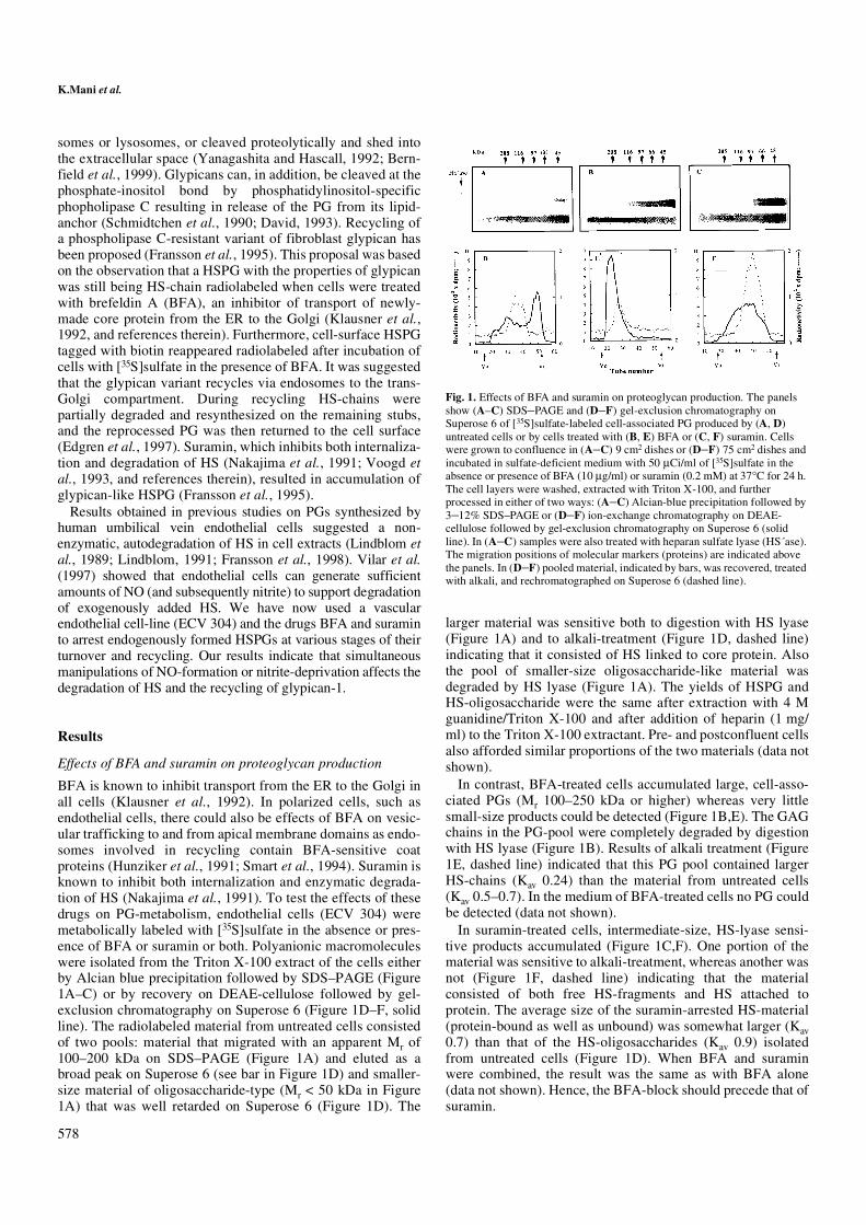
BFA is known to inhibit transport from the ER to the Golgi inall cells (Klausner et al., 1992). In polarized cells, such asendothelial cells, there could also be effects of BFA on vesic-ular trafficking to and from apical membrane domains as endo-somes involved in recycling contain BFA-sensitive coatproteins (Hunziker et al., 1991; Smart et al., 1994). Suramin isknown to inhibit both internalization and enzymatic degrada-tion of HS (Nakajima et al., 1991). To test the effects of thesedrugs on PG-metabolism, endothelial cells (ECV 304) weremetabolically labeled with [35S]sulfate in the absence or pres-ence of BFA or suramin or both. Polyanionic macromoleculeswere isolated from the Triton X-100 extract of the cells eitherby Alcian blue precipitation followed by SDS–PAGE (Figure1A–C) or by recovery on DEAE-cellulose followed by gel-exclusion chromatography on Superose 6 (Figure 1D–F, solidline). The radiolabeled material from untreated cells consistedof two pools: material that migrated with an apparent Mr of100–200 kDa on SDS–PAGE (Figure 1A) and eluted as abroad peak on Superose 6 (see bar in Figure 1D) and smaller-size material of oligosaccharide-type (Mr < 50 kDa in Figure1A) that was well retarded on Superose 6 (Figure 1D). The
larger material was sensitive both to digestion with HS lyase(Figure 1A) and to alkali-treatment (Figure 1D, dashed line)indicating that it consisted of HS linked to core protein. Alsothe pool of smaller-size oligosaccharide-like material wasdegraded by HS lyase (Figure 1A). The yields of HSPG andHS-oligosaccharide were the same after extraction with 4 Mguanidine/Triton X-100 and after addition of heparin (1 mg/ml) to the Triton X-100 extractant. Pre- and postconfluent cellsalso afforded similar proportions of the two materials (data notshown).
In contrast, BFA-treated cells accumulated large, cell-asso-ciated PGs (Mr 100–250 kDa or higher) whereas very littlesmall-size products could be detected (Figure 1B,E). The GAGchains in the PG-pool were completely degraded by digestionwith HS lyase (Figure 1B). Results of alkali treatment (Figure1E, dashed line) indicated that this PG pool contained largerHS-chains (Kav 0.24) than the material from untreated cells(Kav 0.5–0.7). In the medium of BFA-treated cells no PG couldbe detected (data not shown).
In suramin-treated cells, intermediate-size, HS-lyase sensi-tive products accumulated (Figure 1C,F). One portion of thematerial was sensitive to alkali-treatment, whereas another wasnot (Figure 1F, dashed line) indicating that the materialconsisted of both free HS-fragments and HS attached toprotein. The average size of the suramin-arrested HS-material(protein-bound as well as unbound) was somewhat larger (Kav0.7) than that of the HS-oligosaccharides (Kav 0.9) isolatedfrom untreated cells (Figure 1D). When BFA and suraminwere combined, the result was the same as with BFA alone(data not shown). Hence, the BFA-block should precede that ofsuramin.
Fig. 1. Effects of BFA and suramin on proteoglycan production. The panelsshow (A–C) SDS–PAGE and (D–F) gel-exclusion chromatography onSuperose 6 of [35S]sulfate-labeled cell-associated PG produced by (A, D)untreated cells or by cells treated with (B, E) BFA or (C, F) suramin. Cellswere grown to confluence in (A–C) 9 cm2 dishes or (D–F) 75 cm2 dishes andincubated in sulfate-deficient medium with 50 µCi/ml of [35S]sulfate in theabsence or presence of BFA (10 µg/ml) or suramin (0.2 mM) at 37°C for 24 h.The cell layers were washed, extracted with Triton X-100, and furtherprocessed in either of two ways: (A–C) Alcian-blue precipitation followed by3–12% SDS–PAGE or (D–F) ion-exchange chromatography on DEAE-cellulose followed by gel-exclusion chromatography on Superose 6 (solidline). In (A–C) samples were also treated with heparan sulfate lyase (HS´ase).The migration positions of molecular markers (proteins) are indicated abovethe panels. In (D–F) pooled material, indicated by bars, was recovered, treatedwith alkali, and rechromatographed on Superose 6 (dashed line).

Nitric oxide and heparan sulfate degradation
579
Glypican-1 proteoglycan accumulates in BFA-treated cells
The nature of the BFA-arrested HSPGs was determined byimmunodetection after electrophoresis of the core proteins andby direct immunoisolation from the cell extracts. To be able totrace the material during purification, PGs were metabolicallylabeled with [35S]sulfate. Cell-associated PG was extractedfrom the cell layer, recovered by passage over DEAE-cellu-lose, and purified by gel permeation chromatography onSuperose 6 (as in Figure 1E) followed by ion-exchange chro-matography on Mono Q (Figure 2A). PG material (see bar)was pooled and digested with HS lyase followed by SDS–PAGE. Western blot using immunostaining with monoclonalantibodies S1 or 2E9 specific for glypican-1 and syndecan-1/3,respectively, was then performed (David et al., 1990; Mertenset al., 1992). The result (see inset in Figure 2 A) showed onecore protein (66 kDa) reacting with glypican-1 antibody andanother one (125 kDa), which was either that of syndecan-3 oraggregated syndecan-1 core protein (Bernfield et al., 1999).
Radiolabeled, cell-associated glypican-1 was also directlyimmunoisolated using a polyclonal anti-glypican-1 antiserumand subjected to SDS–PAGE (Figure 2B). In untreated cellcultures, a relatively small, [35S]sulfate-labeled glypican-1glycoform (Mr approx. 100–200 kDa) was obtained from thecell extract, whereas smaller amounts of a larger form wererecovered from the medium. In BFA-treated cells a large, butpolydisperse glycoform of glypican-1 (Mr approx. 100–300kDa or higher) accumulated. A separate minor component ofMr approx. 90 kDa was also seen.
The BFA-arrested material is a precursor of the suramin-arrested material
Pulse-labeling of BFA-treated cells with [35S]sulfate followedby extraction, precipitation with Alcian blue and SDS–PAGEindicated that the accumulation of PG increased up to 12 h andthen remained relatively constant (data not shown). In a pulse-chase experiment, cells were incubated with [35S]sulfate for 12h and then chased in either drug-free or suramin-containingnonradioactive medium. Cells were extracted and aliquotswere chromatographed on Superose 6 (Figure 3). After 24 ofchase in drug-free medium, prelabeled PG material had beenconverted to intermediate-size products eluting in fractions29–38 (Figure 3B, solid line) and after 42 h most of this haddisappeared (Figure 3C, solid line). Throughout the chase, aminor portion of PG-material remained undegraded. Oligosac-charides, expected to elute in fractions 40–44, were not seen.No PG or oligosaccharide could be found in the medium (datanot shown). However, during chase in suramin-containingmedium, both intermediate-size products and oligosaccharidesaccumulated (Figure 3B,C, dashed lines). Hence, suramin canblock further degradation of HS-material released from theBFA-block.
Detection of glucosamine residues with unsubstituted aminogroups in heparan sulfate chains
The 35S-labeled HSPG and HSPG-degradation products thataccumulated in BFA- and suramin-treated cells, respectively,were isolated as described in Figure 1 and treated with alkali torelease protein-bound chains and chain fragments. Thesamples were then chromatographed on Superose 6 before andafter deaminative cleavage at pH 3.9 which is specific for
Fig. 2. Identification of glypican-1 PG forms. Cells were grown to confluencein (A) 175 cm2 or (B) 75 cm2 dishes and incubated in sulfate-deficient mediumwith 50 µCi/ml [35S]sulfate for (A) 24 h or (B) 16 h in the absence or presenceof BFA (BFA, 10 µg/ml). The cell layers were washed, extracted with TritonX-100 and (in A) polyanionic material from BFA-treated cells was collectedby passage over DEAE-cellulose, followed by gel-permeation chromatographyon Superose 6 (as in Figure 1E). Macromolecular [35S]material was pooled andsubjected to ion-exchange FPLC on Mono Q. Elution was performed with alinear gradient from 0.3 M NaCl to 1.2 M NaCl (dashed line). The elutionpositions of standard heparan sulfate (HS) and dermatan sulfate (DS) areindicated. The pooled PG material (see bar) was precipitated with ethanol,digested with HS-lyase and subjected to 10% SDS–PAGE under nonreducingconditions (see inset). After blotting to nylon membrane this wasimmunostained with (lane 1) monoclonal S1 antibody against glypican and(lane 2) monoclonal 2E9 antibody against syndecans 1 and 3. Molecular sizemarkers are indicated. Exposure times were 10 sec in lane 1 and 10 min in lane2. In (B) glypican-1 glycoforms were isolated from (M) media and (C) SDS-Triton-deoxycholate extracts of cells by immunoadsorption and subjected to3–12 % SDS–PAGE under reducing conditions. Bands were visualized byautoradiography.

K.Mani et al.
580
glucosamines with unsubstituted amino groups (Lindahl et al.,1973). It is seen (Figure 4A) that the large HS-chains derivedfrom the BFA-arrested PG (solid line) were partially degradedby nitrous acid to yield HS-fragments (dashed line) of the samesize as those accumulating in suramin-treated cells (Figure 4B,solid line). In contrast, the suramin-arrested chain fragmentswere not extensively depolymerized by nitrous acid at pH 3.9(Figure 4B, dashed line). Hence, cleavage at or near theGlcNH2 units of the HS-chains in the BFA-arrested PG seemsto take place when the suramin-arrested material is formed.
Although suramin-arrested HS-degradation products werenot depolymerized by nitrous acid, this does not exclude the
possibility that GlcNH2 units were present in, e.g., non-reducing terminal position. As endogenously produced nitrite
Fig. 3. Conversion of BFA-arrested proteoglycan to degradation productsduring chase. Cells grown to confluence in 25 cm2 dishes were (A) incubatedwith [35S]sulfate in the presence of BFA (10 µg/ml) for 12 h followed by chasein non-radioactive medium for (B) 24 h and (C) 42 h (solid line) withoutfurther additions and (dashed line) with the addition of suramin (0.2 mM). Themedia were removed and the cell layers were extracted with Triton X-100.Aliquots (200 µl) of the extracts were mixed with 8 M guanidine HCl (1:1) andchromatographed on Superose 6.
Fig. 4. Depolymerization at N-unsubstituted glucosamine in heparan sulfatechains. Gel chromatography on Superose 6 of HS-chains/fragments derivedvia alkaline elimination from (A) the BFA-arrested PG and (B) the suramin-arrested material. The products were analyzed (solid line) without furthertreatment and (dashed line) after treatment with HNO2 at pH 3.9. Confluentcells (25 cm2 dishes) were incubated in sulfate-deficient medium containing 50µCi/ml [35S]sulfate in the presence of BFA (10 µg/ml) or suramin (0.2 mM) at37°C for 24 h. The medium was removed and cells were washed and extractedwith Triton X-100. PG and PG-derived material of the cell extract wererecovered by passage over DEAE-cellulose, treated with alkali to releasechains from the core protein and again recovered on DEAE-cellulose.

Nitric oxide and heparan sulfate degradation
581
could remove such units (Lindahl et al., 1973; Vilar et al.,1997) experiments were designed to test whether nitrite depri-vation or generation could affect HSPG metabolism.
Inhibitors of nitrite and nitrite-generation affect proteoglycanand oligosaccharide production
Nitrite is formed by oxidation of NO which, in turn, isproduced by decomposition of arginine catalyzed by NO-
synthases, the constitutive form cNOS and the inducible formiNOS (for review, see Wink et al., 1996). NO is unstable andeither directly converted to nitrite or stored as protein-bound S-nitrosothiols. The latter release NO non-enzymatically in aprocess catalyzed by Cu2+ (for review, see Williams, 1996). Toexplore whether NO-derived nitrite was involved in HS-degra-dation, cells were pre-exposed to the NO-synthase inhibitorsN-methyl-arginine (inhibits both forms of NOS), N-nitro-arginine (inhibits preferentially cNOS) or aminoguanidine(inhibits preferentially iNOS) or to the nitrite-destroying agentammonium sulfamate. Cells were then incubated with radio-sulfate in the low-arginine medium 199 in the continued pres-ence of the respective compounds. As shown in Figure 5A–Etreatment with these compounds reduced generation of HS-oligosaccharides (fractions 40–45). The order of effectivenesswas N-methylarginine = sulfamate > aminoguanidine > N-nitroarginine. There was, however, no concomitant increase inthe radiolabeling of HSPG (fractions 20–25), as was seen whencells not deprived of nitrite were treated with BFA (Figure 5F).
When cells had been pre-treated with suramin, which wouldresult in accumulation of intermediate-size products (seeFigure 1F), and then radiosulfated in the presence of BFAaccumulation of large HSPG was resumed (Figure 5G)suggesting that the suramin-block also precedes the BFA-block in a cyclic process. Moreover, formation of large HSPGunder these conditions (Figure 5G) was markedly reduced bythe simultaneous presence of sulfamate (Figure 5H) suggestingthat a nitrite-dependent step could be located downstream ofthe suramin-block but upstream of the BFA-block. This wasfurther supported by the finding that formation of radiolabeledHSPG in BFA-treated cells was inhibited by pre-incubationwith aminoguanidine (Figure 5I) or sulfamate (Figure 5J).
To test whether nitrite was also involved in the degradationof HS-chain fragments and oligosaccharides, radiosulfate-labeled suramin-arrested material was chased in nonradioac-tive medium with or without sulfamate and the products wereanalyzed as in Figure 3. The turnover of HS-degradation prod-ucts was unaffected by the presence of sulfamate (data notshown). However, as shown above, supply of nitrite appearsnecessary to sustain formation of the BFA-arrested HSPG,perhaps by facilitating HS-chain extension. Hence, we testedwhether HS-chain extension and formation of the BFA-arrested HSPG could be restored in nitrite-deprived cells whenNO-donor was supplied.
Formation of BFA-arrested proteoglycan in nitrite-deprivedcells is restored by NO-donor
Cells were deprived of nitrite by treatment with a combinationof aminoguanidine (to inhibit iNOS), sulfamate (to destroynitrite), and neocuproine (to inhibit release of NO from S-nitrosothiols). The cells were then chased in fresh, BFA-containing medium with [35S]sulfate and increasing concentra-tions of NO-donor (sodium nitroprusside). [35S]PG wasisolated from the cell extracts by ion exchange chromatog-raphy. As seen in Figure 6A (solid circles) formation of BFA-arrested HSPG was gradually stimulated in the presence ofincreasing concentrations of NO-donor. Maximal stimulationoccurred at 300 µM nitroprusside or higher. The stimulatedPG-formation reached the same level as in cells not deprived ofnitrite and not treated with NO-donor (open circles).
Fig. 5. Effects of suramin and inhibitors of nitrite-generation on proteoglycanand oligosaccharide production in untreated and BFA-treated cells. Cellsgrown to confluence in 9 cm2-dishes were preincubated in the arginine-poormedium 199 for 24 h with (A) no addition or in the presence of (B) 10 mM N-methylarginine, (C) 10 mM N-nitroarginine, (D) 10 mM aminoguanidine or(E) 10 mM ammonium sulfamate. After addition of 50 µCi/ml [35S]sulfatecells were incubated for another 24 h (A) without or (B–E) with the samesubstances as during preincubation. In (F–J) cells were preincubated (F)without or with (G, H) 0.2 mM suramin, (I) 10 mM aminoguanidine or (J) 10mM ammonium sulfamate. During the radiolabeling period cells were treatedwith (F, G, I, J) 10 µg/ml of BFA or (H) both 10 µg/ml of BFA and 10 mMsulfamate. See also scheme above the panels. The cells were extracted withTriton X-100 and aliquots (200 µl) of the extracts were mixed with 8 MguanidineHCl (1:1) and directly chromatographed on Superose 6. BFA,brefeldin A; L-NMMA, N-methylarginine; L-NNA, N-nitroarginine; NH2-Gdn, aminoguanidine; NH4SO3NH2, ammonium sulfamate; SUR, suramin.

K.Mani et al.
582
To determine which type(s) of HSPG that is formed duringNO-stimulation, it was necessary to generate both protein- andcarbohydrate-labeled, BFA-arrested HSPG. Pilot experiments
indicated that radiolabeling of untreated cells with[35S]methionine generated immunoreactive and radiolabeledtruncated glypican-1, but radiolabeling was greatly reduced in
Fig. 6. Formation of BFA-arrested glypican proteoglycan is stimulated by NO-donor in nitrite-deprived cells. In (A) cells grown to confluence in 9 cm2 wells wereincubated in regular DMEM with the addition of 10 mM aminoguanidine, 10 mM ammonium sulfamate and 0.01 mM neocuproine for 24 h. The cells were then chasedin sulfate-deficient medium containing 50 µCi/ml [35S]sulfate, 10 µg/ml BFA and with the indicated concentrations of sodium nitroprusside as NO-donor for another15 h. PG-material was recovered from the detergent extracts of the cells by passage over DEAE-cellulose as described in Materials and methods. The eluted materialwas precipitated with ethanol dissolved in 4 M guanidine-HCl, and radioactivity was measured (solid circles). For comparison, the result obtained with cells notsubjected to nitrite-deprivation and chase-labeled in the absence of NO-donor is also shown (open circle). In (B–D) subconfluent cell cultures in 75 cm2 dishes wereincubated in serum-containing, low-methionine DMEM with [35S]methionine/cysteine (50 µCi/ml) until confluent (at least 24 h). Then 10 mM aminoguanidine, 10mM sulfamate and 0.01 mM neocuproine were added and the incubation was continued for another 24 h. Cells were then chased overnight in fresh regular mediumcontaining BFA (10 µg/ml) and [3H]glucosamine (20 µCi/ml) with or without addition of 300 µM sodium nitroprusside as NO-donor. Polyanionic material wasrecovered from the Triton X-100 extracts of both pulse-labeled and chase-labeled cells by passage over DEAE-cellulose. In (B) polyanionic material waschromatographed on Superose 6. The tracings show [3H]glucosamine-labeled material chased with BFA in (dashed line) the absence or (solid line) the presence of 300µM sodium nitroprusside as NO-donor. In (C, D) polyanionic, protein-radiosulfated material from (C) pulse-labeled cells or (D) from cells chase-labeled with[3H]glucosamine in the presence of BFA and NO-donor was first chromatographed on Superose 6 (data not shown), and then PG-like material (fractions 19–29, as inB) was subjected to chromatography on MonoQ. Elution was performed with a linear gradient from 0.3 M NaCl to 1.2 M NaCl (solid line). The elution positions ofstandard heparan sulfate (HS) and dermatan sulfate (DS) are indicated. This purified PG-material was again pooled (fractions 36–66 in C, D), concentrated, dialyzed,digested with HS lyase, precipitated with ethanol and subjected to 3–12% gradient SDS–PAGE under nonreducing conditions. The gel was impregnated withflurophore and dried, and bands were visualized by radioimaging (see insets in C, D). Molecular size markers are indicated.

Nitric oxide and heparan sulfate degradation
583
the BFA-arrested glypican-1 (as in Figure 2B) and insufficientfor core protein analysis (data not shown). Using [3H]leucine,suramin-arrested radiolabeled material (purified on MonoQ)could be detected (data not shown). Although an accumulationof this radiolabel could be seen in large HSPG after chase innonradioactive medium containing BFA, the amounts wereagain insufficient for core protein analysis (data not shown).We therefore tested whether sufficient amounts of radiolabeledcore protein precursor could be obtained by arresting materialat a nitrite-dependent step.
Subconfluent cell cultures were metabolically labeled with[35S]methionine/cysteine until cells were confluent (usuallywithin 24 h) and then radiolabeling was continued for another24 h in the presence of aminoguanidine, sulfamate, and neocu-proin to minimize the endogenous nitrite concentration. Cellswere then chase-labeled overnight in fresh BFA-containingmedium with [3H]glucosamine (to label the HS backbone) andin the absence or presence of 300 µM sodium nitroprusside asNO-donor. Radiolabeled PG was recovered from the cellextracts by passage over DEAE-cellulose and then chromato-graphed on Superose 6 (Figure 6B). It is seen that very little[3H]PG was formed in unstimulated cells (dashed line),whereas a large [3H]PG peak was obtained from stimulatedcells (solid line).
To obtain HSPG that could be used for core protein analysis,further purification by ion exchange chromatography onMonoQ was necessary in order to remove some non-PGprotein (Figure 6C,D). The [35S]methionine/cysteine-labeledPG recovered from (C) pulse-labeled and (D) chase-labeledcells were thus isolated (see bars in Figure 6C,D), HS-chainswere removed by digestion with HS lyase and [35S]coreproteins were subjected to SDS–PAGE (see inserts in Figure6C,D). After the pulse, a HSPG with a core protein of approx.120 kDa was seen (Figure 6C). After the chase in mediumcontaining BFA and NO-donor, an additional HSPG with a
core protein of ∼70 kDa appeared (Figure 6D). This size corre-sponds to that of glypican (David et al., 1990; Schmidtchen etal., 1990).
Discussion
Our results show that a transformed endothelial cell-linederived from human umbilical vein endothelial cells (ECV304) express glypican-1. In unperturbed cells glypican-1 ismostly in the form of HS-chain truncated glycoforms accom-panied by relatively large amounts of HS oligosaccharides(Figure 7). Very little large glypican PG is found associatedwith the cell layer but some is shed into the medium. In BFA-treated cells, large glypican PG accumulates and sheddingceases. Radiolabeling of the core protein in the BFA-arrestedPG was much reduced, as if there is an available pool of nonra-dioactive precursors. These could be HS-chain truncatedglycoforms in various stages of degradation or reconstructionas depicted in Figure 7. According to the proposed recyclingmodel, formation of HS-oligosaccharides should cease whenglypican-1 is arrested in the large PG form. The HS-oligo-saccharide pool then becomes depleted by terminal degrada-tion in lysosomes.
From the recycling model further predictions can be made.The large glypican PG should be the precursor of the suramin-arrested truncated form and recycling back to the BFA-arrestedform should be precluded by nitrite-deprivation. Accordingly,during chase the BFA-arrested glypican-1 PG was degraded byendoglycosidic cleavage, presumably in the vicinity of theGlcNH2 moieties, generating HS-chain fragments andoligosaccharides. Furthermore, during chase in the presence ofsuramin both truncated PG and HS fragments could becaptured. By arresting recycling material at the suramin stageand then chasing in the presence of BFA the large glypican PGwas reformed. This route was also abrogated by nitrite-depri-vation as was subsequent generation of HS-oligosaccharides.Core protein analysis revealed that glypican PG was reformedfrom prelabeled material in nitrite-deprived cells upon additionof NO-donor. During recycling no intermediates were seen inthe medium, suggesting that it was an intracellular process.
Degradation of HS-side chains could take place in severalstages (Figure 7). We propose an initial partial endoglycosidicdegradation on the nonreducing side of the GlcNH2 moieties ofHS, releasing relatively large HS-chain fragments, and gradu-ally generating a truncated PG with the GlcNH2 moieties nearthe nonreducing end of the stubs. Endoglycosidases (hepara-nase) capable of partially depolymerizing HS chains of HSPGhave been demonstrated in many cell types, including endothe-lial cells (Godder et al., 1991). In most cases, these endo-heparanases cleave β-D-glucuronidic linkages which wouldgenerate truncated HSPG with stubs containing nonreducingterminal glucosamine residues (for an extensive list of refer-ences, see Sandbäck-Pikas et al., 1998). We have previouslyobserved that HS oligosaccharides generated by endoglyco-sidic cleavage in fibroblasts had been cleaved at sites locatedclose to the heparinase I cleavage sites, i.e., at the nonreducingside of IdoUA(2-OSO3) (Schmidtchen and Fransson, 1994).Recent studies confirm this observation. CHO cell- (Bai et al.,1997) or human hepatoma- and platelet-derived endohepara-nases (Sandbäck-Pikas et al., 1998), which are not specific for
Fig. 7. Model of glypican-1 glycoforms and their turnover and recycling. TheGPI-anchored glypican-1 (Gpc-1) core protein is posttranslationallysubstituted with HS side-chains that contain a few GlcNH2 residues. It isproposed that glypican is partially degraded by endoglycosidic cleavage of HSforming a truncated PG and HS-oligosaccharides. Degradation proceeds untilthe GlcNH2 moieties are near the non-reducing end of the core protein stubs.These “telosaccharides” are then removed by the action of Nitrite (+) and thetruncated glypican is recycled while the HS-stubs are extended to form newfull-size side-chains. The steps blocked by BFA (BFA-) and suramin (Sur-) areindicated.

K.Mani et al.
584
the type of N-substitution of the adjacent glucosamine, have arequirement for 2-O-sulfate on neighboring hexuronic acidresidues located on the reducing side of the cleavage site (–) in,e.g., the following sequence: -GlcNR-GlcUA–GlcNR-HexUA(2-OSO3)-GlcNR- where R is an unspecified substit-uent and HexUA could be IdoUA. Cleavage of a glucuronidiclinkage a short distance from the non-reducing side of aGlcNH2 would thus generate the non-reducing terminalsequence GlcNR-HexUA(2-OSO3)-[GlcNR-HexUA]n-GlcNH2- in the core protein stubs (Figure 7). When n is smallor zero subsequent deaminative cleavage at the reducing sideof GlcNH2 would result in an undetectable/marginal effect onoverall chain/stub size. It is possible that these short non-reducing terminal GlcNH2-containing “telosaccharides” arecleaved off when sufficiently high concentrations of nitritehave been generated endogenously in a mildly acidic compart-ment providing fresh acceptor sites for HS chain extension(Figure 7). The “telosaccharides” may contain unexpectedfeatures, such as 3-O-sulfation. It is intriguing that certainisoforms of 3-O-sulfatase (e.g. 3-OST-3) recognize IdoUA(2-OSO3)-GlcNH2 repeats (Shukla et al., 1999). It should beadded that in fibroblasts, there may be no need for NO-derivednitrite as fibroblast HS chains do not appear to contain muchGlcNH2 (Fransson et al., 1995).
The intracellular location of the proposed events has notbeen specifically addressed. However, NO, the precursor ofnitrite and thus a potential regulator of glypican recycling, maybe formed both in caveolae and in endosomes. Constitutiveendothelial NO-synthase (cNOS/eNOS) targets to the cyto-plasmic side of caveolae when made lipophilic by acylation(Garcia-Cardenas et al., 1996; Shaul et al., 1996). However,NOS appears to be less active when it is associated with cave-olin, the structural scaffolding protein of caveolae (Feron et al.,1998; Prabhakar et al., 1998). Nevertheless, Vilar et al. (1997)have shown that endothelial cells in culture are capable ofgenerating sufficient amounts of NO (and subsequently nitrite)to support degradation of exogenously added HS. Oxidation ofNO to nitrite increases exponentially with NO-concentration(Wink et al., 1996). Therefore, effects on HS turnover may beseen when the amount of NO has reached a threshold level(e.g., >300 µM) in the local subcellular environment. This mayoccur when recycling glypican passes through the endosomalcompartment where NOS should be fully active. In this wayNO could regulate the level and structure of cell-surface HS.Specific structures in glypican HS-chains contribute to theanticoagulant potency of the intact vascular endothelium bybinding/activation of antithrombin III (Mertens et al., 1992). Itis thus interesting that prolonged inhibition of NO-synthasedecreases the expression of anticoagulant HS on endothelialcells (Irokawa et al., 1997). Recycling glypican may also beinvolved in internalization and recycling of HS-binding growthfactors and enzymes (reviewed in Bernfield et al., 1999) orpolyamines (Belting et al., 1999).
Materials and methods
The endothelial cell line ECV 304 was obtained from Prof.Inge Olsson, Department of Medicine, Lund University.Regular cell culture media, L-glutamine, penicillin-strepto-mycin, trypsin, and fetal bovine serum were obtained from
Life Technologies. Heparin and other GAG were the samepreparations as described earlier (Fransson et al., 1992). BFA,NG-methyl-L-arginine, Nω-nitro-L-arginine, aminoguanidine,neocuproine (2,9-dimethyl-1,10-phenanthrolin), and medium199 were purchased from Sigma, suramin (Germanine) fromBayer, and sodium nitroprusside from Fluka. Na2
35SO4 (1310Ci/mmol), D-[6-3H]glucosamine (40 Ci/mmol), L-[35S]-methionine/L-[35S]-cysteine, 7/3 (>1000 Ci/mmol, Pro-Mix),PVDF-membranes, and Hybond N were from AmershamInternational, UK. HS lyase (heparitinase) was purchased fromthe Seikagaku Corporation, Japan. The prepacked columns(Superose 6 HR 10/30 and Mono Q HR 5/5), protein A–Sepha-rose CL-4B and Dextran T-500 was from Pharmacia-LKB,Sweden, DE-53 DEAE-cellulose from Whatman and Bio-GelP-10 and protein standards from Bio-Rad. Centricon 30 waspurchased from Amicon UK and Alcian blue 8GS fromChroma-Gesellschaft, Germany. A rabbit antiserum againstglypican-1 was obtained after immunization with a 6-Histagged recombinant glypican-1 core protein comprising thesequence Ile 54 to Pro 519. To generate the protein humanglypican-1 cDNA was cleaved with BglII and StuI and ligatedinto the vector pQE32 (Qiagen) digested with BamHI/SmaI.The resulting plasmid was used to transform E.coli M15bacteria. Protein expression was induced with IPTG (GibcoBRC). 6-His tagged protein was purified in guanidineHCl on aNi2+-NTA-agarose column (Qiagen). The antibodies Mab S1and Mab 2E9 were kindly provided by Prof. Guido David,University of Leuven, Belgium. Chemiluminiscent Super-signal substrate was from Pierce. Other chemicals were ofanalytical grade.
Cell culture and radiolabeling
ECV 304 (a transformed vascular endothelial cell line) wascultured as monolayers in Dulbecco’s Modified Earle’sMedium (DMEM) supplemented with 10% (v/v) fetal bovineserum, 2 mM L-glutamine, penicillin (100 U/ml), and strepto-mycin (100 µg/ml) in an incubator with humidified atmosphereand 5% CO2 at 37°C. Confluent cells were preincubated for 1h in labeling medium supplemented with 2 mM glutamine andserum. The labeling medium was low-sulfate (0.05 mM)MgCl2-containing DMEM with serum. When NO-synthaseinhibitors/nitrite-quenchers were tested, cells were incubatedin a low-arginine medium (M 199). Drugs used were: BFA (10µg/ml) or suramin (0.2 mM). Radioactivity was measured byβ-scintillation.
Extraction and isolation of cell-associated proteoglycans
After the incubations, medium was collected and pooled withtwo washings with ice-cold PBS (0.137 M NaCl, 3 mM KCl,8 mM Na2HPO4, 2 mM KH2PO4, pH 7.5). Cells were extractedwith 0.1–0.2 ml/cm2 dish of 0.15 M NaCl, 10 mM EDTA, 2%(v/v) Triton X-100, 10 mM KH2PO4, pH 7.5, 5 µg/ml oval-bumin containing 10 mM N-ethylmaleimide, and 1 mM diiso-propylphosphoro-fluoridate on a slow shaker at 4°C for 10min. Isolation and purification of PG was performed accordingto two different methods. Procedure I included chromatog-raphy on DEAE-cellulose, Superose 6, and Mono Q, andprocedure II consisted of Alcian Blue precipitation (Björnsson,1993) followed by electrophoresis (SDS–PAGE).
In procedure I extracts were mixed with 1.3 vol. of 7 M urea,10 mM Tris, pH 7.5, 0.1% Triton X-100, 10 mM NEM and
Nitric oxide and heparan sulfate degradation
585
passed over a 1 ml column of DE-53 equilibrated with 6 Murea, 0.5 M NaOAc, pH 5.8, 5 µg/ml ovalbumin, 0.1% TritonX-100. After sample application, the columns were washedsuccessively with 10 ml portions of (1) equilibration buffer(see above); (2) 6 M urea, 10 mM Tris, pH 8.0, 5 µg/ml oval-bumin, 0.2% Triton X-100; and (3) 50 mM Tris pH 7.5. Boundmaterial was eluted with 5 × 1 ml 4 M guanidine-HCl, 50 mMNaOAc, pH 5.8, 5 µg/ml ovalbumin, 0.2% Triton X-100. Radi-oactive fractions were pooled, precipitated with 5 vol of 95%ethanol and 100 µg of dextran as carrier, overnight at –20°C.Samples were centrifuged in a Beckman JS-7.5 at 4000 r.p.m.and 4°C for 45 min and dissolved in 4 M guanidine-HCl, 50mM NaOAc, pH 5.8, 0.2% Triton X-100. They were thensubjected to gel permeation FPLC on Superose 6 at a flow rateof 0.4 ml/min in the same buffer. Radioactivity was deter-mined and fractions were pooled. Some fractions were chro-matographed on Mono Q after buffer-change on Centricon 30.The buffer used was 7 M urea, 10 mM Tris, pH 8.0, 0.1%Triton X-100 and the gradient was between 0.3 M NaCl (frac-tion 10) to 1.2 M NaCl (fraction 70) in the same buffer. Radio-active PGs were pooled and precipitated as above.
Immunoisolation
Cell medium was decanted, and the cell layer was washed withPBS and solubilized in PBS containing 0.1% (w/v) SDS, 0.5 %(v/v) Triton X-100, 0.5 % (w/v) sodium deoxycholate bypassage up and down a glass pipette at 4°C for 10 min. Afteraddition of PMSF (a 1/1000 dilution of a saturated solution inethanol), the medium and cell extract were treated with proteinA Sepharose CL-4B (1/100) for at least 1 h on a slow shaker at4°C. The supernatant was collected and treated with anti-glyp-ican-1 antiserum (diluted 1/200) at 4°C overnight. Immunecomplexes were recovered on protein A Sepharose, which waswashed six times with 0.15 M NaCl, 10 mM Tris, pH 7.4containing 0.2% (v/v) Tween 20. Bound material was releasedby boiling in SDS-buffer and subjected to PAGE.
Degradation procedures
GAG chains were released from PG by treatment with 0.5 MNaOH, 0.1 M NaBH4 at room temperature overnight. Sampleswere neutralized with HOAc, freeze-dried, and redissolved foranalysis by gel-permeation chromatography on Superose 6 asabove. Cleavage of N-unsubstituted glucosamines was carriedout with nitrous acid at pH 3.9 (Lindahl et al., 1973). HS-chains were also degraded with HS lyase (3 mU/ml) in thepresence of proteinase inhibitors as described (Fransson et al.,1995). Digestions were terminated by heating at 100°C for 1min. The volume of samples was reduced and buffer changeswere made by centrifugation in Centricon 30. Material wasrecovered by ethanol precipitation, dissolved in SDS-bufferand analyzed on SDS–PAGE. Carrier protein (ovalbumin) andHS or dextran (50–100 µg) were added prior to each purifica-tion and degradation step.
SDS–PAGE
Radiolabeled PG and HS lyase-treated PG were dissolved inSDS-buffer consisting of 5% (w/v) SDS, 20% (v/v) glycerol,4 mM EDTA, 0.04% bromphenol blue, 125 mM Tris–HCl, pH6.8, and 10% (v/v) β-mercaptoethanol. The samples wereboiled for 2 min before loading. For immunostaining gels wereequilibrated in transfer buffer (92 mM glycine, 0.01 M Tris,
pH 8.3, 20% metanol) for 30 min. Transfer to PVDF-membrane was carried out overnight at 4°C and a constantvoltage of 20 V. After a 1 h exposure to PBS containing 10%milk (3% fat), the membrane was incubated with 20 µg/mlMab S1 (anti-glypican) or 20 µg/ml Mab 2E9 (anti-syndecan1+3) in PBS containing 5% milk (washing buffer) for 2 h. Themembrane was rinsed twice for 5 min with washing buffer andfurther incubated for 1 h with horseradish peroxidase–conju-gated anti-mouse IgG diluted 1:5000 in washing buffer. Afterthree washings in washing buffer and two washings in TBS(100 mM Tris, 0.9% NaCl, pH 7.5), the membrane was finallydeveloped with Supersignal substrate for chemiluminiscenceand autoradiographed.
Acknowledgments
We thank Ms. B.Havsmark, S.Persson, and P.Wiik for tech-nical assistance and Prof. Guido David, University of Leuven,for generous gifts of cDNA and antibodies. The SwedishMedical and Technical Research Councils, the Cancer Fund,the Strategic Research Foundation (GLIBS), the J.A.Persson,Kock, Å.Wiberg, and A.Österlund Foundations, the SwedishMedical Society and the Swedish Society for MedicalResearch provided financial support.
Abbreviations
Brefeldin A, BFA; CS, chondroitin sulfate; ECV, endothelialcell-line of vascular origin; ER, endoplasmic reticulum; GAG,glycosaminoglycan; Gal, galactose; GlcNH2, D-glucosamine;GlcNAc, N-acetyl-D-glucosamine; GlcNSO3, N-sulfate-D-glucosamine; GlcNR, glucosamine with unspecified N-substit-uent; GlcUA, D-glucuronic acid; GPI, glycosylphosphatidyli-nositol; HexUA, unspecified hexuronic acid; HS, heparansulfate; HSPG, heparan sulfate proteoglycan; IdoUA, L-iduronic acid; NOS, nitric oxide synthase; PG, proteoglycan; -OSO3, sulfate ester; Xyl, xylose.
References
Anderson,R.G.W. (1998) The caveolae membrane system. Annu. Rev. Bio-chem., 67, 199–225.
Bai,X., Bame,K.J., Habuchi,H., Kimata,K. and Esko,J.D. (1997) Turnover ofheparan sulfate depends on 2-O-sulfation of uronic acids. J. Biol. Chem.,272, 23172–23179.
Belting,M., Persson,S. and Fransson,L.-Å. (1999) Proteoglycan involvementin polyamine uptake. Biochem. J., 338, 317–323.
Bernfield,M., Götte,M., Park,P.W., Reizes,O., Fitzgerald,M.L., Lincecum,J.and Zako,M. (1999) Functions of cell surface heparan sulfate proteogly-cans. Annu. Rev. Biochem., 68, 729–777.
Björnsson,S. (1993) Simultaneous preparation and quantitation of proteogly-cans by precipitation with Alcian Blue. Anal. Biochem., 210, 282–291.
David,G. (1993) Integral membrane heparan-sulfate proteoglycans. FASEB J.,7, 1023–1030.
David,G., Lories,V., Decock,B., Marynen,P., Cassiman J.-J. and Van denBerghe,H. (1990) Molecular cloning of a phosphatidylinositol-anchoredmembrane heparan sulfate proteoglycan from human lung fibroblasts. J.Biol. Chem., 267, 4870–4877.
Edgren,G., Havsmark,B., Jönsson,M. and Fransson L.-Å. (1997) Glypican(heparan sulfate proteoglycan) is palmitoylated, deglycanated and reglyca-nated during recycling in skin fibroblasts. Glycobiology, 7, 103–112.
Feron,O., Saldana,F., Michel,J.B. and Michel,T. (1998) The endothelial nitric-oxide synthase-caveolin regulatory cycle. J. Biol. Chem., 273, 3125–3128.
K.Mani et al.
586
Fransson,L.-Å., Karlsson,P. and Schmidtchen,A. (1992) Effects of cyclohex-imide, brefeldin A, suramin, heparin and primaquine on proteoglycan andglycosaminoglycan biosynthesis in human embryonic skin fibroblasts.Biochim. Biophys. Acta, 1137, 287–297.
Fransson,L.-Å., Edgren,G., Havsmark,B. and Schmidtchen,A. (1995) Recy-cling of a glycosylphosphatidyl-inositol-anchored heparan sulfate prote-oglycan (glypican) in skin fibroblasts. Glycobiology, 5, 407–415.
Fransson,L.-Å., Belting,M., Edgren,G., Jönsson,M., Mani,K., Schmidtchen,A.and Wiik,P. (1998) Degradation and reprocessing of heparan sulfate inrecycling glypican (heparan sulfate proteoglycan). Trends Glycosci. Gly-cotechnol., 10, 81–94.
Garcia-Cardenas,G., Oh,P., Liu,J., Schnitzer,J.E. and Sessa,W.C. (1996) Tar-geting of nitric oxide synthase to endothelial cell caveolae via palmitoyla-tion: implications for nitric oxide signaling. Proc. Natl. Acad. Sci. USA, 93,6448–6453.
Godder,K., Vlodavsky,I., Eldor,A., Weksler,B.B., Haimovitz-Friedmann,A.and Fuks,Z. (1991) Heparanase activity in cultured endothelial cells. J.Cell Physiol., 148, 274–280.
Humphries,D.E., Sullivan,B.M., Aleixo,M.D. and Stow,J.L. (1997) Localiza-tion of human heparan glucosaminyl N-deacetylase/N-sulfotransferase tothe trans-Golgi network. Biochem. J., 325, 351–357.
Hunziker,W., Whitney,A. and Mellman,I. (1991) Selective inhibition of tran-scytosis by BFA in MDCK cells. Cell, 67, 617–627.
Irokawa,M., Nishinaga,M., Ikeda,U., Shinoda,Y., Suematsu,M., Gouda,N.,Ishimura,Y. and Shimada,K. (1997) Endothelial-derived nitric oxide pre-serves anticoagulant heparan sulfate expression in cultured porcineendothelial cells. Atherosclerosis, 135, 9–17.
Klausner,R.D., Donaldson,J.G. and Lippincott-Schwartz,J. (1992) BFA:insight into the control of membrane traffic and organelle structure. J. CellBiol., 116, 1071–1080.
Lindahl,U., Bäckström,G., Jansson,L. and Hallén,A. (1973) Biosynthesis ofheparin II. Formation of sulfamino groups. J. Biol. Chem., 248, 7234–7241.
Lindahl,U., Kusche-Gullberg,M. and Kjellén,L. (1998) Regulated diversity ofheparan sulfate. J. Biol. Chem., 273, 24979–24982.
Lindblom,A. (1991) Structure of endothelial cell proteoglycans. Characteriza-tion of biosynthetic and degradative components in tissue culture. Ph.D.thesis, Lund University, Sweden.
Lindblom,A., Carlstedt,I. and Fransson,L.-Å. (1989) Identification of the coreproteins in proteoglycans synthesized by vascular endothelial cells. Bio-chem. J. 261, 145–153.
Mertens,G., Cassiman,J.-J., Van den Berghe,H., Vermylen,J. and David,G.(1992) Cell surface heparan sulfate proteoglycan from human vascularendothelial cells. J. Biol. Chem., 267, 20435–20443.
Misra,K.B., Kim,K.C., Cho,S.Y., Low,M.G. and Bensadoun,A. (1994) Purifi-cation and characterization of adipocyte heparan sulfate proteoglycanswith affinity for lipoprotein lipase. J. Biol. Chem., 269, 23838–23844.
Nakajima,M., DeChavigny,A., Johnson,C.E., Hamada,J., Stein,C.A. andNicolson,G.L. (1991) Suramin. A potent inhibitor of melanoma hepara-nase and invasion. J. Biol. Chem., 266, 9661–9666.
Norgard-Sumnicht,K. and Varki,A. (1995) Endothelial heparan sulfate prote-oglycans that bind to L-selectin have glucosamine residues with unsubsti-tuted amino groups. J. Biol. Chem., 270, 12012–12024.
Prabhakar,P., Thatte,H.S., Goetz,R.M., Cho,M.R., Golan,D.E. and Michel,T.(1998) Receptor-regulated translocation of endothelial nitric-oxide syn-thase. J. Biol. Chem., 273, 27383–27388.
Sandbäck-Pikas,D., Li,J.-P., Vlodavsky,I. and Lindahl,U. (1998) Substratespecificity of heparanases from human hepatoma and platelets. J. Biol.Chem., 273, 18770–18777.
Schmidtchen,A. and Fransson,L.-Å. (1994) Analysis of heparan sulfate chainsand oligosaccharides from proliferating and quiescent fibroblasts. A pro-posed model for endoheparanase activity. Eur. J. Biochem., 223, 211–221.
Schmidtchen,A., Sundler,R. and Fransson,L.-Å. (1990) A fibroblast heparansulfate proteoglycan with a 70 kDa core protein is linked to membranephosphatidylinositol. Glycoconjugate J., 7, 563–572.
Seidenbecher,C.I., Richter,K., Rauch,U., Fässler,R., Garner,C.C. and Gundelf-inger,E.D. (1995) Brevican, a chondroitin sulfate proteoglycan of rat brain,occurs as secreted and cell surface glycosylphosphatidylinositol-anchoredisoforms. J. Biol. Chem., 270, 27206–27212.
Shaul,P.W., Smart,E.J., Robinson,L.J., German,Z., Yuhanna,I.S., Ying,Y.Anderson,R.G.W. and Michel,T. (1996) Acylation targets endothelialnitric-oxide synthase to plasmalemmal caveolae. J. Biol. Chem., 271,6518–6522.
Shukla,D., Liu,J., Blaiklock,P., Shworak,N.W., Bai,X., Esko,J.D.,Cohen,G.H., Eisenberg,R.J., Rosenberg,R.D. and Spear,P.G. (1999) Anovel role for 3-O-sulfated heparan sulfate in herpes simplex virus 1 entry.Cell, 99, 13–22.
Smart,E.J., Ying,Y.S., Conrad,P.A. and Anderson,R.G. (1994) Caveolinmoves from caveolae to the Golgi apparatus in response to cholesterol oxi-dation. J. Cell Biol., 127, 1185–1197.
Taipale,J. and Keski-Oja,J. (1997) Growth factors in the extracellular matrix.FASEB J., 11, 51–59.
van den Born,J., Gunnarsson,K., Bakker,M.A.H., Kjellén,L., Kusche-Gull-berg,M., Maccarana,M., Berden,J.H.M. and Lindahl,U. (1995) Presence ofN-unsubstituted glucosamine units in native heparan sulfate revealed by amonoclonal antibody. J. Biol. Chem., 270, 31303–31309.
Vilar,R.E., Ghael,D., Li,M., Bhagat,D.D., Arrigo,L.M., Cowman,M.K.,Dweck,H.S. and Rosenfeld,L. (1997) Nitric oxide degradation of heparinand heparan sulfate. Biochem. J., 324, 473–479.
Voogd,T.E., Vansterkenburg,L.M., Wilting,J. and Janssen,L.H.M. (1993)Recent research on the biological activity of suramin. Pharmacol. Rev., 45,177–203.
Williams,D.L.H. (1996) S-Nitrosothiols and role of metal ions in decomposi-tion to nitric oxide. Methods Enzymol., 268, 299–308.
Wink,D.A., Grisham,M.B., Mitchell,J.B. and Ford,P.C. (1996) Direct andindirect effects of nitric oxide in chemical reactions relevant to biology.Methods Enzymol., 268, 12–31.
Yanagashita,M. and Hascall,V.C. (1992) Cell surface heparan sulfate prote-oglycans. J. Biol. Chem., 267, 9451–9454.
Copyright © 2022 FDOKUMEN