Examination of the substrate specificity of heparin and heparan sulfate lyases
7
Biochemistry 1990, 29, 261 1-2617 261 1 Rosing, J., Tans, G., Govers-Riemslag, J. W. P., Zwaal, R. F. A., & Hemker, H. C. (1980) J. Biol. Chem. 255, Rosing, J., vanRijn, J. L. M. L., Bevers, E. M., vanDieijen, G., Comfurius, P., & Zwaal, R. F. A. (1985) Blood 65, Schiffman, S., Rapaport, S. I., & Chong, M. M. Y. (1966) Proc. SOC. Exp. Biol. Med. 123, 736-740. Segel, I. H. (1 975) Enzyme Kinetics. Behavior and Analysis of Rapid Equilibrium and Steady-State Enzyme Systems, p 957, Wiley, New York. Silverberg, S. A., Nemerson, Y., & Zur, M. (1977) J. Biol. Chem. 252, 8481-8488. Sinha, D., Seaman, F. S., Koshy, A., Knight, L. C., & Walsh, P. N. (1984) J .Clin. Invest. 73, 1550-1556. Thompson, A. R. (1977) J. Clin. Invest. 59, 900-910. Tracy, P. B., Peterson, J. M., Nesheim, M. E., McDuffie, F. C., & Mann, K. G. (1979) J. Biof. Chem. 254, 274-283. 3 19-332. 10354-10361. Tracy, P. B., Nesheim, M. E., & Mann, K. G. (1981) J. Biol. Chem. 256, 743-75 1. vanDieijen, G., Tans, G., Rosing, J., & Hemker, H. C. (1981) J. Biol. Chem. 256, 3433-3442. vanDieijen, G., vanRijn, J. L. M. L., Govers-Riemslag, J. W. P., Hemker, H. C., & Rosing, J. (1982) Thromb. Hae- mostasis 53, 396-400. vanRijn, J., Rosing, J., & vanDieijen, G. (1983) Eur. J. Biochem. 133, 1-10. Walsh, P. N. (1978) Br. J. Haematol. 40, 31 1-331. Walsh, P. N., & Biggs, R. (1972) Br. J. Haematol. 22, Walsh, P. N., & Griffin, J. H. (1981) Blood 57, 106-118. Walsh, P. N., Sinha, D., Koshy, A., Seaman, F. S., & Brad- Zur, M., & Nemerson, Y. (1980) J. Biof. Chem. 255, 743-760. ford, H. (1986) Blood 68, 225-230. 5701-5705. Examination of the Substrate Specificity of Heparin and Heparan Sulfate Lyases? R. J. Linhardt,**t J. E. Turnbull,$ H. M. Wang,' D. Loganathan,' and J. T. Gallagher" Division of Medicinal and Natural Products Chemistry, College of Pharmacy, University of Iowa, Iowa City, Iowa 52242, and Department of Clinical Research and Cancer Research Campaign, Department of Medical Oncology, University of Manchester, Christie Hospital and Holt Radium Institute, Wilmslow Road, Manchester M2O 9BX, U.K. Received September 8, 1989; Revised Manuscript Received November 15, 1989 ABSTRACT: We have examined the activities of different preparations of heparin and heparan sulfate lyases from Flavobacterium heparinum. The enzymes were incubated with oligosaccharides of known size and sequence and with complex polysaccharide substrates, and the resulting degradation products were analyzed by strong-anion-exchange high-performance liquid chromatography and by oligosaccharide mapping using gradient polyacrylamide gel electrophoresis. Heparinase (EC 4.2.2.7) purified in our laboratory and a so-called Heparinase I (Hep I) from a commercial source yielded similar oligosaccharide maps with heparin substrates and displayed specificity for di- or trisulfated disaccharides of the structure -4)-a-~-GlcNp2S(6R)( l-+- 4)-a-~-IdoAp2S( 1- (where R = Qsulfo or OH). Oligosaccharide mapping with two different commercial preparations of heparan sulfate lyase [heparitinase (EC 4.2.2.8)] indicated close similarities in their de- polymerization of heparan sulfate. Furthermore, these enzymes only degraded defined oligosaccharides at hexosaminidic linkages with glucuronic acid: -4)-a-~-GlcNpR( 1+4)-P-~-GlcAp( 1- (where R = N-acetamido or N-sulfo). The enzymes showed activity against solitary glucuronate-containing disaccharides in otherwise highly sulfated domains including the saccharide sequence that contains the antithrombin binding region in heparin. A different commercial enzyme, Heparinase I1 (Hep 11), displayed a broad spectrum of activity against polysaccharide and oligosaccharide substrates, but mapping data indicated that it was a separate enzyme rather than a mixture of heparinase and heparitinase/Hep 111. When used in conjunction with the described separation procedures, these enzymes are powerful reagents for the structural/sequence analysis of heparin and heparan sulfate. H e p a r i n and heparan sulfate are complex, sulfated co- polymers of alternating 1-4-linked glucosamine and hexuronic acid. Both biopolymers have been implicated in a diverse range Supported by the Burroughs Wellcome Fund in the form of a Re- search Travel Grant, Cancer Research Campaign (J.T.G.), and by Na- tional Institutes of Health Grants HL29797 and GM38060 (R.J.L.). J.E.T. acknowledges receipt from Christie Hospital of a Research Travel Grant. * To whom correspondence should be addressed. $University of Iowa. 5 Department of Clinical Research, University of Manchester, Christie Hospital and Holt Radium Institute. 'I Cancer Research Campaign, Department of Medical Oncology, University of Manchester, Christie Hospital and Holt Radium Institute. of biological functions and therapeutic uses [for reviews, see Gallagher et al. (1986) and Linhardt and Loganathan (1989)l. Polysaccharide lyases (EC 4.2.2) are a class of enzymes that depolymerize certain acidic polysaccharides through an elim- inative mechanism. This enzymatic reaction results in an unsaturated uronic acid residue at the nonreducing terminal sugar in the resulting oligosaccharide product (Linhardt et al., 1986a). Flavobacterium heparinum is a soil isolate (Payza & Korn, 1956) capable of utilizing either heparin or heparan sulfate as its sole carbon and nitrogen source (Galliher et al., 1981). The growth of this organism on these polymeric sub- strates is dependent on its production of a variety of enzymes including lyases, glucuronidases, sulfoesterases, and sulf- amidases (Galliher et al., 1982). These polysaccharide lyases 0006-2960/90/0429-2611$02.50/0 0 1990 American Chemical Society
Transcript of Examination of the substrate specificity of heparin and heparan sulfate lyases

Biochemistry 1990, 29, 261 1-2617 261 1
Rosing, J., Tans, G., Govers-Riemslag, J. W. P., Zwaal, R. F. A., & Hemker, H. C. (1980) J . Biol. Chem. 255,
Rosing, J., vanRijn, J. L. M. L., Bevers, E. M., vanDieijen, G., Comfurius, P., & Zwaal, R. F. A. (1985) Blood 65,
Schiffman, S . , Rapaport, S. I., & Chong, M. M. Y. (1966) Proc. SOC. Exp. Biol. Med. 123, 736-740.
Segel, I . H. (1 975) Enzyme Kinetics. Behavior and Analysis of Rapid Equilibrium and Steady-State Enzyme Systems, p 957, Wiley, New York.
Silverberg, S. A., Nemerson, Y., & Zur, M. (1977) J . Biol. Chem. 252, 8481-8488.
Sinha, D., Seaman, F. S., Koshy, A., Knight, L. C., & Walsh, P. N. (1984) J .Clin. Invest. 73 , 1550-1556.
Thompson, A. R. (1977) J . Clin. Invest. 59, 900-910. Tracy, P. B., Peterson, J. M., Nesheim, M. E., McDuffie, F.
C., & Mann, K. G. (1979) J . Biof. Chem. 254,
274-283.
3 19-332.
10354-10361. Tracy, P. B., Nesheim, M. E., & Mann, K. G. (1981) J . Biol.
Chem. 256, 743-75 1. vanDieijen, G., Tans, G., Rosing, J., & Hemker, H. C. (1981)
J . Biol. Chem. 256, 3433-3442. vanDieijen, G., vanRijn, J. L. M. L., Govers-Riemslag, J. W.
P., Hemker, H. C., & Rosing, J. (1982) Thromb. Hae- mostasis 53, 396-400.
vanRijn, J., Rosing, J., & vanDieijen, G. (1983) Eur. J . Biochem. 133, 1-10.
Walsh, P. N. (1978) Br. J . Haematol. 40, 31 1-331. Walsh, P. N., & Biggs, R. (1972) Br. J . Haematol. 22,
Walsh, P. N., & Griffin, J. H. (1981) Blood 57, 106-118. Walsh, P. N., Sinha, D., Koshy, A., Seaman, F. S., & Brad-
Zur, M., & Nemerson, Y. (1980) J . Biof. Chem. 255,
743-760.
ford, H. (1986) Blood 68, 225-230.
5701-5705.
Examination of the Substrate Specificity of Heparin and Heparan Sulfate Lyases? R. J. Linhardt,**t J. E. Turnbull,$ H. M. Wang,' D. Loganathan,' and J. T. Gallagher"
Division of Medicinal and Natural Products Chemistry, College of Pharmacy, University of Iowa, Iowa City, Iowa 52242, and Department of Clinical Research and Cancer Research Campaign, Department of Medical Oncology, University of Manchester,
Christie Hospital and Holt Radium Institute, Wilmslow Road, Manchester M2O 9BX, U.K. Received September 8, 1989; Revised Manuscript Received November 15, 1989
ABSTRACT: We have examined the activities of different preparations of heparin and heparan sulfate lyases from Flavobacterium heparinum. The enzymes were incubated with oligosaccharides of known size and sequence and with complex polysaccharide substrates, and the resulting degradation products were analyzed by strong-anion-exchange high-performance liquid chromatography and by oligosaccharide mapping using gradient polyacrylamide gel electrophoresis. Heparinase (EC 4.2.2.7) purified in our laboratory and a so-called Heparinase I (Hep I) from a commercial source yielded similar oligosaccharide maps with heparin substrates and displayed specificity for di- or trisulfated disaccharides of the structure -4)-a-~-GlcNp2S(6R)( l-+- 4)-a-~-IdoAp2S( 1- (where R = Qsulfo or OH). Oligosaccharide mapping with two different commercial preparations of heparan sulfate lyase [heparitinase (EC 4.2.2.8)] indicated close similarities in their de- polymerization of heparan sulfate. Furthermore, these enzymes only degraded defined oligosaccharides a t hexosaminidic linkages with glucuronic acid: -4) -a -~-GlcNpR( 1+4)-P-~-GlcAp( 1- (where R = N-acetamido or N-sulfo). The enzymes showed activity against solitary glucuronate-containing disaccharides in otherwise highly sulfated domains including the saccharide sequence that contains the antithrombin binding region in heparin. A different commercial enzyme, Heparinase I1 (Hep 11), displayed a broad spectrum of activity against polysaccharide and oligosaccharide substrates, but mapping data indicated that it was a separate enzyme rather than a mixture of heparinase and heparitinase/Hep 111. When used in conjunction with the described separation procedures, these enzymes are powerful reagents for the structural/sequence analysis of heparin and heparan sulfate.
H e p a r i n and heparan sulfate are complex, sulfated co- polymers of alternating 1-4-linked glucosamine and hexuronic acid. Both biopolymers have been implicated in a diverse range
Supported by the Burroughs Wellcome Fund in the form of a Re- search Travel Grant, Cancer Research Campaign (J.T.G.), and by Na- tional Institutes of Health Grants HL29797 and GM38060 (R.J.L.). J.E.T. acknowledges receipt from Christie Hospital of a Research Travel Grant.
* To whom correspondence should be addressed. $University of Iowa. 5 Department of Clinical Research, University of Manchester, Christie
Hospital and Holt Radium Institute. 'I Cancer Research Campaign, Department of Medical Oncology,
University of Manchester, Christie Hospital and Holt Radium Institute.
of biological functions and therapeutic uses [for reviews, see Gallagher et al. (1986) and Linhardt and Loganathan (1989)l.
Polysaccharide lyases (EC 4.2.2) are a class of enzymes that depolymerize certain acidic polysaccharides through an elim- inative mechanism. This enzymatic reaction results in an unsaturated uronic acid residue at the nonreducing terminal sugar in the resulting oligosaccharide product (Linhardt et al., 1986a). Flavobacterium heparinum is a soil isolate (Payza & Korn, 1956) capable of utilizing either heparin or heparan sulfate as its sole carbon and nitrogen source (Galliher et al., 1981). The growth of this organism on these polymeric sub- strates is dependent on its production of a variety of enzymes including lyases, glucuronidases, sulfoesterases, and sulf- amidases (Galliher et al., 1982). These polysaccharide lyases
0006-2960/90/0429-2611$02.50/0 0 1990 American Chemical Society

2612
include heparinase (EC 4.2.2.7), which has been purified to homogeneity (Yang et al., 1985), and heparitinase (EC 4.2.2.8). Heparinase acts a t the +4)-cu-~-GlcNp2S6S( 1-- 4)-a-~-ldoApZS( 1- linkage, heparin's major disaccharide repeating unit. Heparinase does not require 6-sulfation to act and can tolerate 3-sulfation in the glucosamine residue (Rice & Linhardt, 1989). The heparitinase enzymes, initially de- scribed as heparitinase l and 2 (Linker & Hovingh, 1972), have not yet been purified to homogeneity. The commercially available heparitinase (EC 4.2.2.8) is described by the man- ufacturer (Seikagaku Kogyo Co.) as capable of cleaving - 4)-a-~-GlcNp(2-acetamido or 2-sulfate)( 1-4)-P-~-GlcAp- (1- and can tolerate 6-sulfation in the glucosamine residue. There is, however, only limited data available on the activity of heparan sulfate lyase against low molecular weight sub- strates (Rice & Linhardt, 1989), and there is some controversy regarding the precise number of different polysaccharide lyases produced by F. heparinum that are capable of acting on either heparin or heparan sulfate.
Recently, several polysaccharide lyases have been introduced onto the market which are capable of acting on heparin and/or heparan sulfate. These enzymes have demonstrated utility in the mapping and sequencing of heparin and heparan sulfate (Linhardt et al., 1988a; Turnbull & Gallagher, 1988) and in the preparation of potentially useful therapeutic agents (Linhardt et al., 1986b; Linhardt & Loganathan, 1989).
I n the present paper we examine all of the commercially available polysaccharide lyases capable of acting on either heparin or heparan sulfate. These enzymes are applied to polymeric substrates in an effort to understand their range of specificities. The products of these reactions are analyzed by both gradient PAGE (Turnbull & Gallagher, 1988; Rice et al., 1987) and SAX-HPLC (Linhardt et al., 1988a) oligo- saccharide mapping techniques.
EXPERIMENTAL PROCEDURES
Materials Heparin, sodium salt, was obtained from Hepar (Porcine
Mucosal, lot ST 82261, 145 units/mg). Heparan sulfate, sodium salt, from porcine intestine (ORG 553) was a gift from Organon, Oss, The Netherlands. Heparinase (heparin lyase, EC 4.2.2.7) was purified from Flavobacterium heparinum [5 units/mg (kmol min-l mg-I)] (Yang et al., 1985) or prepared by Seikagaku Kogyo Co. Ltd., Tokyo, Japan, and obtained from ICN Immunobiologicals, Lisle, IL. Heparinase (He- parinase I) was also obtained from Sigma (1.5 units/mg) or as Heparinase I directly from Grampian Enzymes, Aber- deenshire, Scotland. Heparitinase from F. heparinum (he- paran sulfate lyase, EC 4.2.2.8) was prepared by Seikagaku Kogyo Co. An alternative source of heparitinase (designated as Heparinase 111 or Hep 111) was obtained from Sigma or directly from Grampian Enzymes. Heparinase I1 (no assigned EC number) was obtained from Sigma or directly from Grampian Enzymes. Chondroitinase ABC (chondroitin ABC lyase, EC 4.2.2.4) from P. vulgaris was obtained from ICN Immunobiologicals. Dialysis tubing Spectropore 3500 was from Spectrum Medical Industries, Los Angeles, CA. HPLC was performed on dual Constametric I1 pumps connected through a gradient mixer from LDC, Milton Roy, Riveria Beach, FL. Fixed-loop injector 7125 from Rheodyne, Cotati, CA, and variable-wavelength UV-5 detector from ISCO, Lincoln, NE, were used.
Gradient control and data collection used an Apple IIe microcomputer running Chromatochart software from In- teractive Microware, State College, PA. Strong-anion-ex-
Biochemistry, Vol. 29, No. 10, 1990 Linhardt et al.
change (SAX) HPLC was performed on a Spherisorb (5-pm particle size) column of dimensions 0.46 X 25 cm from Phase Separations, Norwalk, CT. UV spectroscopy used a Shimadzu Model UV- 160 spectrophotometer equipped with a thermo- stated cell. A 32-cm vertical slab gel unit (SE620) was sup- plied by Hoefer Scientific Instruments (San Francisco, CA). Tris and SP-Sephadex C-50 were supplied by Sigma Chemical Co. Acrylamide and N,N'-methylenebis(acry1amide) (Electran grade) were obtained from BDH Chemicals. Bio-Gel P-2 (fine) and Coomassie dye reagent were from Bio-Rad, Rich- mond, CA. All other chemicals were reagent grade.
Methods Preparation of Polymer Substrates. Heparin was prepared
by dissolution of approximately 20 mg into 1 mL of water and dialysis (in M , 3500 cutoff bags at 4 "C) first against 10 volumes of 1 M sodium chloride followed by 3 X 1000 volumes of deionized water. After dialysis the heparin was removed from the dialysis bags and syringe filtered into a preweighed vial, freeze-dried, and stored desiccated over anhydrous cal- cium chloride for 2 days. The heparin sample was then carefully weighed, and the appropriate volume of distilled water was added to obtain a 20 mg/mL stock of solution. Heparan sulfate (1 1 mL at 50 mg/mL) was prepared in 0.25 M sodium acetate and 2.5 mM calcium acetate solution, ad- justed to pH 7.0, and 10 IU of chondroitinase ABC was added. The reaction mixture was incubated for 24 h at 37 "C after which the pH was adjusted to 3.0 with concentrated hydro- chloric acid. The entire reaction mixture was added to a 15 X 0.5 cm SP-Sephadex column (preequilibrated with hydro- chloric acid at pH 3.0). The column was washed with 15 mL of pH 3.0 hydrochloric acid, and the combined eluent was adjusted to pH 7.0 with 1 M sodium hydroxide and dialyzed overnight at 4 "C against 1000 volumes of distilled water. The nondiffusible material was freeze-dried, stored desiccated for 2 days, and prepared in distilled water at 20 mg/mL.
Preparation of Oligomeric Substrates. Heparin (100 mg at 16 mg/mL) was depolymerized at 30 "C with 62 mIU/mL heparinase (prepared and purified in our laboratories) in a solution of 0.2 M sodium chloride and 5 mM sodium phos- phate a t pH 7.0. The reaction was monitored by removing aliquots and measuring A232m after a 1:lOO dilution in 30 mM hydrochloric acid. At 98% reaction completion, the reaction was terminated by heating at 100 OC for 1 min. The sample was desalted on a 35 X 2.5 cm Bio-Gel P-2 column and fractionated on a semipreparative SAX-HPLC column as previously described (Rice & Linhardt, 1989). Five major oligosaccharide products were collected. A second fraction- ation of these oligosaccharides on the same column followed by desalting and freeze-drying resulted in their recovery at >95% purity (Rice & Linhardt, 1989). The structures of these five oligosaccharides (Fl-F5) representing 48, 8.8, 13, 12, and 5.5 wt % of the heparin starting material were determined as described previously (Linhardt et al., 1988a) and are shown in Figure 1.
Heparan sulfate (60 mg at 4 mg/mL) was similarly depo- lymerized with 90 mIU of heparitinase (Seikagaku Kogyo), and two disaccharides were prepared having the structures (6) AUA( 1+4)-a-~-GlcNpAc and (7) AUA( 1-4)-cu-~- GlcNp2S, where AUA is 4-deoxy-cu-~-threo-hex-4-enopyran- uronic acid (Linhardt et al., 1989).
Determination of Enzymatic Activities. Heparin or heparan sulfate (40 pL of 20 mg/mL) was added to 350 pL of 5 mM sodium phosphate and 200 mM sodium chloride solution, adjusted to pH 7.0, at 30 "C in a microcuvette. Enzyme, approximately 15 mIU in 10 pL, was added to substrate and

Heparin and Heparan Sulfate Lyases Biochemistry, Vol. 29, No. IO, 1990 2613
Gradient PAGE of Glycosaminoglycan Oligosaccharides. Polyacrylamide linear gradient resolving gels ( T 20-30% acrylamide) were prepared as described by Turnbull and Gallagher (1988). A linear gradient was formed between the glass plates (32 cm X 16 cm X 0.75 mm) from the bottom (maximum gel concentration, T 30%/C 5%) upward to the top (minimum gel concentration, T 20%/C 0.5%).
Immediately before electrophoresis the gel surface was rinsed three times with stacking-gel buffer and stacking-gel solution ( T 5%/C 0.5% acrylamide in stacking-gel buffer) was applied to the top of the resolving gel. A 15 X 5 mm well former comb was inserted between the glass plates. After polymerization (1 5 min) the comb was removed, and the wells were rinsed three times with electrophoresis buffer, after which the gel unit was placed into the electrophoresis tank.
Oligosaccharide samples (5-30 pL) containing approxi- mately 10% (v/v) glycerol and a trace quantity of Phenol Red were carefully layered onto the bottom of the wells with a microsyringe. Marker samples (10 pL) containing trace quantities of Bromophenol Blue and Phenol Red in 10% (v/v) glycerol were also run on each gel. Electrophoresis was then performed as follows. Samples were initially run into the gel at 150 V for 30 min, followed by electrophoresis at 300 V for 16 h, and finally 1000 V for 1 h (total approximately 6000 V-h). Under these conditions the Phenol Red marker dye migrates to within approximately 3 cm of the bottom of the resolving gel. Throughout the run heat was dissipated by use of a heat exchanger with circulating tap water (10-15 "C). Oligosaccharides were visualized by staining with Alcian blue and Azure A as described previously (Rice et al., 1987).
RESULTS The particular porcine mucosal heparin used as a polymeric
substrate was chosen because it contained no detectable der- matan/chondroitin sulfates (Linhardt et al., 1988a). The mucosal heparan sulfate preparation was selected because of its availability in sufficient quantities to permit the charac- terization of its two major disaccharide products and because of its similarity (by oligosaccharide mapping) to commercially available bovine kidney heparan sulfate. Contaminating dermatan/chondroitin sulfates were removed from this prep- aration by exhaustive treatment with chondroitinase ABC followed by dialysis.
Selective Depolymerization of N-Sulfated Polysaccharides. The activities of the commercially available polysaccharide lyases obtained from Flavobacterium heparinum were mea- sured with heparin and heparan sulfate under saturating conditions (Table I ) . This measurement facilitated the use of equivalent enzymatic treatment (mIU-h) per mole of sub- strate in subsequent experiments. Both heparin and heparan sulfate (1 mg/500 pL) were treated with 15 mIU for 8 h, and the reaction was followed kinetically to demonstrate that it was complete. The results of this study are presented in Table I. These results clearly identify heparin as the primary sub- strate for heparinase and Hep I , while heparan sulfate is the primary substrate for heparitinase and Hep 111. Hep I1 acts to a significant degree on both heparin and heparan sulfate. The polymeric substrates, containing multiple cleavable sites, are substantially better substrates than are the oligomeric substrates having only a single site at which the enzyme can act (Rice & Linhardt, 1989). Kinetic studies involving he- parinase (our preparation) demonstrate a V,,,/apparent K , that was 10000-fold higher for the polymeric substrate heparin (containing on the average 10 cleavable sites) than for oli- gomeric substrates containing a single cleavable site (Rice & Linhardt, 1989). A similar but less pronounced difference of
2 H
5 I nkso;
FIGURE 1 : Structure of oligosaccharide substrates 2-5. Linkages labeled a-e with arrows designate those cleaved by the enzymes as described under Results and in Table 11.
the change in absorbance was measured continuously at 232 nm in a thermostated UV spectrophotometer. The initial rate [dA232 (I-cm path lengthldt) (min)] was measured over the first 9 min of the reaction, and the final absorbance at 232 nm was measured directly after 100 min.
Treatment of Substrates with Enzymes. Polymeric sub- strate, heparin or heparan sulfate, was added to 425 p L of 0.2 M sodium chloride and 5 mM sodium phosphate (50 p L containing 20 mg/mL substrate), pH 7.0. Enzyme (25 pL, 15 mIU) was added to make the total volume of the solution 500 pL. The reactions were run at the appropriate temper- atures (heparinase/Hep I , 30 "C; heparitinase/Hep 111, 43 "C; Hep 11, 30 "C) to completion in 8 h. The depolymeri- zation reactions were terminated by heating at 100 "C for 1 min (Rice et al., 1987). Aliquots (4, 40, and 180 pL) were removed and added to 1 mL of 0.03 M hydrochoric acid to obtain a total volume of 1 mL, and the absorbance at 232 nm was determined.
Oligomeric substrate in 0.2 M sodium chloride and 5 mM sodium phosphate (50 p L containing 12 pg of substrate), pH 7.0, was treated with enzyme (25 mIU), and the reaction was run at the appropriate temperature for 24 h. The reactions were terminated by addition of concentrated hydrochloric acid, resulting in a solution at pH 3.0. The reaction mixture was poured onto an SP-Sephadex microcolumn (100-pL volume) equilibrated with hydrochloric acid at pH 3.0 to remove protein. After the column had been washed with 2 mL of pH 3.0 hydrochloric acid, the combined eluent and washes were adjusted to pH 7.0 with 1 M sodium hydroxide and freeze- dried.
Analytical SAX-HPLC Analysis of Enzyme- Treated Sub- strates. Enzyme-treated heparin, heparan sulfate, or hepa- rin-oligosaccharide was injected in amounts between 4 and 40 pg onto an analytical Spherisorb, 5-pm particle size (0.46 X 25 cm) SAX-HPLC column equilibrated with 0.2 M sodium chloride, pH 3.5. The sample was eluted from the column with a linear gradient [concentration (y, M) at any time (x, s) = 0.0002~ + 0.21 of sodium chloride at pH 3.5 and a flow rate of 1.5 mL/min. The elution profile was monitored by the absorbance at 232 nm at 0.02 absorbance unit full scale (AUFS). The amount of the resulting oligosaccharide prod- ucts was assessed by computer integration of peak area using a standard curve. Peaks were tentatively identified by either coelution with an authentic sample or by retention time and confirmed by comparison of each with purified oligosaccharide standards analyzed on a gradient polyacrylamide gel (Rice et al., 1987).

2614 Biochemistry. Vol. 29. No. IO. 1990 Linhardt et al.
Table I: Susceptibility of Heparin and Hcparan Sulfate to Ravobacterial Polysaccharide Lyases @mol of
product/100 pg av no. of act. (IU/vial)c
heparinase H 0.100 0.100 0.050 7 HS nd nd 0.025 5
Hep I H 0.118 0.200 nd nd HS nd nd nd nd
Hep I1 H 1.686 1.666 0.101 14 HS nd nd 0.117 25
HS 0.324 0.200 0.094 20
HS 1.136 0.800 0.108 23
enzyme? substrate& measured specified of rubstrat& sites cleavede
heparitinase H nd nd 0.007 I
Hep 111 H nd nd 0.007 I
*Substrate is at I mg/500 pL, and enzyme is at I5 mlU/500 pL. & H is heparin; HS is heparan sulfate. <Heparinase prepared in our laboratory by the method of Yang et al. (1986) has ken demonstrated to be identical with Seikagaku Kogyo heparinase purchased from ICN Immunobio- logicals (Linhardt et 81.. 1988a; Rice & Linhardt. 1989). Hep I and Hep I I prepared by Grampian Enzymes were obtained from Sigma. Hepari- tinase prepared by Scikagaku Kogyo was obtained from ICN Immunobiologicals. Heparinase, Hep I, and Hep I I activities were measured with heparin as the polymeric substrate at 30, 30, and 25 OC. respectively. Heparitinase and Hep 111 were assayed with heparan sulfate as the polymeric substrate at 43 OC. Specified activity is the manufacturer's activity converted to international units (IU = 1 mmol/min). None of the enzymes examined had measurable amounts of glycuronidase or sulfatase activity (Yang et 81.. 1986). nd is not determined. dCalculated from A,,, of sample diluted in 30 mM hydrochloric acid with a molar ahrptivity of 5200 (Linhardt et al., 1988a). eCalculated from micromoles of product pr micromoles of substrate. The estimated M, lor H is 14000 and for HS is 21 600.
BE:
DI 3
1 2 3 4 5 6 7 8 9 1 0
..-. ~ ---
. 7
FIGURE 2 Gradient PAGE oligosaccharide maps of polysaccharide lyase depolymerized heparin and heparan sulfate. Polymeric substrates were depolymerized and the resulting oligosaccharide mixtures (100 pg each) separated by gradient PAGE and visualized by staining with Azure A, as described under Methods. Heparan sulfate was depc- lymerized with ( I ) heparinase (Sigma). (2) Hep I I , (3) Hep 111. (4) heparitinax. and (5) simultaneous digestion with heparinase (Sigma) and Hep 111. Heparin was depolymerized with (6) heparinasc (Sigma). (7) Hep 11. (8) Hep 111. (9) heparitinase, and (IO) heparinase (Sigma) followed by Hep 111. The migration positions of Bromophenol Blue (BB). a contaminating product (PR) in BB, and disaccharides (Di) were as indicated. The sample in lane 2 showed no banding between PR and Di, and the samples in lanes 8 and 9 ran above BB, showing no discrete banding. 50-fold was observed for heparitinase (Seikagaku Kogyo) examined on heparan sulfate and oligomeric substrates.
Oligosaccharide Mapping. The products of these reactions were then examined by oligosaccharide mapping using SAX- HPLC and gradient PAGE oligosaccharide mapping as de- scribed under Methods (Figures 2 and 3). In all cases,
I I1 D
/ I B
m 0)
Eluate VOI. (ml) FIGURE 3 SAX-HPLC oligosaccharide map of polysaccharide lyase depolymerized polymeric substrates. Chromatograms plotted as elution volume (mL) versus Am (0.02 AUFS) are of oligosaccharide products formed by (A) heparinase acting on heparin, (B) heparinase acting on heparan sulfate, (C) heparitinase acting on heparan sulfate. (D) heparitinase acting on heparin, (E) Hep I1 acting on heparin, and (F) Hep 11 acting on heparan sulfate.
multiple bands or peaks representing polysaccharide depo- lymerization products were observed. Heparitinase and Hep 111 yielded identical PAGE maps with either heparan sulfate (Figure 2, tracks 3 and 4) or heparin (tracks 8 and 9 ) as substrates, indicating that these are the same enzymes. Clearly, heparinase has quite distinct activity from that of the heparitinases on both substrates (tracks I and 6). The oli- gosaccharide maps obtained for Hep I1 were intriguing. This enzyme brought about extensive degradation of heparin (track 7) and heparan sulfate (track 2) to predominantly di- saccharides. The maps with heparan sulfate as substrate were not reproduced by the combined actions of heparinase and heparitinase whether used simultaneously (track 5 ) or se- quentially in either order (results not shown). Similar results were obtained with heparin as substrate (track IO), confirming the distinctive action of this enzyme. Hep 11 must have a broad specificity (see Table II), but the resistant linkages in the polymeric substrates have not been defined. Recent work suggests that the Hep I1 resistant oligosaccharides in heparan

Heparin and Heparan Sulfate Lyases Biochemistry, Vol. 29, No. 10, 1990 2615 ~~
Table I I : Susceptibility of Defined Oligosaccharide Substrates to Polysaccharide Lyases
enzyme substrate linkage products tested used cleaved recovere&
heparinase 2 3 4 5
Hep 1 2 3 4 5
3 4 5
3 4 5
Hep 111 2 3 4 5
Hep l l 2
heparitinase 2
a 1, 9 none
b 1 none
a 1, 9 none
b 1 none
U 1, 9 C 1, 10 b 1 d , e 1, 8, 11
none C 1, 10
none e 12,11
none C 1, 10
none e 12.11
44)-a-D-GlcNpZS( I+~)-CY-L-I~OAPZS( I + . * 44)-(u-D- GlcNpZS6S( I+4)-c~-~-ldoApZS( 1 4 . C+4)-~-D-GI~Np2S6S( 1-4)- p-~-GlcAp( I+. d+4)-a-~-GlcNp2S6S( I+4)-a-~-IdoAp(l+. e+-
4)-a-~-GlcNpAcbS( 1 -4)-fi-~-GlcAp( I+. f 1 is AUApZS( 1 4 4 ) - ~ - GlcNpZS6S; 8 is AUAp( 1+4)-~-GlcNpAc6S; 9 is AUApZS( 1+4)-~- GlcNpZS; 10 is AUAp( 1+4)-~-GlcNpZS6S; 11 is AUAp( 1 4 4 ) - ~ - GlcNpZS3S6S; 12 is AUApZS( 1-4)-a-D-GlcNpZS6S( 1+4)-a-~- IdoAp( 144)-~-GlcNpAc6S. Where one product is observed, it is obtained as 2 equiv, and where two or three products are observed, each is obtained in 1 equiv with respect to the substrate.
sulfate from human skin fibroblasts are largely composed of sequences of the type 44) -a-~-GlcNpAc( 1+4)-P-~-GlcAp- (1- (Turnbull and Gallagher, unpublished data).
Quantitative data on the oligosaccharide breakdown prod- ucts were acquired with SAX-HPLC mapping procedures (see Methods). The action of heparinase on heparin gave the identified major oligosaccharides, 1-5 and 9, and unidentified minor oligosaccharide components (Figure 3A), and Hep I gave an identical result (result not shown). Heparitinase acting on heparan sulfate gave two disaccharides, 6 and 7, together with additional unidentified oligosaccharide components (Figure 3C), and Hep 111 produced an identical result (result not shown). Hep I1 acts extensively on heparin, giving a mixture of primarily disaccharide products (Figure 3E). Hep I 1 acts on heparan sulfate giving disaccharides 6 and 7, along with a large number of unidentified products (Figure 3F). The patterns obtained for Hep I1 are clearly unlike those obtained with heparinase/Hep I or heparitinase/Hep 111. No combi- nation (simultaneous or sequential in either order) of hepar- inase (or Hep 1) and heparitinase (or Hep 111) on either polymeric substrate can be used to obtain the same product distribution resulting from the action of Hep I1 (results not shown).
Degradation of Defined Oligosaccharide Substrates. He- parin oligosaccharides of known composition and sequence (Figure 1 ) prepared as described under Methods were used as substrates for the heparin and heparan sulfate lyases. Three of these oligosaccharides are tetrasaccharides that contain just one potentially susceptible hexosaminidic linkage. In all of the tetrasaccharides the hexosamine is N-sulfated; the adjacent hexuronate is a-~-IdoAp2S in 2 and 4 and P - D - G ~ c A ~ in 3. The other oligosaccharide, 5, is a hexasaccharide with two internal hexosamines, one being a-~-GlcNp2S6S linked to iduronate and the other being a-~-GlcNpAc6S linked to glucuronate.
I I HNSO; oso;
HNY ox C
Q0Q\ CHzoX Po; HNY OH
FIGURE 4: Glycosidic linkages that can be cleaved by (A) heparinase and Hep I, by (B) Hep 11, and by (C) heparitinase and Hep 111, where X = SO3- or H and Y = SO3- or COCH,. From the defined oli- gomeric substrates available, it is unclear which enzymes, if any, are capable of acting at a linkage containing a 2-sulfated glucuronic acid or a free glucosamine residue.
Heparinase and Hep I only cleave tetrasaccharides 2 and 4, which have internal a-~-IdoAp2S. These structures were resistant to heparitinase and Hep 111 (Table 11). The latter enzymes degraded 3 and 5, but these were poor substrates requiring high enzyme concentrations and long reaction times. Degradation of 5 produced tetrasaccharides and disaccharides of the structure expected if the hexosaminidic linkage was cleaved adjacent to glucuronic acid, Le., -+4)-a-~- GlcNpAc6S( 1-+4)-P-~-GlcAp( 1- (Table 11). Clearly, these enzymes can tolerate 6-0-sulfation of glucosamine (N-sulfated or N-acetylated) as shown by cleavage of 3 and 5, respectively.
The linkage specificities of the enzymes examined, based on our data (both current and previous), are summarized in Figure 4. Details of permissible variations, as far as they have been established, are also given.
DISCUSSION The biological importance of heparin and heparan sulfates
has led a number of research groups to undertake efforts to map and ultimately to sequence these polymers. Several ap- proaches have been applied to these ends. Much has been learned about the structure of heparin and heparan sulfate from studying their biosynthesis in cell culture (Lindahl et al., 1986; Lindahl & KjellCn, 1987). Alternatively, nitrous acid can be used to chemically depolymerize either heparin or heparan sulfate into oligosaccharides, and following frac- tionation, their structure can be determined (Bienkowski & Conrad, 1985). We have primarily exploited polysaccharide lyases to enzymatically depolymerize heparin and heparan sulfate. By use of this approach, oligosaccharides have been prepared, and their structure has been defined (Merchant et al., 1985; Linhardt et al., 1986b, 1989; Loganathan et al., 1990). In addition to preparing oligosaccharide standards, polysaccharide lyases and nitrous acid have been used to de- termine patterns of depolymerization in both heparin (Linhardt et al., 1988a; Loganathan et al., 1990) and heparan sulfate (Gallagher & Walker, 1985; Turnbull & Gallagher, 1988) with oligosaccharide mapping techniques.
Heparinase (EC 4.2.2.7) and heparitinase (EC 4.2.2.8) first became commerically available in the early 1980s. In the past 2 years, Hep I, 11, and 111 made their commercial debut and a series of heparitinase [I and I1 (from F. heparinum) and IV and V (from Flavobacterium sp. Hp206)] may soon be
2616
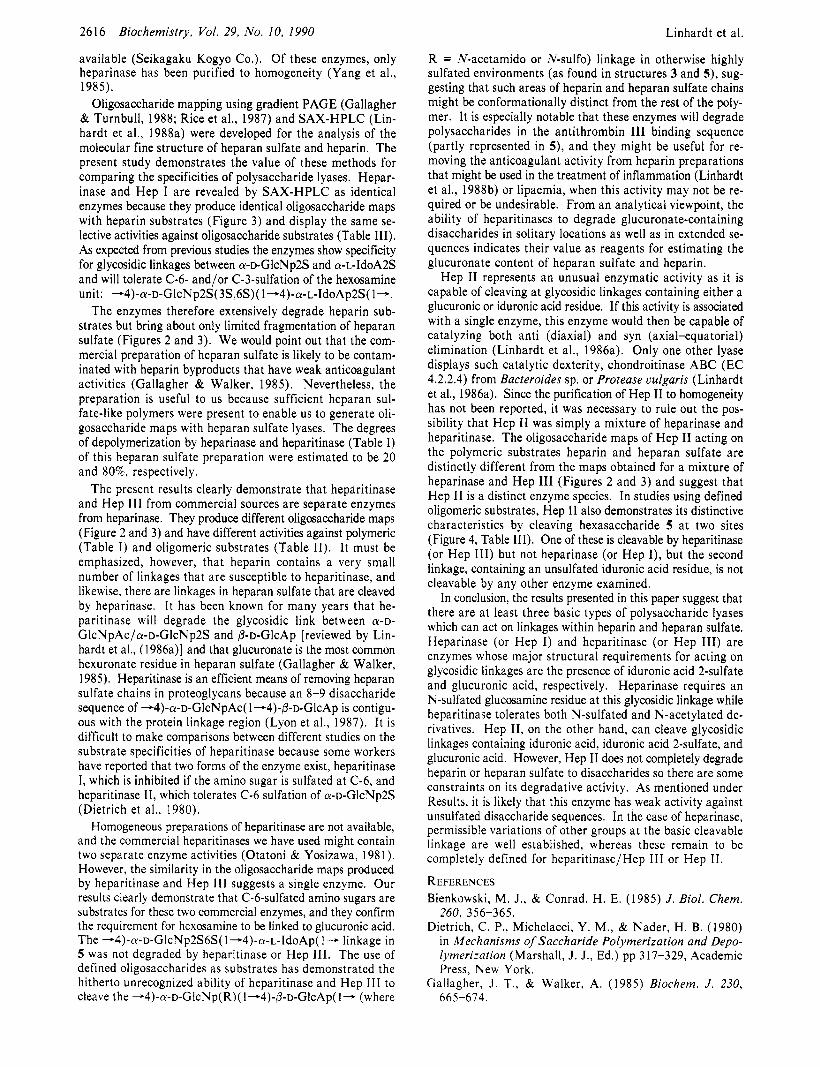
available (Seikagaku Kogyo Co.). Of these enzymes, only heparinase has been purified to homogeneity (Yang et al., 1985).
Oligosaccharide mapping using gradient PAGE (Gallagher & Turnbull, 1988; Rice et al., 1987) and SAX-HPLC (Lin- hardt et al., 1988a) were developed for the analysis of the molecular fine structure of heparan sulfate and heparin. The present study demonstrates the value of these methods for comparing the specificities of polysaccharide lyases. Hepar- inase and Hep I are revealed by SAX-HPLC as identical enzymes because they produce identical oligosaccharide maps with heparin substrates (Figure 3) and display the same se- lective activities against oligosaccharide substrates (Table 111). As expected from previous studies the enzymes show specificity for glycosidic linkages between a-~-GlcNp2S and a-~-IdoA2S and will tolerate C-6- and/or C-3-sulfation of the hexosamine unit: -4)-a-~-GlcNp2S(3S,6S)( 1+4)-a-~-IdoAp2S( 1-.
The enzymes therefore extensively degrade heparin sub- strates but bring about only limited fragmentation of heparan sulfate (Figures 2 and 3). We would point out that the com- mercial preparation of heparan sulfate is likely to be contam- inated with heparin byproducts that have weak anticoagulant activities (Gallagher & Walker, 1985). Nevertheless, the preparation is useful to us because sufficient heparan sul- fate-like polymers were present to enable us to generate oli- gosaccharide maps with heparan sulfate lyases. The degrees of depolymerization by heparinase and heparitinase (Table I) of this heparan sulfate preparation were estimated to be 20 and 80%, respectively.
The present results clearly demonstrate that heparitinase and Hep 111 from commercial sources are separate enzymes from heparinase. They produce different oligosaccharide maps (Figure 2 and 3) and have different activities against polymeric (Table I ) and oligomeric substrates (Table 11). It must be emphasized, however, that heparin contains a very small number of linkages that are susceptible to heparitinase, and likewise, there are linkages in heparan sulfate that are cleaved by heparinase. It has been known for many years that he- paritinase will degrade the glycosidic link between WD- GlcNpAc/a-~-GlcNp2S and P - D - G ~ c A ~ [reviewed by Lin- hardt et al., (1986a)l and that glucuronate is the most common hexuronate residue in heparan sulfate (Gallagher & Walker, 1985). Heparitinase is an efficient means of removing heparan sulfate chains in proteoglycans because an 8-9 disaccharide sequence of --4)-a-~-GlcNpAc( 1 -4)-P-~-GlcAp is contigu- ous with the protein linkage region (Lyon et al., 1987). It is difficult to make comparisons between different studies on the substrate specificities of heparitinase because some workers have reported that two forms of the enzyme exist, heparitinase I, which is inhibited if the amino sugar is sulfated a t (2-6, and heparitinase TI, which tolerates C-6 sulfation of a-~-GlcNp2S (Dietrich et al., 1980).
Homogeneous preparations of heparitinase are not available, and the commercial heparitinases we have used might contain two separate enzyme activities (Otatoni & Yosizawa, 198 1). However, the similarity in the oligosaccharide maps produced by heparitinase and Hep 111 suggests a single enzyme. Our results clearly demonstrate that C-6-sulfated amino sugars are substrates for these two commercial enzymes, and they confirm the requirement for hexosamine to be linked to glucuronic acid. The -4)-a-~-GlcNp2S6S( 1+4)-a-~-IdoAp( 1- linkage in 5 was not degraded by heparitinase or Hep 111. The use of defined oligosaccharides as substrates has demonstrated the hitherto unrecognized ability of heparitinase and Hep 111 to cleave the +4)-a-~-GlcNp(R)( 1 -4)-P-~-GlcAp( 1- (where
Biochemistry, Vol. 29, No. 10, 1990 Linhardt et al.
R = N-acetamido or N-sulfo) linkage in otherwise highly sulfated environments (as found in structures 3 and 5 ) , sug- gesting that such areas of heparin and heparan sulfate chains might be conformationally distinct from the rest of the poly- mer. It is especially notable that these enzymes will degrade polysaccharides in the antithrombin I11 binding sequence (partly represented in 5) , and they might be useful for re- moving the anticoagulant activity from heparin preparations that might be used in the treatment of inflammation (Linhardt et al., 1988b) or lipaemia, when this activity may not be re- quired or be undesirable. From an analytical viewpoint, the ability of heparitinases to degrade glucuronate-containing disaccharides in solitary locations as well as in extended se- quences indicates their value as reagents for estimating the glucuronate content of heparan sulfate and heparin.
Hep I1 represents an unusual enzymatic activity as it is capable of cleaving at glycosidic linkages containing either a glucuronic or iduronic acid residue. If this activity is associated with a single enzyme, this enzyme would then be capable of catalyzing both anti (diaxial) and syn (axial-equatorial) elimination (Linhardt et al., 1986a). Only one other lyase displays such catalytic dexterity, chondroitinase ABC (EC 4.2.2.4) from Bacteroides sp. or Protease vulgaris (Linhardt et al., 1986a). Since the purification of Hep I1 to homogeneity has not been reported, it was necessary to rule out the pos- sibility that Hep I1 was simply a mixture of heparinase and heparitinase. The oligosaccharide maps of Hep I1 acting on the polymeric substrates heparin and heparan sulfate are distinctly different from the maps obtained for a mixture of heparinase and Hep 111 (Figures 2 and 3) and suggest that Hep I1 is a distinct enzyme species. In studies using defined oligomeric substrates, Hep I1 also demonstrates its distinctive characteristics by cleaving hexasaccharide 5 a t two sites (Figure 4, Table 111). One of these is cleavable by heparitinase (or Hep 111) but not heparinase (or Hep I), but the second linkage, containing an unsulfated iduronic acid residue, is not cleavable by any other enzyme examined.
In conclusion, the results presented in this paper suggest that there are a t least three basic types of polysaccharide lyases which can act on linkages within heparin and heparan sulfate. Heparinase (or Hep I) and heparitinase (or Hep 111) are enzymes whose major structural requirements for acting on glycosidic linkages are the presence of iduronic acid 2-sulfate and glucuronic acid, respectively. Heparinase requires an N-sulfated glucosamine residue at this glycosidic linkage while heparitinase tolerates both N-sulfated and N-acetylated de- rivatives. Hep 11, on the other hand, can cleave glycosidic linkages containing iduronic acid, iduronic acid 2-sulfate, and glucuronic acid. However, Hep I1 does not completely degrade heparin or heparan sulfate to disaccharides so there are some constraints on its degradative activity. As mentioned under Results, it is likely that this enzyme has weak activity against unsulfated disaccharide sequences. In the case of heparinase, permissible variations of other groups a t the basic cleavable linkage are well established, whereas these remain to be completely defined for heparitinase/Hep 111 or Hep 11.
REFERENCES Bienkowski, M. J., & Conrad, H. E. (1985) J. Biol. Chem.
Dietrich, C. P., Michelacci, Y. M., & Nader, H. B. (1980) in Mechanisms of Saccharide Polymerization and Depo- lymerization (Marshall, J. J., Ed.) pp 317-329, Academic Press, New York.
Gallagher, J. T., & Walker, A. (1985) Biochem. J. 230,
260, 356-365.
66 5-674.

Biochemistry 1990, 29, 261 7-2622 2617
Gallagher, J. T., Lyon, M., & Steward, W. P. ( 1 986) Biochem. J . 236, 313-325.
Galliher, P. M., Cooney, C. L., Langer, R., & Linhardt, R. J. (1 98 1 ) Appl. Environ. Microbiol. 41, 360-365.
Galliher, P. M., Linhardt, R. J., Conway, L. J., Langer, R., & Cooney, C. L. (1982) Eur. J . Appl. Microbiol. 15,
Lindahl, U., & Kjellin, L. (1987) in The Biology of the Ex- tracellular Matrix: Biology of Proteoglycans (Wight, T. N., & Mecham, R. P., Eds.) pp 59-104, Academic Press, New York.
Lindahl, U., Feingold, D. S., & Rodin, L. (1986) Trends Biochem. Sci. 11, 221-225.
Linhardt, R. J., & Loganathan, D. (1989) in Biomimetic Polymers (Gebelein, G., Ed.) Plenum Press, New York (in press).
Linhardt, R. J., Cooney, C. L., & Galliher, P. M. (1986a) Appl. Biochem. Biotechnol. 12, 135-177.
Linhardt, R. J., Rice, K. G., Merchant, Z. M., Kim, Y. S., & Lohse, D. L. (1986b) J . Biol. Chem. 261, 14448-14454.
Linhardt, R. J., Rice, K. G., Kim, Y. S., Lohse, D. L., Wang, H. M., & Loganathan, D. (1988a) Biochem. J . 254,
Linhardt, R. J., Rice, K. G., Kim, Y. S., Engelken, J., &
252-257.
78 1-787.
Weiler, J. (1988b) J . Biol. Chem. 263, 13090-13096. Linhardt, R. J., Gu, K. N., Loganathan, D., & Carter, S. R.
(1989) Anal. Biochem. 181, 288-296. Linker, A., & Hovingh, P. (1972) Methods Enzymol. 28,
Loganathan, D., Wang, H. M., Mallis, L. M., & Linhardt, R. J. (1990) Biochemistry (in press).
Lyon, M., Steward, W. P., Hampson, I. N., & Gallagher, J. T. (1987) Biochem. J . 242, 493-498.
Merchant, Z. M., Kim, Y. S., Rice, K. G., & Linhardt, R. J. (1985) Biochem. J . 229, 369-377.
Ototani, N., & Yosizawa, Z. (1981) Glycoconjugates, Vlth International Symposium (Yamakawa, T., Osawa, T., & Handa, S., Eds.) pp 41 1-412, Japan Scientific Societies Press, Tokyo.
Rice, K. G., & Linhardt, R. J. (1989) Carbohydr. Res. 190,
Rice, K. G., Rottink, M. K., & Linhardt, R. J. (1987) Bio-
Turnbull, J. E., & Gallagher, J. T. (1988) Biochem. J . 251,
Yang, V. C., Linhardt, R. J., Bernstein, H., Cooney, C. L., & Langer, R. (1985) J . Biol. Chem. 260, 1849-1857.
902-9 1 1.
2 19-233.
chem. J . 244, 515-522.
597-608.
Direct Measurement of Agonist Binding to Genetically Engineered Peptides of the Acetylcholine Receptor by Selective T I NMR Relaxation?
Yigal Fraenkel,: Gil Navon,*it Ami Aronheim,s and Jonathan M. Gershoni*,s School of Chemistry, Tel- Aviv University, Ramat Auiu. Tel-Auiu 69978, Israel, and Department of Biophysics, The Weizmann
Institute of Science, Rehovot 76100, Israel Received June 30, 1989; Revised Manuscript Received September 25, 1989
ABSTRACT: Interactions of four ligands of the nicotinic acetylcholine receptor with genetically engineered peptides have been studied by NMR. A recombinant cholinergic binding site was prepared as a fusion protein between a truncated form of the bacterial protein trpE and a peptide corresponding to the sequence a184-200 from the Torpedo californica receptor. This construct binds a-bungarotoxin while the trpE protein alone does not, and thus serves as a negative control [Aronheim, A, , Eshel, Y., Mosckovitz, R., & Gershoni, J. M. (1988) J . Biol. Chem. 263, 9933-99371. In this study agonist binding to (~184-200 is demonstrated by monitoring the T , relaxation of the ligand’s protons in the presence and absence of the recombinant binding site. This binding is specific as it can be competed with a-bungarotoxin. Quantitative analyses of such competitions yielded the concentration of binding sites, which corresponded to 3.3% and 16.5% of the total protein, for partially purified and affinity-purified a 184-200 constructs, respectively. The KD values for the binding of acetylcholine, nicotine, d-tubocurarine, and gallamine to the affinity-purified construct were 1.4, 1.4, 0.20, and 0.21 mM, respectively, while KD’s with the nontoxin binding protein were all above 10 mM. Thus, this is a direct demonstration that the toxin binding domain a184-200 may comprise a major component of the cholinergic agonist site.
K t h the advent of recombinant DNA technoloaies much lished (Ballivet et al.. 1983: Numa et al.. 1983: Boulter et al.. has been learned about the structure of the nicotinic acetyl- choline receptor (AChR)’ [for recent reviews, see Popot and Changeux (1984), Hucho (1986), McCarthy et al. (l986), and Lentz and Wilson (1988)l. Since 1982, the complete amino acid sequences of a wide variety of AChR’s have been pub-
1985, 1988; Bossy et’al., 1988). From these studies, not only has the subunit composition of the AChR been confirmed, but it has become clear that all the receptor subunits thus far analyzed are common to one gene family. Furthermore, new types of subunits have been discovered (Takai et al., 1985). The “next step” in studying the structure and function of the
‘This work was supported in part by grants to J.M.G. by the Minerva
4 Tel-Aviv University. #The Weizmann Institute of Science.
Foundation and the Israel Institute of Psychobiology. I Abbreviations: ACh, acetylcholine; AChR, nicotinic acetylcholine receptor; BTX, a-bungarotoxin; GA, gallamine; NMR, nuclear magnetic resonance; TC, d-tubocurarine.
0006-2960/90/0429-26 17$02.50/0 0 1990 American Chemical Society