Spermatozoa capture HIV1 through heparan sulfate and efficiently transmit the virus to dendritic...
17
Article The Rockefeller University Press $30.00 J. Exp. Med. Vol. 206 No. 12 2717-2733 www.jem.org/cgi/doi/10.1084/jem.20091579 2717 The UNAIDS/World Health Organization AIDS epidemic update estimated that 33 million peo- ple were living with HIV at the end of 2007. New infections have been occurring in the past few years at a rate of 3.2 million per year, and almost 2 million individuals succumbed to AIDS-related diseases in 2007 (UNAIDS, 2007). Most infec- tions are acquired through sexual transmission during vaginal or anal intercourse, with semen be- ing the major transmission vector for HIV-1 (Miller and Shattock, 2003; Pope and Haase, 2003; Haase, 2005; Lederman et al., 2006). Semen contains three major sources of infec- tious virus: free virions, spermatozoa-associated virions, and infected leukocytes (Miller and Shattock, 2003; Gupta and Klasse, 2006; Lederman et al., 2006; Hladik and McElrath, 2008). The role of each of these sources in sex- ual transmission of HIV-1 is not well defined. Few studies have addressed this important ques- tion. Free virus and seminal infected leukocytes appear to play an important role in sexual trans- mission of HIV-1 (Miller and Shattock, 2003; Gupta and Klasse, 2006; Lederman et al., 2006; Hladik and McElrath, 2008). The role of sper- matozoa, however, has been a matter of debate (Mermin et al., 1991; Dussaix et al., 1993; Quayle et al., 1997; Pudney et al., 1998), in spite of the fact that the presence of viral particles and/or nucleic acids in spermatozoa from HIV-1– infected men has been largely demonstrated us- ing a variety of techniques (Baccetti et al., 1994, 1998; Bagasra et al., 1994; Nuovo et al., 1994; Dulioust et al., 1998; Muciaccia et al., 1998, 2007; Barboza et al., 2004). CORRESPONDENCE Jorge Geffner: larageffner@ yahoo.com.ar Abbreviations used: BMA, biotin-labeled mannose coupled to albumin; DC-SIGN, DC- specific intercellular adhesion molecule 3-grabbing noninte- grin; HS, heparan sulfate; MFI, mean fluorescence intensity; MR, mannose receptor; PLC, phospholipase C. Spermatozoa capture HIV-1 through heparan sulfate and efficiently transmit the virus to dendritic cells Ana Ceballos, 1 Federico Remes Lenicov , 1 Juan Sabatté, 1 Christian Rodríguez Rodrígues, 1 Mercedes Cabrini, 1 Carolina Jancic, 2 Silvina Raiden, 2 Mónica Donaldson, 3 Rodolfo Agustín Pasqualini Jr., 3 Clara Marin-Briggiler, 4 Mónica Vazquez-Levin, 4 Francisco Capani, 1 Sebastián Amigorena, 5 and Jorge Geffner 1,2 1 Centro Nacional de Referencia para el SIDA, Facultad de Medicina, Universidad de Buenos Aires, Buenos Aires C1121ABG, Argentina 2 Instituto de Investigaciones Hematológicas, Academia Nacional de Medicina, Buenos Aires C1425ASU, Argentina 3 Instituto Médico Halitus, Buenos Aires C1122AAF, Argentina 4 Instituto de Biología y Medicina Experimental, Buenos Aires C1428DN, Argentina 5 Institut National de la Sante et de la Recherche Medicale U365, Immunite et Cancer, Institut Curie, Paris F-75248, France Semen is the main vector for HIV-1 dissemination worldwide. It contains three major sources of infectious virus: free virions, infected leukocytes, and spermatozoa-associated virions. We focused on the interaction of HIV-1 with human spermatozoa and dendritic cells (DCs). We report that heparan sulfate is expressed in spermatozoa and plays an impor- tant role in the capture of HIV-1. Spermatozoa-attached virus is efficiently transmitted to DCs, macrophages, and T cells. Interaction of spermatozoa with DCs not only leads to the transmission of HIV-1 and the internalization of the spermatozoa but also results in the phenotypic maturation of DCs and the production of IL-10 but not IL-12p70. At low values of extracellular pH ( 6.5 pH units), similar to those found in the vaginal mucosa after sexual intercourse, the binding of HIV-1 to the spermatozoa and the consequent transmis- sion of HIV-1 to DCs were strongly enhanced. Our observations support the notion that far from being a passive carrier, spermatozoa acting in concert with DCs might affect the early course of sexual transmission of HIV-1 infection. © 2009 Ceballos et al. This article is distributed under the terms of an Attribu- tion–Noncommercial–Share Alike–No Mirror Sites license for the first six months after the publication date (see http://www.jem.org/misc/terms.shtml). After six months it is available under a Creative Commons License (Attribution–Noncom- mercial–Share Alike 3.0 Unported license, as described at http://creativecommons .org/licenses/by-nc-sa/3.0/).
-
Upload
independent -
Category
Documents
-
view
0 -
download
0
Transcript of Spermatozoa capture HIV1 through heparan sulfate and efficiently transmit the virus to dendritic...

Article
The Rockefeller University Press $30.00
J. Exp. Med. Vol. 206 No. 12 2717-2733
www.jem.org/cgi/doi/10.1084/jem.20091579
2717
The UNAIDS/World Health Organization AIDS epidemic update estimated that 33 million peo-ple were living with HIV at the end of 2007. New infections have been occurring in the past few years at a rate of 3.2 million per year, and almost 2 million individuals succumbed to AIDS-related diseases in 2007 ( UNAIDS, 2007 ). Most infec-tions are acquired through sexual transmission during vaginal or anal intercourse, with semen be-ing the major transmission vector for HIV-1 ( Miller and Shattock, 2003 ; Pope and Haase, 2003 ; Haase, 2005 ; Lederman et al., 2006 ).
Semen contains three major sources of infec-tious virus: free virions, spermatozoa-associated virions, and infected leukocytes ( Miller and Shattock, 2003 ; Gupta and Klasse, 2006 ; Lederman et al., 2006 ; Hladik and McElrath, 2008 ). The role of each of these sources in sex-ual transmission of HIV-1 is not well defi ned. Few studies have addressed this important ques-
tion. Free virus and seminal infected leukocytes appear to play an important role in sexual trans-mission of HIV-1 ( Miller and Shattock, 2003 ; Gupta and Klasse, 2006 ; Lederman et al., 2006 ; Hladik and McElrath, 2008 ). The role of sper-matozoa, however, has been a matter of debate ( Mermin et al., 1991 ; Dussaix et al., 1993 ; Quayle et al., 1997 ; Pudney et al., 1998 ), in spite of the fact that the presence of viral particles and/or nucleic acids in spermatozoa from HIV-1–infected men has been largely demonstrated us-ing a variety of techniques ( Baccetti et al., 1994 , 1998 ; Bagasra et al., 1994 ; Nuovo et al., 1994 ; Dulioust et al., 1998 ; Muciaccia et al., 1998 , 2007 ; Barboza et al., 2004 ).
CORRESPONDENCE
Jorge Geffner: larageffner@
yahoo.com.ar
Abbreviations used: BMA,
biotin-labeled mannose coupled
to albumin; DC-SIGN, DC-
specifi c intercellular adhesion
molecule 3-grabbing noninte-
grin; HS, heparan sulfate; MFI,
mean fl uorescence intensity;
MR, mannose receptor; PLC,
phospholipase C.
Spermatozoa capture HIV-1 through heparan sulfate and effi ciently transmit the virus to dendritic cells
Ana Ceballos , 1 Federico Remes Lenicov , 1 Juan Sabatté , 1 Christian Rodríguez Rodrígues , 1 Mercedes Cabrini , 1 Carolina Jancic , 2 Silvina Raiden , 2 Mónica Donaldson , 3 Rodolfo Agustín Pasqualini Jr. , 3 Clara Marin-Briggiler , 4 Mónica Vazquez-Levin , 4 Francisco Capani , 1 Sebastián Amigorena , 5 and Jorge Geff ner 1,2
1 Centro Nacional de Referencia para el SIDA, Facultad de Medicina, Universidad de Buenos Aires, Buenos Aires C1121ABG, Argentina
2 Instituto de Investigaciones Hematológicas, Academia Nacional de Medicina, Buenos Aires C1425ASU, Argentina
3 Instituto Médico Halitus, Buenos Aires C1122AAF, Argentina
4 Instituto de Biología y Medicina Experimental, Buenos Aires C1428DN, Argentina
5 Institut National de la Sante et de la Recherche Medicale U365, Immunite et Cancer, Institut Curie, Paris F-75248, France
Semen is the main vector for HIV-1 dissemination worldwide. It contains three major
sources of infectious virus: free virions, infected leukocytes, and spermatozoa-associated
virions. We focused on the interaction of HIV-1 with human spermatozoa and dendritic
cells (DCs). We report that heparan sulfate is expressed in spermatozoa and plays an impor-
tant role in the capture of HIV-1. Spermatozoa-attached virus is effi ciently transmitted to
DCs, macrophages, and T cells. Interaction of spermatozoa with DCs not only leads to the
transmission of HIV-1 and the internalization of the spermatozoa but also results in the
phenotypic maturation of DCs and the production of IL-10 but not IL-12p70. At low values
of extracellular pH ( � 6.5 pH units), similar to those found in the vaginal mucosa after
sexual intercourse, the binding of HIV-1 to the spermatozoa and the consequent transmis-
sion of HIV-1 to DCs were strongly enhanced. Our observations support the notion that far
from being a passive carrier, spermatozoa acting in concert with DCs might affect the early
course of sexual transmission of HIV-1 infection.
© 2009 Ceballos et al. This article is distributed under the terms of an Attribu-tion–Noncommercial–Share Alike–No Mirror Sites license for the fi rst six months after the publication date (see http://www.jem.org/misc/terms.shtml). After six months it is available under a Creative Commons License (Attribution–Noncom-mercial–Share Alike 3.0 Unported license, as described at http://creativecommons.org/licenses/by-nc-sa/3.0/).

2718 Spermatozoa capture and transmit HIV-1 to DCs | Ceballos et al.
tected in 60% of healthy women after consensual intercourse ( Norvell et al., 1984 ; Guimarães et al., 1997 ), suggesting that the presence of genital microlesions represent a frequent sce-nario for the transmission of HIV-1. Anal intercourse is also often associated with mucosal trauma and, because the rectal epithelium is only one cell layer thick, it provides a low de-gree of protection against trauma, favoring the access of virus to the underlying target cells ( Shattock and Moore, 2003 ). Moreover, the access of HIV-1 to target cells may be facili-tated by other mechanisms such as the binding of HIV-1 to DC projections that extend to the luminal surface ( Pope and Haase, 2003 ; Haase, 2005 ; Wu and KewalRamani, 2006 ; Sharkey et al., 2007 ). Transmission may also be favored by the induction of a local infl ammatory response triggered by semen ( Pandya and Cohen 1985 ; Thompson et al., 1992 ; Berlier et al., 2006 ; Sharkey et al., 2007 ).
In this paper, we analyze the interaction of HIV-1 with human spermatozoa and the ability of spermatozoa to trans-mit the virus to immature DCs. We found that spermatozoa capture HIV-1 and effi ciently transmit the virus to DCs through a mechanism that requires cell-to-cell contacts. DC–sperma-tozoa contacts lead to the phenotypic maturation of the DCs and the production of IL-10 (but not IL-12). Acidic values of extracellular pH similar to those found in the vaginal mucosa after sexual intercourse markedly increased both the attachment of HIV-1 to the spermatozoa and the consequent transmission of HIV-1 to DCs. Our results suggest that spermatozoa-associated virus may play a central role in the sexual transmis-sion of HIV-1.
RESULTS
Spermatozoa capture HIV-1 through heparan sulfate (HS)
In a fi rst set of experiments, we analyzed the capture of HIV-1 BAL (R5 tropic) and HIV-1 IIIB (X4 tropic) by human sper-matozoa. Fig. 1 (A and B) shows that spermatozoa capture both strains in a similar fashion. Fig. 1 A also shows that
The identity of the receptor for HIV-1 expressed in the spermatozoa remains unclear. On the basis of its ability to recognize the HIV gp120, it has been proposed that mannose receptors (MRs) might function as HIV-1 receptors on the spermatozoa ( Bandivdekar et al., 2003 ; Cardona-Maya et al., 2006 ; Fanibunda et al., 2008 ). It has also been shown that the gp120 can be recognized by galactosyl-alkyl-acylglycerol, a glycolipid expressed on the spermatozoa, suggesting that, as described for keratinocytes and epithelial cells, this molecule also contributes to the attachment of HIV-1 to the spermato-zoa ( Brogi et al., 1996 , 1998 ).
After deposition of HIV-1 on the mucosa, the virus must cross the mucosal epithelium to interact with T CD4 + lym-phocytes, macrophages, and DCs, which are the most impor-tant targets of infection ( Mermin et al., 1991 ; Miller and Shattock, 2003 ; Haase, 2005 ; Gupta and Klasse, 2006 ; Lederman et al., 2006 ; Hladik and McElrath, 2008 ). These cells express the HIV-1 receptors CD4 and coreceptors CCR5 or CXCR4 which are required for infection ( Haase, 2005 ; Gupta and Klasse, 2006 ; Lederman et al., 2006 ; Hladik and McElrath, 2008 ). It is now clear that DCs are able to capture HIV-1 at entry sites and transport the virus to draining lymph nodes, where HIV-1 is effi ciently transmitted to T CD4 + cells, which be-come the center of viral replication ( Geijtenbeek et al., 2000 ; Gurney et al., 2005 ; Wilkinson and Cunningham, 2006 ; Wu and KewalRamani, 2006 ).
The pathways used by HIV-1 to cross the mucosal epi-thelium are not well defi ned. The virions may transcytose through the genital epithelium ( Gupta and Klasse, 2006 ; Hladik and McElrath, 2008 ) or may pass the barrier through genital lesions, either as cell-free or cell-associated virus ( Piot and Laga, 1989 ; Serwadda et al., 2003 ; Galvin and Cohen, 2004 ). The latter possibility could account for the association of HIV infections with sexually transmitted diseases ( Miller and Shattock, 2003 ; Haase, 2005 ; Lederman et al., 2006 ). It is of note that epithelial microabrasions in the vagina are de-
Figure 1. Capture of HIV-1 by human spermatozoa. Spermatozoa (1.5 × 10 6 /200 μl) were incubated with different amounts of HIV-1 BAL
(R5-tropic; A) or HIV-1 IIIB (X4-tropic; B) for 60 min at 37 or 4°C. Cells were then washed thoroughly, lysed, and assayed for p24 antigen by ELISA. Results
are the mean ± SEM of six to eight experiments performed in triplicate. In A, spermatozoa was cultured with HIV-1 BAL containing 75 ng p24 for 60 min
at 37°C, washed thoroughly, treated with 1,000 U/ml trypsin for 15 min at 37°C, lysed, and assayed for p24 antigen by ELISA. ( � ) sp represents the values
of p24 found in wells in which spermatozoa were omitted (nonspecifi c attachment of HIV-1 to the wells). The asterisk represents statistical signifi cance
(P < 0.05 vs. untreated spermatozoa incubated with 75 ng HIV-1 BAL).

JEM VOL. 206, November 23, 2009
Article
2719
Figure 2. MRs do not play a major role in the capture of HIV-1 by spermatozoa. (A) Spermatozoa were treated with 1,000 U/ml trypsin or 20 μg/ml
pronase for 15 min at 37°C, washed thoroughly, and 1.5 × 10 6 /200 μl were incubated with HIV-1 BAL containing 25 ng p24 for 60 min at 37°C. Cells
were then washed, lysed, and assayed for p24 antigen by ELISA. Results are the mean ± SEM of fi ve experiments performed in triplicate. Asterisks repre-
sent statistical signifi cance (P < 0.05 vs. controls). (B) The expression of MRs in the spermatozoa was analyzed by measuring the binding of albumin
bovine- � - D -mannopyranosylphenyl isothiocyanate-BMA revealed by streptavidin-FITC. Assays were performed by incubating spermatozoa for 30 min at
37°C with 100 μg/ml BMA in the absence or presence of 5 mg/ml mannan. The gray histogram represents spermatozoa incubated only with streptavidin-
FITC (isotype). A representative experiment ( n = 4) is shown. (C) Spermatozoa (1.5 × 10 6 /200 μl) were incubated with HIV-1 BAL containing 25 ng p24 for
60 min at 37°C in the absence or presence of mannan, mannose-BSA, or blocking antibodies directed to MR. Cells were washed thoroughly, lysed, and
assayed for p24 antigen by ELISA. Results are the mean ± SEM of four to eight experiments performed in duplicate. (D) Human macrophages (10 5 /100 μl),
obtained from monocytes cultured with GM-CSF for 5 d, were incubated with HIV-1 BAL containing 25 ng p24 for 60 min at 37°C in the absence or pres-
ence of mannan, mannose-BSA, or blocking antibodies directed to MR. Cells were washed thoroughly, lysed, and assayed for p24 antigen by ELISA. Re-
sults are the mean ± SEM of three to four experiments performed in duplicate. (E) B-THP-1-DC-SIGN + cells (2.5 × 10 5 /200 μl) were incubated with HIV-1
BAL containing 25 ng p24 for 60 min at 37°C in the absence or presence of mannan or mannose-BSA, washed thoroughly, lysed, and assayed for p24
antigen by ELISA. Results are the mean ± SEM of three to fi ve experiments performed in duplicate. Asterisks in C–E represent statistical signifi cance (P <
0.05 vs. controls). The capture mediated by THP-1 cells, which do not express DC-SIGN, is also shown (black bar).

2720 Spermatozoa capture and transmit HIV-1 to DCs | Ceballos et al.
Figure 3. Capture of HIV-1 by spermatozoa is mainly mediated through HS. (A) The expression of HS on spermatozoa was analyzed by fl ow cy-
tometry using anti–HS-specifi c antibodies (clone 10E4). Isotype control (gray histogram) and 10E4 labeled (open histogram) are depicted. A representative
experiment ( n = 7) is shown. (B) Spermatozoa (1.5 × 10 6 /200 μl) were incubated with HIV-1 BAL containing 25 ng of p24 for 60 min at 37°C in the ab-
sence or presence of 10 and 100 U/ml heparin, washed thoroughly, lysed, and assayed for p24 antigen by ELISA. Results are the mean ± SEM of six experi-
ments performed in triplicate. Asterisks represent statistical signifi cance (P < 0.05 vs. controls). (C) Spermatozoa were treated with 5 U/ml heparinase II
for 60 min at 25°C or 1,000 U/ml trypsin for 15 min at 37°C. Then the expression of HS was analyzed by fl ow cytometry. The results are expressed as the
MFI ± SEM of fi ve experiments performed in duplicate. Asterisks represent statistical signifi cance (P < 0.05 vs. the expression of HS in controls). (D) Sper-
matozoa were treated with 1 and 5 U/ml heparinase II for 60 min at 25°C. Then their ability to capture HIV-1 was assayed as described for Fig. 2 A .

JEM VOL. 206, November 23, 2009
Article
2721
Previous studies have shown that HS interacts with gp120 ( de Parseval et al., 2005 ; Crublet et al., 2008 ), allowing the at-tachment of HIV-1 to macrophages ( Saphire et al., 2001 ), DCs ( de Witte et al., 2007 ), epithelial cells ( Wu et al., 2003 ), and endothelial cells ( Argyris et al., 2003 ). Studies performed by fl ow cytometry showed that spermatozoa express HS ( Fig. 3 A ). Heparin, an agent able to inhibit the attachment of HIV-1 to HS ( Saphire et al., 2001 ; de Parseval et al., 2005 ; Parish, 2006 ; Crublet et al., 2008 ), prevented the capture of HIV-1 by sper-matozoa ( Fig. 3 B ) without aff ecting the binding of BMA (MFI values of 269 ± 64 and 281 ± 43 for binding assays per-formed in the absence or presence of 100 U/ml heparin, re-spectively; mean ± SEM; n = 4). Heparinase II, which removes HS from heparin-like glycosaminoglycans ( Parish, 2006 ; Bishop et al., 2007 ), reduced the expression of HS and the capture of HIV-1 ( Fig. 3, C and D ). As expected, treatment with trypsin, which almost completely prevented HIV capture ( Fig. 2 A ), also resulted in the abrogation of HS expression ( Fig. 3 C ). Be-cause the expression of HS detected in the spermatozoa surface is relatively low, we then analyzed whether this result could be related to the antibody used to detect HS (mAb 10E4). Ruling out this possibility, Fig. S1 shows high levels of HS revealed by the mAb 10E4 in the human intestinal epithelial cell line HT-29 and a marked reduction in cell staining after heparinase II treatment. Cleavage of HS by heparinase III (heparitinase) re-sults not only in the destruction of the epitope recognized by the anti-HS mAb 10E4 but also in the expression of a neoepit-ope recognized by the mAb 3G10 ( Jones et al., 2005 ; Kureishy et al., 2006 ). Consistent with these studies, we found that treat-ment of spermatozoa with heparinase III resulted in a dimin-ished expression of the HS epitope 10E4 (69 ± 13% decrease in the MFI; mean ± SEM; n = 4; P < 0.05) and the neoexpres-sion of the epitope 3G10 ( Fig. 3 E ).
Similar to the observations made with HIV-1 BAL and HIV-1 IIIB, experiments performed with primary HIV-1 isolates ( Table S1 ) indicated that heparin strongly inhibited the capture of HIV-1 by spermatozoa, whereas mannan exerted only a weak inhibitory eff ect ( Fig. 3 F ). Moreover, Fig. 3 G shows that HIV-1 env � virus binds poorly to the spermatozoa surface, supporting a critical role for gp160 in the binding of HIV-1 to spermatozoa.
Two classes of HS are found in mammalian cells; synde-cans and glypicans ( Kreuger et al., 2006 ; Parish, 2006 ; Bishop et al., 2007 ). All syndecans are able to mediate the attachment
spermatozoa capture similar amounts of HIV-1 at 37°C and 4°C and that treatment with trypsin eff ectively removed bound HIV-1, indicating that the majority of the spermatozoa-attached virus remains at the cell surface.
Fig. 2 A shows that pretreatment of spermatozoa with either trypsin or pronase almost completely prevented the binding of HIV-1, suggesting that a protein receptor medi-ates HIV binding. We then analyzed the expression of the diff erent known receptors for HIV-1 in human spermatozoa. Consistent with previously published data ( el-Demiry et al., 1986 ; Wolff and Anderson, 1988 ; Kim et al., 1999 ), CD4 expression was not detected on the spermatozoa (unpublished data). Other published studies have shown that spermatozoa express MR ( Benoff et al., 1993 ; Chen et al., 1995 ). Consis-tent with these studies, we found that spermatozoa bind biotin-labeled mannose coupled to albumin (BMA) and that the binding of BMA to spermatozoa was almost completely inhibited by mannan, an inhibitor of the family of C-type lectin receptors, including MR and DC-specifi c intercellular adhesion molecule 3-grabbing nonintegrin (DC-SIGN; Hong et al., 2002 ; Nguyen and Hildreth, 2003 ; Fig. 2 B ). Treatment of spermatozoa with 1,000 U/ml trypsin for 15 min at 37°C resulted in a diminished ability to bind BMA, with a 79 ± 18% decrease in the mean fl uorescence intensity (MFI; mean ± SEM; n = 4; P < 0.01, untreated vs. trypsin-treated spermatozoa).
It has been proposed that MR is an attachment receptor for HIV-1 on spermatozoa ( Bandivdekar et al., 2003 ; Cardona-Maya et al., 2006 ; Fanibunda et al., 2008 ). Using three MR inhibitors, mannan, mannose-BSA, and blocking antibodies directed to MR, we observed only a slight inhibitory eff ect on the binding of HIV-1 to spermatozoa ( Fig. 2 C ). The effi -ciency of these agents to block MR was tested using macro-phages diff erentiated from peripheral blood monocytes. It has been shown that � 60% of the capture of HIV by macrophages is MR dependent ( Nguyen and Hildreth, 2003 ). Consistent with these data, we observed � 50% inhibition of HIV-1 cap-ture using either mannan, BSA-mannose, and blocking anti-bodies directed to MR ( Fig. 2 D ). Moreover, both mannan and mannose-BSA almost completely abrogate the binding of HIV-1 to B-THP-1–DC-SIGN–positive cells ( Fig. 2 E ), thus confi rming the effi ciency of the inhibitors used. We conclude that MR does not contribute critically to the attachment of HIV-1 to spermatozoa.
Results are the mean ± SEM of fi ve experiments performed in triplicate. Asterisks represent statistical signifi cance (P < 0.05 vs. controls). (E) Spermatozoa
were treated with 5 U/ml heparinase III for 60 min at 25°C. Then the expression of the neoepitope 3G10 was analyzed by fl ow cytometry. Isotype (gray
histogram) and 3G10-labeled untreated (control) or heparinase III–treated spermatozoa (open histograms) are shown. Isotype controls were similar for
untreated and heparinase III–treated spermatozoa. A representative experiment ( n = 4) is shown. (F) Spermatozoa (1.5 × 10 6 /200 μl) were incubated with
different primary HIV-1 isolates containing 25 ng of p24 for 60 min at 37°C in the absence or presence of 100 U/ml heparin or 5 mg/ml mannan, washed
thoroughly, lysed, and assayed for p24 antigen by ELISA. Results are the mean ± SEM of three to fi ve experiments performed in triplicate. Asterisks repre-
sent statistical signifi cance (P < 0.05 vs. controls). (G) HIV-1 pseudotypes were produced as described in Materials and methods. Spermatozoa (1.5 ×
10 6 /200 μl) were incubated with HIV-1 env � or HIV-1 env + pseudotypes containing 40 ng of p24 for 60 min at 37°C in the absence or presence of 5 mg/ml
mannan or 100 U/ml heparin, washed thoroughly, lysed, and assayed for p24 antigen by ELISA. Results are the mean ± SEM of three to four experiments
performed in triplicate. Asterisks represent statistical signifi cance (P < 0.05 vs. controls). (H) The expression of syndecans 1–4 was analyzed by fl ow
cytometry. Gray histograms correspond to isotype controls. In each case, a representative experiment ( n = 3–6) is shown.
2722 Spermatozoa capture and transmit HIV-1 to DCs | Ceballos et al.
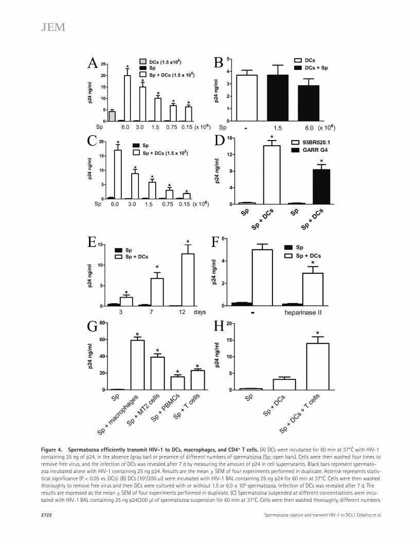
Figure 4. Spermatozoa effi ciently transmit HIV-1 to DCs, macrophages, and CD4 + T cells. (A) DCs were incubated for 60 min at 37°C with HIV-1
containing 25 ng of p24, in the absence (gray bar) or presence of different numbers of spermatozoa (Sp; open bars). Cells were then washed four times to
remove free virus, and the infection of DCs was revealed after 7 d by measuring the amount of p24 in cell supernatants. Black bars represent spermato-
zoa incubated alone with HIV-1 containing 25 ng p24. Results are the mean ± SEM of four experiments performed in duplicate. Asterisk represents statis-
tical signifi cance (P < 0.05 vs. DCs). (B) DCs (10 5 /200 μl) were incubated with HIV-1 BAL containing 25 ng p24 for 60 min at 37°C. Cells were then washed
thoroughly to remove free virus and then DCs were cultured with or without 1.5 or 6.0 × 10 6 spermatozoa. Infection of DCs was revealed after 7 d. The
results are expressed as the mean ± SEM of four experiments performed in duplicate. (C) Spermatozoa suspended at different concentrations were incu-
bated with HIV-1 BAL containing 25 ng p24/200 μl of spermatozoa suspension for 60 min at 37°C. Cells were then washed thoroughly, different numbers

JEM VOL. 206, November 23, 2009
Article
2723
spermatozoa incubated with HIV-1 in the absence of DCs ( Fig. 4 A , black bar). As shown in Fig. 4 A , the presence of spermatozoa markedly enhanced the infection of DCs at all ratios. No enhancing eff ect on DC infection was observed when spermatozoa were added to DCs previously incubated with HIV-1 ( Fig. 4 B ), suggesting that spermatozoa might increase the attachment and/or entry of the virus into DCs.
We then analyzed whether the facilitation of DC infec-tion involves the transfer of HIV-1 from the spermatozoa to DCs. Spermatozoa were incubated fi rst with HIV-1 BAL, washed four times to remove free virus, and cultured for 7 d with DCs at diff erent spermatozoa/DC ratios. As shown in Fig. 4 C , spermatozoa bound HIV-1–infected DCs effi ciently (eff ective infection was observed even at spermatozoa/DC ratios as low as 1:1). Similar results were observed using pri-mary HIV-1 isolates instead of HIV-1 BAL ( Fig. 4 D ). A time course of infection of DCs after exposure to spermatozoa-bound HIV-1 is shown in Fig. 4 E . Supporting a role for HS in the transmission of HIV-1 from the spermatozoa to DCs, we found that treatment with heparinase II signifi cantly im-paired the ability of spermatozoa to transmit the infection to DCs ( Fig. 4 F ). In all cases, studies of DC viability performed by fl uorescence microscopy after 7 d of culture revealed lev-els of viability >80%.
To gain insight into the capacity of spermatozoa to act as HIV carriers, we analyzed their ability to transmit HIV-1 to macrophages, PBMCs, and T cells. Spermatozoa were fi rst incubated with HIV-1 for 60 min at 37°C, washed four times to remove free virus, and cultured for 7 d with macrophages, the CD4 + T cell line MT2, activated PBMCs, and activated T cells purifi ed from peripheral blood of normal donors. Our results showed that spermatozoa effi ciently transmit HIV-1 to macrophages, MT2 cells, PBMCs, and normal T cells ( Fig. 4 G ). Lastly, we analyzed whether the capture of spermatozoa-bound
of HIV-1 to nonpermissive cells via HS chains ( Gallay, 2004 ; de Parseval et al., 2005 ). Fig. 3 H shows that spermatozoa do not express syndecans 1 and 2 but express syndecans 3 and 4. The second class of cellular HS is glypicans. There are six family members of glypicans in mammals. All of them are at-tached to the outer cell surface of the plasma membrane by a glycosylphosphatidylinositol anchor ( Kreuger et al., 2006 ; Bishop et al., 2007 ). Thus, they can be released from the cell surface by phospholipase C (PLC). To determine the possible in-volvement of glypicans in the attachment of HIV-1, we de-termined whether treatment of spermatozoa with PLC resulted in the inhibition of HIV-1 capture. Fig. S2 shows that treat-ment with 2 U/ml PLC did not prevent the binding of HIV-1 to the spermatozoa, suggesting that it does not in-volve glypicans. The effi cacy of PLC treatment was tested in neutrophils. As expected, treatment with 2 U/ml PLC al-most completely removed CD16, a molecule attached by a glycosylphosphatidylinositol anchor, as detected by fl ow cy-tometry (unpublished data).
Spermatozoa effi ciently transmit HIV-1 to DCs,
macrophages, and CD4 + T cells
DCs are able to capture HIV-1 at entry sites and transport the virus to draining lymph nodes where HIV-1 is transmitted to T CD4 + cells ( Geijtenbeek et al., 2000 ; Gurney et al., 2005 ; Wilkinson and Cunningham, 2006 ; Wu and KewalRamani, 2006 ). We analyzed whether spermatozoa were able to mod-ulate the course of DC infection by HIV-1, using DCs ob-tained from monocytes cultured for 5 d with GM-CSF plus IL-4 ( Sallusto and Lanzavecchia, 1994 ). DCs were incubated for 60 min at 37°C with HIV-1 BAL, in the absence or pres-ence of diff erent numbers of spermatozoa. Cells were then washed four times to remove free virus, and the infection of DCs was revealed after 7 d of culture. Controls included
of HIV-1–treated spermatozoa were incubated with (open bars) or without DCs (black bars) over 7 d, and the infection of DCs was then analyzed. Results
are the mean ± SEM of four to fi ve experiments performed in duplicate. Asterisks represent statistical signifi cance (P < 0.05 vs. spermatozoa cultured
without DCs). (D) Spermatozoa (1.5 × 10 6 /200 μl) were incubated with the primary HIV-1 isolates 93BR020.1 or GARR-G4 containing 25 ng p24 for
60 min at 37°C, and then cells were washed thoroughly. Spermatozoa and DCs were co-cultured during 7 d at a spermatozoa/DC ratio of 10:1, and the
infection of DCs was then analyzed. Results are the mean ± SEM of three experiments performed in duplicate. Asterisks represent statistical signifi cance
(P < 0.05 vs. controls). (E) Spermatozoa (1.5 × 10 6 /200 μl) were incubated with HIV-1 BAL containing 25 ng p24 for 60 min at 37°C, washed thoroughly, and
co-cultured with 10 5 DCs during 3, 7, and 12 d at a spermatozoa/DC ratio of 10:1. Infection of DCs was then analyzed. Black bars represent the amount of
p24 found in the supernatants of HIV-1–treated spermatozoa cultured without DCs. Results are the mean ± SEM of four experiments performed in dupli-
cate. Asterisks represent statistical signifi cance (P < 0.05 vs. controls). (F) Spermatozoa (1.5 × 10 6 /200 μl) were treated with 5 U/ml heparinase II for
60 min at 25°C, incubated with HIV-1 BAL containing 25 ng p24 for 60 min at 37°C, washed thoroughly, and co-cultured during 7 d with DCs at a spermato-
zoa/DC ratio of 10:1. Infection of DCs was then revealed as described in A. Black bars represent the levels of p24 antigen in the supernatants of HIV-1–
treated spermatozoa cultured alone. Results are the mean ± SEM of four experiments performed in duplicate. The asterisk represents statistical
signifi cance (P < 0.05 vs. controls). (G) Spermatozoa (1.5 × 10 6 /200 μl) were incubated with 25 ng HIV-1 BAL or HIV-1 IIIB for 60 min at 37°C. Cells were
then washed thoroughly and incubated with macrophages, the T cell line MT2, PHA plus IL-2–activated PBMCs, or purifi ed CD3 + T cells activated by PHA
plus IL-2 (1.5 × 10 5 cells/200 μl). Spermatozoa treated with HIV-1 BAL were used in macrophage cultures, whereas spermatozoa treated with HIV-1 IIIB
were used for MT2 cells, PBMCs, and purifi ed T cells. Cellular infection was analyzed after 7 d of culture. Results are the mean ± SEM of three to four
experiments performed in duplicate. Asterisks represent statistical signifi cance (P < 0.05 vs. spermatozoa cultured alone). (H) Spermatozoa (1.5 × 10 6 /200 μl)
were incubated with HIV-1 IIIB containing 25 ng of p24 for 60 min at 37°C, and then cells were washed thoroughly. Spermatozoa and DCs were co-
cultured during 60 min at 37°C at a spermatozoa/DC ratio of 10:1. Cells were then treated with 1,000 U/ml trypsin for 15 min at 37°C, washed, and cul-
tured with or without 3.0 × 10 5 T cells purifi ed from peripheral blood and activated with PHA plus IL-2. The amount of p24 antigen in cell supernatants
was analyzed after 7 d of culture. Results are the mean ± SEM of three experiments performed in duplicate. The asterisk represents statistical signifi cance
(P < 0.05 vs. Sp + DCs).

2724 Spermatozoa capture and transmit HIV-1 to DCs | Ceballos et al.
Figure 5. Spermatozoa strongly interact with DCs. (A) Spermatozoa (5 × 10 6 /400 μl) were incubated with 50 ng HIV-1 BAL for 60 min at 37°C. Cells
were then washed thoroughly. Experiments were performed in 24-transwell plates with a polycarbonate fi lter (0.2-μm pore size). 5 × 10 6 HIV-1–treated
spermatozoa were in the upper compartment and 5 × 10 5 DCs were seeded in the lower compartment. Controls were performed by incubating together
HIV-1–treated spermatozoa and DCs in the lower compartment. Cells were cultured for 7 d and the infection of DCs was evaluated by measuring the
amount of p24 in the supernatants of DC cultures. Results are the mean ± SEM of fi ve experiments performed in triplicate. The asterisk represents statisti-
cal signifi cance (P < 0.05 vs. controls). (B) CFSE-labeled spermatozoa (1.5 × 10 6 /200 μl) were incubated with or without HIV-1 BAL containing 25 ng p24 for
60 min at 37°C. Cells were then washed thoroughly and were incubated with unlabeled DCs at a spermatozoa/DC ratio of 10:1 during 1 h at 37°C. The in-
teraction of spermatozoa and DCs were then analyzed by fl ow cytometry in the gate of DCs, which could be easily distinguished from spermatozoa because

JEM VOL. 206, November 23, 2009
Article
2725
spermatozoa were phagocytosed by DCs, additional studies were performed by laser confocal microscopy and electron micros-copy. These studies showed that a signifi cant fraction of the spermatozoa was actually taken up by DCs ( Fig. 5, D and E ). We then performed a new set of experiments to defi ne the frac-tion of ingested and attached spermatozoa. As expected, when spermatozoa and DCs were incubated at 4°C, no ingestion of spermatozoa was observed and bound spermatozoa were re-leased from DCs by trypsin treatment (unpublished data). We then used trypsin to distinguish internalized versus attached sper-matozoa. Spermatozoa and DCs were incubated together for 60 min at 37°C. After washing, cells were treated or not with 1,000 U/ml trypsin for 15 min at 37°C, and the number of spermatozoa associated with DCs was determined by fl uores-cence microscopy. Treatment with trypsin resulted in a marked reduction in the number of spermatozoa associated with DCs (47 ± 13% reduction; n = 4; mean ± SEM), suggesting that al-most half of the spermatozoa associated to DCs are not ingested and remain attached to the DC surface.
Although the relative contribution of attached versus ingested spermatozoa in the infection of DCs remains to be defi ned, we speculated that ingestion of spermatozoa might provide an alternative pathway for the infection of DCs, en-abling HIV-1 to infect DCs without the participation of re-ceptors classically involved in HIV-1 attachment or infection, such as DC-SIGN and CD4 ( Turville et al., 2003 ; Piguet and Steinman, 2007 ; Sabatté et al., 2007 ). Transmission of HIV-1 from the spermatozoa to DCs was markedly impaired by block-ing antibodies directed to either DC-SIGN or CD4 ( Fig. 5 F ), suggesting that phagocytosis of spermatozoa does not provide an alternative pathway for the infection of DCs.
The capacity of HIV-1 to hijack DCs for viral dissemina-tion depends not only on the capture of HIV-1 by DCs but also on the migration of DCs to lymph nodes, a response asso-ciated with the phenotypic maturation of DCs ( Turville et al., 2003 ; Piguet and Steinman, 2007 ). We therefore investigated the eff ect of spermatozoa on the maturation of DCs. Sperma-tozoa and DCs were cultured for 24 h and the phenotype of DCs was then analyzed by fl ow cytometry. Fig. 6 A shows the phenotype of immature DCs used in these experiments. As expected, cells were CD1a positive, CD14 negative, and CD83 and CCR7 negative and expressed low to intermediate levels of HLA-DR, CD86, and CD40. Spermatozoa eff ectively trig-ger the phenotypic maturation of DCs, as indicated by the
HIV-1 enables DCs to transmit the virus to T cells. HIV-1–treated spermatozoa and DCs were incubated for 60 min at 37°C. To remove spermatozoa-bound HIV-1, cells were then treated with trypsin to minimize the transmission of HIV-1 from spermatozoa directly to T cells. Cells were then washed thoroughly and cultured with or without activated T cells for 7 d. Fig. 4 H shows that the addition of T cells resulted in a marked increase in p24 levels in culture supernatants, whereas fl ow cytometry revealed T cell infection by p24 intracellular immunostaining (not depicted).
Interaction of spermatozoa with DCs leads
to the internalization of the spermatozoa
and the phenotypic maturation of DCs
To examine whether transmission of HIV-1 required the contact between spermatozoa and DCs, experiments were performed using 24-transwell chambers with a polycarbonate fi lter (0.2-μm pore size). Spermatozoa were incubated with HIV-1 containing 50 ng p24 for 60 min at 37°C, washed four times to remove free virus, and included in the upper cham-ber of the transwell system (5 × 10 6 spermatozoa/well). The lower chamber included 5 × 10 5 DCs. Controls were per-formed by incubating together HIV-treated spermatozoa and DCs in the lower compartment. Infection of DCs was evalu-ated after 7 d of co-culture. Our results shown that DCs re-main uninfected when spermatozoa and DCs were placed in diff erent chambers of the transwell system ( Fig. 5 A ), suggest-ing that the transmission of HIV-1 from the spermatozoa to DCs actually requires cell-to-cell contact.
The requirement for direct cell–cell contact for HIV trans-mission suggests that the DCs may actually phagocytose the spermatozoa. To evaluate internalization, we labeled the sper-matozoa with CFSE. Binding of the CFSE-labeled spermatozoa to the DCs was quantifi ed using fl ow cytometry. As shown in Fig. 5 B , virtually all DCs bind the green spermatozoa, regardless of the presence of HIV on the spermatozoa. The interaction of DCs with spermatozoa was also analyzed by fl uorescence micro s-copy, using CFSE-labeled spermatozoa and DCs labeled with PE-labeled IgG anti–HLA-DR antibodies (ratio of 10:1). A rep-resentative image is shown in Fig. 5 C , with arrows indicating spermatozoa attached and/or ingested by DCs. Consistent with fl ow cytometer data, microscopic analysis revealed that >90% of DCs bind or ingest at least one spermatozoa with a mean of 2.7 ± 1.1 (mean ± SE; n = 6) per DC. To determine whether
of their higher values of forward light scatter. Histograms from a representative experiment ( n = 7) are shown. (C and D) CFSE-labeled spermatozoa (1.5 ×
10 6 /200 μl) were incubated during 60 min at 37°C with DCs, previously labeled with PE–anti–HLA-DR antibodies, at a spermatozoa/DC ratio of 10:1. The
interaction of spermatozoa and DCs were then analyzed by fl uorescence microscopy (C) or laser confocal microscopy (D). Bars, 10 μm. (E) Spermatozoa
(1.5 × 10 6 /200 μl) were incubated during 60 min at 37°C with DCs at a spermatozoa/DC ratio of 10:1. The interaction of spermatozoa and DCs were then
analyzed by electron microscopy. Representative images are shown. Arrows in C–E indicate spermatozoa attached to or ingested by DCs. Bars: (top) 0.5 μm;
(bottom) 1.5 μm. (F) Spermatozoa (1.5 × 10 6 /200 μl) were incubated with HIV-1 BAL containing 25 ng p24 for 60 min at 37°C and washed thoroughly. DCs
were pretreated, or not, with blocking antibodies directed to either CD4 or DC-SIGN for 15 min at 4°C. Blocking antibodies were used at a concentration
three- to fi vefold higher than those needed to saturate all binding sites, as determined by FACS analysis. Spermatozoa and DCs were co-cultured during 7 d
at a spermatozoa/DC ratio of 10:1, and the infection of DCs was then analyzed by measuring the levels of p24 antigen in cell supernatants. Also shown in
the fi gure is the amount of p24 found in the supernatants of HIV-1–treated spermatozoa cultured for 7 d without DCs (gray bar). The results are expressed
as the mean ± SEM of four experiments performed in duplicate. Asterisks represent statistical signifi cance (P < 0.05 vs. controls).

2726 Spermatozoa capture and transmit HIV-1 to DCs | Ceballos et al.
marked increase in the expression of HLA-DR, CD86, CD40, CD83, and CCR7, the chemokine receptor responsible for the homing of DCs to lymph nodes ( Fig. 6 B ). As expected, LPS also induced the phenotypic maturation of DCs, as indi-cated by the increase in the expression of all the markers ana-lyzed. The maturation of DCs induced by spermatozoa cannot be attributed to contaminating LPS in the spermatozoa suspen-sions because supernatants collected from spermatozoa suspen-sions were completely unable to change the phenotype of DCs (unpublished data). Moreover, spermatozoa lysates and sper-matozoa supernatants were shown to be free of endotoxin (2 × 10 7 spermatozoa/100 μl; <0.5 endotoxin U/ml) as determined using a Limulus amebocyte lysate test. Fig. 6 (C and D) shows that the interaction with spermatozoa not only induced the phenotypic maturation of DCs but also the production of high levels of IL-10 but not IL-12 p70 (in contrast with LPS which induced both cytokines). We conclude that the spermatozoa induce DCs to mature phenotypically and to produce IL-10.
Extracellular acidosis markedly enhances the attachment
of HIV-1 to spermatozoa and the consequent transmission
of HIV-1 to DCs
The healthy vaginal environment is markedly acidic with pH values between 4.0 and 6.0 ( García-Closas et al., 1999 ;
Thinkhamrop et al., 1999 ). In contrast, normal values of semen pH vary between 7.2 and 8.0 ( Harraway et al., 2000 ), and it is well known that deposition of semen in the vaginal mucosa raises the pH of vaginal secretion to slightly acidic values rang-ing from 6.0 to 7.0 ( Masters and Johnson, 1961 ; Bouvet et al., 1997 ). We therefore analyzed whether these pH values modu-lated the capture of HIV-1 by spermatozoa. Spermatozoa were cultured with HIV-1 during 60 min at 37°C at extracellular pH values of 7.3 (controls), 6.8, 6.5, and 6.0, and the capture of HIV-1 was then analyzed. Fig. 7 A shows that the capture of HIV-1 by spermatozoa was markedly increased (over fourfold) at pH 6.5 or 6.0 compared with neutral pH. Similar results were observed using primary HIV-1 isolates ( Fig. 7 B ). No changes in sperm viability were observed at pH 6.8 and 6.5, whereas a slight decrease was found at pH 6.0 (unpublished data). In contrast, Fig. 7 C shows that the capture of the virus by the spermatozoa at pH 6.5 was markedly inhibited by heparin, suggesting that, as at neutral pH, the binding of HIV-1 to the spermatozoa at pH 6.5 is dependent on the expression of HS by the spermato-zoa. Moreover, Fig. 7 D shows that the expression of HS did not increase in spermatozoa incubated at pH 6.5, supporting the idea that the enhanced attachment of HIV-1 to spermatozoa observed under acidic conditions is not related to changes in the amount of HS expressed at the spermatozoa surface.
Figure 6. Interaction with spermatozoa leads to the phenotypic maturation of DCs and the production of IL-10. (A) Representative histograms
of the phenotype of immature DCs. Gray histograms represent isotype controls. (B) Spermatozoa (Sp; 1.5 × 10 6 /200 μl) were incubated for 24 h at 37°C
with DCs at a spermatozoa/DC ratio of 10:1, and the phenotype of DCs was then analyzed by fl ow cytometry. The phenotype of DCs incubated alone or in
the presence of spermatozoa or 100 ng/ml LPS during 24 h at 37°C is also shown. Results are expressed as MFI values and represent the arithmetic mean
± SEM of 11 experiments. Asterisks represent statistical signifi cance (P < 0.05 vs. controls). (C and D) The production of IL-10 and IL-12p70 was evaluated
in cell supernatants of DCs cultured alone or in the presence of spermatozoa or 100 ng/ml LPS during 24 h at 37°C. Results are expressed in picograms
per milliliter and represent the arithmetic mean ± SEM of 10 experiments. Asterisks represent statistical signifi cance (P < 0.05 vs. controls).

JEM VOL. 206, November 23, 2009
Article
2727
Figure 7. Acidic values of extracellular pH increase HIV-1 binding to spermatozoa and the subsequent transmission of HIV-1 to DCs.
(A) Spermatozoa (Sp; 1.5 × 10 6 /200 μl) were incubated with HIV-1 BAL containing 25 ng of p24 for 60 min at 37°C at pH 7.3 (controls), 6.8, 6.5, or 6.0.
Cells were then washed thoroughly, lysed, and assayed for p24 antigen by ELISA. The results are expressed as the mean ± SEM of fi ve experiments per-
formed in triplicate. Asterisks represent statistical signifi cance (P < 0.05 vs. pH 7.3). (B) Spermatozoa (1.5 × 10 6 /200 μl) were incubated with different
primary HIV-1 isolates containing 25 ng p24 for 60 min at 37°C at pH 7.3 or 6.5. Cells were then washed thoroughly, lysed, and assayed for p24 antigen
by ELISA. A representative experiment performed in duplicate ( n = 2–3) is shown. (C) Spermatozoa (1.5 × 10 6 /200 μl) were incubated with HIV-1 BAL con-
taining 25 ng p24 for 60 min at 37°C at pH 7.3 (controls) or 6.5, in the absence or presence of 10 U/ml heparin. Cells were then washed thoroughly, lysed,
and assayed for p24 antigen by ELISA. The results are expressed as the mean ± SEM of four experiments performed in triplicate. *, P < 0.05, heparin vs.
controls, at either pH 7.3 or 6.5; **, P < 0.05, controls at pH 6.5 vs. controls at 7.3. (D) Spermatozoa (1.5 × 10 6 /200 μl) were incubated for 60 min at 37°C
at pH 7.3 or 6.5, and the expression of HS was then analyzed by fl ow cytometry. The gray histogram represents isotype control. A representative experi-
ment ( n = 3) is shown. (E) 100 μl of whole semen were diluted 1:1 with culture medium, and the pH was adjusted to 7.3 (control) or 6.5. HIV-1 BAL was
added containing 25 ng p24, and the samples were incubated for 60 min at 37°C. After centrifugation, cells pellets were washed thoroughly, lysed, and
assayed for p24 antigen by ELISA. The results are expressed as the mean ± SEM of four experiments performed in triplicate. The asterisk represents statis-
tical signifi cance (P < 0.05 vs. pH 7.3). (F) DCs (1.5 × 10 5 /200 μl) were incubated with HIV-1 BAL containing 25 ng p24 for 60 min at 37°C at pH 7.3 (con-
trols), 6.8, 6.5, or 6.0. Cells were then washed thoroughly, lysed, and assayed for p24 antigen by ELISA. The results are expressed as the mean ± SEM of
three experiments performed in triplicate. (G) Spermatozoa (1.5 × 10 6 /200 μl) were incubated with HIV-1 BAL containing 25 ng p24 for 60 min at 37°C, at
pH 7.3 or 6.5. Cells were then washed thoroughly and incubated with or without DCs for 7 d at a spermatozoa/DC ratio of 10:1. The infection of DCs was
then analyzed by measuring the levels of p24 antigen by ELISA in cell supernatants. The results are expressed as the mean ± SEM of four experiments
performed in duplicate. Asterisks represent statistical signifi cance (P < 0.05 vs. spermatozoa cultured without DCs).

2728 Spermatozoa capture and transmit HIV-1 to DCs | Ceballos et al.
Microabrasions of the mucosal surface induced either by mechanical stress during intercourse ( Norvell et al., 1984 ; Guimarães et al., 1997 ) or by genital ulcer diseases ( Piot and Laga, 1989 ; Serwadda et al., 2003 ; Galvin and Cohen, 2004 ) may allow spermatozoa to directly access DCs as well as mac-rophages and CD4 + T cells, the three major targets of HIV-1 infection. It is of note that this is not an unusual scenario; in fact, epithelial microabrasions in the vagina are usually de-tected in 60% of healthy women after consensual intercourse ( Norvell et al., 1984 ). The access of spermatozoa to DCs may also be facilitated by an alternative mechanism: the interac-tion of spermatozoa with DC projections that extend to, or near, the luminal surface of the mucosa ( Shattock and Moore, 2003 ; Chieppa et al., 2006 ).
HS is a ubiquitous linear glycosaminoglycan composed of 30–400 repeats of a sulfated disaccharide motif with distinct sugar sequences and sulfation patterns ( Parish, 2006 ; Bishop et al., 2007 ). Previous studies have shown that HS functions as an ancillary attachment factor for HIV-1 in DCs, macro-phages, and epithelial and endothelial cells ( Saphire et al., 2001 ; Wu et al., 2003 ; Argyris et al., 2003 ; Gallay, 2004 ; de Witte et al., 2007 ). The ability of HS to recognize HIV-1 appears to depend on four HS binding domains identifi ed in the V2 and V3 loops, in the C-terminal domain, and within the CD4-induced bridging sheet of the gp120 ( de Parseval et al., 2005 ; Crublet et al., 2008 ).
We found that human spermatozoa express HS. More-over, we observed that treatment of spermatozoa with hepa-rinase II reduced the attachment of HIV-1 supporting the involvement of HS. Interestingly, it has been shown recently that human papillomavirus 16 effi ciently binds to the sperma-tozoa surface and also that the binding is inhibited by heparin and carrageenan, supporting the participation of HS-like mole-cules in the mechanisms responsible for virus attachment ( Pérez Andino et al., 2009 ).
Our results support the notion that HS might enable sper-matozoa to capture HIV-1 and to transmit the virus to CD4 + target cells promoting the spreading of the infection. A similar role has been proposed for HS expressed by epithelial ( Wu et al., 2003 ) and endothelial ( Argyris et al., 2003 ) cells. Inter-estingly, although previous studies have shown that the avid-ity of gp120 for HS is usually higher for X4 virus than R5 virus ( Moulard et al., 2000 ; Fletcher et al., 2006 ), our obser-vations indicated that both HIV-1 BAL (R5) and HIV-1 IIIB (X4) interact with spermatozoa with similar effi ciency. This suggests that, besides HS, other attachment factors might con-tribute to the binding of HIV-1 to the spermatozoa surface.
MRs expressed by leukocytes belong to the family of C-type lectin receptors, which recognize mannose-type carbo-hydrates found on virus, bacteria, and fungi ( Figdor et al., 2002 ; Cambi and Figdor, 2003 ). Theses receptors play an im-portant role in the capture of HIV-1 by macrophages and DCs ( Figdor et al., 2002 ; Cambi and Figdor, 2003 ; Trujillo et al., 2007 ). Our results support that MR plays a complemen-tary role in the attachment of HIV-1 to spermatozoa because mannan, mannose-BSA, and blocking antibodies directed to
To analyze the infl uence of extracellular pH on the cap-ture of HIV-1 in a more physiological setting, experiments were done using whole semen instead of isolated spermato-zoa. 100 μl of semen were diluted 1:1 with culture medium, the pH was adjusted to values of 7.3 (control) and 6.5, and capture assays were performed by incubating semen and HIV-1 containing 25 ng p24 for 60 min at 37°C. The cell pellet was then washed four times and the attachment of HIV-1 was evaluated. As shown in Fig. 7 E , and as observed for isolated spermatozoa, incubation of HIV-1 with whole semen at pH 6.5 also resulted in a marked increase in cellular attachment of HIV-1. To confi rm that the virus was actually attached to spermatozoa and not to seminal leukocytes, sper-matozoa were fi rst purifi ed from the semen by the technique of swim-up and were then resuspended in autologous semi-nal plasma diluted 1:1 with culture medium. The pH was ad-justed to values of 7.3 and 6.5, and capture assays were performed as described in the Fig. 7 A legend. A marked in-crease in the attachment of HIV-1 at pH 6.5 was also ob-served under these experimental conditions, with a 585 ± 132% increase in the attachment of HIV-1 at pH 6.5 versus 7.3 (mean ± SEM; n = 4; P < 0.01). This result suggest that the enhancement of HIV-1 capture observed at pH 6.5 in assays performed in whole semen actually refl ects an increased interaction between virus and spermatozoa. Interestingly, the enhancing eff ect of acidic values of pH on the cellular bind-ing of HIV-1 does not appear to represent a general phenom-enon. In fact, no enhancing eff ect was observed when the attachment of HIV-1 to DCs was analyzed ( Fig. 7 F ).
Because previous studies have shown that acidic values of pH are able to induce the inactivation of HIV-1 ( Kempf et al., 1991 ; Connor, 2006 ), we investigated if the HIV-1 captured by the spermatozoa at pH 6.5 retained its ability to infect DCs. As shown in Fig. 7 G the increased capture of HIV-1 by sper-matozoa at pH 6.5, as compared with pH 7.3, resulted in a marked increase (more than fourfold) in the transmission of HIV-1 from the spermatozoa to DCs. Together, these results suggest that, after sexual intercourse, the mildly acidic pH of the vagina might favor the dissemination of HIV-1 infection via the concerted action of spermatozoa and DCs.
DISCUSSION
In the present study, we show that human spermatozoa cap-ture HIV-1 and efficiently transmit the virus to DCs in a process that requires cell-to-cell contact. The interaction of spermatozoa and DCs not only leads to the uptake of the spermatozoa but also results in the phenotypic maturation of DCs. In addition, we show that acidic values of extracellular pH, similar to those found in the vaginal mucosa after sexual intercourse, markedly increase the attachment of HIV-1 to the spermatozoa and the consequent transmission of HIV-1 to DCs. Interestingly, transmission of spermatozoa-bound HIV-1 was not restricted to DCs. It was also observed for macrophages, PBMCs, and normal T cells.
How could spermatozoa gain access to DCs at the female genital tract? This might occur under two diff erent scenarios.

JEM VOL. 206, November 23, 2009
Article
2729
MR modestly inhibited the binding of HIV-1 to spermato-zoa. A recent study ( Fanibunda et al., 2008 ) has shown that MR expressed by the spermatozoa bind the HIV gp120, sug-gesting that these receptors might play an important role in the binding of HIV-1. Our results do not support this view and reinforce the notion that the ability of a given receptor to bind soluble recombinant gp120 is not necessarily predictive of its ability to capture HIV-1. In fact, elegant studies per-formed in cell lines transfected with the C-type lectin re-ceptors langerin, DC-SIGN, and LSECTin (lymph node sinusoidal endothelial cell C-type lectin) have clearly shown that all of these receptors eff ectively recognize recombinant soluble gp120, whereas only DC-SIGN demonstrated capac-ity to attach the virus ( Gramberg et al., 2008 ). Diff erences in the content or the surface exposure of gp120 glycans and/or in the multimerization status of the gp120 between soluble recombinant gp120 and the gp120 expressed on the virus sur-face may explain these apparently contrasting results.
DCs play a crucial role in the induction of adaptive im-munity ( Guermonprez et al., 2002 ). Owing to their unique localization at mucosal surfaces and their extraordinary ability to capture antigens, DCs are among the fi rst potential targets of HIV infection during sexual transmission ( Wilkinson and Cunningham, 2006 ; Wu and KewalRamani, 2006 ). Thus, after vaginal inoculation with simian immunodefi ciency virus in macaques, DCs appear to be the predominant cell type in-fected during the early phase of the infection ( Hu et al., 2000 ). Moreover, studies performed in infected men showed that DCs account for >90% of the virus bound to total mono-nuclear cells from the rectal mucosa, in spite of the fact that DCs represent <5% of these mononuclear cells ( Gurney et al., 2005 ). A large body of evidence supports the notion that HIV-1 hijacks DC function. Instead of, or at the same time as, stimulating the adaptive immune response against HIV-1, DCs mediate HIV-1 transmission to T CD4 + cells in the lymphoid tissues, contributing to the spreading of infection ( Wilkinson and Cunningham, 2006 ; Wu and KewalRamani, 2006 ). We found that spermatozoa effi ciently transfer the virus to immature DCs, which transmit the virus to T cells. Additional experiments performed in transwell chambers showed that transmission of HIV-1 from the spermatozoa to DCs requires cell-to-cell contact, whereas studies performed by fl ow cytometry, fl uorescence microscopy, confocal mi-croscopy, and electron microscopy revealed that spermatozoa strongly interact with DCs. Interestingly, this interaction leads to both the endocytosis of the spermatozoa (almost 50% of the attached spermatozoa were internalized) and the phe-notypic maturation of DCs. These processes were not related to the presence of HIV-1 on the spermatozoa because they were similarly observed using either untreated or HIV-treated spermatozoa. Importantly, as reported for DCs challenged with free HIV-1 ( Turville et al., 2003 ; Piguet and Steinman, 2007 ; Sabatté et al., 2007 ), we found that trans-infection of DCs mediated by spermatozoa was markedly prevented by blocking antibodies directed to either DC-SIGN or CD4, supporting the notion that this mechanism do not provide an
alternative route for the infection of DCs by HIV-1. Interest-ingly, our results support the notion that DC-SIGN expressed by DCs interacts with HIV-1 on the spermatozoa surface. The mechanisms through which DC-SIGN promotes the in-fection of DCs by spermatozoa-bound HIV-1 are not yet de-fi ned. DC-SIGN might promote the attachment of HIV-1 to DCs ( Geijtenbeek and van Kooyk, 2003 ). Moreover, it might protect the virus from degradation ( Geijtenbeek and van Kooyk, 2003 ) and might also activate signaling pathways in DCs able to promote HIV spreading ( Hodges et al., 2007 ).
Our results are consistent with previous studies ( Scofi eld et al., 1992 ; Scofi eld, 1998 ) showing that spermatozoa inter-act with B lymphocytes, activated T cells, and monocytes. The authors showed that the binding of leukocytes and sper-matozoa appeared to be dependent on the interaction of HLA-DR molecules expressed by leukocytes and a CD4-like structure expressed by the spermatozoa because it was im-paired by antibodies directed to either HLA-DR or CD4. We analyzed whether a similar mechanism was involved in the interaction between spermatozoa and DCs. Consistent with previous published data ( el-Demiry et al., 1986 ; Wolff and Anderson, 1988 ; Kim et al., 1999 ), we found no expres-sion of CD4 in the spermatozoa. However, because we can-not rule out that spermatozoa express very low amounts of CD4, undetectable by fl ow cytometry, we performed a set of experiments using blocking antibodies directed to either CD4 (clone SIM.4) or HLA-DR (clone L243). Only a low inhibition of the interaction of spermatozoa and DCs (< 30%) was observed when it was performed in the presence of both blocking antibodies (unpublished data), raising the ques-tion of the existence of HLA-DR–independent mechanisms responsible for the interaction between spermatozoa and DCs. Supporting this possibility, we observed that HLA-DR–negative cells, such as resting neutrophils, were also able to interact with viable spermatozoa (unpublished data).
It is usually assumed that the acidic environment of the vagina provides a protective mechanism against sexual trans-mission of HIV-1. In fact, the pH of vaginal secretions usually ranged from 4.0 to 6.0 ( García-Closas et al., 1999 ; Thinkhamrop et al., 1999 ), and early studies showed that the exposure of free HIV-1 to pH values lower than 5.0 results in virus inactivation ( Kempf et al., 1991 ). More recent studies performed with primary HIV-1 isolates instead of laboratory strains showed considerable variability to inactivation by acidic pH, and for some isolates it was observed that the in-fectivity of HIV-1 was actually enhanced after exposure at pH 4.0–5.0 ( Connor, 2006 ). Regarding the infl uence of vag-inal pH on the infectivity of HIV-1, it is very important to consider that semen does not only carry the virus but also acts as a buff er of vaginal secretions, leading the pH of the vaginal mucosa to values usually ranged from 6.0 to 7.0 for several hours after sexual intercourse ( Masters and Johnson, 1961 ; Bouvet et al., 1997 ). We found that the attachment of HIV-1 to spermatozoa was markedly increased when it was assayed at pH 6.0–7.0 instead of neutral pH (pH 7.3). Importantly, these results challenge the view that acidic environment of

2730 Spermatozoa capture and transmit HIV-1 to DCs | Ceballos et al.
at 37°C. During this incubation, the osmolarity of the medium was gradually
increased from 290 to 360 osmol/liter by the addition of 9% NaCl. Three
diff erent Percoll fractions were layered in polypropylene tubes: 50% at the
bottom, followed by 46 and 40%. PBMCs (5 × 10 6 /ml) were layered at the
top, and they were centrifuged at 400 g for 20 min at 4°C. Monocytes were
recovered at the 50/46% interface. The purity was checked by FACS analysis
using an anti-CD14 mAb and was found to be >85%. To obtain DCs, mono-
cytes were cultured in RPMI 1640 medium (Life Technologies) supple-
mented with 10% fetal calf serum, 50 U/ml penicillin, 50 μg/ml streptomycin,
and 0.1 mM nonessential amino acids (complete culture medium; all from
Life Technologies) at 10 6 cells/ml with 10 ng/ml IL-4 and 10 ng/ml
GM-CSF, as previously described ( Sallusto and Lanzavecchia, 1994 ). On day 6,
the cells were analyzed by FACS. To obtain macrophages, monocytes were
cultured in complete culture medium with 50 ng/ml GM-CSF for 5 d.
Cell lines and virus. The HIV-1 BAL using CCR5, the HIV-1 IIIB using
CXCR4, the primary HIV-1 isolates HIV-1 93BR020.1 and HIV-1
96USHIPS4, and the cell lines MT2, B-THP-1, and B-THP-1-DC-SIGN +
were obtained from the AIDS Research and Reference Reagent program.
The cell line HT29 (American Tissue Culture Collection) derived from
human colon adenocarcinoma was maintained as a subconfl uent monolayer in
DMEM/F12 nutrient mixture, supplemented with 2 mmol/liter glutamine,
10% fetal bovine serum, 50 U/ml penicillin, and 50 μg/ml streptomycin
(Thermo Fisher Scientifi c). The primary HIV-1 isolates GARR G4 and
GARR G2 were obtained from the Laboratory of Cellular Biology and
Retrovirus (Hospital de Pediatría Juan P. Garrahan, Buenos Aires, Argen-
tine). The HIV isolates BAL, 93BR020.1, 96USHIPS4, GARR G4, and
GARR G2 were grown on 10 U/ml IL-2 (R&D Systems) plus PBMC
stimulated with 10 μg/ml PHA. The HIV-1 IIIB isolate was obtained from
H9HTLV-IIIB supernatants. The viruses were concentrated by ultracentri-
fugation at 28,000 rpm for 90 min at 4°C (L2-65B ultracentrifuge; Beckman
Coulter), and the virus pellet was suspended in RPMI 1640 medium. p24
antigen levels were determined by ELISA (Abbott Diagnostics), and virus
input into assays was a function of p24 antigen concentration. Pseudotypes
were produced, as previously described, by transiently cotransfecting (Lipo-
fectamine 2000; Invitrogen) human embryonic kidney 293T cells with the
proviral pNL-Luc-E-R + vector ( Connor et al., 1995 ), which lacks the env
gene, and the expression vector pCMV harboring the gene coding for the
HIV-1 R5 (BAL) envelope protein. 293T cells were also transfected only
with pNL-Luc-E-R + . Supernatants from 293T cells were harvested 72 h
after transfection or cotransfection and p24 levels were measured by ELISA.
Quantitation of cellular apoptosis and viability by fl uorescence
microscopy. Quantitation was performed using 100 μg/ml of the fl uorescent
DNA-binding dye acridine orange to determine the percentage of cells that
had undergone apoptosis and 100 μg/ml ethidium bromide to diff erentiate
between viable and nonviable cells. With this method, nonapoptotic cell
nuclei show structure or variations in fl uorescence intensity that refl ect the
distribution of euchromatin and heterochromatin. In contrast, apoptotic nu-
clei exhibit highly condensed chromatin that is uniformly stained by acridine
orange. In fact, the entire apoptotic nucleus is present as bright spherical
beads. To assess the percentage of cells showing morphological features of
apoptosis, at least 200 cells were scored in each experiment.
Flow cytometry. Fluorescein isothiocyanate– or phycoerythrin-conju-
gated mAbs directed to CD1a, CD14, CD4, CD80, CD86, HLA-DR,
CD83, and CCR7 were obtained from BD. mAb 10E4 (mouse IgM) di-
rected to HS chains and mAb 3G10 (mouse IgG) directed to a neoepitope
expressed by heparinase III–treated HS were obtained from Seikagaku Cor-
poration. Rat IgG monoclonal antibodies directed to human syndecan-1
(clone 359103) and syndecan-2 (clone 305515), as well as goat polyclonal
antibodies directed to human syndecan-3 and syndecan-4, were obtained
from R&D Systems. FITC-conjugated IgG antibodies directed to rat IgG
or goat IgG were obtained from Sigma-Aldrich. Analysis was performed by
using a FACS fl ow cytometer and CellQuest software (BD).
the vagina provides a protective mechanism against sexual transmission of HIV-1 and support the notion that after sex-ual intercourse a fraction of free HIV-1 found in semen be-comes attached to the spermatozoa, thus enhancing its intrinsic ability to infect CD4 + cells.
We speculate that spermatozoa, in addition to their ability to transmit the virus to DCs, might infl uence the course of the immune response against HIV by modulating the func-tion of mucosal DCs in the receptive partner. Although this hypothesis remains to be tested, our results showing that spermatozoa not only induce the phenotypic maturation of DCs but also the production of IL-10, but not IL-12 p70, and the expansion of Foxp3-expressing CD4 + CD25 + regula-tory T cells (unpublished data) support the notion that the interaction with spermatozoa might drive mucosal DCs into a tolerogenic profi le.
There are no candidate vaccines for HIV that could in-duce sterilizing immunity and protect against HIV infection. The best opportunity to prevent sexual transmission of HIV-1 clearly lies at the sites of mucosal entry. However, a better understanding of the earliest events governing the mucosal transmission of HIV is needed to develop rational strategies that prevent HIV infection. Vasectomized men are still able to transmit HIV-1 ( Coombs et al., 2003 ), indicating that sexual transmission of the virus can proceed without spermatozoa. However, spermatozoa might be able to modulate the sexual transmission of HIV-1. Our observations support that far from being a passive carrier, spermatozoa acting in concert with DCs might aff ect the course of sexual transmission of HIV-1 infection by infl uencing both viral spreading and, perhaps, the profi le of the adaptive immune response directed to HIV-1.
MATERIALS AND METHODS Reagents. LPS from Escherichia coli , recombinant human IL-4, recombinant
human GM-CSF, trypsin, pronase, PHA, heparin, heparinase II, heparinase III,
PLC, mannan, and mannose-BSA were obtained from Sigma-Aldrich.
Ficoll-Hypaque and Percoll were obtained from GE Healthcare. Blocking
IgG monoclonal antibodies directed to either DC-SIGN (clone 120526) or
CD4 (SIM.4) were obtained from the AIDS Research and Reference Re-
agent Program (AIDS Division, National Institute of Allergy and Infectious
Disease, National Institutes of Health). Blocking IgG monoclonal antibody
to MR (LS-C16312) was obtained from Lifespan Biosciences.
Semen samples and spermatozoa isolation. All protocols using human
cells were approved by the Ethical Committee of the National Academy of
Medicine (Buenos Aires, Argentina). Fresh semen samples were obtained from
healthy fertile volunteer donors (aged 25–45 yr) after 3–5 d of abstinence. In-
formed consent was obtained from each patient before spermatozoa collection.
Semen samples were obtained by masturbation from normozoospermic do-
nors, according to World Health Organization standards ( WHO, 1999 ). After
complete liquefaction, semen samples were subjected to spermatozoa selection
using the swim-up procedure ( WHO, 1999 ). Motile cells were resuspended in
RPMI 1640 medium supplemented with 0.3% BSA. Only ejaculates deter-
mined to have normal semen parameters ( WHO, 1999 ) were used.
Preparation of human DCs and macrophages. PBMCs were isolated
from healthy volunteers by standard density gradient centrifugation on Ficoll-
Hypaque. Monocytes were purifi ed by centrifugation on a discontinuous
Percoll gradient. In brief, PBMCs were suspended in Ca2 + /Mg2 + -free Ty-
rode solution supplemented with 0.2% EDTA and incubated during 30 min

JEM VOL. 206, November 23, 2009
Article
2731
microscope (1011; JEOL) at 60 kV. Images were recorded with a cooled
charge-coupled device digital camera (MegaView III; Olympus).
Production of IL-10 and IL-12p70 by DCs. Spermatozoa (1.5 × 10 6 /200 μl)
were incubated for 24 h at 37°C with DCs, at a spermatozoa/DC ratio of
10:1, and the production of IL-10 and IL-12p70 was evaluated by ELISA
(R&D Systems), according to the manufacturer’s recommendations.
Statistics. All statistical comparisons were performed by using analysis of vari-
ance. P-values of <0.01 and <0.05 were considered statistically signifi cant.
Online supplemental material. Fig. S1 shows the expression of HS in the
human intestinal epithelial cell line HT-29. Fig. S2 shows that spermatozoa
treatment with PLC did not prevent HIV-1 capture. Table S1 illustrates the
properties of primary HIV-1 isolates. Online supplemental material is avail-
able at http://www.jem.org/cgi/content/full/jem.20091579/DC1.
We thank Mabel Horvat and Beatriz Loria for their technical assistance.
This study was supported by grants from the Consejo Nacional de
Investigaciones Científi cas y Técnicas (CONICET), the Buenos Aires University School
of Medicine, and the Agencia Nacional de Promoción Cientıfi ca y Tecnológica
(Argentina). F. Remes Lenicov was supported by Fogarty AIDS International Training
and Research Program grant 5D43 TW0010137.
The authors have no confl icting fi nancial interests.
Submitted: 20 July 2009
Accepted: 28 September 2009
REFERENCES Argyris , E.G. , E. Acheampong , G. Nunnari , M. Mukhtar , K.J. Williams ,
and R.J. Pomerantz . 2003 . Human immunodefi ciency virus type 1 en-ters primary human brain microvascular endothelial cells by a mech-anism involving cell surface proteoglycans independent of lipid rafts. J. Virol. 77 : 12140 – 12151 . doi:10.1128/JVI.77.22.12140-12151.2003
Baccetti , B. , A. Benedetto , A.G. Burrini , G. Collodel , E.C. Ceccarini , N. Crisà , A. Di Caro , M. Estenoz , A.R. Garbuglia , A. Massacesi , et al . 1994 . HIV-particles in spermatozoa of patients with AIDS and their transfer into the oocyte. J. Cell Biol. 127 : 903 – 914 . doi:10.1083/jcb.127.4.903
Baccetti , B. , A. Benedetto , G. Collodel , A. di Caro , A.R. Garbuglia , and P. Piomboni . 1998 . The debate on the presence of HIV-1 in human gametes. J. Reprod. Immunol. 41 : 41 – 67 . doi:10.1016/S0165-0378(98)00048-5
Bagasra , O. , H. Farzadegan , T. Seshamma , J.W. Oakes , A. Saah , and R.J. Pomerantz . 1994 . Detection of HIV-1 proviral DNA in sperm from HIV-1-infected men. AIDS . 8 : 1669 – 1674 . doi:10.1097/00002030-199412000-00005
Bandivdekar , A.H. , S.M. Velhal , and V.P. Raghavan . 2003 . Identifi cation of CD4-independent HIV receptors on spermatozoa. Am. J. Reprod. Immunol. 50 : 322 – 327 . doi:10.1034/j.1600-0897.2003.00096.x
Barboza , J.M. , H. Medina , M. Doria , L. Rivero , L. Hernández , and N.V. Joshi . 2004 . Use of atomic force microscopy to reveal sperm ultra-structure in HIV-patients on highly active antiretroviral therapy. Arch. Androl. 50 : 121 – 129 . doi:10.1080/01485010490269524
Benoff , S. , I. Hurley , G.W. Cooper , F.S. Mandel , D.L. Rosenfeld , and A. Hershlag . 1993 . Head-specifi c mannose-ligand receptor expression in human spermatozoa is dependent on capacitation-associated membrane cholesterol loss. Hum. Reprod. 8 : 2141 – 2154 .
Berlier , W. , M. Cremel , H. Hamzeh , R. Lévy , F. Lucht , T. Bourlet , B. Pozzetto , and O. Delézay . 2006 . Seminal plasma promotes the attrac-tion of Langerhans cells via the secretion of CCL20 by vaginal epithe-lial cells: involvement in the sexual transmission of HIV. Hum. Reprod. 21 : 1135 – 1142 . doi:10.1093/humrep/dei496
Bishop , J.R. , M. Schuksz , and J.D. Esko . 2007 . Heparan sulphate pro-teoglycans fi ne-tune mammalian physiology. Nature . 446 : 1030 – 1037 . doi:10.1038/nature05817
Bouvet , J.P. , G. Grésenguet , and L. Bélec . 1997 . Vaginal pH neutralization by semen as a cofactor of HIV transmission. Clin. Microbiol. Infect. 3 : 19 – 23 . doi:10.1111/j.1469-0691.1997.tb00246.x
Brogi , A. , R. Presentini , D. Solazzo , P. Piomboni , and E. Costantino-Ceccarini . 1996 . Interaction of human immunodefi ciency virus type 1
Enzymatic treatments. Spermatozoa were treated with the following:
1,000 U/ml trypsin for 15 min at 37°C, 20 μg/ml pronase for 15 min at
37°C, 1 and 5 U/ml heparinase II for 1 h at 25°C, 5 U/ml heparinase III for
1 h at 25°C, or 2 U/ml PLC for 90 min at 37°C. Cells were then washed
and used in subsequent experiments.
HIV-1 capture assays. Spermatozoa were suspended in culture complete
medium at a concentration of 1.5 × 10 6 /200 μl and were incubated with HIV-1
stocks containing the indicated amounts of p24 antigen for 60 min at 37°C.
Cells were then washed thoroughly, pelleted, lysed, and assayed for HIV p24
antigen by ELISA. Controls of nonspecifi c attachment of HIV-1 were per-
formed by incubating HIV-1 in the absence of spermatozoa. When indicated,
capture assays were performed at acidic values of extracellular pH. In these ex-
periments, 1.5 × 10 6 spermatozoa were suspended in 200 μl of culture com-
plete medium previously adjusted to pH values of 6.8, 6.5, and 6.0 by the
addition of a precalculated volume of isotonic HCl solution. HIV-1 stocks
containing the indicated amounts of p24 antigen were then added, and capture
assays were performed as described in this section. Capture assays were also
performed using whole semen instead of isolated spermatozoa. In these experi-
ments, 100 μl of whole semen were diluted 1:1 with culture medium, and the
pH was adjusted to 7.3 (control) or 6.5 by the addition of a precalculated vol-
ume of isotonic HCl solution. HIV-1 was then added and the samples were
incubated for 60 min at 37°C. After centrifugation, cells pellets were washed
thoroughly, lysed, and assayed for p24 antigen by ELISA.
HIV-1 trans-infection assays. To analyze whether spermatozoa were able
to transmit HIV-1 to DCs, 1.5 × 10 6 spermatozoa cells in 200 μl of complete
medium were fi rst incubated with HIV-1 for 60 min at 37°C. Cells were
then washed thoroughly and coincubated with DCs suspended in culture
complete medium supplemented with 10 ng/ml IL-4 and 10 ng/ml
GM-CSF at diff erent spermatozoa/DC ratios in a fi nal volume of 200 μl in
96-well U-bottom plates. Supernatants, harvested at 7 d of culture, were
assayed for p24 antigen by ELISA. Trans-infection of macrophages and MT2
cells was performed by incubating 1.5 × 10 6 spermatozoa cells in 200 μl of com-
plete medium with HIV-1 for 60 min at 37°C. Cells were then washed
thoroughly and were coincubated with macrophages or MT2 cells at a sper-
matozoa/macrophage MT2 ratio of 10:1 in a fi nal volume of 200 μl. Super-
natants, harvested at 7 d of culture, were assayed for p24 antigen by ELISA.
Trans-infection of PBMCs and T cells purifi ed from PBMCs by positive
magnetic selection (purity >95%; CD3 MicroBeads; Miltenyi Biotec) was
performed as described in this section, using cells activated by 10 U/ml IL-2
plus 10 μg/ml PHA for 72 h.
When indicated, trans-infection assays were performed in 24-transwell
chambers with a polycarbonate fi lter (0.2-μm pore size). In these experiments
spermatozoa were incubated with HIV-1 containing 50 ng/ml of p24 for
60 min at 37°C, washed four times to remove free virus, and included in the up-
per chamber of the transwell system (5 × 10 6 spermatozoa/well). 5 × 10 5 DCs
were seeded in the lower chamber, and infection was evaluated after 7 d of cul-
ture by measuring the amount of p24 in the supernatants of the DC chamber.
Analysis of the interaction between spermatozoa and DCs. Analysis
was performed by fl ow cytometry, fl uorescence microscopy, confocal mi-
croscopy, and electronic microscopy. Analysis by fl ow cytometry was per-
formed using spermatozoa labeled with 5 μM of CFSE for 15 min at 37°C.
CFSE-labeled spermatozoa (1.5 × 10 5 /200 μl) were incubated in the absence
or presence of HIV-1 BAL containing 25 ng of p24 for 60 min at 37°C.
Cells were then washed four times and were cultured for 60 min at 37°C
with unlabeled DCs, using a spermatozoa/DC ratio of 10:1. DCs display
values of forward light scatter that are markedly higher than those of sperma-
tozoa, enabling us to defi ne a gate for DCs which exclude free spermatozoa.
Interaction of spermatozoa and DCs was also analyzed by fl uorescence mi-
croscopy (Carl Zeiss, Inc.) and confocal microscopy (Eclipse E800 laser con-
focal microscope; Nikon), using CFSE-labeled spermatozoa and DCs labeled
with PE–anti–HLA-DR antibodies, using a spermatozoa/DC ratio of 10:1.
Studies using electron microscopy were performed with a transmission electron

2732 Spermatozoa capture and transmit HIV-1 to DCs | Ceballos et al.
Galvin , S.R. , and M.S. Cohen . 2004 . The role of sexually transmitted dis-eases in HIV transmission. Nat. Rev. Microbiol. 2 : 33 – 42 . doi:10.1038/nrmicro794
García-Closas , M. , R. Herrero , C. Bratti , A. Hildesheim , M.E. Sherman , L.A. Morera , and M. Schiff man . 1999 . Epidemiologic determinants of vaginal pH. Am. J. Obstet. Gynecol. 180 : 1060 – 1066 . doi:10.1016/S0002-9378(99)70595-8
Geijtenbeek , T.B. , and Y. van Kooyk . 2003 . Pathogens target DC-SIGN to infl uence their fate DC-SIGN functions as a pathogen receptor with broad specifi city. APMIS . 111 : 698 – 714 . doi:10.1034/j.1600-0463.2003.11107803.x
Geijtenbeek , T.B. , D.S. Kwon , R. Torensma , S.J. van Vliet , G.C. van Duijnhoven , J. Middel , I.L. Cornelissen , H.S. Nottet , V.N. KewalRamani , D.R. Littman , et al . 2000 . DC-SIGN, a dendritic cell-specifi c HIV-1-binding protein that enhances trans-infection of T cells. Cell . 100 : 587 – 597 . doi:10.1016/S0092-8674(00)80694-7
Gramberg , T. , E. Soilleux , T. Fisch , P.F. Lalor , H. Hofmann , S. Wheeldon , A. Cotterill , A. Wegele , T. Winkler , D.H. Adams , and S. Pöhlmann . 2008 . Interactions of LSECtin and DC-SIGN/DC-SIGNR with viral ligands: Diff erential pH dependence, internalization and virion binding. Virology . 373 : 189 – 201 . doi:10.1016/j.virol.2007.11.001
Guermonprez , P. , J. Valladeau , L. Zitvogel , C. Théry , and S. Amigorena . 2002 . Antigen presentation and T cell stimulation by dendritic cells. Annu. Rev. Immunol. 20 : 621 – 667 . doi:10.1146/annurev.immunol.20.100301.064828
Guimarães , M.D. , D. Vlahov , and E.A. Castilho ; Rio de Janeiro Heterosexual Study Group . 1997 . Postcoital vaginal bleeding as a risk factor for transmission of the human immunodefi ciency virus in a het-erosexual partner study in Brazil. Arch. Intern. Med. 157 : 1362 – 1368 . doi:10.1001/archinte.157.12.1362
Gupta , K. , and P.J. Klasse . 2006 . How do viral and host factors modulate the sexual transmission of HIV? Can transmission be blocked? PLoS Med. 3 : e79 . doi:10.1371/journal.pmed.0030079
Gurney , K.B. , J. Elliott , H. Nassanian , C. Song , E. Soilleux , I. McGowan , P.A. Antón , and B. Lee . 2005 . Binding and transfer of human immuno-defi ciency virus by DC-SIGN+ cells in human rectal mucosa. J. Virol. 79 : 5762 – 5773 . doi:10.1128/JVI.79.9.5762-5773.2005
Haase , A.T. 2005 . Perils at mucosal front lines for HIV and SIV and their hosts. Nat. Rev. Immunol. 5 : 783 – 792 .
Harraway , C. , N.G. Berger , and N.H. Dubin . 2000 . Semen pH in pa-tients with normal versus abnormal sperm characteristics. Am. J. Obstet. Gynecol. 182 : 1045 – 1047 . doi:10.1067/mob.2000.106051
Hladik , F. , and M.J. McElrath . 2008 . Setting the stage: host invasion by HIV. Nat. Rev. Immunol. 8 : 447 – 457 . doi:10.1038/nri2302
Hodges , A. , K. Sharrocks , M. Edelmann , D. Baban , A. Moris , O. Schwartz , H. Drakesmith , K. Davies , B. Kessler , A. McMichael , and A. Simmons . 2007 . Activation of the lectin DC-SIGN induces an immature dendritic cell phenotype triggering Rho-GTPase activity required for HIV-1 rep-lication. Nat. Immunol. 8 : 569 – 577 . doi:10.1038/ni1470
Hong , P.W. , K.B. Flummerfelt , A. de Parseval , K. Gurney , J.H. Elder , and B. Lee . 2002 . Human immunodefi ciency virus envelope (gp120) bind-ing to DC-SIGN and primary dendritic cells is carbohydrate dependent but does not involve 2G12 or cyanovirin binding sites: implications for structural analyses of gp120-DC-SIGN binding. J. Virol. 76 : 12855 – 12865 . doi:10.1128/JVI.76.24.12855-12865.2002
Hu , J. , M.B. Gardner , and C.J. Miller . 2000 . Simian immunodefi ciency virus rapidly penetrates the cervicovaginal mucosa after intravaginal inoc-ulation and infects intraepithelial dendritic cells. J. Virol. 74 : 6087 – 6095 . doi:10.1128/JVI.74.13.6087-6095.2000
Jones , K.S. , C. Petrow-Sadowski , D.C. Bertolette , Y. Huang , and F.W. Ruscetti . 2005 . Heparan sulfate proteoglycans mediate attachment and entry of human T-cell leukemia virus type 1 virions into CD4+ T cells. J. Virol. 79 : 12692 – 12702 . doi:10.1128/JVI.79.20.12692-12702.2005
Kempf , C. , P. Jentsch , F.B. Barre-Sinoussi , B. Poirier , J.J. Morgenthaler , A. Morell , and D. Germann . 1991 . Inactivation of human immunodefi -ciency virus (HIV) by low pH and pepsin. J. Acquir. Immune Defi c. Syndr. 4 : 828 – 830 .
Kim , L.U. , M.R. Johnson , S. Barton , M.R. Nelson , G. Sontag , J.R. Smith , F.M. Gotch , and J.W. Gilmour . 1999 . Evaluation of sperm
envelope glycoprotein gp120 with a galactoglycerolipid associated with human sperm. AIDS Res. Hum. Retroviruses . 12 : 483 – 489 . doi:10.1089/aid.1996.12.483
Brogi , A. , R. Presentini , E. Moretti , M. Strazza , P. Piomboni , and E. Costantino-Ceccarini . 1998 . New insights into the interaction between the gp120 and the HIV receptor in human sperm (human.sperm/gp120/galactoglycerolipid/antigalactosylceramide/seminolip id/spermatogonia). J. Reprod. Immunol. 41 : 213 – 231 . doi:10.1016/S0165-0378(98)00060-6
Cambi , A. , and C.G. Figdor . 2003 . Dual function of C-type lectin-like receptors in the immune system. Curr. Opin. Cell Biol. 15 : 539 – 546 . doi:10.1016/j.ceb.2003.08.004
Cardona-Maya , W. , A. López-Herrera , P. Velilla-Hernández , M.T. Rugeles , and A.P. Cadavid . 2006 . The role of mannose receptor on HIV-1 entry into human spermatozoa. Am. J. Reprod. Immunol. 55 : 241 – 245 . doi:10.1111/j.1600-0897.2005.00340.x
Chen , J.S. , G.F. Doncel , C. Alvarez , and A.A. Acosta . 1995 . Expression of mannose-binding sites on human spermatozoa and their role in sperm-zona pellucida binding. J. Androl. 16 : 55 – 63 .
Chieppa , M. , M. Rescigno , A.Y. Huang , and R.N. Germain . 2006 . Dynamic imaging of dendritic cell extension into the small bowel lumen in re-sponse to epithelial cell TLR engagement. J. Exp. Med. 203 : 2841 – 2852 . doi:10.1084/jem.20061884
Connor , R.I. 2006 . Sensitivity of non-clade B primary HIV-1 isolates to mildly acidic pH. J. Acquir. Immune Defi c. Syndr. 43 : 499 – 501 . doi:10.1097/01.qai.0000243048.50451.18
Connor , R.I. , B.K. Chen , S. Choe , and N.R. Landau . 1995 . Vpr is re-quired for effi cient replication of human immunodefi ciency virus type-1 in mononuclear phagocytes. Virology . 206 : 935 – 944 . doi:10.1006/viro.1995.1016
Coombs , R.W. , P.S. Reichelderfer , and A.L. Landay . 2003 . Recent obser-vations on HIV type-1 infection in the genital tract of men and women. AIDS . 17 : 455 – 480 . doi:10.1097/00002030-200303070-00001
Crublet , E. , J.-P. Andrieu , R.R. Vivès , and H. Lortat-Jacob . 2008 . The HIV-1 envelope glycoprotein gp120 features four heparan sulfate bind-ing domains, including the co-receptor binding site. J. Biol. Chem. 283 : 15193 – 15200 . doi:10.1074/jbc.M800066200
de Parseval , A. , M.D. Bobardt , A. Chatterji , U. Chatterji , J.H. Elder , G. David , S. Zolla-Pazner , M. Farzan , T.H. Lee , and P.A. Gallay . 2005 . A highly conserved arginine in gp120 governs HIV-1 binding to both syn-decans and CCR5 via sulfated motifs. J. Biol. Chem. 280 : 39493 – 39504 . doi:10.1074/jbc.M504233200
de Witte , L. , M. Bobardt , U. Chatterji , G. Degeest , G. David , T.B. Geijtenbeek , and P. Gallay . 2007 . Syndecan-3 is a dendritic cell-specifi c attachment receptor for HIV-1. Proc. Natl. Acad. Sci. USA . 104 : 19464 – 19469 . doi:10.1073/pnas.0703747104
Dulioust , E. , A. Tachet , M. De Almeida , L. Finkielsztejn , S. Rivalland , D. Salmon , D. Sicard , C. Rouzioux , and P. Jouannet . 1998 . Detection of HIV-1 in seminal plasma and seminal cells of HIV-1 seropositive men. J. Reprod. Immunol. 41 : 27 – 40 . doi:10.1016/S0165-0378(98)00047-3
Dussaix , E. , D. Guetard , C. Dauguet , M. D’Almeida , J. Auer , A. Ellrodt , L. Montagnier , and M. Auroux . 1993 . Spermatozoa as potential carriers of HIV. Res. Virol. 144 : 487 – 495 . doi:10.1016/S0923-2516(06)80064-6
el-Demiry , M.I. , T.B. Hargreave , A. Busuttil , K. James , and G.D. Chisholm . 1986 . Identifying leucocytes and leucocyte subpopulations in semen using monoclonal antibody probes. Urology . 28 : 492 – 496 . doi:10.1016/0090-4295(86)90150-0
Fanibunda , S.E. , S.M. Velhal , V.P. Raghavan , and A.H. Bandivdekar . 2008 . CD4 independent binding of HIV gp120 to mannose receptor on human spermatozoa. J. Acquir. Immune Defi c. Syndr. 48 : 389 – 397 . doi:10.1097/QAI.0b013e318179a0fb
Figdor , C.G. , Y. van Kooyk , and G.J. Adema . 2002 . C-type lectin receptors on dendritic cells and Langerhans cells. Nat. Rev. Immunol. 2 : 77 – 84 . doi:10.1038/nri723
Fletcher , P.S. , G.S. Wallace , P.M.M. Mesquita , and R.J. Shattock . 2006 . Candidate polyanion microbicides inhibit HIV-1 infection and dissemina-tion pathways in human cervical explants. Retrovirology . 3 : 46 . doi:10.1186/1742-4690-3-46
Gallay , P. 2004 . Syndecans and HIV-1 pathogenesis. Microbes Infect. 6 : 617 – 622 . doi:10.1016/j.micinf.2004.02.004

JEM VOL. 206, November 23, 2009
Article
2733
washing as a potential method of reducing HIV transmission in HIV-discordant couples wishing to have children. AIDS . 13 : 645 – 651 . doi:10.1097/00002030-199904160-00004
Kreuger , J. , D. Spillmann , J.P. Li , and U. Lindahl . 2006 . Interactions be-tween heparan sulfate and proteins: the concept of specifi city. J. Cell Biol. 174 : 323 – 327 . doi:10.1083/jcb.200604035
Kureishy , N. , D. Faruque , and C.D. Porter . 2006 . Primary attachment of mu-rine leukaemia virus vector mediated by particle-associated heparan sul-fate proteoglycan. Biochem. J. 400 : 421 – 430 . doi:10.1042/BJ20060825
Lederman , M.M. , R.E. Off ord , and O. Hartley . 2006 . Microbicides and other topical strategies to prevent vaginal transmission of HIV. Nat. Rev. Immunol. 6 : 371 – 382 . doi:10.1038/nri1848
Masters , W.H. , and V.E. Johnson . 1961 . The physiology of the vaginal repro-ductive function. West J Surg Obstet Gynecol . 69 : 105 – 120 .
Mermin , J.H. , M. Holodniy , D.A. Katzenstein , and T.C. Merigan . 1991 . Detection of human immunodefi ciency virus DNA and RNA in semen by the polymerase chain reaction. J. Infect. Dis. 164 : 769 – 772 .
Miller , C.J. , and R.J. Shattock . 2003 . Target cells in vaginal HIV transmission. Microbes Infect. 5 : 59 – 67 . doi:10.1016/S1286-4579(02)00056-4
Moulard , M. , H. Lortat-Jacob , I. Mondor , G. Roca , R. Wyatt , J. Sodroski , L. Zhao , W. Olson , P.D. Kwong , and Q.J. Sattentau . 2000 . Selective interactions of polyanions with basic surfaces on human immunodefi -ciency virus type 1 gp120. J. Virol. 74 : 1948 – 1960 . doi:10.1128/JVI.74.4.1948-1960.2000
Muciaccia , B. , S. Uccini , A. Filippini , E. Ziparo , F. Paraire , C.D. Baroni , and M. Stefanini . 1998 . Presence and cellular distribution of HIV in the testes of seropositive subjects: an evaluation by in situ PCR hybridization. FASEB J. 12 : 151 – 163 .
Muciaccia , B. , S. Corallini , E. Vicini , F. Padula , L. Gandini , G. Liuzzi , A. Lenzi , and M. Stefanini . 2007 . HIV-1 viral DNA is present in ejaculated abnormal spermatozoa of seropositive subjects. Hum. Reprod. 22 : 2868 – 2878 . doi:10.1093/humrep/dem288
Nguyen , D.G. , and J.E. Hildreth . 2003 . Involvement of macrophage mannose receptor in the binding and transmission of HIV by macrophages. Eur. J. Immunol. 33 : 483 – 493 . doi:10.1002/immu.200310024
Norvell , M.K. , G.I. Benrubi , and R.J. Thompson . 1984 . Investigation of mi-crotrauma after sexual intercourse. J. Reprod. Med. 29 : 269 – 271 .
Nuovo , G.J. , J. Becker , A. Simsir , M. Margiotta , G. Khalife , and M. Shevchuk . 1994 . HIV-1 nucleic acids localize to the spermatogonia and their progeny. A study by polymerase chain reaction in situ hybridization. Am. J. Pathol. 144 : 1142 – 1148 .
Pandya , I.J. , and J. Cohen . 1985 . The leukocytic reaction of the human uterine cervix to spermatozoa. Fertil. Steril. 43 : 417 – 421 .
Parish , C.R. 2006 . The role of heparan sulphate in infl ammation. Nat. Rev. Immunol. 6 : 633 – 643 . doi:10.1038/nri1918
Pérez-Andino , J. , C.B. Buck , and K. Ribbeck . 2009 . Adsorption of human papillomavirus 16 to live human sperm. PLoS One . 4 : e5847 . doi:10.1371/journal.pone.0005847
Piguet , V. , and R.M. Steinman . 2007 . The interaction of HIV with dendritic cells: outcomes and pathways. Trends Immunol. 28 : 503 – 510 . doi:10.1016/j.it.2007.07.010
Piot , P. , and M. Laga . 1989 . Genital ulcers, other sexually transmitted diseases, and the sexual transmission of HIV. BMJ . 298 : 623 – 624 . doi:10.1136/bmj.298.6674.623
Pope , M. , and A.T. Haase . 2003 . Transmission, acute HIV-1 infection and the quest for strategies to prevent infection. Nat. Med. 9 : 847 – 852 . doi:10.1038/nm0703-847
Pudney , J. , H. Nguyen , C. Xu , and D.J. Anderson . 1998 . Microscopic evidence against HIV-1 infection of germ cells or attachment to sperm. J. Reprod. Immunol. 41 : 105 – 125 . doi:10.1016/S0165-0378(98)00052-7
Quayle , A.J. , C. Xu , K.H. Mayer , and D.J. Anderson . 1997 . T lymphocytes and macrophages, but not motile spermatozoa, are a signifi cant source of human immunodefi ciency virus in semen. J. Infect. Dis. 176 : 960 – 968 . doi:10.1086/516541
Sabatté , J. , A. Ceballos , S. Raiden , M. Vermeulen , K. Nahmod , J. Maggini , G. Salamone , H. Salomón , S. Amigorena , and J. Geff ner . 2007 . Human seminal plasma abrogates the capture and transmission of human immuno-defi ciency virus type 1 to CD4+ T cells mediated by DC-SIGN. J. Virol. 81 : 13723 – 13734 . doi:10.1128/JVI.01079-07
Sallusto , F. , and A. Lanzavecchia . 1994 . Effi cient presentation of soluble antigen by cultured human dendritic cells is maintained by granulocyte/macrophage colony-stimulating factor plus interleukin 4 and downregulated by tumor ne-crosis factor � . J. Exp. Med. 179 : 1109 – 1118 . doi:10.1084/jem.179.4.1109
Saphire , A.C. , M.D. Bobardt , Z. Zhang , G. David , and P.A. Gallay . 2001 . Syndecans serve as attachment receptors for human immunodefi ciency virus type 1 on macrophages. J. Virol. 75 : 9187 – 9200 . doi:10.1128/JVI.75.19.9187-9200.2001
Scofi eld , V.L. 1998 . Sperm as infection-potentiating cofactors in HIV transmission. J. Reprod. Immunol. 41 : 359 – 372 . doi:10.1016/S0165-0378(98)00071-0
Scofi eld , V.L. , R. Clisham , L. Bandyopadhyay , P. Gladstone , L. Zamboni , and R. Raghupathy . 1992 . Binding of sperm to somatic cells via HLA-DR. Modulation by sulfated carbohydrates. J. Immunol. 148 : 1718 – 1724 .
Serwadda , D. , R.H. Gray , N.K. Sewankambo , F. Wabwire-Mangen , M.Z. Chen , T.C. Quinn , T. Lutalo , N. Kiwanuka , G. Kigozi , F. Nalugoda , et al . 2003 . Human immunodefi ciency virus acquisition associated with genital ulcer disease and herpes simplex virus type 2 infection: a nested case-control study in Rakai, Uganda. J. Infect. Dis. 188 : 1492 – 1497 . doi:10.1086/379333
Sharkey , D.J. , A.M. Macpherson , K.P. Tremellen , and S.A. Robertson . 2007 . Seminal plasma diff erentially regulates infl ammatory cytokine gene expression in human cervical and vaginal epithelial cells. Mol. Hum. Reprod. 13 : 491 – 501 . doi:10.1093/molehr/gam028
Shattock , R.J. , and J.P. Moore . 2003 . Inhibiting sexual transmission of HIV-1 infection. Nat. Rev. Microbiol. 1 : 25 – 34 . doi:10.1038/nrmicro729
Thinkhamrop , J. , P. Lumbiganon , P. Thongkrajai , C. Chongsomchai , and M. Pakarasang . 1999 . Vaginal fl uid pH as a screening test for vaginitis. Int. J. Gynaecol. Obstet. 66 : 143 – 148 . doi:10.1016/S0020-7292(99)00057-0
Thompson , L.A. , C.L. Barratt , A.E. Bolton , and I.D. Cooke . 1992 . The leu-kocytic reaction of the human uterine cervix. Am. J. Reprod. Immunol. 28 : 85 – 89 .
Trujillo , J.R. , R. Rogers , R.M. Molina , F. Dangond , M.F. McLane , M. Essex , and J.D. Brain . 2007 . Noninfectious entry of HIV-1 into periph-eral and brain macrophages mediated by the mannose receptor. Proc. Natl. Acad. Sci. USA . 104 : 5097 – 5102 . doi:10.1073/pnas.0611263104
Turville , S. , J. Wilkinson , P. Cameron , J. Dable , and A.L. Cunningham . 2003 . The role of dendritic cell C-type lectin receptors in HIV patho-genesis. J. Leukoc. Biol. 74 : 710 – 718 . doi:10.1189/jlb.0503208
UNAIDS/World Health Organization . 2007 AIDs epidemic update. Joint United Nations Programme on HIV/AIDS, Geneva, Switzerland. http://data.unaids.org/pub/EPISlides/2007/2007_epiupdate_en.pdf (accessed June 25, 2009).
Wilkinson , J. , and A.L. Cunningham . 2006 . Mucosal transmission of HIV-1: fi rst stop dendritic cells. Curr. Drug Targets . 7 : 1563 – 1569 . doi:10.2174/138945006779025482
Wolff , H. , and D.J. Anderson . 1988 . Male genital tract infl ammation as-sociated with increased numbers of potential human immunodefi ciency virus host cells in semen. Andrologia . 20 : 404 – 410 .
World Health Organization . 1999 WHO laboratory manual for the examina-tion of human semen and sperm-cervical mucus interaction. Cambridge University Press, Cambridge, UK. 138 pp.
Wu , L. , and V.N. KewalRamani . 2006 . Dendritic-cell interactions with HIV: infection and viral dissemination. Nat. Rev. Immunol. 6 : 859 – 868 . doi:10.1038/nri1960
Wu , Z. , Z. Chen , and D.M. Phillips . 2003 . Human genital epithelial cells capture cell-free human immunodefi ciency virus type 1 and transmit the virus to CD4+ Cells: implications for mechanisms of sexual transmis-sion. J. Infect. Dis. 188 : 1473 – 1482 . doi:10.1086/379248