Heparan sulfate-protein interactions: therapeutic potential through structure-function insights
15
Review Heparan sulfate-protein interactions: therapeutic potential through structure-function insights D. R. Coombe a, * and W. C. Kett b a Molecular Immunology, School of Biomedical Sciences, Curtin University of Technology, Level 5 MRF Building, Rear 50 Murray Street, Perth 6000 (Western Australia), Fax: +61 8 9224 0360, e-mail: [email protected] b GlycoFi Inc., 21 Lafayette Street, Suite 200, Lebanon, New Hampshire 03766 (USA) Received 6 July 2004; received after revision 16 September 2004; accepted 28 September 2004 Abstract. Heparin and the related glycosaminoglycan, heparan sulfate, bind a myriad of proteins. The structural diversity of heparin and heparan sulfates is enormous, but differences in the conformational flexibility of the mono- saccharide constituents add extra complexity and may influence protein binding. Silencing genes for heparin/ heparan sulfate biosynthetic enzymes profoundly affects mammalian development. Thus, altering the structure of heparan sulfate chains can alter protein binding and embryo development. Different heparan sulfate struc- tures are located in particular tissue sites, and these CMLS, Cell. Mol. Life Sci. 62 (2005) 410–424 1420-682X/05/040410-15 DOI 10.1007/s00018-004-4293-7 © Birkhäuser Verlag, Basel, 2005 CMLS Cellular and Molecular Life Sciences structures are recognised by different sets of proteins. Regulation of certain heparan sulfate-protein interactions by pH or cations is described. Heparin/heparan sulfate structures are viewed as potential therapeutics for a variety of diseases. An understanding at the molecular and functional levels of the specificity and affinity of heparan sulfate-protein interactions is crucial for design- ing heparin-inspired drugs. How the development of synthesis techniques is facilitating structure-function analyses and drug development is discussed. Key words. Heparan sulfate; heparin; heparin-like therapeutics; heparan sulfate structure; biosynthetic enzymes; binding; protein interactions. Introduction Although heparin has been in clinical use for decades, the extent of the importance of heparin and the related glycosaminoglycans (GAGs), heparan sulfates, in biol- ogy and medicine has not been recognised until recently. Heparan sulfate and heparin-like structures appeared very early in metazoan evolution and have been preserved in modern organisms. Virtually all cells secrete, or have associated with their cell surface, a type of glycosamino- glycan. This means that all proteins outside of the cell, regardless of function, have evolved in the presence of sulfated polysaccharides. It is thus not surprising that * Corresponding author. there are large numbers of heparin/heparan sulfate binding proteins and that protein-GAG interactions have profound effects on vertebrate and invertebrate physiol- ogy. Heparin or heparan sulfate family members have been detected in a wide range of marine invertebrates [1]. Perhaps the best illustration that heparan sulfate-protein interactions were fundamental to metazoan development comes from the finding that a protein binding a sulfated polysaccharide mediates cell-cell adhesion in the sim- plest of all metazoans, a marine sponge [2]. Data from a partial characterisation of the sulfated polysaccharide revealed the presence of glucuronic acid, N-sulfated glucosamine and O-sulfates. As the polysaccharide was cleaved by nitrous acid, collectively these data point to it being a heparan sulfate family member [2]. Although
-
Upload
independent -
Category
Documents
-
view
2 -
download
0
Transcript of Heparan sulfate-protein interactions: therapeutic potential through structure-function insights
Review
Heparan sulfate-protein interactions: therapeutic potentialthrough structure-function insightsD. R. Coombea,* and W. C. Kettb
a Molecular Immunology, School of Biomedical Sciences, Curtin University of Technology, Level 5 MRF Building,Rear 50 Murray Street, Perth 6000 (Western Australia), Fax: +61 8 9224 0360, e-mail: [email protected] GlycoFi Inc., 21 Lafayette Street, Suite 200, Lebanon, New Hampshire 03766 (USA)
Received 6 July 2004; received after revision 16 September 2004; accepted 28 September 2004
Abstract. Heparin and the related glycosaminoglycan,heparan sulfate, bind a myriad of proteins. The structuraldiversity of heparin and heparan sulfates is enormous, butdifferences in the conformational flexibility of the mono-saccharide constituents add extra complexity and may influence protein binding. Silencing genes for heparin/heparan sulfate biosynthetic enzymes profoundly affectsmammalian development. Thus, altering the structure of heparan sulfate chains can alter protein binding and embryo development. Different heparan sulfate struc-tures are located in particular tissue sites, and these
CMLS, Cell. Mol. Life Sci. 62 (2005) 410–4241420-682X/05/040410-15DOI 10.1007/s00018-004-4293-7© Birkhäuser Verlag, Basel, 2005
CMLS Cellular and Molecular Life Sciences
structures are recognised by different sets of proteins.Regulation of certain heparan sulfate-protein interactionsby pH or cations is described. Heparin/heparan sulfatestructures are viewed as potential therapeutics for a variety of diseases. An understanding at the molecularand functional levels of the specificity and affinity of heparan sulfate-protein interactions is crucial for design-ing heparin-inspired drugs. How the development of synthesis techniques is facilitating structure-functionanalyses and drug development is discussed.
Key words. Heparan sulfate; heparin; heparin-like therapeutics; heparan sulfate structure; biosynthetic enzymes;binding; protein interactions.
Introduction
Although heparin has been in clinical use for decades, theextent of the importance of heparin and the related glycosaminoglycans (GAGs), heparan sulfates, in biol-ogy and medicine has not been recognised until recently.Heparan sulfate and heparin-like structures appearedvery early in metazoan evolution and have been preservedin modern organisms. Virtually all cells secrete, or haveassociated with their cell surface, a type of glycosamino-glycan. This means that all proteins outside of the cell, regardless of function, have evolved in the presence ofsulfated polysaccharides. It is thus not surprising that
* Corresponding author.
there are large numbers of heparin/heparan sulfate binding proteins and that protein-GAG interactions haveprofound effects on vertebrate and invertebrate physiol-ogy. Heparin or heparan sulfate family members havebeen detected in a wide range of marine invertebrates [1].Perhaps the best illustration that heparan sulfate-proteininteractions were fundamental to metazoan developmentcomes from the finding that a protein binding a sulfatedpolysaccharide mediates cell-cell adhesion in the sim-plest of all metazoans, a marine sponge [2]. Data from apartial characterisation of the sulfated polysaccharide revealed the presence of glucuronic acid, N-sulfated glucosamine and O-sulfates. As the polysaccharide wascleaved by nitrous acid, collectively these data point to itbeing a heparan sulfate family member [2]. Although

CMLS, Cell. Mol. Life Sci. Vol. 62, 2005 Review Article 411
many invertebrates have also been reported to have chondroitin sulfates, it has been proposed that the wide,virtually ubiquitous distribution of heparan sulfate-typestructures indicates that this was the original GAG in themetazoan lineage [3]. It is now accepted that the heparin/heparan sulfate classof GAGs binds to a wide range of proteins of diversefunction. Heparin was initially discovered because of itsprofound effect on coagulation, and it was in that capac-ity that in 1935 it was used in clinical trials and subse-quently in the clinic [4]. Heparin was later found to bindto antithrombin III, causing a conformational changewithin that protein, which enhanced the neutralization ofthrombin leading to anticoagulant effects [5]. However,only one-third of the chains in commercial heparins havethis capacity, which indicated that heparin chains withhigh affinity for antithrombin III must contain particularoligosaccharide sequences. Subsequent structural analy-sis revealed that a unique pentasaccharide (fig. 1) was required for high-affinity binding and that the relativelyrare modification of a 3-O-sulfate on the glucosamine,the third monosaccharide in the sequence, was essential[6]. The possibility that other proteins may bind heparinor heparan sulfate with similar exquisite specificity is acontinuing source of interest and controversy. In recent years, technological advances in the structuralanalyses of heparin and heparan-sulfate oligosaccharides[7, 8] and in the modeling of GAG-protein binding events[9, 10] have assisted our understanding of structural aspects of these interactions. However, studies to unravelthe functional relevance of GAG-protein interactionsmust be performed alongside structural analyses beforethe biological implications can be ascertained. In somecases, the interaction with GAGs serves to regulate protein stability and activity. In others, the interaction ofproteins with GAGs acts to sequester proteins or infec-tious agents to particular locations. Growth factors, particularly those of the fibroblast growth factor (FGF)family, are a well-studied example of the types of proteinsthat bind GAGs. It was the finding in 1991 that heparan sulfate is requiredfor the binding of basic FGF (or FGF-2) to its high-affinity receptor and for receptor activation [11, 12] thatspearheaded heparan sulfate into a centre-stage position
in cell biology. The idea that particular heparan sulfatestructures of a certain length may be required to bind various FGF family members followed shortly thereafter[13]. In 1993 a paper was published entitled ‘Minimal se-quence in heparin/heparan sulfate required for binding ofbasic fibroblast growth factor’ [14]. Indeed, different heparin/heparan sulfate structures were required to bindand activate different FGFs [15–17]. The possibility thatheparin/heparan sulfate mediated dimerisation of FGFmolecules triggered or facilitated receptor dimerisation,and hence activation, was proposed to explain the role ofthese oligsaccharides in receptor activation [13, 18–20]. Afurther key finding was that heparan sulfate also boundFGF receptors (FGFR). This was first demonstrated withFGFR-1 [21]. Crystal structures of FGF FGFR-heparincomplexes confirmed that heparin makes contact with boththe growth factor and the receptor [22, 23]. It is now knownthat the heparan sulfate structure and length required foractivating FGFs is dictated by the particular FGF-FGFRpair [24]. The FGF-FGFR-heparin story is a complex one,and its pre-eminence in heparan sulfate biology hasshaped, rightly or wrongly, much of current thinking in relation to heparin/heparan sulfate growth factor interac-tions. However, it is probable that not all growth factor interactions with these GAGs will be reminiscent of that ofthe FGF family. As we move into an era where heparin/heparan sulfate-like structures are being examined fortheir therapeutic potential, it is important not to have our thinking unduly biased by the FGF-FGFR-heparinstory. Nevertheless, as a result of the huge body of work on theFGF-FGFR-heparin/heparan sulfate interaction, GAGsand particularly heparin/heparan sulfate-like structures arenow attracting considerable interest as a source of newtherapeutics for the treatment of infectious diseases, inflammation and allergic diseases, and cancers. Crucialissues to be understood if the potential of GAGs as thera-peutics is to be realised include how GAG-protein interac-tions are regulated in the tissues, whether particular GAGepitopes are localised within tissues, whether biologicalactivity requires high-affinity GAG-protein binding, andhow specific GAG-protein interactions are in vivo. Someof these issues will be examined in the course of this review.
Structure of heparin and heparan sulfates
Saccharide composition and arrangementHeparin and heparan sulfates are mixtures of linear chainsthat display extraordinary structural diversity, the differentchains of these molecules having different patterns of sulfation. Yet, the underlying structural regularities of heparin-like-GAGs (HL-GAGs) allows the deduction ofstructural details from molecular weight data. Heparin andFigure 1. The antithrombin III-binding pentasaccharide.
412 D. R. Coombe and W. C. Kett Heparan sulfate-protein interactions
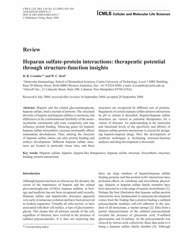
heparan sulfates consist of repeating disaccharide unitscomprising a hexuronic acid (HexA) and a D-glucosamine(GlcN) linked to each other and to other disaccharides by1Æ4 linkages. The uronic acid may be either a b-D-glu-curonic acid (GlcA) or a-L-iduronic acid (IdoA). Both oc-cur as unmodified monosaccharides or as 2-O-sulfatedresidues (GlcA2S and IdoA2S). The glucosamine may beeither N-sulfated or N-acetylated or, rarely, may exist as afree amine. N-sulfated-glucosamines (GlcNSO3) may beO-sulfated at C3 (GlcNSO33S) (rarely), or at C6 (Glc-NSO36S), or both C3 and C6 (GlcNSO33S6S), or carry nosulfates. Similarly, the N-acetylated glucosamines (Glc-NAc) may be O-sulfated at C6 (GlcNAc6S) or unsulfated.The combination of these different structural units into dis-accharides and the arrangement of the disaccharides alonga chain creates an extraordinarily large potential for struc-tural diversity. However, the theoretical diversity is not realised because of constraints manifest during chainbiosynthesis.Heparin and heparan sulfate chains are all synthesised attached to a core protein. Heparin is cleaved from its coreprotein, the mast cell protein serglycin, and is secretedfrom mast cells as a glycosaminoglycan chain. In contrast,heparan sulfates are attached to a protein core, to give astructure called a proteoglycan. Heparan sulfate proteogly-cans are expressed by virtually all mammalian cells, anddepending on the core protein, they may either be associ-ated with the cell surface or deposited in the extracellularmatrix. Heparin and heparan sulfate chains are synthesisedas a non-sulfated precursor that is linked to a serine residuein the core protein by a tetrasaccharide linker: bGlcA-1,3Æ bGal-1,3 Æ bGal-1,4 Æ bXyl-1 Æ Ser [25]. The poly-saccharide chain is modified sequentially by a series of enzymic reactions, none of which goes to completion.Complex patterns of sulfation result. It is the regulated expression and activity of a number of glycotransferases,sulfotransferases and an epimerase that determines the finestructure of both heparin and heparan sulfate chains. He-paran sulfate structures are not dependent upon the coreprotein, as different structures are produced when the samecore protein is expressed in different cell types [26]. Theprocess of heparan sulfate biosynthesis that gives rise tothis structural diversity has been reviewed in detail by Eskoand Lindahl [27].
Heparin and heparan sulfates are structurally distinct. Heparan sulfate has a well-defined domain organisationthat is not present in heparin [28, 29]. Commercial heparin is isolated from mast cell-rich tissues and com-prises that HL-GAG fraction that has the highest anticoag-ulant activity. Heparan sulfates are extracted from tissues that contain few, if any mast cells, and make up thetotal HL-GAG pool isolated. However, the most highly sul-fated fractions of heparan sulfate fit many of the criteriaused to describe mast cell heparin. Generally the ratio ofiduronic acid to glucuronic acid is low in heparan sulfates, and the numbers of N-sulfated-glucosamines and N-acetylated glucosamines are approximately equal.In contrast, heparin has a high ratio of iduronic acid to glucuronic acid, more N-sulfated glucosamines than N-acetylated glucosamines and is more highly sulfatedthan heparan sulfates [25]. Common features of heparinchains are stretches where the tri-sulfated disaccharide,IdoA2SGlcNSO36S-IdoA2SGlcNSO36S, is repeated [30].Heparan sulfate chains have IdoA and GlcNSO3 contain-ing regions that are highly sulfated (S-domains) alternat-ing with regions that are predominantly GlcA and rela-tively low in sulfates (fig. 2). There are also a relatively minor proportion of mixed sequences, which contain both GlcNSO3 and GlcNAc. The S-domains may containheparin-like trisulfated disaccharides as well as the disul-fated disaccharide, Ido2SGlcNSO3. C-6 sulfation of GlcNSO3 also occurs variably in the mixed regions. Therare modifications of C-3 O-sulfation of GlcNSO3 occursin the mixed regions and the S-domains [31], and thedeacetylation of GlcNAc to yield an unsubstituted amineoccurs in the mixed regions and the N-acetylated-domains[32].
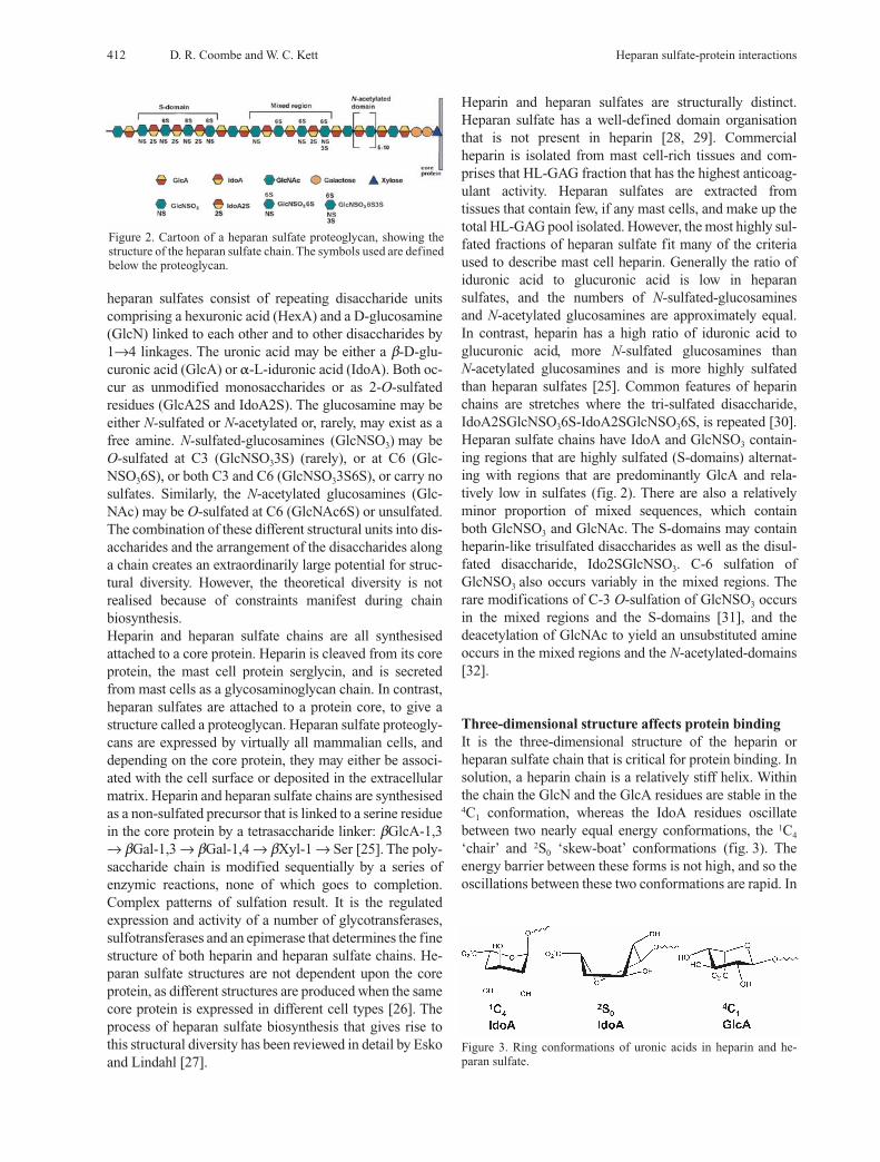
Three-dimensional structure affects protein bindingIt is the three-dimensional structure of the heparin or heparan sulfate chain that is critical for protein binding. Insolution, a heparin chain is a relatively stiff helix. Withinthe chain the GlcN and the GlcA residues are stable in the4C1 conformation, whereas the IdoA residues oscillate between two nearly equal energy conformations, the 1C4
‘chair’ and 2S0 ‘skew-boat’ conformations (fig. 3). The energy barrier between these forms is not high, and so the oscillations between these two conformations are rapid. In
Figure 2. Cartoon of a heparan sulfate proteoglycan, showing thestructure of the heparan sulfate chain. The symbols used are definedbelow the proteoglycan.
Figure 3. Ring conformations of uronic acids in heparin and he-paran sulfate.

CMLS, Cell. Mol. Life Sci. Vol. 62, 2005 Review Article 413
the main, internal IdoA residues favour the 2S0 conforma-tion, because in the 1C4 form the bulky carboxyl group isequatorial and all other substituents are in axial positions[28]. However, the iduronate conformation is influencedby the substitution pattern of the glucosamine attached to its non-reducing end. For example, the 1C4 chair form predominates when Ido2S has a GlcNAc attached at the 4-position [28]. The glycosidic linkages are quite stiff, and this means that the shape of the heparin chaindoes not change according to the conformation of theIdoA ring [10]. Moreover, the glycosidic linkage confor-mations remain similar regardless of the sulfation pattern[33].The two conformations of IdoA orientate the 2-O-sulfateand carboxyl groups in different positions relative to thehelix, and this can have profound effects on protein bind-ing (fig. 4). The arrangement of the heparin helix is suchthat for sequences of repeating trisulfated disaccharides,the three sulfates are clustered on one side of the chain,with a similar cluster forming on the other side of thechain for the next trisulfated disaccharide in the se-quence [10, 34]. The distance between sulfate clusters onone side of the chain is about 17Å [10]. However, whenIdoA2S is in the 1C4 chair configuration, the clusters ofsulfates appear more dispersed than when IdoA2S is askew-boat [34]. In solution these two conformations arein equilibrium; thus, it may be expected that a bindingprotein will perturb this equilibrium to select the confor-mation that is most energetically favourable for a stableinteraction. Experimental data indicate that this is thecase. The heparin pentasaccharide that binds antithrom-bin III has a single internal IdoA (fig. 1). A study usingorganic synthesis to lock IdoA derivatives in differentconformations demonstrated a critical role for the skew-boat 2S0 conformer in the activation of antithrom-bin by heparin [35]. In the case of FGF-2 binding to a heparin hexasaccharide, crystal structures of the com-plex revealed the IdoA at position 3 in the saccharidewas in the 1C4 conformation, whilst the IdoA at residue 5was in the 2S0 conformation [36]. The sulfate group inthe IdoA locked in a particular conformation is not always directly involved in interacting with the basicamino acids of the binding protein. The 2-O-S on theIdoA of the antithrombin III binding pentasaccharide isnot directly involved in binding; rather the skew-boatconformation of this IdoA facilitates electrostatic interactions between carboxyl groups in the saccharideand basic amino acids in the antithrombin III proteinbinding site [34]. In contrast, the Ido2S that is not directly involved in binding to FGF-2 adopts a 1C4
conformation, whereas the one that is involved in bind-ing adopts the skew-boat conformation [34]. These examples illustrate how similar monosaccharide sequences could display different conformations whenbound to different proteins.
There is very little information as to the three-dimensionalstructure of heparan sulfate. Although the highly sulfatedS-domains of heparan sulfate presumably would adopt thehelical structure of heparin, the likely conformation of theGlcA-GlcNAc domains is less certain. Clearly, these sequences lack the internal flexibility that is generated byIdoA, but they retain an ability to rotate about their glyco-sidic linkages. It appears that the degree of rotation thatcan occur at GlcA linkages, when GlcA is in the 4C1 con-formation, are greater than that observed with IdoA. Thisled Conrad [25] to argue that stretches of GlcA-contain-ing disaccharides may bend more readily than IdoA- containing disaccharide sequences. Mulloy and Forsterutilised data from model oligosaccharides, the K5 poly-saccharide (GlcNAc-GlcA-n), maltose and cellobiose.They argued that because more than one low-energy link-age conformation was detected for both linkage types,GlcNAc-GlcA and GlcA-GlcNAc, it is probable that theflexibility of regions rich in GlcA-GlcNAc repeating disaccharides would facilitate the appropriate positioningof two S-domains within a heparan sulfate chain onto aninteracting protein [10]. Indeed, a heptadecasaccharidechain consisting of two S-domains linked by three GlcA-GlcNAc repeats has been proposed as a heparan sulfatebinding domain for the chemokine macrophage inflam-matory protein 1a (MIP-1a) [37]. This structure was modeled using coordinates of the solution structure of heparin to form the S-domains, with the non-sulfated mid-
Figure 4. Space-filled model of the solution structure of heparin de-termined by nuclear magnetic resonance spectroscopy [132] andfirst published by Mulloy and Forster [10]. The iduronates areshown in their 1C4 chair (left) and 2S0 skew-boat (right) conforma-tions. Sulfates are displayed in red and yellow. Reproduced withpermission from [10].

414 D. R. Coombe and W. C. Kett Heparan sulfate-protein interactions
dle region being modeled as an extended chain in whichthe glycosidic linkages adopt sterically allowed conforma-tions. This type of model appears to be appropriate for heparan sulfate structures that bind other proteins, for example platelet factor 4, transforming growth factor b(TGF-b), interleukin-8 (IL-8) and regulated on secretion,normal T-cell expressed and secreted (RANTES) [38–41].
Heparan-sulfate chain configuration is affected by protein bindingThe binding of some proteins to heparin and heparan sul-fate is not only reliant on interactions between the sulfategroups on the saccharide and basic residues on the protein,but van der Waals contacts contribute substantially [22,42]. The best example comes from FGF family members.Studies revealed that although the GAG chain maintainedits overall helical structure when bound to the FGF, a kinkin the helical axis appears upon binding. Moreover, this‘kink’ is retained in the FGF-2-FGFR1-GAG complex andin the FGF-1-FGFR2-heparin complex [22, 42]. The extentof the kink is exaggerated by the 1C4 conformation of theiduronic acid that is favoured upon FGF binding because itorientates the glycosidic bonds axially. Calculations of theinteraction energies of a kinked oligosaccharide and anoligosaccharide with a standard helical structure indicatedthat the kinked oligosaccharide provides more favourableionic and van der Waals contacts [42]. The biological im-plications of this seem to lie in the specificity of the GAGstructure recognised. For FGF-1, a number of differentstructures were found to bind, but with graded affinities[43]. A detailed structural analysis of the oligosaccharidesthat bind FGF-1 indicated that although the number ofsulfated groups is about equal, the difference lies in the abilities of these oligosaccharides to form a kink comprising an iduronic acid in the 1C4 conformationflanked by two glucosamines. Optimal binding requiresthe sulfation pattern of the kink-spanning trisaccharide to be an N-sulfate on the non-reducing end glucosamine,2-O-sulfate on iduronate and 6-O-sulfate on the reduc-ing end glucosamine [42]. Analyses of other protein-oligosaccharide co-crystal structures also indicate a kinkin the oligosaccharide chain, and this positions the sulfates optimally for ionic and van der Waals interac-tions. These proteins include antithrombin III, and theNK1 domain of hepatocyte growth factor [42].
Altering heparan sulfate structures has profound biological outcomes
Morphological consequences of silencing biosynthetic enzymes Recent genetic experiments have thrust heparan sulfate-protein interactions into a centre-stage position in thefield of developmental biology. The genes EXT1 and
EXT2 encode glycosyltransferases that are required forheparan sulfate chain elongation in mammals. The EXTproteins transfer GlcA or GlcNAc residues to the nonre-ducing end of the polysaccharide. Heparan sulfate syn-thesis does not take place in EXT1–/– ES cells [44]. Simi-larly, when EXT2 expression is diminished in mam-malian cells by gene-silencing techniques, heparansulfate synthesis is blocked [45]. Mice rendered EXT1deficient by gene targeting failed to gastrulate and lackedorganised mesoderm and extra-embryonic tissues. Dis-ruption of the EXT1 gene selectively in the murine ner-vous system caused death in the first day of life [46].Moreover, mutations in either EXT1 or EXT2 causehereditary multiple exostoses, an autosomal dominantbone disorder [44, 45]. These studies have indicated thatheparan sulfate chain biosynthesis is critical for normalembryonic development.The effects of alterations in heparan sulfate structure ondevelopment have been investigated by making knockoutmice that lack expression of the enzymes required formodifying the heparan sulfate chain. The first step in themodification of the heparin/heparan sulfate chain is theremoval of acetyl groups from GlcNAc residues to givefree amino groups, which are then sulfated. The enzymescatalyzing these reactions are one of four isoforms of N-acetylglucosamine N-deacetylase/N-sulfotransferase(NDST). Knockout mice have been generated for two ofthese enzymes. Although NDST-2 is widely distributedduring development and in the adult, mice lacking thisenzyme had a phenotype that was restricted to connec-tive-tissue mast cells. Heparan sulfates from the liver ofNDST-2–/– mice show no real differences in the N-sulfa-tion pattern from control mice, but heparin was absent in-dicating the essential role of this enzyme for heparinbiosynthesis [47]. In contrast, a lack of NDST-1 is lethal.The heparan sulfates produced by NDST-1–/– mice havereduced N-sulfation and O-sulfation, and the epimeriza-tion of GlcA to IdoA occurs at reduced levels [48].Around a third of embryos die during the prenatal period,whilst new-born pups have abnormal lungs that produceinsufficient surfactants, and as a consequence of lungfailure, the pups die shortly after birth [48, 49]. Skeletaldefects and other defects which contribute to embryonicdeath are reviewed in Grobe et al. [48]. Loss of glucuronyl C5-epimerase activity is also lethalfor neonates. Targeted disruption of the murine glu-curonyl C5-epimerase gene (Hsepi) caused biosynthesisof heparan sulfate chains devoid of IdoA and with an abnormal sulfation pattern [50]. The phenotype of theHsepi–/– mice was loss of kidneys, poorly inflated and immature lungs, bilateral iris coloboma, abundant skele-tal abnormalities but normal brain, heart, liver, gastroin-testinal tract, pancreas and skin [50]. Hs2st is the singlegene that encodes heparan sulfate 2-O-sulfotransferase,and mice lacking this enzyme die in the neonatal period.

CMLS, Cell. Mol. Life Sci. Vol. 62, 2005 Review Article 415
The phenotype of Hs2st–/– mice has some similaritieswith that of Hsepi–/– mice. Given that neither of theseknockout mice have heparan sulfate with 2-O-sulfatedIdoA, it is probably not surprising that they both fail todevelop kidneys and display several skeletal abnormali-ties resembling those of Hsepi–/– mice [50, 51]. Complications in the interpretation of data from biosyn-thetic enzyme silencing experiments may arise when onemember of a multi-enzyme family is silenced. There aresix members of the 3-O-sulfotransferase (3-OST) family,and these enzymes transfer sulfate groups to the 3-OH of glucosamine. Interestingly, the different enzyme isoforms preferentially recognise different saccharidestructures around the glucosamine that is to be sulfated[52]. As these isoforms are expressed at different levels indifferent tissues this finding provides an explanation forthe formation of tissue specific saccharide structures. The3-OST-1 isoform is primarily responsible for 3-O-sulfa-tion of the glucosamine within the antithrombin III-bind-ing pentasaccharide. Thus, it may be expected that silenc-ing this gene (Hs3st1) would give rise to mice with a pro-coagulant phenotype. This was not the case, even thoughheparan sulfate isolated from various tissues of Hs3st1–/–
mice had markedly reduced anti-Xa activity compared toheparan sulfates isolated from normal mice [53]. Unex-pectedly, Hs3st1–/– mice exhibited intrauterine growth retardation and genetic background-specific lethality.Possibly, in the absence of 3-OST-1 other members of thisfamily perform the 3-O-sulfation of the glucosamine to alevel that is sufficient to protect against thrombosis butnot against the other abnormalities. The finding that the3-OST-5 isoform is capable of generating antithrombinIII-binding heparan sulfate in a cell line supports thisview [54].
Functional consequences of silencing biosynthetic enzymes Although the knockout studies indicate that the correctbiosynthesis of heparan sulfate chains is critical for development, they also reveal how little is understoodabout the role of particular heparan sulfate structures inthese processes. Experiments designed to address whatstructural changes in the heparan sulfate chains mean forprotein binding and signaling are required. These studiesare beginning. An analysis of heparan sulfate structuresproduced in Hs2st–/– embryos revealed that the domainstructure is conserved but the N-sulfates are clusteredinto longer S-domains. Despite the lack of 2-O-sulfate,the charge density is maintained by a dramatic increase of6-O-sulfate in the GlcNS repeat regions [55]. The abilityof the mutant heparan sulfate to bind fibronectin and hepatocyte growth factor (HGF) is maintained, but bind-ing to FGF-1 and -2 was weaker. It would be interestingto perform X-ray crystallography on FGF-2 binding to
oligosaccharides derived from Hs2st mutant mice to determine how structures lacking 2-O-sulfation, but withincreased 6-O-sulfation, make contacts with amino acidsin the GAG binding site. Unexpectedly, growth factor signaling was very similar for FGF-1, -2 and HGF [55].Clearly the strength of FGF-1 and -2 binding to heparansulfate without 2-O-sulfate is sufficient for signaling in embryonic fibroblasts. This finding indicates that invitro assessments of the strength of protein-heparin/heparan sulfate interactions do not necessarily predict thefunctional outcomes of that interaction. It also illustratesthat novel heparan sulfate structures produced as a con-sequence of silencing heparan sulfate biosynthetic en-zymes may have unexpected binding activities, furthercomplicating interpretation of the molecular basis of thephenotypes of mice in which these genes have been silenced.Intact heparin and heparan sulfate chains are also modi-fied by extracellular endosulfatases, and these enzymesappear to play a role in regulating embryo patterning. Twoendosulfatases, designated HSulf-1 and HSulf-2, havebeen cloned in mice and humans, as well as a quailhomologue, QSulf1 [56, 57]. These enzymes remove sulfate from the 6-position of glucosamine in the disac-charide IdoA2S-GlcNS6S and to a lesser extent in GlcA-GlcNS6S disaccharides, recognising only a small subsetof these disaccharides, but saccharide sequences contain-ing IdoA are not recognised [56, 58]. Data from anotherstudy suggest that Qsulf1 recognises the 6-O-sulfateswhen GlcNS6S is flanked by IdoA2S. That is, the activ-ity of Qsulf1 is confined to the S-domains [59]. An explanation for the increase in 6-O-sulfation in heparansulfates from Hs2st mutant mice could be a marked reduction in the activity of HSulf-1 or HSulf-2, as theseenzymes primarily recognise the IdoA2S-containingtrisulfated disaccharide which is common in heparin andS-domains of heparan sulfate. Given the role of FGF family members in development,and that heparan sulfate is an obligate cofactor forFGF/receptor signaling, it is logical that discussion of the molecular basis for the phenotypes observed when genes encoding heparan sulfate biosynthetic enzymes are silenced should focus on FGF family members. Merryand Wilson [60] have examined phenotypes of mice car-rying targeted mutations in growth factors and receptors,principally of the FGF family and FGF receptors, with aview to determining whether a mutation in these mole-cules recapitulates the Hs2st mutant phenotype. Althoughthe Hs2st mutant phenotype overlaps with the phenotypeof mice depleted of some of these growth factors and receptors, no single mutation gives rise to a phenotypethat resembles the Hs2st mutant. This is not unexpectedgiven the large number of vertebrate proteins that bind toheparan sulfate, but which are not FGF family membersor their receptors. Some of these, like NCAM [61], are
416 D. R. Coombe and W. C. Kett Heparan sulfate-protein interactions
involved in cell adhesion and cell migration during embryogenesis, whilst others are extracellular matrixproteins, e.g. fibronectin, some laminins, thrombo-spondin and BM-40 [62–65]. Furthermore, other hep-aran sulfate-binding proteins may indirectly influenceembryogenesis via the proteins they bind, e.g. the heparansulfate-binding protein follistatin binds and neutralisesactivin, a molecule that plays a critical role in differenti-ation and early embryo development [66]. Unravelingwhich vertebrate molecular pathways are affected by mutations of heparan sulfate biosynthetic enzymes willbe complex and challenging.
Tissue specific heparan sulfate structures are functionally relevant
The heparan sulfate enzyme knockout experiments areunable to demonstrate whether subtle differences in heparan sulfate structure are found in particular tissuesites and whether they have biological relevance. Theseare crucial, yet difficult questions to address because ofthe problems of isolating the very small quantities of heparan sulfates synthesised by different cell types, andof the difficulty of performing structural and biologicalanalyses with very little material that has underlying heterogeneity. The use of epitope specific antibodiesshould assist in examining these questions. Heparan sulfates are generally poor immunogens, but therecent use of phage display technology has allowed thegeneration of a panel of apparently epitope specific anti-heparan sulfate and anti-heparin antibodies [67–69].Screening of these antibodies against panels of modifiedheparan sulfate and heparin molecules [67–69] as well asagainst heparan sulfate oligosaccharides of known sequence [68] indicated that the antibodies had differentbinding patterns. Assessment of antibody reactivity to heparan sulfate oligosaccharides of known sequence alsoprovided data on the type of structure likely to be contained in the preferred epitope [68]. However, two ofthe antibodies selected against lung heparan sulfate wereidentical to antibodies selected against bovine kidney andhuman skeletal muscle, indicating that these tissues shareheparan sulfate epitopes [70]. Antibody staining has revealed defined topological distributions of various heparan sulfate epitopes in the rat kidney and spleen, human lung, and human, rat and mouse skeletal muscle[67, 68, 70, 71]. In the spleen some of the antibodies co-localised with interleukin-2 which was bound to heparan sulfate [71]. Similarly, in the lung some of the an-tibodies blocked FGF-2 and VEGF (vascular endothelialgrowth factor) binding [70]. Collectively, these data indi-cate that heparan sulfate biosynthesis is controlled anddifferently regulated by the cell types within tissues, prob-ably creating specific extracellular microenvironments.
If particular heparan sulfate epitopes are expressed in tissue sites, then are these different epitopes selectivelyrecognised by heparan sulfate binding proteins? Twostudies by Allen and colleagues suggest that they are [72,73]. They have generated a series of probes for heparansulfates based on the fact that both FGFs and their recep-tors bind heparan sulfates to form a signaling complex.The probes used were FGF-2, FGF-4 and their receptors:soluble FGF receptor 1-IIIc (FR1c) and FGF receptor 2-IIIc (FR2c), and in the second study, FGF-1 and FGF-8b with FR2c and soluble FGF receptor 3-IIIc (FR3c).Collectively, the data from these studies indicated thatthere are changes in the heparan sulfate structures expressed during development, and these changes are reflected in the different binding patterns of the variousFGFs and FGF/receptor complexes. As is expected fromin vitro studies of various FGFs binding to different heparan sulfate structures, the binding patterns of thegrowth factors differed. FGF-2 was found to bind heparan sulfate in a ubiquitous fashion in the developingmouse embryo, whereas FGF-4 was more selective, failing to bind heparan sulfate in the heart and large bloodvessels, nor to aortic endothelial cells in culture [72]. The complex pattern with which each FGF/receptor pairbound heparan sulfates in the embryos suggested that thedifferent FGF/receptor combinations recognised distinctheparan sulfate structures that are spatially and tempo-rally regulated [72, 73]. Analysis of whether 2-O-sulfa-tion or 6-O-sulfation is a requirement of the heparan sulfate that is involved in the particular FGF/receptor/heparan sulfate signaling complex indicated differencesin sulfation requirements between FGF/receptor com-plexes [73]. Importantly, the data suggest that the heparansulfate binding site displayed by an FGF/receptor pairdiffers from that displayed when the same FGF combineswith a different receptor. These data are discussed in thelight of the finding that FGF-1 can signal using heparansulfate from Hs2st–/– mice. It is suggested that for FGF-1, the heparan sulfate structure that binds FGF-1 requires2-O-sulfation, similarly 2-O-sulfation is required for it toform a complex with heparan sulfate and FR2c, but thisis not the case if it forms a complex with heparan sulfateand FR2b [73]. Although the sulfation patterns involvedhave not been determined, the situation could be similarfor FGF-4 and its receptors. Considering the pattern withwhich these probes bind embryo sections, it appears thatthe heparan sulfate structures required to bind FGF-4/FR1c complexes differ from those that bind FGF-4 alone.On the other hand, FR2c seems to be less selective, recog-nising all FGF-4-heparan sulfate complexes [72]. Thus,the heparan sulfate structure that binds the various FGFsin isolation will not necessarily be the same structure thatis required for signaling. Another example of subtly different heparan sulfatestructures displaying markedly variant capabilities for
CMLS, Cell. Mol. Life Sci. Vol. 62, 2005 Review Article 417
FGF signaling comes from a study on murine embryos.The structure of heparan sulfates isolated from murineembryonic day 10 (E10) and embryonic day 12 (E12)neuroepithelial cells differed. There were differences inthe levels of 2-O-sulfation, the patterns of 6-O-sulfation,total chain length and the number of sulfated domains perchain [74]. E10 heparan sulfate was strongly active insupporting FGF-8 signaling via FR3c, whereas the E12heparan sulfate had no activity. In contrast, both heparansulfate preparations supported FGF-2 signaling via FR1c[75]. These data are in concordance with the develop-mental stages at which FGF-8 and FGF-2 function in theembryo. Interestingly, clear differences were also evidentin the levels of 2-O-sulfotransferase expressed and theisoforms of 6-O-sulfotransferases expressed in E10 andE12 neuroepithelial cells. Moreover, there were differ-ences in the isoforms of NDSTs expressed [75], consis-tent with the finding of altered patterns of N-sulfation (re-flected in the differences in sulfated domains) betweenE10 and E12 heparan sulfate. This study provides evi-dence in support of isozyme expression patterns givingrise to certain heparan sulfate structures that are func-tionally specific and is an important adjunct to the en-zyme knockout experiments.An example of heparan sulfates in a particular tissue location preferentially binding a protein is that of thechemokine MCP-1 binding. Although heparan sulfatesare also found in the extracellular matrix secreted bythese cells, MCP-1 focused on the apical surface evenwhen added to the basal side of the endothelial cell layer[76]. Presumably the secreted heparan sulfate is struc-turally different from the cell surface-bound heparan sulfate, and this is reflected in the pattern of MCP-1 bind-ing. Clearly, studies of the binding patterns of otherchemokines and cytokines that bind heparan sulfate arewarranted. Although little structural data on the types ofheparan sulfate structures recognised by these proteinsare available, it is frequently hypothesised that particularheparan sulfates act to localise chemokines and cytokinesto specific tissue sites and by so doing contribute to reg-ulating their function. This appears to be true for FGFfamily members, but whether it is true for a range of cy-tokines and chemokines remains to be determined.
Regulation of heparan sulfate-protein interactions in the tissues
The data indicate that certain heparan sulfate structuresbind particular proteins both in vivo and in vitro but dothe in vitro data reflect what is happening biologically?The ionic nature of buffers used for in vitro binding as-says, for example, may not always reflect the extracellu-lar milieu in which proteins bind GAGs in vivo. There arenumerous proteins, which bind heparin/heparan sulfate
with higher affinity if cations are present, particularlyzinc or copper ions. These proteins include beta-amyloidprecursor protein, histidine-proline-rich glycoprotein(HPRG), interleukin-5, high molecular weight kininogen,prion protein, heparin cofactor II and endostatin, to namea few [77–83]. Occasionally, differences in the bindingpatterns of a particular protein to heparin can be explained by variations in the losses of cations associatedwith the protein, depending on the purification methodused. For example, variable binding to GAGs of differentendostatin preparations appeared to be due to differinglosses of cations (probably zinc) according to the purifi-cation protocol employed, and the existence, or other-wise, of zinc-dependent dimers [83]. Although oligomer-ization may be stabilised by cations, and this facilitatesinteractions with heparin or heparan sulfate [81], cationsmay also induce a conformational change in a proteinwhich assists an interaction with heparin. For example,Zn++ induces a conformational change in heparin cofactorII that enhances its interaction with heparin [82]. In the above examples the cations are bound by the protein, but heparin/heparan sulfate chains also bindstrongly to divalent metal ions [25]. The binding ofcations to GAG chains is not always a simple electrosta-tic interaction between the negatively charged groups onthe carbohydrate and the positively charged cation because Zn++ binds selectively to heparin rather than toother GAGs [84]. NMR evidence indicates that iduronicacid is the main binding site in heparin for heavy metalcations. Moreover, the spectral data suggest that Zn++
binding alters the ring conformation of iduronic acid such that the 1C4 conformation is stabilised over the 2S0
conformation [85–87]. If metal ion binding similarly controls the ring conformation of iduronate in heparinand heparan sulfate under physiological conditions, thismay be expected to influence the specificity and affinityof protein interactions. The concentration of Zn++ in bodyfluids is generally quite low, but platelets contain zinc at30–60-fold higher concentrations than plasma, and abun-dant zinc binding proteins such as decorin or biglycancould serve as storage pools for these ions [88, 89]. It isfeasible that local concentrations of Zn++ may be muchhigher than that of plasma, particularly around sites ofplatelet activation. Thus, microenvironmental concentra-tions of cations are likely to influence/regulate the in vivoaffinity and specificity of numerous heparan sulfate-protein interactions. In vitro binding assays are almost invariably performed atneutral pH; however, in vivo the local pH is not alwaysneutral. For example, local interstitial acidification iscommonly associated with inflammatory lesions, whichis attributed to primarily the local increase in lactic acidproduction caused by the anaerobic glycolysis of infil-trating neutrophils [90]. Measurements of the pH of fluids drained from sites of inflammation document
418 D. R. Coombe and W. C. Kett Heparan sulfate-protein interactions
extracellular pH values as low as 6.1 [91]. Moreover, ithas been known for many years that tumour microenvi-ronments are usually more acidic than normal, with pHvalues as low as 5.5 [92, 93]. Hypoxia or ischemia is com-monly associated with local acidosis. Alteration of pHcan have profound effects on the ability of some proteinsto bind heparin or heparan sulfate. This is particularly sowhen the GAG binding site involves histidines. Histidine-proline-rich glycoprotein is a prime example, for at neu-tral pH binding to heparin is minimal, but increases to amaximum at pH 6.5. However, zinc ions supplant the requirement for low pH. At intermediate pH, both proto-nation of histidine and the binding of zinc promote the interaction with heparin [94]. Prion protein also bindsGAGs in a pH- and metal ion-dependent fashion; at pH values above the histidine pKa, prion protein-GAG complexes are stabilised by Cu++ or Zn++ [81]. Other proteins that bind GAGs in a pH-dependent fashioninclude the non-fibrillar form of beta-amyloid peptide,selenoprotein P, granulocyte macrophage colony stimu-lating factor (GM-CSF) and VEGF [95–98]. These pro-teins are dissimilar in overall structure, but their bindingsites for GAGs involve one or more histidines. A VEGFisoform that lacks the native heparin binding domain(VEGF121) was found only to bind heparin and heparansulfate at low pH, whereas binding of the isoform,VEGF165, to heparin increased at acidic pH. Thus, low pHappears to expose a new heparin binding site within theregions shared by the two isoforms [98]. The binding ofboth these VEGF isoforms to fibronectin also increasedat low pH, an effect that was further enhanced by heparin.Under hypoxic conditions, like those found in and aroundtumours or wounds, the generation of an acidic extracel-lular environment may lead to storage of VEGF in a stable complex of fibronectin and heparan sulfate proteo-glycans. As at neutral pH active VEGF is readily released,the pH-sensitive matrix storage and release of VEGFcould set up a VEGF gradient which directs and stimu-lates the growth of new blood vessels into hypoxic or ischemic regions in tissues [99]. GM-CSF is a cytokine involved in regulating haemo-poiesis in the bone marrow and at extramedullar sites. Inthe late 1980s it was suggested that an interaction of GM-CSF with stromal heparan sulfates contributed to itsbiological activity [100, 101], but there were no follow-up reports directly demonstrating heparan sulfate struc-tures bound to GM-CSF. Other data indicated that even inthe presence of cytokines, not all stromal cells could sustain myelopoiesis. The nature of the glycoconjugatesin the stromal cell layer was found to be a determinant[102]. If GM-CSF undergoes a pH-induced conforma-tional change that allows heparin to bind, as has been suggested [97], this could explain the lack of in vitrobinding data. The interaction of haemopoietic cells withsupporting stroma leads to an accumulation of sialylated
glycoconjugates and proteoglycans at the interface of thetwo cell types [97, 103]. This may produce a local acidicmicroenvironment that supports the binding of GM-CSFto membrane heparan sulfates. Within the bone marrow,precursor cells of particular lineages are known to favour particular sites giving foci of developing cells ofone lineage [104]. Thus, local pH and the presence of particular GAG structures may act together to regulate GM-CSF localisation, thereby producing the microenvi-ronmental niche necessary for sustained myelopoiesis.Indeed, highly O-sulfated stromal cell heparan sulfate isan important component of the bone marrow ‘niche’ thatacts with cytokines and chemokines to regulate cell proliferation and differentiation [105].
GAG chain presentation and activity
Many in vitro assays do not take into account the fact thatthe presentation of GAG chains may alter their activity. Invivo more than one heparan sulfate chain is frequently attached to a core protein, and proteoglycans may be expressed on cell surfaces which permit molecular clus-tering. Soluble and cell membrane forms of syndecan-1and glypican-1 differed markedly in their ability to stimulate FGF-2-induced FGFR1 phosphorylation [106].Membrane associated forms were active, whereas corre-sponding soluble forms were inactive. Interestingly, cellsexpressing a mutant glypican-1 that carried only one heparan sulfate chain also strongly stimulated FGF-2 induced FGFR1 signaling, suggesting that multivalencyof heparan sulfate chains on the same core protein is nota requirement for the activity of membrane-associatedproteoglycans in this system [106]. In contrast, when the syndecan-1 functions of collagen binding, cell-celladhesion and cell invasion of collagen gels were assayed,multivalency was important. The function of syndecan-1was modulated according to the number of heparan sulfate chains it carried, and the position of these chainson the core protein, even though the overall levels of cellsurface heparan sulfate expression did not vary apprecia-bly [107].
Towards the use of heparin or heparan structures as therapeutics
Heparin and heparan sulfates bind a multitude of differ-ent proteins that have a variety of biological functions.The requirement for these GAGs in mammalian develop-ment has been demonstrated. It is also clear that tissue-specific, different heparan sulfate structures bind differ-ent sets of proteins. Frequently the local tissue environ-ment modulates the affinity of the GAG-proteininteraction, and this may have biological outcomes in

CMLS, Cell. Mol. Life Sci. Vol. 62, 2005 Review Article 419
terms of establishing gradients of cytokines or chemo-kines. If the goal is to define a GAG structure that hastherapeutic applications, it is appropriate to determineoptimal GAG structures that bind the protein in questionwith high affinity when GAG chains are in solution, as anoligosaccharide that binds tightly in vivo is the require-ment. Thus, the pH of the target tissue and the possibilitythat cations will contribute to the binding affinity deserveconsideration.There is enormous potential for the development of heparin-like structures as drugs for a range of diseases inaddition to the current antithrombotic target. The mostobvious of these are cancer, inflammatory diseases andvirus infections [108]. There have been a number of approaches to the development of heparin-based thera-peutics. The production of mixtures of heparin-like struc-tures that interact with and alter the function of numerousproteins is one approach. A drug currently in clinical trials in cancer patients that was designed through this approach is the phosphosulfomannan, PI-88 [109]. PI-88is structurally heterogeneous [110], and as well as inhibiting heparanase it also binds FGF-1, FGF-2 andVEGF [111]. Another approach is the synthesis of particular struc-tures based on a heparin/heparan sulfate template that are designed to bind specifically to the target protein. Although the diversity of heparin/heparan sulfates andthe nature of their monosaccharide components makesynthesis a major challenge for chemists, the first syn-thetic molecules were produced 20 years ago. Pioneers inthe field were Choay, Petitou and colleagues who synthe-sised tetrasaccharides and pentasaccharides to under-stand the structural basis for heparin’s antithrombin III-binding, and anticoagulant activity [112–114]. Thisearly work has led to the registration of the first fully synthetic heparin structure for clinical use. Fondaparinux (Arixtra, Sanofi-Synthelabo) has been approved for usein thromboprophylaxis following orthopedic surgery. It isthe antithrombin III-binding pentasaccharide sequence(fig. 1), but with a methyl group stabilizing the anomericend [115]. Fondaparinux does not bind to platelet factor 4(PF4) and does not cross-react with antibodies generatedas a result of heparin-induced thrombocytopenia. Nordoes fondaparinux stimulate endotoxin-induced inter-leukin-8 production by monocytes [116]. The data alsosuggest that fondaparinux will be well tolerated by patients who have a tendency to develop delayed-type hypersensitivity reactions to subcutaneously injected heparin, probably because fondaparinux does not binddermal proteins [117, 118]. Clearly, careful selection of aheparin structure for activity against a target protein, inthis case antithrombin III, does eliminate many of the undesirable effects of heparin therapy. Other synthetic heparin antithrombin III pentasaccharideanalogues, e.g. Idraparinux (SANORG 34006), are cur-
rently in clinical development [115, 119]. A second generation of heparin mimetics designed to treat variousthrombotic disorders are in the pipeline. These structuresconsist of two functional domains, an antithrombin IIIbinding domain and a thrombin binding domain sepa-rated by a spacer [120]. The goal was to obtain a mimeticwith a favourable antithrombotic/bleeding ratio butwhich does not bind to PF4 or lead to heparin-inducedthrombocytopenia. The preclinical data on a synthetichexadecasaccharide indicate it is more active than heparin in in vivo models of thrombosis, yet it did not activate platelets or compete with heparin for binding toPF4 [121, 122]. A few laboratories are investigating utilising a modularapproach for the synthesis of heparin oligosaccharides.The ready synthesis of heparin-like structures in quanti-ties suitable for biological and biochemical assays, andeventually drugs, is the aim. The modular approach is feasible because different combinations of 20 disaccha-rides arranged in a linear sequence can determine thestructure of native heparan sulfate chains. The labora-tories of Seeberger and Boons have independently published their strategies for synthesis of six mono-saccharide building blocks which contain different chem-ical protecting groups on key positions that are involvedin chain linkage or modification by carboxyl or sulfategroups [123–125]. The linkage of these monosaccha-rides, first into disaccharides and then into larger well-defined oligosaccharides that display the variety of structures found in native heparan sulfate, is being explored. Rosenberg and colleagues developed a method utilisingthe polysaccharide isolated from Escherichia coli K5 as a starting material for the synthesis of classical and non-classical heparan sulfate-like structures [126]. Theproduction of N-sulfated glucosamines was performedchemically, but all subsequent modifications were per-formed by a set of recombinant heparan sulfate biosyn-thetic enzymes. The authors suggest this ‘chemosyn-thetic’ approach will allow the generation of libraries ofhomogeneous oligosaccharides, the definition of criticalfunctional groups for target proteins, and eventually thedesign of heparan sulfate-like drugs [126]. Using thismethod they have prepared non-classical heparan sulfate-like structures that lack IdoA2S groups but are 3-O- and6-O-sulfated. These structures possess anticoagulant activity indicating that the 2-O-sulfate of IdoA2S is a minor contributor to antithrombin III binding [126]. AsIdoA2S groups seem critical for PF4 binding to GAGsand for heparanase cleavage, these non-classical antico-agulants should be more biologically active than their native counterparts. Idraparinux is an O-methylated, O-sulfated pentasaccharide, which although modeled onthe antithrombin III pentasaccharide (fig. 1) similarlylacks a IdoA2S, but binds antithrombin III with an affin-

420 D. R. Coombe and W. C. Kett Heparan sulfate-protein interactions
ity 10 times that of the natural structure [119]. These data highlight the importance of critical groups within aGAG structure for specific protein binding rather than the native heparin/heparan sulfate sequence itself beingcritical.
Concluding remarks
The study of heparin/heparan sulfate-protein interactionsis now poised to make major advances in the next fewyears spearheaded by new technologies being developedfor the structural analysis of heparin and heparan sulfatesand the synthesis of heparin-like structures. These newtechnologies will greatly assist the development of microarrays of structurally defined GAG fragments.Such tools will assist in answering crucial questions as tothe specificity of many of the GAG-protein interactionsand whether motifs that possess a similar level of speci-ficity as displayed by the antithrombin III-binding pentasaccharide exist for other heparin/heparan sulfate-binding proteins. However, binding specificity and affin-ity do not fully address questions of function. The exam-ple of the heparan sulfate produced by Hs2st–/– mice being able to initiate FGF-1 and FGF-2 signaling despiteits reduced binding affinity [55] demonstrates that in biology, increased affinity does not always directly corre-late with increased activity. Often once a certain thresh-old of binding stability is achieved, further increases inaffinity are functionally immaterial. Moreover, an appro-priate multivalent presentation of heparan sulfate struc-tures each with suboptimal binding affinity may producethe same stability of binding, and hence biological activ-ity, as a more specific but monomeric heparan sulfatestructure. It is important to consider the biological role of a partic-ular heparin/heparan sulfate-protein interaction whendiscussing specificity and affinity issues. The recent discovery that heparan sulfate proteoglycans onmacrophages, endothelia and genital epithelial cells capture cell-free human immunodeficiency virus (HIV)and for the latter two cells types facilitate the transfer ofvirus to CD4+ T lymphocytes is an example where exquisite specificity appears not to be required for bio-logical activity [127–129]. In contrast, herpes simplexvirus type 1 (HSV-1) envelope glycoprotein D (gD)recognises an octasaccharide that includes the rare 3-O-sulfated glucosamine generated by the 3-O-sulfotrans-ferase isoforms 3 and 5 [130, 131]. The gD-octasaccha-ride interaction is a critical event for HSV-1 entry intopermissive cells, and as the virus infects only mucosal epithelium and very rarely neuronal cells, a quite specificinteraction may be expected. Clearly, some GAG-proteininteractions have evolved to be relatively non-specific,whereas others are quite specific.
The biosynthetic-enzyme silencing experiments havetaught us a number of things. First, that heparan sulfatechains are critical for normal development. Second, thatthe structure of those chains is critical. Third, how littleis understood about the tissue-specific regulation of the various isoforms of these enzymes, and hence whatstructures are expressed where, and finally how little isknown of the biological consequences of normal heparan sulfate-protein interactions, the loss of which causes thephenotypes that are observed. As has been discussed, it is clear that particular heparan sulfate structures are expressed in different tissue types and at different timesduring development, and these different structures are selectively recognised by heparan sulfate-binding pro-teins. The tissue environment may also contribute to theregulation of GAG-protein interactions by changes in pH or cation composition. Thus, in vivo the specificity question takes on a different complexion because the microenvironmental milieu could prohibit binding, ormany proteins may never encounter particular heparansulfate structures. The fact that a protein may bind astructure in vitro that it would never encounter in vivo would normally have no biological relevance, but itcould be important in a pharmaceutical context. The design of an effective GAG-based therapeuticshould be appropriate for the heparan sulfate-protein interaction that is targeted. A novel, very specific GAGstructure may be inappropriate for inhibiting the locali-sation of HIV, for example. Frequently the side effects ofa relatively non-specific GAG-based drug can be min-imised by the choice of the drug delivery route. For example, an anti-HIV intravaginal application could obviate the lack of specificity. Among the multitude ofheparan sulfate-protein interactions that exist in biology,there will be a spectrum of affinities and specificities.Nevertheless, because the diversity of heparan sulfatestructures and the complexity of their biosynthetic path-ways has been maintained throughout animal evolution,it is likely that many of these heparan sulfate-protein interactions will require a pronounced level of speci-ficity. The newly evolving technologies in GAG synthe-sis and structural analysis will assist in resolving the relationship between structure and activity. An under-standing of structure-activity relationships could welllead to the design of well-tolerated drugs based aroundheparan sulfate structures that target a range of diseasesoutside of the thrombosis-anticoagluation axis. The nextdecade is likely to be an exciting time for heparan sulfate-inspired therapeutics.
Acknowledgements. We would like to thank our colleague SandraStevenson for her comments on this manuscript. We also thank DrBarbara Mulloy for making her pictures of the heparin helix available.

CMLS, Cell. Mol. Life Sci. Vol. 62, 2005 Review Article 421
1 Medeiros G. F., Mendes A., Castro R. A., Bau E. C., Nader H. B. and Dietrich C. P. (2000) Distribution of sulfated glycosaminoglycans in the animal kingdom: widespread occurrence of heparin-like compounds in invertebrates.Biochim. Biophys. Acta 1475: 287–294
2 Parish C. R., Jakobsen K. B., Coombe D. R. and Bacic A.(1991) Isolation and characterization of cell adhesion mole-cules from the marine sponge, Ophlitaspongia tenuis.Biochim. Biophys. Acta 1073: 56–64
3 DeAngelis P. L. (2002) Evolution of glycosaminoglycans andtheir glycosyltransferases: implications for the extracellularmatrices of animals and the capsules of pathogenic bacteria.Anat. Rec. 268: 317–326
4 Linhardt R. J. (1991) Heparin: an important drug enters itsseventh decade. Chem. Ind. 2: 45–50
5 Whisstock J. C., Pike R. N., Jin L., Skinner R., Pei X. Y., Carrell R. W. and Lesk A. M. (2000) Conformational changesin serpins: II. The mechanism of activation of antithrombin byheparindagger. J. Mol. Biol. 301: 1287–1305
6 Lindahl U., Thunberg L., Backstrom G., Riesenfeld J.,Nordling K. and Bjork I. (1984) Extension and structural vari-ability of the antithrombin-binding sequence in heparin. J.Biol. Chem. 259: 12368–12376
7 Merry C. L., Lyon M., Deakin J. A., Hopwood J. J. and Gallagher J. T. (1999) Highly sensitive sequencing of the sulfated domains of heparan sulfate. J. Biol. Chem. 274:18455–18462
8 Venkataraman G., Shriver Z., Raman R. and Sasisekharan R.(1999) Sequencing complex polysaccharides. Science 286:537–542
9 Lortat-Jacob H., Grosdidier A. and Imberty A. (2002) Struc-tural diversity of heparan sulfate binding domains inchemokines. Proc. Natl. Acad. Sci. USA 99: 1229–1234
10 Mulloy B. and Forster M. J. (2000) Conformation and dynam-ics of heparin and heparan sulfate. Glycobiology 10:1147–1156
11 Yayon A., Klagsbrun M., Esko J. D., Leder P. and Ornitz D. M.(1991) Cell surface, heparin-like molecules are required forbinding of basic fibroblast growth factor to its high affinity receptor. Cell 64: 841–848
12 Rapraeger A. C., Krufka A. and Olwin B. B. (1991) Require-ment of heparan sulfate for bFGF-mediated fibroblast growthand myoblast differentiation. Science 252: 1705–1708
13 Ornitz D. M., Yayon A., Flanagan J. G., Svahn C. M., Levi E.and Leder P. (1992) Heparin is required for cell-free bindingof basic fibroblast growth factor to a soluble receptor and formitogenesis in whole cells. Mol. Cell Biol. 12: 240–247
14 Maccarana M., Casu B. and Lindahl U. (1993) Minimal sequence in heparin/heparan sulfate required for binding ofbasic fibroblast growth factor. J. Biol. Chem. 268: 23898–23905
15 Guimond S., Maccarana M., Olwin B. B., Lindahl U. andRapraeger A. C. (1993) Activating and inhibitory heparin sequences for FGF-2 (basic FGF). Distinct requirements forFGF-1, FGF-2 and FGF-4. J. Biol. Chem. 268: 23906–23914
16 Aviezer D., Levy E., Safran M., Svahn C., Buddecke E.,Schmidt A. et al. (1994) Differential structural requirementsof heparin and heparan sulfate proteoglycans that promote binding of basic fibroblast growth factor to its receptor. J.Biol. Chem. 269: 114–121
17 Ishihara M., Takano R., Kanda T., Hayashi K., Hara S.,Kikuchi H. et al. (1995) Importance of 6-O-sulfate groups ofglucosamine residues in heparin for activation of FGF-1 andFGF-2. J. Biochem. 118: 1255–1260
18 Schlessinger J., Lax I. and Lemmon M. (1995) Regulation ofgrowth factor activation by proteoglycans: what is the role ofthe low affinity receptors? Cell 83: 357–360
19 Venkataraman G., Sasisekharan V., Herr A. B., Ornitz D. M.,Waksman G., Cooney C. L. et al. (1996) Preferential self-
association of basic fibroblast growth factor is stabilized byheparin during receptor dimerization and activation. Proc.Natl. Acad. Sci. USA 93: 845–850
20 Herr A. B., Ornitz D. M., Sasisekharan R., Venkataraman G.and Waksman G. (1997) Heparin-induced self-association offibroblast growth factor-2. Evidence for two oligomerizationprocesses. J. Biol. Chem. 272: 16382–16389
21 Kan M., Wang F., Xu J., Crabb J. W., Hou J. and McKeehan W.L. (1993) An essential heparin-binding domain in the fibro-blast growth factor receptor kinase. Science 259: 1918–1921
22 Pellegrini L., Burke D. F., von Delft F., Mulloy B. and Blundell T. L. (2000) Crystal structure of fibroblast growthfactor receptor ectodomain bound to ligand and heparin. Nature 407: 1029–1034
23 Schlessinger J., Plotnikov A. N., Ibrahimi O. A., EliseenkovaA. V., Yeh B. K., Yayon A. et al. (2000) Crystal structure of aternary FGF-FGFR-heparin complex reveals a dual role forheparin in FGFR binding and dimerization. Mol. Cell. 6:743–750
24 Ostrovsky O., Berman B., Gallagher J., Mulloy B., Fernig D.G., Delehedde M. et al. (2002) Differential effects of heparinsaccharides on the formation of specific fibroblast growthfactor (FGF) and FGF receptor complexes. J. Biol. Chem.277: 2444–2453
25 Conrad H. E. (1998) Heparin-Binding Proteins, AcademicPress, San Diego, P. 527
26 Sanderson R. D., Turnbull J. E., Gallagher J. T. and Lander A.D. (1994) Fine structure of heparan sulfate regulates syn-decan-1 function and cell behavior. J. Biol. Chem. 269:13100–13106
27 Esko J. D. and Lindahl U. (2001) Molecular diversity of heparan sulfate. J. Clin. Invest. 108: 169–173.
28 Capila I. and Linhardt R. J. (2002) Heparin-protein interac-tions. Angew Chem. Int. Ed. Engl. 41: 391–412
29 Gallagher J. T. (2001) Heparan sulfate: growth control with arestricted sequence menu. J. Clin. Invest. 108: 357–361.
30 Yamada S., Yamane Y., Tsuda H., Yoshida K. and Sugahara K.(1998) A major common trisulfated hexasaccharide core sequence, hexuronic acid(2-sulfate)-glucosamine(N-sulfate)-iduronic acid-N-acetylglucosamine-glucuronic acid-glu-cosamine(N-sulfate), isolated from the low sulfated irregularregion of porcine intestinal heparin. J. Biol. Chem. 273:1863–1871
31 Chen J., Duncan M. B., Carrick K., Pope R. M. and Liu J.(2003) Biosynthesis of 3-O-sulfated heparan sulfate: uniquesubstrate specificity of heparan sulfate 3-O-sulfotransferaseisoform 5. Glycobiology 13: 785–794
32 Westling C. and Lindahl U. (2002) Location of N-unsubsti-tuted glucosamine residues in heparan sulfate. J. Biol. Chem.277: 49247–49255
33 Mulloy B., Forster M. J., Jones C., Drake A. F., Johnson E. A.and Davies D. B. (1994) The effect of variation of substitutionon the solution conformation of heparin: a spectroscopic andmolecular modelling study. Carbohydr. Res. 255: 1–26
34 Hricovini M., Guerrini M., Bisio A., Torri G., Naggi A. andCasu B. (2002) Active conformations of glycosaminoglycans.NMR determination of the conformation of heparin sequencescomplexed with antithrombin and fibroblast growth factors insolution. Semin. Thromb. Hemost. 28: 325–334
35 Das S. K., Mallet J. M., Esnault J., Driguez P. A., DuchaussoyP., Sizun P. et al. (2001) Synthesis of conformationally lockedL-iduronic acid derivatives: direct evidence for a critical roleof the skew-boat 2S0 conformer in the activation of anti-thrombin by heparin. Chemistry 7: 4821–4834
36 Faham S., Hileman R. E., Fromm J. R., Linhardt R. J. and ReesD. C. (1996) Heparin structure and interactions with basic fibroblast growth factor. Science 271: 1116–1120
37 Stringer S. E., Forster M. J., Mulloy B., Bishop C. R., GrahamG. J. and Gallagher J. T. (2002) Characterization of the bind-
422 D. R. Coombe and W. C. Kett Heparan sulfate-protein interactions
ing site on heparan sulfate for macrophage inflammatory protein 1alpha. Blood 100: 1543–1550
38 Stringer S. E. and Gallagher J. T. (1997) Specific binding ofthe chemokine platelet factor 4 to heparan sulfate. J. Biol.Chem. 272: 20508–20514
39 Lyon M., Rushton G. and Gallagher J. T. (1997) The interac-tion of the transforming growth factor-betas with heparin/he-paran sulfate is isoform-specific. J. Biol. Chem. 272:18000–18006
40 Spillmann D., Witt D. and Lindahl U. (1998) Defining the interleukin-8-binding domain of heparan sulfate. J. Biol.Chem. 273: 15487–15493
41 Vives R. R., Sadir R., Imberty A., Rencurosi A. and Lortat-Jacob H. (2002) A kinetics and modeling study ofRANTES(9–68) binding to heparin reveals a mechanism ofcooperative oligomerization. Biochemistry 41: 14779–14789
42 Raman R., Venkataraman G., Ernst S., Sasisekharan V. andSasisekharan R. (2003) Structural specificity of heparin binding in the fibroblast growth factor family of proteins.Proc. Natl. Acad. Sci. USA 100: 2357–2362
43 Kreuger J., Salmivirta M., Sturiale L., Gimenez-Gallego G.and Lindahl U. (2001) Sequence analysis of heparan sulfateepitopes with graded affinities for fibroblast growth factors 1and 2. J. Biol. Chem. 276: 30744–30752
44 Lin X., Wei G., Shi Z., Dryer L., Esko J. D., Wells D. E. andMatzuk M. M. (2000) Disruption of gastrulation and heparansulfate biosynthesis in EXT1-deficient mice. Dev. Biol. 224:299–311
45 Zak B. M., Crawford B. E. and Esko J. D. (2002) Hereditarymultiple exostoses and heparan sulfate polymerization.Biochim. Biophys. Acta 1573: 346–355
46 Inatani M., Irie F., Plump A. S., Tessier-Lavigne M. and Yam-aguchi Y. (2003) Mammalian brain morphogenesis and midline axon guidance require heparan sulfate. Science 302:1044–1046
47 Forsberg E., Pejler G., Ringvall M., Lunderius C., Tomasini-Johansson B., Kusche-Gullberg M. et al. (1999) Abnormalmast cells in mice deficient in a heparin-synthesizing enzyme.Nature 400: 773–776
48 Grobe K., Ledin J., Ringvall M., Holmborn K., Forsberg E.,Esko J. D. et al. (2002) Heparan sulfate and development: differential roles of the N-acetylglucosamine N-deacety-lase/N-sulfotransferase isozymes. Biochim. Biophys. Acta1573: 209–215
49 Forsberg E. and Kjellen L. (2001) Heparan sulfate: lessonsfrom knockout mice. J. Clin. Invest. 108: 175–180
50 Li J. P., Gong F., Hagner-McWhirter A., Forsberg E., AbrinkM., Kisilevsky R. et al. (2003) Targeted disruption of a murineglucuronyl C5-epimerase gene results in heparan sulfate lack-ing L-iduronic acid and in neonatal lethality. J. Biol. Chem.278: 28363–28366
51 Bullock S. L., Fletcher J. M., Beddington R. S. and Wilson V.A. (1998) Renal agenesis in mice homozygous for a gene trapmutation in the gene encoding heparan sulfate 2-sulfotrans-ferase. Genes Dev. 12: 1894–1906
52 Liu J., Shworak N. W., Sinay P., Schwartz J. J., Zhang L., FritzeL. M. et al. (1999) Expression of heparan sulfate D-glu-cosaminyl 3-O-sulfotransferase isoforms reveals novel sub-strate specificities. J. Biol. Chem. 274: 5185–5192.
53 HajMohammadi S., Enjyoji K., Princivalle M., Christi P.,Lech M., Beeler D. et al. (2003) Normal levels of anticoagu-lant heparan sulfate are not essential for normal hemostasis. J.Clin. Invest. 111: 989–999
54 Duncan M. B., Chen J., Krise J. P. and Liu J. (2004) Thebiosynthesis of anticoagulant heparan sulfate by the heparansulfate 3-O-sulfotransferase isoform 5. Biochim. Biophys.Acta 1671: 34–43
55 Merry C. L., Bullock S. L., Swan D. C., Backen A. C., LyonM., Beddington R. S. et al. (2001) The molecular phenotype
of heparan sulfate in the Hs2st–/– mutant mouse. J. Biol.Chem. 276: 35429–35434
56 Morimoto-Tomita M., Uchimura K., Werb Z., Hemmerich S.and Rosen S. D. (2002) Cloning and characterization of twoextracellular heparin-degrading endosulfatases in mice andhumans. J. Biol. Chem. 277: 49175–49185
57 Dhoot G. K., Gustafsson M. K., Ai X., Sun W., Standiford D.M. and Emerson C. P. Jr (2001) Regulation of Wnt signalingand embryo patterning by an extracellular sulfatase. Science293: 1663–1666
58 Ai X., Do A. T., Lozynska O., Kusche-Gullberg M., LindahlU. and Emerson C. P. Jr. (2003) QSulf1 remodels the 6-O sulfation states of cell surface heparan sulfate proteoglycansto promote Wnt signaling. J. Cell Biol. 162: 341–351
59 Viviano B. L., Paine-Saunders S., Gasiunas N., Gallagher J.and Saunders S. (2004) Domain-specific modification of heparan sulfate by Qsulf1 modulates the binding of the bonemorphogenetic protein antagonist Noggin. J. Biol. Chem. 279:5604–5611
60 Merry C. L. and Wilson V. A. (2002) Role of heparan sulfate-2-O-sulfotransferase in the mouse. Biochim. Biophys. Acta1573: 319–327
61 Cotman S. L., Halfter W. and Cole G. J. (1999) Identificationof extracellular matrix ligands for the heparan sulfate proteo-glycan agrin. Exp. Cell Res. 249: 54–64
62 Sachchidanand, Lequin O., Staunton D., Mulloy B., Forster M.J., Yoshida K. et al. (2002) Mapping the heparin-binding siteon the 13–14F3 fragment of fibronectin. J. Biol. Chem. 277:50629–50635
63 Yamaguchi H., Yamashita H., Mori H., Okazaki I., NomizuM., Beck K. et al. (2000) High and low affinity heparin-binding sites in the G domain of the mouse laminin alpha 4chain. J. Biol. Chem. 275: 29458–29465
64 Guo N. H., Krutzsch H. C., Negre E., Zabrenetzky V. S. and Roberts D. D. (1992) Heparin-binding peptides from the type I repeats of thrombospondin. Structural require-ments for heparin binding and promotion of melanoma cell adhesion and chemotaxis. J. Biol. Chem. 267: 19349–19355
65 Hohenester E., Maurer P. and Timpl R. (1997) Crystal struc-ture of a pair of follistatin-like and EF-hand calcium-bindingdomains in BM-40. EMBO J. 16: 3778–3786
66 Hashimoto O., Nakamura T., Shoji H., Shimasaki S., HayashiY. and Sugino H. (1997) A novel role of follistatin, an activin-binding protein, in the inhibition of activin action in rat pituitary cells. Endocytotic degradation of activin and its acceleration by follistatin associated with cell-surface heparansulfate. J. Biol. Chem. 272: 13835–13842
67 Jenniskens G. J., Oosterhof A., Brandwijk R., Veerkamp J. H.and van Kuppevelt T. H. (2000) Heparan sulfate heterogeneityin skeletal muscle basal lamina: demonstration by phage display-derived antibodies. J. Neurosci. 20: 4099–4111
68 Dennissen M. A., Jenniskens G. J., Pieffers M., Versteeg E.M., Petitou M., Veerkamp J. H. et al. (2002) Large, tissue-regulated domain diversity of heparan sulfates demonstratedby phage display antibodies. J. Biol. Chem. 277: 10982–10986
69 van de Westerlo E. M., Smetsers T. F., Dennissen M. A., Linhardt R. J., Veerkamp J. H., van Muijen G. N. et al. (2002)Human single chain antibodies against heparin: selection,characterization, and effect on coagulation. Blood 99:2427–2433
70 Smits N. C., Robbesom A. A., Versteeg E. M., van de WesterloE. M., Dekhuijzen P. N. and van Kuppevelt T. H. (2004) Heterogeneity of heparan sulfates in human lung. Am. J.Respir. Cell Mol. Biol. 30: 166–173
71 ten Dam G. B., Hafmans T., Veerkamp J. H. and van KuppeveltT. H. (2003) Differential expression of heparan sulfate domains in rat spleen. J. Histochem. Cytochem. 51: 727–739

CMLS, Cell. Mol. Life Sci. Vol. 62, 2005 Review Article 423
72 Allen B. L., Filla M. S. and Rapraeger A. C. (2001) Role of he-paran sulfate as a tissue-specific regulator of FGF-4 and FGFreceptor recognition. J. Cell Biol. 155: 845–858
73 Allen B. L. and Rapraeger A. C. (2003) Spatial and temporalexpression of heparan sulfate in mouse development regu-lates FGF and FGF receptor assembly. J. Cell Biol. 163:637–648
74 Brickman Y. G., Ford M. D., Gallagher J. T., Nurcombe V.,Bartlett P. F. and Turnbull J. E. (1998) Structural modificationof fibroblast growth factor-binding heparan sulfate at a determinative stage of neural development. J. Biol. Chem.273: 4350–4359
75 Ford-Perriss M., Guimond S. E., Greferath U., Kita M., GrobeK., Habuchi H. et al. (2002) Variant heparan sulfates synthe-sized in developing mouse brain differentially regulate FGFsignaling. Glycobiology 12: 721–727
76 Hardy L. A., Booth T. A., Lau E. K., Handel T. M., Ali S. andKirby J. A. (2004) Examination of MCP-1 (CCL2) partition-ing and presentation during transendothelial leukocyte migra-tion. Lab. Invest. 84: 81–90
77 Bush A. I., Pettingell W. H. Jr, de Paradis M., Tanzi R. E. andWasco W. (1994) The amyloid beta-protein precursor and itsmammalian homologues. Evidence for a zinc-modulated heparin-binding superfamily. J. Biol. Chem. 269: 26618–26621
78 Olsen H. M., Parish C. R. and Altin J. G. (1996) Histidine-richglycoprotein binding to T-cell lines and its effect on T-cell sub-stratum adhesion is strongly potentiated by zinc. Immunology88: 198–206
79 Lipscombe R. J., Nakhoul A. M., Sanderson C. J. and CoombeD. R. (1998) Interleukin-5 binds to heparin/heparan sulfate. Amodel for an interaction with extracellular matrix. J. Leukoc.Biol. 63: 342–350
80 Renne T., Dedio J., David G. and Muller-Esterl W. (2000)High molecular weight kininogen utilizes heparan sulfate proteoglycans for accumulation on endothelial cells. J. Biol.Chem. 275: 33688–33696
81 Gonzalez-Iglesias R., Pajares M. A., Ocal C., Espinosa J. C.,Oesch B. and Gasset M. (2002) Prion protein interaction withglycosaminoglycan occurs with the formation of oligomericcomplexes stabilized by Cu(II) bridges. J. Mol. Biol. 319:527–540
82 Eckert R. and Ragg H. (2003) Zinc ions promote the interac-tion between heparin and heparin cofactor II. FEBS Lett. 541:121–125
83 Ricard-Blum S., Feraud O., Lortat-Jacob H., Rencurosi A.,Fukai N., Dkhissi F., Vittet D et al. (2004) Characterization ofendostatin binding to heparin and heparan sulfate by surfaceplasmon resonance and molecular modeling: role of divalentcations. J. Biol. Chem. 279: 2927–2936
84 Parrish R. F. and Fair W. R. (1981) Selective binding of zincions to heparin rather than to other glycosaminoglycans.Biochem. J. 193: 407–410
85 Whitfield D. M. and Sarkar B. (1991) Metal binding to heparin monosaccharides: D-glucosamine-6-sulphate, D-glucuronic acid, and L-iduronic acid. J. Inorg. Biochem. 41:157–170
86 Whitfield D. M., Choay J. and Sarkar B. (1992) Heavy metalbinding to heparin disaccharides. I. Iduronic acid is the mainbinding site. Biopolymers 32: 585–596
87 Whitfield D. M. and Sarkar B. (1992) Heavy metal binding toheparin disaccharides. II. First evidence for zinc chelation.Biopolymers 32: 597–619
88 Kluszynski B. A., Kim C. and Faulk W. P. (1997) Zinc as a cofactor for heparin neutralization by histidine-rich glycopro-tein. J. Biol. Chem. 272: 13541–13547
89 Yang V. W., LaBrenz S. R., Rosenberg L. C., McQuillan D. andHook M. (1999) Decorin is a Zn2+ metalloprotein. J. Biol.Chem. 274: 12454–12460
90 Lardner A. (2001) The effects of extracellular pH on immunefunction. J. Leukoc. Biol. 69: 522–530
91 Trevani A. S., Andonegui G., Giordano M., Lopez D. H.,Gamberale R., Minucci F. and Geffner J. R. (1999) Extracel-lular acidification induces human neutrophil activation. J. Im-munol. 162: 4849–4857
92 Young P. R. and Spevacek S. M. (1993) Substratum acidifica-tion and proteinase activation by murine B16F10 melanomacultures. Biochim. Biophys. Acta 1182: 69–74
93 Helmlinger G., Yuan F., Dellian M. and Jain R. K. (1997) In-terstitial pH and pO2 gradients in solid tumors in vivo: high-resolution measurements reveal a lack of correlation. Nat.Med. 3: 177–182
94 Borza D. B. and Morgan W. T. (1998) Histidine-proline-richglycoprotein as a plasma pH sensor. Modulation of its inter-action with glycosaminoglycans by ph and metals. J. Biol.Chem. 273: 5493–5499
95 Gupta-Bansal R., Frederickson R. C. and Brunden K. R.(1995) Proteoglycan-mediated inhibition of A beta proteoly-sis. A potential cause of senile plaque accumulation. J. Biol.Chem. 270: 18666–18671
96 Arteel G. E., Franken S., Kappler J. and Sies H. (2000) Bind-ing of selenoprotein P to heparin: characterization with sur-face plasmon resonance. Biol. Chem. 381: 265–268
97 Wettreich A., Sebollela A., Carvalho M. A., Azevedo S. P.,Borojevic R., Ferreira S. T. et al. (1999) Acidic pH modulatesthe interaction between human granulocyte-macrophagecolony-stimulating factor and glycosaminoglycans. J. Biol.Chem. 274: 31468–31475
98 Goerges A. L. and Nugent M. A. (2003) Regulation of vascu-lar endothelial growth factor binding and activity by extracel-lular pH. J. Biol. Chem. 278: 19518–19525
99 Goerges A. L. and Nugent M. A. (2004) pH regulates vascularendothelial growth factor binding to fibronectin: a mechanismfor control of extracellular matrix storage and release. J. Biol.Chem. 279: 2307–2315
100 Gordon M. Y., Riley G. P., Watt S. M. and Greaves M. F.(1987) Compartmentalization of a haematopoietic growthfactor (GM-CSF) by glycosaminoglycans in the bone marrowmicroenvironment. Nature 326: 403–405
101 Roberts R., Gallagher J., Spooncer E., Allen T. D., BloomfieldF. and Dexter T. M. (1988) Heparan sulphate bound growthfactors: a mechanism for stromal cell mediated haemopoiesis.Nature 332: 376–378.
102 Carvalho M. A., Arcanjo K., Silva L. C. and Borojevic R.(2000) The capacity of connective tissue stromas to sustainmyelopoiesis depends both upon the growth factors and the lo-cal intercellular environment. Biol. Cell 92: 605–614
103 Borojevic R., Carvalho M. A., Correa-Junior J. D., Arcanjo K.,Gomes L., Joazeiro P. P. et al. (2003) Stroma-mediated granu-locyte-macrophage colony-stimulating factor (GM-CSF) con-trol of myelopoiesis: spatial organisation of intercellular inter-actions. Cell Tissue Res. 313: 55–62
104 Coombe D. R. (1996) The role of stromal cell heparan sulphatein regulating haemopoiesis. Leuk. Lymphoma 21: 399–406
105 Gupta P., Oegema T. R. Jr, Brazil J. J., Dudek A. Z., SlungaardA. and Verfaillie C. M. (2000) Human LTC-IC can be main-tained for at least 5 weeks in vitro when interleukin-3 and asingle chemokine are combined with O-sulfated heparan sulfates: requirement for optimal binding interactions of heparan sulfate with early-acting cytokines and matrix pro-teins. Blood 95: 147–155
106 Zhang Z., Coomans C. and David G. (2001) Membrane heparan sulfate proteoglycan-supported FGF2-FGFR1 signal-ing: evidence in support of the ‘cooperative end structures’model. J. Biol. Chem. 276: 41921–41929
107 Langford J. K., Stanley M. J., Cao D. and Sanderson R. D.(1998) Multiple heparan sulfate chains are required for opti-mal syndecan-1 function. J. Biol. Chem. 273: 29965–29971

424 D. R. Coombe and W. C. Kett Heparan sulfate-protein interactions
108 Lever R. and Page C. P. (2002) Novel drug development opportunities for heparin. Nat. Rev. Drug Discov. 1: 140–148
109 Yu G., Gunay N. S., Linhardt R. J., Toida T., Fareed J., Hoppensteadt D. A. et al. (2002) Preparation and anticoagu-lant activity of the phosphosulfomannan PI-88. Eur. J. Med.Chem. 37: 783–791
110 Kett W. C. and Coombe D. R. (2004) A structural analysis ofheparin-like glycosaminoglycans using MALDI-TOF massspectrometry. Spectroscopy 18: 185–201
111 Cochran S., Li C., Fairweather J. K., Kett W. C., Coombe D. R.and Ferro V. (2003) Probing the interactions of phosphosulfo-mannans with angiogenic growth factors by surface plasmonresonance. J. Med. Chem. 46: 4601–4608
112 Choay J., Petitou M., Lormeau J. C., Sinay P., Casu B. andGatti G. (1983) Structure-activity relationship in heparin: asynthetic pentasaccharide with high affinity for antithrombinIII and eliciting high anti-factor Xa activity. Biochem. Biophys. Res. Commun. 116: 492–499
113 Choay J. (1985) Biologic studies on chemically synthesizedpentasaccharide and tetrasaccharide fragments. Semin.Thromb. Hemost. 11: 81–85
114 Petitou M., Duchaussoy P., Lederman I., Choay J., Sinay P.,Jacquinet J. C. et al. (1986) Synthesis of heparin fragments. Achemical synthesis of the pentasaccharide O-(2-deoxy-2-sul-famido-6-O-sulfo-alpha-D-glucopyranosyl)-(1-4)-O-(beta-D-glucopyranosyluronic acid)-(1-4)-O-(2-deoxy-2-sulfamido-3,6-di-O-sulfo-alpha-D-glu copyranosyl)-(1-4)-O-(2-O-sulfo-alpha-L-idopyranosyluronic acid)-(1-4)-2-deoxy-2-sulfamido-6-O-sulfo-D-glucopyranose decasodium salt, a heparin frag-ment having high affinity for antithrombin III. Carbohydr.Res. 147: 221–236
115 Bauer K. A. (2003) New pentasaccharides for prophylaxis ofdeep vein thrombosis: pharmacology. Chest 124: 364S–370S
116 Heinzelmann M. and Bosshart H. (2004) Fondaparinuxsodium lacks immunomodulatory effects of heparin. Am. J.Surg. 187: 111–113
117 Ludwig R. J., Beier C., Lindhoff-Last E., Kaufmann R. andBoehncke W. H. (2003) Tolerance of fondaparinux in a patientallergic to heparins and other glycosaminoglycans. ContactDermatitis 49: 158–159
118 Maetzke J., Hinrichs R., Staib G. and Scharffetter-KochanekK. (2004) Fondaparinux as a novel therapeutic alternative in apatient with heparin allergy. Allergy 59: 237–238
119 Herbert J. M., Herault J. P., Bernat A., van Amsterdam R. G.,Lormeau J. C., Petitou M., et al. (1998) Biochemical andpharmacological properties of SANORG 34006, a potent andlong-acting synthetic pentasaccharide. Blood 91: 4197–4205
120 Petitou M., Herault J. P., Bernat A., Driguez P. A., DuchaussoyP., Lormeau J. C. et al. (1999) Synthesis of thrombin-inhibit-
ing heparin mimetics without side effects. Nature 398:417–422
121 Herault J. P., Cappelle M., Bernat A., Millet L., Bono F., Schaeffer P. et al. (2003) Effect of SanOrg123781A, a synthetic hexadecasaccharide, on clot-bound thrombin andfactor Xa in vitro and in vivo. J. Thromb. Haemost. 1:1959–1965
122 Herbert J. M., Herault J. P., Bernat A., Savi P., Schaeffer P.,Driguez P. A., et al. (2001) SR123781A, a synthetic heparinmimetic. Thromb. Haemost. 85: 852–860
123 Orgueira H. A., Bartolozzi A., Schell P., Litjens R. E., Palmacci E. R. and Seeberger P. H. (2003) Modular synthesisof heparin oligosaccharides. Chemistry 9: 140–169
124 Lohman G. J., Hunt D. K., Hogermeier J. A. and Seeberger P.H. (2003) Synthesis of iduronic acid building blocks for themodular assembly of glycosaminoglycans. J. Org. Chem. 68:7559–7561
125 Prabhu A., Venot A. and Boons G. J. (2003) New set of orthogonal protecting groups for the modular synthesis of heparan sulfate fragments. Org. Lett. 5: 4975–4978
126 Kuberan B., Beeler D. L., Lech M., Wu Z. L. and RosenbergR. D. (2003) Chemoenzymatic synthesis of classical and non-classical anticoagulant heparan sulfate polysaccharides.J. Biol. Chem. 278: 52613–52621
127 Saphire A. C., Bobardt M. D., Zhang Z., David G. and GallayP. A. (2001) Syndecans serve as attachment receptors for human immunodeficiency virus type 1 on macrophages. J. Virol. 75: 9187–9200
128 Bobardt M. D., Saphire A. C., Hung H. C., Yu X., Van derSchueren B., Zhang Z. et al. (2003) Syndecan captures, protects, and transmits HIV to T lymphocytes. Immunity 18:27–39
129 Wu Z., Chen Z. and Phillips D. M. (2003) Human genital epithelial cells capture cell-free human immunodeficiencyvirus type 1 and transmit the virus to CD4+ Cells: implica-tions for mechanisms of sexual transmission. J. Infect. Dis.188: 1473–1482
130 Liu J., Shriver Z., Pope R. M., Thorp S. C., Duncan M. B.,Copeland R. J. et al. (2002) Characterization of a heparan sulfate octasaccharide that binds to herpes simplex virus type1 glycoprotein D. J. Biol. Chem. 277: 33456–33467
131 Xia G., Chen J., Tiwari V., Ju W., Li J. P., Malmstrom A. et al.(2002) Heparan sulfate 3-O-sulfotransferase isoform 5 gener-ates both an antithrombin-binding site and an entry receptorfor herpes simplex virus, type 1. J. Biol. Chem. 277:37912–37919
132 Mulloy B., Forster M. J., Jones C. and Davies D. B. (1993)N.m.r. and molecular-modelling studies of the solution conformation of heparin. Biochem. J. 293: 849–858