Heparan sulfate proteoglycans are increased during skeletal muscle regeneration: requirement of...
12
Introduction Post-natal skeletal muscle formation occurs in mammals during the body growth period and also throughout the rest of the life as a damage-induced regenerative response. Satellite cells, mononuclear cells located at the periphery of mature myofibers and beneath its basal lamina, constitute the main source of muscle precursor cells after birth. Satellite cells normally rest in a quiescent state, but signals released after skeletal muscle injury induce their activation, proliferation and differentiation into mature myofibers in a process that resembles, in some aspects, embryonic myogenesis (Buckingham, 1992). Skeletal muscle differentiation is regulated by the expression of specific combinations of muscle regulatory transcription factors by precursor cells. Among them, a family of basic helix-loop-helix transcription factors called muscle regulatory factors (MRFs) is crucial for muscle differentiation (Seale and Rudnicki, 2000). A complete understanding of the molecular mechanisms that regulate satellite cell activation and muscle differentiation has remained elusive. In vivo, the onset and progression of skeletal muscle regeneration are controlled by a complex set of interactions between muscle precursor cells and their environment. The presence of extracellular matrix (ECM), as has been demonstrated by in vitro studies (Melo et al., 1996; Osses and Brandan, 2001), is essential for normal myogenesis, both through direct interactions of ECM molecules with plasma membrane receptors and through the modulation of growth factor activities. Growth factors are crucial regulators of skeletal muscle differentiation. Among them, some heparin- binding growth factors such as members of the fibroblast growth factor (FGF) family, hepatocyte growth factor/scatter factor (HGF) and transforming growth factor β (TGFβ) have been implicated in satellite cell activation, migration, proliferation and as regulators of skeletal muscle differentiation (Hawke and Garry, 2001). Heparan sulfate proteoglycans, which are present in almost all ECMs and on most eukaryotic cell surfaces, influence biological processes by interacting with a large number of physiologically important macromolecules. Cell-surface heparan sulfate proteoglycans can sequester soluble ligands thereby increasing their local concentration, and they can also modulate ligand-receptor encounters (Bernfield et al., 1999). HGF and FGF signaling is markedly enhanced by heparan sulfate. In particular, FGF2 shows an absolute requirement for heparan sulfate in order to transduce an intracellular signal through its receptors (Rapraeger et al., 1991; Yayon et al., 1991). Heparan sulfate interactions with other growth factors such as TGFβ isoforms, do not affect their binding to their membrane receptors, but protect the growth factors from inactivation (Lyon et al., 1997). 73 Skeletal muscle regeneration is a highly complex and regulated process that involves muscle precursor proliferation and differentiation and probably requires the participation of heparin binding growth factors such as FGFs, HGF and TGFβ. Heparan sulfate proteoglycans, key components of cell-surfaces and ECM, modulate growth factor activities and influence cell growth and differentiation. Their expression in forming muscle masses during development and in cell culture, suggest their participation in the regulation of myogenesis. In the present study, heparan sulfate proteoglycan expression in skeletal muscle regeneration induced by barium chloride injection was evaluated. Expression of muscle differentiation markers and neuromuscular junction (NMJ) components was characterized. Immunoblots with anti-∆-heparan sulfate antibody showed that four major species – perlecan, glypican, syndecan-3 and syndecan-4 – were transiently up-regulated. The first three were detected at the surface or basement membranes of newly formed myotubes by specific indirect immunofluorescence. Syndecan-3, a satellite cell marker, showed the earliest and most significant increase. Experiments involving myoblast grafting into regenerating muscle showed that C2C12 cell clones, with inhibited syndecan-3 expression resulting from antisense transfection, presented a normal proliferation rate but an impaired capacity to fuse and form skeletal muscle fibers. These data constitute the first in vivo evidence suggesting the requirement of a specific heparan sulfate proteoglycan for successful skeletal muscle regeneration. Key words: Proteoglycans, Myogenesis, Heparan sulfate, Perlecan, Syndecan-3, Skeletal muscle regeneration Summary Heparan sulfate proteoglycans are increased during skeletal muscle regeneration: requirement of syndecan-3 for successful fiber formation Juan Carlos Casar, Claudio Cabello-Verrugio, Hugo Olguin, Rebeca Aldunate, Nibaldo C. Inestrosa and Enrique Brandan* Centro de Regulación Celular y Patología, Facultad de Ciencias Biológicas, MIFAB, P. Universidad Católica de Chile, Santiago, Chile *Author for correspondence (e-mail: [email protected]) Accepted 8 August 2003 Journal of Cell Science 117, 73-84 Published by The Company of Biologists 2004 doi:10.1242/jcs.00828 Research Article JCS ePress online publication date 19 November 2003
Transcript of Heparan sulfate proteoglycans are increased during skeletal muscle regeneration: requirement of...
IntroductionPost-natal skeletal muscle formation occurs in mammalsduring the body growth period and also throughout the rest ofthe life as a damage-induced regenerative response. Satellitecells, mononuclear cells located at the periphery of maturemyofibers and beneath its basal lamina, constitute the mainsource of muscle precursor cells after birth. Satellite cellsnormally rest in a quiescent state, but signals released afterskeletal muscle injury induce their activation, proliferationand differentiation into mature myofibers in a processthat resembles, in some aspects, embryonic myogenesis(Buckingham, 1992). Skeletal muscle differentiation isregulated by the expression of specific combinations of muscleregulatory transcription factors by precursor cells. Amongthem, a family of basic helix-loop-helix transcription factorscalled muscle regulatory factors (MRFs) is crucial for muscledifferentiation (Seale and Rudnicki, 2000).
A complete understanding of the molecular mechanisms thatregulate satellite cell activation and muscle differentiation hasremained elusive. In vivo, the onset and progression of skeletalmuscle regeneration are controlled by a complex set ofinteractions between muscle precursor cells and theirenvironment. The presence of extracellular matrix (ECM), ashas been demonstrated by in vitro studies (Melo et al., 1996;Osses and Brandan, 2001), is essential for normal myogenesis,
both through direct interactions of ECM molecules withplasma membrane receptors and through the modulation ofgrowth factor activities. Growth factors are crucial regulatorsof skeletal muscle differentiation. Among them, some heparin-binding growth factors such as members of the fibroblastgrowth factor (FGF) family, hepatocyte growth factor/scatterfactor (HGF) and transforming growth factor β (TGFβ)have been implicated in satellite cell activation, migration,proliferation and as regulators of skeletal muscledifferentiation (Hawke and Garry, 2001).
Heparan sulfate proteoglycans, which are present in almostall ECMs and on most eukaryotic cell surfaces, influencebiological processes by interacting with a large number ofphysiologically important macromolecules. Cell-surfaceheparan sulfate proteoglycans can sequester soluble ligandsthereby increasing their local concentration, and they can alsomodulate ligand-receptor encounters (Bernfield et al., 1999).HGF and FGF signaling is markedly enhanced by heparansulfate. In particular, FGF2 shows an absolute requirement forheparan sulfate in order to transduce an intracellular signalthrough its receptors (Rapraeger et al., 1991; Yayon et al.,1991). Heparan sulfate interactions with other growth factorssuch as TGFβ isoforms, do not affect their binding to theirmembrane receptors, but protect the growth factors frominactivation (Lyon et al., 1997).
73
Skeletal muscle regeneration is a highly complex andregulated process that involves muscle precursorproliferation and differentiation and probably requires theparticipation of heparin binding growth factors such asFGFs, HGF and TGFβ. Heparan sulfate proteoglycans,key components of cell-surfaces and ECM, modulategrowth factor activities and influence cell growth anddifferentiation. Their expression in forming muscle massesduring development and in cell culture, suggest theirparticipation in the regulation of myogenesis. In the presentstudy, heparan sulfate proteoglycan expression in skeletalmuscle regeneration induced by barium chloride injectionwas evaluated. Expression of muscle differentiationmarkers and neuromuscular junction (NMJ) componentswas characterized. Immunoblots with anti-∆-heparansulfate antibody showed that four major species – perlecan,
glypican, syndecan-3 and syndecan-4 – were transientlyup-regulated. The first three were detected at the surface orbasement membranes of newly formed myotubes by specificindirect immunofluorescence. Syndecan-3, a satellite cellmarker, showed the earliest and most significant increase.Experiments involving myoblast grafting into regeneratingmuscle showed that C2C12 cell clones, with inhibitedsyndecan-3 expression resulting from antisensetransfection, presented a normal proliferation rate but animpaired capacity to fuse and form skeletal muscle fibers.These data constitute the first in vivo evidence suggestingthe requirement of a specific heparan sulfate proteoglycanfor successful skeletal muscle regeneration.
Key words: Proteoglycans, Myogenesis, Heparan sulfate, Perlecan,Syndecan-3, Skeletal muscle regeneration
Summary
Heparan sulfate proteoglycans are increased duringskeletal muscle regeneration: requirement ofsyndecan-3 for successful fiber formationJuan Carlos Casar, Claudio Cabello-Verrugio, Hugo Olguin, Rebeca Aldunate, Nibaldo C. Inestrosa andEnrique Brandan*Centro de Regulación Celular y Patología, Facultad de Ciencias Biológicas, MIFAB, P. Universidad Católica de Chile, Santiago, Chile*Author for correspondence (e-mail: [email protected])
Accepted 8 August 2003Journal of Cell Science 117, 73-84 Published by The Company of Biologists 2004doi:10.1242/jcs.00828
Research Article
JCS ePress online publication date 19 November 2003
74
The role of heparan sulfate proteoglycans in skeletal musclephysiology and during skeletal muscle differentiation has beenpreviously evaluated. In mature skeletal muscle tissue, heparansulfate proteoglycans also act as co-receptors for theasymmetric form of acetyl cholinesterase (AChE) at the NMJ(Brandan et al., 1985; Peng et al., 1999). Several studies havebeen performed with C2C12 cells, a satellite cell line derivedfrom regenerating adult mouse skeletal muscle that undergoesin vitro terminal myogenic differentiation after serum removalfrom the culture medium (Blau et al., 1985; Yaffe and Saxel,1977). Inhibition of proteoglycan sulfation by chloratetreatment of C2C12 cultures (Melo et al., 1996; Osses andBrandan, 2001) or intact myofibers (Cornelison et al., 2001)affects the proper progression of the myogenic program invitro. Moreover, in vivo administration of synthetic polymersthat imitate heparan sulfate glycosaminoglycans acceleratesboth the regeneration and re-innervation of skeletal muscle(Desgranges et al., 1999).
Heparan sulfate proteoglycans are diverse and can betransmembrane, as syndecans, bound by a glycosylphosphatidylinositol (GPI) linkage to plasma membrane lipid,as glypicans, or secreted into the basement membrane, asperlecan and agrin (Perrimon and Bernfield, 2000). Glypicansand syndecans represent the two main cell-surface heparansulfate proteoglycans, and, in mammals, four separatesyndecan and six separate glypican genes have been identified.We have shown that during C2C12 myogenesis the expressionof perlecan, syndecan-1 and syndecan-3 is down-regulated(Fuentealba et al., 1999; Larrain et al., 1997a; Larrain et al.,1997b), whereas the expression of glypican is up-regulated(Brandan et al., 1996). This differential expression may reflectdiffering functions or macromolecular specificity during theprocess. Syndecans have been reported to modulate FGF2activity in in vitro myogenesis (Fuentealba et al., 1999; Larrainet al., 1998; Rapraeger et al., 1991) and to participate in themodulation of growth factor activities, cell-cell adhesion andcell-matrix adhesion in developmental and adult wound repairprocesses (reviewed by Bernfield et al., 1999; Carey, 1997;Rapraeger, 2001). It has been reported recently that syndecan-3 and syndecan-4 are expressed by developing myocytesduring embryonic limb skeletal muscle formation (Cornelisonet al., 2001; Olguin and Brandan, 2001), and that theexpression continues in adult muscle tissue but is restricted tosatellite cells (Cornelison et al., 2001). Glypican-1 colocalizeswith laminin in adult rat skeletal muscle (Brandan et al., 1996;Campos et al., 1993). Perlecan is an intrinsic constituent ofskeletal muscle basal membrane that participates in the bindingof α-dystroglycan (Peng et al., 1998) and ECM moleculessuch as laminin (Yamagata et al., 1993), and it is present atNMJs where it is necessary for AChE localization (Arikawa-Hirasawa et al., 2002). In this study, heparan sulfateproteoglycan expression was studied in a model of skeletalmuscle regeneration induced by barium chloride injection, andthe functional participation of syndecan-3 was evaluated bymyoblast transfer experiments into regenerating muscle withantisense clones.
Materials and MethodsAnimals and experimental muscle injuryC57BL/10 ScSn male mice 8- to 12-weeks old were studied. The
animals were kept at room temperature with a 24 hour night-day cycleand fed with pellets and water ad libitum. Injury of normal muscleswas performed by barium-chloride injection (Caldwell et al., 1990) inmice under ketamine/xylazine anesthesia [80/12 mg/kg body weight(b.w.), intraperitoneally (i.p.)]. Briefly, 60 µl of an aqueous 1.2%mass/volume (m/w) BaCl2 solution were injected along the wholelength of the left tibialis anterior muscle (TA). Contralateral non-injected muscles were used as controls. After different recovery times(range 2-15 days), TAs were dissected and removed under anesthesia,and the animals were sacrificed. Tissues were rapidly frozen andstored at –80°C until processing. All protocols were conducted instrict accordance and with the formal approval of the Animal EthicsCommittee of the P. Universidad Católica de Chile.
Western blot analysis of proteoglycansProtein extracts were prepared with a protocol slightly modified fromthat previously described by Brandan and Inestrosa (Brandan andInestrosa, 1987). Briefly, skeletal muscle was homogenized in 4 Mguanidine-HCl, 0.05 M sodium acetate (pH 5.8) and 1 mM PMSF at4°C and maintained under agitation for 18 hours. The supernatant wasequilibrated by dialysis with 8 M urea, 0.2 M NaCl, 0.05 M sodiumacetate and 0.5% Triton X-100 to remove guanidine. Samples wereconcentrated by DEAE-Sephacel anion-exchange chromatography,equilibrated and washed with the same urea buffer and eluted with 1.0M NaCl. The extracts were finally equilibrated with heparitinasebuffer (100 mM Tris-HCl, 50 mM NaCl, pH 7.5). Protein content wasdetermined as described previously (Riquelme et al., 2001).
Appropriate samples, containing equivalent amounts of proteins,were digested with heparitinase or chondroitinase ABC (Seikagaku,Tokio, Japan) and then analyzed by SDS-PAGE using a 4-15%acrylamide gradient in the separation gel. Proteins wereelectrophoretically transferred to nitrocellulose membranes, detectedwith anti-∆-heparan sulfate monoclonal antibody 3G10 (Seikagaku,Tokio, Japan) or anti-perlecan polyclonal antibody as describedpreviously (Olguin and Brandan, 2001), and visualized by enhancedchemiluminescence (Pierce, IL, USA). In parallel, blots ofchondroitinase ABC-treated samples were detected with anti-decorinpolyclonal antibodies [LF-113; kindly donated by Dr L. Fisher, NIDR,NIH, Bethesda, MD (Fisher et al., 1995)]. Corresponding samplesfrom the total homogenate in urea buffer were also analyzed byimmunoblot as described above with monoclonal antibodies againstmyogenin (Olguin and Brandan, 2001) and embryonic myosin F1.652[developed by Dr H. Blau (Silberstein et al., 1986) and obtained fromthe Developmental Studies Hybridoma Bank, developed under theauspices of the NICHD and maintained by The University of Iowa,Department of Biological Sciences, Iowa City, IA, USA].
RNA isolation and northern blot analysisTotal RNA was isolated from skeletal muscles as described previously(Brandan et al., 1992; Chomczynski and Sacchi, 1987). Twentymicrograms RNA samples were electrophoresed in 1.2%agarose/formaldehyde gels and transferred to Nytran membranes(Schleicher & Shuell, Dassel, Germany) and hybridized with randomprimed [32P]dCTP-labeled cDNA probes for syndecan-3, glypican-1and perlecan in hybridization buffer at 65°C as described previously(Brandan et al., 1996; Fuentealba et al., 1999; Larrain et al., 1997a).Hybridized membranes were washed twice at 65°C and exposed toKodak X-ray film. To normalize signal intensity, blots were laterstripped and rehybridized with GAPDH or 18S 32P-labeled probeskindly donated by Dr J. Chianale (Department of Gastroenterology,Faculty of Medicine, P. Universidad Católica de Chile, Chile).
Immunohistochemistry Cryostat sections (6 µm) of control and treated TAs at different
Journal of Cell Science 117 (1)

75HS proteoglycans in muscle regeneration
times after BaCl2 injection were fixed for 20 minutes in 3%paraformaldehyde in phosphate-buffered saline (PBS), pH 7.4,blocked with 8% BSA in PBS and incubated at 4°C overnight withprimary antibodies as described previously for syndecan-3 at a 1:50dilution (Fuentealba et al., 1999), glypican (1:150) (Brandan et al.,1996) and perlecan (1:1500) (Larrain et al., 1997a). Anti-∆-heparansulfate antibody was used in a 1:500 dilution, and anti-α-dystroglycan(Upstate Biotechnology, NY, USA) and anti-embryonic myosin wereused at 1:100 dilutions. Sections were then washed and incubated witheither anti-rabbit-FITC, anti-mouse-TRITC or anti-mouse alkalinephosphatase-conjugated secondary antibodies (all diluted 1:100;Pierce, IL, USA) for 1 hour at room temperature. For nuclear staining,sections were incubated with 1 µg/ml Hoechst 33258 in PBS for 10minutes. After rinsing, the sections were mounted with fluorescentmounting medium (Dako Corporation, CA, USA) under glasscoverslips and viewed and photographed with a Nikon Eclipsemicroscope equipped for epifluorescence. Alkaline phosphataseactivity was detected with NBT/BCIP (Sigma Chemical Co, MO,USA) and developed for 2-5 minutes at room temperature accordingto the manufacturer instructions.
For staining with anti-∆-heparan sulfate antibody, the sections werepre-treated with 2.5 mU heparitinase for 2 hours at 37°C (Friedl etal., 1997). For detection of nicotinic acetyl choline receptors (AChR),sections were first fixed for 5 minutes in 1% paraformaldehyde, rinsedand incubated in rhodamine-conjugated α-bungarotoxin (MolecularProbes, OR, USA) for 1 hour (Bowe et al., 2000). Sections were thenwashed, fixed and processed as described above.
Constitutive β-galactosidase expressing myoblastsThe mouse skeletal muscle cell line C2C12 (ATCC, VA, USA) wasgrown in Dulbecco’s Modified Eagle’s medium (DMEM) containing10% fetal calf serum and 0.5% chick embryo extract (Larrain et al.,1998; Larrain et al., 1997b). For the antisense stable transfectants,400µg/ml G418 was added to the growth medium. C2C12 myoblastswith an inhibited syndecan-3 expression [clones AN-10 and AN-8(Fuentealba et al., 1999)] and wild-type C2C12 myoblasts were infectedwith a replication-defective retroviral vector containing the lacZsequence (kindly provided by Dr María Rosa Bono, MIFAB andImmunology Laboratory, Universidad de Chile, Chile). This vector canonly replicate in the NB5 mouse fibroblast cell line. Briefly, myoblastswere grown on six-well culture plates until reaching semi-confluence.Cells were incubated for 2 hours (37°C, 8% CO2 and agitation) with0.5 ml per well of NB5 conditioned medium from overnight culture,plus 2 µl Polibrene (4 mg/ml; Sigma Chemical Co, MO, USA). Afterincubation, 2 ml of myoblast growth medium were added and the wholeculture medium was replaced by fresh growth medium the day after.
In all cases, β-galactosidase-positive clones were isolated afterclonal dilution by testing both the β-galactosidase activity and thepersistence of the parental phenotype by morphological and specificmolecular skeletal muscle markers, and will be herein referred to asC2C12i, AN-10i and AN-8i.
Myoblast transfer experimentsOnce stable clones of C2C12i, AN-10i and AN-8i cells were obtained,cells were multiplied to obtain large populations for injection. Thecells were removed with 0.05% trypsin-0.53 mM EDTA (LifeTechnologies, MA) and collected by centrifugation at 1,300 g for 10minutes. 106 cells from each group were resuspended in 20 µl ofHBSS and separately injected with a 702 Hamilton syringe into miceTAs 24 hours after barium chloride injection. Myoblasts were injectedslowly when the needle was being withdrawn from the muscle, afterinserting it longitudinally, so they were delivered along the wholelength of the TA. Three to seven muscles were injected with each ofthe clones for every different experimental time point, in eachexperimental series. Muscles were dissected out 1, 3, 5 and 7 days
later and frozen in isopentane chilled in liquid nitrogen. Cryostatsections (16 µm) were obtained at the proximal and middle thirds ofeach muscle.
Histochemical detection of β-galactosidase activitylacZ reporter gene-derived β-gal activity was detected usingthe chromogenic substrate 5-bromo-4-chloro-3-indolyl β-D-galactopyranoside (X-gal). X-gal was used at a final concentration of1 mg/ml in PBS containing 25 mM potassium ferrocyanide, 25 mMpotassium ferricyanide, 2 mM MgCl2, 0.01% sodium deoxycholateand 0.02% Nonidet P-40. Sections were fixed in 3.7% formaldehydein PBS, pH 7.4 containing 2 mM MgCl2 and 1.25 mM EGTA for10 minutes and incubated in X-gal solution overnight at 37°C.Sections were post-fixed and skeletal muscle fibers detected byimmunohistochemistry against α-dystroglycan or embryonic myosinas described above.
β-galactosidase-positive cells and myotubes positive for both β-galactosidase and α-dystroglycan or embryonic myosin were countedin 3-12 sections from the different zones of each muscle. Statisticaldifference among the fraction of β-galactosidase-positive fibers fromthe different clones was evaluated by unpaired Student’s t-tests. Toassess the diameter of β-galactosidase-positive fibers, measurementswere made from at least five photomicrographs, taken with a 40×objective lens, for each of the counted sections from day 7 aftertransplantation. The minimum diameter of all β-galactosidase-positive fibers was measured by an independent observer afterimporting the images into Adobe Photoshop 5.0 software.
Cell proliferation studiesProliferation of the transplanted cells was evaluated by 5-bromo-2′-deoxyuridine (BrdU) labeling with the In Situ Cell Proliferation Kit,FLUOS (Roche Molecular Biochemicals, Mannheim, Germany).Briefly, animals were injected 20 hours after cell transplantation with100 µmol/kg b.w. BrdU i.p., and 4 hours later the mice were sacrificedand TA were harvested and processed as described above. After β-galactosidase detection, BrdU incorporation was determinedaccording to the manufacturer instructions. The preparations werefinally incubated with anti-BrdU FLUOS fluorescein-conjugatedF(ab′)2 antibody fragments for 45 minutes at 37°C, rinsed andmounted. A pre-incubation with 20 µg/ml sulphorhodamin 101(Molecular Probes, OR, USA) was used to reduce non-specificfluorescence. Four muscles from each group were studied. Two to foursections from each muscle were completely scanned and images wereacquired with bright-field and fluorescence microscopy. Cells positivefor β-galactosidase and for BrdU and β-galactosidase were countedafter merging the images with Adobe Photoshop 5.0 software. In vitroproliferation was estimated after plating the same number of cells(5,000 cells/cm2) of each clone and then removing them with 0.05%trypsin-0.53 mM EDTA at different time points. After collection ofthe cells by centrifugation at 1,300 g, they were resuspended in thesame volume of growth medium and a sample was counted in aNeubauer chamber.
ResultsMorphological and molecular characterization of themuscle necrotic-regeneration modelInjection of a 1.2% barium chloride solution induces necrosisof skeletal muscle fibers followed by the regeneration of thetissue (Caldwell et al., 1990). As can be observed in Fig. 1A,in the first 3 days after the injection abundant mononuclearcells, predominantly polimorphonuclear leukocytes andmacrophages, are present in the enlarged intercellular spaceseparating necrotic fibers. Spindle-shaped mononuclear cells
76
that may represent activated satellite cells are apparent by thethird day after the injection, and the first regenerated myotubes– slim and with the nucleus in a central position – appear byday 4 or 5. By the seventh day, most of the fibers have a centralnucleus but their diameters are still highly heterogeneous.Myotubes continue maturing and by day 15 after the injectionthe tissue has recovered most of its normal morphology, withpersistence of central nuclei and a slight increase in theinterstitial spaces. The process of regeneration after bariumchloride-induced damage, as evaluated by the proportion offibers with centrally located nuclei, is homogeneous along thewhole TA length and involves over 90% of the fibers (data notshown).
Basement membranes are initially preserved after myofiberdegeneration and are supposed to serve a scaffolding functionfor muscle precursor cells and to conserve positionalinformation of the original NMJ that facilitates re-innervation(Sanes et al., 1978). Its components are replaced at specifictimes in the new basement membranes synthesized by theregenerating fibers (Gulati et al., 1983). α-Dystroglycan is anextracellular glycoprotein that is part of the dystrophin-associated protein complex in the sarcolemma of skeletalmuscle fibers and has been reported to participate, togetherwith heparan sulfate proteoglycans, in asymmetric AChE
clustering at the synaptic basal lamina. α-Dystroglycan andAChR localization were followed during skeletal muscleregeneration by double-labeling with immunofluorescenceand binding of α-bungarotoxin conjugated with rhodamine,respectively (Fig. 1A). α-Dystroglycan is present at the surfaceof the myofibers and concentrates at NMJ as is observed incontrol TA and in regenerated muscle 15 days after bariumchloride injection. As has been previously described(Kaariainen et al., 2000; Vater et al., 1995), after damage, α-dystroglycan staining can be observed firstly associated withresidual basal lamina (day 3) and then at the surface ofregenerating myotubes (days 5 and 7). α-Bungarotoxinlabeling is localized in NMJ in control sections. Three daysafter barium chloride injection, AChR clusters are still detected(although in a very reduced numbers, data not shown),probably associated with remnants of degenerating fibers. Bythe fifth day, α-bungarotoxin labels the perimeter of some ofthe regenerating myotubes, but in others patches are observed.After 15 days, AChR clusters, albeit still reduced in number,are morphologically similar to those observed in controlmuscle.
During barium chloride-induced regeneration the myogenicprocess may be followed in vivo by the expression of markerssuch as MRF myogenin or the transient expression of the
Journal of Cell Science 117 (1)
Fig. 1.Time course of skeletal muscle regeneration afterbarium chloride-induced injury. (A) Skeletal muscleregeneration is completed 10-15 days after the injection of60µl of 1.2% w/v BaCl2 into mice TA muscle. Degeneratingmyofibers together with abundant mononuclear cells, mostlyinflammatory and muscle precursor cells, are observed the firstdays after the injection. Regenerated fibers, with nuclei in acentral position, appear by the fourth or fifth day after theinjection. Haematoxylin and Eosin stained cross sections fromcontrol and regenerating muscles at different times after bariumchloride injection are presented in the first column, and thedouble-detection of α-dystroglycan by immunofluorescenceand AChRs by labeling with rhodamine-conjugated α-bungarotoxin is presented in the second and third column,respectively. α-Dystroglycan staining surrounds normal andregenerating muscle fibers and is associated with the remnantsof basement membranes 3 days after the injection. Clusters ofAChR can be detected throughout the whole process,associated with necrotic fibers or regenerating myotubes, buttheir number is reduced after barium chloride induced damage.Scale bars: 50 µm. (B) Transient protein expression ofmyogenin and embryonic myosin during skeletal muscle
regeneration. Aliquots of extracts containing equivalent amounts of protein, obtained from control TA on different days after injury weresubjected to SDS-PAGE, transferred to a nitrocellulose membrane and analyzed by western blot as described in the Materials and Methods.
77HS proteoglycans in muscle regeneration
embryonic isoform of skeletal muscle myosin. These markersare absent from normal adult muscle, but, after injury, transientup-regulation of myogenin was detected from days 3 to 5 afterbarium chloride injection, by western blot analysis, whileembryonic myosin appeared as a later differentiation markerand was only detected between days 5 and 7 (Fig. 1B).
Thus, after barium chloride injection the process of skeletalmuscle regeneration occurs during a precise time window andcan be followed by its morphological features and theexpression of molecular markers of differentiation.
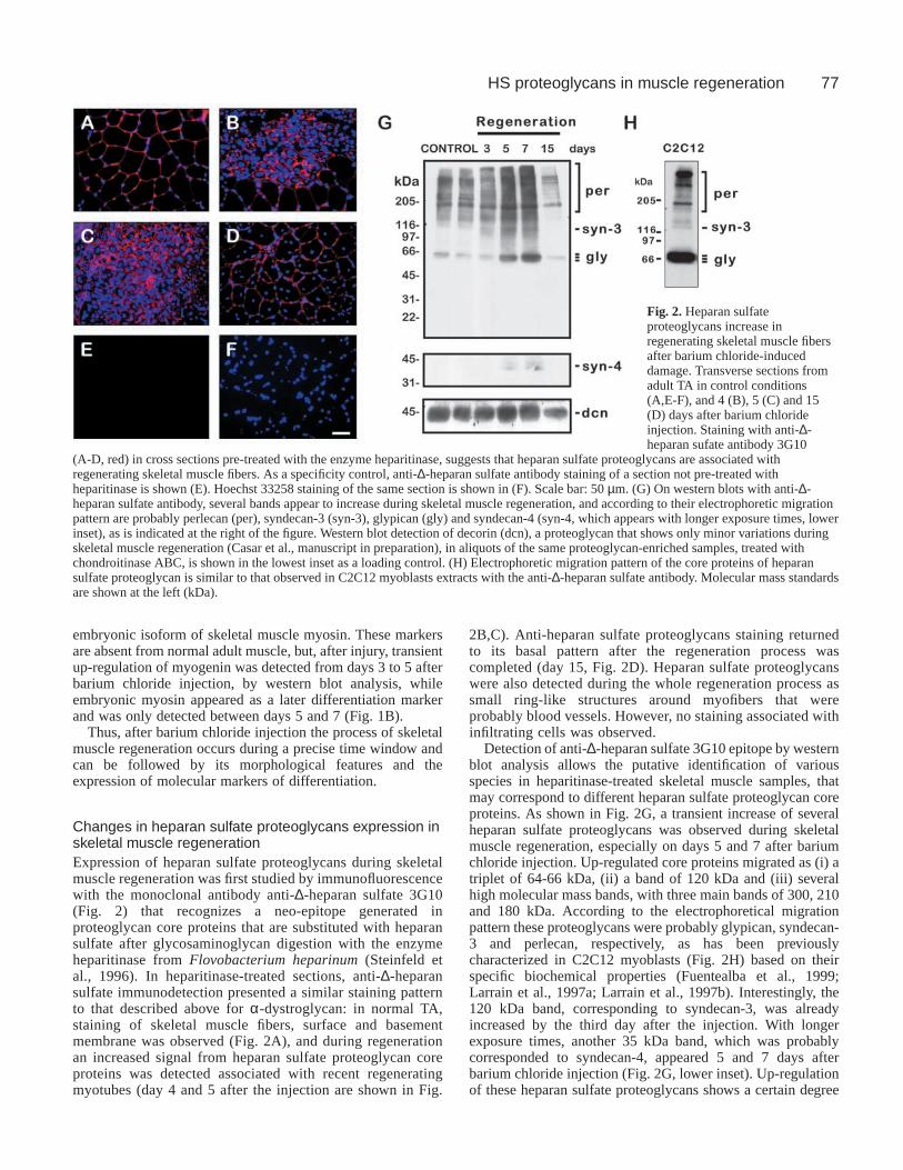
Changes in heparan sulfate proteoglycans expression inskeletal muscle regenerationExpression of heparan sulfate proteoglycans during skeletalmuscle regeneration was first studied by immunofluorescencewith the monoclonal antibody anti-∆-heparan sulfate 3G10(Fig. 2) that recognizes a neo-epitope generated inproteoglycan core proteins that are substituted with heparansulfate after glycosaminoglycan digestion with the enzymeheparitinase from Flovobacterium heparinum(Steinfeld etal., 1996). In heparitinase-treated sections, anti-∆-heparansulfate immunodetection presented a similar staining patternto that described above for α-dystroglycan: in normal TA,staining of skeletal muscle fibers, surface and basementmembrane was observed (Fig. 2A), and during regenerationan increased signal from heparan sulfate proteoglycan coreproteins was detected associated with recent regeneratingmyotubes (day 4 and 5 after the injection are shown in Fig.
2B,C). Anti-heparan sulfate proteoglycans staining returnedto its basal pattern after the regeneration process wascompleted (day 15, Fig. 2D). Heparan sulfate proteoglycanswere also detected during the whole regeneration process assmall ring-like structures around myofibers that wereprobably blood vessels. However, no staining associated withinfiltrating cells was observed.
Detection of anti-∆-heparan sulfate 3G10 epitope by westernblot analysis allows the putative identification of variousspecies in heparitinase-treated skeletal muscle samples, thatmay correspond to different heparan sulfate proteoglycan coreproteins. As shown in Fig. 2G, a transient increase of severalheparan sulfate proteoglycans was observed during skeletalmuscle regeneration, especially on days 5 and 7 after bariumchloride injection. Up-regulated core proteins migrated as (i) atriplet of 64-66 kDa, (ii) a band of 120 kDa and (iii) severalhigh molecular mass bands, with three main bands of 300, 210and 180 kDa. According to the electrophoretical migrationpattern these proteoglycans were probably glypican, syndecan-3 and perlecan, respectively, as has been previouslycharacterized in C2C12 myoblasts (Fig. 2H) based on theirspecific biochemical properties (Fuentealba et al., 1999;Larrain et al., 1997a; Larrain et al., 1997b). Interestingly, the120 kDa band, corresponding to syndecan-3, was alreadyincreased by the third day after the injection. With longerexposure times, another 35 kDa band, which was probablycorresponded to syndecan-4, appeared 5 and 7 days afterbarium chloride injection (Fig. 2G, lower inset). Up-regulationof these heparan sulfate proteoglycans shows a certain degree
Fig. 2. Heparan sulfateproteoglycans increase inregenerating skeletal muscle fibersafter barium chloride-induceddamage. Transverse sections fromadult TA in control conditions(A,E-F), and 4 (B), 5 (C) and 15(D) days after barium chlorideinjection. Staining with anti-∆-heparan sufate antibody 3G10
(A-D, red) in cross sections pre-treated with the enzyme heparitinase, suggests that heparan sulfate proteoglycans are associated withregenerating skeletal muscle fibers. As a specificity control, anti-∆-heparan sulfate antibody staining of a section not pre-treated withheparitinase is shown (E). Hoechst 33258 staining of the same section is shown in (F). Scale bar: 50 µm. (G) On western blots with anti-∆-heparan sulfate antibody, several bands appear to increase during skeletal muscle regeneration, and according to their electrophoretic migrationpattern are probably perlecan (per), syndecan-3 (syn-3), glypican (gly) and syndecan-4 (syn-4, which appears with longer exposure times, lowerinset), as is indicated at the right of the figure. Western blot detection of decorin (dcn), a proteoglycan that shows only minor variations duringskeletal muscle regeneration (Casar et al., manuscript in preparation), in aliquots of the same proteoglycan-enriched samples, treated withchondroitinase ABC, is shown in the lowest inset as a loading control. (H) Electrophoretic migration pattern of the core proteins of heparansulfate proteoglycan is similar to that observed in C2C12 myoblasts extracts with the anti-∆-heparan sulfate antibody. Molecular mass standardsare shown at the left (kDa).

78
of specificity because other proteoglycans, e.g. the chondroitin/dermatan sulfate proteoglycan decorin, remain relativelyunchanged (Fig. 2G, lowest inset) (Casar et al., manuscript inpreparation).
Specific heparan sulfate proteoglycans expression inskeletal muscle regeneration: early up-regulation ofsyndecan-3In order to advance the identification of the specific heparansulfate proteoglycans that are up-regulated during skeletalmuscle regeneration, the localization of the specific abovementioned proteoglycans in the tissue was studied by indirectimmunofluorescence with polyclonal antibodies against thecorresponding species (Fig. 3).
In control muscle, syndecan-3, glypican and perlecan weredetected at the sarcolemma or endomisium in the periphery ofmyofibers and were absent from the perimisium (Fig. 3A,E andI, respectively). Syndecan-3 reactivity was weak and there wasalso some cytoplasmic staining for glypican (Fig. 3E). Afterinjury, an increased staining for these three proteoglycans wasapparent from day 4, preferentially surrounding the newlyformed myotubes rather than with remnant myofibers in thesame sections (Fig. 3B,F,J). This pattern of expression wasmaintained and more evident at day 5, where staining wasabsent from interstitial strips with abundant cellular infiltration(Fig. 3C,G,K). By the 15th day, the expression of syndecan-3,glypican and perlecan returned to basal levels (Fig. 3D,H,L).
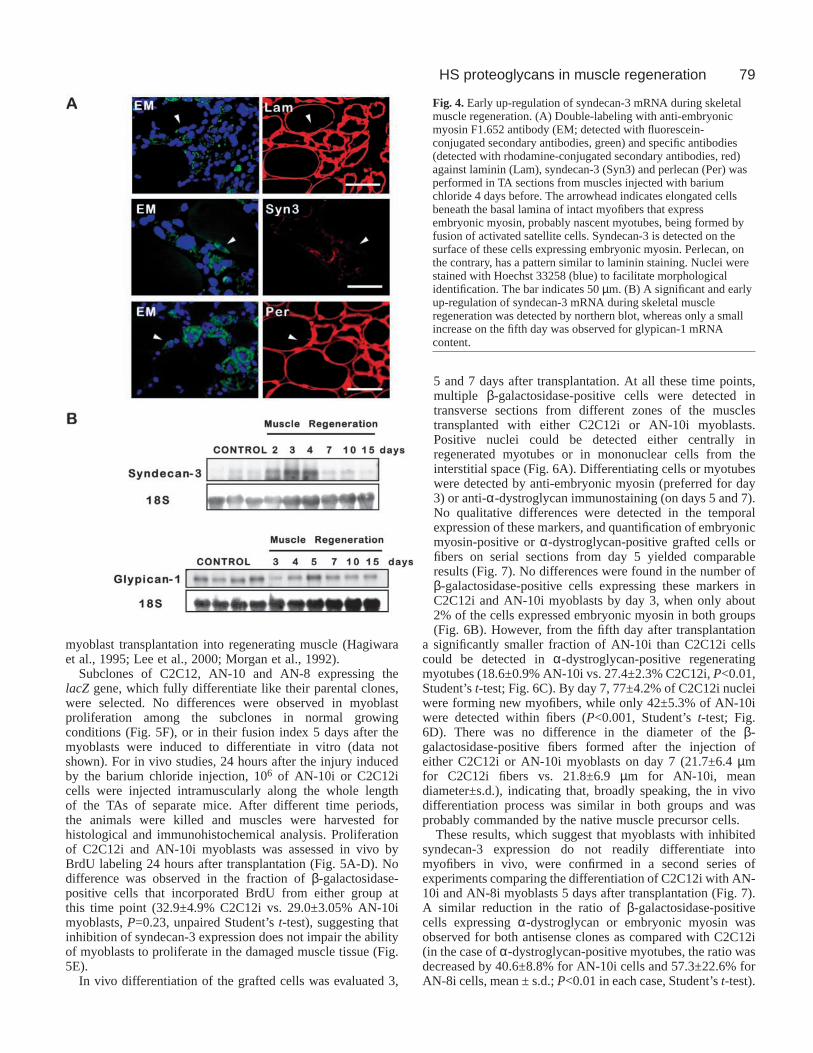
Beneath the basal lamina of injured myofibers, until thefourth day after barium chloride injection, a number ofelongated cells, positive for the expression of desmin (notshown) or embryonic myosin, could be observed (Fig. 4A).From their position, their elongated morphology and theexpression of the markers described above these cells areprobably nascent myotubes or myoblasts starting todifferentiate (Gorza et al., 1983; Lawson-Smith andMcGeachie, 1998; Sartore et al., 1982). Interestingly,syndecan-3, the heparan sulfate proteoglycan that showed anearlier increase (Fig. 2G), and could be detected at the surface
of these cells, whereas perlecan was mainly detected in thebasal lamina of the myofiber.
An early increase of syndecan-3 transcripts was alsoobserved during regeneration: a transient up-regulation ofsyndecan-3 steady state mRNA levels was observed betweendays 2 and 5 after barium chloride injection (Fig. 4B). Theexpression returned to basal, almost undetectable levels afterthe seventh day. Similar changes in mRNA expression were notobserved for the other heparan sulfate proteoglycans studied.Glypican-1 mRNA showed a slight decrease in its expressionthe first days after the injection, followed by a transientincrease on day 5 and then recovered its basal level ofexpression (Fig. 4B), and perlecan mRNA expression was notdetectable with this assay, although its up-regulation wasconfirmed by western blot with specific antibodies (data notshown).
Taken together, these results confirm the up-regulation ofdifferent heparan sulfate proteoglycans in young myotubes andthe early expression of syndecan-3 in myoblast cells and youngmyotubes.
Syndecan-3 expression in myoblast plasma membraneis important for successful fiber formation in myoblasttransplant studiesAmong the heparan sulfate proteoglycans that increase duringskeletal muscle regeneration, syndecan-3 stands out as apossible regulator of the process for its low expression innormal muscle, its expression by quiescent and activatedsatellite cells (Cornelison et al., 2001), its early increase inregeneration (Figs 3 and 4) and for its localization in forminglimb skeletal muscle masses during embryonic development(Olguin and Brandan, 2001). We have previously describedtwo sub-clones of the skeletal muscle cell line C2C12 in which,by the stable transfection of an antisense-sequence encodingplasmid, syndecan-3 expression is inhibited (Fuentealba et al.,1999). In view of the lack of studies on the role of heparansulfate proteoglycans in skeletal muscle regeneration in vivo,the specific role of syndecan-3 in this process was studied by
Journal of Cell Science 117 (1)
Fig. 3. Expression of syndecan-3,glypican and perlecan by skeletalmuscle fibers during TAregeneration. Heparan sulfateproteoglycans syndecan-3 (A-D),glypican-1 (E-H) and perlecan(I-L) were detected byimmunofluorescence with thecorresponding specific polyclonalantibodies in cross sections fromcontrol TA (A,E,I), and TA 4(B,F,J), 5 (C,G,K) and 15 (D,H,L)days after barium chlorideinjection. Heparan sulfateproteoglycans are localized at thesurface or basal lamina ofmyofibers, and increase in newlyregenerated myotubes. Scale bar:50 µm.
79HS proteoglycans in muscle regeneration
myoblast transplantation into regenerating muscle (Hagiwaraet al., 1995; Lee et al., 2000; Morgan et al., 1992).
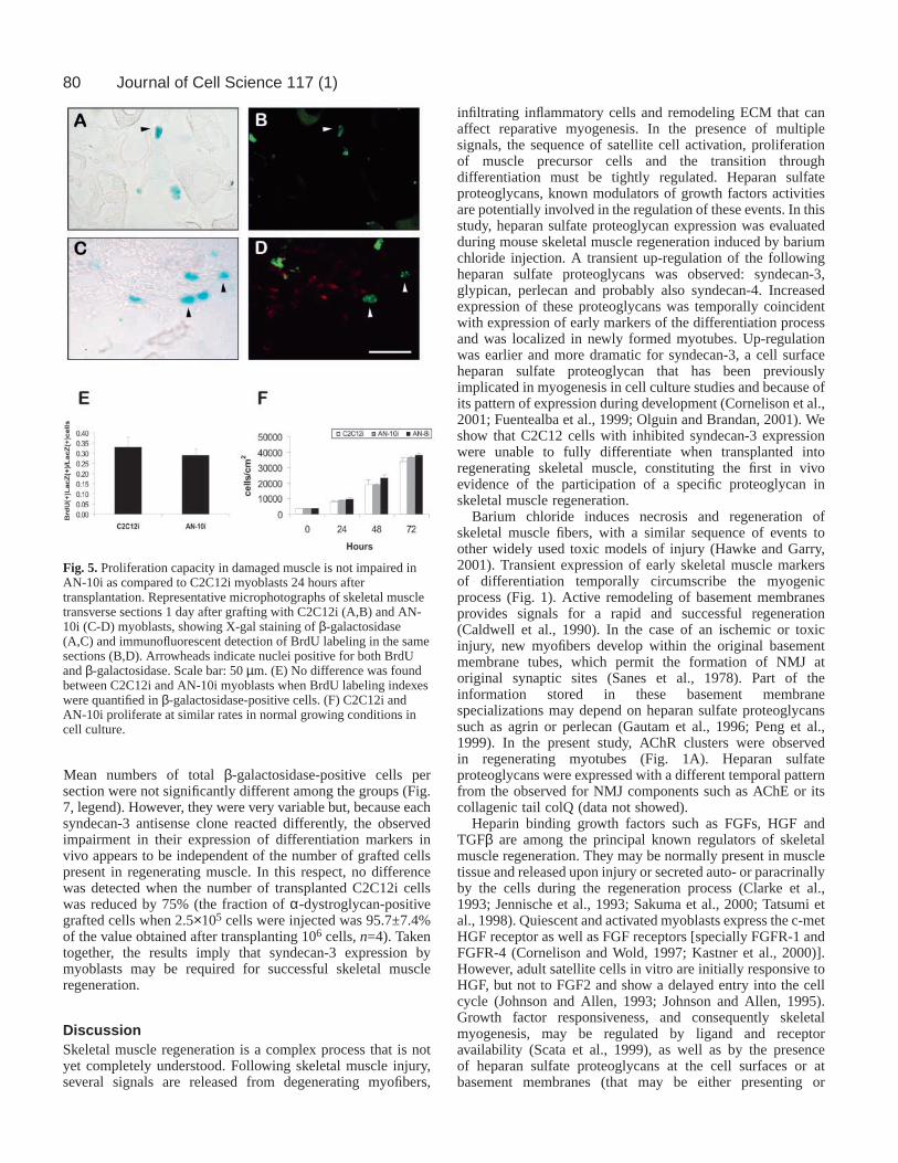
Subclones of C2C12, AN-10 and AN-8 expressing thelacZ gene, which fully differentiate like their parental clones,were selected. No differences were observed in myoblastproliferation among the subclones in normal growingconditions (Fig. 5F), or in their fusion index 5 days after themyoblasts were induced to differentiate in vitro (data notshown). For in vivo studies, 24 hours after the injury inducedby the barium chloride injection, 106 of AN-10i or C2C12icells were injected intramuscularly along the whole lengthof the TAs of separate mice. After different time periods,the animals were killed and muscles were harvested forhistological and immunohistochemical analysis. Proliferationof C2C12i and AN-10i myoblasts was assessed in vivo byBrdU labeling 24 hours after transplantation (Fig. 5A-D). Nodifference was observed in the fraction of β-galactosidase-positive cells that incorporated BrdU from either group atthis time point (32.9±4.9% C2C12i vs. 29.0±3.05% AN-10imyoblasts, P=0.23, unpaired Student’s t-test), suggesting thatinhibition of syndecan-3 expression does not impair the abilityof myoblasts to proliferate in the damaged muscle tissue (Fig.5E).
In vivo differentiation of the grafted cells was evaluated 3,
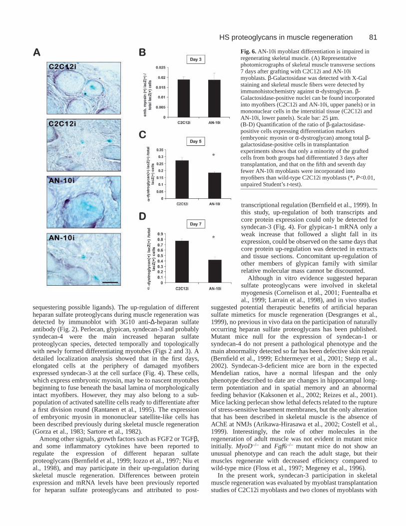
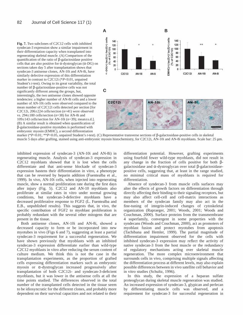
5 and 7 days after transplantation. At all these time points,multiple β-galactosidase-positive cells were detected intransverse sections from different zones of the musclestransplanted with either C2C12i or AN-10i myoblasts.Positive nuclei could be detected either centrally inregenerated myotubes or in mononuclear cells from theinterstitial space (Fig. 6A). Differentiating cells or myotubeswere detected by anti-embryonic myosin (preferred for day3) or anti-α-dystroglycan immunostaining (on days 5 and 7).No qualitative differences were detected in the temporalexpression of these markers, and quantification of embryonicmyosin-positive or α-dystroglycan-positive grafted cells orfibers on serial sections from day 5 yielded comparableresults (Fig. 7). No differences were found in the number ofβ-galactosidase-positive cells expressing these markers inC2C12i and AN-10i myoblasts by day 3, when only about2% of the cells expressed embryonic myosin in both groups(Fig. 6B). However, from the fifth day after transplantation
a significantly smaller fraction of AN-10i than C2C12i cellscould be detected in α-dystroglycan-positive regeneratingmyotubes (18.6±0.9% AN-10i vs. 27.4±2.3% C2C12i, P<0.01,Student’s t-test; Fig. 6C). By day 7, 77±4.2% of C2C12i nucleiwere forming new myofibers, while only 42±5.3% of AN-10iwere detected within fibers (P<0.001, Student’s t-test; Fig.6D). There was no difference in the diameter of the β-galactosidase-positive fibers formed after the injection ofeither C2C12i or AN-10i myoblasts on day 7 (21.7±6.4 µmfor C2C12i fibers vs. 21.8±6.9 µm for AN-10i, meandiameter±s.d.), indicating that, broadly speaking, the in vivodifferentiation process was similar in both groups and wasprobably commanded by the native muscle precursor cells.
These results, which suggest that myoblasts with inhibitedsyndecan-3 expression do not readily differentiate intomyofibers in vivo, were confirmed in a second series ofexperiments comparing the differentiation of C2C12i with AN-10i and AN-8i myoblasts 5 days after transplantation (Fig. 7).A similar reduction in the ratio of β-galactosidase-positivecells expressing α-dystroglycan or embryonic myosin wasobserved for both antisense clones as compared with C2C12i(in the case of α-dystroglycan-positive myotubes, the ratio wasdecreased by 40.6±8.8% for AN-10i cells and 57.3±22.6% forAN-8i cells, mean ± s.d.; P<0.01 in each case, Student’s t-test).
Fig. 4. Early up-regulation of syndecan-3 mRNA during skeletalmuscle regeneration. (A) Double-labeling with anti-embryonicmyosin F1.652 antibody (EM; detected with fluorescein-conjugated secondary antibodies, green) and specific antibodies(detected with rhodamine-conjugated secondary antibodies, red)against laminin (Lam), syndecan-3 (Syn3) and perlecan (Per) wasperformed in TA sections from muscles injected with bariumchloride 4 days before. The arrowhead indicates elongated cellsbeneath the basal lamina of intact myofibers that expressembryonic myosin, probably nascent myotubes, being formed byfusion of activated satellite cells. Syndecan-3 is detected on thesurface of these cells expressing embryonic myosin. Perlecan, onthe contrary, has a pattern similar to laminin staining. Nuclei werestained with Hoechst 33258 (blue) to facilitate morphologicalidentification. The bar indicates 50 µm. (B) A significant and earlyup-regulation of syndecan-3 mRNA during skeletal muscleregeneration was detected by northern blot, whereas only a smallincrease on the fifth day was observed for glypican-1 mRNAcontent.
80
Mean numbers of total β-galactosidase-positive cells persection were not significantly different among the groups (Fig.7, legend). However, they were very variable but, because eachsyndecan-3 antisense clone reacted differently, the observedimpairment in their expression of differentiation markers invivo appears to be independent of the number of grafted cellspresent in regenerating muscle. In this respect, no differencewas detected when the number of transplanted C2C12i cellswas reduced by 75% (the fraction of α-dystroglycan-positivegrafted cells when 2.5×10⁵ cells were injected was 95.7±7.4%of the value obtained after transplanting 106 cells, n=4). Takentogether, the results imply that syndecan-3 expression bymyoblasts may be required for successful skeletal muscleregeneration.
DiscussionSkeletal muscle regeneration is a complex process that is notyet completely understood. Following skeletal muscle injury,several signals are released from degenerating myofibers,
infiltrating inflammatory cells and remodeling ECM that canaffect reparative myogenesis. In the presence of multiplesignals, the sequence of satellite cell activation, proliferationof muscle precursor cells and the transition throughdifferentiation must be tightly regulated. Heparan sulfateproteoglycans, known modulators of growth factors activitiesare potentially involved in the regulation of these events. In thisstudy, heparan sulfate proteoglycan expression was evaluatedduring mouse skeletal muscle regeneration induced by bariumchloride injection. A transient up-regulation of the followingheparan sulfate proteoglycans was observed: syndecan-3,glypican, perlecan and probably also syndecan-4. Increasedexpression of these proteoglycans was temporally coincidentwith expression of early markers of the differentiation processand was localized in newly formed myotubes. Up-regulationwas earlier and more dramatic for syndecan-3, a cell surfaceheparan sulfate proteoglycan that has been previouslyimplicated in myogenesis in cell culture studies and because ofits pattern of expression during development (Cornelison et al.,2001; Fuentealba et al., 1999; Olguin and Brandan, 2001). Weshow that C2C12 cells with inhibited syndecan-3 expressionwere unable to fully differentiate when transplanted intoregenerating skeletal muscle, constituting the first in vivoevidence of the participation of a specific proteoglycan inskeletal muscle regeneration.
Barium chloride induces necrosis and regeneration ofskeletal muscle fibers, with a similar sequence of events toother widely used toxic models of injury (Hawke and Garry,2001). Transient expression of early skeletal muscle markersof differentiation temporally circumscribe the myogenicprocess (Fig. 1). Active remodeling of basement membranesprovides signals for a rapid and successful regeneration(Caldwell et al., 1990). In the case of an ischemic or toxicinjury, new myofibers develop within the original basementmembrane tubes, which permit the formation of NMJ atoriginal synaptic sites (Sanes et al., 1978). Part of theinformation stored in these basement membranespecializations may depend on heparan sulfate proteoglycanssuch as agrin or perlecan (Gautam et al., 1996; Peng et al.,1999). In the present study, AChR clusters were observedin regenerating myotubes (Fig. 1A). Heparan sulfateproteoglycans were expressed with a different temporal patternfrom the observed for NMJ components such as AChE or itscollagenic tail colQ (data not showed).
Heparin binding growth factors such as FGFs, HGF andTGFβ are among the principal known regulators of skeletalmuscle regeneration. They may be normally present in muscletissue and released upon injury or secreted auto- or paracrinallyby the cells during the regeneration process (Clarke et al.,1993; Jennische et al., 1993; Sakuma et al., 2000; Tatsumi etal., 1998). Quiescent and activated myoblasts express the c-metHGF receptor as well as FGF receptors [specially FGFR-1 andFGFR-4 (Cornelison and Wold, 1997; Kastner et al., 2000)].However, adult satellite cells in vitro are initially responsive toHGF, but not to FGF2 and show a delayed entry into the cellcycle (Johnson and Allen, 1993; Johnson and Allen, 1995).Growth factor responsiveness, and consequently skeletalmyogenesis, may be regulated by ligand and receptoravailability (Scata et al., 1999), as well as by the presenceof heparan sulfate proteoglycans at the cell surfaces or atbasement membranes (that may be either presenting or
Journal of Cell Science 117 (1)
Fig. 5. Proliferation capacity in damaged muscle is not impaired inAN-10i as compared to C2C12i myoblasts 24 hours aftertransplantation. Representative microphotographs of skeletal muscletransverse sections 1 day after grafting with C2C12i (A,B) and AN-10i (C-D) myoblasts, showing X-gal staining of β-galactosidase(A,C) and immunofluorescent detection of BrdU labeling in the samesections (B,D). Arrowheads indicate nuclei positive for both BrdUand β-galactosidase. Scale bar: 50 µm. (E) No difference was foundbetween C2C12i and AN-10i myoblasts when BrdU labeling indexeswere quantified in β-galactosidase-positive cells. (F) C2C12i andAN-10i proliferate at similar rates in normal growing conditions incell culture.
81HS proteoglycans in muscle regeneration
sequestering possible ligands). The up-regulation of differentheparan sulfate proteoglycans during muscle regeneration wasdetected by immunoblot with 3G10 anti-∆-heparan sulfateantibody (Fig. 2). Perlecan, glypican, syndecan-3 and probablysyndecan-4 were the main increased heparan sulfateproteoglycan species, detected temporally and topologicallywith newly formed differentiating myotubes (Figs 2 and 3). Adetailed localization analysis showed that in the first days,elongated cells at the periphery of damaged myofibersexpressed syndecan-3 at the cell surface (Fig. 4). These cells,which express embryonic myosin, may be to nascent myotubesbeginning to fuse beneath the basal lamina of morphologicallyintact myofibers. However, they may also belong to a sub-population of activated satellite cells ready to differentiate aftera first division round (Rantanen et al., 1995). The expressionof embryonic myosin in mononuclear satellite-like cells hasbeen described previously during skeletal muscle regeneration(Gorza et al., 1983; Sartore et al., 1982).
Among other signals, growth factors such as FGF2 or TGFβ,and some inflammatory cytokines have been reported toregulate the expression of different heparan sulfateproteoglycans (Bernfield et al., 1999; Iozzo et al., 1997; Niu etal., 1998), and may participate in their up-regulation duringskeletal muscle regeneration. Differences between proteinexpression and mRNA levels have been previously reportedfor heparan sulfate proteoglycans and attributed to post-
transcriptional regulation (Bernfield et al., 1999). Inthis study, up-regulation of both transcripts andcore protein expression could only be detected forsyndecan-3 (Fig. 4). For glypican-1 mRNA only aweak increase that followed a slight fall in itsexpression, could be observed on the same days thatcore protein up-regulation was detected in extractsand tissue sections. Concomitant up-regulation ofother members of glypican family with similarrelative molecular mass cannot be discounted.
Although in vitro evidence suggested heparansulfate proteoglycans were involved in skeletalmyogenesis (Cornelison et al., 2001; Fuentealba etal., 1999; Larrain et al., 1998), and in vivo studies
suggested potential therapeutic benefits of artificial heparansulfate mimetics for muscle regeneration (Desgranges et al.,1999), no previous in vivo data on the participation of naturallyoccurring heparan sulfate proteoglycans has been published.Mutant mice null for the expression of syndecan-1 orsyndecan-4 do not present a pathological phenotype and themain abnormality detected so far has been defective skin repair(Bernfield et al., 1999; Echtermeyer et al., 2001; Stepp et al.,2002). Syndecan-3-deficient mice are born in the expectedMendelian ratios, have a normal lifespan and the onlyphenotype described to date are changes in hippocampal long-term potentiation and in spatial memory and an abnormalfeeding behavior (Kaksonen et al., 2002; Reizes et al., 2001).Mice lacking perlecan show lethal defects related to the ruptureof stress-sensitive basement membranes, but the only alterationthat has been described in skeletal muscle is the absence ofAChE at NMJs (Arikawa-Hirasawa et al., 2002; Costell et al.,1999). Interestingly, the role of other molecules in theregeneration of adult muscle was not evident in mutant miceinitially. MyoD–/– and Fgf6–/– mutant mice do not show anunusual phenotype and can reach the adult stage, but theirmuscles regenerate with decreased efficiency compared towild-type mice (Floss et al., 1997; Megeney et al., 1996).
In the present work, syndecan-3 participation in skeletalmuscle regeneration was evaluated by myoblast transplantationstudies of C2C12i myoblasts and two clones of myoblasts with
Fig. 6. AN-10i myoblast differentiation is impaired inregenerating skeletal muscle. (A) Representativephotomicrographs of skeletal muscle transverse sections7 days after grafting with C2C12i and AN-10imyoblasts. β-Galactosidase was detected with X-Galstaining and skeletal muscle fibers were detected byimmunohistochemistry against α-dystroglycan. β-Galactosidase-positive nuclei can be found incorporatedinto myofibers (C2C12i and AN-10i, upper panels) or inmononuclear cells in the interstitial tissue (C2C12i andAN-10i, lower panels). Scale bar: 25 µm.(B-D) Quantification of the ratio of β-galactosidase-positive cells expressing differentiation markers(embryonic myosin or α-dystroglycan) among total β-galactosidase-positive cells in transplantationexperiments shows that only a minority of the graftedcells from both groups had differentiated 3 days aftertransplantation, and that on the fifth and seventh dayfewer AN-10i myoblasts were incorporated intomyofibers than wild-type C2C12i myoblasts (*, P<0.01,unpaired Student’s t-test).
82
inhibited expression of syndecan-3 (AN-10i and AN-8i) inregenerating muscle. Analysis of syndecan-3 expression inC2C12 myoblasts showed that it is lost when the cellsdifferentiate and that anti-sense blockade of syndecan-3expression hastens their differentiation in vitro, a phenotypethat can be reversed by heparin addition (Fuentealba et al.,1999). In vivo, AN-10i cells, when injected into regeneratingmuscle, show a normal proliferation rate during the first daysafter injury (Fig. 5). C2C12 and AN-10 myoblasts alsoproliferate at similar rates in vitro under normal growingconditions, but syndecan-3-defective myoblasts have adecreased proliferative response to FGF2 (L. Fuentealba andE.B., unpublished results). This suggests that, in vivo, thespecific contribution of FGF2 to myoblast proliferation isprobably redundant with the several other mitogens that arepresent in the tissue.
Both antisense clones, AN-10i and AN-8i, showed adecreased capacity to form or be incorporated into newmyotubes in vivo (Figs 6 and 7), suggesting at least a partialsyndecan-3 requirement for a successful regeneration. Wehave shown previously that myoblasts with an inhibitedsyndecan-3 expression differentiate earlier than wild-typeC2C12 myoblasts in vitro after reducing the serum content ofculture medium. We think this is not the case in thetransplantation experiments, as the proportion of graftedcells expressing differentiation markers such as embryonicmyosin or α-dystroglycan increased progressively aftertransplantation of both C2C12i- and syndecan-3-deficientmyoblasts, but it was lower in the antisense cells at all thetime points studied. The differences observed in the totalnumber of the transplanted cells detected in the tissue seemto be idiosyncratic for the different clones, and probably moredependent on their survival capacities and not related to their
differentiation potential. However, grafting experimentsusing fourfold fewer wild-type myoblasts, did not result inany change in the fraction of cells positive for both β-galactosidase and α-dystroglycan over total β-galactosidase-positive cells, suggesting that, at least in the range studied,no minimal critical mass of myoblasts is required fordifferentiation.
Absence of syndecan-3 from muscle cells surfaces mayalter the effects of growth factors on differentiation throughdirectly affecting their binding to their signaling receptors, butmay also affect cell-cell and cell-matrix interactions asmembers of the syndecan family may also act in thefine-tuning of integrin-induced changes of cytoskeletalorganization (Rapraeger, 2001; Woods, 2001; Woods andCouchman, 2000). Surface proteins from the transmembrane4 superfamily, convergent in some properties with thesyndecans (Woods and Couchman, 2000), act as promoters ofmyoblast fusion and protect myotubes from apoptosis(Tachibana and Hemler, 1999). The partial magnitude ofdifferentiation impairment observed for the cells withinhibited syndecan-3 expression may reflect the activity ofnative syndecan-3 from the host muscle or the redundancyof regulatory mechanisms acting over skeletal muscleregeneration. The more complex microenvironment thatsurrounds cells in vivo, comprising multiple signals affectingthe differentiation process at different levels, may also explainpossible differences between in vivo satellite cell behavior andin vitro studies (Schultz, 1996).
In this study, the expression of a heparan sulfateproteoglycan during skeletal muscle regeneration was studied.An increased expression of syndecan-3, glypican and perlecanby differentiating muscle cells was observed, and arequirement for syndecan-3 for successful regeneration in
Journal of Cell Science 117 (1)
Fig. 7. Two subclones of C2C12 cells with inhibitedsyndecan-3 expression show a similar impairment intheir differentiation capacity when transplanted intoregenerating skeletal muscle. (A) Comparison of thequantification of the ratio of β-galactosidase positivecells that are also positive for α-dystroglycan (α-DG) onsections taken day 5 after transplantation shows thatsyndecan-3 antisense clones, AN-10i and AN-8i, havesimilarly defective expression of this differentiationmarker in contrast to C2C12i (*P<0.01, unpairedStudent’s t-test). Owing to its great variability, the totalnumber οf β-galactosidase-positive cells was notsignificantly different among the groups, but,interestingly, the two antisense clones showed oppositetendencies: a higher number of AN-8i cells and a lowernumber of AN-10i cells were observed compared to themean number of C2C12i cells detected per section [forC2C12i, 206±224 cells/section (n=41) were observedvs. 294±180 cells/section (n=38) for AN-8i and109±143 cells/section for AN-10i (n=39); mean±s.d.].(B) A similar result is obtained when quantification ofβ-galactosidase-positive myotubes is performed withembryonic myosin (EMHC), a second differentiationmarker (*P<0.01; **P<0.05, unpaired Student’s t-test). (C) Representative transverse sections of β-galactosidase-positive cells in skeletalmuscle 5 days after grafting, stained using anti-embryonic myosin histochemistry, for C2C12i, AN-10i and AN-8i myoblasts. Scale bar: 25 µm.
83HS proteoglycans in muscle regeneration
myoblast transplant experiments was found. Future studiesshould explore the specific mechanism of syndecan-3participation in skeletal muscle regeneration, and address thefunction of the other up-regulated proteoglycans in thisprocess.
The authors are indebted to Dr John R. Hassell for generouslyproviding anti-perlecan antibody and to Dr David J. Carey forgenerously providing anti-glypican and anti-syndecan-3 antibodies.This work was supported in part by grants from FONDAP-Biomedicine No. 13980001, FONDECYT 1990151 (to E.B.), and2000113 (to J.C.C.). The research of E.B. was supported in part byan International Research Scholars grant from the Howard HughesMedical Institute. E.B. and N.I. were supported by a PresidentialChair in Science from the Chilean Government. The MilleniumInstitute for Fundamental and Applied Biology (MIFAB) is financedin part by the Ministerio de Planificación y Cooperación (Chile).
ReferencesArikawa-Hirasawa, E., Rossi, S. G., Rotundo, R. L. and Yamada, Y.
(2002). Absence of acetylcholinesterase at the neuromuscular junctions ofperlecan-null mice. Nat. Neurosci.5, 119-123.
Bernfield, M., Gotte, M., Park, P. W., Reizes, O., Fitzgerald, M. L.,Lincecum, J. and Zako, M. (1999). Functions of cell surface heparansulfate proteoglycans. Annu. Rev. Biochem.68, 729-777.
Blau, H., Grace, K., Pavlath, E. C., Hardeman, C.-P. C., Silberstein, L.,Webster, S. G., Miller, S. C. and Webster, C. (1985). Plasticity of thedifferentiated state. Science230, 758-766.
Bowe, M. A., Mendis, D. B. and Fallon, J. R. (2000). The small leucine-richrepeat proteoglycan biglycan binds to alpha-dystroglycan and is upregulatedin dystrophic muscle. J. Cell Biol.148, 801-810.
Brandan, E., Carey, D. J., Larrain, J., Melo, F. and Campos, A. (1996).Synthesis and processing of glypican during differentiation of skeletalmuscle cells. Eur. J. Cell Biol.71, 171-176.
Brandan, E., Fuentes, M. E. and Andrade, W. (1992). Decorin, achondroitin/dermatan sulfate proteoglycan, is under neural control in ratskeletal muscle. J. Neurosci. Res.32, 51-59.
Brandan, E. and Inestrosa, N. C. (1987). Isolation of the heparan sulfateproteoglycans from the extracellular matrix of rat skeletal muscle. J.Neurobiol.18, 271-282.
Brandan, E., Maldonado, M., Garrido, J. and Inestrosa, N. C.(1985).Anchorage of collagen-tailed acetylcholinesterase to the extracellular matrixis mediated by heparan sulfate proteoglycans. J. Cell Biol.101, 985-992.
Buckingham, M. (1992). Making muscle in mammals. Trends Genet.8, 144-149.
Caldwell, C. J., Mattey, D. L. and Weller, R. O. (1990). Role of the basementmembrane in the regeneration of skeletal muscle. Neuropathol. Appl.Neurobiol.16, 225-238.
Campos, A., Nuñez, R., Koenig, C., Carey, D. J. and Brandan, E. (1993).A lipid-anchored heparan sulfate proteoglycan is present in the surface ofdifferentiated skeletal muscle cells. Eur. J. Cell Biol.216, 587-595.
Carey, D. J. (1997). Syndecans: multifunctional cell-surface co-receptors.Biochem. J.327, 1-16.
Chomczynski, P. and Sacchi, N.(1987). Single-step method of RNA isolationby acid guanidinium thiocyanate-phenol-chloroform extraction. Anal.Biochem.162, 156-159.
Clarke, M. S., Khakee, R. and McNeil, P. L. (1993). Loss of cytoplasmicbasic fibroblast growth factor from physiologically wounded myofibers ofnormal and dystrophic muscle. J. Cell Sci.106, 121-133.
Cornelison, D. D. and Wold, B. J. (1997). Single-cell analysis of regulatorygene expression in quiescent and activated mouse skeletal muscle satellitecells. Dev. Biol.191, 270-283.
Cornelison, D. D. W., Filla, M. S., Stanley, H. M., Rapraeger, A. C. andOlwin, B. B. (2001). Syndecan-3 and syndecan-4 specifically mark skeletalmuscle satellite cells and are implicated in satellite cell maintenance andmuscle regeneration. Dev. Biol.239, 79-94.
Costell, M., Gustafsson, E., Aszodi, A., Morgelin, M., Bloch, W., Hunziker,E., Addicks, K., Timpl, R. and Fassler, R.(1999). Perlecan maintains theintegrity of cartilage and some basement membranes. J. Cell Biol. 147,1109-1122.
Desgranges, P., Barbaud, C., Caruelle, J. P., Barritault, D. and Gautron, J.(1999). A substituted dextran enhances muscle fiber survival and regenerationin ischemic and denervated rat EDL muscle. FASEB J.13, 761-766.
Echtermeyer, F., Streit, M., Wilcox-Adelman, S., Saoncella, S., Denhez, F.,Detmar, M. and Goetinck, P. (2001). Delayed wound repair and impairedangiogenesis in mice lacking syndecan- 4. J. Clin. Invest.107, R9-R14.
Fisher, L. W., Stubbs, J. T., 3rd and Young, M. F.(1995). Antisera andcDNA probes to human and certain animal model bone matrixnoncollagenous proteins. Acta Orthop. Scand.Supplement266, 61-65.
Floss, T., Arnold, H. H. and Braun, T.(1997). A role for FGF-6 in skeletalmuscle regeneration. Genes Dev.11, 2040-2051.
Friedl, A., Chang, Z., Tierney, A. and Rapraeger, A. C.(1997). Differentialbinding of fibroblast growth factor-2 and –7 to basement membrane heparansulfate: comparison of normal and abnormal human tissues. Am. J. Pathol.150, 1443-1455.
Fuentealba, L., Carey, D. J. and Brandan, E.(1999). Antisense inhibitionof syndecan-3 expression during skeletal muscle differentiation acceleratesmyogenesis through a basic fibroblast growth factor-dependent mechanism.J. Biol. Chem.274, 37876-37884.
Gautam, M., Noakes, P. G., Moscoso, L., Rupp, F., Scheller, R. H., Merlie,J. P. and Sanes, J. R.(1996). Defective neuromuscular synaptogenesis inagrin-deficient mutant mice. Cell 85, 525-535.
Gorza, L., Sartore, S., Triban, C. and Schiaffino, S.(1983). Embryonic-likemyosin heavy chains in regenerating chicken muscle. Exp. Cell Res.143,395-403.
Gulati, A. K., Reddi, A. H. and Zalewski, A. A. (1983). Changes in thebasement membrane zone components during skeletal muscle fiberdegeneration and regeneration. J. Cell Biol.97, 957-962.
Hagiwara, Y., Mizuno, Y., Takemitsu, M., Matsuzaki, T., Nonaka, I. andOzawa, E.(1995). Dystrophin-positive muscle fibers following C2 myoblasttransplantation into mdx nude mice. Acta Neuropathol.90, 592-600.
Hawke, T. J. and Garry, D. J. (2001). Myogenic satellite cells: physiologyto molecular biology. J. Appl. Physiol.91, 534-551.
Iozzo, R. V., Pillarisetti, J., Sharma, B., Murdoch, A. D., Danielson, K. G.,Uitto, J. and Mauviel, A. (1997). Structural and functional characterizationof the human perlecan gene promoter. Transcriptional activation bytransforming growth factor-beta via a nuclear factor 1-binding element. J.Biol. Chem.272, 5219-5228.
Jennische, E., Ekberg, S. and Matejka, G. L. (1993). Expression ofhepatocyte growth factor in growing and regenerating rat skeletal muscle.Am. J. Physiol.265, C122-C128.
Johnson, S. E. and Allen, R. E.(1993). Proliferating cell nuclear antigen(PCNA) is expressed in activated rat skeletal muscle satellite cells. J. CellPhysiol.154, 39-43.
Johnson, S. E. and Allen, R. E.(1995). Activation of skeletal muscle satellitecells and the role of fibroblast growth factor receptors. Exp. Cell Res.219,449-453.
Kaariainen, M., Kaariainen, J., Jarvinen, T. L., Nissinen, L., Heino, J.,Jarvinen, M. and Kalimo, H. (2000). Integrin and dystrophin associatedadhesion protein complexes during regeneration of shearing-type muscleinjury. Neuromuscular Disord.10, 121-132.
Kaksonen, M., Pavlov, I., Voikar, V., Lauri, S. E., Hienola, A., Riekki, R.,Lakso, M., Taira, T. and Rauvala, H.(2002). Syndecan-3-deficient miceexhibit enhanced LTP and impaired hippocampus- dependent memory. Mol.Cell Neurosci.21, 158-172.
Kastner, S., Elias, M. C., Rivera, A. J. and Yablonka-Reuveni, Z.(2000).Gene expression patterns of the fibroblast growth factors and their receptorsduring myogenesis of rat satellite cells. J. Histochem. Cytochem.48, 1079-1096.
Larrain, J., Alvarez, J., Hassell, J. R. and Brandan, E. (1997a). Expressionof perlecan, a proteoglycan that binds myogenic inhibitory basic fibroblastgrowth factor, is down regulated during skeletal muscle differentiation. Exp.Cell Res.234, 405-412.
Larrain, J., Carey, D. J. and Brandan, E. (1998). Syndecan-1 expressioninhibits myoblast terminal differentiation through a basic fibroblast growthfactor-dependent mechanism. J. Biol. Chem.273, 32288-32296.
Larrain, J., Cizmeci-Smith, G., Troncoso, V., Stahl, R. C., Carey, D. J. andBrandan, E. (1997b). Syndecan-1 expression is down-regulated duringmyoblast terminal differentiation. Modulation by growth factors and retinoicacid. J. Biol. Chem.272, 18418-18424.
Lawson-Smith, M. J. and McGeachie, J. K.(1998). The identification ofmyogenic cells in skeletal muscle, with emphasis on the use of tritiatedthymidine autoradiography and desmin antibodies. J. Anat.192, 161-171.
Lee, J. Y., Qu-Petersen, Z., Cao, B., Kimura, S., Jankowski, R., Cummins,

84
J., Usas, A., Gates, C., Robbins, P., Wernig, A. et al.(2000). Clonalisolation of muscle-derived cells capable of enhancing muscle regenerationand bone healing. J. Cell Biol.150, 1085-1100.
Lyon, M., Rushton, G. and Gallagher, J. T.(1997). The interaction of thetransforming growth factor-betas with heparin/heparan sulfate is isoform-specific. J. Biol. Chem.272, 18000-18006.
Megeney, L. A., Kablar, B., Garrett, K., Anderson, J. E. and Rudnicki, M.A. (1996). MyoD is required for myogenic stem cell function in adultskeletal muscle. Genes Dev.10, 1173-1183.
Melo, F., Carey, D. J. and Brandan, E.(1996). Extracellular matrix isrequired for skeletal muscle differentiation but not myogenin expression. J.Cell Biochem.62, 227-239.
Morgan, J. E., Moore, S. E., Walsh, F. S. and Partridge, T. A. (1992).Formation of skeletal muscle in vivo from the mouse C2 cell line. J. CellSci.102, 779-787.
Niu, S., Bahl, J. J., Adamson, C. and Morkin, E. (1998). Structure,regulation and function of avian glypican. J. Mol. Cell Cardiol. 30,537-550.
Olguin, H. and Brandan, E. (2001). Expression and localization ofproteoglycans during limb myogenic activation. Dev. Dyn.221, 106-115.
Osses, N. and Brandan, E.(2001). ECM is required for skeletal muscledifferentiation independently of muscle regulatory factor expression. Am. J.Physiol. Cell Physiol.282, C383-394.
Peng, H. B., Ali, A. A., Daggett, D. F., Rauvala, H., Hassell, J. R. andSmalheiser, N. R. (1998). The relationship between perlecan anddystroglycan and its implication in the formation of the neuromuscularjunction. Cell Adhes. Commun.5, 475-489.
Peng, H. B., Xie, H., Rossi, S. G. and Rotundo, R. L. (1999).Acetylcholinesterase clustering at the neuromuscular junction involvesperlecan and dystroglycan. J. Cell Biol.145, 911-921.
Perrimon, N. and Bernfield, M. (2000). Specificities of heparan sulphateproteoglycans in developmental processes. Nature404, 725-728.
Rantanen, J., Hurme, T., Lukka, R., Heino, J. and Kalimo, H. (1995).Satellite cell proliferation and the expression of myogenin and desmin inregenerating skeletal muscle: evidence for two different populations ofsatellite cells. Lab. Invest.72, 341-347.
Rapraeger, A. C. (2001). Molecular interactions of syndecans duringdevelopment. Semin. Cell Dev. Biol.12, 107-116.
Rapraeger, A. C., Krufka, A. and Olwin, B. B. (1991). Requirement ofheparan sulfate for bFGF-mediated fibroblast growth and myoblastdifferentiation. Science252, 1705-1708.
Reizes, O., Lincecum, J., Wang, Z., Goldberger, O., Huang, L., Kaksonen,M., Ahima, R., Hinkes, M. T., Barsh, G. S., Rauvala, H. et al.(2001).Transgenic expression of syndecan-1 uncovers a physiological control offeeding behavior by syndecan-3. Cell 106, 105-116.
Riquelme, C., Larrain, J., Schonherr, E., Henriquez, J. P., Kresse, H. andBrandan, E. (2001). Antisense inhibition of decorin expression in myoblastsdecreases cell responsiveness to transforming growth factor beta andaccelerates skeletal muscle differentiation. J. Biol. Chem.276, 3589-3596.
Sakuma, K., Watanabe, K., Sano, M., Kitajima, S., Sakamoto, K.,Uramoto, I. and Totsuka, T. (2000). The adaptive response of transforminggrowth factor-beta 2 and -beta RII in the overloaded, regenerating anddenervated muscles of rats. Acta Neuropathol.99, 177-185.
Sanes, J. R., Marshall, L. M. and McMahan, U. J. (1978). Reinnervation ofmuscle fiber basal lamina after removal of myofibers. Differentiation ofregenerating axons at original synaptic sites. J. Cell Biol.78, 176-198.
Sartore, S., Gorza, L. and Schiaffino, S. (1982). Fetal myosin heavy chainsin regenerating muscle. Nature298, 294-296.
Scata, K. A., Bernard, D. W., Fox, J. and Swain, J. L. (1999). FGF receptoravailability regulates skeletal myogenesis. Exp. Cell Res.250, 10-21.
Schultz, E. (1996). Satellite cell proliferative compartments in growingskeletal muscles. Dev. Biol.175, 84-94.
Seale, P. and Rudnicki, M. A.(2000). A new look at the origin, function, and‘stem-cell’ status of muscle satellite cells. Dev. Biol.218, 115-124.
Silberstein, L., Webster, S. G., Travis, M. and Blau, H. M.(1986).Developmental progression of myosin gene expression in cultured musclecells. Cell 46, 1075-1081.
Steinfeld, R., van den Berghe, H. and David, G.(1996). Stimulation offibroblast growth factor receptor-1 occupancy and signaling by cell surfaceassociated syndecans and glypican. J. Cell Biol.133, 405-416.
Stepp, M. A., Gibson, H. E., Gala, P. H., Iglesia, D. D., Pajoohesh-Ganji,A., Pal-Ghosh, S., Brown, M., Aquino, C., Schwartz, A. M., Goldberger,O. et al.(2002). Defects in keratinocyte activation during wound healing inthe syndecan-1-deficient mouse. J. Cell Sci.115, 4517-4531.
Tachibana, I. and Hemler, M. E. (1999). Role of transmembrane 4superfamily (TM4SF) proteins CD9 and CD81 in muscle cell fusion andmyotube maintenance. J. Cell Biol.146, 893-904.
Tatsumi, R., Anderson, J. E., Nevoret, C. J., Halevy, O. and Allen, R. E.(1998). HGF/SF is present in normal adult skeletal muscle and is capableof activating satellite cells. Dev. Biol.194, 114-128.
Vater, R., Harris, J. B., Anderson, V. B., Roberds, S. L., Campbell, K. P.and Cullen, M. J. (1995). The expression of dystrophin-associatedglycoproteins during skeletal muscle degeneration and regeneration. Animmunofluorescence study. J. Neuropathol. Exp. Neurol.54, 557-569.
Woods, A. (2001). Syndecans: transmembrane modulators of adhesion andmatrix assembly. J. Clin. Invest.107, 935-941.
Woods, A. and Couchman, J. R.(2000). Integrin modulation by lateralassociation. J. Biol. Chem.275, 24233-24236.
Yaffe, D. and Saxel, O.(1977). Serial passaging and differentiation ofmyogenic cells isolated from dystrophic mouse muscle. Nature270, 725-727.
Yamagata, M., Saga, S., Kato, M., Bernfield, M. and Kimata, K. (1993).Selective distributions of proteoglycans and their ligands in pericellularmatrix of cultured fibroblasts. Implications for their roles in cell-substratumadhesion. J. Cell Sci.106, 55-65.
Yayon, A., Klagsbrun, M., Esko, J. D., Leder, P. and Ornitz, D. M.(1991).Cell surface, heparin-like molecules are required for binding of basicfibroblast growth factor to its high affinity receptor. Cell 64, 841-848.
Journal of Cell Science 117 (1)