Cerebroglycan, a Developmentally Regulated Cell-Surface Heparan Sulfate Proteoglycan, Is Expressed...
13
DEVELOPMENTAL BIOLOGY 184, 320 –332 (1997) ARTICLE NO. DB978532 Cerebroglycan, a Developmentally Regulated Cell- Surface Heparan Sulfate Proteoglycan, Is Expressed on Developing Axons and Growth Cones Jonathan K. Ivins,* ,1 E. David Litwack,² ,2 Asli Kumbasar,* , ² Christopher S. Stipp,² ,3 and Arthur D. Lander* *Department of Cell and Developmental Biology and Developmental Biology Center, University of California at Irvine, Irvine, California 92697; and ²Department of Biology, Massachusetts Institute of Technology, Cambridge, Massachusetts 02139 Cerebroglycan is a glycosylphosphatidylinositol-linked integral membrane heparan sulfate proteoglycan found exclusively in the developing nervous system. In the rodent, cerebroglycan mRNA first appears in regions containing newly generated neurons and typically disappears 1 to several days later (Stipp et al., 1994, J. Cell Biol. 124:149 –160). To gain insight into the roles that cerebroglycan plays in the developing nervous system, monospecific antibodies were prepared and used to localize cerebroglycan protein. In the rat, cerebroglycan was prominantly expressed on axon tracts throughout the devel- oping brain and spinal cord, where it was found at times when axons are actively growing, but generally not after axons have reached their targets. Cerebroglycan was also found on neuronal growth cones both in vivo and in vitro. Interestingly, cerebroglycan immunoreactivity was rarely seen in or around neuronal cell bodies. Indeed, by examining the hippocampus at a late stage in development— when most neurons no longer express cerebroglycan but newly generated granule neurons do— evidence was obtained that cerebroglycan is strongly polarized to the axonal, and excluded from the somatodendritic, compartment of neurons. The timing and pattern of cerebroglycan expression are consistent with a role for this cell-surface heparan sulfate proteoglycan in regulating the growth or guidance of axons. q 1997 Academic Press INTRODUCTION Lander, 1990; Lander, 1993), little is known about their physiological functions. Molecular cloning suggests that the core proteins of most cell surface HSPGs fall into two Heparan sulfate proteoglycans (HSPGs) have been hy- families: the syndecan family of transmembrane HSPGs and pothesized to play important roles in nervous system devel- the glypican family of glycosylphosphatidylinositol (GPI)- opment (Lander, 1993) and have been implicated in such anchored HSPGs. To date, four syndecan family members diverse processes as cell– cell adhesion (Reyes et al., 1990; have been described (Bernfield et al., 1992). One member of Stanley et al., 1995), cell-substratum adhesion (LeBaron et the syndecan family, syndecan-3, is highly expressed in the al., 1988; Haugen et al., 1992), neurite outgrowth (Wang developing brain (Carey et al., 1992; Gould et al., 1995). and Denburg, 1992), and growth factor signaling (Yayon et The glypican family is currently composed of four mam- al., 1991; Kan et al., 1993; Aviezer and Yayon, 1994; Zion- malian proteins: glypican, cerebroglycan, OCI-5, and K- check et al., 1995). glypican, as well as the product of the Drosophila dally Although several distinct cell surface HSPGs are ex- gene (Lander et al., 1996). The amino acid sequences of pressed in the developing nervous system (Herndon and these GPI-anchored HSPGs show considerable homology throughout their length, including an invariant pattern of 14 cysteines that confers upon these molecules a compact, 1 To whom correspondence should be addressed. Fax: (714) 824- disulfide-stabilized tertiary structure. Two of these HSPGs, 4709. E-mail: [email protected]. cerebroglycan and glypican, have been directly isolated 2 Present address: Molecular Neurobiology Laboratory, 10010 N. from developing rat brain (Karthikeyan et al., 1992, 1994; Torrey Pines Road, Salk Institute, La Jolla, CA. Litwack et al., 1994; Stipp et al., 1994), and it appears that 3 Present address: Department of Molecular and Cell Biology, Harvard University, 16 Divinity Ave., Cambridge, MA. these, along with syndecan-3, are the major carriers of cell 320 0012-1606/97 $25.00 Copyright q 1997 by Academic Press All rights of reproduction in any form reserved.
-
Upload
istanbultek -
Category
Documents
-
view
7 -
download
0
Transcript of Cerebroglycan, a Developmentally Regulated Cell-Surface Heparan Sulfate Proteoglycan, Is Expressed...

DEVELOPMENTAL BIOLOGY 184, 320–332 (1997)ARTICLE NO. DB978532
Cerebroglycan, a Developmentally Regulated Cell-Surface Heparan Sulfate Proteoglycan, Is Expressedon Developing Axons and Growth Cones
Jonathan K. Ivins,*,1 E. David Litwack,†,2 Asli Kumbasar,*,†Christopher S. Stipp,†,3 and Arthur D. Lander**Department of Cell and Developmental Biology and Developmental Biology Center,University of California at Irvine, Irvine, California 92697; and †Department of Biology,Massachusetts Institute of Technology, Cambridge, Massachusetts 02139
Cerebroglycan is a glycosylphosphatidylinositol-linked integral membrane heparan sulfate proteoglycan found exclusivelyin the developing nervous system. In the rodent, cerebroglycan mRNA first appears in regions containing newly generatedneurons and typically disappears 1 to several days later (Stipp et al., 1994, J. Cell Biol. 124:149–160). To gain insight intothe roles that cerebroglycan plays in the developing nervous system, monospecific antibodies were prepared and used tolocalize cerebroglycan protein. In the rat, cerebroglycan was prominantly expressed on axon tracts throughout the devel-oping brain and spinal cord, where it was found at times when axons are actively growing, but generally not after axonshave reached their targets. Cerebroglycan was also found on neuronal growth cones both in vivo and in vitro. Interestingly,cerebroglycan immunoreactivity was rarely seen in or around neuronal cell bodies. Indeed, by examining the hippocampusat a late stage in development—when most neurons no longer express cerebroglycan but newly generated granule neuronsdo—evidence was obtained that cerebroglycan is strongly polarized to the axonal, and excluded from the somatodendritic,compartment of neurons. The timing and pattern of cerebroglycan expression are consistent with a role for this cell-surfaceheparan sulfate proteoglycan in regulating the growth or guidance of axons. q 1997 Academic Press
INTRODUCTION Lander, 1990; Lander, 1993), little is known about theirphysiological functions. Molecular cloning suggests thatthe core proteins of most cell surface HSPGs fall into twoHeparan sulfate proteoglycans (HSPGs) have been hy-families: the syndecan family of transmembrane HSPGs and
pothesized to play important roles in nervous system devel-the glypican family of glycosylphosphatidylinositol (GPI)-
opment (Lander, 1993) and have been implicated in suchanchored HSPGs. To date, four syndecan family members
diverse processes as cell–cell adhesion (Reyes et al., 1990;have been described (Bernfield et al., 1992). One member of
Stanley et al., 1995), cell-substratum adhesion (LeBaron et the syndecan family, syndecan-3, is highly expressed in theal., 1988; Haugen et al., 1992), neurite outgrowth (Wang developing brain (Carey et al., 1992; Gould et al., 1995).and Denburg, 1992), and growth factor signaling (Yayon et The glypican family is currently composed of four mam-al., 1991; Kan et al., 1993; Aviezer and Yayon, 1994; Zion- malian proteins: glypican, cerebroglycan, OCI-5, and K-check et al., 1995). glypican, as well as the product of the Drosophila dally
Although several distinct cell surface HSPGs are ex- gene (Lander et al., 1996). The amino acid sequences ofpressed in the developing nervous system (Herndon and these GPI-anchored HSPGs show considerable homology
throughout their length, including an invariant pattern of14 cysteines that confers upon these molecules a compact,1 To whom correspondence should be addressed. Fax: (714) 824-disulfide-stabilized tertiary structure. Two of these HSPGs,4709. E-mail: [email protected] and glypican, have been directly isolated2 Present address: Molecular Neurobiology Laboratory, 10010 N.from developing rat brain (Karthikeyan et al., 1992, 1994;Torrey Pines Road, Salk Institute, La Jolla, CA.Litwack et al., 1994; Stipp et al., 1994), and it appears that3 Present address: Department of Molecular and Cell Biology,
Harvard University, 16 Divinity Ave., Cambridge, MA. these, along with syndecan-3, are the major carriers of cell
320
0012-1606/97 $25.00Copyright q 1997 by Academic Press
All rights of reproduction in any form reserved.
AID DB 8532 / 6x21$$$$61 03-26-97 12:18:03 dba

321Axonal Expression of Cerebroglycan
quick-frozen in isopentane on dry ice. Twenty-micrometer cryostatsurface heparan sulfate in the developing brain. By in situsections of all samples were collected on Probe-On Plus slideshybridization, cerebroglycan expression is restricted to the(Fisher) and stored at 0807C. Unfixed tissue sections were postfixeddeveloping nervous system where it is transiently expressedby immersion in 4% paraformaldehyde in PBS for 10 min at roomin regions containing newly postmitotic neurons (Stipp ettemperature prior to storage.al., 1994). In contrast, glypican mRNA is expressed in ven-
For immunohistochemistry, sections were incubated in blockingtricular zones in the developing rat brain and by many pro- solution (2% bovine serum albumin [BSA]; 3% goat serum; 100jection neurons throughout development and into adult life mM Tris–Cl, pH 7.5; 150 mM NaCl; 0.3% Triton X-100). Sections(Karthikeyan et al., 1992, 1994; Litwack et al., 1994). The were washed in TBS (100 mM Tris, pH 7.5; 150 mM NaCl) and, ifmRNAs encoding K-glypican and OCI-5 have also been de- sections were to be developed with horseradish peroxidase, treated
twice for 30 min each in 0.3% H2O2 in methanol and washed againtected at low levels in the developing and adult rodent brainin TBS. Affinity-purified 521-2 antibody, diluted in blocking solu-(Watanabe et al., 1995), and although their expression pat-tion, was applied overnight at room temperature at a concentrationterns have not been thoroughly characterized to date, K-of 2.5–5 mg/ml. For immunofluorescence, a Cy3-conjugated goatglypican mRNA has been detected in the ventricular zoneanti-rabbit antibody (Jackson Immunoresearch) was used at a dilu-of the developing cerebral cortex (Watanabe et al., 1995).tion of 1:100 in blocking solution as a secondary antibody. SectionsThe distribution of OCI-5 is currently unknown.were coverslipped with a solution of ProLong antifade reagent (Mo-
To investigate the roles that HSPGs of the glypican family lecular Probes) in GelMount (Biomeda). For immunoperoxidaseplay in neuronal growth and development, we have raised staining, a biotin-conjugated goat anti-rabbit antibody (Vector) di-specific polyclonal antibodies to the core protein of cerebro- luted 1:300 in blocking solution was used as a secondary antibody.glycan and used these antibodies to localize cerebroglycan Sections were washed in TBS and then incubated in ABC reagent
(Vector) diluted in 0.1 M Tris (pH 7.5). Sections were washed againduring the development of the rodent central nervous sys-in 0.1 M Tris and developed in 50 mg/ml DAB/0.3% H2O2/0.1 Mtem. As described below, cerebroglycan is localized primar-Tris. Sections were washed in H2O, dehydrated, cleared in xylenes,ily, if not exclusively, to the growing axons and growthand coverslipped using Permount. In some cases, sections werecones of newly generated neurons, a result that suggests acounterstained with either methyl green (0.1%) or bisbenzamidefunction for this HSPG in axonal growth and/or guidance.(20 mg/ml) prior to dehydration and mounting. Nonspecific stainingwas defined as staining that occurred in the absence of primaryantiserum. Sections were examined on a Zeiss Axiophot micro-
METHODS scope equipped for brightfield, DIC, and epifluorescence.
Antipeptide AntibodiesProteoglycan Isolation and Radioiodination
The peptide CRPPRPPRPPRRDGL, which corresponds to aminoProteoglycans (PGs) were isolated from detergent extracts of neo-acids 521–535 of rat cerebroglycan (Stipp et al., 1994), was synthe-
natal rat brain membranes by sequential salt elution from DEAE-sized and purified by reverse-phase HPLC (Biopolymers Lab, M.I.T.)Sephacel (Pharmacia) essentially as described (Herndon and Lander,and coupled to keyhole limpet hemocyanin (KLH) as follows: 201990). For radioiodination, PGs were rebound to approximately 50mg of KLH (Pierce Chemical Co., Rockford, IL) was activated byml of DEAE-Spectra/Gel (Spectrum) in 3–4 volumes of 0.05 M Tris,reaction with 2 mg sulfo-SMCC (Pierce) at room temperature withpH 8.0, containing 0.5% CHAPS overnight at 47C. The beads werestirring for 30 min. Activated KLH was purified over a Sephadexisolated, washed with 0.05 M Tris, pH 8.0, to remove detergent,G-25 column (Pharmacia) equilibrated in 0.1 M sodium phosphateand transferred to a polypropylene tube with a minimum volume(pH 6.0). Twenty-two milligrams of peptide 521 was dissolved inof buffer. PGs were iodinated with 5 mCi of Na125I (NEN Dupont).the same buffer and allowed to react overnight at room temperatureIodinations were initiated with the addition of 4 ml of a 1 mg/mlwith the activated KLH. KLH-521 complexes were purified over asolution of chloramine T, allowed to proceed for 3 min at roomSephadex G-25 column. Rabbits were injected intradermally withtemperature, and terminated with the addition of 43 ml each of 102.5 mg KLH-521 in complete Freund’s adjuvant and boosted twicemM sodium metabisulfite and 10 mM KI. Beads were transferredintramuscularly with 2.5 mg KLH-521 in incomplete Freund’s adju-to a column, washed with 20 ml of 0.05 M Tris, pH 8.0; 0.5%vant. Antisera were collected and subsequently affinity-purifiedCHAPS; 150 mM NaCl and eluted with 8 1 50-ml aliquots of 0.05over peptide 521 coupled to Sulfo-Link (Pierce); the resulting affin-M Tris, pH 8.0; 0.5% CHAPS; 750 mM NaCl. Fractions containingity-purified antibody was designated 521-2. An anti-glypican anti-the peak radioactivity were pooled and brought to 1 mg/ml BSA.body, designated 343-1 and directed against amino acids 343–360Aliquots were flash frozen on dry ice/ethanol and stored at 0807C.of rat glypican, was prepared in a similar fashion (E. D. Litwack etProtein determinations were made prior to the addition of BSAal., in preparation).using the amido black method essentially as described (Herndonand Lander, 1990) with crystalline BSA as standard, except thatproteins were precipitated with 10% trichloroacetic acid (TCA).Immunohistochemistry
Embryonic Sprague–Dawley rats were removed from timed preg-nant females and fixed by overnight immersion in 4% paraformal- Glycosaminoglycan (GAG) Lyase Digestion,dehyde in Ca2//Mg2/-free Dulbecco’s phosphate-buffered saline SDS–PAGE, and Western Blotting(PBS) at 47C and subsequently cryoprotected in 15% sucrose in PBS.Date of sperm positivity was considered to be Embryonic Day 1 Digestions with heparitinase, chondroitinase ABC (Seikagaku),
or both enzymes were performed on 125I-labeled PGs as described(E1). Brains from postnatal animals were quickly removed and
Copyright q 1997 by Academic Press. All rights of reproduction in any form reserved.
AID DB 8532 / 6x21$$$$61 03-26-97 12:18:03 dba

322 Ivins et al.
(Herndon and Lander, 1990), in the presence of 1 mM EDTA, 1 mg/ containing the full open reading frame of the cerebroglycan cDNA(Stipp et al., 1994) into the multicloning site of the pLNCX retrovi-ml phenylmethylsulfonyl fluoride (PMSF), 25 mg/ml N-ethyl malei-
imide (NEM), and 1 mg/ml pepstatin A, in a volume of 30 ml. All ral expression vector (Miller and Rosman, 1989). C2 cells weretransfected with pLNCX-CBGD3* using lipofectamine (Gibco BRL)incubations were carried out at 377C except digestions with hepari-
tinase alone, which were performed at 437C to inhibit any contami- according to the instructions of the manufacturer. Conditioned me-dium from the transfected C2 cells was used to infect C2 cellsnating chondroitinase activity. After 4–6 hr, digestions were termi-
nated by the addition of 7.5 ml 51 SDS sample buffer, heated to treated with 200 ng/ml tunicamycin and 8 mg/ml polybrene, creat-ing stable, virus-producing cell lines.957C for 10 min, and analyzed by SDS–PAGE. For Western blotting,
neonatal brain membranes were subjected to GAG lyase digestion PC12 cells were grown in RPMI 1640 with 10% heat-inactivatedhorse serum, 5% fetal calf serum, glutamine, and penicillin/strepto-in the presence of protease inhibitors, resolved by SDS–PAGE, and
electrophoretically transferred to Immobilon P. To block nonspe- mycin on Vitrogen (Celltrix)-coated plates. Cells expressing cere-broglycan were generated by infecting PC12 cells with conditionedcific protein binding, the membrane was incubated in TBS con-
taining 3% goat serum, 2% BSA, and 0.3% Tween 20 for 30 –60 medium, containing 8 mg/ml polybrene, from C2 producer celllines. Infected PC12 cells were selected by growth in 1 mg/ml G418min, followed by incubation overnight in the same buffer con-
taining 2.5mg/ml 521-2 antibody. Incubation with enhanced chemi- and maintained as an uncloned population by growth in 0.5 mg/ml G418; the cells were designated PC12infuCBGD3*. For immuno-luminescence reagents was performed according to the instructions
of the manufacturer (Amersham, Arlington Heights, IL). histochemistry, PC12infuCBGD3* cells were grown on laminin-coated coverslips (20 mg/ml). Cells were rinsed once in prewarmedRPMI and then fixed for 10 min at room temperature in 4% para-
Immunoprecipitation formaldehyde, 5% sucrose in PBS and processed for immunohisto-chemistry as described above using a Cy3-conjugated second anti-
Immunoprecipitations were performed from 125I-labeled PGs body (Jackson Immunoresearch) at a dilution of 1:100. Coverslipsstarting with 0.51 106 TCA-precipitable cpm of material. PGs were were washed in TBS, mounted in Aquamount, and imaged on asubjected to digestion with appropriate GAG lyases, brought to a confocal microscope (MRC-1024) using identical image acquisitionvolume of 200 ml with the addition of 0.05 M Tris, pH 8.0; 0.5% parameters for both infected and parental control cell lines.CHAPS; 150 mM NaCl (buffer C) containing EDTA and PMSF, and Embryonic Day 18 retinal neurons were cultured using timedcleared by the addition of 30 ml of a 50 mg/ml solution of protein pregnant rats as described (Calof et al., 1994). Neurons were platedA–Sepharose beads (Sigma) in the same buffer. After rocking for
onto merosin (Life Technologies; 40 mg/ml)-coated glass coverslips.30–60 min at 47C, beads were pelleted by centrifugation and the
After 16–20 hr they were fixed and processed for immunofluores-supernatants transferred to fresh tubes. Antibodies were added and
cence as described above.the tubes were incubated overnight at 47C with rocking. Immunecomplexes were collected on protein A–Sepharose for several hoursat 47C by the addition of 50 ml protein A–Sepharose. Beads were
RESULTScollected by centrifugation, washed once with 1 ml buffer C, oncewith buffer C containing 750 mM NaCl, and again with buffer C.Beads were eluted with 30 ml 21 SDS sample buffer at 957C for 10 Production and Characterization of Antipeptidemin and the eluate was analyzed by SDS–PAGE. In some experi- Antibodiesments, samples were washed before elution with 50 mM Tris, pH
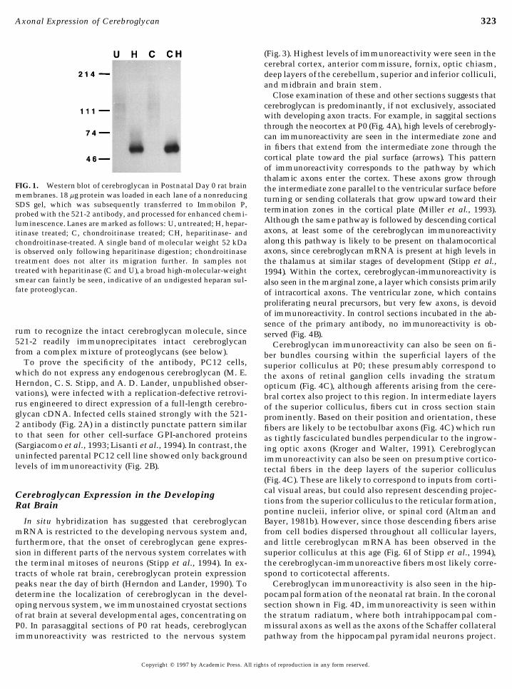
A synthetic peptide corresponding to amino acids 521–8.0; 150 mM NaCl; 1.0% SDS; 1 mM EDTA, 1 mg/ml PMSF; 25 mg/ml NEM; and 1 mg/ml pepstatin A. Identical results were obtained. 535 of the cerebroglycan coding sequence was synthesized,
coupled to KLH, and used to produce an antiserum in rab-bits. The resulting serum, 521-2, was affinity-purified
Growth Cone Particle Preparation against the same peptide. To confirm the specificity of theaffinity-purified antibody, 40 mg of neonatal rat brain mem-Growth cone particles (GCPs) were prepared from Postnatal Day
0 (P0) rat brains as described (Greenberger and Pfenninger, 1986; brane protein was digested with either heparitinase or chon-Skene et al., 1986; Simkowitz et al., 1989). Briefly, brains (including droitinase or a mixture of both GAG lyases and subjectedolfactory bulbs) were removed into ice-cold PBS, dissected free of to Western blot analysis (Fig. 1). A single 52-kDa band wasmeninges, and homogenized in 10 volumes GCP buffer (10 mM seen following heparitinase digestion, but not in untreatedTES, pH 7.4; 1 mM MgCl2; 0.32 M sucrose; PMSF (1 mg/ml); pep- samples or samples subjected to chondroitinase digestionstatin A (1 mg/ml); and N-ethyl maleiimide (2.5 mg/ml). Homoge- alone. In samples not treated with heparitinase (lanes Cnates were clarified by centrifugation at 5000g for 15 min at 47C
and U), a broad high-molecular-weight smear can faintly beand gently layered onto discontinuous sucrose gradients consistingseen, indicative of an undigested heparan sulfate proteogly-of 0.83 M, 1.0 M, and 2.0 M sucrose steps in GCP buffer. Gradientscan. Digestion with both chondroitinase and heparitinasewere centrifuged at 242,000g in a swinging bucket rotor at 47C fordid not alter the mobility of the immunoreactive band fur-40 min. GCPs were isolated from a band of white material forming
at the first sucrose interface. ther than was observed with heparitinase digestion alone,consistent with previous observations that cerebroglycanbears only heparan sulfate GAG chains (Herndon and
Primary Cell Culture and Establishment of C2 and Lander, 1990). The poor immunostaining of undigestedPC12 Cell Lines Expressing Cerebroglycan cerebroglycan (lanes C and U) is most likely due to the poor
binding of GAG-bearing PGs to protein blotting membranesA cerebroglycan expression vector, pLNCX-CBGD3*, was con-structed by subcloning the 1835-bp EagI/NarI restriction fragment (Rapraeger et al., 1985) and not to an inability of the antise-
Copyright q 1997 by Academic Press. All rights of reproduction in any form reserved.
AID DB 8532 / 6x21$$$$61 03-26-97 12:18:03 dba
323Axonal Expression of Cerebroglycan
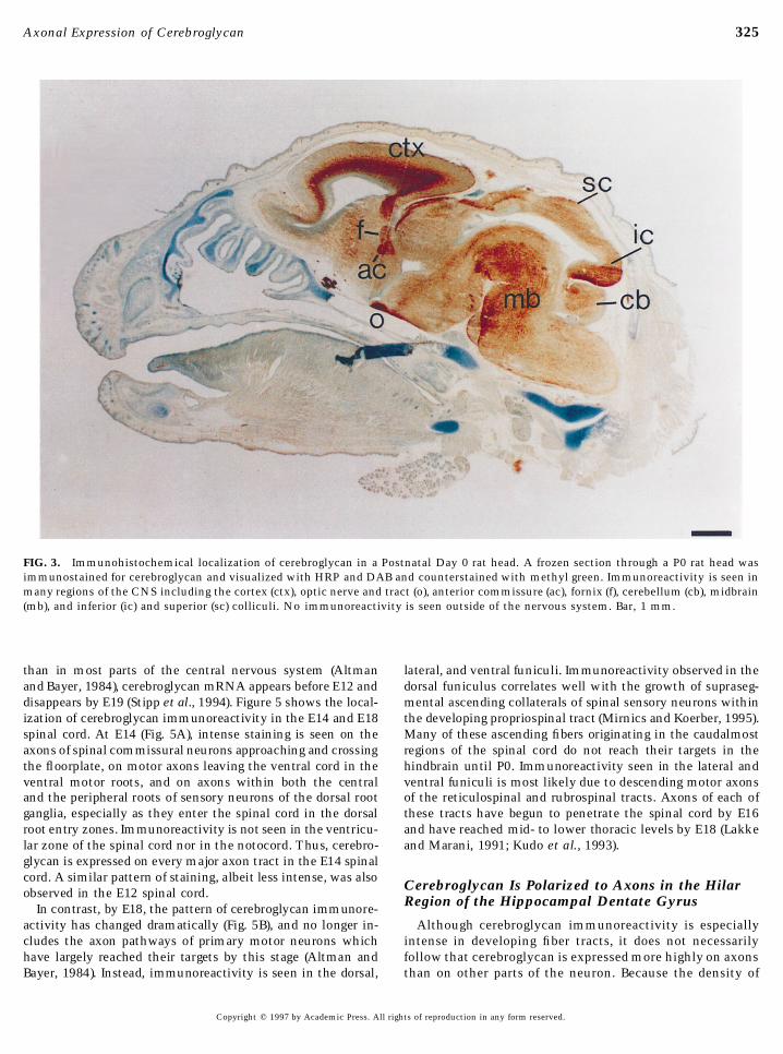
(Fig. 3). Highest levels of immunoreactivity were seen in thecerebral cortex, anterior commissure, fornix, optic chiasm,deep layers of the cerebellum, superior and inferior colliculi,and midbrain and brain stem.
Close examination of these and other sections suggests thatcerebroglycan is predominantly, if not exclusively, associatedwith developing axon tracts. For example, in saggital sectionsthrough the neocortex at P0 (Fig. 4A), high levels of cerebrogly-can immunoreactivity are seen in the intermediate zone andin fibers that extend from the intermediate zone through thecortical plate toward the pial surface (arrows). This patternof immunoreactivity corresponds to the pathway by whichthalamic axons enter the cortex. These axons grow through
FIG. 1. Western blot of cerebroglycan in Postnatal Day 0 rat brain the intermediate zone parallel to the ventricular surface beforemembranes. 18 mg protein was loaded in each lane of a nonreducing turning or sending collaterals that grow upward toward theirSDS gel, which was subsequently transferred to Immobilon P,
termination zones in the cortical plate (Miller et al., 1993).probed with the 521-2 antibody, and processed for enhanced chemi-Although the same pathway is followed by descending corticalluminescence. Lanes are marked as follows: U, untreated; H, hepar-axons, at least some of the cerebroglycan immunoreactivityitinase treated; C, chondroitinase treated; CH, heparitinase- andalong this pathway is likely to be present on thalamocorticalchondroitinase-treated. A single band of molecular weight 52 kDaaxons, since cerebroglycan mRNA is present at high levels inis observed only following heparitinase digestion; chondroitinase
treatment does not alter its migration further. In samples not the thalamus at similar stages of development (Stipp et al.,treated with heparitinase (C and U), a broad high-molecular-weight 1994). Within the cortex, cerebroglycan-immunoreactivity issmear can faintly be seen, indicative of an undigested heparan sul- also seen in the marginal zone, a layer which consists primarilyfate proteoglycan. of intracortical axons. The ventricular zone, which contains
proliferating neural precursors, but very few axons, is devoidof immunoreactivity. In control sections incubated in the ab-sence of the primary antibody, no immunoreactivity is ob-
rum to recognize the intact cerebroglycan molecule, since served (Fig. 4B).521-2 readily immunoprecipitates intact cerebroglycan Cerebroglycan immunoreactivity can also be seen on fi-from a complex mixture of proteoglycans (see below). ber bundles coursing within the superficial layers of the
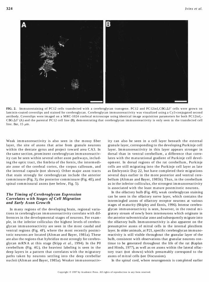
To prove the specificity of the antibody, PC12 cells, superior colliculus at P0; these presumably correspond towhich do not express any endogenous cerebroglycan (M. E. the axons of retinal ganglion cells invading the stratumHerndon, C. S. Stipp, and A. D. Lander, unpublished obser- opticum (Fig. 4C), although afferents arising from the cere-vations), were infected with a replication-defective retrovi- bral cortex also project to this region. In intermediate layersrus engineered to direct expression of a full-length cerebro- of the superior colliculus, fibers cut in cross section stainglycan cDNA. Infected cells stained strongly with the 521- prominently. Based on their position and orientation, these2 antibody (Fig. 2A) in a distinctly punctate pattern similar fibers are likely to be tectobulbar axons (Fig. 4C) which runto that seen for other cell-surface GPI-anchored proteins as tightly fasciculated bundles perpendicular to the ingrow-(Sargiacomo et al., 1993; Lisanti et al., 1994). In contrast, the ing optic axons (Kroger and Walter, 1991). Cerebroglycanuninfected parental PC12 cell line showed only background immunoreactivity can also be seen on presumptive cortico-levels of immunoreactivity (Fig. 2B). tectal fibers in the deep layers of the superior colliculus
(Fig. 4C). These are likely to correspond to inputs from corti-cal visual areas, but could also represent descending projec-Cerebroglycan Expression in the Developingtions from the superior colliculus to the reticular formation,Rat Brainpontine nucleii, inferior olive, or spinal cord (Altman andBayer, 1981b). However, since those descending fibers ariseIn situ hybridization has suggested that cerebroglycan
mRNA is restricted to the developing nervous system and, from cell bodies dispersed throughout all collicular layers,and little cerebroglycan mRNA has been observed in thefurthermore, that the onset of cerebroglycan gene expres-
sion in different parts of the nervous system correlates with superior colliculus at this age (Fig. 6I of Stipp et al., 1994),the cerebroglycan-immunoreactive fibers most likely corre-the terminal mitoses of neurons (Stipp et al., 1994). In ex-
tracts of whole rat brain, cerebroglycan protein expression spond to corticotectal afferents.Cerebroglycan immunoreactivity is also seen in the hip-peaks near the day of birth (Herndon and Lander, 1990). To
determine the localization of cerebroglycan in the devel- pocampal formation of the neonatal rat brain. In the coronalsection shown in Fig. 4D, immunoreactivity is seen withinoping nervous system, we immunostained cryostat sections
of rat brain at several developmental ages, concentrating on the stratum radiatum, where both intrahippocampal com-missural axons as well as the axons of the Schaffer collateralP0. In parasaggital sections of P0 rat heads, cerebroglycan
immunoreactivity was restricted to the nervous system pathway from the hippocampal pyramidal neurons project.
Copyright q 1997 by Academic Press. All rights of reproduction in any form reserved.
AID DB 8532 / 6x21$$$$61 03-26-97 12:18:03 dba
324 Ivins et al.
FIG. 2. Immunostaining of PC12 cells transfected with a cerebroglycan transgene. PC12 and PC12infuCBGD3* cells were grown onlaminin-coated coverslips and stained for cerebroglycan. Cerebroglycan immunoreactivity was visualized using a Cy3-conjugated secondantibody. Coverslips were imaged on a MRC-1024 confocal microscope using identical image acquisition parameters for both PC12infu-CBGD3* (A) and the parental PC12 cell line (B), demonstrating that cerebroglycan immunoreactivity is only seen in the transfected cellline. Bar, 15 mm.
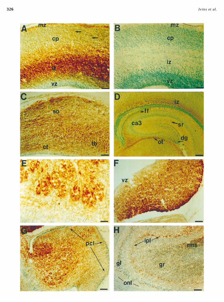
Weak immunoreactivity is also seen in the mossy fiber ity can also be seen in a cell layer beneath the externalgranule layer, corresponding to the developing Purkinje celllayer, the site of axons that arise from granule neurons
within the dentate gyrus and project toward area CA3. In layer. Immunoreactivity in this layer appears stronger indorsal than in ventral cerebellum, a difference that corre-the same section, prominent cerebroglycan immunoreactiv-
ity can be seen within several other axon pathways, includ- lates with the maturational gradient of Purkinje cell devel-opment. In dorsal regions of the rat cerebellum, Purkinjeing the optic tract, the fimbria of the fornix, the intermedi-
ate zone of the cerebral cortex, the corpus callosum, and cells are still migrating into the Purkinje cell layer as lateas Embryonic Day 22, but have completed their migrationsthe internal capsule (not shown). Other major axon tracts
that stain strongly for cerebroglycan include the anterior several days earlier in the more posterior and ventral cere-bellum (Altman and Bayer, 1985b). Thus, in the cerebellumcommissure (Fig. 3), the posterior commissure (Fig. 4E), and
spinal commissural axons (see below, Fig. 5). as in the inferior colliculus, the strongest immunoreactivityis associated with the least mature postmitotic neurons.
In the olfactory bulb (Fig. 4H), weak cerebroglycan stainingThe Timing of Cerebroglycan Expression can be seen in the olfactory nerve layer, which contains theCorrelates with Stages of Cell Migration intermingled axons of olfactory receptor neurons at variousand Early Axon Growth stages of maturity (Shipley and Ennis, 1996). Intense cerebro-
glycan immunoreactivity is seen, however, in the rostral mi-In many regions of the developing brain, regional varia-gratory stream of newly born interneurons which originate intions in cerebroglycan immunoreactivity correlate with dif-the anterior subventricular zone and subsequently migrate intoferences in the developmental stages of neurons. For exam-the olfactory bulb. Immunoreactivity is also apparent on theple, in the inferior colliculus the highest levels of cerebro-presumptive axons of mitral cells in the internal plexiformglycan immunoreactivity are seen in the most caudal andlayer. In older animals, at P21, specific cerebroglycan immuno-ventral regions (Fig. 4F), where the most recently postmi-reactivity is still visible throughout the granular layer of thetotic neurons are located (Altman and Bayer, 1981a). Thesebulb, consistent with observations that granular neurons con-are also the regions that hybridize most strongly for cerebro-tinue to be generated throughout the life of the rat (Kaplanglycan mRNA at this stage (Stipp et al., 1994). In the P0and Hinds, 1977), as well as on axons within the lateral olfac-cerebellum (Fig. 4G), the heaviest labeling is seen in thetory tract (not shown) which presumably correspond to thedeep layers in a pattern that correlates with the migratoryaxons of mitral cells (see Discussion).paths taken by neurons settling into the deep cerebellar
nucleii (Altman and Bayer, 1985a). Weaker immunoreactiv- In the spinal cord, where neurogenesis is completed earlier
Copyright q 1997 by Academic Press. All rights of reproduction in any form reserved.
AID DB 8532 / 6x21$$$$61 03-26-97 12:18:03 dba
325Axonal Expression of Cerebroglycan
FIG. 3. Immunohistochemical localization of cerebroglycan in a Postnatal Day 0 rat head. A frozen section through a P0 rat head wasimmunostained for cerebroglycan and visualized with HRP and DAB and counterstained with methyl green. Immunoreactivity is seen inmany regions of the CNS including the cortex (ctx), optic nerve and tract (o), anterior commissure (ac), fornix (f), cerebellum (cb), midbrain(mb), and inferior (ic) and superior (sc) colliculi. No immunoreactivity is seen outside of the nervous system. Bar, 1 mm.
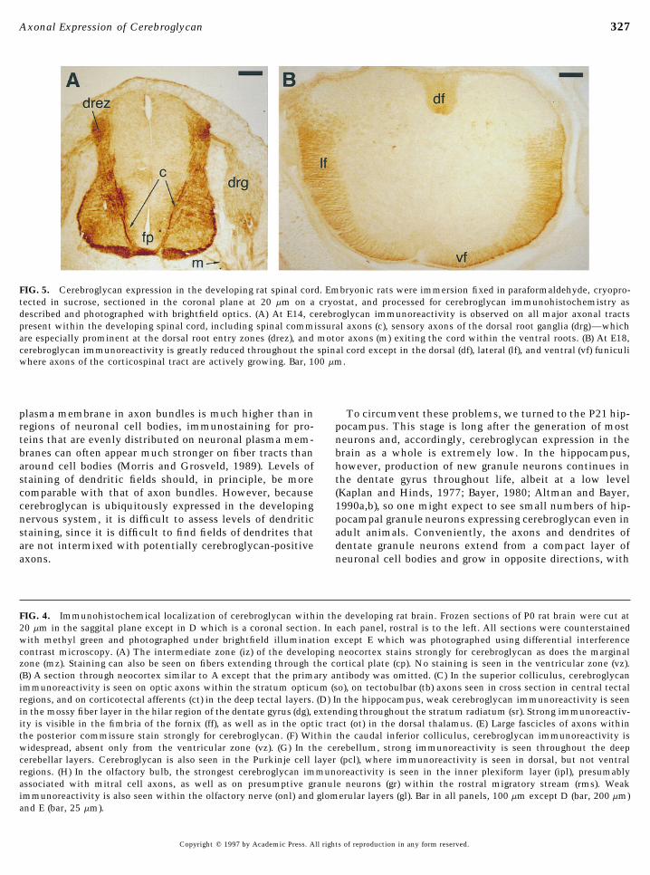
than in most parts of the central nervous system (Altman lateral, and ventral funiculi. Immunoreactivity observed in thedorsal funiculus correlates well with the growth of supraseg-and Bayer, 1984), cerebroglycan mRNA appears before E12 and
disappears by E19 (Stipp et al., 1994). Figure 5 shows the local- mental ascending collaterals of spinal sensory neurons withinthe developing propriospinal tract (Mirnics and Koerber, 1995).ization of cerebroglycan immunoreactivity in the E14 and E18
spinal cord. At E14 (Fig. 5A), intense staining is seen on the Many of these ascending fibers originating in the caudalmostregions of the spinal cord do not reach their targets in theaxons of spinal commissural neurons approaching and crossing
the floorplate, on motor axons leaving the ventral cord in the hindbrain until P0. Immunoreactivity seen in the lateral andventral funiculi is most likely due to descending motor axonsventral motor roots, and on axons within both the central
and the peripheral roots of sensory neurons of the dorsal root of the reticulospinal and rubrospinal tracts. Axons of each ofthese tracts have begun to penetrate the spinal cord by E16ganglia, especially as they enter the spinal cord in the dorsal
root entry zones. Immunoreactivity is not seen in the ventricu- and have reached mid- to lower thoracic levels by E18 (Lakkeand Marani, 1991; Kudo et al., 1993).lar zone of the spinal cord nor in the notocord. Thus, cerebro-
glycan is expressed on every major axon tract in the E14 spinalcord. A similar pattern of staining, albeit less intense, was also Cerebroglycan Is Polarized to Axons in the Hilarobserved in the E12 spinal cord.
Region of the Hippocampal Dentate GyrusIn contrast, by E18, the pattern of cerebroglycan immunore-activity has changed dramatically (Fig. 5B), and no longer in- Although cerebroglycan immunoreactivity is especially
intense in developing fiber tracts, it does not necessarilycludes the axon pathways of primary motor neurons whichhave largely reached their targets by this stage (Altman and follow that cerebroglycan is expressed more highly on axons
than on other parts of the neuron. Because the density ofBayer, 1984). Instead, immunoreactivity is seen in the dorsal,
Copyright q 1997 by Academic Press. All rights of reproduction in any form reserved.
AID DB 8532 / 6x21$$$$61 03-26-97 12:18:03 dba
326 Ivins et al.
03-26-97 12:18:03 dba
327Axonal Expression of Cerebroglycan
FIG. 5. Cerebroglycan expression in the developing rat spinal cord. Embryonic rats were immersion fixed in paraformaldehyde, cryopro-tected in sucrose, sectioned in the coronal plane at 20 mm on a cryostat, and processed for cerebroglycan immunohistochemistry asdescribed and photographed with brightfield optics. (A) At E14, cerebroglycan immunoreactivity is observed on all major axonal tractspresent within the developing spinal cord, including spinal commissural axons (c), sensory axons of the dorsal root ganglia (drg)—whichare especially prominent at the dorsal root entry zones (drez), and motor axons (m) exiting the cord within the ventral roots. (B) At E18,cerebroglycan immunoreactivity is greatly reduced throughout the spinal cord except in the dorsal (df), lateral (lf), and ventral (vf) funiculiwhere axons of the corticospinal tract are actively growing. Bar, 100 mm.
plasma membrane in axon bundles is much higher than in To circumvent these problems, we turned to the P21 hip-pocampus. This stage is long after the generation of mostregions of neuronal cell bodies, immunostaining for pro-
teins that are evenly distributed on neuronal plasma mem- neurons and, accordingly, cerebroglycan expression in thebrain as a whole is extremely low. In the hippocampus,branes can often appear much stronger on fiber tracts than
around cell bodies (Morris and Grosveld, 1989). Levels of however, production of new granule neurons continues inthe dentate gyrus throughout life, albeit at a low levelstaining of dendritic fields should, in principle, be more
comparable with that of axon bundles. However, because (Kaplan and Hinds, 1977; Bayer, 1980; Altman and Bayer,1990a,b), so one might expect to see small numbers of hip-cerebroglycan is ubiquitously expressed in the developing
nervous system, it is difficult to assess levels of dendritic pocampal granule neurons expressing cerebroglycan even inadult animals. Conveniently, the axons and dendrites ofstaining, since it is difficult to find fields of dendrites that
are not intermixed with potentially cerebroglycan-positive dentate granule neurons extend from a compact layer ofneuronal cell bodies and grow in opposite directions, withaxons.
FIG. 4. Immunohistochemical localization of cerebroglycan within the developing rat brain. Frozen sections of P0 rat brain were cut at20 mm in the saggital plane except in D which is a coronal section. In each panel, rostral is to the left. All sections were counterstainedwith methyl green and photographed under brightfield illumination except E which was photographed using differential interferencecontrast microscopy. (A) The intermediate zone (iz) of the developing neocortex stains strongly for cerebroglycan as does the marginalzone (mz). Staining can also be seen on fibers extending through the cortical plate (cp). No staining is seen in the ventricular zone (vz).(B) A section through neocortex similar to A except that the primary antibody was omitted. (C) In the superior colliculus, cerebroglycanimmunoreactivity is seen on optic axons within the stratum opticum (so), on tectobulbar (tb) axons seen in cross section in central tectalregions, and on corticotectal afferents (ct) in the deep tectal layers. (D) In the hippocampus, weak cerebroglycan immunoreactivity is seenin the mossy fiber layer in the hilar region of the dentate gyrus (dg), extending throughout the stratum radiatum (sr). Strong immunoreactiv-ity is visible in the fimbria of the fornix (ff), as well as in the optic tract (ot) in the dorsal thalamus. (E) Large fascicles of axons withinthe posterior commissure stain strongly for cerebroglycan. (F) Within the caudal inferior colliculus, cerebroglycan immunoreactivity iswidespread, absent only from the ventricular zone (vz). (G) In the cerebellum, strong immunoreactivity is seen throughout the deepcerebellar layers. Cerebroglycan is also seen in the Purkinje cell layer (pcl), where immunoreactivity is seen in dorsal, but not ventralregions. (H) In the olfactory bulb, the strongest cerebroglycan immunoreactivity is seen in the inner plexiform layer (ipl), presumablyassociated with mitral cell axons, as well as on presumptive granule neurons (gr) within the rostral migratory stream (rms). Weakimmunoreactivity is also seen within the olfactory nerve (onl) and glomerular layers (gl). Bar in all panels, 100 mm except D (bar, 200 mm)and E (bar, 25 mm).
Copyright q 1997 by Academic Press. All rights of reproduction in any form reserved.
AID DB 8532 / 6x21$$$$61 03-26-97 12:18:03 dba

328 Ivins et al.
a single axon heading basally into the hilar region (to joinother mossy fibers) and a single dendrite heading apically(Bayer, 1980; Gaarskjaer, 1986). Thus, at P21 it should bepossible to individually examine cerebroglycan expressionon well-separated axons, cell bodies, and dendrites of smallnumbers of granule neurons, without interfering immuno-staining from any other cell type.
As shown in Fig. 6, this was indeed the case. In sections ofthe P21 hippocampus, small numbers of thin cerebroglycan-immunoreactive fibers were scattered throughout the hilarregion; these fibers had the appearance of individual axons.In contrast, little if any cerebroglycan immunoreactivitywas detected among the granule cell bodies nor among thelayer containing their dendrites. These data demonstratethat, at least for some neurons, cerebroglycan is restrictedto the axonal compartment and suggest that this polarizedexpression pattern may be a general feature of cerebroglycanexpression.
Cerebroglycan Is Expressed on NeuronalGrowth Cones
The strong localization of cerebroglycan to the axons ofnewly generated neurons raises the possibility that it couldfunction on axonal growth cones. To address whether cere-broglycan is expressed on growth cones in vitro and in vivo,E18 retinal neurons were cultured on merosin-coated glasscoverslips for 20 hr and immunostained for cerebroglycan.As can be seen in Fig. 7, strong cerebroglycan immunoreac-tivity was observed on growing neurites, including thegrowth cones. Heavy staining was seen along the leadingedge of veil-rich regions (Fig. 7A), as well as along filopodia,where highly punctate staining (Figs. 7B and 7C), consistentwith clustering of cerebroglycan in the membrane, was typi-cally observed.
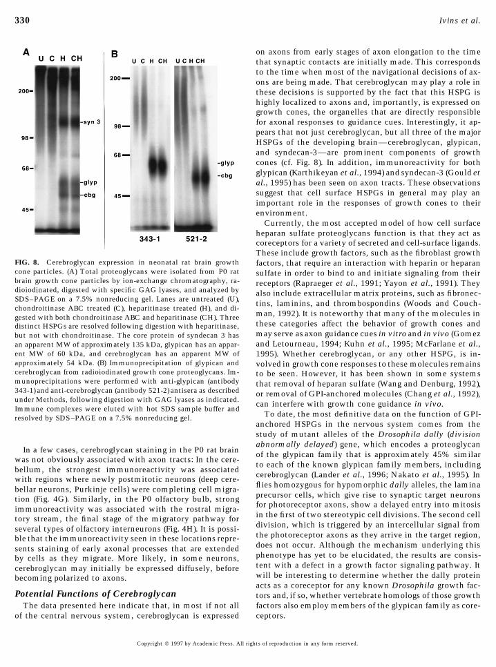
To determine if cerebroglycan is also a prominent compo-nent of growth cones in vivo, total proteoglycans were puri-fied from freshly isolated growth cone particles and radioio-dinated, and their core proteins were analyzed by SDS–PAGE following digestion with specific GAG lyases (Fig.8A). Three major HSPGs were seen in these preparations,with apparent core protein molecular weights of approxi-mately 135, 60, and 54 kDa, corresponding to syndecan 3,glypican, and cerebroglycan (formerly known as HSPGs M7,M12, and M13, respectively; Herndon and Lander, 1990).Identical results were obtained in five independent prepara-tions. The identification of the 60- and 54-kDa species as
FIG. 6. Cerebroglycan expression in the hilar region of the Post-glypican and cerebroglycan, respectively, was based on thenatal Day 21 rat hippocampus. Saggital sections of fresh-frozenability of anti-glypican and anti-cerebroglycan antisera toP21 rat brain were cut at 12 mm on a cryostat, postfixed, andspecifically immunoprecipitate these molecules (Fig. 8B).stained with the 521-2 antibody. Antibody binding was visual-The identification of HSPG M7 as syndecan 3 was madeized using a Cy3-conjugated anti-rabbit second antibody, and
on the basis of an anti-syndecan 3 antiserum (generously the section was counterstained with bisbenzamide to allow vi-provided by Dr. M. Bernfield) to specifically immunoprecip- sualization of nucleii. (A) Schematic of the cellular anatomy ofitate the 135-kDa species (not shown). Although the relative the dentate gyrus. (B) Nuclei of granule neurons in the dentateproportions of cerebroglycan, glypican, and syndecan-3 in gyrus labeled with bisbenzamide. (C) Cerebroglycan immunore-
activity demonstrated on mossy fibers by immunofluorescence.growth cone particles are similar to those in whole-brainBar, 40 mm.membranes (cf. Herndon and Lander, 1990), PGs as a whole
Copyright q 1997 by Academic Press. All rights of reproduction in any form reserved.
AID DB 8532 / 6x21$$$$61 03-26-97 12:18:03 dba

329Axonal Expression of Cerebroglycan
FIG. 7. Cerebroglycan expression on retinal growth cones in vitro. Retinal neurons from E18 rats were cultured overnight on merosin-coated glass coverslips. After fixation, they were immunostained for cerebroglycan, visualized using a Cy3-conjugated second antibody,and photographed at 631 under rhodamine fluorescence. Three separate examples of neurites bearing growth cones are shown in A, B,and C. The cell bodies from which these neurites extended were also immunoreactive (not shown). Bar, 15 mm.
were enriched in the growth cone fraction. Specifically, in only transiently expressed by growing axons is the lateralolfactory tract, which primarily consists of axons of thegrowth cones of neonatal rat brain, PGs constituted be-mitral cells of the olfactory bulb, but also contains fiberstween 1.21 and 1.69% of total protein (1.43 { 0.24%, n Åthat originate in the raphe nuclei and locus coeruleus and3), whereas in whole-brain membranes they were approxi-innervate the olfactory bulb (Shipley and Ennis, 1996).mately three times less abundant (0.45% of total protein, nCerebroglycan immunoreactivity was detected in the lat-Å 2).eral olfactory tract as late as P21 (unpublished observations),despite the fact that mitral cells are born between E14 andE16 (Bayer et al., 1993) and raphe and locus coeruleus neu-DISCUSSIONrons are born by E16 and E14, respectively (Bayer et al.,1993). Other than this, the only evidence of cerebroglycan
To investigate the roles of cell surface HSPGs in neural expression at P21 was associated with regions of very latedevelopment, we generated specific antibodies against neurogenesis (dentate granule neurons of the hippocampuscerebroglycan, a GPI-anchored HSPG expressed specifically [Fig. 6]; granule neurons of the olfactory bulb [not shown]),within the developing nervous system. The antibodies are or regions where small numbers of axons had not yet fin-directed against an epitope near the C-terminus of the ma- ished growing (e.g., the internal capsule, which containsture polypeptide and recognize both the core protein and some very late-developing axons of the corticospinal tractthe intact HSPG (Figs. 1, 2, and 8). Immunostaining of the [not shown]).developing rat nervous system indicates that cerebroglycan In at least one cell type, the hippocampal granule neuron,is highly localized to developing axon tracts and growth expression of cerebroglycan is specifically polarized to thecones. Furthermore, these data indicate that cerebroglycan axonal compartment, as no dendritic or cell-body stainingis predominantly, if not exclusively, a product of neurons could be detected (Fig. 6). This could reflect either preferen-(rather than glial cells), a result that in situ hybridization tial sorting or preferential stabilization of the cerebroglycandata had suggested (Stipp et al., 1994). molecule. In several types of cells, GPI–anchors have been
As long as they were examined at stages when axons are shown to target proteins to specific cellular domains, in-actively growing, all axon tracts appeared to be immunore- cluding the axonal compartment of hippocampal pyramidalactive for cerebroglycan, suggesting that cerebroglycan may neurons in vitro (Dotti and Simons, 1990). Indeed axonin-be a general marker expressed by immature neurons. In 1/TAG-1 is another example of a GPI-anchored glycoproteinmost cases, axon tracts became cerebroglycan negative that appears to be preferentially associated with axonswithin several days after the final mitosis of their neurons (Dodd et al., 1988). Nevertheless, it cannot be that GPIof origin (cf. Fig. 5), usually around the time that axons anchorage targets cerebroglycan or other proteins to axons,arrived at their targets. For instance, the axons of spinal since the GPI-anchored glycoprotein Thy-1 is actually ex-sensory neurons, which reach their central targets between cluded from growing axons in vivo (Xue et al., 1991). It willE16 and E18 (Mirnics and Koerber, 1995b), stained strongly be interesting to determine whether particular structuralfor cerebroglycan at E14, but had largely lost cerebroglycan features in the core protein and/or glycosaminoglycanimmunoreactivity by E18. chains of cerebroglycan play a direct role in controlling the
unique localization of this proteoglycan.An apparent exception to the rule that cerebroglycan is
Copyright q 1997 by Academic Press. All rights of reproduction in any form reserved.
AID DB 8532 / 6x21$$$$61 03-26-97 12:18:03 dba
330 Ivins et al.
on axons from early stages of axon elongation to the timethat synaptic contacts are initially made. This correspondsto the time when most of the navigational decisions of ax-ons are being made. That cerebroglycan may play a role inthese decisions is supported by the fact that this HSPG ishighly localized to axons and, importantly, is expressed ongrowth cones, the organelles that are directly responsiblefor axonal responses to guidance cues. Interestingly, it ap-pears that not just cerebroglycan, but all three of the majorHSPGs of the developing brain—cerebroglycan, glypican,and syndecan-3—are prominent components of growthcones (cf. Fig. 8). In addition, immunoreactivity for bothglypican (Karthikeyan et al., 1994) and syndecan-3 (Gould etal., 1995) has been seen on axon tracts. These observationssuggest that cell surface HSPGs in general may play animportant role in the responses of growth cones to theirenvironment.
Currently, the most accepted model of how cell surfaceheparan sulfate proteoglycans function is that they act ascoreceptors for a variety of secreted and cell-surface ligands.These include growth factors, such as the fibroblast growth
FIG. 8. Cerebroglycan expression in neonatal rat brain growth factors, that require an interaction with heparin or heparancone particles. (A) Total proteoglycans were isolated from P0 rat sulfate in order to bind to and initiate signaling from theirbrain growth cone particles by ion-exchange chromatography, ra- receptors (Rapraeger et al., 1991; Yayon et al., 1991). Theydioiodinated, digested with specific GAG lyases, and analyzed by also include extracellular matrix proteins, such as fibronec-SDS–PAGE on a 7.5% nonreducing gel. Lanes are untreated (U), tins, laminins, and thrombospondins (Woods and Couch-chondroitinase ABC treated (C), heparitinase treated (H), and di-
man, 1992). It is noteworthy that many of the molecules ingested with both chondroitinase ABC and heparitinase (CH). Threethese categories affect the behavior of growth cones anddistinct HSPGs are resolved following digestion with heparitinase,may serve as axon guidance cues in vitro and in vivo (Gomezbut not with chondroitinase. The core protein of syndecan 3 hasand Letourneau, 1994; Kuhn et al., 1995; McFarlane et al.,an apparent MW of approximately 135 kDa, glypican has an appar-
ent MW of 60 kDa, and cerebroglycan has an apparent MW of 1995). Whether cerebroglycan, or any other HSPG, is in-approximately 54 kDa. (B) Immunoprecipitation of glypican and volved in growth cone responses to these molecules remainscerebroglycan from radioiodinated growth cone proteoglycans. Im- to be seen. However, it has been shown in some systemsmunoprecipitations were performed with anti-glypican (antibody that removal of heparan sulfate (Wang and Denburg, 1992),343-1) and anti-cerebroglycan (antibody 521-2) antisera as described or removal of GPI-anchored molecules (Chang et al., 1992),under Methods, following digestion with GAG lyases as indicated. can interfere with growth cone guidance in vivo.Immune complexes were eluted with hot SDS sample buffer and
To date, the most definitive data on the function of GPI-resolved by SDS–PAGE on a 7.5% nonreducing gel.anchored HSPGs in the nervous system comes from thestudy of mutant alleles of the Drosophila dally (divisionabnormally delayed) gene, which encodes a proteoglycan
In a few cases, cerebroglycan staining in the P0 rat brain of the glypican family that is approximately 45% similarwas not obviously associated with axon tracts: In the cere-
to each of the known glypican family members, includingbellum, the strongest immunoreactivity was associated
cerebroglycan (Lander et al., 1996; Nakato et al., 1995). Inwith regions where newly postmitotic neurons (deep cere-flies homozygous for hypomorphic dally alleles, the laminabellar neurons, Purkinje cells) were completing cell migra-precursor cells, which give rise to synaptic target neuronstion (Fig. 4G). Similarly, in the P0 olfactory bulb, strongfor photoreceptor axons, show a delayed entry into mitosisimmunoreactivity was associated with the rostral migra-in the first of two stereotypic cell divisions. The second celltory stream, the final stage of the migratory pathway fordivision, which is triggered by an intercellular signal fromseveral types of olfactory interneurons (Fig. 4H). It is possi-the photoreceptor axons as they arrive in the target region,ble that the immunoreactivity seen in these locations repre-does not occur. Although the mechanism underlying thissents staining of early axonal processes that are extendedphenotype has yet to be elucidated, the results are consis-by cells as they migrate. More likely, in some neurons,tent with a defect in a growth factor signaling pathway. Itcerebroglycan may initially be expressed diffusely, beforewill be interesting to determine whether the dally proteinbecoming polarized to axons.acts as a coreceptor for any known Drosophila growth fac-
Potential Functions of Cerebroglycan tors and, if so, whether vertebrate homologs of those growthfactors also employ members of the glypican family as core-The data presented here indicate that, in most if not all
of the central nervous system, cerebroglycan is expressed ceptors.
Copyright q 1997 by Academic Press. All rights of reproduction in any form reserved.
AID DB 8532 / 6x21$$$$61 03-26-97 12:18:03 dba
331Axonal Expression of Cerebroglycan
T. M. (1988). Spatial regulation of axonal glycoprotein expressionACKNOWLEDGMENTSon subsets of embryonic spinal neurons. Neuron 1, 105–116.
Dotti, C. G., and Simons, K. (1990). Polarized sorting of viral glyco-We are grateful to Merton Bernfield for the gift of antiserum proteins to the axon and dendrites of hippocampal neurons in
to syndecan-3. We thank Tim Fritz and Cathy Krull for helpful culture. Cell 62, 63 –72.comments on the manuscript. This work was supported by NIH Gaarskjaer, F. B. (1986). The organization and development of theGrant NS26862 to A.D.L. hippocampal mossy fiber system. Brain Res. 11, 335–357.
Gomez, T. M., and Letourneau, P. C. (1994). Filopodia initiatechoices made by sensory neuron growth cones at laminin/fibro-nectin borders in vitro. J. Neurosci. 14, 5959–5972.
REFERENCES Gould, S. E., Upholt, W. B., and Kosher, R. A. (1995). Characteriza-tion of chicken syndecan-3 as a heparan sulfate proteoglycan andits expression during embryogenesis. Dev. Biol. 168, 438–451.
Altman, J., and Bayer, S. A. (1981a). Time of origin of neurons of Greenberger, L. M., and Pfenninger, K. H. (1986). Membrane glyco-the rat inferior colliculus and the relations between cytogenesis proteins of the nerve growth cone: Diversity and growth regula-and tonotopic order in the auditory pathway. Exp. Brain Res. 42, tion of oligosaccharides. J. Cell Biol. 103, 1369–1382.411–423. Haugen, P. K., Letourneau, P. C., Drake, S. L., Furcht, L. T., and
Altman, J., and Bayer, S. A. (1981b). Time of origin of neurons of McCarthy, J. B. (1992). A cell-surface heparan sulfate proteogly-the rat superior colliculus in relation to other components of the can mediated neural cell adhesion and spreading on a definedvisual and visuomotor pathways. Exp. Brain Res. 42, 424–434. sequence from the c-terminal and heparin binding domain of
Altman, J., and Bayer, S. A. (1984). The development of the rat fibronectin, FN-C/H II. J. Neurosci. 12, 2597–2608.spinal cord. Adv. Anat. Embryol. Cell Biol. 85, 1–160. Herndon, M. E., and Lander, A. D. (1990). A diverse set of develop-
Altman, J., and Bayer, S. A. (1985a). Embryonic development of the mentally regulated proteoglycans is expressed in the rat centralrat cerebellum. II. Translocation and regional distribution of the nervous system. Neuron 4, 949–961.deep neurons. J. Comp. Neurol. 231, 27–41. Kan, M., Wang, F. X. J., Crabb, J. W., Hou, J., and McKeehan, W. L.
Altman, J., and Bayer, S. A. (1985b). Embryonic development of the (1993). An essential heparin-binding domain in the fibroblastrat cerebellum. lll. Regional differences in the time of origin, growth factor receptor kinase. Science 259, 1918–1921.migration, and settling of Purkinje cells. J. Comp. Neurol. 231,
Kaplan, M. S., and Hinds, J. W. (1977). Neurogenesis in the adult42–65.
rat: Electron microscopic analysis of light radiographs. ScienceAltman, J., and Bayer, S. A. (1990a). Migration and distribution of
197, 1092–1094.two populations of hippocampal granule cell precursors during
Karthikeyan, L., Flad, M., Engel, M., Meyer-Puttlitz, B., and Mar-the perinatal and postnatal periods. J. Comp. Neurol. 301, 365–
golis, R. U. (1994). Immunocytochemical and in situ hybridiza-381.
tion studies of the heparan sulfate proteoglycan, glypican, in ner-Altman, J., and Bayer, S. A. (1990b). Mosaic organization of the
vous tissue. J. Cell Sci. 107, 3213–3222.hippocampal neuroepithelium and the multiple germinal sources
Karthikeyan, L., Maurel, P., Rauch, U., Margolis, R. K., and Mar-of dentate granule cells. J. Comp. Neurol. 301, 325–342.golis, R. U. (1992). Cloning of a major heparan sulfate proteogly-Aviezer, D., and Yayon, A. (1994). Heparin-dependent binding andcan from brain and identification as the rat form of glypican.autophosphorylation of epidermal growth factor (EGF) receptorBiochem. Biophys. Res. Commun. 188, 395–401.by heparin-binding EGF-like growth factor but not by EGF. Proc.
Kroger, S., and Walter, J. (1991). Molecular mechanisms separatingNatl. Acad. Sci. USA 91, 12173–12177.two axonal pathways during embryonic development of the avianBayer, S. A. (1980). Development of the hippocampal region in theoptic tectum. Neuron 6, 291–303.rat. I. Neurogenesis examined with 3H-thymidine autordiogra-
Kudo, N., Furukawa, F., and Okado, N. (1993). Development ofphy. J. Comp. Neurol. 190, 87–114.descending fibers to the rat embryonic spinal cord. Neurosci. Res.Bayer, S. A., Altman, J., Russo, R. J., and Zhang, X. (1993). Timeta-16, 131–141.bles of neurogenesis in the human brain based on experimentally
Kuhn, T. B., Schmidt, M. F., and Kater, S. B. (1995). Laminin anddetermined patterns in the rat. Neurotoxicology 14, 83–144.fibronectin guideposts signal sustained but opposite effects toBernfield, M., Kokenyesi, R., Kato, M., Hinkes, M. T., Gallo, R. L.,passing growth cones. Neuron 14, 175–285.and Lose, E. J. (1992). Biology of the syndecans: A family of trans-
Lakke, E. A. J. F., and Marani, E. (1991). Prenatal descent of rubro-membrane heparan sulfate proteoglycans. Annu. Rev. Cell Biol.spinal fibers through the spinal cord of the rat. J. Comp. Neurol.8, 365–393.314, 67–78.Calof, A. L., Campanero, M. R., O’Rear, J. J., Yurchenco, P. D., and
Lander, A. D. (1993). Proteoglycans in the nervous system. Curr.Lander, A. D. (1994). Domain-specific activation of neuronal mi-Opin. Neurobiol. 3, 716–723.gration and neurite outgrowth-promoting activities of laminin.
Lander, A. D., Stipp, C. S., and Ivins, J. K. (1996). The glypican fam-Neuron 13, 117–130.ily of heparan sulfate proteoglycans: Major cell-surface proteogly-Carey, D. J., Evans, D. M., Stahl, R. C., Asundi, V. K., Conner, K. J.,cans of the developing nervous system. Perspect. Dev. Neurobiol.Garbes, P., and Cizmeci-Smith, G. (1992). Molecular cloning and3/4, 347 –359.characterization of N-syndecan, a novel transmembrane heparan
LeBaron, R. G., Esko, J. D., Woods, A., Johansson, S., and Hook, M.sulfate proteoglycan. J. Cell Biol. 117, 191 –201.(1988). Adhesion of glycosaminoglycan-deficient Chinese ham-Chang, W. S., Serikawa, K., Allen, K., and Bentley, D. (1992). Dis-ster ovary cell mutants to fibronectin substrata. J. Cell Biol. 106,ruption of pioneer growth cone guidance in vivo by removal of945–952.glycosylphosphatidylinositol-anchored cell surface proteins. De-
Lisanti, M. P., Scherer, P. E., Vidugiriene, J., Tang, Z., Hermanow-velopment 114, 507–519.Dodd, J., Morton, S. B., Karagogeos, D., Yamamoto, M., and Jessell, ski-Vosatka, A., Tu, Y.-H., Cook, R. F., and Sargiacomo, M.
Copyright q 1997 by Academic Press. All rights of reproduction in any form reserved.
AID DB 8532 / 6x21$$$$61 03-26-97 12:18:03 dba

332 Ivins et al.
(1994). Characterization of caveoli-rich membrane domains iso- Shipley, M. T., and Ennis, M. (1996). Functional organization ofolfactory system. J. Neurobiol. 30, 123–176.lated from an endothelial-rich source: Implications for human
disease. J. Cell Biol. 126, 111–126. Simkowitz, P., Ellis, L., and Pfenninger, K. H. (1989). Membraneproteins of the nerve growth cone and their developmental regu-Litwack, E. D., Stipp, C. S., Kumbasar, A., and Lander, A. D. (1994).lation. J. Neurosci. 9, 1004–1017.Neuronal expression of glypican, a cell-surface glycosylphospha-
Skene, J. H. P., Jacoson, R. D., Snipes, G. J., McGuire, C. B., Norden,tidylinositol-anchored heparan sulfate proteoglycan, in the adultJ. J., and Freeman, J. A. (1986). A protein induced during nerverat nervous system. J. Neurosci. 14, 3713–3724.growth (GAP-43) is a major component of growth cone mem-McFarlane, S., McNeill, L., and Holt, C. E. (1995). FGF signalingbranes. Science 233, 783–785.and target recognition in the developing Xenopus visual system.
Stanley, M. J., Liebersbach, B. F., Liu, W., Anhalt, D. J., and Sand-Neuron 15, 1017–1028.erson, R. D. (1995). Heparan sulfate-mediated cell aggregation:Miller, A. D., and Rosman, G. J. (1989). Improved retroviral vectorsSyndecans-1 and -4 mediate intercellular adhesion followingfor gene transfer and expression. Biotechniques 7, 980–990.their transfection into human B lymphoid cells. J. Biol. Chem.Miller, B., Chou, L., and Finlay, B. L. (1993). The early development270, 5077–5083.of thalamocortical and corticothalamic projections. J. Comp.
Stipp, C. S., Litwack, E. D., and Lander, A. D. (1994). Cerebrogly-Neurol. 335, 16–41.can: An integral membrane heparan sulfate proteoglycan that isMirnics, K., and Koerber, R. (1995). Prenatal development of ratunique to the developing nervous system and expressed specifi-primary afferent fibers. II. Central projections. J. Comp. Neurol.cally during neuronal differentiation. J. Cell Biol. 124, 149–160.355, 601–614.
Wang, L., and Denburg, J. L. (1992). A role for proteoglycans in theMorris, R., and Grosveld, F. (1989). Expression of thy-1 in the ner-guidance of a subset of pioneer axons in cultured embryos of thevous system of the rat and mouse. In ‘‘Cell Surface Antigen Thy-cockroach. Neuron 8, 701 –714.1: Immunology, Neurology, and Therapeutic Applications’’
Watanabe, K., Yamada, H., and Yamaguchi, Y. (1995). K-glypican:(A. E. Reif and M. Schlesinger, Eds.), pp. 121–148. Dekker, NewA novel GPI-anchored heparan sulfate proteoglycan that is highlyYork.expressed in developing brain and kidney. J. Cell Biol. 130, 1207–Nakato, H., Futch, T. A., and Selleck, S. B. (1995). The division1218.abnormally delayed (dally) gene: A putative integral membrane
Woods, A., and Couchman, J. (1992). Heparan sulfate proteoglycansproteoglycan required for cell division patterning during postem-and signalling in cell adhesion. Adv. Exp. Med. Biol. 313, 87 –96.bryonic development of the nervous system in Drosophila. De-
Xue, G. P., Pliego Rivero, B., and Morris, R. J. (1991). The surfacevelopment 121, 3687–3702.glycoprotein Thy-1 is excluded from growing axons during devel-Rapraeger, A., Jalkanen, M., Endo, E., Koda, J., and Bernfield, M.opment: A study of the expression of Thy-1 during axogenesis(1985). The cell surface proteoglycan from mouse mammary epi-in hippocampus and hindbrain. Development 112, 161–176.thelial cells bears chondroitin sulfate and heparan sulfate glycos-
Yayon, A., Klagsbrun, M., Esko, J. D., Leder, P., and Ornitz, D. M.aminoglycans. J. Biol. Chem. 260, 11046–11052.(1991). Cell surface, heparin-like molecules are required for bind-Rapraeger, A. C., Krufka, A., and Olwin, B. B. (1991). Requirementing of basic fibroblast growth factor to its high affinity receptor.of heparan sulfate for bFGF-mediated fibroblast and myoblastCell 64, 841–848.differentiation. Science 252, 1705–1708.
Zioncheck, T. F., Richardson, L., Liu, J., Chang, L., King, K. L.,Reyes, A. A., Akeson, R., Brezina, L., and Cole, G. J. (1990). Struc-Bennett, G. L., Fugedi, P., Chamow, S. M., Schwall, R. H., andtural requirements for neural cell adhesion molecule-heparin in-Stack, R. J. (1995). Sulfated oligosaccharides promote hepatocyteteraction. Cell Regul. 1, 567–576.growth factor association and govern its mitogenic activity. J.Sargiacomo, M., Sudol, M., Tang, Z., and Lisanti, M. P. (1993). Sig-Biol. Chem. 270, 16871–16878.nal transducing molecules and glycosylphosphatidylinositol-
linked proteins form a caveolin-rich complex in MDCK cells. J. Received for publication August 28, 1996Accepted February 7, 1997Cell Biol. 122, 789–807.
Copyright q 1997 by Academic Press. All rights of reproduction in any form reserved.
AID DB 8532 / 6x21$$$$61 03-26-97 12:18:03 dba