Obligatory Role for Endothelial Heparan Sulphate Proteoglycans and Caveolae Internalization in...
11
Research Article Obligatory Role for Endothelial Heparan Sulphate Proteoglycans and Caveolae Internalization in Catestatin-Dependent eNOS Activation Sara Fornero, 1 Eleonora Bassino, 1 Roberta Ramella, 1 Clara Gallina, 1 Sushil K. Mahata, 2,3 Bruno Tota, 4,5 Renzo Levi, 1 Giuseppe Alloatti, 1,5 and Maria Pia Gallo 1 1 Department of Life Sciences and Systems Biology, University of Turin, Via Accademia Albertina 13, 10123 Turin, Italy 2 Veterans Affairs San Diego Healthcare System, 3350 La Jolla Village Drive, San Diego, CA 92161, USA 3 Department of Medicine, University of California, 9500-0838 Gilman Drive, La Jolla, San Diego, CA 92093, USA 4 DIBEST Department, University of Calabria, 87030 Arcavacata di Rende, Italy 5 National Institute for Cardiovascular Research, Via Irnerio 48, 40126 Bologna, Italy Correspondence should be addressed to Maria Pia Gallo; [email protected] Received 8 April 2014; Revised 26 June 2014; Accepted 27 June 2014; Published 20 July 2014 Academic Editor: Paraskevi Heldin Copyright © 2014 Sara Fornero et al. is is an open access article distributed under the Creative Commons Attribution License, which permits unrestricted use, distribution, and reproduction in any medium, provided the original work is properly cited. e chromogranin-A peptide catestatin modulates a wide range of processes, such as cardiovascular functions, innate immunity, inflammation, and metabolism. We recently found that the cardiac antiadrenergic action of catestatin requires a PI3K-dependent NO release from endothelial cells, although the receptor involved is yet to be identified. In the present work, based on the cationic properties of catestatin, we tested the hypothesis of its interaction with membrane heparan sulphate proteoglycans, resulting in the activation of a caveolae-dependent endocytosis. Experiments were performed on bovine aortic endothelial cells. Endocytotic vesicles trafficking was quantified by confocal microscopy using a water-soluble membrane dye; catestatin colocalization with heparan sulphate proteoglycans and caveolin 1 internalization were studied by fluorimetric measurements in live cells. Modulation of the catestatin-dependent eNOS activation was assessed by immunofluorescence and immunoblot analysis. Our results demonstrate that catestatin (5 nM) colocalizes with heparan sulphate proteoglycans and induces a remarkable increase in the caveolae-dependent endocytosis and caveolin 1 internalization, which were significantly reduced by both heparinase and wortmannin. Moreover, catestatin was unable to induce Ser 1179 eNOS phosphorylation aſter pretreatments with heparinase and methyl--cyclodextrin. Taken together, these results highlight the obligatory role for proteoglycans and caveolae internalization in the catestatin-dependent eNOS activation in endothelial cells. 1. Introduction Chromogranin A (CgA) is a 48 kDa acidic glycoprotein [1– 3] and the main component of granins, a family of proteins abundantly expressed in large dense core vesicles of neuroen- docrine cells, neurons, and other secreting cells including cardiac cells [4]. In the heart CgA is costored and cosecreted, respectively, with catecholamines and natriuretic peptides [4]. Catestatin (CST: hCgA 352−372 ) is a 21 amino acid cationic and hydrophobic peptide derived from the proteolytic cleav- age of CgA. It was firstly discovered as an endogenous allosteric nicotinic-cholinergic antagonist [5], but it is now established as a multifunctional peptide modulating several organs/systems, including the cardiovascular system [6, 7]. In particular, it has been shown that CST administration induces vasodilation by multiple mechanisms. One mechanism relies on the CST-dependent inhibition of catecholamine secretion, through noncompetitive binding at the nicotinic cholinergic receptor [5, 8]. Another potential mechanism for the CST- induced vasodilation is through histamine release [9, 10], which has been shown in vitro in mast cells mimicking the receptor-independent peptidergic pathway proposed for mastoparan and for other cationic and amphipathic peptides [10]. Finally, CST has been shown to decrease sympathetic Hindawi Publishing Corporation BioMed Research International Volume 2014, Article ID 783623, 10 pages http://dx.doi.org/10.1155/2014/783623
Transcript of Obligatory Role for Endothelial Heparan Sulphate Proteoglycans and Caveolae Internalization in...
Research ArticleObligatory Role for Endothelial HeparanSulphate Proteoglycans and Caveolae Internalization inCatestatin-Dependent eNOS Activation
Sara Fornero,1 Eleonora Bassino,1 Roberta Ramella,1 Clara Gallina,1 Sushil K. Mahata,2,3
Bruno Tota,4,5 Renzo Levi,1 Giuseppe Alloatti,1,5 and Maria Pia Gallo1
1 Department of Life Sciences and Systems Biology, University of Turin, Via Accademia Albertina 13, 10123 Turin, Italy2 Veterans Affairs San Diego Healthcare System, 3350 La Jolla Village Drive, San Diego, CA 92161, USA3Department of Medicine, University of California, 9500-0838 Gilman Drive, La Jolla, San Diego, CA 92093, USA4DIBEST Department, University of Calabria, 87030 Arcavacata di Rende, Italy5 National Institute for Cardiovascular Research, Via Irnerio 48, 40126 Bologna, Italy
Correspondence should be addressed to Maria Pia Gallo; [email protected]
Received 8 April 2014; Revised 26 June 2014; Accepted 27 June 2014; Published 20 July 2014
Academic Editor: Paraskevi Heldin
Copyright © 2014 Sara Fornero et al. This is an open access article distributed under the Creative Commons Attribution License,which permits unrestricted use, distribution, and reproduction in any medium, provided the original work is properly cited.
The chromogranin-A peptide catestatin modulates a wide range of processes, such as cardiovascular functions, innate immunity,inflammation, and metabolism. We recently found that the cardiac antiadrenergic action of catestatin requires a PI3K-dependentNO release from endothelial cells, although the receptor involved is yet to be identified. In the present work, based on thecationic properties of catestatin, we tested the hypothesis of its interaction with membrane heparan sulphate proteoglycans,resulting in the activation of a caveolae-dependent endocytosis. Experiments were performed on bovine aortic endothelialcells. Endocytotic vesicles trafficking was quantified by confocal microscopy using a water-soluble membrane dye; catestatincolocalization with heparan sulphate proteoglycans and caveolin 1 internalization were studied by fluorimetric measurements inlive cells. Modulation of the catestatin-dependent eNOS activation was assessed by immunofluorescence and immunoblot analysis.Our results demonstrate that catestatin (5 nM) colocalizes with heparan sulphate proteoglycans and induces a remarkable increasein the caveolae-dependent endocytosis and caveolin 1 internalization, which were significantly reduced by both heparinase andwortmannin. Moreover, catestatin was unable to induce Ser1179 eNOS phosphorylation after pretreatments with heparinase andmethyl-𝛽-cyclodextrin. Taken together, these results highlight the obligatory role for proteoglycans and caveolae internalization inthe catestatin-dependent eNOS activation in endothelial cells.
1. Introduction
Chromogranin A (CgA) is a 48 kDa acidic glycoprotein [1–3] and the main component of granins, a family of proteinsabundantly expressed in large dense core vesicles of neuroen-docrine cells, neurons, and other secreting cells includingcardiac cells [4]. In the heart CgA is costored and cosecreted,respectively, with catecholamines and natriuretic peptides[4]. Catestatin (CST: hCgA
352−372) is a 21 amino acid cationic
and hydrophobic peptide derived from the proteolytic cleav-age of CgA. It was firstly discovered as an endogenousallosteric nicotinic-cholinergic antagonist [5], but it is now
established as a multifunctional peptide modulating severalorgans/systems, including the cardiovascular system [6, 7]. Inparticular, it has been shown thatCST administration inducesvasodilation by multiple mechanisms. One mechanism relieson the CST-dependent inhibition of catecholamine secretion,through noncompetitive binding at the nicotinic cholinergicreceptor [5, 8]. Another potential mechanism for the CST-induced vasodilation is through histamine release [9, 10],which has been shown in vitro in mast cells mimickingthe receptor-independent peptidergic pathway proposed formastoparan and for other cationic and amphipathic peptides[10]. Finally, CST has been shown to decrease sympathetic
Hindawi Publishing CorporationBioMed Research InternationalVolume 2014, Article ID 783623, 10 pageshttp://dx.doi.org/10.1155/2014/783623
2 BioMed Research International
vascular tone by a direct excitatory effect on the GABAer-gic inhibitory neurons of the caudal ventrolateral medulla(CVLM), resulting in decreased sympathetic drive and a sub-sequent fall in arterial pressure and heart rate [11]. Therefore,CST plays crucial roles in the regulation and development ofhypertension. In humans, CST plasma levels are decreasednot only in hypertensive patients but also in their still-normotensive offsprings [12]. However, plasma CST level hasrecently been shown to be elevated in patients with heartfailure [13] or with coronary heart diseases [14].
Several studies indicate that exogenous CST rescueshypertension [15] and improves baroreflex sensitivity [16] andheart rate variability [17] in CgA knockout mice.
In addition to its important role in the control of bloodpressure, CST is now emerging as a peptide that has directcardiovascular actions under both basal and stimulatedconditions, suggesting that CST-induced negative inotro-pism and lusitropism may be important components of itshypotensive action [6, 18].
In particular in a previous study we showed a PI3K-dependent nitric oxide (NO) release induced by CST inendothelial cells [18], suggesting one of the intracellularmechanisms underlying the cardiac antiadrenergic action ofthis peptide.
Based on these findings, the aim of the present study wasto look into the initiating step required for this intracellularcascade, as up to now, high affinity membrane receptors forCST remain unknown. We therefore tested the hypothesis ofa receptor-independent cell membrane interaction.
This hypothesis was supported by several evidences: CSTadopts a 𝛽-sheet structure when interacting with negativelycharged membranes and may thus directly pass through cellplasma membrane [19], and this property is believed to beresponsible for the antimicrobial activity against a wide arrayof skin pathogens, including bacteria, yeast, and fungi [20,21]. Moreover, in endothelial cells the CgA-derived peptidevasostatin-1 (VS-1), bearing cationic and amphipathic prop-erties, interacts with cell-surface proteoglycans and activateseNOS phosphorylation on Ser1179 residue through a PI3K-dependent endocytosis-coupled mechanism [22, 23]. Theabove findings prompted us to test whether CST, by virtue ofits cationic and amphipathic properties, can work like VS-1.
2. Materials and Methods
2.1. Cell Culture, Solutions, and Drugs. Bovine AorticEndothelial (BAE-1) cells (European Collection of CellCultures, Salisbury, Wiltshire, UK) were maintained inDulbecco’s Modified Eagle’s medium (DMEM, Sigma, St.Louis, MO, USA) added with 10% heat-inactivated fetal calfserum (FCS, Biowhittaker, Verviers, Belgium, lot 1SB0019),100U/mL penicillin, 100 𝜇g/mL streptomycin, and 2mMglutamine, at 37∘C, 5% CO
2. Cells were used at passages 2–
6 and maintained in 1% FCS 24 h before the experiments.M𝛽CD, H:ase, and Wm were purchased from Sigma.
Tyrode’s standard solution used for cell washes in colo-calization experiments contained (mM) 154 NaCl, 4 KCl, 2
CaCl2, 1 MgCl
2, 5.5 D-glucose, 5 Hepes, pH adjusted to 7.34
with NaOH.CST was a kind gift by Professor Mahata and was
synthesized by the solid-phase method, using 9-fluorenylme-thoxycarbonyl protection chemistry [24]. The concentrationof CST tested on BAE-1 cells was 5 nM, in the range of thecirculating levels of CgA (1.36 nM) found in healthy humans[12].
Cy3-CST was purchased from Phoenix Pharmaceuticals.
2.2. Antibodies. The expressions of total eNOS and 𝛽-actinwere detected with monoclonal antibodies (Invitrogen andBD Biosciences, resp.), while PSer1179eNOS and Caveolin 1(Cav1) were evidenced with polyclonal antibodies that werepurchased, respectively, from Invitrogen and Sigma. heparansulphate was stained with a mouse monoclonal antibody(MAB2040, Millipore) that we labeled with Alexa Fluor 488using the APEX Antibody Labeling Kit (Invitrogen).
The secondary antibodies employed for immunofluores-cence experiments were Alexa Fluor 488 anti-mouse (Molec-ular Probes) for total eNOS and Cy3 anti-rabbit (Sigma) forPSer1179eNOS and Cav1. For Western blot experiments weused horseradish peroxidase-conjugated secondary antibod-ies: anti-mouse for 𝛽-actin (Invitrogen) and anti-rabbit forPSer1179eNOS (Amersham).
2.3. Immunofluorescence and Confocal Microscopy. Cellsgrown on cover slides were fixed for 20 minutes in 4%paraformaldehyde in 0.1M phosphate buffer (PB), pH 7.3.After three washes with Dulbecco’s phosphate buffer saline(PBS), cells were incubated 20 minutes with 0.3% Tritonand 1% bovine serum albumin (BSA, Sigma) in PBS andstained with the primary antibody 24 h at 4∘C. Coverslides were washed twice with PBS and incubated 1 h atroom temperature with the secondary antibody. After twowashes in PBS cover slides were mounted on standardslides with DABCO (Sigma) and observed after 24 h underconfocal microscope. Fluorimetric measurements were alsoperformed with confocal microscopy, using an OlympusFluoview 200 laser scanning confocal system (OlympusAmerica Inc., Melville, NY, USA) mounted on an invertedIX70 Olympus microscope, equipped with a 60X Uplan Fl(NA 1.25) and a 100X Uplan Fl (NA 1.3) oil-immersionobjectives. Image processing and analysis were performedwith ImageJ software (Rasband, W.S., U.S. National Institutesof Health, Bethesda, MA, http://rsb.info.nih.gov/ij/, 1997–2013).
2.4. Western Blot Analysis. BAE-1 cells were lysed with lysisbuffer (100mMTris HCl, pH 8.0, 1mMMgCl
2, plus inhibitor
cocktail) and incubated at −80∘C overnight. An equal volumeof sucrose buffer containing 20mM Tris Hepes pH 7.4 and315mM sucrose plus inhibitor cocktail was added and celllysate was forced throughout a 25-gauge needle attachedon a 1mL syringe for several times. The inhibitor cocktailcontained 2 𝜇g/mL aprotinin, 0.1mM PMSF, 1mM sodiumorthovanadate, and 20mM sodium fluoride. Protein lysates
BioMed Research International 3
(15 𝜇g of protein per lane) were run on 8% gradient SDS-PAGE gel, transferred to a polyvinylidene fluoridemembrane(PVDF; Millipore), and blocked overnight in TBST (10mMTris-HCl pH 7.5, 0.1M NaCl, and 0.1% Tween 20) plus 5%nonfat dry milk (Biorad). PVDF was incubated, with gentleagitation, 1 h at 30∘C with a polyclonal anti-PSer1179eNOSantibody. Membranes were washed three times with TBSTandwere incubated 1 h at room temperature with horseradishperoxidase-conjugated secondary antibodies before beingwashed again three times with TBST. Protein band detectionwas performed by chemiluminescence using the Super SignalWest Pico Kit (Pierce).
2.5. Endocytotic Vesicles Trafficking. Thewater-soluble mem-brane dye N-(3-triethylaminopropyl)-4-(p-dibutylaminosty-ryl) pyridinium dibromide (FM 1-43, Invitrogen) was used tolabel plasmalemma-derived vesicles, as previously described[25], and therefore to quantify endocytosis by confocalmicroscopy. BAE-1 cells grown on glass-bottom dishes (Mat-Tek Corporation) were incubated at 37∘C-5% CO
2for 15min
in PBS containing 5 𝜇g/mL FM 1-43, for control conditionand in Hepes buffered saline solution (HBSS) plus 5 nM CSTfor experimental condition. Before fixation for 20 minutes in4% paraformaldehyde, cells were washed three times in ice-cold dye-free PBS containing BSA (6mg/mL, fraction𝑉, 99%pure, endotoxin free; Sigma) to remove all unincorporatedfluorescent probes from the external surface of endothelialcells. To investigate the involvement of heparan sulphateproteoglycans (HSPGs) in CST-dependent processes, wepretreated BAE-1 cells with 2U/mL Heparinase III (H:ase)from Flavobacterium heparinum (Sigma) for 3 h at 37∘C,5% CO
2. After two washes with PBS, cells were incubated
for 15min with PBS plus 5 nM CST and we proceeded asdescribed above. To investigate the role of PI3K pathway inthe formation of vesicles, we pretreated BAE-1 cells with thePI3K inhibitor Wortmannin (Wm, 100 nM) for 20min andthen we added 5 nM CST. For each experiment we randomlyacquired three fields/sample. Endocytosis quantification wasperformed with ImageJ software: briefly, after creating the 𝑧-axis reconstruction (i.e., average of the slices) of the stack,the quantity of intracellular stained vesicles was analyzed byevaluating the fluorescence intensity/cell/field.
2.6. Colocalization Studies with CST and HSPGs. Mono-clonal anti-heparin/heparan sulfate antibody (anti-HSPGsMAB2040, Millipore) was conjugated with Alexa Fluor 488using a commercial kit (APEX Antibody Labeling Kit, Invit-rogen); briefly 10 𝜇L of Alexa Fluor-anti-HSPGs was freshlyprepared before the experiment. Fluorescent CST (Cy3-CST)was purchased from Phoenix Pharmaceuticals. BAE-1 cellsgrown on 35mm glass bottom dishes were treated at 4∘Cfor 10min with Alexa Fluor-anti-HSPGs, followed by a rapid(2min) exposure to Cy3-CST (5 nM), and after two washesin Tyrode’s solution cells were rapidly transferred to the stageof the confocal microscope. Single fields were captured (60xobjective) using separately 488 and 568 nm laser lines toavoid bleed through and ensure specific fluorescence. Images
were analysed with the ImageJ/Fiji command “ColocalizationThreshold.”
2.7. Transfection with GFP-Cav1. Transfection of BAE-1 cellswas performed with the cationic liposome Lipofectamine(Lipofectamine 2000TM, Life Technologies, Carlsbad, CA).Endothelial cells were seeded into P-10 flasks (Costar, Cam-bridge, MA) at 30% confluence and allowed to attach andgrow to reach 90% confluence; transfection was performedusing 1𝜇L of Lipofectamine 2000TM and 1𝜇g of GFP-Cav1plasmid DNA in 120𝜇L of Opti-MEM (Life Technologies)for 6 h at 37∘C, 5% CO
2. Before observations, transfected
BAE-1 cells were incubated 24 h to allow protein expres-sion. Confocal fluorimetric measurements were performedas described in the “Immunofluorescence and ConfocalMicroscopy” section.
2.8. Statistical Analysis. All values are presented as themean ± S.E.M. Statistical comparisons were performed withANOVA analysis followed by Bonferroni correction for posthoc tests. Significance was accepted at a 𝑃 level < 0.05.
3. Results
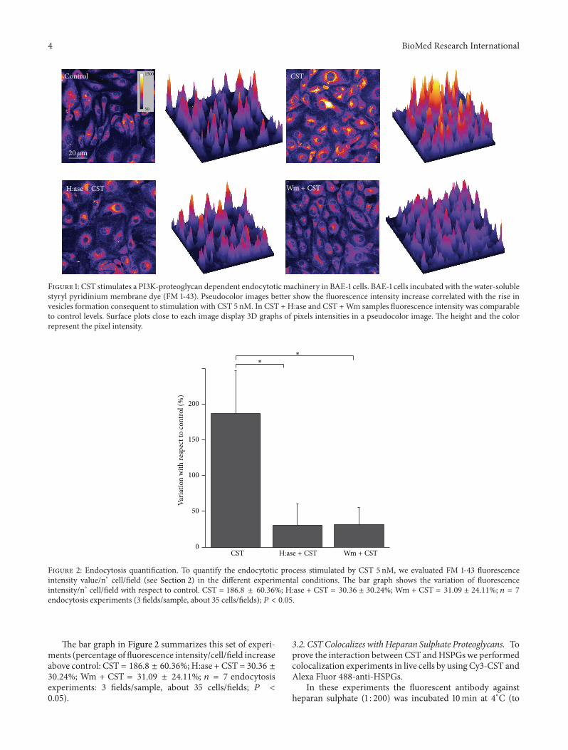
3.1. CST Induces Endocytotic Vesicles Formation That Is Abol-ished by Both Heparinase and Wortmannin. The role of CSTin the endocytotic process was evaluated by incubating BAE-1 cells with the styryl pyridinium membrane probe FM 1-43(5 𝜇g/mL), in order to visualize plasmalemma-derived endo-cytotic vesicles.
To quantify these results we randomly acquired threefields per sample in each experiment and then evaluated thefluorescence intensity value/n∘ cell/field (see Section 2), incontrol condition and after CST stimulation.
We observed that CST (5 nM) induced a significantincrease in the FM 1-43 fluorescence (Figure 1), indicating astimulation of the endocytotic process.
As the molecular properties of CST (little, amphipathic,and cationic peptide) resemble those of the polycationicpeptides (cell penetrating peptides orCPPs), we hypothesizedthat the first contact of this peptide with the cell surface takesplace through proteoglycans, as we previously demonstratedfor VS-1 [22]; internalization mechanisms mediated byHSPGs interaction involving different routes of endocytosishave indeed been described for several CPPs [26].
In order to verify thismechanism,HSPGswere selectivelyremoved from the cell surface by pretreatment of cells withH:ase (2U/mL). After this step, CST strongly reduced itsability to stimulate endocytosis (Figure 1), suggesting thatCST binding toHSPGs is fundamental to start its intracellularcascade.
In a recent work we showed that in BAE-1 cells CSTinduced a PI3K-dependent NO release [18]. To investigatethe role of PI3K pathway also in CST-dependent formationof endocytotic vesicles, we performed FM 1-43 detection inthe presence of the PI3K inhibitor Wm (100 nM). As shownin Figure 1, pretreatment withWm strongly reduced the CST-activated endocytosis.
4 BioMed Research International
Wm + CST
CST1500
50
20𝜇m
Control
H:ase + CST
Figure 1: CST stimulates a PI3K-proteoglycan dependent endocytotic machinery in BAE-1 cells. BAE-1 cells incubated with the water-solublestyryl pyridinium membrane dye (FM 1-43). Pseudocolor images better show the fluorescence intensity increase correlated with the rise invesicles formation consequent to stimulation with CST 5 nM. In CST + H:ase and CST +Wm samples fluorescence intensity was comparableto control levels. Surface plots close to each image display 3D graphs of pixels intensities in a pseudocolor image. The height and the colorrepresent the pixel intensity.
0
50
100
150
200
CST
∗∗
Wm + CSTH:ase + CST
Varia
tion
with
resp
ect t
o co
ntro
l (%
)
Figure 2: Endocytosis quantification. To quantify the endocytotic process stimulated by CST 5 nM, we evaluated FM 1-43 fluorescenceintensity value/n∘ cell/field (see Section 2) in the different experimental conditions. The bar graph shows the variation of fluorescenceintensity/n∘ cell/field with respect to control. CST = 186.8 ± 60.36%; H:ase + CST = 30.36 ± 30.24%; Wm + CST = 31.09 ± 24.11%; 𝑛 = 7endocytosis experiments (3 fields/sample, about 35 cells/fields); 𝑃 < 0.05.
The bar graph in Figure 2 summarizes this set of experi-ments (percentage of fluorescence intensity/cell/field increaseabove control: CST = 186.8 ± 60.36%; H:ase + CST = 30.36 ±30.24%; Wm + CST = 31.09 ± 24.11%; 𝑛 = 7 endocytosisexperiments: 3 fields/sample, about 35 cells/fields; 𝑃 <0.05).
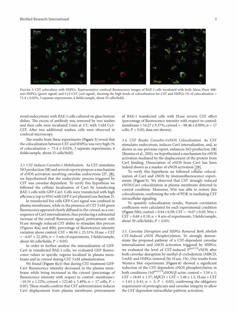
3.2. CST Colocalizes with Heparan Sulphate Proteoglycans. Toprove the interaction between CST andHSPGswe performedcolocalization experiments in live cells by using Cy3-CST andAlexa Fluor 488-anti-HSPGs.
In these experiments the fluorescent antibody againstheparan sulphate (1 : 200) was incubated 10min at 4∘C (to
BioMed Research International 5
Anti-HSPGs Cy3-CST Merge
20𝜇m
Figure 3: CST colocalizes with HSPGs. Representative confocal fluorescence images of BAE-1 cells incubated with both Alexa Fluor 488-anti-HSPGs (green signal) and Cy3-CST (red signal), showing the high levels of colocalization for CST and HSPGs (% of colocalization =73.4 ± 0.02%, 3 separate experiments, 6 fields/sample, about 25 cells/field).
avoid endocytosis) with BAE-1 cells cultured on glass bottomdishes. The excess of antibody was removed by two washesand then cells were incubated 2min at 4∘C with 5 nM Cy3-CST. After two additional washes, cells were observed inconfocal microscopy.
The results from these experiments (Figure 3) reveal thatthe colocalization between CST andHSPGs was very high (%of colocalization = 73.4 ± 0.02%, 3 separate experiments, 6fields/sample, about 25 cells/field).
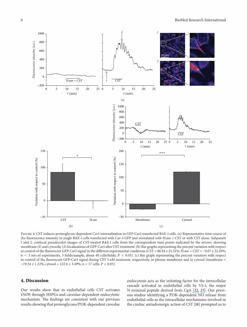
3.3. CST Induces Caveolin-1 Mobilization. As CST stimulatesNOproduction [18] and several reports propose amechanismof eNOS activation involving caveolae endocytosis [27, 28],we hypothesized that the endocytotic process triggered byCST was caveolae-dependent. To verify this hypothesis wefollowed the cellular localization of Cav1 by transfectingBAE-1 cells with GFP-Cav1. Cells were transfected with highefficiency (up to 95%)withGFP-Cav1 plasmid (see Section 2).
In transfected live cells GFP-Cav1 signal was confined inplasmamembranes, while in the presence of CST 5 nM greenfluorescence appeared clearly diffused in the cytosol, as a con-sequence ofCav1 internalization, thus producing a substantialincrease of the overall fluorescent signal; pretreatment withH:ase strongly reduced CST ability to stimulate this process(Figures 4(a) and 4(b); percentage of fluorescence intensityvariation above control: CST = 86.94 ± 25.51%; H:ase + CST= −0.07 ± 22.20%; 𝑛 = 3 sets of experiments, 3 fields/sample,about 40 cells/fields; 𝑃 < 0.05).
In order to further analyze the internalization of GFP-Cav1 in transfected BAE-1 cells, we evaluated GFP fluores-cence values in specific regions localized in plasma mem-brane and in cytosol during CST 5 nM administration.
We found (Figure 4(c)) that during CST treatment GFP-Cav1 fluorescence intensity decreased in the plasma mem-brane while being increased in the cytosol (percentage offluorescence intensity with respect to control: membrane=−19.34 ± 1.22%; cytosol = 122.60 ± 3.49%; 𝑛 = 17 cells; 𝑃 <0.05). These results confirm that CST administration inducesCav1 displacement from plasma membrane; pretreatment
of BAE-1 transfected cells with H:ase reverts CST effect(percentage of fluorescence intensity with respect to control:membrane = 54.27± 9.37%; cytosol = −98.46± 0.09%; 𝑛 = 17cells; 𝑃 < 0.05; data not shown).
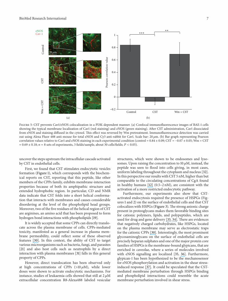
3.4. CST Breaks Caveolin-1/eNOS Colocalization. As CSTstimulates endocytosis, induces Cav1 internalization, and, asshown in our previous report, enhances NO production [18](Bassino et al., 2011), we hypothesized amechanism for eNOSactivation mediated by the displacement of the protein fromCav1 binding. Dissociation of eNOS from Cav1 has beenindeed shown as a marker of eNOS activation [29].
To verify this hypothesis we followed cellular colocal-ization of Cav1 and eNOS by immunofluorescence experi-ments (Figure 5). We observed that CST strongly reducedeNOS/Cav1 colocalization at plasma membrane detected incontrol condition. Moreover, Wm was able to restore thiscolocalization, confirming the role of PI3K in mediating CSTintracellular signaling.
To quantify colocalization results, Pearson correlationcoefficients were calculated for each experimental condition(Figure 5(b); control = 0.84±0.09; CST = −0.07±0.05; Wm +CST = 0.69 ± 0.18; 𝑛 = 8 sets of experiments, 3 fields/sample,about 30 cells/fields; 𝑃 < 0.05).
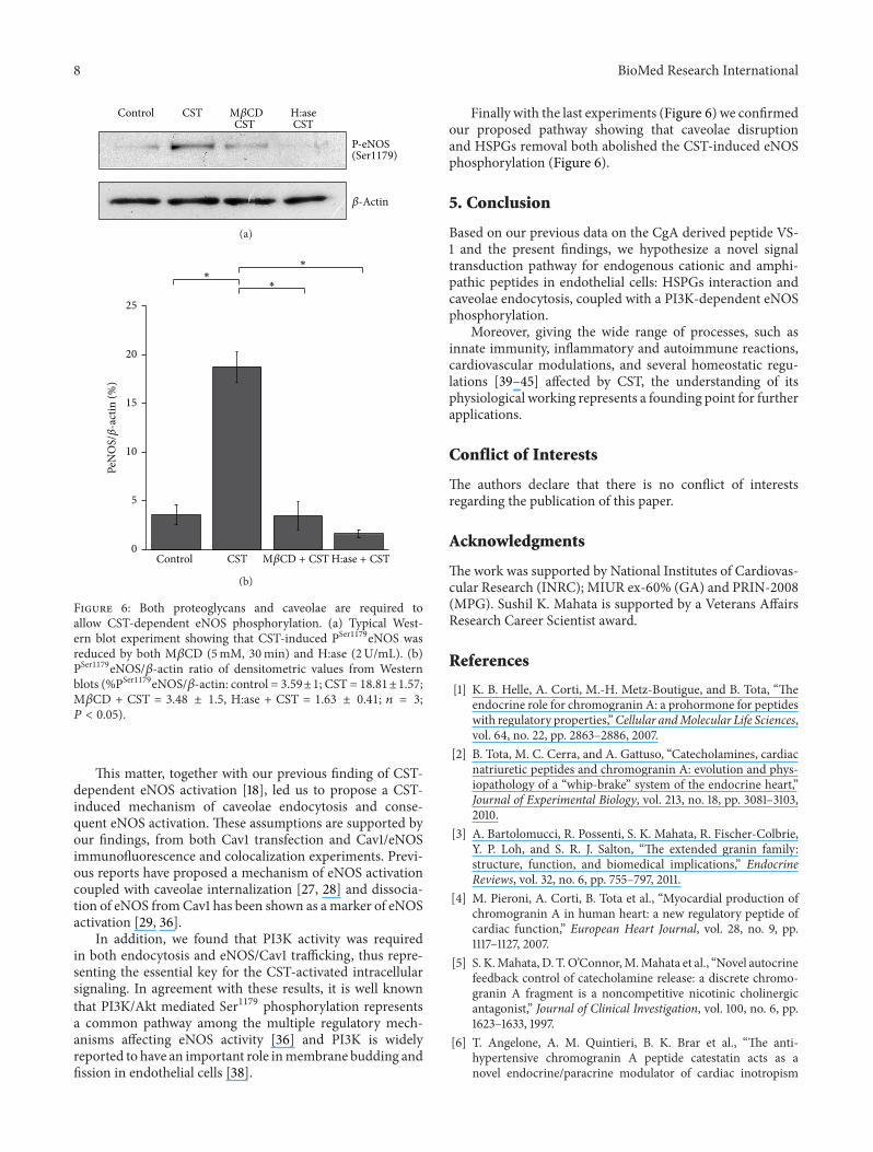
3.5. Caveolae Disruption and HSPGs Removal Both AbolishCST-Induced eNOS Phosphorylation. To strongly demon-strate the proposed pathway of a CST-dependent caveolaeinternalization and eNOS activation triggered by HSPGs,we evaluated the level of CST-induced PSer1179eNOS afterboth caveolae disruption by methyl-𝛽-cyclodextrin (MBCD,5mM) and HSPGs removal (by H:ase, 3 h). Our results fromWestern blot experiments (Figure 6) showed a significantreduction of the CST-dependent eNOS phosphorylation inboth conditions (%PSer1179eNOS/𝛽-actin: control = 3.59 ± 1;CST = 18.81 ± 1.57; M𝛽CD + CST = 3.48 ± 1.5, H:ase + CST= 1.63 ± 0.41; 𝑛 = 3; 𝑃 < 0.05), confirming the obligatoryrequirement of proteoglycans and caveolae integrity to allowthe CST-dependent intracellular pathway activation.
6 BioMed Research International
CST
2
1
1
2
0 5 10 15 20 25 0 5 10 15 20 25
0
200
400
600
800
1000Fl
uore
scen
ce in
tens
ity (a
.u.)
−200
t (min) t (min)
20𝜇m
H:ase + CST
(a)
0
50
100
150
CST−50
∗
H:ase
Varia
tion
with
resp
ect t
o co
ntro
l (%
)
(b)
0200400600800
1000
0 5 10 15 20 25 0 5 10 15 20 25
CST
CST
Fluo
resc
ence
inte
nsity
(a.u
.)
0
50
100
150
200
Membrane Cytosol
−200
−50
t (min) t (min)
∗∗∗
Varia
tion
with
resp
ect t
o co
ntro
l (%
)
(c)
Figure 4: CST induces proteoglycan-dependent Cav1 internalization in GFP-Cav1 transfected BAE-1 cells. (a) Representative time course ofthe fluorescence intensity in single BAE-1 cells transfected with Cav-1-GFP and stimulated with H:ase + CST or with CST alone. Subpanels1 and 2: confocal pseudocolor images of CST-treated BAE-1 cells from the correspondent time points indicated by the arrows, showingmembrane (1) and cytosolic (2) localizations of GFP-Cav1 after CST treatment. (b) Bar graphs representing the percent variation with respectto control of the fluorescent GFP-Cav1 signal in the different experimental conditions (CST = 86.94± 25.51%; H:ase + CST = −0.07±22.20%;𝑛 = 3 sets of experiments, 3 fields/sample, about 40 cells/fields; 𝑃 < 0.05). (c) Bar graph representing the percent variation with respectto control of the fluorescent GFP-Cav1 signal during CST 5 nM treatment, respectively, in plasma membrane and in cytosol (membrane =−19.34 ± 1.22%; cytosol = 122.6 ± 3.49%; 𝑛 = 17 cells; 𝑃 < 0.05).
4. Discussion
Our results show that in endothelial cells CST activateseNOS through HSPGs and caveolae-dependent endocytoticmechanism. The findings are consistent with our previousresults showing that proteoglycans/PI3K-dependent caveolae
endocytosis acts as the initiating factor for the intracellularcascade activated in endothelial cells by VS-1, the majorN-terminal peptide derived from CgA [22, 23]. Our previ-ous studies identifying a PI3K-dependent NO release fromendothelial cells as the intracellular mechanisms involved inthe cardiac antiadrenergic action of CST [18] prompted us to
BioMed Research International 7
eNOSCav-1
Wm
+CS
TC
ontro
lCS
T
20𝜇m
(a)
0
0.2
0.4
0.6
0.8
1
Pear
son
corr
elat
ion
valu
e
Wm + CSTControl CST−0.2
∗∗∗∗∗
(b)
Figure 5: CST prevents Cav1/eNOS colocalization in a PI3K-dependent manner. (a) Confocal immunofluorescence images of BAE-1 cellsshowing the typical membrane localization of Cav1 (red staining) and eNOS (green staining). After CST administration, Cav1 dissociatedfrom eNOS and staining diffused in the cytosol. This effect was reversed by Wm pretreatment. Immunofluorescence detection was carriedout using Alexa Fluor 488 anti-mouse for total eNOS and Cy3 anti-rabbit for Cav1. Scale bar: 20𝜇m. (b) Bar graph representing Pearsoncorrelation values relative to Cav1 and eNOS staining in each experimental condition (control = 0.84 ± 0.09; CST = −0.07 ± 0.05; Wm + CST= 0.69 ± 0.18; 𝑛 = 8 sets of experiments, 3 fields/sample, about 30 cells/fields; 𝑃 < 0.05).
uncover the steps upstream the intracellular cascade activatedby CST in endothelial cells.
First, we found that CST stimulates endocytotic vesiclesformation (Figure 1), which corresponds with the biochem-ical reports on CST, reporting that this peptide, like othermembers of the CPPs family, exhibits membrane-interactionproperties because of both its amphipathic structure andextended hydrophobic region. In particular, CD and NMRdata indicate that CST folds into a short helical conforma-tion that interacts with membranes and causes considerabledisordering at the level of the phospholipid head groups.Moreover, two of the five residues of the helical region of CSTare arginines, an amino acid that has been proposed to formhydrogen bond interactions with phospholipids [19].
It is widely accepted that some CPPs can directly translo-cate across the plasma membrane of cells. CPPs-mediatedtoxicity, manifested as a general increase in plasma mem-brane permeability, could reflect some of these observedfeatures [30]. In this context, the ability of CST to targetvariousmicroorganisms such as bacteria, fungi, and parasites[21] and also host cells such as neutrophils by a directinteraction with plasma membranes [31] falls in this generalproperty of CPPs.
However, direct translocation has been observed onlyat high concentrations of CPPs (>10 𝜇M), while lowerdoses were shown to activate endocytotic mechanisms. Forinstance, studies of leukaemia cells showed that still at 2 𝜇Mextracellular concentration R8-Alexa488 labeled vesicular
structures, which were shown to be endosomes and lyso-somes. Upon raising the concentration to 10𝜇M, instead, thepeptide was seen to flood into cells giving, in most cases,uniform labeling throughout the cytoplasm and nucleus [32].In this perspective our results with CST 5 nM, higher than butcomparable to the circulating concentrations of CgA foundin healthy humans [12] (0.5–2 nM), are consistent with theactivation of a more restricted endocytotic pathway.
Furthermore, our experiments also show that CST-activated endocytosis required the presence of HSPGs (Fig-ures 1 and 2) on the surface of endothelial cells and that CSTcolocalizes with HSPGs (Figure 3).The strong anionic chargepresent in proteoglycans makes them favorable binding sitesfor cationic polymers, lipids, and polypeptides, which areused for drug and gene delivery [33, 34]. There are evidencesthat negatively charged carbohydrates, like HSPGs, locatedon the plasma membrane may serve as electrostatic trapsfor the cationic CPPs [30]. Interestingly, the most prominentglycosaminoglycans on the surface of endothelial cells areprecisely heparan sulphates and one of themajor protein corefamilies of HSPGs is themembrane-bound glypicans, that areenriched in caveolae, where a series of molecules involvedwith eNOS signalling are localized [35, 36]. Furthermore,glypican-1 has been hypothesized to be the mechanosensorfor eNOS phosphorylation and activation in the shear stress-induced response [37]. It could be speculated that the CST-mediated membrane perturbation through HSPGs bindingand phospholipid interactions could resemble the acutemembrane perturbation involved in shear stress.
8 BioMed Research International
Control CSTCST CST
P-eNOS(Ser1179)
M𝛽CD
𝛽-Actin
H:ase
(a)
0
5
10
15
20
25
Control CST
∗
∗∗
M𝛽CD + CST
PeN
OS/𝛽
-act
in (%
)
H:ase + CST
(b)
Figure 6: Both proteoglycans and caveolae are required toallow CST-dependent eNOS phosphorylation. (a) Typical West-ern blot experiment showing that CST-induced PSer1179eNOS wasreduced by both M𝛽CD (5mM, 30min) and H:ase (2U/mL). (b)PSer1179eNOS/𝛽-actin ratio of densitometric values from Westernblots (%PSer1179eNOS/𝛽-actin: control = 3.59±1; CST = 18.81±1.57;M𝛽CD + CST = 3.48 ± 1.5, H:ase + CST = 1.63 ± 0.41; 𝑛 = 3;𝑃 < 0.05).
This matter, together with our previous finding of CST-dependent eNOS activation [18], led us to propose a CST-induced mechanism of caveolae endocytosis and conse-quent eNOS activation. These assumptions are supported byour findings, from both Cav1 transfection and Cav1/eNOSimmunofluorescence and colocalization experiments. Previ-ous reports have proposed a mechanism of eNOS activationcoupled with caveolae internalization [27, 28] and dissocia-tion of eNOS fromCav1 has been shown as amarker of eNOSactivation [29, 36].
In addition, we found that PI3K activity was requiredin both endocytosis and eNOS/Cav1 trafficking, thus repre-senting the essential key for the CST-activated intracellularsignaling. In agreement with these results, it is well knownthat PI3K/Akt mediated Ser1179 phosphorylation representsa common pathway among the multiple regulatory mech-anisms affecting eNOS activity [36] and PI3K is widelyreported to have an important role inmembrane budding andfission in endothelial cells [38].
Finally with the last experiments (Figure 6) we confirmedour proposed pathway showing that caveolae disruptionand HSPGs removal both abolished the CST-induced eNOSphosphorylation (Figure 6).
5. Conclusion
Based on our previous data on the CgA derived peptide VS-1 and the present findings, we hypothesize a novel signaltransduction pathway for endogenous cationic and amphi-pathic peptides in endothelial cells: HSPGs interaction andcaveolae endocytosis, coupled with a PI3K-dependent eNOSphosphorylation.
Moreover, giving the wide range of processes, such asinnate immunity, inflammatory and autoimmune reactions,cardiovascular modulations, and several homeostatic regu-lations [39–45] affected by CST, the understanding of itsphysiological working represents a founding point for furtherapplications.
Conflict of Interests
The authors declare that there is no conflict of interestsregarding the publication of this paper.
Acknowledgments
The work was supported by National Institutes of Cardiovas-cular Research (INRC); MIUR ex-60% (GA) and PRIN-2008(MPG). Sushil K. Mahata is supported by a Veterans AffairsResearch Career Scientist award.
References
[1] K. B. Helle, A. Corti, M.-H. Metz-Boutigue, and B. Tota, “Theendocrine role for chromogranin A: a prohormone for peptideswith regulatory properties,”Cellular andMolecular Life Sciences,vol. 64, no. 22, pp. 2863–2886, 2007.
[2] B. Tota, M. C. Cerra, and A. Gattuso, “Catecholamines, cardiacnatriuretic peptides and chromogranin A: evolution and phys-iopathology of a “whip-brake” system of the endocrine heart,”Journal of Experimental Biology, vol. 213, no. 18, pp. 3081–3103,2010.
[3] A. Bartolomucci, R. Possenti, S. K. Mahata, R. Fischer-Colbrie,Y. P. Loh, and S. R. J. Salton, “The extended granin family:structure, function, and biomedical implications,” EndocrineReviews, vol. 32, no. 6, pp. 755–797, 2011.
[4] M. Pieroni, A. Corti, B. Tota et al., “Myocardial production ofchromogranin A in human heart: a new regulatory peptide ofcardiac function,” European Heart Journal, vol. 28, no. 9, pp.1117–1127, 2007.
[5] S. K.Mahata, D. T.O’Connor,M.Mahata et al., “Novel autocrinefeedback control of catecholamine release: a discrete chromo-granin A fragment is a noncompetitive nicotinic cholinergicantagonist,” Journal of Clinical Investigation, vol. 100, no. 6, pp.1623–1633, 1997.
[6] T. Angelone, A. M. Quintieri, B. K. Brar et al., “The anti-hypertensive chromogranin A peptide catestatin acts as anovel endocrine/paracrine modulator of cardiac inotropism
BioMed Research International 9
and lusitropism,” Endocrinology, vol. 149, no. 10, pp. 4780–4793,2008.
[7] S. K. Mahata, M. Mahata, M. M. Fung, and D. T. O’Connor,“Catestatin: a multifunctional peptide from chromogranin A,”Regulatory Peptides, vol. 162, no. 1–3, pp. 33–43, 2010.
[8] S. K. Mahata, M. Mahata, R. J. Parmer, and D. T. O’Connor,“Desensitization of catecholamine release: the novel cate-cholamine release-inhibitory peptide catestatin (chromograninA344-364) acts at the receptor to prevent nicotinic cholinergictolerance,” The Journal of Biological Chemistry, vol. 274, no. 5,pp. 2920–2928, 1999.
[9] B. P. Kennedy, S. K. Mahata, D. T. O’Connor, and M. G. Ziegler,“Mechanism of cardiovascular actions of the chromogranin Afragment catestatin in vivo,”Peptides, vol. 19, no. 7, pp. 1241–1248,1998.
[10] P. Kruger, S. K. Mahata, and K. B. Helle, “Catestatin (CgA344-364) stimulates rat mast cell release of histamine in a mannercomparable to mastoparan and other cationic charged neu-ropeptides,” Regulatory Peptides, vol. 114, no. 1, pp. 29–35, 2003.
[11] A. H. Gaede and P. M. Pilowsky, “Catestatin, a chromograninA-derived peptide, is sympathoinhibitory and attenuates sym-pathetic barosensitivity and the chemoreflex in rat CVLM,”American Journal of Physiology-Regulatory Integrative andCom-parative Physiology, vol. 302, no. 3, pp. R365–R372, 2012.
[12] D. T. O’Connor,M. T. Kailasam, B. P. Kennedy,M. G. Ziegler, N.Yanaihara, and R. J. Parmer, “Early decline in the catecholaminerelease-inhibitory peptide catestatin in humans at genetic risk ofhypertension,” Journal of Hypertension, vol. 20, no. 7, pp. 1335–1345, 2002.
[13] L. Liu, W. Ding, R. Li et al., “Plasma levels and diagnostic valueof catestatin in patients with heart failure,” Peptides, vol. 46, pp.20–25, 2013.
[14] L. Liu, W. Ding, F. Zhao, L. Shi, Y. Pang, and C. Tang, “Plasmalevels and potential roles of catestatin in patients with coronaryheart disease,” Scandinavian Cardiovascular Journal, vol. 47, no.4, pp. 217–224, 2013.
[15] N. R. Mahapatra, D. T. O’Connor, S. M. Vaingankar et al.,“Hypertension from targeted ablation of chromogranin Acan be rescued by the human ortholog,” Journal of ClinicalInvestigation, vol. 115, no. 7, pp. 1942–1952, 2005.
[16] J. R. Gayen, Y. Gu, D. T. O’Connor, and S. K. Mahata, “Globaldisturbances in autonomic function yield cardiovascular insta-bility and hypertension in the chromogranin A null mouse,”Endocrinology, vol. 150, no. 11, pp. 5027–5035, 2009.
[17] N. B. Dev, J. R. Gayen, D. T. O’Connor, and S. K. Mahata,“Chromogranin A and the autonomic system: decompositionof heart rate variability and rescue by its catestatin fragment,”Endocrinology, vol. 151, no. 6, pp. 2760–2768, 2010.
[18] E. Bassino, S. Fornero, M. P. Gallo et al., “A novel catestatin-induced antiadrenergic mechanism triggered by the endothe-lial PI3KeNOS pathway in the myocardium,” CardiovascularResearch, vol. 91, no. 4, pp. 617–624, 2011.
[19] M. Sugawara, J. M. Resende, C. M. Moraes et al., “Membranestructure and interactions of human catestatin by multidimen-sional solution and solid-state NMR spectroscopy,” The FASEBJournal, vol. 24, no. 6, pp. 1737–1746, 2010.
[20] K. A. Radek, B. Lopez-Garcia, M. Hupe et al., “The neu-roendocrine peptide catestatin is a cutaneous antimicrobialand induced in the skin after injury,” Journal of InvestigativeDermatology, vol. 128, no. 6, pp. 1525–1534, 2008.
[21] J. Briolat, S. D. Wu, S. K. Mahata et al., “New antimicrobialactivity for the catecholamine release-inhibitory peptide from
chromogranin A,” Cellular and Molecular Life Sciences, vol. 62,no. 3, pp. 377–385, 2005.
[22] R. Ramella, O. Boero, G. Alloatti, T. Angelone, R. Levi, andM. P. Gallo, “Vasostatin 1 activates eNOS in endothelial cellsthrough a proteoglycan-dependent mechanism,” Journal ofCellular Biochemistry, vol. 110, no. 1, pp. 70–79, 2010.
[23] S. Fornero, E. Bassino, M. P. Gallo, R. Ramella, R. Levi, and G.Alloatti, “Endothelium dependent cardiovascular effects of thechromogranin a-derived peptides vasostatin-1 and catestatin,”Current Medicinal Chemistry, vol. 19, no. 24, pp. 4059–4067,2012.
[24] S. K. Mahata, M. Mahata, A. R. Wakade, and D. T. O’Connor,“Primary structure and function of the catecholamine releaseinhibitory peptide catestatin (chromogranin A344-364): iden-tification of amino acid residues crucial for activity,”MolecularEndocrinology, vol. 14, no. 10, pp. 1525–1535, 2000.
[25] W. D. Niles and A. B.Malik, “Endocytosis and exocytosis eventsregulate vesicle traffic in endothelial cells,” Journal of MembraneBiology, vol. 167, no. 1, pp. 85–101, 1999.
[26] M. C. Morris, S. Deshayes, F. Heitz, and G. Divita, “Cell-penetrating peptides: frommolecular mechanisms to therapeu-tics,” Biology of the Cell, vol. 100, no. 4, pp. 201–217, 2008.
[27] N. A. Maniatis, V. Brovkovych, S. E. Allen et al., “Novel mecha-nism of endothelial nitric oxide synthase activation mediatedby caveolae internalization in endothelial cells,” CirculationResearch, vol. 99, no. 8, pp. 870–877, 2006.
[28] F. A. Sanchez, D. D. Kim, R. G. Duran, C. J. Meininger, and W.N. Duran, “Internalization of eNOS via caveolae regulates PAF-induced inflammatory hyperpermeability to macromolecules,”The American Journal of Physiology: Heart and CirculatoryPhysiology, vol. 295, no. 4, pp. H1642–H1648, 2008.
[29] R. D. Minshall, W. C. Sessa, R. V. Stan, R. G. W. Anderson,and A. B. Malik, “Caveolin regulation of endothelial function,”American Journal of Physiology-Lung Cellular and MolecularPhysiology, vol. 285, no. 6, pp. L1179–L1183, 2003.
[30] A. T. Jones and E. J. Sayers, “Cell entry of cell penetratingpeptides: tales of tails wagging dogs,” Journal of ControlledRelease, vol. 161, no. 2, pp. 582–591, 2012.
[31] D. Zhang, P. Shooshtarizadeh, B. Laventie et al., “Two chromo-granin a-derived peptides induce calcium entry in human neu-trophils by calmodulin-regulated calcium independent phos-pholipase A2,” PLoS ONE, vol. 4, no. 2, Article ID e4501, 2009.
[32] M. M. Fretz, N. A. Penning, S. Al-Taei et al., “Temperature-,concentration- and cholesterol-dependent translocation of L-and D-octa-arginine across the plasma and nuclear membraneof CD34+ leukaemia cells,” Biochemical Journal, vol. 403, no. 2,pp. 335–342, 2007.
[33] D. L. Rabenstein, “Heparin and heparan sulfate: structure andfunction,” Natural Product Reports, vol. 19, no. 3, pp. 312–331,2002.
[34] M. Belting, “Heparan sulfate proteoglycan as a plasma mem-brane carrier,” Trends in Biochemical Sciences, vol. 28, no. 3, pp.145–151, 2003.
[35] J.M. Tarbell, “Shear stress and the endothelial transport barrier,”Cardiovascular Research, vol. 87, no. 2, pp. 320–330, 2010.
[36] I. Fleming, “Molecular mechanisms underlying the activationof eNOS,” Pflugers Archiv European Journal of Physiology, vol.459, no. 6, pp. 793–806, 2010.
[37] S. V. Lopez-Quintero, R. Amaya, M. Pahakis, and J. M. Tarbell,“The endothelial glycocalyx mediates shear-induced changes in
10 BioMed Research International
hydraulic conductivity,” The American Journal of Physiology—Heart and Circulatory Physiology, vol. 296, no. 5, pp. H1451–H1456, 2009.
[38] P. Mellor, L. A. Furber, J. N. K. Nyarko, and D. H. Anderson,“Multiple roles for the p85𝛼 isoform in the regulation and func-tion of PI3K signalling and receptor trafficking,” BiochemicalJournal, vol. 441, no. 1, pp. 23–37, 2012.
[39] R. Aslam, M. Atindehou, T. Lavaux, Y. Haıkel, F. Schneider, andM.-H. Metz-Boutigue, “Chromogranin a-derived peptides areinvolved in innate immunity,”CurrentMedicinal Chemistry, vol.19, no. 24, pp. 4115–4123, 2012.
[40] G. Aung, F. Niyonsaba, H. Ushio et al., “Catestatin, a neu-roendocrine antimicrobial peptide, induces human mast cellmigration, degranulation and production of cytokines andchemokines,” Immunology, vol. 132, no. 4, pp. 527–539, 2011.
[41] K. B. Helle, “Regulatory peptides from chromogranin A andsecretogranin II: putative modulators of cells and tissuesinvolved in inflammatory conditions,” Regulatory Peptides, vol.165, no. 1, pp. 45–51, 2010.
[42] K. B. Helle, “The chromogranin A-derived peptides vasostatin-I and catestatin as regulatory peptides for cardiovascular func-tions,” Cardiovascular Research, vol. 85, no. 1, pp. 9–16, 2010.
[43] K. B. Helle, “Chromogranins A and B and secretogranin II asprohormones for regulatory peptides from the diffuse neuroen-docrine system,” Results and Problems in Cell Differentiation,vol. 50, pp. 21–44, 2010.
[44] N. R.Mahapatra, “Catestatin is a novel endogenous peptide thatregulates cardiac function and blood pressure,” CardiovascularResearch, vol. 80, no. 3, pp. 330–338, 2008.
[45] G. K. Bandyopadhyay, C. U. Vu, S. Gentile et al., “Catestatin(Chromogranin A
352−372) and novel effects on mobilization of
fat from adipose tissue through regulation of adrenergic andleptin signaling,” Journal of Biological Chemistry, vol. 287, no.27, pp. 23141–23151, 2012.

Submit your manuscripts athttp://www.hindawi.com
PainResearch and TreatmentHindawi Publishing Corporationhttp://www.hindawi.com Volume 2014
The Scientific World JournalHindawi Publishing Corporation http://www.hindawi.com Volume 2014
Hindawi Publishing Corporationhttp://www.hindawi.com
Volume 2014
ToxinsJournal of
VaccinesJournal of
Hindawi Publishing Corporation http://www.hindawi.com Volume 2014
Hindawi Publishing Corporationhttp://www.hindawi.com Volume 2014
AntibioticsInternational Journal of
ToxicologyJournal of
Hindawi Publishing Corporationhttp://www.hindawi.com Volume 2014
StrokeResearch and TreatmentHindawi Publishing Corporationhttp://www.hindawi.com Volume 2014
Drug DeliveryJournal of
Hindawi Publishing Corporationhttp://www.hindawi.com Volume 2014
Hindawi Publishing Corporationhttp://www.hindawi.com Volume 2014
Advances in Pharmacological Sciences
Tropical MedicineJournal of
Hindawi Publishing Corporationhttp://www.hindawi.com Volume 2014
Medicinal ChemistryInternational Journal of
Hindawi Publishing Corporationhttp://www.hindawi.com Volume 2014
AddictionJournal of
Hindawi Publishing Corporationhttp://www.hindawi.com Volume 2014
Hindawi Publishing Corporationhttp://www.hindawi.com Volume 2014
BioMed Research International
Emergency Medicine InternationalHindawi Publishing Corporationhttp://www.hindawi.com Volume 2014
Hindawi Publishing Corporationhttp://www.hindawi.com Volume 2014
Autoimmune Diseases
Hindawi Publishing Corporationhttp://www.hindawi.com Volume 2014
Anesthesiology Research and Practice
ScientificaHindawi Publishing Corporationhttp://www.hindawi.com Volume 2014
Journal of
Hindawi Publishing Corporationhttp://www.hindawi.com Volume 2014
Pharmaceutics
Hindawi Publishing Corporationhttp://www.hindawi.com Volume 2014
MEDIATORSINFLAMMATION
of