Obligatory Additive Particle on Negation Ishani Guha MIT 1 ...
An arachidonic acid-preferring acyl-CoA synthetase is ahormone-dependent and obligatory protein in the signaltransduction pathway of steroidogenic hormones
Fabiana Cornejo Maciel, Paula Maloberti, Isabel Neuman, Florencia Cano, Rocío Castilla,Fernanda Castillo, Cristina Paz and Ernesto J PodestáDepartment of Biochemistry, School of Medicine, University of Buenos Aires, Paraguay 2155, 5th (C1121ABG) Buenos Aires, Argentina
(Requests for offprints should be addressed to F Cornejo Maciel; Email: [email protected])
Abstract
We have described that, in adrenal and Leydig cells, the hormonal regulation of free arachidonic acid (AA)concentration is mediated by the concerted action of two enzymes: an acyl-CoA thioesterase (MTE-I or ARTISt) and anacyl-CoA synthetase (ACS4). In this study we analyzed the potential regulation of these proteins by hormonal actionin steroidogenic cells. We demonstrated that ACS4 is rapidly induced by adrenocorticotropin (ACTH) and cAMP inY1 adrenocortical cells. The hormone and its second messenger increased ACS4 protein levels in a time andconcentration dependent way. Maximal concentration of ACTH (10 mIU/ml) produced a significant effect after 15 min oftreatment and exerted the highest increase (3-fold) after 30 min. Moreover, 35S-methionine incorporation showed thatthe increase in ACS4 protein levels is due to an increase in the de novo synthesis of the protein. On the contrary MTE-Iprotein levels in Y1 and MA-10 cells did not change after steroidogenic stimuli. In contrast with the effect observed onprotein levels, stimulation of both cell lines did not change ACS4 RNA levels during the first hour of treatment,indicating that the effect of both stimuli is exerted at the level of ACS4 protein synthesis.
StAR protein induction has a key role on the activation of steroidogenesis since this protein increases the rate of thelimiting step of the whole process. In agreement with the fact that the inhibition of ACS4 activity by triacsin C blockscAMP-stimulated progesterone production by MA-10 Leydig cells, here we demonstrated that ACS4 inhibition alsoreduces StAR protein levels. Moreover, exogenous AA was able to overcome the effect of triacsin C on both events,StAR induction and steroidogenesis. These results were confirmed by experiments using ACS4-targeted siRNA whichresult in a reduction in both ACS4 and StAR protein levels. The concomitant decrease in steroid production wasovercome by the addition of AA to the knocked-out cells. In summary, this study suggests that in adrenal and Leydigcells the hormonal action prompts the synthesis of a labile protein, ACS4, which activity is involved in the regulation ofAA release and is essential for steroidogenesis and StAR protein induction.
Journal of Molecular Endocrinology (2005) 34, 655–666
Introduction
In the different steroidogenic tissues the appropriatetrophic hormone activates the mitochondrial conversionof cholesterol to pregnenolone. This reaction is catalyzedby the side-chain splitting cytochrome P450 enzyme(P450 scc) located in the matrix side of the innermitochondrial membrane. The access of free cholesterolto the active site of P450 scc, and not the enzymaticactivity itself, is the true rate-limiting step in immediatesteroidogenesis (Crivello & Jefcoate 1980, Privalle et al.1983). Numerous reports indicate that this event iscontrolled by the steroidogenic acute regulatory protein(StAR protein) (Clark et al. 1994, Stocco & Clark 1996,Wang et al. 2000). In addition, it is well known thatsteroid biosynthesis requires de novo protein synthesis of alabile factor that has a short half-life, and the process isinhibited by cycloheximide (CHX) (Garren et al. 1965,
Cooke et al. 1975). In this regard, the furthercharacterization of StAR has revealed that this proteinmay be considered as the ‘labile’ factor that mediates thesteroidogenic response to hormone action in adrenal andLeydig cells, as well as in other steroidogenic systemsstudied to date. Thus, StAR is rapidly synthesized inresponse to the respective trophic hormones, it iscycloheximide-sensitive, its active form has a very shorthalf-life and it rapidly increases the transport ofcholesterol to the site of P450 scc action (Stocco & Clark1996, Reinhart et al. 1999).
In adrenal and Leydig cells, the increase of 3�,5�-cyclic-adenosine monophosphate (cAMP) levels andcAMP-dependent protein kinase (PKA) phosphorylationevents are accepted as intermediate steps in theadrenocorticotropin (ACTH) and luteinizing hormone(LH) action (Dufau et al. 1977, Sala et al. 1979). It is alsoaccepted that cAMP and PKA regulate arachidonic acid
655
Journal of Molecular Endocrinology (2005) 34, 655–6660952–5041/05/034–655 © 2005 Society for Endocrinology Printed in Great Britain
DOI: 10.1677/jme.1.01691Online version via http://www.endocrinology-journals.org
(AA) release. Following LH stimulation, AA is releasedwithin one minute in rat testicular Leydig cells (Dix et al.1984, Didolkar & Sundaram 1987, Cooke et al. 1991).Also, AA release occurs in a dose and time dependentmanner in human chorionic gonadotropin (hCG)-stimulated Leydig (Moraga et al. 1997) and ACTH-stimulated adrenal cells (Dada et al. 1996). In addition,previous studies have reported that inhibition of AArelease abrogates the effect of LH- (Mele et al. 1997) andACTH-stimulated steroid production (Solano et al.1988). Recently, we have presented results suggestingthat in steroidogenic tissues the release of AA involvesthe action of two enzymes, an acyl-CoA synthetase andan acyl-CoA thioesterase (Finkielstein et al. 1998,Maloberti et al. 2002). The acyl-CoA thioesterase,MTE-I (Svensson et al. 1998) or ARTISt (Finkielsteinet al. 1998), is a mitochondrial phosphoprotein includedin a family of acyl-CoA thioesterases that displayspreferential activity towards very long chain acyl-CoA(Svensson et al. 1998). The protein was first identified byits capacity to increase mitochondrial steroidogenesis ina cell-free assay (Paz et al. 1994). It is present in adrenaland testicular Leydig cells, and ovary, brain andplacenta among other tissues (Finkielstein et al. 1996),and its activity would be regulated by ACTH (Malobertiet al. 2002).
The second enzyme involved in AA release insteroidogenic tissues, the acyl-CoA synthetase, is anenzyme designed ACS4 that belongs to a five-memberfamily. ACS4 shares 68% of its amino acid sequencewith ACS3, another member of this family, although thissequence is poorly related to the other family members(Kang et al. 1997). The purified enzyme utilizesarachidonate as substrate most preferentially amongother C8-C22 saturated fatty acids and C4-C22unsaturated fatty acids. The striking feature of ACS4 isits abundance in steroidogenic tissues, especially adrenalgland and ovary. ACS4 immunoreactivity was detectedin the zona fasciculata (ZF) and reticularis of the adrenalcortex, in the corpus luteum and stromal luteinized cellsof the ovary and in the Leydig cells of the testis (Kanget al. 1997).
Given that AA release is linked to hormone-dependent StAR induction and steroidogenesis (Wanget al. 2000), the present study was undertaken toinvestigate whether AA release in steroidogenic tissuesby hormone action involves the regulation of the levelsof the acyl-CoA synthetase and/or thioesterase.
In the present study the results demonstrate thathormone treatment of adrenal and Leydig cells does notmodify MTE-I protein levels while it induces ACS4protein. This enzyme appears rapidly in response to therespective trophic hormone and seems to have a shorthalf-life. The data are consistent with the presence of anew factor, ACS4, which is essential for StAR inductionand steroidogenesis.
Materials and methods
Materials
ACTH was a kind gift of Elea Laboratories (BuenosAires, Argentina). Dexamethasone (9-fluoro-11�, 17,21-trihydroxy-16�-methyl-pregna-1,4-diene-3,20-dione)was a kind gift from Ciba Geigy (Basel, Switzerland).Media, sera, antibiotics, Trypsin-EDTA, TriZol reagentand Lipofectamine 2000 were from Gibco-Life Tech-nologies (Carlsbad, CA, USA); plastic flasks and disheswere provided by Corning-Costar (Corning, NY, USA).Acrylamide, bis-acrylamide, cycloheximide (CHX), 8Br-3�, 5�-cyclic-adenosine-monophosphate (8Br-cAMP),arachidonic acid (AA), trypsin from bovine pancreas,soybean trypsin inhibitor, agarose, formaldehyde, bovineseroalbumin (BSA) and fatty acid-free BSA wereobtained from Sigma-Aldrich Fine Chemicals (St Louis,MO, USA). Triacsin C was bought from ICN (Aurora,OH, USA). Polyclonal antibodies against MTE-I werepreviously obtained at our laboratory (Maloberti et al.2002). Anti-ACS4 antibodies were obtained fromimmunizing rabbits with recombinant ACS4 protein.The protein was obtained in E.coli (BL21) transfectedwith a plasmid (pGEX-4T3) containing ACS4 sequence,kindly provided by Dr T Yamamoto (University ofTohoku, Sendai, Japan). In Y1 and MA-10 cells,Western blot analysis utilizing this antiserum results inonly one band of the expected molecular weight(74 kDa), which is not detected by pre-immune serum orwhen the antiserum is pre-adsorbed with the immuno-gen. Moreover, its specificity is demonstrated by the factthat in adult cardiac tissue, where ACS4 is notexpressed, the antibody does not reveal any band,although this tissue expresses ACS3, another synthetaseisoform. Anti-StAR antibody was generously providedby Dr Douglas Stocco (Texas Tech University, Lubbock,Texas, USA). Anti-�-tubulin monoclonal antibody wasbought from Upstate (Lake Placid, NY, USA).Electrophoresis supplies, polyvinylidendifluoride mem-brane and secondary antibody (horseradish peroxidase-conjugated goat antibody) were bought to Bio-RadLaboratories Inc. (Hercules, CA, USA). ECL kit andHybond N+ membrane were provided by AmershamPharmacia Biotech (Buckinghamshire, UK). All otherchemicals were commercial products of the highestgrade available.
Animals
The use of animals complies with the guidelinesapproved by the Animal Welfare and Health Committeeof the University of Buenos Aires. Male Wistar rats(90-day-old) were used throughout. Adrenal gland or ZFcells were obtained from animals supplied withdexamethasone (10 µg/ml, ad libitum) in the drinkingwater for 16 h before they were killed, as previously
F CORNEJO MACIEL and others · ACS4: a new labile protein involved in steroid synthesis656
www.endocrinology-journals.orgJournal of Molecular Endocrinology (2005) 34, 655–666
described (Neher et al. 1982). Animals were killed bydecapitation and adrenal glands were excised and kepton ice.
Adrenal in vivo stimulation
Following dexamethasone treatment, animals wereinjected s.c. with 18 IU ACTH per kg body weight andkilled at the indicated times. Adrenal glands wereremoved, zona glomerulosa (ZG) and ZF were obtainedas described (Solano et al. 1988) and homogenized in270 mM mannitol, 10 mM Tris pH 7·4 containing acocktail of inhibitors (100 mM NaF, 1 µg/ml pepstatin,200 µM leupeptin, 2 µg/ml aprotinin, 2 mM phenyl-methyl-sulfonyl fluoride (PMSF)). Cellular proteins ofthe homogenates were analyzed by Western blot.Steroid production was measured in the peripheralblood in order to determine the efficiency of ACTHtreatment (Neher et al. 1982).
Cell preparation and stimulation
Adrenal ZF cells were obtained by trypsin digestion,following published procedures (Solano et al. 1988). ZFcells were suspended in Krebs-Ringer-Bicarbonatebuffer, pH 7·4, containing 10 mM glucose and 0·5%BSA at a final concentration of 105 cells/ml and theywere maintained throughout under carbogen (95% O2:5% CO2) atmosphere. Adrenal ZF cells were incubatedwith 1 mM 8Br-cAMP for 15, 30 and 60 min at 37 �Cunder carbogen atmosphere with gentle shaking. Aftercooling of the tubes in an ice/water bath, cells werepelleted by centrifugation at 1000 g for 20 min.Incubation media were stored at –20 �C until cortico-sterone determination. Cellular pellets were suspendedin 270 mM mannitol, 10 mM Tris pH 7·4, plus thecocktail of inhibitors described above and lysed insample buffer. The samples were kept to analyze cellularproteins by Western blot.
Cell cultures
Murine Y1 adrenocortical tumor cells, generouslyprovided by Dr Bernard Schimmer (University ofToronto, Toronto, Canada) were maintained inHam-F10 medium, supplemented with 12·5% heat-inactivated horse serum and 2·5% heat-inactivated fetalbovine serum, 1·2 g/l NaHCO3, 200 IU/ml penicillinand 200 mg/ml streptomycin sulfate (Schimmer 1981).
The MA-10 Leydig cell line is a clonal strain of mouseLeydig tumor cells, generously provided by Dr MarioAscoli (University of Iowa, College of Medicine, IowaCity, IA, USA). These cells were maintained inWaymouth MB752/1, supplemented with 15% horseserum, 1·2 g/l NaHCO3, 20 mM Hepes, 50 µg/mlgentamicin (Ascoli 1981).
Both cell lines were kept at 36 �C in a 5% CO2humidified atmosphere. After replacing the media byfresh-serum free medium, Y1 cultures were incubatedwith or without ACTH or 8Br-cAMP and MA-10 cellswith or without 8Br-cAMP, as stated in the legend of thecorresponding figures. When indicated, AA was addedtogether with 8Br-cAMP. In some cases, previous to thestimulations, the cells were preincubated with 10 µg/mlCHX (30 min) or 1 µM triacsin C (240 min). Followingtreatments, media was kept to determine steroidproduction by radioimmunoassay. Y1 or MA-10 cellswere washed with PBS and scraped into a buffercontaining 35 mM Tris pH 7·4, 5 mM EDTA, 5 mMMgCl2, 200 mM sucrose, 0·5% Triton-X 100 and thecocktail of inhibitors described above. The suspensionwas incubated on ice for 10 min, vortexed for 1 min andcentrifuged at 11 000 g at 4 �C for 10 min. Thesupernatant (total lysate) was kept to analyze cellularproteins by Western blot.
35S-Methionine incorporation and ACS4immunoprecipitation
Y1 cell cultures were incubated with the inclusion of400 µCi/ml 35S-methionine (specific activity 1175 Ci/mmol; New England Nuclear, Perkin-Elmer, Boston,MA, USA) for 45 min. Following this pre-incubationperiod, the cells were stimulated with 10 mIU/mlACTH. After 30 min of stimulation, the cells werewashed and then homogenized in a buffer consisting of10 mM Tris–HCl pH 7·4, 5 mM EDTA, 5 mM EGTA.Proteins (400 µg) of the homogenate were incubatedovernight at 4 �C with 10 µl of polyclonal anti-ACS4and 30 µl of protein A/G plus-agarose in a final volumeof 0·4 ml in the following buffer 10 mM Tris, pH 7·4,130 mM NaCl, 5 mM EDTA, 5 mM EGTA, 1% TritonX-100, 0·5% Nonidet P40, 0·5% sodium deoxycolate,0·1% SDS, 10 mM sodium pyrophosphate, 10 mM NaF(immunoprecipitation buffer). After the incubation, thesamples were centrifuged at 12 000 g for 4 min. Thepellets were washed four times with 0·5 ml of immuno-precipitation buffer prior to boiling in SDS loadingbuffer. Samples were then analyzed by SDS-PAGE(10% acrylamide), and dried gels exposed to autoradiog-raphy films to detect 35S-methionine incorporated intonewly synthesized ACS4.
Western blot analysis
Equal amounts of protein were separated by SDS-PAGE(10% acrylamide for ACS4 and MTE-I analysis or 12%for StAR analysis), as described by Laemmli (1970) andtransferred to polyvinylidene difluoride membranesaccording to the procedure described by Towbin et al.(1979). ACS4, MTE-I or StAR proteins were detectedusing the specific antibodies and immunoreactive bands
ACS4: a new labile protein involved in steroid synthesis · F CORNEJO MACIEL and others 657
www.endocrinology-journals.org Journal of Molecular Endocrinology (2005) 34, 655–666
were visualized by enhanced chemiluminescence. In allWestern blots, detection of �-tubulin was used as loadingcontrol.
Northern blot analysis
For Northern blot analysis, total RNA was isolated fromY1 and MA-10 cells by the guanidinium isothiocyanatemethod using TriZol reagent, according to themanufacturer protocol. Total RNA (24 µg) was separ-ated by electrophoresis on 1·5% agarose gels and blottedonto Hybond N+ membranes. ACS4 mRNA wasdetected using a specific 32P-labeled cDNA probe (Kang
et al. 1997). The levels of 28S RNA were also detectedand used as loading control.
siRNA transfection
siRNAs targeting ACS4 and MTE-I coding sequenceswere custom-designed by Dharmacon (Lafayette,CO, USA). One day before transfection, Y1 cells(5�105 cells/well) were grown up to 80% confluenceonto 24-well plates. Transfection was performed usingsiRNA (350 nM) with Opti-MEM medium and 2 µlLipofectamine 2000 reagent according to the instruc-tions of the manufacturer. Cells were placed into normal
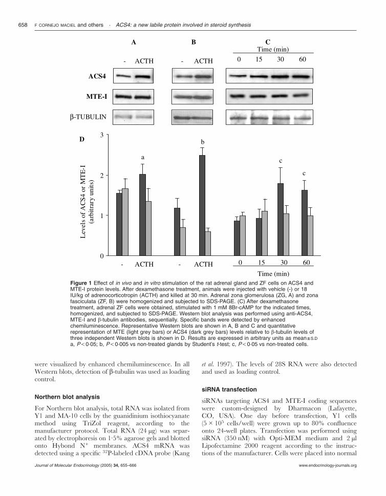
Figure 1 Effect of in vivo and in vitro stimulation of the rat adrenal gland and ZF cells on ACS4 andMTE-I protein levels. After dexamethasone treatment, animals were injected with vehicle (-) or 18IU/kg of adrenocorticotropin (ACTH) and killed at 30 min. Adrenal zona glomerulosa (ZG, A) and zonafasciculata (ZF, B) were homogenized and subjected to SDS-PAGE. (C) After dexamethasonetreatment, adrenal ZF cells were obtained, stimulated with 1 mM 8Br-cAMP for the indicated times,homogenized, and subjected to SDS-PAGE. Western blot analysis was performed using anti-ACS4,MTE-I and �-tubulin antibodies, sequentially. Specific bands were detected by enhancedchemiluminescence. Representative Western blots are shown in A, B and C and quantitativerepresentation of MTE (light grey bars) or ACS4 (dark grey bars) levels relative to �-tubulin levels ofthree independent Western blots is shown in D. Results are expressed in arbitrary units as mean±S.D
a, P,0·05; b, P,0·005 vs non-treated glands by Student’s t-test; c, P,0·05 vs non-treated cells.
F CORNEJO MACIEL and others · ACS4: a new labile protein involved in steroid synthesis658
www.endocrinology-journals.orgJournal of Molecular Endocrinology (2005) 34, 655–666
culture medium for 6 h after transfection and furthergrown for 48 h. Y1 cells were stimulated with 5 mIU/mlACTH in culture medium containing 0·1% fattyacid-free BSA, in the presence or absence of 0·3 mMAA. Following treatments, media was kept to determinesteroid production by radioimmunoassay.
Protein determination
Protein concentrations were determined by Bradfordassay, using BSA as standard (Bradford 1976).
Statistics
Results are shown as the mean�S.D Unless otherwiseindicated, statistical significance was evaluated usingANOVA followed by Tukey test. P<0·05 was consideredsignificant.
Results
Given that ACTH regulates intracellular concentrationof AA in adrenal cells and that ACS4 and MTE-I are
enzymes involved in AA release, the first experimentswere performed in order to determine whether ACTHhas any effect on ACS4 and MTE-I protein levels. Withthis purpose, we analyzed the levels of both proteins inadrenal glands from animals stimulated with a singledose of ACTH for 30 min. As shown in Fig. 1, Westernblot analysis revealed an increase in ACS4 protein levelsin both ZG (Fig. 1, panels A and D) and ZF (Fig. 1,panels B and D). In addition, when isolated rat adrenalZF cells were stimulated in vitro with 8Br-cAMP(15–60 min), we also observed an increase in the protein(Fig. 1, panels C and D). In contrast, neither in vivoACTH treatment nor in vitro 8Br-cAMP incubationmodifies the levels of MTE-I (Fig. 1).
In order to further characterize the effect of hormoneaction on ACS4 levels, we used two well recognizedsteroidogenic cell lines, adrenocortical Y1 and LeydigMA10 cells. The levels of ACS4 and MTE-I wereanalyzed in Y1 cells stimulated with different concen-trations of ACTH for different periods of time. None ofthe concentrations or times tested (1–10 mIU/ml,15–45 min) proved to modify the intracellular content ofMTE-I (data not shown). However, ACTH produces an
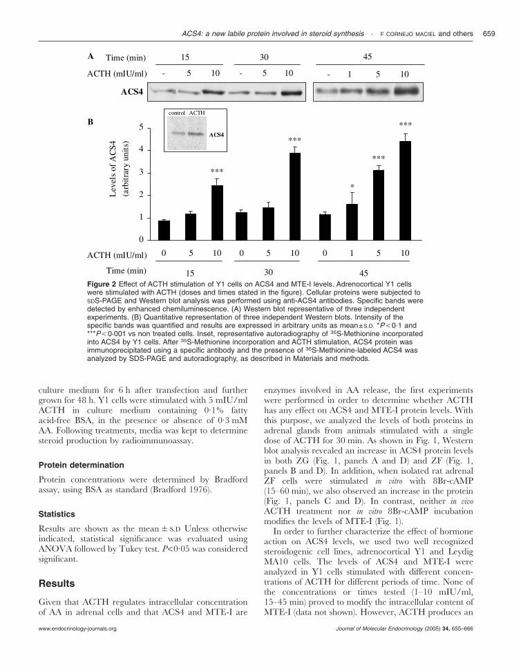
Figure 2 Effect of ACTH stimulation of Y1 cells on ACS4 and MTE-I levels. Adrenocortical Y1 cellswere stimulated with ACTH (doses and times stated in the figure). Cellular proteins were subjected toSDS-PAGE and Western blot analysis was performed using anti-ACS4 antibodies. Specific bands weredetected by enhanced chemiluminescence. (A) Western blot representative of three independentexperiments. (B) Quantitative representation of three independent Western blots. Intensity of thespecific bands was quantified and results are expressed in arbitrary units as mean±S.D. *P,0·1 and***P,0·001 vs non treated cells. Inset, representative autoradiography of 35S-Methionine incorporatedinto ACS4 by Y1 cells. After 35S-Methionine incorporation and ACTH stimulation, ACS4 protein wasimmunoprecipitated using a specific antibody and the presence of 35S-Methionine-labeled ACS4 wasanalyzed by SDS-PAGE and autoradiography, as described in Materials and methods.
ACS4: a new labile protein involved in steroid synthesis · F CORNEJO MACIEL and others 659
www.endocrinology-journals.org Journal of Molecular Endocrinology (2005) 34, 655–666
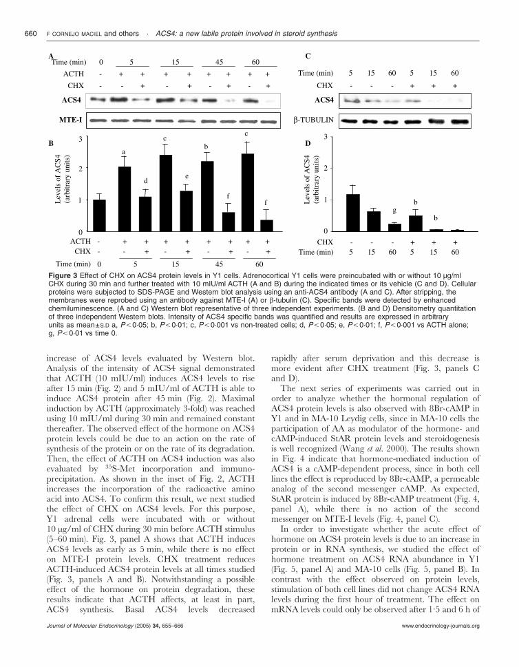
increase of ACS4 levels evaluated by Western blot.Analysis of the intensity of ACS4 signal demonstratedthat ACTH (10 mIU/ml) induces ACS4 levels to riseafter 15 min (Fig. 2) and 5 mIU/ml of ACTH is able toinduce ACS4 protein after 45 min (Fig. 2). Maximalinduction by ACTH (approximately 3-fold) was reachedusing 10 mIU/ml during 30 min and remained constantthereafter. The observed effect of the hormone on ACS4protein levels could be due to an action on the rate ofsynthesis of the protein or on the rate of its degradation.Then, the effect of ACTH on ACS4 induction was alsoevaluated by 35S-Met incorporation and immuno-precipitation. As shown in the inset of Fig. 2, ACTHincreases the incorporation of the radioactive aminoacid into ACS4. To confirm this result, we next studiedthe effect of CHX on ACS4 levels. For this purpose,Y1 adrenal cells were incubated with or without10 µg/ml of CHX during 30 min before ACTH stimulus(5–60 min). Fig. 3, panel A shows that ACTH inducesACS4 levels as early as 5 min, while there is no effecton MTE-I protein levels. CHX treatment reducesACTH-induced ACS4 protein levels at all times studied(Fig. 3, panels A and B). Notwithstanding a possibleeffect of the hormone on protein degradation, theseresults indicate that ACTH affects, at least in part,ACS4 synthesis. Basal ACS4 levels decreased
rapidly after serum deprivation and this decrease ismore evident after CHX treatment (Fig. 3, panels Cand D).
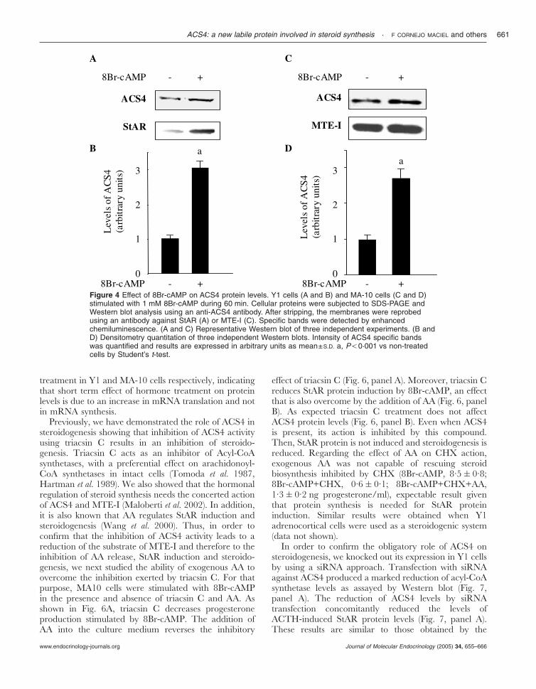
The next series of experiments was carried out inorder to analyze whether the hormonal regulation ofACS4 protein levels is also observed with 8Br-cAMP inY1 and in MA-10 Leydig cells, since in MA-10 cells theparticipation of AA as modulator of the hormone- andcAMP-induced StAR protein levels and steroidogenesisis well recognized (Wang et al. 2000). The results shownin Fig. 4 indicate that hormone-mediated induction ofACS4 is a cAMP-dependent process, since in both celllines the effect is reproduced by 8Br-cAMP, a permeableanalog of the second messenger cAMP. As expected,StAR protein is induced by 8Br-cAMP treatment (Fig. 4,panel A), while there is no action of the secondmessenger on MTE-I levels (Fig. 4, panel C).
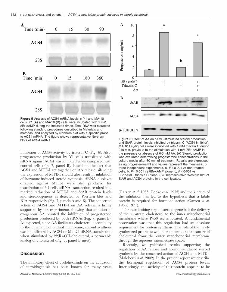
In order to investigate whether the acute effect ofhormone on ACS4 protein levels is due to an increase inprotein or in RNA synthesis, we studied the effect ofhormone treatment on ACS4 RNA abundance in Y1(Fig. 5, panel A) and MA-10 cells (Fig. 5, panel B). Incontrast with the effect observed on protein levels,stimulation of both cell lines did not change ACS4 RNAlevels during the first hour of treatment. The effect onmRNA levels could only be observed after 1·5 and 6 h of
Figure 3 Effect of CHX on ACS4 protein levels in Y1 cells. Adrenocortical Y1 cells were preincubated with or without 10 µg/mlCHX during 30 min and further treated with 10 mIU/ml ACTH (A and B) during the indicated times or its vehicle (C and D). Cellularproteins were subjected to SDS-PAGE and Western blot analysis using an anti-ACS4 antibody (A and C). After stripping, themembranes were reprobed using an antibody against MTE-I (A) or �-tubulin (C). Specific bands were detected by enhancedchemiluminescence. (A and C) Western blot representative of three independent experiments. (B and D) Densitometry quantitationof three independent Western blots. Intensity of ACS4 specific bands was quantified and results are expressed in arbitraryunits as mean±S.D a, P,0·05; b, P,0·01; c, P,0·001 vs non-treated cells; d, P,0·05; e, P,0·01; f, P,0·001 vs ACTH alone;g, P,0·01 vs time 0.
F CORNEJO MACIEL and others · ACS4: a new labile protein involved in steroid synthesis660
www.endocrinology-journals.orgJournal of Molecular Endocrinology (2005) 34, 655–666
treatment in Y1 and MA-10 cells respectively, indicatingthat short term effect of hormone treatment on proteinlevels is due to an increase in mRNA translation and notin mRNA synthesis.
Previously, we have demonstrated the role of ACS4 insteroidogenesis showing that inhibition of ACS4 activityusing triacsin C results in an inhibition of steroido-genesis. Triacsin C acts as an inhibitor of Acyl-CoAsynthetases, with a preferential effect on arachidonoyl-CoA synthetases in intact cells (Tomoda et al. 1987,Hartman et al. 1989). We also showed that the hormonalregulation of steroid synthesis needs the concerted actionof ACS4 and MTE-I (Maloberti et al. 2002). In addition,it is also known that AA regulates StAR induction andsteroidogenesis (Wang et al. 2000). Thus, in order toconfirm that the inhibition of ACS4 activity leads to areduction of the substrate of MTE-I and therefore to theinhibition of AA release, StAR induction and steroido-genesis, we next studied the ability of exogenous AA toovercome the inhibition exerted by triacsin C. For thatpurpose, MA10 cells were stimulated with 8Br-cAMPin the presence and absence of triacsin C and AA. Asshown in Fig. 6A, triacsin C decreases progesteroneproduction stimulated by 8Br-cAMP. The addition ofAA into the culture medium reverses the inhibitory
effect of triacsin C (Fig. 6, panel A). Moreover, triacsin Creduces StAR protein induction by 8Br-cAMP, an effectthat is also overcome by the addition of AA (Fig. 6, panelB). As expected triacsin C treatment does not affectACS4 protein levels (Fig. 6, panel B). Even when ACS4is present, its action is inhibited by this compound.Then, StAR protein is not induced and steroidogenesis isreduced. Regarding the effect of AA on CHX action,exogenous AA was not capable of rescuing steroidbiosynthesis inhibited by CHX (8Br-cAMP, 8·5�0·8;8Br-cAMP+CHX, 0·6�0·1; 8Br-cAMP+CHX+AA,1·3�0·2 ng progesterone/ml), expectable result giventhat protein synthesis is needed for StAR proteininduction. Similar results were obtained when Y1adrenocortical cells were used as a steroidogenic system(data not shown).
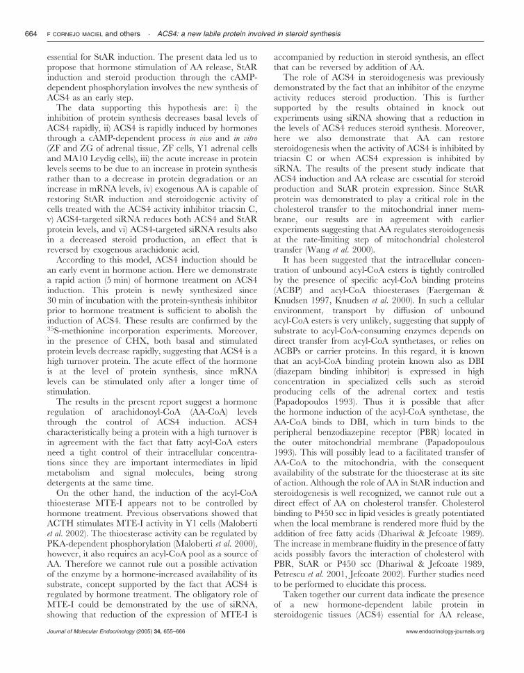
In order to confirm the obligatory role of ACS4 onsteroidogenesis, we knocked out its expression in Y1 cellsby using a siRNA approach. Transfection with siRNAagainst ACS4 produced a marked reduction of acyl-CoAsynthetase levels as assayed by Western blot (Fig. 7,panel A). The reduction of ACS4 levels by siRNAtransfection concomitantly reduced the levels ofACTH-induced StAR protein levels (Fig. 7, panel A).These results are similar to those obtained by the
Figure 4 Effect of 8Br-cAMP on ACS4 protein levels. Y1 cells (A and B) and MA-10 cells (C and D)stimulated with 1 mM 8Br-cAMP during 60 min. Cellular proteins were subjected to SDS-PAGE andWestern blot analysis using an anti-ACS4 antibody. After stripping, the membranes were reprobedusing an antibody against StAR (A) or MTE-I (C). Specific bands were detected by enhancedchemiluminescence. (A and C) Representative Western blot of three independent experiments. (B andD) Densitometry quantitation of three independent Western blots. Intensity of ACS4 specific bandswas quantified and results are expressed in arbitrary units as mean±S.D. a, P,0·001 vs non-treatedcells by Student’s t-test.
ACS4: a new labile protein involved in steroid synthesis · F CORNEJO MACIEL and others 661
www.endocrinology-journals.org Journal of Molecular Endocrinology (2005) 34, 655–666
inhibition of ACS4 activity by triacsin C (Fig. 6). Also,progesterone production by Y1 cells transfected withsiRNA against ACS4 was inhibited when compared withcontrol cells (Fig. 7, panel B). Based on the fact thatACS4 and MTE-I act together on AA release, silencingthe expression of MTE-I should also result in inhibitionof hormone-induced steroid synthesis. siRNA duplexesdirected against MTE-I were also produced fortransfection of Y1 cells. siRNA transfection resulted in amarked reduction of MTE-I and StAR protein levelsand steroidogenesis as detected by Western blot andRIA respectively (Fig. 7, panels A and B). The concertedaction of ACS4 and MTE-I on AA release is firmlysupported by the experiments showing that addition ofexogenous AA blunted the inhibition of progesteroneproduction produced by both siRNAs (Fig. 7, panel B).As expected, since AA facilitates cholesterol accessibilityto the inner mitochondrial membrane, steroid synthesiswas not affected by ACS4 or MTE-I siRNA transfectionwhen stimulated by 22(R)-OH-cholesterol, a permeableanalog of cholesterol (Fig. 7, panel B inset).
Discussion
The inhibitory effect of cycloheximide on the activationof steroidogenesis has been known for many years
(Garren et al. 1965, Cooke et al. 1975) and the kinetics ofthe inhibition has led to the hypothesis that a labileprotein is required for hormone action (Garren et al.1965, 1971).
The rate limiting step in steroidogenesis is the deliveryof the substrate cholesterol to the inner mitochondrialmembrane where P450 scc is located. A fundamentalobservation was that this regulation had an absoluterequirement for protein synthesis. The role of the newlysynthesized protein(s) would be to mediate the transfer ofcholesterol from the outer mitochondrial membranethrough the aqueous intermediate space.
Recently, we published results supporting theregulation of AA release and hormone-induced steroidsynthesis by the concerted action of ACS4 and MTE-I(Maloberti et al. 2002). In the present report we describethe hormonal regulation of ACS4 protein levels.Interestingly, the activity of this protein appears to be
Figure 5 Analysis of ACS4 mRNA levels in Y1 and MA-10cells. Y1 (A) and MA-10 (B) cells were incubated with 1 mM8Br-cAMP during the indicated times. Total RNA was extractedfollowing standard procedures described in Materials andmethods, and analyzed by Northern blot with a specific probeto ACS4 mRNA. The figure shows representative Northernblots of ACS4 mRNA. Figure 6 Effect of AA on cAMP-stimulated steroid production
and StAR protein levels inhibited by triacsin C (ACS4 inhibitor).MA-10 Leydig cells were incubated with 1 mM triacsin C during240 min, previous to the stimulation with 1 mM 8Br-cAMP inthe presence or absence of 0·3 mM AA. (A) Steroid productionwas evaluated determining progesterone concentrations in theculture media after 60 min of treatment. Results are expressedas ng progesterone/ml and values represent the mean±S.D. ofthree independent experiments. a, P,0·001 vs non treatedcells; b, P,0·001 vs 8Br-cAMP alone, c, P,0·001 vs8Br-cAMP+triacsin C alone. (B) Representative Western blot ofStAR and ACS4 proteins in the cell lysates.
F CORNEJO MACIEL and others · ACS4: a new labile protein involved in steroid synthesis662
www.endocrinology-journals.orgJournal of Molecular Endocrinology (2005) 34, 655–666
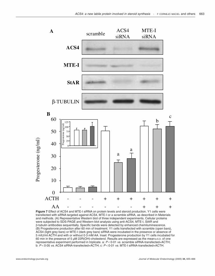
Figure 7 Effect of ACS4 and MTE-I siRNA on protein levels and steroid production. Y1 cells weretransfected with siRNA targeted against ACS4, MTE-I or a scramble siRNA, as described in Materialsand methods. (A) Representative Western blot of three independent experiments. Cellular proteinswere subjected to SDS-PAGE and Western blot analysis using anti-ACS4, MTE-I, StAR and�-tubulin antibodies sequentially. Specific bands were detected by enhanced chemiluminescence.(B) Progesterone production after 60 min of treatment. Y1 cells transfected with scramble (open bars),ACS4 (light grey bars) or MTE-I (dark grey bars) siRNA were incubated in the presence or absence of5 mIU/ml ACTH and with or without 0·3 mM AA. Inset: Progesterone production by Y1 cells incubated for60 min in the presence of 5 µM 22R(OH)-cholesterol. Results are expressed as the mean±S.D. of onerepresentative experiment performed in triplicate. a: P,0·01 vs. scramble siRNA-transfected+ACTH;b: P,0·05 vs. ACS4 siRNA-transfected+ACTH; c: P,0·01 vs. MTE-I siRNA-transfected+ACTH.
ACS4: a new labile protein involved in steroid synthesis · F CORNEJO MACIEL and others 663
www.endocrinology-journals.org Journal of Molecular Endocrinology (2005) 34, 655–666
essential for StAR induction. The present data led us topropose that hormone stimulation of AA release, StARinduction and steroid production through the cAMP-dependent phosphorylation involves the new synthesis ofACS4 as an early step.
The data supporting this hypothesis are: i) theinhibition of protein synthesis decreases basal levels ofACS4 rapidly, ii) ACS4 is rapidly induced by hormonesthrough a cAMP-dependent process in vivo and in vitro(ZF and ZG of adrenal tissue, ZF cells, Y1 adrenal cellsand MA10 Leydig cells), iii) the acute increase in proteinlevels seems to be due to an increase in protein synthesisrather than to a decrease in protein degradation or anincrease in mRNA levels, iv) exogenous AA is capable ofrestoring StAR induction and steroidogenic activity ofcells treated with the ACS4 activity inhibitor triacsin C,v) ACS4-targeted siRNA reduces both ACS4 and StARprotein levels, and vi) ACS4-targeted siRNA results alsoin a decreased steroid production, an effect that isreversed by exogenous arachidonic acid.
According to this model, ACS4 induction should bean early event in hormone action. Here we demonstratea rapid action (5 min) of hormone treatment on ACS4induction. This protein is newly synthesized since30 min of incubation with the protein-synthesis inhibitorprior to hormone treatment is sufficient to abolish theinduction of ACS4. These results are confirmed by the35S-methionine incorporation experiments. Moreover,in the presence of CHX, both basal and stimulatedprotein levels decrease rapidly, suggesting that ACS4 is ahigh turnover protein. The acute effect of the hormoneis at the level of protein synthesis, since mRNAlevels can be stimulated only after a longer time ofstimulation.
The results in the present report suggest a hormoneregulation of arachidonoyl-CoA (AA-CoA) levelsthrough the control of ACS4 induction. ACS4characteristically being a protein with a high turnover isin agreement with the fact that fatty acyl-CoA estersneed a tight control of their intracellular concentra-tions since they are important intermediates in lipidmetabolism and signal molecules, being strongdetergents at the same time.
On the other hand, the induction of the acyl-CoAthioesterase MTE-I appears not to be controlled byhormone treatment. Previous observations showed thatACTH stimulates MTE-I activity in Y1 cells (Malobertiet al. 2002). The thioesterase activity can be regulated byPKA-dependent phosphorylation (Maloberti et al. 2000),however, it also requires an acyl-CoA pool as a source ofAA. Therefore we cannot rule out a possible activationof the enzyme by a hormone-increased availability of itssubstrate, concept supported by the fact that ACS4 isregulated by hormone treatment. The obligatory role ofMTE-I could be demonstrated by the use of siRNA,showing that reduction of the expression of MTE-I is
accompanied by reduction in steroid synthesis, an effectthat can be reversed by addition of AA.
The role of ACS4 in steroidogenesis was previouslydemonstrated by the fact that an inhibitor of the enzymeactivity reduces steroid production. This is furthersupported by the results obtained in knock outexperiments using siRNA showing that a reduction inthe levels of ACS4 reduces steroid synthesis. Moreover,here we also demonstrate that AA can restoresteroidogenesis when the activity of ACS4 is inhibited bytriacsin C or when ACS4 expression is inhibited bysiRNA. The results of the present study indicate thatACS4 induction and AA release are essential for steroidproduction and StAR protein expression. Since StARprotein was demonstrated to play a critical role in thecholesterol transfer to the mitochondrial inner mem-brane, our results are in agreement with earlierexperiments suggesting that AA regulates steroidogenesisat the rate-limiting step of mitochondrial cholesteroltransfer (Wang et al. 2000).
It has been suggested that the intracellular concen-tration of unbound acyl-CoA esters is tightly controlledby the presence of specific acyl-CoA binding proteins(ACBP) and acyl-CoA thioesterases (Faergeman &Knudsen 1997, Knudsen et al. 2000). In such a cellularenvironment, transport by diffusion of unboundacyl-CoA esters is very unlikely, suggesting that supply ofsubstrate to acyl-CoA-consuming enzymes depends ondirect transfer from acyl-CoA synthetases, or relies onACBPs or carrier proteins. In this regard, it is knownthat an acyl-CoA binding protein known also as DBI(diazepam binding inhibitor) is expressed in highconcentration in specialized cells such as steroidproducing cells of the adrenal cortex and testis(Papadopoulos 1993). Thus it is possible that afterthe hormone induction of the acyl-CoA synthetase, theAA-CoA binds to DBI, which in turn binds to theperipheral benzodiazepine receptor (PBR) located inthe outer mitochondrial membrane (Papadopoulous1993). This will possibly lead to a facilitated transfer ofAA-CoA to the mitochondria, with the consequentavailability of the substrate for the thioesterase at its siteof action. Although the role of AA in StAR induction andsteroidogenesis is well recognized, we cannot rule out adirect effect of AA on cholesterol transfer. Cholesterolbinding to P450 scc in lipid vesicles is greatly potentiatedwhen the local membrane is rendered more fluid by theaddition of free fatty acids (Dhariwal & Jefcoate 1989).The increase in membrane fluidity in the presence of fattyacids possibly favors the interaction of cholesterol withPBR, StAR or P450 scc (Dhariwal & Jefcoate 1989,Petrescu et al. 2001, Jefcoate 2002). Further studies needto be performed to elucidate this process.
Taken together our current data indicate the presenceof a new hormone-dependent labile protein insteroidogenic tissues (ACS4) essential for AA release,
F CORNEJO MACIEL and others · ACS4: a new labile protein involved in steroid synthesis664
www.endocrinology-journals.orgJournal of Molecular Endocrinology (2005) 34, 655–666
StAR induction and steroidogenesis. This study furthersupports the new concept in the regulation ofintracellular distribution of AA through a mechanismdifferent from the classical phospholipase A2-mediatedpathway that involves the activity of a hormone-inducedacyl-CoA synthetase and a hormone-regulated acyl-CoAthioesterase.
Acknowledgements
This work was supported by grants from University ofBuenos Aires (UBA) to EJP (M064), CP (M059) andFCM (M079) and Agencia Nacional de PromociónCientífica y Tecnológica (ANPCYT) to EJP (PICT6738). Thanks are also due to Carlos F Méndez forcritical reading. The authors declare that there is noconflict of interest that would prejudice the impartialityof this scientific work.
ReferencesAscoli M 1981 Characterization of several clonal lines of cultured
Leydig tumor cells gonadotropin receptors and steroidogenicresponses. Endocrinology 108 88–95.
Bradford MM 1976 A rapid and sensitive method for quantitation ofmicrogram quantities of protein utilizing the principle ofprotein-dye binding. Analytical Biochemistry 72 248–254.
Clark BJ, Wells J, King SR & Stocco D 1994 The purification,cloning, and expression of a novel luteinizing hormone-inducedmitochondrial protein in MA-10 mouse Leydig tumor cells.Characterization of the steroidogenic acute regulatory protein(StAR). Journal of Biological Chemistry 269 28314–28322.
Cooke BA, Janszen FH, Clotscher WF & van der Molen HJ 1975Effect of protein-synthesis inhibitors on testosterone production inrat testis interstitial tissue and Leydig-cell preparations. BiochemicalJournal 150 413–418.
Cooke BA, Dirami G, Chaudry L, Choi MSK, Abayasekara DRE &Phipp L 1991 Release of arachidonic acid and the effects ofcorticosteroids on steroidogenesis in rat testis Leydig cells. Journalof Steroid Biochemistry and Molecular Biology 40 465–471.
Crivello JF & Jefcoate CR 1980 Intracellular movement ofcholesterol in rat adrenal cells. Kinetic and effect of inhibitors.Journal of Biological Chemistry 255 8144–8151.
Dada L, Cornejo Maciel F, Neuman I, Mele PG, Maloberti P, PazC, Cymeryng C, Finkielstein C, Mendez CF & Podestá EJ 1996Cytosolic and mitochondrial proteins as possible targets ofcycloheximide effect on adrenal steroidogenesis. Endocrine Research22 533–539.
Dhariwal MS & Jefcoate CR 1989 Cholesterol metabolism bypurified cytochrome P-450 scc is highly stimulated by octylglucoside and stearic acid exclusively in large unilamellarphospholipid vesicles. Biochemistry 28 8397–8402.
Didolkar A & Sundaram K 1987 Arachidonic acid is involved in theregulation of hCG induced steroidogenesis in rat Leydig cells. LifeSciences 41 471–477.
Dix CJ, Habberfield AD, Sullivan MH & Cooke BA 1984 Inhibitionof steroid production in Leydig cells by non-steroidalanti-inflammatory and related compounds evidence for theinvolvement of lipoxygenase products in steroidogenesis.Biochemical Journal 219 529–537.
Dufau ML, Tsuruhara T, Horner K, Podestá E & Catt K 1977Intermediate role of adenosine 3� 5�-cyclic monophosphate andprotein kinase during gonadotropin-induced steroidogenesis intesticular interstitial cells. PNAS 4 3419–3423.
Faergeman NJ & Knudsen J 1997 Role of long-chain fatty acyl-CoAesters in the regulation of metabolism and in cell signaling.Biochemical Journal 323 1–12.
Finkielstein C, Cymeryng C, Paz C, Neuman I, Dada L, CornejoMaciel F, Mele PG, Mendez CF Maloberti P, Solano A,Schimmer BP & Podestá EJ 1996 Characterization of the cDNAcorresponding to a phosphoprotein (p43) intermediary in theaction of ACTH. Endocrine Research 22 521–532.
Finkielstein C, Maloberti P, Mendez CF, Paz C, Cornejo Maciel F,Cymeryng C, Neuman I, Dada L, Mele PG, Solano A & PodestáEJ 1998 An adrenocorticotropin regulated phosphoproteinintermediary in steroid synthesis is similar to an acyl-CoAthioesterase enzyme. European Journal of Biochemistry 256 60–66.
Garren LD, Ney RL & Davis WW 1965 Studies on the role ofprotein synthesis in the regulation of corticosterone production byadrenocorticotropic hormone in vivo. PNAS 53 1443–1450.
Garren LD, Gill GN, Masui H & Walton GM 1971 On themechanism of action of ACTH. Recent Progress in Hormone Research27 433–478.
Hartman EJ, Omura S & Laposata M 1989 Triacsin C a diferentialinhibitor of arachidonoyl-CoA synthetase and nonspecific longchain acyl-CoA synthetase. Prostaglandins 37 655–671.
Jefcoate C 2002 High-flux mitochondrial cholesterol trafficking, aspecialized function of the adrenal cortex. Journal of ClinicalInvestigation 110 881–890.
Kang M, Fujino T, Sasano H, Minekura H, Yabuki N, Nagura H,Iijima H & Yamamoto T 1997 A novel arachidonate-preferringacyl-CoA synthetase is present in steroidogenic cells of the ratadrenal, ovary, and testis. PNAS 94 2880–2884.
Knudsen J, Neergaard TBF, Gaigg B, Jensen MV & Hansen JK2000 Role of acyl-CoA binding protein in acyl-CoA metabolismand acyl-CoA mediated cell signaling. Symposium The role oflong chain fatty acyl-CoAs as signaling molecules in cellularmetabolism. American Society for Nutritional Sciences. Journal ofNutrition 130 294S–298S.
Laemmli UK 1970 Cleavage of structural proteins during theassembly of the head of bacteriophage T4. Nature 227 680–685.
Maloberti P, Lozano RC, Mele PG, Cano F, Colonna C, MendezCF, Paz C & Podestá EJ 2002 Concerted regulation of freearachidonic acid and hormone-induced steroid synthesis byacyl-CoA thioesterase and Acyl-CoA synthetases in adrenal cells.European Journal of Biochemistry 269 5599–5607.
Maloberti P, Mele PG, Neuman I, Cornejo Maciel F, Cano F, BeyP, Paz C & Podestá EJ 2000 Regulation of arachidonic acidrelease in steroidogenesis role of a new acyl-CoA thioesterase(ARTISt). Endocrine Research 26 653–662.
Mele PF, Dada LA, Paz C, Neuman I, Cymeryng CB, Mendez CF,Finkielstein CV, Cornejo Maciel F & Podestá EJ 1997Involvement of arachidonic acid and the lipoxygenase pathway inmediating luteinizing hormone-induced testosterone synthesis inrat Leydig cells. Endocrine Research 23 15–26.
Moraga PF, Llanos MN & Ronco AM 1997 Arachidonic acidrelease from rat Leydig cells depends on the presence ofluteinizing hormone/human chorionic gonadotropin receptors.Journal of Endocrinology 154 201–209.
Neher R, Milani A, Solano AR & Podestá EJ 1982Compartmentalization of corticotropin-dependent steroidogenicfactors in adrenal cortex evidence for a post-translationalcascade in stimulation of cholesterol side chain split. PNAS 791727–1731.
Papadopoulos V 1993 Peripheral-type benzodiazepine/diazepambinding inhibitor receptor biological role in steroidogenic cellfunction. Endocrine Reviews 14 222–240.
Paz C, Dada LA, Cornejo Maciel F, Mele PG, Cymeryng CB,Neuman I, Mendez CF, Finkielstein CV, Solano AR & PodestáEJ 1994 Purification of a novel 43-kDa protein (p43) intermediaryin the activation of steroidogenesis from rat adrenal gland.European Journal of Biochemistry 224 709–716.
ACS4: a new labile protein involved in steroid synthesis · F CORNEJO MACIEL and others 665
www.endocrinology-journals.org Journal of Molecular Endocrinology (2005) 34, 655–666

Petrescu AD, Gallegos AM, Okamura Y, Strauss JF III & SchroederF 2001 Steroidogenic acute regulatory protein binds cholesteroland modulates mitochondrial membrane sterol domain dynamics.Journal of Biological Chemistry 276 36970–36982.
Privalle CT, Crivello JF & Jefcoate CR 1983 Regulation ofintramitochondrial cholesterol transfer to side-chain cleavagecytochrome P-450 scc in rat adrenal gland. PNAS 80 702–706.
Reinhart AJ, Williams SC & Stocco DM 1999 Transcriptionalregulation of the StAR gene. Molecular and Cellular Endocrinology151 161–169.
Sala G, Hayoshi K, Catt K & Dufau M 1979 The intermediate roleof cyclic AMP in stimulation of corticosterone synthesis. Journal ofBiological Chemistry 254 3861–3865.
Schimmer BP 1981 The adrenocortical tumor cell line Y1. InFunctionally differentiated cell lines, pp 61–92. Ed. G Sato. New York:Alan R. Liss Inc.
Solano AR, Dada L & Podestá EJ 1988 Lipoxygenase products ascommon intermediates in cyclic AMP-dependent and-independent adrenal steroidogenesis in rats. Journal of MolecularEndocrinology 1 147–154.
Stocco DM & Clark BJ 1996 Regulation of the acute production ofsteroids in steroidogenic cells. Endocrine Reviews 17 221–244.
Svensson LT, Endberg ST, Aoyama T, Usuda N, Alexon SEH &Hashimoto T 1998 Molecular cloning and characterization of amitochondrial peroxisome proliferator-induced acyl-CoAthioestease from rata liver. Biochemical Journal 329 601–608.
Tomoda H, Igarashi K & Omura S 1987 Inhibition of acyl-CoAsynthetase by triacsins. Biochimica et Biophysica Acta 921 595–598.
Towbin H, Stachelin T & Gordon J 1979 Electrophoretic transfer ofproteins from polyacrylamide gels to nitrocellulose sheetsprocedure and some applications. PNAS 76 4350–4354.
Wang X, Walsh LP, Reinhart AJ & Stocco DM 2000 The role ofarachidonic acid in steroidogenesis and steroidogenic acuteregulatory (StAR) gene and protein expression. Journal of BiologicalChemistry 275 20204–20209.
Received 16 March 2005Accepted 18 March 2005Made available online as an Accepted Preprint 31 March 2005
F CORNEJO MACIEL and others · ACS4: a new labile protein involved in steroid synthesis666
www.endocrinology-journals.orgJournal of Molecular Endocrinology (2005) 34, 655–666
Copyright © 2022 FDOKUMEN