Disruption of the Jak1 Gene Demonstrates Obligatory and Nonredundant Roles of the Jaks in...
11
Cell, Vol. 93, 373–383, May 1, 1998, Copyright 1998 by Cell Press Disruption of the Jak1 Gene Demonstrates Obligatory and Nonredundant Roles of the Jaks in Cytokine-Induced Biologic Responses A role for Jaks in cytokine signaling was initially identi- fied using mutagenized human fibrosarcoma cell lines that lacked individual Jak proteins and displayed unique patterns of biologic unresponsiveness to IFNa or IFNg (Velazquez et al., 1992; Mu ¨ ller et al., 1993; Watling et Scott J. Rodig,* Marco A. Meraz,* J. Michael White,* Pat A. Lampe, ² Joan K. Riley,* Cora D. Arthur,* Kathleen L. King, ‡ Kathleen C. F. Sheehan,* Li Yin,* Diane Pennica, ‡ Eugene M. Johnson, Jr., ² al., 1993). This work demonstrated that distinct combi- and Robert D. Schreiber* § nations of Jaks were required for the induction of cellular * Center for Immunology and Department of Pathology responses to type I versus type II interferons. However, ² Department of Molecular Biology and Pharmacology subsequent studies that sought to define a role for Jaks Washington University School of Medicine in signaling by other cytokine receptors monitored only St. Louis, Missouri 63110 whether particular Jaks became phosphorylated and/ ‡ Departments of Molecular Oncology and or activated following in vitro treatment of a cell with Protein Chemistry different cytokines. This approach often produced con- Genentech, Inc. flicting results and failed to define a causal relationship South San Francisco, California 94080 between cytokine-dependent Jak activation and the in- duction of a biologic response (Ihle et al., 1995). This problem has been most evident for the family of recep- Summary tors that utilizes the gp130 receptor subunit (i.e., the receptors for IL-6, IL-11, leukemia inhibitory factor [LIF], Herein we report the generation of mice lacking the oncostatin M [OSM], ciliary neurotrophic factor [CNTF], ubiquitously expressed Janus kinase, Jak1. Jak1 2/2 and cardiotrophin-1 [CT-1] [Kishimoto et al., 1995]). By mice are runted at birth, fail to nurse, and die perina- monitoring the ability of ligands of the gp130 receptor tally. Although Jak1 2/2 cells are responsive to many family to induce Jak and STAT activation in cells engi- cytokines, they fail to manifest biologic responses to neered to overexpress particular Jaks, one group con- cytokines that bind to three distinct families of cyto- cluded that Jaks can associate in a nonselective manner kine receptors. These include all class II cytokine re- with certain cytokine receptor subunits (Stahl et al., ceptors, cytokine receptors that utilize the g c subunit 1994). In contrast, using a Jak1-deficient tumor cell line, for signaling, and the family of cytokine receptors that another group reported that Jak1 was indeed required depend on the gp130 subunit for signaling. Our results for IL-6–dependent induction of the IRF-1 transcription thus demonstrate that Jak1 plays an essential and factor in vitro (Guschin et al., 1995). However, no sub- nonredundant role in promoting biologic responses sequent experiments were performed to determine induced by a select subset of cytokine receptors, in- whether Jak1-deficient cells were capable of develop- cluding those in which Jak utilization was thought to ing a full biologic response. Thus, it remains unclear be nonspecific. whether, under physiologic conditions, Jaks participate in a specific manner in effecting Jak-STAT pathway– Introduction dependent biologic responses. One problem impeding the elucidation of the physio- Cytokines that bind to class I or class II cytokine recep- logic roles of these enzymes is the absence of mamma- tors employ the Jak-STAT signaling pathway to manifest lian models of Jak protein deficiency. To date, the only many of their biologic effects on cells (Darnell et al., known Jak abnormality is that found in Jak3 gene– 1994; Ihle et al., 1995; Darnell, 1997; O’Shea, 1997). This targeted mice and Jak3-deficient human SCID patients pathway consists of four Janus family protein tyrosine (Nosaka et al., 1995; Park et al., 1995; Russell et al., 1995; kinases (Jaks) and a family of seven latent cytosolic Thomis et al., 1995). The analysis of these individuals has transcription factors known as signal transducers and revealed that Jak3 associates exclusively with a single activators of transcription (STATs). cytokine receptor subunit (g c ) and functions predomi- Recent work using mutagenized cell lines and gene- nantly to promote lymphopoiesis. Thus, due to its highly targeted mice has shown that the STATs play a major restricted nature, Jak3 deficiency does not provide a role in determining cytokine signaling specificity (Bach suitable system to evaluate the generalized physiologic et al., 1997; Darnell, 1997; O’Shea, 1997). Distinct STAT functions of the Jaks. proteins are recruited to individual ligated cytokine re- In contrast, the other members of the Janus kinase ceptors, where they undergo activation and dimeriza- family (Jak1, Jak2, and Tyk2) are ubiquitously expressed tion. Activated STAT dimers then translocate to the nu- and interact with many different cytokine receptor sub- cleus, where they bind to the promoter regions of units (Darnell et al., 1994; Ihle et al., 1995; O’Shea, 1997). specific genes and activate gene transcription. Although We therefore decided to generate Jak1-deficient mice the Jaks are known to play a critical role in this process, using homologous recombination techniques and deter- the physiologic functions and fine specificities of these mine whether Jak1 deficiency leads to distinct biologic enzymes remain poorly defined (Ihle et al., 1995; O’Shea, response deficits. In this report, we demonstrate that 1997). Jak1 indeed plays an essential and nonredundant role in promoting biologic responses induced only by specific subsets of cytokine receptors, including those in which § To whom correspondence should be addressed.
-
Upload
independent -
Category
Documents
-
view
0 -
download
0
Transcript of Disruption of the Jak1 Gene Demonstrates Obligatory and Nonredundant Roles of the Jaks in...
Cell, Vol. 93, 373–383, May 1, 1998, Copyright 1998 by Cell Press
Disruption of the Jak1 Gene DemonstratesObligatory and Nonredundant Roles of the Jaksin Cytokine-Induced Biologic Responses
A role for Jaks in cytokine signaling was initially identi-fied using mutagenized human fibrosarcoma cell linesthat lacked individual Jak proteins and displayed uniquepatterns of biologic unresponsiveness to IFNa or IFNg(Velazquez et al., 1992; Muller et al., 1993; Watling et
Scott J. Rodig,* Marco A. Meraz,*J. Michael White,* Pat A. Lampe,†Joan K. Riley,* Cora D. Arthur,*Kathleen L. King,‡ Kathleen C. F. Sheehan,*Li Yin,* Diane Pennica,‡ Eugene M. Johnson, Jr.,†
al., 1993). This work demonstrated that distinct combi-and Robert D. Schreiber*§
nations of Jaks wererequired for the induction of cellular*Center for Immunology and Department of Pathologyresponses to type I versus type II interferons. However,†Department of Molecular Biology and Pharmacologysubsequent studies that sought to define a role for JaksWashington University School of Medicinein signaling by other cytokine receptors monitored onlySt. Louis, Missouri 63110whether particular Jaks became phosphorylated and/‡Departments of Molecular Oncology andor activated following in vitro treatment of a cell withProtein Chemistrydifferent cytokines. This approach often produced con-Genentech, Inc.flicting results and failed to define a causal relationshipSouth San Francisco, California 94080between cytokine-dependent Jak activation and the in-duction of a biologic response (Ihle et al., 1995). Thisproblem has been most evident for the family of recep-Summarytors that utilizes the gp130 receptor subunit (i.e., thereceptors for IL-6, IL-11, leukemia inhibitory factor [LIF],Herein we report the generation of mice lacking theoncostatin M [OSM], ciliary neurotrophic factor [CNTF],ubiquitously expressed Janus kinase, Jak1. Jak12/2
and cardiotrophin-1 [CT-1] [Kishimoto et al., 1995]). Bymice are runted at birth, fail to nurse, and die perina-monitoring the ability of ligands of the gp130 receptortally. Although Jak12/2 cells are responsive to manyfamily to induce Jak and STAT activation in cells engi-cytokines, they fail to manifest biologic responses toneered to overexpress particular Jaks, one group con-cytokines that bind to three distinct families of cyto-cluded that Jaks can associate in a nonselective mannerkine receptors. These include all class II cytokine re-with certain cytokine receptor subunits (Stahl et al.,ceptors, cytokine receptors that utilize the gc subunit1994). In contrast, using a Jak1-deficient tumor cell line,for signaling, and the family of cytokine receptors thatanother group reported that Jak1 was indeed requireddepend on the gp130 subunit for signaling. Our resultsfor IL-6–dependent induction of the IRF-1 transcriptionthus demonstrate that Jak1 plays an essential andfactor in vitro (Guschin et al., 1995). However, no sub-nonredundant role in promoting biologic responsessequent experiments were performed to determineinduced by a select subset of cytokine receptors, in-whether Jak1-deficient cells were capable of develop-cluding those in which Jak utilization was thought toing a full biologic response. Thus, it remains unclearbe nonspecific.whether, under physiologic conditions, Jaks participatein a specific manner in effecting Jak-STAT pathway–Introductiondependent biologic responses.
One problem impeding the elucidation of the physio-Cytokines that bind to class I or class II cytokine recep-
logic roles of these enzymes is the absence of mamma-tors employ the Jak-STAT signaling pathway to manifest
lian models of Jak protein deficiency. To date, the onlymany of their biologic effects on cells (Darnell et al.,
known Jak abnormality is that found in Jak3 gene–1994; Ihle et al., 1995; Darnell, 1997; O’Shea, 1997). This targeted mice and Jak3-deficient human SCID patientspathway consists of four Janus family protein tyrosine
(Nosaka et al., 1995; Park et al., 1995; Russell et al., 1995;kinases (Jaks) and a family of seven latent cytosolic
Thomis et al.,1995). The analysis of these individuals hastranscription factors known as signal transducers and
revealed that Jak3 associates exclusively with a singleactivators of transcription (STATs). cytokine receptor subunit (gc) and functions predomi-
Recent work using mutagenized cell lines and gene- nantly to promote lymphopoiesis. Thus, due to its highlytargeted mice has shown that the STATs play a major restricted nature, Jak3 deficiency does not provide arole in determining cytokine signaling specificity (Bach suitable system to evaluate the generalized physiologicet al., 1997; Darnell, 1997; O’Shea, 1997). Distinct STAT functions of the Jaks.proteins are recruited to individual ligated cytokine re- In contrast, the other members of the Janus kinaseceptors, where they undergo activation and dimeriza- family (Jak1, Jak2, and Tyk2) are ubiquitously expressedtion. Activated STAT dimers then translocate to the nu- and interact with many different cytokine receptor sub-cleus, where they bind to the promoter regions of units (Darnell et al., 1994; Ihle et al., 1995; O’Shea, 1997).specific genes and activate gene transcription. Although We therefore decided to generate Jak1-deficient micethe Jaks are known to play a critical role in this process, using homologous recombination techniques and deter-the physiologic functions and fine specificities of these mine whether Jak1 deficiency leads to distinct biologicenzymes remain poorly defined (Ihle et al., 1995; O’Shea, response deficits. In this report, we demonstrate that1997). Jak1 indeed plays an essential and nonredundant role in
promoting biologic responses induced only by specificsubsets of cytokine receptors, including those in which§To whom correspondence should be addressed.
Cell374
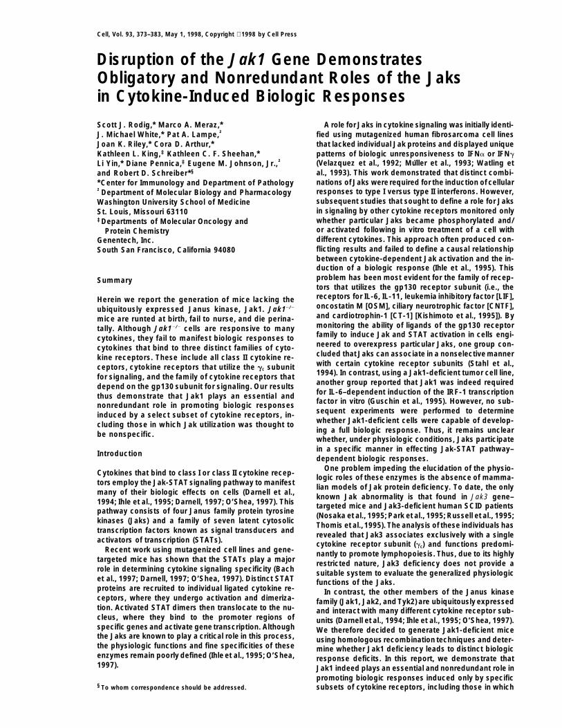
generate animals heterozygote for the targeted deletion.Heterozygote 3 heterozygote breedings produced wild-type, heterozygote, and homozygote embryos as identi-fied by Southern blot analyses (Figure 1B). Immunopre-cipitation and Western blot analyses using Jak1 antiserashowed the presence of the 130 kDa Jak1 protein inlysates of embryonic fibroblasts (EFs) derived from wild-type embryos but not embryos homozygous for the dis-rupted Jak1 gene (Figure 1C). EFs derived from hetero-zygote embryos contained reduced amounts of Jak1relative to wild-type EFs.
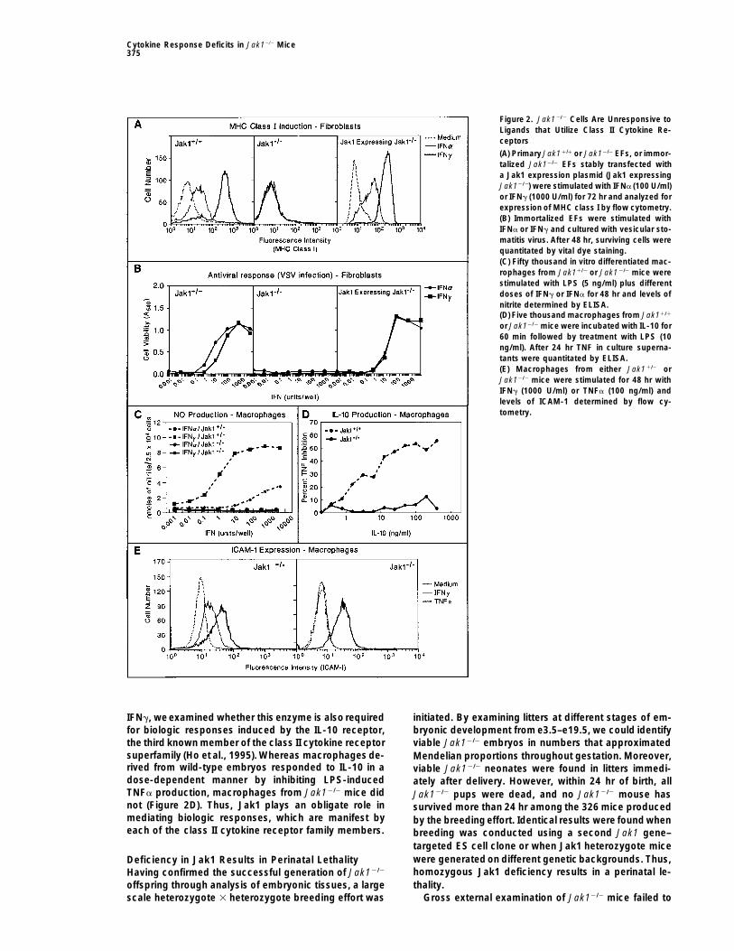
Cells from Jak1-Deficient Mice Fail To DevelopBiologic Responses to Ligands ofClass II Cytokine ReceptorsPreviously published work using a randomly mutagen-ized human fibrosarcoma cell line indicated that Jak1was required for both signaling and biologic responseinduction by IFNa and IFNg (Muller et al., 1993). Toextend these observations to primary cells with a tar-geted deletion of Jak1, we monitored the ability of theinterferons to enhance MHC class I expression on EFsderived from wild-type and Jak1 null mice. Whereaswild-type EFs exposed to either IFNa or IFNg expressedenhanced levels of H2-Kb, Jak12/2 EFs were unrespon-sive to either cytokine (Figure 2A). Similar results wereobtained when antiviral activity was monitored as a sec-ond IFN-dependent biologic response (data not shown).
Figure 1. Targeted Disruption of the Jak1 Gene Ablates Expression To document that the unresponsive state was spe-of Jak1 Protein cifically due to the absence of Jak1, immortalized EFs(A) The genomic locus of the wild-type Jak1 gene, the targeting from Jak11/1 and Jak12/2 embryos were analyzed forvector, and the disrupted Jak1 gene. The first three exons (I, II, IFN responsiveness before and after reconstitution withand III) encoding the 59 region of the Jak1 protein, the neomycin Jak1. Immortalized Jak11/1 EFs responded to eitherresistance cassette (Neo), and the thymidine kinase cassette (TK)
IFNa or IFNg in a dose-dependent manner and resistedare marked by shaded boxes. The orientation of transcription ofthe cytopathic effects of vesicular stomatitis virus (Fig-Neo and TK are indicated by arrows. The restriction fragment usedure 2B). In contrast, neither IFNa nor IFNg protectedas a probe external (Ext. Probe) to the targeting vector in Southern
analyses is marked. The locations of restriction sites for Eco RI (E), Jak12/2 fibroblasts from viral infection. However, IFNPst I (P), and Xba I (X) are indicated. responsiveness was restored to immortalized gene-tar-(B) Southern blot analysis of Eco RI digested DNA isolated from the geted cells following stable transfection with a murinetails of e16.5 embryos derived from the mating of heterozygote mice.
Jak1 expression plasmid. Reconstituted Jak12/2 EFsThe bands at 6.7 and 3.6 kb correspond to the endogenous andalso recovered the ability to up-regulate MHC class Imutant Jak1 genes, respectively.expression in response to IFNa or IFNg (Figure 2A). Thus,(C) Two million EFs of each genotype were lysed and analyzed by
immunoprecipitation and Western blotting for the presence of Jak1. the IFN unresponsiveness observed in fibroblasts of ourgene-targeted mice is specifically due to the absenceof Jak1.
Jak utilization was originally thought to be nonspecific. We also tested whether Jak1 was required for devel-These results thus demonstrate that the Jaks are utilized opment of IFN-dependent biologic responses in otherby cytokine receptors in a functionally dedicated man- cell types. Whereas macrophages from Jak11/1 em-ner to promote development of cytokine-specific bio- bryos responded to the combination of LPS and eitherlogic responses. IFNa or IFNg and produced nitric oxide, macrophages
from Jak12/2 embryos did not (Figure 2C). Jak12/2 mac-rophages also failed to up-regulate ICAM-1 expressionResults(Figure 2E) or induce expression of the third componentof complement (data not shown) in response to IFNg.Generation of Jak1-Deficient Mice
To ablate the mouse Jak1 gene, a targeting construct However, Jak12/2 macrophages were not generally cy-tokine unresponsive because they up-regulated ICAM-1was designed to replace the first two exons of the gene
and 1.6 kb of 59 upstream sequence with a neomycin following treatment with TNFa in a manner similar tothat of wild-type macrophages. Thus, the absence ofresistance cassette (Figure 1A). Following electropora-
tion and selection, 3 out of 235 ES cell clones were Jak1 results in a generalized cellular unresponsivenessto type I and type II interferons.identified by Southernblot analysis as having undergone
successful gene targeting. Chimeric animals from two Based on the obligate requirement of Jak1 for biologicresponses induced through the receptors for IFNa andof these clones transmitted the mutation germline to
Cytokine Response Deficits in Jak12/2 Mice375
Figure 2. Jak12/2 Cells Are Unresponsive toLigands that Utilize Class II Cytokine Re-ceptors
(A) Primary Jak11/1 or Jak12/2 EFs, or immor-talized Jak12/2 EFs stably transfected witha Jak1 expression plasmid (Jak1 expressingJak12/2) were stimulated with IFNa (100 U/ml)or IFNg (1000 U/ml) for 72 hr and analyzed forexpression of MHC class I by flow cytometry.(B) Immortalized EFs were stimulated withIFNa or IFNg and cultured with vesicular sto-matitis virus. After 48 hr, surviving cells werequantitated by vital dye staining.(C) Fifty thousand in vitro differentiated mac-rophages from Jak11/2 or Jak12/2 mice werestimulated with LPS (5 ng/ml) plus differentdoses of IFNg or IFNa for 48 hr and levels ofnitrite determined by ELISA.(D) Five thousand macrophages from Jak11/1
or Jak12/2 mice were incubated with IL-10 for60 min followed by treatment with LPS (10ng/ml). After 24 hr TNF in culture superna-tants were quantitated by ELISA.(E) Macrophages from either Jak11/2 orJak12/2 mice were stimulated for 48 hr withIFNg (1000 U/ml) or TNFa (100 ng/ml) andlevels of ICAM-1 determined by flow cy-tometry.
IFNg, we examined whether this enzyme is also required initiated. By examining litters at different stages of em-for biologic responses induced by the IL-10 receptor, bryonic development from e3.5–e19.5, we could identifythe third known member of the class II cytokine receptor viable Jak12/2 embryos in numbers that approximatedsuperfamily (Ho et al., 1995). Whereas macrophages de- Mendelian proportions throughout gestation. Moreover,rived from wild-type embryos responded to IL-10 in a viable Jak12/2 neonates were found in litters immedi-dose-dependent manner by inhibiting LPS-induced ately after delivery. However, within 24 hr of birth, allTNFa production, macrophages from Jak12/2 mice did Jak12/2 pups were dead, and no Jak12/2 mouse hasnot (Figure 2D). Thus, Jak1 plays an obligate role in survived more than 24 hr among the 326 mice producedmediating biologic responses, which are manifest by by the breeding effort. Identical results were found wheneach of the class II cytokine receptor family members. breeding was conducted using a second Jak1 gene–
targeted ES cell clone or when Jak1 heterozygote micewere generated on different geneticbackgrounds. Thus,Deficiency in Jak1 Results in Perinatal Lethalityhomozygous Jak1 deficiency results in a perinatal le-Having confirmed the successful generation of Jak12/2
thality.offspring through analysis of embryonic tissues, a largescale heterozygote 3 heterozygote breeding effort was Gross external examination of Jak12/2 mice failed to

Cell376
Figure 3. Jak12/2 Mice Exhibit Deficits inLymphopoiesis but Not Myelopoiesis
(A, C, and E) Cellular recoveries from thymi(A) spleens (C) and weight of livers (E) derivedfrom newborn Jak11/1 (open boxes) andJak12/2 (filled boxes) mice.(B) Thymocytes derived from newbornJak11/1 (left panel) and Jak12/2 (right panel)mice were stained with FITC anti-CD4 and PEanti-CD8 and analyzed by flow cytometry.(D) Splenocytes derived from newbornJak11/1 (left panel) and Jak12/2 (right panel)mice were stained with FITC anti-IgM and PEanti-B220 and the gated lymphoid populationanalyzed by flow cytometry.(F) Fetal liver cells derived from e18.5 Jak11/1
(left panel) and Jak12/2 (right panel) animalswere stained with FITC anti-Mac-1 and PEanti-Gr-1 and the gated myeloid populationsanalyzed by flow cytometry.The percentage of the total gated cells ana-lyzed is given within each quadrant.
reveal significant developmental abnormalities com- reduction in cellularity. Nevertheless, the distribution ofCD4- and CD8-positive cells within the wild-type andpared to Jak1-sufficient newborns. However, newborn
Jak12/2 mice (0.99 6 0.11 g) weighed 40% less than Jak12/2 thymocyte populations was similar (Figure 3B).The thymi of gene-targeted mice contained a slightlytheir heterozygote (1.61 6 0.16 g) or wild-type (1.65 6
0.09 g) littermates. Moreover, Jak1-deficient neonates higher percentage of CD42CD82 cells relative to con-trols. However, these cells most likely represent stromalfailed to nurse despite a normal nurturing behavior of
the mothers even when these mice were part of very cells, which contaminate the limited number of thymo-cytes that can be isolated from newborn Jak12/2 mice.small litters or when all Jak1 sufficient siblings were
removed from the litter (data not shown). Thus, Jak1 deficiency results in a primary defect in thy-mocyte production rather than a definitive block in thy-mocyte maturation.Severely Impaired Lymphocytic Development
in Jak1-Deficient Mice To determine whether B cell production and/or devel-opment was also dependent on Jak1, the B cell lineagesBlood from newborn Jak12/2 mice contained normal
numbers of erythrocytes, mononuclear phagocytes, neu- present in the spleens and livers of wild-type and gene-targeted mice were quantitated. No significant differ-trophils, and platelets (data not shown). Moreover, upon
necropsy of newborn Jak1 null pups, no gross abnor- ences in total splenocyte numbers were observed be-tween newborn wild-type and Jak12/2 mice (Figure 3C).malities were found in several organs, such as heart,
liver, lungs, kidney, and brain. In contrast, thymi from This result is consistent with the fact that spleens ofnewborn mice contain only a limited number of lymphoidnewborn Jak12/2 mice were of severely reduced size
compared to those isolated from control newborn lit- cells. However, when the B cells present in the spleensof Jak1-sufficient and Jak1-deficient mice were quanti-termates. Histologic examination of comparable cross
sections of Jak1-sufficient and Jak1-deficient litter- tated by monitoring expression of the B220 and IgM cellsurface markers, Jak1 null mice were found to displaymates revealed the presence of abundant developing
thymocytes in wild-type mice but a severely reduced profound abnormalities in the B cell compartment.Compared to spleens of wild-type littermate controls,number of thymocytes in Jak1 null mice. Both types of
animals displayed normal thymic epithelial architecture. Jak12/2 spleens contained 40-fold fewer double positiveB cellsand 7- to10-fold fewer B2201, IgM2 B cell precur-To examine the thymocyte deficiency in detail, the
cellularities of thymi from wild-type and Jak1 null mice sors (Figure 3D). Identical results were obtained using Bcells from fetal livers (data not shown). Since the B2201,were compared (Figure 3A). An average thymus from a
newborn wild-type mouse contained 182 6 85 3 105 IgM2 cell population in normal mice contains pro– andpre–B cells, NK cells, and a subpopulation of T cells,(mean 6 SD, n 5 12) thymocytes. In contrast, an average
thymus from a Jak12/2 mouse containedonly 0.7 6 0.6 3 we analyzed it for expression of CD43, a marker that isexpressed on pro–B cells but not on pre–B cells (Hardy105 thymocytes (n 5 17), which represents a 260-fold

Cytokine Response Deficits in Jak12/2 Mice377
et al., 1991). Whereas 49.7% of the wild-type cells inthis population expressed the CD43 marker, more than90% of the B2201, IgM2 splenocytes derived from Jak1null mice were CD43 positive (data not shown). Thus,Jak1 deficiency leads to a developmental blockade ofB lymphocyte differentiation at the pro–B to pre–B celltransition step and results in a profound deficit in thegeneration of mature B lymphocytes.
In contrast to the lymphoid compartment deficiencies,no significant abnormalities were observed in total liverweight or in Mac-11 and/or Gr-11 myeloid cells derivedfrom fetal liver or newborn spleen of Jak1 null mice(Figures 3E and 3F). Thus, the hematopoietic defects ofJak12/2 mice are limited exclusively to the lymphoidcompartment. To date, we have been unable to repopu-late the lymphoid compartment of irradiated Jak11/1
mice with Jak12/2 fetal liver cells despite successfulreconstitution with wild-type fetal liver cells (data notshown).
Abnormal Lymphoid Cell Development Is Dueto IL-7 Receptor Dysfunction Causedby the Absence of Jak1A number of cytokines that promote hematopoiesis, in-
Figure 4. Jak12/2 Hematopoietic Cells Exhibit Selective Unrespon-cluding IL-3, IL-5, GM-CSF, G-CSF, and IL-7, have beensiveness to Lymphopoietic Cytokines that Use Receptors that Con-reported toactivate Jak1 using different in vitro signalingtain the gc Receptor Subunit
assay systems (Ihle et al., 1995). To obtain insights into(A) Fetal liver cells from e15.5–e17.5 Jak1-sufficient or Jak1-deficientthe molecular basis of the lymphopoietic defects in theembryos were plated in duplicate methylcellulose cultures supple-
Jak1 null mice, we utilized methylcellulose colony form- mented with either no factor, IL-7 (10 ng/ml), GM-CSF (12 ng/ml),ing assays to determine whether any of the receptors IL-3 (16 ng/ml), IL-5 (100 ng/ml), G-CSF (13.3 U/ml), or M-CSF (33.3for the known hematopoietic cytokines display an oblig- U/ml). After 7–8 days of culture, total colonies (.50 cells) visible at
103 magnification were enumerated.atory requirement for Jak1 for growth and/or survival of(B) Duplicate cultures of 1 3 105 thymocytes from newborn Jak11/1
hematopoietic precursor cells (Figure 4A). Fetal liver(open bars), Jak11/2 (light shaded bars), or Jak12/2 (dark shadedcells from Jak1-sufficient and Jak1-deficient embryosbars) mice were stimulated with medium, PMA, or PMA in combina-
formed equivalent numbers of colonies in response to tion with muIL-7 (100 ng/ml), muIL-2 (25 ng/ml), muIL-4 (25 ng/M-CSF, G-CSF, GM-CSF, and IL-5. IL-3 also induced ml), or ionomycin, and proliferative responses were determined bycolony formation in Jak1 null fetal liver cells; however, quantitating 3H-thymidine uptake.these colonies were somewhat reduced in number andsignificantly reduced in size (Figure 4A and data notshown). In contrast, whereas wild-type and heterozy- from Jak1 null mice expressed the gc subunit and the
IL-4 receptor a subunit at levels comparable to wild-gote fetal liver cells formed colonies when incubatedwith IL-7 at 10 ng/ml, no colonies were observed in type cells (data not shown). Thus, Jak1 deficiency leads
to the ablation of lymphopoiesis in response to many ifcultures of IL-7–treated Jak12/2 cells. Similar experi-ments performed with higher doses of IL-7 (50 ng/ml) not all cytokine receptors that utilize the gc, including
the IL-7 receptor. Since IL-7 is known to act early andor using cells isolated from newborn spleen instead offetal liver yielded identical results (data not shown). specifically during lymphocyte development to promote
lymphocyte survival (Akashi et al., 1997), the lymphopoi-The lack of IL-7–induced Jak12/2 cell colony formationwas confirmed using cell proliferation assays in liquid etic deficiencies in the Jak12/2 mouse can be linked to
the absence of IL-7 receptor function.culture (Figure 4B). Whereas Jak1-sufficient thymocytesproliferated when cultured with IL-7 plus phorbol ester,Jak1 null thymocytes did not. Since the IL-7 receptor Signaling via the IL-6 and LIF Receptors Is Deficient
but Not Abolished in Jak1 Null Cellsbelongs to the family of cytokine receptors that alsoincludes the receptors for IL-2, IL-4, IL-9, and IL-15 that The aforementioned data showed that Jak1 is used in
a highly specific and restricted manner by particularshare a common subunit (the gc chain), the proliferationof Jak1-sufficient and Jak1-deficient thymocytes in re- cytokine receptors. We therefore asked whether Jak1
played a similar dedicated role in mediating signalingsponse to IL-2 and IL-4 was also examined. As was thecase for IL-7, IL-2 and IL-4 induced proliferation of Jak1- and biologic response induction by the gp130 family of
cytokine receptors that has been reported to use thesufficient thymocytes but not Jak1 null thymocytes. In-terestingly, Jak12/2 thymocytes were also unresponsive Jaks in a nonspecific manner (Stahl et al., 1994).
To test whether Jak1 is required for gp130 receptorto the combination of PMA and ionomycin, a result thatsuggests that Jak12/2 thymocytes may have an inherent family signaling, macrophages, EFs, and cardiomyo-
cytes from Jak1-sufficient and Jak1-deficient mice weresurvival defect. FACS analysis revealed that thymocytes

Cell378
clearly point to a major quantitative contribution of Jak1to the signaling response. As expected, STATs wereactivated in wild-type but not Jak12/2 macrophages,and EFs were exposed to IFNg. Moreover, Jak12/2 mac-rophages retained the ability to activate STATs in re-sponse to IL-3 and GM-CSF (Figure 5A), and Jak12/2
fetal liver cells activated STATs when treated with GM-CSF, IL-3, or EPO (data not shown).
Lack of Biologic Response Induction to gp130Receptor Family Ligands in Jak12/2 MiceSince Jak1 null cells displayed only a limited capacityto activate the Jak-STAT signaling pathway when chal-lenged with ligands for the gp130 receptor family, weasked whether partial signaling was of sufficient magni-tude to induce biologic responses in the treated cells.Cardiomyocytes from control newborn mice manifest ahypertrophic response when incubated with either IL-6,LIF, or CT-1 (Figure 6), similar to that previously reportedfor normal rat cardiomyocytes (King et al., 1996). In con-trast, no hypertrophy was induced in cells from Jak12/2
mice. Thus, the quantitative signaling deficiency ob-served in the Jak12/2 cardiomyocytes translates into aqualitative biologic response deficit.
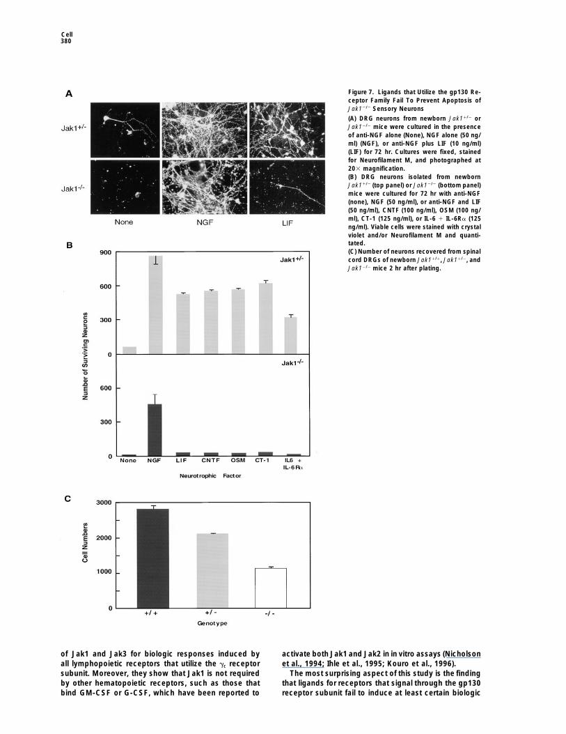
Ligands of the gp130 receptor family play importantroles in neuronal development and survival (Dechiara etal., 1995; Li et al., 1995; Ware et al., 1995). We thereforeexamined whether Jak12/2 neurons were capable of re-sponding to neurotrophic cytokines. Neurons from dor-sal root ganglions (DRGs) of control newborn mice cul-tured in the absence of growth factor died after 72 hrin culture, as evidenced by the absence of immunostain-ing for Neurofilament M (Figure 7A). However, neuronalviability was maintained if the cells were cultured in thepresence of either nerve growth factor (NGF) or LIF.Viability of neurons from newborn Jak12/2 mice was alsopreserved by NGF. However, almost no viable Jak12/2
neurons were detected following 3 day culture in thepresence of LIF.
Usingthis assay system, the response of DRG sensoryFigure 5. Diminished but Detectable STAT Activation following
neurons to a wider range of gp130 receptor family li-Stimulation of Jak12/2 Cells with Ligands that Utilize gp130 and/organds was quantitated. Whereas LIF, CNTF, OSM, CT-1,LIFRb
and the combination of IL-6 1 IL-6Ra individually pre-Five million macrophages (A) derived from Jak11/2 or Jak12/2 mice,served theviability of Jak1-sufficient neurons to approx-one million EFs (B) derived from Jak11/2 or Jak12/2 mice, or one
million cardiomyocytes (C) derived from Jak11/2 or Jak12/2 mice imately 75% of that induced by NGF (Figure 7B, topwere stimulated with either buffer alone, human IL-6 (400 ng/ml) panel), none of these cytokines was able to maintainand human IL-6Ra (500 ng/ml), murine LIF (400 ng/ml), murine IL-3 viability of Jak1-deficient neurons in culture (Figure 7B,(200 ng/ml), or murine GM-CSF (200 ng/ml) for 15 min or murine
bottom panel). In contrast, NGF was capable of pre-IFNg (1000 u/ml) for 10 min. Cells were lysed and DNA bindingventing apoptosis of Jak12/2 neurons. In situ hybridiza-activity quantitated by EMSA.tion revealed the presence of LIF receptor b chain mRNAin DRG neurons of either wild-type or Jak12/2 mice (data
stimulated in vitro with different cytokines and Stat acti- not shown). These results thus demonstrate a centralvation assessed using an electrophoretic mobility shift role for Jak1 in neuronal development and/or survivalassay (Figure 5). All cell types derived from Jak1-suffi- and strongly suggest that gp130 receptor family mem-cient animals activated Stat3 (and in some cases Stat1) bers display an obligate requirement for Jak1 to inducein response toeither IL-6 or LIF. In contrast, Stat3 activa- at least a subset of their cellular responses.tion was diminished but detectable in Jak12/2 cells stim- Interestingly, the total number of Jak1 null neuronsulated with IL-6 (50%, 85%, and .95% reduction for surviving 3 day culture with NGF was 33%–50% lowermacrophages, EFs, and cardiomyocytes, respectively) than that of Jak1-sufficient neurons. This result sug-and was almost completely abolished (.95%) but still gested that Jak12/2 mice might have an inherent neu-detectable in Jak12/2 cells treated with LIF. Whereas ronal deficit. To examine this possibility, we quantitatedthese results are consistent with the observations of recovery of DRG sensory neurons from wild-type, het-
erozygote, or Jak1 null mice examined 2, 24, or 72 hrothers (Stahl et al., 1994; Guschin et al., 1995), they

Cytokine Response Deficits in Jak12/2 Mice379
Figure 6. Jak12/2 Cardiomyocytes Fail ToUndergo a Hypertrophic Response to LIF,CT-1, or IL-6
(A) One representative dose response experi-ment of Jak11/2 (open symbols) or Jak12/2
(closed symbols) cardiomyocytes plated induplicate cultures and stimulated with the in-dicated amounts of cytokine for 48 hr. Cul-tures were fixed, stained, and the surfaceareas of 30 cells/dose quantitated and av-eraged.(B) Mean values of three independent experi-ments of Jak11/2 (open boxes) and Jak12/2
(shaded boxes) cardiomyocyte surface areasfollowing treatment with LIF (1 nM), CT-1 (1nM), or IL-6 1 IL-6Ra (500 ng/ml).
after isolation and culture in NGF. At each time point, the IFNs requires Jak-STAT pathway signaling (Durbinet al., 1996; Meraz et al., 1996). However, the demon-significantly lower numbers of neurons were found in thestration that Jak1 is needed for induction of IL-10–depen-Jak12/2 cultures compared to cultures established withdent responses has not been previously demonstrated.Jak1-sufficient cells (Figure 7C and data not shown).This result thus establishes that Jak1 is the commonThese results indicate that Jak1 may be necessary inJak-STAT signaling pathway component, which is sharedvivo to promote survival of at least certain subsets ofby all members of the class II cytokine receptor family.neurons.
Jak1 deficiency in mice led to severely reduced num-bers of thymocytes, pre–B cells, and mature T and BDiscussionlymphocytes but did not lead to alterations in the devel-opment of other hematopoietic lineages. This result thusAlthough Jak1 is a globally expressed Janus kinase,demonstrates that in hematopoiesis, Jak1 is required
which is activated by a wide variety of cytokines, itsonly by cytokine receptors that selectively induce devel-
requirement in the induction of cytokine-specific bio-opment of lymphoid cell populations. Through the use
logic responses remains largely undefined (Ihle et al.,of colony forming assays and proliferation assays, Jak1
1995; O’Shea, 1997). The purpose of this study was towas shown to be required for IL-7–dependent lympho-
identify those cytokine receptors that demonstrate anpoietic responses. Cells from Jak12/2 mice thus show
obligate dependency on Jak1 for induction of cellulara hematopoietic deficit similar to that observed in mice
responses. Our results show that Jak1 plays an indis- that lack IL-7, the IL-7 receptor a chain, orJak3 (Peschonpensable role in mediating biologic responses induced et al., 1994; Nosaka et al., 1995; Park et al., 1995; Thomisby three major cytokine receptor subfamilies: class II et al., 1995; von Freeden-Jeffry et al., 1995). These micecytokine receptors (i.e., the receptors for IFNa/b, IFNg, exhibit substantial reductions in T and B cell numbersand IL-10), cytokine receptors that utilize the gc receptor but do not display an absolute block in lymphocyte de-subunit (i.e., the receptors for IL-2, IL-4, IL-7, IL-9, and velopment. Recently, the deficits expressed in IL-7 unre-IL-15), and receptors that utilize the gp130 subunit (i.e., sponsive mice have been shown to be a result of thereceptors for IL-6, IL-11, LIF, OSM, CNTF, and CT-1). absence of a survival signal provided to developing cellsThese results demonstrate that under physiologic con- through the IL-7 receptor (Akashi et al., 1997). Our re-ditions, the Jaks play dedicated and nonredundant roles sults reveal that IL-7 receptor–induced responses re-in effecting cytokine specific biologic responses. quire Jak1 as well as Jak3 and demonstrate that Jak1
The results from analyses of cells and tissues derived is the obligate partner for Jak3 for inducing the survivalfrom Jak12/2 mice demonstrate that there is an absolute signal. In addition, thymocytes from Jak1 null mice failedrequirement for Jak1 in mediating biologic responses to respond to other lymphopoietic cytokines such asto IFNa and IFNg. This result is thus in agreement with IL-2 and IL-4 that associate with distinct ligand bindingearlier studies showing that mutant human tumor cell receptor subunits but also incorporate gc as part of theirlines which lack Jak1 are IFN-unresponsive (Muller et activated receptor complexes (Leonard et al., 1995).
These results demonstrate the generality of the pairingal., 1993) and that induction of all biologic responses to
Cell380
Figure 7. Ligands that Utilize the gp130 Re-ceptor Family Fail To Prevent Apoptosis ofJak12/2 Sensory Neurons
(A) DRG neurons from newborn Jak11/2 orJak12/2 mice were cultured in the presenceof anti-NGF alone (None), NGF alone (50 ng/ml) (NGF), or anti-NGF plus LIF (10 ng/ml)(LIF) for 72 hr. Cultures were fixed, stainedfor Neurofilament M, and photographed at203 magnification.(B) DRG neurons isolated from newbornJak11/2 (top panel) or Jak12/2 (bottom panel)mice were cultured for 72 hr with anti-NGF(none), NGF (50 ng/ml), or anti-NGF and LIF(50 ng/ml), CNTF (100 ng/ml), OSM (100 ng/ml), CT-1 (125 ng/ml), or IL-6 1 IL-6Ra (125ng/ml). Viable cells were stained with crystalviolet and/or Neurofilament M and quanti-tated.(C) Number of neurons recovered from spinalcord DRGs of newborn Jak11/1, Jak11/2, andJak12/2 mice 2 hr after plating.
of Jak1 and Jak3 for biologic responses induced by activate both Jak1 and Jak2 in in vitro assays (Nicholsonet al., 1994; Ihle et al., 1995; Kouro et al., 1996).all lymphopoietic receptors that utilize the gc receptor
subunit. Moreover, they show that Jak1 is not required The most surprising aspect of this study is the findingthat ligands for receptors that signal through the gp130by other hematopoietic receptors, such as those that
bind GM-CSF or G-CSF, which have been reported to receptor subunit fail to induce at least certain biologic
Cytokine Response Deficits in Jak12/2 Mice381
responses in Jak1-deficient cells. These results thus (Li et al., 1995; Ware et al., 1995). Both types of gene-targeted mice are significantly smaller than their lit-directly address the controversy concerning the dedi-
cated usage of Jaks by this family of cytokine receptors. termates at birth, fail to nurse, and die perinatally. Inaddition, neurons from both types of mice are unableExtensive work using cultured tumor cell lines and over-
expression approaches led to the concept that Jaks to respond to the ligands of the gp130 receptor familyand die by apoptosis. In the absence of the LIFRb sub-may be used in a nonspecific manner by the receptors
for IL-6, IL-11, LIF, OSM, CNTF, and CT-1 to initiate unit, newborn mice display decreased numbers of motorneurons. Although we have not yet quantitated motorsignaling (Stahl et al., 1994). Although this receptor fam-
ily is known to control a wide variety of important devel- neurons in the Jak12/2 mice, it is striking that the num-bers of sensory neurons isolated from these mice wereopmental, immune, and inflammatory responses (Kishi-
moto et al., 1995), no biologic responses were examined reduced compared to those isolated from Jak1-suffi-cient mice. Thus, it is possible that a neuronal defect inin the earlier studies. This concept was subsequently
challenged by an independent study showing that Jak1 the Jak1 null mice secondary to a lack of signalingthrough receptors that employ the LIFRb subunit is thewas necessary for IL-6–dependent induction of an im-
mediate early gene (IRF-1) in a human fibrosarcoma cell cause of the perinatal lethality. Work is currently under-way to examine this possibility.line (Guschin et al., 1995). Our results show that ligands
that bind to gp130 receptor family members, althoughExperimental Procedurescapable of inducing weak activation of the Jak-STAT
pathway in different Jak1-deficient cell types, fail to pro-Cytokines, Reagents, and General Proceduresvoke a biologic response in primary nontransformedPurified recombinant cytokines were obtained from the followingcardiomyocytes or neurons. Importantly, in the case ofsources: MuIFNg and MuCT-1 from Genentech (South San Fran-
cardiomyocytes, the discordance between initiation of cisco, CA); HuIFNaA/D from Dr. Michael Brunda, Hoffmann-LaRochesignaling and the induction of biologic responses was (Nutley, NJ); HuIL-6 and IL-6Ra chain was kindly provided by Dr.
Tetsuya Taga of Osaka University (Osaka, Japan); GM-CSF wasnoted in the same cell type. Thus, although other Jaksprovided by Dr. Satwant Narula, Schering-Plough Research Institutemay indeed be able to compensate to some degree for(Kenilworth, NJ); HuM-CSF, MuIL-3, and human and murine IL-7the absence of Jak1 to initiate the signaling process,from Genzyme Corporation (Cambridge, MA); MuIL-5, HuEPO, and
this compensation is not sufficient to result in the devel- MuOSM from R&D Systems (Minneapolis, MN); LIF from GIBCO/opment of a full cellular response. These observations BRL (Gaithersburg, MD); NGF from Harlan Bioproducts (Madison,establish that development of biologic responses in- WI); and rat CNTF was provided by Dr. Richard Scott and Dr. Sheryl
Meyer, Cephalon (West Chester,PA). A cDNA-encoding murine Jak1duced via the Jak-STAT pathway occur only when thewas provided by Dr. S. Nagata of Osaka Bioscience Institute (Osaka,level of signaling exceeds some, as yet undefined,Japan). All experiments presented were performed at least threethreshold value.times.Interestingly, the phenotype of Jak12/2 mice is distinct
from that displayed by mice with targeted disruption of Targeting Vectorgp130 (Yoshida et al., 1996) or Jak2 (Neubauer et al., Genomic clones containing portions of the murine Jak1 gene were1998; Parganas et al., 1998 [both in this issue of Cell]). isolated from a Lambda Fx II murine 129/Sv genomic library (Stra-
tagene, LaJolla, CA). To generate the Jak1 targeting vector, a 5.5 kbDeficiency in gp130 results in malformation of the ven-Xba-Xba fragment containing exon 3 was inserted into the targetingtricular walls of the heart and leads to embryonic lethalityvector pTK.NEO.UMS as previously described (Meraz et al., 1996)between e14.5–e17.5 p.c. Jak12/2 mice exhibited no de-such that its 39 end was adjacent to the polyoma enhancer promoter
velopmental heart abnormalities and survived to the first driven herpes simplex virus thymidine kinase gene. The 59 end of thepostnatal day of life. Moreover, no heart abnormality targeting construct was generated from a 1.6 kb PstI-PstI fragmentwas found in Jak12/2 pups at the time of their perinatal upstream of exon I (Figure 1).death. These results show that Jak1 is not required for
Transfection of ES Cells and Generationat least certain gp130-dependent events that lead toof Jak1-Deficient Micecardiac development and thereby lend support to theThe GS-1 embryonic stem cell line was cultured as described(Huangconcept that gp130 can activate multiple signal trans-et al., 1993). ES cells were electroporated with 20 mg of Sac II
duction pathways that give rise to distinct biologic re- linearized targeting construct per 1 3 107 cells and grown undersponses (Kishimoto et al., 1995). Jak2 deficiency results double selection as described (Meraz et al., 1996). Two hundred andin an embryonic lethality due to a lack of definitive eryth- thirty five colonies were analyzed for homologous recombination by
PCR using a sequence derived from the external probe (Figure 1).ropoiesis. Importantly, cells from Jak22/2 mice displayThree positive clones (C4, L3, and 1A15) were identified by PCR,cytokine response deficits that are nearly complemen-confirmed by Southern blotting, and microinjected into blastocysts.tary to those observed in our Jak12/2 mice. Like Jak12/2
Two clones (C4 and 1A15) transmitted the mutation germline andcells, Jak22/2 cells do not respond to IFNg. However, thus produced two independent lines of Jak12/2 mice.unlike Jak12/2 cells, Jak22/2 cells manifest biologicresponses to IL-6 and IFNa/b and can repopulate the Genotyping by Southern Blot and PCRlymphoid compartment of irradiated lymphocyte-defi- Southern blot analysis was performed on 25 mg of Eco RI digested
genomic DNA as described (Meraz et al., 1996). Filters were probedcient mice but fail to respond to erythropoietin, IL-3,with a 32P-labeled external probe (Figure 1). To genotype mice byor thrombopoietin. Thus, the combined data obtainedPCR, 0.5 mg of genomic DNA was amplified with one set of primersthrough the analysis of Jak1- and Jak2-deficient micederived from the neomycin gene (59 sequence: CACGACGGGCGTT
unequivocally demonstrate the specific utilization of the CCTTGCGCAG, 39 sequence: CCTGATGCTCTTCGTCCAGATCAT)Jaks by cytokine receptors. and a second set of primers derived from the second exon of the
In contrast, the phenotypes of the Jak12/2 mice and Jak1 gene deleted by the targeting construct (59 sequence: GAGGACTGCAATGCCATGGCGTTC, 39 sequence: CACTCCTGGGCGGCCCmice lacking the LIFRb subunit are remarkably similar
Cell382
TGATG). The samples were amplified in the following manner: 1 60 min. Medium (MEM supplemented with 10% FCS, 2 mM L-gluta-mine, 100 mg/ml penicillin and streptomycin, 20 mM fluorodeoxyuri-cycle at 948C for 1 s; 25 cycles at the following conditions: 948C for
30 s, 628C for 45 s, and 728C for 45 s; and finally 1 cycle at 728C for dine, and 20 mM uridine) was then added and the neurons trituratedusing a Pasteur pipette. Dissociated ganglia were plated into colla-2 min. A 200 bp fragment derived from exon 2 and a 250 bp fragment
derived from the neomycin gene wereresolved on a 2% TBE agarose gen-coated 24-well tissue culture dishes or collagen-coated glasschamber slides in MEM media supplemented with cytokine and/orgel.goat anti-murine NGF at a final dilution of 1/500. After 72 hr, cellswere fixed in 4% formalin, stained with crystal violet or anti-Neuro-Jak1 Western Blotsfilament M (Chemicon International, Temecula, CA), and the numberImmunoprecipitation and Western blotting were performed on ly-of viable cells enumerated by microscopy (Gribaudo et al., 1985).sates of EFs as described (Meraz et al., 1996) using different Jak1-
specific antisera obtained from Upstate Biotechnology Institute(Lake Placid, NY) and Santa Cruz Biotechnology (Santa Cruz, CA). Acknowledgments
Cell Culture and Transfection We are grateful to Michel Aguet (ISREC, Lausanne, Switzerland) forImmortalized normal and Jak12/2 EFs were generated using the helpful discussions, William Snider and Jeffrey Elliot (Departmentmethod of Todaro and Green (1963). Jak12/2 EFs were reconstituted of Neurology and Neurological Surgery, Washington Universityby cotransfecting 1 3 106 EFs with 20 mg of a murineJak1 expression School of Medicine) for conducting the in situ hybridization studiesplasmid and 5 mg of the pMON Hygromycin B resistance plasmid for LIFRb expression, and Jeffrey Saffitz (Department of Pathology,by electroporation at 400 V, 320 mF on a Biorad gene pulser (Her- Washington University School of Medicine) for pathology studiescules, CA). Transfected cells were selected in 0.1 mg/ml Hygromycin on the Jak12/2 mice. We also thank Paul Allen, Kenneth Murphy,for 3 weeks and then cloned by limiting dilution. Macrophages de- Andrey Shaw, and Emil Unanue (Department of Pathology, Washing-rived from e16.5–18.5 fetal liver were prepared in a manner similar ton University School of Medicine) for helpful suggestions. The stud-to that used for growing bone marrow derived macrophages (Celada ies conducted in the R. D. S. laboratory were supported by Nationalet al., 1984). Institutes of Health grant CA43059 and grants from Genentech, Inc.
and Abbott Laboratories. Studies conducted in the E. M. J. labora-tory were supported by National Institutes of Health grant AG12947.Induction of Cytokine-Dependent Cellular Responses
on EFs, Macrophages, and LymphocytesEFs and macrophages were stimulated with designated doses of Received February 20, 1998; revised March 13, 1998.cytokines andassayed for expression of cell surface proteins, antivi-ral activity, iNOS induction, or inhibition of LPS-induced TNF pro-duction as previously described (Meraz et al., 1996). Thymocytes Referencesisolated from newborn mice were stained with FITC anti-CD4 andPE anti-CD8 (Pharmingen, San Diego, CA) as described (Dighe et Akashi, K., Kondo, M., vonFreeden-Jeffry, U., Murray, R., and Weiss-al., 1995). Fetal liver cells and newborn splenocytes were stained man, I.L. (1997). Bcl-2 rescues T lymphopoiesis in interleukin-7 re-after blocking Fc receptors with FITC anti-IgM and PE anti-B220 ceptor-deficient mice. Cell 89, 1033–1041.(Pharmingen, San Diego, CA). The capacity of thymocytes to prolif- Bach, E.A., Aguet, M., and Schreiber, R.D. (1997). The IFNg receptor:erate in response to IL-2, IL-4, IL-7, or ionomycin in the presence a paradigm for cytokine receptor signaling. Annu. Rev. Immunol.of PMA was determined as described (Park et al., 1995). 15, 563–591.
Celada, A., Gray, P.W., Rinderknecht, E., and Schreiber, R.D. (1984).Colony Forming Assays Performed in Methylcellulose Evidence for a gamma-interferon receptor that regulates macro-Cytokine-dependent fetal liver cell colony formation was assessed phage tumoricidal activity. J. Exp. Med. 160, 55–74.as described (Suda et al., 1989) using a commercial assay system Darnell, J.E., Jr. (1997). STATs and gene regulation. Science 277,(Stem Cell Technologies, Vancouver, Canada). The following analy- 1630–1635.ses were used: 1.0 3 105 cells/plate with M-CSF, 1.25 3 105 cells/
Darnell, J.E., Jr., Kerr, I.M., and Stark, G.R. (1994). Jak-STAT path-plate with GM-CSF or IL-3, 3 3 105 cells/plate with G-CSF, and 5 3ways and transcriptional activation in response to IFNs and other105 cells/plate with IL-5. For IL-7 cultures, 5 3 105 cells/plate wereextracellular signaling proteins. Science 264, 1415–1421.cultured either in pre–B cell medium (Stem Cell Technologies), whichDechiara, T.M., Vejsada, R., Poueymirou, W.T., Acheson, A., Suri,contained 10 ng/ml HuIL-7, or in normal basal methylcellulose me-C., Conover, J.C., Friedman, B., McClain, J., Pan, L., Stahl, N., etdium supplemented with up to 50 ng/ml MuIL-7.al. (1995). Mice lacking the CNTF receptor, unlike mice lacking CNTF,exhibit profound motor neuron deficits at birth. Cell 83, 313–322.Electrophoretic Mobility Shift AssayDighe, A.S., Campbell, D., Hsieh, C.-S., Clarke, S., Greaves, D.R.,Cells in 1 ml PBS/10% FCS were stimulated with different cytokinesGordon, S., Murphy, K.M., and Schreiber, R.D. (1995). Tissue specificand analyzed by an EMSA that used a 32P-labeled 18 base pair GRRtargeting of cytokine unresponsiveness in transgenic mice. Immu-probe (Greenlund et al., 1994).nity 3, 657–666.
Durbin, J.E., Hackenmiller, R., Simon, M.C., and Levy, D.E. (1996).Cardiomyocyte HypertrophyTargeted disruption of the mouse Stat1 gene results in compromisedThis assaywas performed as described for rat cardiomyocytes (Kinginnate immunity to viral infection. Cell 84, 443–450.et al., 1996). Cardiomyocytes isolated from the hearts of newbornGreenlund, A.C., Farrar, M.A., Viviano, B.L., and Schreiber, R.D.mice were plated overnight at a density of 1.25 3 104 cells per well(1994). Ligand induced IFNg receptor phosphorylation couples thein 96-well tissue culture plates in DMEM/F12 medium containingreceptor to its signal transduction system (p91). EMBO J. 13, 1591–insulin, transferrin, and aprotinin. Cultures were stimulated with LIF1600.or CT-1 or mixtures of IL-6 plus soluble IL-6 receptor a chain. Forty
eight hours later, cells were fixed, stained with crystal violet, and Gribaudo, G., Cofano, F., Prat, M., Baiocchi, C., Cavallo, G., andcell surface areas quantitated by digital microscopy (King et al., Landolfo, S. (1985). Natural murine interferon-gamma: evidence for1996). post-translational proteolytic processing. J. Biol. Chem. 260, 9936–
9940.
Guschin, D., Rogers, N., Briscoe, J., Witthuhn, B., Watling, D., Horn,Neuronal CulturesThese assays employed an adaptation of the culture system re- F., Pellegrini, S., Yasukawa, K., Heinrich, P., Stark, G.R., Ihle, J.N.,
and Kerr, I.M. (1995). A major role for the protein tyrosine kinaseported by others (Dechiara et al., 1995). DRG from individual new-born mice were dissected, placed in L-15 medium (Sigma Chemical Jak1 in the Jak/STAT signal transduction pathway in response to
interleukin-6. EMBO J. 14, 1421–1429.Company, St. Louis, MO), and dissociated with trypsin (0.05%) for

Cytokine Response Deficits in Jak12/2 Mice383
Hardy, R.R., Carmack, C.E., Shinton, S.A., Kemp, J.D., and Haya- O’Shea, J.J., and Leonard, W.J. (1995). Mutation of Jak3 in a patientwith SCID: essential role of Jak3 in lymphoid development. Sciencekawa, K. (1991). Resolution and characterization of pro-B and pre-
pro-B cell stages in normal mouse bone marrow. J. Exp. Med. 173, 270, 797–800.1213–1225. Stahl, N., Boulton, T.G., Farruggella, T., Ip, N.Y., Davis, S., Witthuhn,
B.A., Quelle, F.W., Silvennoinen, O., Barbiere, G., Pellegrini, S., Ihle,Ho, A.S.-Y., Wei, S.H.-Y., Mui, A.L.-F., Miyajima, A., and Moore, K.W.(1995). Functional regions of the mouse IL-10 receptor cytoplasmic J.N., and Yancopoulos, G.D. (1994). Association and activation of
Jak-Tyk kinases by CNTF-LIF-OSM-IL-6 beta receptor components.domain. Mol. Cell. Biol. 15, 5043–5053.Science 263, 92–95.Huang, S., Hendriks, W., Althage, A., Hemmi, S., Bluethmann, H.,
Kamijo, R., Vilcek, J., Zinkernagel, R., and Aguet, M. (1993). Immune Suda, T., Okada, S., Suda, J., Miura, Y., Ito, Y., Sudo, T., Hayashi,S., Nishikawa, S., and Nakauchi, H. (1989). A stimulatory effect ofresponse in mice that lack the interferon-g receptor. Science 259,
1742–1745. recombinant murine interleukin-7 (IL-7) on B-cell colony formationand an inhibitory effect of IL-1a. Blood 74, 1936–1941.Ihle, J.N., Witthuhn, B.A., Quelle, F.W., Yamamoto, K., and Silven-
noinen, O. (1995). Signaling through the hematopoietic cytokine re- Thomis, D.C., Gurniak, C.B., Tivol, E., Sharpe, A.H., and Berg, L.J.ceptors. Annu. Rev. Immunol. 13, 369–398. (1995). Defects in B lymphocyte maturation and T lymphocyte acti-
vation in mice lacking Jak3. Science 270, 794–797.King, K.L., Lai, J., Winer, J., Luis, E., Yen, R., Hooley, J., Williams,P.M., and Mather, J.P. (1996). Cardiac fibroblasts produce leukemia Todaro, G.J., andGreen, H. (1963). Quantitative studies of thegrowthinhibitory factor and endothelin, which combine to induce cardiac of mouse embryo cells in culture and their development in estab-myocyte hypertrophy in vitro. Endocrine 5, 85–93. lished lines. J. Cell Biol. 17, 299–313.Kishimoto, T., Akira, S., Narazaki, M., and Taga, T. (1995). Interleu- Velazquez, L., Fellous, M., Stark, G.R., and Pellegrini, S. (1992). Akin-6 family of cytokines and gp130. Blood 86, 1243–1254. protein tyrosine kinase in the interferon a/b signaling pathway. Cell
70, 313–322.Kouro, T., Kikuchi, Y., Kanazawa, H., Hirokawa, K., Harada, N., Shi-iba, M., Wakao, H., Takaki, S., and Takatsu, K. (1996). Critical proline von Freeden-Jeffry, U., Vieira, P., Lucian, L.A., McNeil, T., Burdach,residues in the cytoplasmic domain of the IL-5 receptor alpha chain S.E., and Murray, R. (1995). Lymphopenia in interleukin (IL)-7 gene-and its function in IL-5-mediated activation of JAK kinase and deleted mice identifies IL-7 as a nonredundant cytokine. J. Exp.STAT5. Int. Immunol. 8, 237–245. Med. 181, 1519–1526.Leonard, W.J., Shores, E.W., and Love, P.E. (1995). Role of the Ware, C.B., Horowitz, M.C., Renshaw, B.R., Hunt, J.S., Liggit, D.,common cytokine receptor g chain in cytokine signaling and lym- Koblar, S.A., Gliniak, B.C., McKenna, H.J., Papayannopoulou, T.,phoid development. Immunol. Rev. 148, 97–114. Thoma, B., et al. (1995). Targeted disruption of the low-affinity leuke-
mia inhibitory factor receptor gene causes placental, skeletal, neu-Li, M., Sendtner, M., and Smith, A. (1995). Essential function of LIFreceptor in motor neurons. Nature 378, 724–727. ral, and metabolic defects and results in perinatal death. Develop-
ment 121, 1283–1299.Meraz, M.A., White, J.M., Sheehan, K.C.F., Bach, E.A., Rodig, S.J.,Dighe, A.S., Kaplan, D.H., Riley, J.K., Greenlund, A.C., Campbell, Watling, D., Guschin, D., Muller, M., Silvennoinen, O., Witthuhn, B.A.,D., et al. (1996). Targeted disruption of the STAT1 gene in mice Quelle, F.W., Rogers, N.C., Schindler, C., Stark, G.R., Ihle, J.N. andreveals unexpected physiologic specificity in the Jak-STAT signaling Kerr, I.M. (1993). Complementation by the protein tyrosine kinasepathway. Cell 84, 431–442. JAK2 of a mutant cell line defective in the interferon-g signal trans-
duction pathway. Nature 366, 166–170.Muller, M., Briscoe, J., Laxton, C., Guschin, D., Ziemiecki, A., Silven-noinen, O., Harpur, A.G., Barbier, G., Witthuhn, B.A., Schindler, C., Yoshida, K., Taga, T., Saito, M., Suematsu, S., Kumanogoh, A., Ta-et al. (1993). The protein tyrosine kinase JAK1 complements a mu- naka, T., Fujiwara, H., Hirata, M., Yamagami, T., Nakahata, T., et al.tant cell line defective in the interferon-a/b and -g signal transduc- (1996). Targeted disruption of gp130, a common signal transducertion pathways. Nature 366, 129–135. for IL-6-family cytokines, leads to myocardial and hematological
disorders. Proc. Natl. Acad. Sci. USA 93, 407–411.Neubauer, H., Cumano, A., Muller, M., Wu, H., Huffstadt, U., andPfeffer, K. (1998). Jak2 deficiency defines an essential devel-opmental checkpoint in definitive hematopoiesis. Cell 93, this issue,397–409.
Nicholson, A.E., Oates, A.C., Harpus, A.G., Ziemiecki, A., Wilks, A.F.and Layton, J.E. (1994). Tyrosine kinase Jak1 is associated with thegranulocyte-colony-stimulating factor receptor and both becometyrosine-phosphorylated after receptor activation. Proc. Natl. Acad.Sci. USA 91, 2985–2988.
Nosaka, T., van Deursen, J.M.A., Tripp, R.A., Thierfelder, W.E., Wit-thuhn, B.A., McMickle, A.P., Doherty, P.C., Grosveld, G.C., and Ihle,J.N. (1995). Defective lymphoid development in mice lacking Jak3.Science 270, 800–802.
O’Shea, J.J. (1997). Jaks, STATs, cytokine signal transduction, andimmunoregulation: are we there yet? Immunity 7, 1–11.
Parganas, E., Wang, D., Stravopodis, D., Topham, D.J., Marine, J.-C.,Teglund, S., Vanin, E., Bodner, S., Colamonici, O.R., van Deursen,J.M., Grosveld, G., andIhle, J.N. (1998). Jak2 is essential for signalingthrough a variety of cytokine receptors. Cell 93, this issue, 385–395.
Park, S.Y., Saijo, K., Takahashi, T., Osawa, M., Arase, H., Hirayama,N., Miyake, K., Nakauchi, H., Shirasawa, T., and Saito, T. (1995).Developmental defects of lymphoid cells in Jak3 kinase-deficientmice. Immunity 3, 771–782.
Peschon, J.J., Morrissay,P.J., Grabstein, K.H.,Ramsdell, F.J., Mara-skovsky, E., Gliniak, B.C., Park, L.S., Ziegler, S.F., Williams, D.E.,Ware, C.B., et al. (1994). Early lymphocyte expansion is severelyimpaired in interleukin 7 receptor-deficient mice. J. Exp. Med. 180,1955–1960.
Russell, S.M., Tayebi, N., Nakajima, H., Riedy, M.C., Roberts, J.L.,Aman, M.J., Migone, T., Noguchi, M., Markert, M.L., Buckley, R.H.,


![Quantitative analysis of [11C]-erlotinib PET demonstrates specific binding for activating mutations of the EGFR kinase domain](https://static.fdokumen.com/doc/165x107/6345ca446cfb3d406409d7f9/quantitative-analysis-of-11c-erlotinib-pet-demonstrates-specific-binding-for-activating.jpg)