Human relaxin-2: historical perspectives and role in cancer biology
Upload
independentCategory
view
4download
0
pubs.acs.org/biochemistry Published on Web 01/13/2011 r 2011 American Chemical Society
1368 Biochemistry 2011, 50, 1368–1375
DOI: 10.1021/bi1013968
H3 Relaxin Demonstrates Antifibrotic Properties via the RXFP1 Receptor†
Mohammed Akhter Hossain,‡,§,# Bryna Chow Suet Man,‡, ),# Chongxin Zhao,‡ Qi Xu,^ Xiao-Jun Du,^ John D. Wade,‡,§
and Chrishan S. Samuel*,‡, )
‡Howard Florey Institute, §School of Chemistry, and )Department of Biochemistry and Molecular Biology, The University ofMelbourne, Parkville, Victoria 3010, Australia, and ^Baker IDI Heart and Diabetes Institute, St. Kilda Road Central, Melbourne,
Victoria 8008, Australia. #M.A.H. and B.C.S.M. contributed equally to the manuscript
Received August 30, 2010; Revised Manuscript Received January 11, 2011
ABSTRACT: Human gene 3 (H3) relaxin is the most recently discovered member of the relaxin peptide familyand can potentially bind all of the defined relaxin family peptide receptors (RXFP1-4). While its effects as aneuromodulator are being increasingly studied through its primary receptor, RXFP3, its actions via otherRXFPs are poorly understood. Hence, we specifically determined the antifibrotic effects and mechanisms ofaction of H3 relaxin via the RXFP1 receptor using primary rat ventricular fibroblasts in vitro, which naturallyexpress RXFP1, but not RXFP3, and a mouse model of fibrotic cardiomyopathy in vivo. Transforminggrowth factor β1 (TGF-β1) administration to ventricular fibroblasts significantly increased Smad2 phos-phorylation, myofibroblast differentiation, and collagen deposition (all p<0.05 vs untreated controls), whilehaving no marked effect on matrix metalloproteinase (MMP) 9, MMP-13, tissue inhibitor of metallopro-teinase (TIMP) 1, or TIMP-2 expression over 72 h. H3 relaxin (at 100 and 250 ng/mL) almost completelyabrogated the TGF-β1-stimulated collagen deposition over 72 h, and its effects at 100 ng/mL were equivalentto that of the same dose of H2 relaxin. Furthermore, H3 relaxin (100 ng/mL) significantly inhibited TGF-β1-stimulated cardiac myofibroblast differentiation and TIMP-1 and TIMP-2 expression to an equivalent extentas H2 relaxin (100 ng/mL), while also inhibiting Smad2 phosphorylation to approximately half the extent ofH2 relaxin (all p < 0.05 vs TGF-β1). Lower doses of H3 (50 ng/mL) and H2 (50 ng/mL) relaxin additivelyinhibited TGF-β1-stimulated collagen deposition in vitro, while H3 relaxin was also found to reverse leftventricular collagen overexpression in the model of fibrotic cardiomyopathy in vivo. These combined findingsdemonstrate that H3 relaxin exerts antifibrotic actions via RXFP1 and may enhance the collagen-inhibitoryeffects of H2 relaxin.
Three nonallelic relaxin genes have been identified in humans,which produce H1, H2,1 and H3 relaxin, respectively (1). Mostother mammals including rodents, however, contain only tworelaxin genes, which produce relaxin (equivalent to H2 relaxin)and relaxin-3 (equivalent to H3 relaxin) (1). H2 relaxin (andrelaxin in other species) represent the major stored and circulat-ing forms of relaxin and have been the most investigated todate (1-7). A plethora of studies have demonstrated that H2relaxin has several biological actions in the body, which arecentered around its antiinflammatory, antifibrotic, antiapopto-tic, antihypertrophic, cardioprotective, vasodilatory, and proan-giogenic actions (1-7), many of which are mediated through its
primary G-protein-coupled receptor, relaxin family peptidereceptor 1 (RXFP1) (8).
On the other hand, comparatively little is known about themore recently discovered H3 relaxin (9, 10) and its highlyconserved species equivalent, relaxin-3 (11). While the dis-tribution of H3 relaxin in humans remains unknown, anato-mical studies of relaxin-3 in rodents and primates suggest thatit is predominantly expressed in the brain, particularly withinneurons of the nucleus insertus (9, 10, 12, 13) in addition tonerve fibers and terminals within the cortex, hippocampus,thalamus, hypothalamus, and midbrain. Thus, H3 relaxin andrelaxin-3 are thought to primarily function as neuromodula-tors and have been implicated in regulating arousal, feeding,learning, and memory and central responses to physiologicalstressors (14-16).
Studies using recombinant (17) and chemically synthesized (18)H3 relaxin have identified it as the cognate ligand for relaxinfamily peptide receptor 3 (RXFP3; formerly known as GPCR-135), which is also widely distributed in the brain (12, 13, 17).However, it has been demonstrated thatH3 relaxin can bind to allfour defined receptors of the relaxin family of peptides, includingRXFP1 (18), RXFP2 (the primary receptor for insulin-likepeptide 3) (18), and RXFP4 (the primary receptor for insulin-like peptide 5) (19, 20), suggesting that it may have additionalroles through these receptors, other than its neuromodulatoryactions via RXFP3.
†This study was supported by a John T. Reid Charitable TrustsFellowship to M.A.H., National Health andMedical Research Council(NHMRC) of Australia Senior Research Fellowships to X.-J.D. andJ.D.W., a National Heart Foundation of Australia/NHMRC RDWright Fellowship to C.S.S., and a NHMRC Project Grant (508995)to J.D.W. and by the Victorian Government’s Operational Infrastruc-ture Support Program.*To whom correspondence should be addressed at the Howard
Florey Institute, The University of Melbourne. Phone: þ 61 3 83440416. Fax: þ61 3 9348 1707. E-mail: [email protected].
1Abbreviations: AR, adrenergic receptors; H2, human gene 2; H3,human gene 3; GPCR, G-protein-coupled receptor; MALDI-TOF,matrix-assisted laser desorption ionization time of flight; MMP, matrixmetalloproteinase; RP-HPLC, reverse-phase high-performance liquidchromatography; RXFP, relaxin family peptide receptor; SMA, smoothmuscle actin; TGF-β1, transforming growth factor β1; TIMP, tissueinhibitor of metalloproteinase.
Article Biochemistry, Vol. 50, No. 8, 2011 1369
Indeed H3 relaxin has been shown to promote matrix metal-loproteinase (MMP) 2 levels in a dose-dependent manner, whenadministered to rat ventricular fibroblasts in vitro, and stimulatewater drinking in rats in vivo (18) in a comparable manner to thatof H2 relaxin, via a high-affinity interaction with RXFP1. Thesefindings imply that H3 relaxin maymimic the matrix-remodelingand tissue homeostasis-related actions of H2 relaxin through theRXFP1 receptor. Consistent with the effects of H3 relaxin onMMP-2 activity in vitro, relaxin-3 has been found to inhibitcardiac fibrosis and improve cardiac dysfunction in an experi-mental rat model of cardiac toxicity in vivo (21), again suggestingthat H3 relaxin/relaxin-3 may possess similar matrix-remodelingand antifibrotic actions to that of H2 relaxin via RXFP1.
On the basis of these previous observations (18, 21), we havesought to determine the mechanisms by which H3 relaxinmediates its antifibrotic actions via the RXFP1 receptor inneonatal rat ventricular fibroblasts in vitro (which only expressRXFP1 but not RXFP3) (22) and in a mouse model of fibroticcardiomyopathy in vivo (22, 23), which have both been used toevaluate the antifibrotic actions of H2 relaxin (22, 23). Addition-ally, we evaluated if H2 and H3 relaxin had any synergisticantifibrotic actions via the RXFP1 receptor.
EXPERIMENTAL PROCEDURES
Materials. The H3 relaxin used in these studies was chemi-cally synthesized and characterized as described previously (18).Recombinant H2 relaxin was kindly provided by Corthera Inc.(SanMateo, CA), andTGF-β1was obtained fromR&DSystems(Minneapolis, MN).Animals. Neonate (1-day-old) Sprague-Dawley rats were used
for tissue collection and subsequent cardiac cell isolation andpreparation. The 4-5-month-old male wild-type and heterozygousβ2-adrenergic receptor (β2-AR) mice used in these studies weregenerated from heterozygous (C57Blk6JxSJL) parents (22-24).The animals were housed in a controlled environment andmaintained on a 14 h light/10 h dark schedule with access torodent lab chow (Barastock Stockfeeds, Pakenham, Victoria,Australia) andwater. The detailed experimentswere approved bytheHowardFlorey Institute’s and Baker IDIHeart andDiabetesInstitute’s Animal Ethics Committees, which adhere to theAustralian code of practice for the care and use of laboratoryanimals for scientific purposes.Cell Culture. Ventricular fibroblasts were obtained from
neonate rats using standard collagenase digestion methods,isolated from myocytes, and subsequently prepared for experi-mentation as described before (25). These preparations containedmore than 95% cardiac fibroblasts as determined by morpholo-gical appearance and immunocytochemical staining and werepreviously demonstrated to express RXFP1 but notRXFP3 (22).The cells were maintained in DMEM and supplemented with10% fetal calf serum and the antibiotics penicillin (50 units/mL)and streptomycin (50 μg/mL) (DMEM-FBS). These fibroblastswere used between passages 2 and 4 for all studies, while alldescribed experiments were performed separately at least three toseven times in duplicate.H3 Relaxin Dose-Response Studies on Collagen De-
position. To determine the optimal dose(s) at which H3 relaxininhibited TGF-β1-stimulated collagen deposition, ventricularfibroblasts were plated at an equal density of 1 � 106 cells insix-well plate wells and supplemented with 1 mL (per well) ofDMEM-FBS. The cells were then either untreated (controls)or treated for 72 hwith TGF-β1 (3 ng/mL) alone or with TGF-β1
(3 ng/mL) plus increasing concentrations of H3 relaxin (10, 50,100, 250, and 500 ng/mL) or with TGF-β1 (3 ng/mL) plus H2relaxin (100 ng/mL; used as a positive control at a concentrationthat had previously been used to inhibit TGF-β1-stimulatedcollagen deposition (22)). The optimal dose at which H3 relaxininhibited TGF-β1-stimulated collagen deposition was also testedalone over a 72 h culture period to determine if it affected basalcollagen deposition. After 72 h, the deposited collagen into thecell layers (from each well) was isolated and hydrolyzed with 6Mhydrochloric acid formeasurement of hydroxyproline content, asdescribed previously (26, 27). Hydroxyproline values were thenconverted to total collagen content by multiplying by a factor of6.94 (based on hydroxyproline representing approximately 14.4%of the amino acid composition of collagen in most mammaliantissues (28)).Western Blot Analysis of Smad2 Phosphorylation and
R-SmoothMuscle Actin (SMA) andMMP-13 Expression.To determine if H3 relaxin had any effects on Smad2 phosphor-ylation (pSmad2; a key mediator of TGF-β1 signal transduction)and R-SMA expression (a marker of myofibroblast differentia-tion), which are both downregulated by H2 relaxin (22, 29-31),and MMP-13 (collagenase-3, an enzyme involved in collagendegradation), which is upregulated by H2 relaxin (32, 33), ven-tricular fibroblasts were plated at an equal density of 1� 105 cellsin 12-well plate wells and supplemented with 1mLDMEM-FBS.The cells were then either untreated or treated for 72 hwith TGF-β1 (3 ng/mL) alone or with TGF-β1 (3 ng/mL) plus H3 relaxin(100 ng/mL) or with TGF-β1 (3 ng/mL) plus H2 relaxin (100 ng/mL; positive control). After 72 h, the cell layers were subjected toTrizol reagent, and total protein was extracted from each sample(according to the manufacturer’s instructions; Life Technologies,Gaithesburg, MD) and quantified by the Bio-Rad dye-bindingprotein assay (Bio-Rad Laboratories, Richmond, CA).
Equal protein extracts (in 1% sodium dodecyl sulfate; 10-15μg of total protein/lane) were analyzed by electrophoresis undernonreducing conditions on 12.5% acrylamide gels, as describedbefore (22). Western blot analysis was then performed withprimary monoclonal antibodies to either pSmad2 (no. 3108;1:1000 dilution; Cell Signaling Technology, Danvers, MA),R-SMA (M0851; 1:1000 dilution; Dako Corp., Carpinteria, CA),or MMP-13 (IM78T; 1:1000 dilution; Calbiochem, San Diego,CA) and either goat anti-rabbit (for pSmad2; 1:2500 dilution; Bio-Rad) or goat anti-mouse (for R-SMA and MMP-13; 1:2000dilution; Bio-Rad) secondary antibodies as detailed previously(22, 30, 33). Membranes probed for pSmad2 were stripped andreprobed with a Smad2 polyclonal antibody (no. 3102; 1:750dilution; Cell Signaling Technology) for determination of basalSmad2 levels and equal loadingof the samples,while amonoclonalantibody to R-tubulin (05-829; 1:8000 dilution; Millipore Corp.,Bedford,MA) was used to determine equal loading of samples forR-SMA and MMP-13. Densitometry of the pSmad, R-SMA, andMMP-13 bands was performed using a Bio-RadGS710 calibratedimaging densitometer and Quantity-One software (Bio-Rad).Gelatin Zymography of MMP-9 Expression and Activ-
ity. Of the main collagen degrading enzymes (MMP-2, -9, and -13) that have been shown to be upregulated by H2 relaxin inrodentmodels of disease (5, 22, 29, 31-33),MMP-2has previouslybeen shown to be stimulated in a dose-dependent manner by H3relaxin (18). Hence we decided to focus on the effects of H3relaxin on MMP-9 and -13 in this study.
To assess the effects of H3 relaxin on the latent and activelevels of MMP-9, ventricular fibroblasts were plated at an equal

1370 Biochemistry, Vol. 50, No. 8, 2011 Hossain et al.
density of 1� 105 cells in 12-well plate wells and supplementedwith1 mL (per well) of DMEM-FBS. The cells were then either untrea-ted (controls) or treated for 72 h with TGF-β1 (3 ng/mL) alone orwith TGF-β1 (3 ng/mL) plusH3 relaxin (100 ng/mL) orwith TGF-β1 (3 ng/mL) plus H2 relaxin (100 ng/mL; positive control), thefinal 24 h of treatment under serum-free conditions (as seruminterferes with zymographic analysis of MMPs). Equal aliquots ofthe collectedmediawere thenanalyzedonzymogramgels consistingof 7.5% acrylamide and 1 mg/mL gelatin, and the gels weresubsequently treated as previously detailed (22, 33). Clear bandsindicated gelatinolytic activity, while densitometry of the (latent andactive) MMP-9 bands was performed as described above.Reverse Zymography of Tissue Inhibitor of Metallopro-
teinase (TIMP) 1 andTIMP-2Expression.To determine theeffects of H3 relaxin on TIMP-1 (the natural inhibitor ofMMP-9and MMP-13) and TIMP-2 (the natural inhibitor of MMP-2)expression, equal aliquots of the same media samples (fromuntreated and TGF-β1 (3 ng/mL) ( H3 relaxin (100 ng/mL) orH2 relaxin (100 ng/mL) treated fibroblasts) that were used toidentify changes in MMP-9 activity (by gelatin zymography)were also assessed by reversed zymography as described be-fore (34). Densitometry of the TIMP-1 and TIMP-2 bands wasperformed as described above.Assessment of the Synergistic Effects of H2 and H3
Relaxin on Collagen Deposition. To determine if H3 relaxinhad any synergistic effects to the collagen inhibitory actions ofH2relaxin (via RXFP1 (22)), fibroblasts were plated at an equaldensity of 1 � 106 cells in six-well plate wells and supplementedwith 1 mL of DMEM-FBS. The cells were then either untreatedor treated for 72 hwith TGF-β1 (3 ng/mL) alone or with TGF-β1(3 ng/mL) plus H3 relaxin (50, 100 ng/mL), TGF-β1 (3 ng/mL)plus H2 relaxin (50, 100 ng/mL), or TGF-β1 (3 ng/mL) plus H3relaxin (50 ng/mL) plus H2 relaxin (50 ng/mL). The collagendeposited into the cell layer of each sample over 72 h was thenassessed by the hydroxyproline assay, as described above.Assessment of the Antifibrotic Effects of H3 Relaxin in
an Experimental Model of Fibrotic Cardiomyopathy inVivo. The ability of H3 relaxin to reduce myocardial collagenaccumulation in vivowas investigated in a well-established modelof cardiac fibrosis, induced by cardiac-restricted transgenic over-expression of β2-AR, a model which had previously been used toevaluate the effects of H2 relaxin (22, 23). The age at whichcardiac fibrosis was already established in this model (4-5months of age) was chosen for treatment studies, based on ourprevious observations (22, 23). Transgenic animals (n=6-7 pergroup) were subcutaneously implanted with osmotic minipumps(model 2002; DURECTCorp., Cupertino, CA) containing eithera vehicle (20 mM sodium acetate buffer, pH 5.0) or synthetic H3relaxin in vehicle at a concentration of 0.5 mg kg-1 day-1 (aconcentration at which H2 relaxin has previously been used toassess its antifibrotic effects (22, 35, 36)). Age-matched wild-typelittermate mice were used as controls. After 14 days, the leftventricle (LV) of each animal was isolated for hydroxyprolineassessment of collagen content (26, 27).Statistical Analysis. The results were analyzed using a one-
way ANOVA and the Newman-Keuls test for multiple compar-isons between groups. All data in this study are presented as themean ( SEM, with p < 0.05 considered statistically significant.
RESULTS
Synthesis and Characterization of H3 Relaxin. The hu-man H3 relaxin A- and B-chains were synthesized separately by
continuous flow Fmoc (fluoren-9-ylmethoxycarbonyl) solid-phase peptide synthesis (18). The two chains were combinedusing previously reported sequential and directed disulfide bondformation strategy (18, 37, 38). The final product was purified bypreparative RP (reverse-phase) HPLC. The high purity of thepeptide was confirmed by analytical RP-HPLC, and the identitywas confirmed by MALDI-TOF mass spectrometry (calculatedMHþ, 5499.52; found MHþ, 5498.51).Effects of H3 Relaxin on TGF-β1-Stimulated Collagen
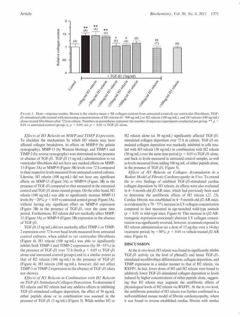
Deposition. The optimal dose (10-500 ng/mL) at which H3relaxin inhibited TGF-β1-stimulated collagen deposition over72 h in vitrowas determined by hydroxyproline analysis of fibro-blast cell layers (Figure 1). TGF-β1 (3 ng/mL) alone significantlyincreased collagen deposition by 40-50% (p<0.01 vs untreatedcells) over 72 h in culture, consistent with its profibrotic actionsand previously documented effects on rat cardiac fibroblasts (22).An inverse bell-shaped dose-response curve was observed withH3 relaxin treatment, where it did not have anymarked effects onTGF-β1-stimulated collagen deposition at 10 and 50 ng/mL,significantly inhibited TGF-β1-stimulated collagen deposition at100 and 250 ng/mL (back to levels measured in untreated controlsamples; both p < 0.05 vs TGF-β1 alone), but only induced atrend toward a decrease in TGF-β1-stimulated collagen deposi-tion at 500 ng/mL (no significant difference vs TGF-β1 alone;Figure 1) over 72 h, with its optimal inhibitory actions beingmediated at 100 ng/mL. At this dose, H3 relaxin was able toinhibit TGF-β1-stimulated collagen deposition to a similar extentas 100 ng/mL H2 relaxin (both p < 0.05 vs TGF-β1 alone, andback to levels measured in untreated control samples) over 72 h(Figure 1). However, H3 relaxin alone (100 ng/mL) did not affectbasal collagen expression in the absence of TGF-β1 (Figure 1).Thus, 100 ng/mL H3 relaxin was used for subsequent experi-ments to assess its mechanisms of action (via RXFP1).Effects of H3 Relaxin on pSmad2 and R-SMA Expres-
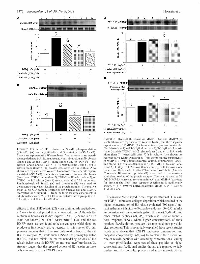
sion. To elucidate themechanisms bywhichH3 relaxinmay haveaffected TGF-β1-stimulated collagen production, its effects onpSmad2 and R-SMA expression were assessed by Westernblotting. pSmad2 expression was barely detectable from un-treated control fibroblasts but significantly upregulated byTGF-β1 (3 ng/mL) administration (which increased the pSmad2/Smad2 ratio by 120-130%; p<0.01 vs untreated control group;Figure 2A) over 72 h of culture, consistent with pSmad2 being apositive regulator of TGF-β1 signaling (39). H3 relaxin (100 ng/mL) significantly inhibited TGF-β1-stimulated pSmad2 levels by∼40% (p<0.05 vs TGF-β1 alone) but not to the same extent asH2 relaxin (100 ng/mL), which inhibited TGF-β1-stimulatedpSmad2 expression by ∼80% (p < 0.01 vs TGF-β1 alone;Figure 2A) over the same time period. pSmad2 expression wasagain barely detectable/undetectable in samples treated with H3relaxin (100 ng/mL) alone, suggesting that H3 relaxin did notaffect pSmad2 in the absence of TGF-β1 (Figure 2A).
TGF-β1 (3 ng/mL) administration to ventricular fibroblastsalso significantly increased R-SMA expression by 70-80% over72 h in vitro (p < 0.01 vs untreated control group; Figure 2B),consistent with its effects on collagen deposition (Figure 1) andpSmad2 (Figure 2A). H3 relaxin (100 ng/mL) was able tosignificantly inhibit TGF-β1-stimulated R-SMA expression to asimilar extent as H2 relaxin (100 ng/mL) over 72 h in culture(both p < 0.05 vs TGF-β1 alone, and no difference to levelsmeasured in untreated control samples; Figure 2B).However, H3relaxin did not affect R-SMA expression in the absence of TGF-β1 (data not shown).
Article Biochemistry, Vol. 50, No. 8, 2011 1371
Effects of H3 Relaxin on MMP and TIMP Expression.To elucidate the mechanisms by which H3 relaxin may haveaffected collagen breakdown, its effects on MMP-9 (by gelatinzymography), MMP-13 (by Western blotting), and TIMP-1 andTIMP-2 (by reverse zymography) were determined in the presenceor absence of TGF-β1. TGF-β1 (3 ng/mL) administration to ratventricular fibroblasts did not have any marked effects on MMP-13 (Figure 3A) or MMP-9 (Figure 3B) levels over 72 h comparedto their respective levelsmeasured fromuntreated control cultures.Likewise, H3 relaxin (100 ng/mL) did not have any significanteffects on MMP-13 (Figure 3A) or MMP-9 (Figure 3B) in thepresence of TGF-β1 compared to that measured in the untreatedcontrol andTGF-β1 alone treated groups.On the other hand,H2relaxin (100 ng/mL) was able to significantly increase MMP-13levels (by∼20%; p<0.05 vs untreated control group; Figure 3A),without having any significant effect on MMP-9 expression(Figure 3B) in the presence of TGF-β1, over the same timeperiod. Furthermore, H3 relaxin did not markedly affect MMP-13 (Figure 3A) or MMP-9 (Figure 3B) expression in the absenceof TGF-β1.
TGF-β1 (3 ng/mL) did not markedly affect TIMP-1 or TIMP-2 expression over 72 h over basal levels measured from untreatedcontrol cultures, when added to rat ventricular fibroblasts(Figure 4). H3 relaxin (100 ng/mL) was able to significantlyinhibit both TIMP-1 and TIMP-2 expression (by 50-55%) inthe presence of TGF-β1 over 72 h (both p < 0.05 vs TGF-β1alone and untreated control groups) and to a similar extent asthat of H2 relaxin (100 ng/mL) in the presence of TGF-β1(Figure 4). H3 relaxin (100 ng/mL), however, did not affectTIMP-1 or TIMP-2 expression in the absence of TGF-β1 (datanot shown).Effects of H3 Relaxin in Combination with H2 Relaxin
onTGF-β1-StimulatedCollagenDeposition.Todetermine ifH3 relaxin and H2 relaxin had any additive effects in inhibitingTGF-β1-stimulated collagen deposition, the effect of 50 ng/mLeither peptide alone or in combination was assessed in thepresence of TGF-β1 (3 ng/mL) (Figure 5). While neither H3 or
H2 relaxin alone (at 50 ng/mL) significantly affected TGF-β1-stimulated collagen deposition over 72 h in culture, TGF-β1-sti-mulated collagen deposition was markedly inhibited in cells trea-ted with H3 relaxin (50 ng/mL) in combination with H2 relaxin(50 ng/mL) over the same time period (p<0.05 vs TGF-β1 alone,and back to levels measured in untreated control samples, as wellas levels measured from adding 100 ng/mL of either peptide alone,in the presence of TGF-β1; Figure 5).Effects of H3 Relaxin on Collagen Accumulation in a
RodentModel of Fibrotic Cardiomyopathy in Vivo. To extendthe in vitro findings of inhibited TGF-β1-stimulated cardiaccollagen deposition by H3 relaxin, its effects were also evaluatedin 4-5-month-old β2-AR mice, which had previously been usedto determine the antifibrotic effects of H2 relaxin (22, 23).Cardiac fibrosis was established in 4-5-month-old β2-AR mice,as evidenced by a 70-75% increase in LV collagen concentrationcompared to that measured in age-matched wild-type animals(p < 0.01 vs wild-type mice; Figure 6). This increase in (β2-AR-transgenic expression-associated) aberrant LV collagen concen-trationwas significantly reversed, however, in animals exposed toH3 relaxin administration (at a dose of 15 μg/day over a 14-daytreatment period; by ∼50%; p < 0.05 vs vehicle-treated β2-ARmice; Figure 6).
DISCUSSION
At the in vitro level, H3 relaxinwas found to significantly inhibitTGF-β1 activity (at the level of pSmad2) and hence TGF-β1-stimulated myofibroblast differentiation, collagen deposition, andTIMP expression in a similar manner to that of H2 relaxin, viaRXFP1. In fact, lower doses of H3 and H2 relaxin were found toadditively lower TGF-β1-stimulated collagen deposition to levelsinduced by higher concentrations of either peptide alone, suggest-ing that H3 relaxin may augment the antifibrotic effects ofphysiological levels of H2 relaxin via RXFP1. At the in vivo level,the antifibrotic potential of H3 relaxin was further confirmed in awell-established mouse model of fibrotic cardiomyopathy, whereit was found to reverse established cardiac fibrosis with similar
FIGURE 1: Dose-response studies. Shown is the relative mean( SE collagen content from untreated (control) rat ventricular fibroblasts, TGF-β1-stimulated cells treated with increasing concentrations of H3 relaxin (0-500 ng/mL) or H2 relaxin (100 ng/mL), and H3 relaxin (100 ng/mL)alone treated fibroblasts after 72 h in culture.Numbers in parentheses represent the number of separate experiments conducted per group. **, p<0.01 vs untreated/control group; #, p< 0.05; ##, p< 0.01 vs TGF-β1 alone.
1372 Biochemistry, Vol. 50, No. 8, 2011 Hossain et al.
efficacy to that of H2 relaxin (22) when continuously applied overa 2 week treatment period at an equivalent dose. Although theventricular fibroblasts studied express RXFP1 (22) and RXFP2(data not shown), but not RXFP3 mRNA (18), and the ratRXFP4 gene has been found to be a pseudogene which does notproduce a functionally active receptor in this species(40), ourprevious findings that H3 relaxin only weakly binds to the ratRXFP2 receptor (18), while human INSL3 (the primary ligand forRXFP2) did not mimic the matrix remodeling actions of H2relaxin (which acts via RXFP1) on rat renal myofibroblasts (30),strongly suggest that the reported actions of H3 relaxin on thesecells were mediated via RXFP1 alone.
The inverse “bell-shaped” dose-response effects of H3 relaxinon TGF-β1-stimulated collagen deposition, which resulted in thehighest concentration of H3 relaxin evaluated (500 ng/mL) nothaving the same inhibitory effects as lower doses (100-250 ng/mL),are consistentwithprevious findings forH2 relaxin (35, 41-43) andother related peptides (44, 45), which also produce biphasicdose-response curves, where higher concentrations of thesepeptides likewise do not produce the same maximum physiolo-gical responses. This is potentially explained from recent studieswhich have shown that RXFP1 undergoes dimerization and“negative cooperativity” (45, 46) to accelerate the dissociationrate of relaxin peptides with ascending concentrations, leadingto lower physiological responses of these peptides at higherconcentrations. Additional studies though are required to fullyunderstand this complex process and more importantly in
FIGURE 2: Effects of H3 relaxin on Smad2 phosphorylation(pSmad2) (A) and myofibroblast differentiation (R-SMA) (B).Shown are representative Western blots (from three separate experi-ments) of pSmad2 (A) fromuntreated/control ventricular fibroblasts(lanes 1 and 2) and TGF-β1 alone (lanes 3 and 4), TGF-β1 þ H3relaxin (lanes 5 and 6), TGF-β1 þ H2 relaxin (lanes 7 and 8), or H3relaxin alone (lanes 9-10) treated cells after 72 h in culture. Alsoshown are representative Western blots (from three separate experi-ments) of R-SMA (B) from untreated/control ventricular fibroblasts(lane 1) andTGF-β1-alone (lane 2), TGF-β1þH3 relaxin (lane 3), orTGF-β1 þ H2 relaxin (lane 4) treated cells after 72 h in culture.Unphosphorylated Smad2 (A) and R-tubulin (B) were used todemonstrate equivalent loading of the protein samples. The relativemean ( SE OD pSmad2 (corrected for Smad2) (A) and R-SMA(corrected for R-tubulin) (B) from the three separate experiments isadditionally shown. **, p<0.01 vs untreated/control group; #, p<0.05; ##, p< 0.01 vs TGF-β1 alone.
FIGURE 3: Effects of H3 relaxin on MMP-13 (A) and MMP-9 (B)levels. Shown are representative Western blots (from three separateexperiments) of MMP-13 (A) from untreated/control ventricularfibroblasts (lane 1) andTGF-β1 alone (lane 2), TGF-β1þH3 relaxin(lanes 3 and 4), TGF-β1 þ H2 relaxin (lanes 5 and 6), or H3 relaxinalone (lane 7) treated cells after 72 h in culture. Also shown arerepresentative gelatin zymographs (from three separate experiments)ofMMP-9 (B) fromuntreated/control ventricular fibroblasts (lanes 1and 2) andTGF-β1alone (lanes 3 and4),TGF-β1þH3relaxin (lanes5 and 6), TGF-β1 þ H2 relaxin (lanes 7 and 8), or H3 relaxin alone(lanes 9 and 10) treated cells after 72 h in culture.R-Tubulin (A) and aCoomassie Blue-stained protein (B) were used to demonstrateequivalent loading of the protein samples. The relative mean ( SEOD MMP-13 (corrected for R-tubulin) (A) and MMP-9 (correctedfor protein) (B) from three separate experiments is additionallyshown. *, p < 0.05 vs untreated/control group; #, p < 0.05 vsTGF-β1 alone.

Article Biochemistry, Vol. 50, No. 8, 2011 1373
physiological cell systems (such as fibroblasts) that have lowreceptor numbers (31, 47). As H3 relaxin has a lower affinity forRXFP1 (18) and reduced potency for accelerating its dissociationin comparison to H2 relaxin, it was thought that H3 relaxin wasless likely to induce biphasic dose-response effects compared toH2 relaxin. Nevertheless, the findings of this study confirmedthat H3 relaxin could significantly inhibit TGF-β1-stimulatedbut not basal collagen deposition, indicating that it exhibitedrapidly acting but safe antifibrotic properties that mimicked theactions of H2 relaxin through binding to RXFP1.
Given that the affinity of H3 relaxin for the rat RXFP1receptor is approximately 30-50-fold lower than that for H2relaxin (18), it was not surprising that H3 relaxin was only able to
inhibit TGF-β1-mediated Smad2 phosphorylation by half theextent of H2 relaxin (∼40% vs ∼80%), while not being able tostimulateMMP-13 levels to the same extent asH2 relaxin. Intere-stingly though, H3 relaxin (at 100 ng/mL) was able to reduceTGF-β1-stimulated R-SMA expression (myofibroblast differen-tiation), collagen deposition, and TIMP-1 and TIMP-2 levelsover 72 h in culture to a similar extent as the comparative dose ofH2 relaxin. These findings may be attributed to the con-stant exposure of these peptides to RXFP1 receptors expressedon rat ventricular fibroblasts over the 72 h experimental period(with their activities stabilized and maintained by FBS), henceallowing them to exert inhibitory actions with similar efficacyover long-term culture periods in vitro. Additionally, althoughmost GPCRs undergo desensitization in response to prolongedexposure to an agonist, it has recently been shown that prolongedRXFP1 signaling (toH2 relaxin (48), and presumablyH3 relaxin)is likely attributed to its weak phosphorylation and poor inter-nalization (in response to ligand binding) and the lack of itsability to recruit/traffic β-arrestins, which contributes to RXFP1not being desensitized in the same manner as other GPCRs,allowingH2 andH3 relaxin to have comparable long-term effectsvia this receptor (on the downstream targets of TGF-β1/Smad2).
TGF-β1 is thought to have dual actions in response to cardiacinjury, initially acting as an antiinflammatory mediator tostimulate ECM production during cardiac inflammation topromote wound healing (49), while its constant presence at thesite of injury and its ability to auto-upregulate itself eventuallyresults in an overproduction of matrix components (primarilycollagen) and, hence, fibrosis (50).Hence, our findings that TGF-β1 promoted Smad2 phosphorylation, myofibroblast differentia-tion, and collagen deposition when administered to ventricularfibroblasts are consistent with its previously reported primary ac-tions (22, 29, 35, 50). On the other hand, our findings that TGF-β1 did not influence MMP-9, MMP-13, TIMP-1, or TIMP-2levels in rat ventricular fibroblasts are consistent with its inabilityto alter MMP-9 and TIMP-2 activity in human lung fibro-blasts (51) but differ from other studies which have shown thatTGF-β1 suppresses collagen degradation by differentially reg-ulating MMP activity (52) and upregulating TIMP-1 levels inhuman fibroblast cultures (51) and experimental models of heartdisease (53). Additionally, although TGF-β1 was previously
FIGURE 4: Effects of H3 relaxin on TIMP-1 and TIMP-2 levels.Shown is a representative reverse zymograph (from three separateexperiments) of TIMP-1 and TIMP-2 from untreated/control ven-tricular fibroblasts (lanes 1 and 2) and TGF-β1 alone (lanes 3 and 4),TGF-β1þH3 relaxin (lanes 5 and 6), or TGF-β1þH2 relaxin (lanes7 and 8) treated cells after 72 h in culture. A Coomassie Blue-stainedprotein was used to demonstrate equivalent loading of the proteinsamples. The relative mean ( SE OD TIMP-1 and TIMP-2(corrected for protein) from three separate experiments is also shown.*, p<0.05 vs untreated/control group; #, p<0.05 vsΤGF-β1 alone.
FIGURE 5: Synergistic effects of H3 and H2 relaxin on TGF-β1-stimulated collagen deposition. Shown is the relative mean ( SErelative collagen content from untreated (control) rat ventricularfibroblasts and TGF-β1-stimulated cells treated with H3 relaxin (50,100 ng/mL) orH2 relaxin (50, 100 ng/mL), orH3 (50 ng/mL) andH2relaxin (50 ng/mL), after 72 h in culture. Numbers in parenthesesrepresent the number of separate experiments conducted per group.**, p<0.01 vs untreated/control group; ‡, p<0.05; ##, p<0.01 vsTGF-β1 alone.
FIGURE 6: Effects of H3 relaxin in a mouse model of fibroticcardiomyopathy in vivo. Shown is the relative collagen content (μg/mgdryweight LV tissue) from4- to 5-month-old wild-type (WT) andage-matched littermate β2-AR transgenic (TG) mice treated withvehicle or synthetic H3 relaxin over a 14 day period. Numbers inparentheses represent the number of animals used per group. *, p<0.05; **, p < 0.01 vs WT group; #, p < 0.05 vs β2-AR TG/vehiclegroup.

1374 Biochemistry, Vol. 50, No. 8, 2011 Hossain et al.
found to promote MMP-2 expression and activity when admi-nistered to these rat ventricular fibroblasts (18, 22) and otherfibroblast culture models (52), it remains to be determined if thisinteraction is more consistent with its antiinflammatory andwound healing actions (54) or with its matrix-promoting effects.These combined findings suggest that the effects of TGF-β1 onMMPs and TIMPs (which remain controversial) are species,organ, and/or cell specific, or specific to the MMP and TIMPbeing studied, and are secondary to its effects on upregulatingmyofibroblast differentiation and collagen/matrix production.
Consistent with this, by suppressing TGF-β1 signaling (at thelevel of Smad2 phosphorylation), it is proposed that H3 and H2relaxin mediate their antifibrotic actions by primarily inhibitingthe predominant actions of TGF-β1 (on myofibroblast differ-entiation and collagen deposition), and to a lesser extent thesecondary actions of TGF-β1 on collagen degradation, viaregulation of the MMPs and TIMPs. This is the first study todemonstrate a H2 and H3 relaxin-mediated inhibition of Smad2phosphorylation from nonrenal fibroblasts (29, 30), implyingthat the regulation of pSmad2 is pivotal to the actions of thesepeptides in several organs outside the kidney. Despite not havingany significant effects on MMP-9 activity (which is thought toplay a role as an inflammatory mediator rather than as aregulator of fibrogenesis), our demonstration that H2 and H3relaxin could promote MMP-2 levels (18) and downregulateTIMP-1 and TIMP-2 expression in TGF-β1-stimulated ventri-cular fibroblasts (while H2 relaxin could also increase MMP-13levels) suggests that these peptides can additionally increase theMMP/TIMPbalance that would favor a net promotion ofmatrixdegradation in the presence of TGF-β1. The added finding thatH3 relaxin did not affect basal collagen deposition (in the absenceof TGF-β1) further suggests that H3 and H2 relaxins (22) do nothave any direct effects on collagen per se.
At the in vivo level, continuous H3 relaxin infusion into amousemodel of fibrotic cardiomyopathy in which the etiology ofcardiac fibrosis was independent of relaxin (i.e., due to cardiac-restricted overexpression of β2-adrenoreceptors) significantlyreversed established collagen deposition by approximately50%, again with similar efficacy to the 58% reduction in fibrosisthat was seen with the same dose of H2 relaxin treatment of thismodel (22). These findings are consistent with the ability of H3relaxin to ameliorate aberrant collagen deposition in an isopro-terenol-induced model of cardiac toxicity in rats (21), whencontinuously applied over a 10 day period. The encouragingfindings of these studies are that they demonstrate the rapid andspecific reversal of cardiac fibrosis by short-term continuoustreatment of H3 relaxin (over 10-14 days), which appear to beadvantages to the modest effects of angiotensin convertingenzyme inhibitors, which usually require longer treatment peri-ods to demonstrate efficacy (55).
In conclusion, these studies have demonstrated that H3 relaxincanmimic the antifibrotic actions ofH2 relaxin viaRXFP1,mostlikely by interfering with TGF-β1 signaling and, hence, theinfluence of TGF-β1 on myofibroblast differentiation, collagendeposition, and collagen degradation (via regulation of theTIMPs). Although it has a lower affinity for RXFP1 comparedtoH2 relaxin, H3 relaxinwas found to have similar efficacy toH2relaxin when continuously exposed to fibroblast cultures in vitroand to an animal model of fibrotic cardiomyopathy in vivo.Furthermore, lower doses of H3 and H2 relaxin were found toadditively inhibit TGF-β1-stimulated collagen deposition to thesame extent as higher doses of each peptide alone, suggesting that
H3 relaxin (acting through a similarmechanismviaRXFP1)mayprovide a suitable adjunct therapy to H2 relaxin to maintain itsefficacy at lower doses. Elucidating the signal transductionpathways that are activated by H3 relaxin to mediate itsantifibrotic actions will be an important future step in determin-ing the strengths and limitations of this peptide as a therapeuticagent and identifying novel therapeutic targets that may be usedto enhance its antifibrotic potential. The findings of this studyalso demonstrated that H3 relaxin has important biologicalactions through other GPCRs outside its role(s) as a neuropep-tide via RXFP3, where it was found to promote significant (butsafe) matrix remodeling actions via RXFP1.
REFERENCES
1. Bathgate, R. A. D., Hsueh, A. J., and Sherwood, O. D. (2006)Physiology and Molecular Biology of the Relaxin Peptide Family,in Physiology of Reproduction (Neill, J. D., Ed.) pp 679-770, Elsevier,San Diego.
2. Sherwood, O. D. (2004) Relaxin’s physiological roles and otherdiverse actions. Endocr. Rev. 25, 205–234.
3. Samuel, C. S., Du, X. J., Bathgate, R. A. D., and Summers, R. J.(2006) “Relaxin” the stiffened heart and arteries: the therapeuticpotential of relaxin in the treatment of cardiovascular disease. Phar-macol. Ther. 112, 529–552.
4. Dschietzig, T., Bartsch, C., Baumann, G., and Stangl, K. (2006)Relaxin;a pleiotropic hormone and its emerging role for experi-mental and clinical therapeutics. Pharmacol. Ther. 112, 38–56.
5. Samuel, C. S., Hewitson, T. D., Unemori, E. N., and Tang, M. L.(2007) Drugs of the future: the hormone relaxin. Cell. Mol. Life Sci.64, 1539–1557.
6. Jeyabalan, A., Shroff, S. G., Novak, J., and Conrad, K. P. (2007) Thevascular actions of relaxin. Adv. Exp. Med. Biol. 612, 65–87.
7. Bani, D., Yue, S. K., and Bigazzi, M. (2009) Clinical profile of relaxin,a possible new drug for human use. Curr. Drug Saf. 4, 238–249.
8. Bathgate, R. A., Ivell, R., Sanborn, B. M., Sherwood, O. D., andSummers, R. J. (2006) International Union of Pharmacology LVII:recommendations for the nomenclature of receptors for relaxin familypeptides. Pharmacol. Rev. 58, 7–31.
9. Bathgate, R. A., Samuel, C. S., Burazin, T. C., Layfield, S., Claasz,A. A., Reytomas, I. G., Dawson, N. F., Zhao, C., Bond, C., Summers,R. J., Parry, L. J., Wade, J. D., and Tregear, G. W. (2002) Humanrelaxin gene 3 (H3) and the equivalentmouse relaxin (M3) gene.Novelmembers of the relaxin peptide family. J. Biol. Chem. 277, 1148–1157.
10. Burazin, T. C., Bathgate, R. A., Macris, M., Layfield, S., Gundlach,A. L., and Tregear, G.W. (2002) Restricted, but abundant, expressionof the novel rat gene-3 (R3) relaxin in the dorsal tegmental region ofbrain. J. Neurochem. 82, 1553–1557.
11. Wilkinson, T.N., Speed, T. P., Tregear, G.W., andBathgate, R.A.D.(2005) Evolution of the relaxin-like peptide family. BMC Evol. Biol.5, 14.
12. Ma, S., Bonaventure, P., Ferraro, T., Shen, P. J., Burazin, T. C.,Bathgate, R. A., Liu, C., Tregear, G. W., Sutton, S. W., andGundlach, A. L. (2007) Relaxin-3 in GABA projection neurons ofnucleus incertus suggests widespread influence on forebrain circuitsvia G-protein-coupled receptor-135 in the rat.Neuroscience 144, 165–190.
13. Ma, S., Sang, Q., Lanciego, J. L., and Gundlach, A. L. (2009)Localization of relaxin-3 in brain of Macaca fascicularis: identifica-tion of a nucleus incertus in primate. J. Comp. Neurol. 517, 856–872.
14. McGowan, B. M., Stanley, S. A., Smith, K. L., White, N. E.,Connolly, M. M., Thompson, E. L., Gardiner, J. V., Murphy,K. G., Ghatei, M. A., and Bloom, S. R. (2005) Central relaxin-3administration causes hyperphagia inmaleWistar rats.Endocrinology146, 3295–3300.
15. McGowan, B. M., Stanley, S. A., Smith, K. L., Minnion, J. S.,Donovan, J., Thompson, E. L., Patterson, M., Connolly, M. M.,Abbott, C.R., Small, C. J.,Gardiner, J. V.,Ghatei,M.A., andBloom,S. R. (2006) Effects of acute and chronic relaxin-3 on food intake andenergy expenditure in rats. Regul. Pept. 136, 72–77.
16. Ma, S., Olucha-Bordonau, F. E., Hossain, M. A., Lin, F., Kuei, C.,Liu, C., Wade, J. D., Sutton, S. W., Nunez, A., and Gundlach, A. L.(2009) Modulation of hippocampal theta oscillations and spatialmemory by relaxin-3 neurons of the nucleus incertus. Learn. Mem.16, 730–742.

Article Biochemistry, Vol. 50, No. 8, 2011 1375
17. Liu, C., Eriste, E., Sutton, S., Chen, J., Roland, B., Kuei, C., Farmer,N., Jornvall,H., Sillard,R., andLovenberg, T.W. (2003) Identificationof relaxin-3/INSL7 as an endogenous ligand for the orphanG-protein-coupled receptor GPCR135. J. Biol. Chem. 278, 50754–50764.
18. Bathgate, R. A. D., Lin, F., Hanson, N. F., Otvos, L., Jr., Guidolin,A., Giannakis, C., Bastiras, S., Layfield, S. L., Ferraro, T., Ma, S.,Zhao, C., Gundlach, A. L., Samuel, C. S., Tregear, G.W., andWade,J. D. (2006) Relaxin-3: improved synthesis strategy and demonstra-tion of its high-affinity interaction with the relaxin receptor LGR7both in vitro and in vivo. Biochemistry 45, 1043–1053.
19. Liu, C., Chen, J., Sutton, S., Roland, B., Kuei, C., Farmer, N., Sillard,R., and Lovenberg, T. W. (2003) Identification of relaxin-3/INSL7 asa ligand for GPCR142. J. Biol. Chem. 278, 50765–50770.
20. Hossain, M. A., Bathgate, R. A., Kong, C. K., Shabanpoor, F., Zhang,S., Haugaard-Jonsson, L. M., Rosengren, K. J., Tregear, G. W., andWade, J. D. (2008) Synthesis, conformation, and activity of humaninsulin-like peptide 5 (INSL5). ChemBioChem 9, 1816–1822
21. Zhang, J., Qi, Y. F., Geng, B., Pan, C. S., Zhao, J., Chen, L., Yang, J.,Chang, J. K., and Tang, C. S. (2005) Effect of relaxin on myocardialischemia injury induced by isoproterenol. Peptides 26, 1632–1639.
22. Samuel, C. S., Unemori, E. N., Mookerjee, I., Bathgate, R. A.,Layfield, S. L., Mak, J., Tregear, G. W., and Du, X. J. (2004) Relaxinmodulates cardiac fibroblast proliferation, differentiation, and col-lagen production and reverses cardiac fibrosis in vivo. Endocrinology145, 4125–4133.
23. Bathgate, R. A., Lekgabe, E. D., McGuane, J. T., Su, Y., Pham, T.,Ferraro, T., Layfield, S., Hannan, R. D., Thomas, W. G., Samuel,C. S., and Du, X. J. (2008) Adenovirus-mediated delivery of relaxinreverses cardiac fibrosis. Mol. Cell. Endocrinol. 280, 30–38.
24. Gao, X. M., Agrotis, A., Autelitano, D. J., Percy, E., Woodcock,E.A., Jennings,G. L.,Dart, A.M., andDu,X. J. (2003) Sex hormonesand cardiomyopathic phenotype induced by cardiac beta 2-adrenergicreceptor overexpression. Endocrinology 144, 4097–4105.
25. Gray,M.O., Long, C. S., Kalinyak, J. E., Li, H. T., andKarliner, J. S.(1998) Angiotensin II stimulates cardiac myocyte hypertrophy viaparacrine release of TGF-beta 1 and endothelin-1 from fibroblasts.Cardiovasc. Res. 40, 352–363.
26. Samuel, C. S., Butkus, A., Coghlan, J. P., and Bateman, J. F. (1996)The effect of relaxin on collagen metabolism in the nonpregnant ratpubic symphysis: the influence of estrogen and progesterone inregulating relaxin activity. Endocrinology 137, 3884–3890.
27. Samuel, C. S. (2009) Determination of collagen content, concentra-tion, and sub-types in kidney tissue.MethodsMol. Biol. 466, 223–235.
28. Gallop, P. M., and Paz, M. A. (1975) Posttranslational proteinmodifications, with special attention to collagen and elastin. Physiol.Rev. 55, 418–487.
29. Heeg, M. H., Koziolek, M. J., Vasko, R., Schaefer, L., Sharma, K.,Muller, G. A., and Strutz, F. (2005) Posttranslational protein mod-ifications, with special attention to collagen and elastin. Kidney Int.68, 96–109.
30. Mookerjee, I., Hewitson, T. D., Halls,M. L., Summers, R. J.,Mathai,M. L., Bathgate, R. A., Tregear, G. W., and Samuel, C. S. (2009)Relaxin inhibits renal myofibroblast differentiation via RXFP1, thenitric oxide pathway, and Smad2. FASEB J. 23, 1219–1229.
31. Masterson, R., Hewitson, T. D., Kelynack, K., Martic, M., Parry, L.,Bathgate, R., Darby, I., and Becker, G. (2004) Relaxin down-regulates renal fibroblast function and promotes matrix remodellingin vitro. Nephrol. Dial. Transplant. 19, 544–552.
32. Bennett, R. G., Kharbanda, K. K., and Tuma, D. J. (2003) Inhibitionof markers of hepatic stellate cell activation by the hormone relaxin.Biochem. Pharmacol. 66, 867–874.
33. Samuel, C. S., Hewitson, T. D., Zhang, Y., and Kelly, D. J. (2008)Relaxin ameliorates fibrosis in experimental diabetic cardiomyopa-thy. Endocrinology 149, 3286–3293.
34. Jeyabalan, A., Kerchner, L. J., Fisher, M. C., McGuane, J. T., Doty,K. D., and Conrad, K. P. (2006) Matrix metalloproteinase-2 activity,protein, mRNA, and tissue inhibitors in small arteries from pregnantand relaxin-treated nonpregnant rats. J. Appl. Physiol. 100, 1955–1963.
35. Unemori, E. N., Pickford, L. B., Salles, A. L., Piercy, C. E., Grove,B. H., Erikson, M. E., and Amento, E. P. (1996) Relaxin induces anextracellular matrix-degrading phenotype in human lung fibroblastsin vitro and inhibits lung fibrosis in a murine model in vivo. J. Clin.Invest. 98, 2739–2745.
36. Lekgabe, E. D., Kiriazis, H., Zhao, C., Xu, Q., Moore, X. L., Su, Y.,Bathgate, R. A., Du, X. J., and Samuel, C. S. (2005) Relaxin reversescardiac and renal fibrosis in spontaneously hypertensive rats. Hyper-tension 46, 412–418.
37. Hossain, M. A., Rosengren, K. J., Haugaard-Jonsson, L. M., Zhang,S., Layfield, S., Ferraro, T., Daly, N. L., Tregear, G.W., Wade, J. D.,and Bathgate, R. A. (2008) The A-chain of human relaxin familypeptides has distinct roles in the binding and activation of the differentrelaxin family peptide receptors. J. Biol. Chem. 283, 17287–17297.
38. Hossain, M. A., Lin, F., Zhang, S., Ferraro, T., Bathgate, R. A. D.,Tregear, G. W., and Wade, J. D. (2006) Regioselective disulfide solidphase synthesis, chemical characterization and in vitro receptorbinding activity of equine relaxin. Int. J. Pept. Res. Ther. 12, 211–215.
39. Kretzschmar, M., and Massague, J. (1998) SMADs: mediators andregulators of TGF-beta signaling.Curr. Opin. Genet. Dev. 8, 103–111.
40. Chen, J., Kuei, C., Sutton, S. W., Bonaventure, P., Nepomuceno, D.,Eriste, E., Sillard, R., Lovenberg, T. W., and Liu, C. (2005) Pharma-cological characterization of relaxin-3/INSL7 receptors GPCR135and GPCR142 from different mammalian species. J. Pharmacol. Exp.Ther. 312, 83–95.
41. Halls, M. L., Bathgate, R. A., and Summers, R. J. (2006) Relaxinfamily peptide receptors RXFP1 andRXFP2modulate cAMP signal-ing by distinct mechanisms. Mol. Pharmacol. 70, 214–226.
42. Danielson, L. A., and Conrad, K. P. (2003) Time course and doseresponse of relaxin-mediated renal vasodilation, hyperfiltration, andchanges in plasma osmolality in conscious rats. J. Appl. Physiol. 95,1509–1514.
43. Teerlink, J. R., Metra, M., Felker, G. M., Ponikowski, P., Voors,A. A., Weatherley, B. D., Marmor, A., Katz, A., Grzybowski, J.,Unemori, E., Teichman, S. L., and Cotter, G. (2009) Relaxin for thetreatment of patients with acute heart failure (Pre-RELAX-AHF): amulticentre, randomised, placebo-controlled, parallel-group, dose-finding phase IIb study. Lancet 373, 1429–1439.
44. De Meyts, P. (1976) Cooperative properties of hormone receptors incell membranes. J. Supramol. Struct. 4, 241–258.
45. Svendsen, A. M., Vrecl, M., Knudsen, L., Heding, A., Wade, J. D.,Bathgate, R. A., De Meyts, P., and Nohr, J. (2009) Dimerization andnegative cooperativity in the relaxin family peptide receptors. Ann.N.Y. Acad. Sci. 1160, 54–59.
46. Svendsen, A. M., Zalesko, A., Konig, J., Vrecl, M., Heding, A.,Kristensen, J. B., Wade, J. D., Bathgate, R. A., De Meyts, P., andNohr, J. (2008) Negative cooperativity in H2 relaxin binding to adimeric relaxin family peptide receptor 1. Mol. Cell. Endocrinol. 296,10–17.
47. Palejwala, S., Stein, D., Wojtczuk, A., Weiss, G., and Goldsmith,L. T. (1998) Demonstration of a relaxin receptor and relaxin-stimu-lated tyrosine phosphorylation in human lower uterine segmentfibroblasts. Endocrinology 139, 1208–1212.
48. Callander, G. E., Thomas, W. G., and Bathgate, R. A. (2009)Prolonged RXFP1 and RXFP2 signaling can be explained by poorinternalization and a lack of beta-arrestin recruitment.Am. J. Physiol.Cell. Physiol. 296, C1058–1066.
49. Pender, B. S., Chen, H., Ashton, S., Wise, W. C., Zingarelli, B.,Cusumano, V., and Cook, J. A. (1996) Transforming growth factorbeta 1 alters rat peritoneal macrophage mediator production andimproves survival during endotoxic shock. Eur. Cytokine Netw. 7,137–142.
50. Lijnen, P. J., Petrov, V. V., and Fagard, R. H. (2000) Induction ofcardiac fibrosis by transforming growth factor-beta(1). Mol. Genet.Metab. 71, 418–435.
51. Papakonstantinou, E., Aletras, A. J., Roth, M., Tamm, M., andKarakiulakis, G. (2003) Hypoxia modulates the effects of transform-ing growth factor-beta isoforms on matrix-formation by primaryhuman lung fibroblasts. Cytokine 24, 25–35.
52. Philips, N., Keller, T., and Gonzalez, S. (2004) TGF beta-like regula-tion ofmatrixmetalloproteinases by anti-transforming growth factor-beta, and anti-transforming growth factor-beta 1 antibodies in dermalfibroblasts: implications for wound healing.Wound Repair Regen. 12,53–59.
53. Zhao, W., Zhao, T., Chen, Y., Ahokas, R. A., and Sun, Y. (2008)Oxidative stress mediates cardiac fibrosis by enhancing transforminggrowth factor-beta1 in hypertensive rats. Mol. Cell. Biochem. 317,43–50.
54. Lechapt-Zalcman, E., Pruliere-Escabasse, V., Advenier, D., Galiacy,S., Charriere-Bertrand, C., Coste, A., Harf, A., d’Ortho, M. P., andEscudier, E. (2006) Transforming growth factor-beta1 increases air-way wound repair via MMP-2 upregulation: a new pathway forepithelial wound repair? Am. J. Physiol. Lung Cell Mol. Physiol.290, L1277–1282.
55. Lijnen, P. J., and Petrov, V. V. (2003) Role of intracardiac renin-angiotensin-aldosterone system in extracellular matrix remodeling.Methods Find. Exp. Clin. Pharmacol. 25, 541–564.
Copyright © 2022 FDOKUMEN


![Quantitative analysis of [11C]-erlotinib PET demonstrates specific binding for activating mutations of the EGFR kinase domain](https://static.fdokumen.com/doc/165x107/6345ca446cfb3d406409d7f9/quantitative-analysis-of-11c-erlotinib-pet-demonstrates-specific-binding-for-activating.jpg)