Improved outcome after spinal cord compression injury in mice treated with docosahexaenoic acid
RESEARCH PAPER
Novel Nanostructured Lipid Carrier Co-LoadedwithDoxorubicinand Docosahexaenoic Acid Demonstrates Enhanced in VitroActivity and Overcomes Drug Resistance in MCF-7/Adr Cells
Samuel V. Mussi & Rupa Sawant & Federico Perche & Mônica C. Oliveira & Ricardo B. Azevedo & Lucas A. M. Ferreira & Vladimir P. Torchilin
Received: 24 October 2013 /Accepted: 31 December 2013# Springer Science+Business Media New York 2014
ABSTRACTPurpose To develop a nanostructured lipid carrier (NLC) co-loaded with doxorubicin and docosahexaenoic acid (DHA) and toevaluate its potential to overcome drug resistance and to increaseantitumoral effect in MCF-7/Adr cancer cell line.Methods The NLC was prepared by a hot homogenizationmethod and characterized for size, zeta potential, entrapmentefficiency (EE) and drug loading (DL). Drug release was evaluatedby dialysis in complete DMEM, and NLC aggregation was assayedin the presence of serum. The cytotoxicity of formulations, doxo-rubicin uptake or penetration were evaluated in MCF-7 andMCF-7/Adr as monolayer or spheroid models.Results The formulation had a size of about 80 nm, negative zetapotential, EE of 99%, DL of 31 mg/g, a controlled drug release inDMEM and no particles aggregation in presence of serum. TheNLC loaded with doxorubicin and DHA showed the sameactivity as free drugs against MCF-7 but a stronger activity againstMCF-7/Adr cells. In monolayer model, the doxorubicin uptake asfree and encapsulated form was similar in MCF-7 but higher forthe encapsulated drug in MCF-7/Adr, suggesting a bypassing of P-glycoprotein bomb efflux. For spheroids, the NLC loaded withdoxorubicin and DHA showed a prominent cytotoxicity and agreater penetration of doxorubicin.
Conclusions These findings suggest that the co-encapsulation ofdoxorubicin and DHA in NLC enhances the cytotoxicity andovercomes the doxorubicin resistance in MCF-7/Adr.
KEY WORDS cancer therapy . docosahexaenoic acid .doxorubicin . nanostructured lipid carrier . resistance overcoming
ABREVIATIONSDDS Drug delivery systemDHA Docosahexaenoic acidDL Drug loadingDOX DoxorubicinEE Entrapment efficiencyEPR Enhanced permeability and retentionNE NanoemulsionNLC Nanostructured lipid carrierPO Peanut oilSLN Solid lipid nanoparticlesTEA Triethanolamine
INTRODUCTION
Doxorubicin is an anthracycline with broad-spectrum anti-cancer activity that is widely used in cancer therapy but hasserious side effects highlighted by potentially fatalcardiotoxicity (1,2). Its low tumor penetration and limiteddistribution within solid tumors are the main causes of itsfailures as a therapeutic agent (3,4). Anthracycline-based com-bination chemotherapy regimens have shown improvement inactivity compared to a single anthracycline, including com-bined doxorubicin or epirubicin with cyclophosphamide,doxorubicin with cyclophosphamide and fluorouracil,epirubicin with cyclophosphamide and fluorouracil. Howev-er, despite the greater activity, these regimens are also moretoxic than single agent regimens (1,5).
S. V. Mussi :M. C. Oliveira : L. A. M. FerreiraDepartment of Pharmaceutics, Faculty of PharmacyFederal University of Minas Gerais (UFMG)Belo Horizonte, Minas Gerais, Brazil
S. V. Mussi : R. Sawant : F. Perche : V. P. Torchilin (*)Center for Pharmaceutical Biotechnology and NanomedicineNortheastern University, Boston, Massachusetts, USAe-mail: [email protected]
R. B. AzevedoDepartment of Genetic and Morphology, Biological Sciences InstituteBrasília University, Brasília, DF, Brazil
Pharm ResDOI 10.1007/s11095-013-1290-2
Among alternatives, the combination of docosahexaenoicacid (DHA) and doxorubicin has been described as increasingthe sensitivity of tumors to chemotherapy compared to doxo-rubicin alone (6–8). DHA is a long-chain (C22) polyunsaturatedfatty acid (omega 3) that enhances oxidative stress and subse-quent lipid peroxidation of tumor cells (9). Several studies havereported an increase in the sensitivity of mammary tumors inrodents to doxorubicin when accompanied by a prolongedsupplementation with DHA (7,10) and others have verified thatthis is probably due to the increased level of oxidative stress,particularly in cancer cells (11). In addition, DHA attenuatesdrug resistance and improves anticancer drug efficacy in resis-tant cell lines (8,9). Recently, a phase II trial verified that adietary DHA supplementation improved the outcome of che-motherapy in metastatic breast cancer patients (12). However,the varieties of drug administration schedules have complicatedthe management of the pharmacokinetic and pharmacody-namic profiles, and obtaining uniform temporal and spatialco-delivery remains challenging. Thus, the combination ofdrugs co-delivered in drug delivery systems (DDSs) hasemerged as an attractive alternative (13).
DDS-based combination therapy offers many advantages,such as more synchronized, controlled pharmacokinetics ofthe drugs, improved drug solubility and bioavailability andthe potential to bypass mechanisms of multidrug resistance.Overall, these combinations can result in improved drugefficacy with a single formulation (13,14). Among these DDSs,nanocarriers including liposomes, polymeric micelles and lipidnanoparticles have been described. A few DDSs have reachedthe market, e.g. liposomal doxorubicin. Such nanocarrierswith sizes of 50–200 nm enhance the concentration of drugsin the tumor by a mechanism known as the “enhanced per-meability and retention (EPR) effect” (15–17).
Lipid nanoparticles such as solid lipid nanoparticles (SLNs)and nanostructured lipid carrier (NLC) have gained muchattention in recent years. SLNs are derived from nanoemulsionswhere the liquid lipid is replaced by a solid lipid. NLC repre-sents an improved generation of SLNs that consists of solid lipidmatrices with spatially distributed liquid lipids, resulting in astructure with imperfections in the crystal structure and canbetter accommodate the drug (19). NLC and SLN share ad-vantages including biocompatibility and the possibility of pro-duction on an industrial scale, since they do not require the useof organic solvents and due to their similarity to parenteralnutrition manufacturing methods (18–20). However, the con-trolled drug release for NLC can be problematical depending ofthe solid/liquid lipid ratio (19,21). A disadvantage of SLN/NLCis the low entrapment of hydrophilic drugs. To increase the drugencapsulation, the formation of an ion pairing with a lipophiliccounter-ion has been proposed as alternative (22–24).
This work aims to develop a new NLC co-loaded withdoxorubicin and DHA triglyceride and to evaluate the physi-cochemical characteristics, drug release and the in vitro efficacy
in sensitive and resistant human breast cancer cell lines in a cellmonolayer model and resistant cells in a spheroid model.
MATERIALS AND METHODS
Materials
Doxorubicin hydrochloride (DOX) was purchased from ACICChemicals (Ontario, Canada). Doxorubicin liposome injection(Lipodox®) was purchased from Sun Pharma Global FZE(Gujarat, India). Triethanolamine (TEA), oleic acid (OA),ethylenediamine tetraacetic acid sodium (EDTA) were pur-chased from Sigma-Aldrich (Missouri, USA). Glycerylbehenate (Compritol 888 ATO®) was kindly provided byGattefossé (Rhône-Alpes, France). Monooleate of sorbitanethoxylated (Super refinedTM polysorbate 80; Tween TM 80),docosahexaenoic acid (DHA) as triglyceride (Incromega™DHA 500TG) and peanut oil (PO) (Super Refined TM PeanutOil) were kindly provided by Croda Inc. (New Jersey, USA).Dulbecco’s Modified Eagle Medium (DMEM), antibiotic stocksolution (10,000 I.U. of penicillin+10,000 μg/mL of strepto-mycin) and 0.25% Trypsin-EDTA were purchased fromCellGro (Virginia, USA). Heat inactivated fetal bovine serum(FBS) was purchased from Atlanta Biologicals (Georgia, USA).The Cell Titer Blue assay kit and CytoTox 96® Non-Radioactive Cytotoxicity Assay were purchased from Promega(Wisconsin, USA). All other chemicals were of analytical grade.
Preparation of Formulation
The formulation was prepared by the hot melting homogeni-zation method using emulsification-ultrasound. The compo-sition was very similar to the one previously described (24) butwith some modifications. The DHA was used in a triglycerideform (Incromega® DHA 500 TG). The formation of the ionpairing to improve the doxorubicin encapsulation was obtain-ed with oleic acid. Briefly, for a batch of 10 ml of formulation,the oily phase (OP) was prepared with 100 mg of Tween 80,10 mg of oleic acid, 5 mg of doxorubicin, 6 mg oftriethanolamine and 150 mg of matrix lipid composed of110 mg of Compritol and 40 mg of DHA (0.4% w/v). Theaqueous phase (AP) was composed of 4 mg of EDTA andpurified water. About 8 mL of water was added at the begin-ning of preparation and at the end volume was completed to10 mL. This formulation was termed NLC-DOX+DHA0.4%. A NLC containing DHA at 0.8% (70 mg of Compritoland 80 mg of DHA; NLC-DOX+DHA 0.8%) and ananoemulsion containing only DHA as lipid matrix (150 mgof DHA; NE-DOX+DHA 1.5%), were also prepared toevaluate the influence of the lipid liquid on doxorubicinrelease. Furthermore, a NLC loaded with DOX withoutDHA was prepared replacing the 0.4% of DHA by a liquid
Mussi et al.
lipid with a low amount of unsaturated lipids (peanut oil−PO)(NLC-DOX+PO0.4%). To prepare them, firstly the OP andthe AP were heated separately to 80°C. After OP melting at80°C, the AP was gently dropped into the OP and homoge-nized using a glass rod for 1 min. This emulsion was immedi-ately subjected to intense probe sonication (8 W) for 5 min,using a high intensity ultrasonic processor (Fisher F60 SonicDismembrator Fisher F-6; Fisher Scientific, Pennsylvania,USA). The pH was adjusted to 7.0 with 0.1 M HCl or0.1 M NaOH and the volume was adjusted to 10 mL. Theformulation was stored at 4°C, protected from light in anitrogen atmosphere.
Characterization of Formulations
Themean particle diameter and zeta potential were measuredby dynamic light scattering (DLS) and electrophoretic mobil-ity, respectively, using the Zeta PLUS particle size analyzer(Brookhaven Instruments; Brookhaven, USA). All measure-ments were performed in triplicate.
The encapsulation efficiency (EE) and drug loading (DL) ofdoxorubicin in NLC were determined by an ultrafiltrationmethod using centrifugal devices (Amicon® Ultra - 0.5 mL100 k;Millipore,USA). To eliminate the binding of doxorubicinon the devices, a pretreating of the filters was performed aspreviously described (24). The devices were soaked in a passiv-ating solution (TweenTM 20, 5% w/v), maintained overnight atroom temperature, and washed with distilled water prior to use.
The EE and DL were calculated using the followingequations:
EE %ð Þ ¼ CT−CAPð Þ=CT � 100DL mg=gð Þ ¼ WDL=WNP
where CT=total doxorubicin concentration in NLC, CAP=doxorubicin concentration in aqueous phase (non-encapsulated), WDL=mg of drug loaded in nanoparticlesand WNP=g of nanoparticles (lipids).
Briefly, the CT, CAP andWDLwere evaluated as follows. TheCT was analyzed by dissolving an aliquot of the NLC dispersionin a mixture of tetrahydrofuran (THF)/methanol (MeOH)40:60 v/v, centrifugation for 10 min at 2,400 g and analysis ofthe supernatant by spectrophotometry at 480 nm (UV-mini1240; Shimadzu, Japan). The CAP was evaluated from analiquot of the aqueous phase separated from the NLC disper-sion by ultrafiltration (10 min at 2,400 g), dilution with THF/MeOHand analysis byUV-visible. TheWDL was derived usingthe calculated EE x mg total of doxorubicin added.
Serum Stability
Serum stability of the NLCwas evaluated by incubation of theNLC with FBS 1:1 at 37°C with shaking (250 rpm) for up to
24 h. Aliquots were sampled, diluted in purified water and themeasurement of zeta potential and size were performed usingthe Zeta Plus Analyzer.
Release of Doxorubicin from NLC
The release study was conducted by dialysis in DMEM con-taining 10% fetal bovine serum (FBS) (25). Dialysis tubes with acutoff size of 100 KDa (Cellulose ester membrane; SpectrumLaboratories; Rancho Dominguez, USA) were filled with 3 mLof formulations diluted in media (1:2), sealed and incubatedwith 50ml of the media for up to 48 h at 37°C, with continuousshaking at 250 rpm. A doxorubicin aqueous solution(0.5 mg/mL) was used as a control. At various time points,aliquots were withdrawn and doxorubicin concentration wasmeasuredwith the SynergyHTMulti-ModeMicroplate Reader(Biotek; Winnooski, USA) at 485/590 ex/em wavelengths. Thevalues were plotted as cumulative percentage of drug release.
Cell Culture
MCF-7 were purchased from The American Type CultureCollection (ATCC; Manassas, USA) and MCF-7/Adr wasobtained from The National Cancer Institute (Frederick,MD, USA). Cells were grown and maintained in DMEM,pH 7.4, supplemented with 10% (v/v) FBS and 1% ofpenicillin/streptomycin stock solution in a humidified, 5%(v/v) CO2 atmosphere at 37°C.
Adherent Monolayer Cells
Cell Viability
Cell viability was assayed with the CellTiter-Blue® Cell Via-bility Assay kit (CTB) following the manufacturer’s protocol.Cells were seeded in 96-well tissue culture plates at 5×10³/well about 24 h before the treatment. The drug solutions werefreshly prepared by dissolving doxorubicin in purified waterand DHA in ethanol. We evaluated the cytotoxicity of theencapsulated drug compared to the free drug with doxorubi-cin concentrations between 0.5 and 16 μM and DHA at 3.5–112 μM. The cells were incubated with treatments for 24 h,washed thrice with complete DMEM, supplemented with100 μL of DMEM and 20 μL of the CTB reagent. Theincubation was performed for 2 h and the fluorescence inten-sity was measured using a Synergy HT Multi-Mode Micro-plate Reader at 530/590 nm excitation/emission wave-lengths. The solvent controls were done.
Doxorubicin Uptake
The cellular uptake of doxorubicin was assessed by flowcytometry by using the natural fluorescence of the drug. Cells
NLC Co-Loaded with Doxorubicin and DHA
were seeded in 12 well plates at a density of 1×10⁵ cells/well24 h before the treatment. The seeding medium was thenremoved and replaced by media containing 16 μM of doxo-rubicin. After 2 h incubation, the cells were washed thrice withPBS, trypsinized, centrifuged at 2000 g for 5 min, re-suspended in PBS pH 7.4 and analyzed using a BDFACSCalibur flow cytometer. The cells were gated usingforward (FSC-H) versus side-scatter (SSC-H) to exclude debrisand dead cells before analysis of 10,000 cell counts. The meanfluorescence intensity (MFI) of the cells was measured at 488/585 nm (FL2 channel) excitation/emission wavelengths.
Spheroids
Preparation of Spheroids
MCF-7/Adr were used to prepare spheroids of 400–500 μmdiameter from 10,000 cells in 96 well plates by a liquid overlaymethod (26). Briefly, DMEMwith 1.5% of agarose was addedto each well and the cells were seeded. After this, the plateswere centrifuged for 15 min at 1,500 RCF. Spheroid forma-tion was monitored using a Nikon Eclipse E400 microscope(Nikon Inc., Melville, USA) at 10× magnification with a SpotInsight™ 3.2.0 camera with Spot Advanced™ software (SpotImaging, Sterling Heights, MI).
Cytotoxicity
Cytotoxicity was measured with a Cytotox 96 Non-Radioactive Cytotoxicity kit (Promega; Fitchburg, USA)(26). Briefly, both the lactate dehydrogenase (LDH) releasedinto the medium and the LDH from spheroids dissociatedafter incubation for 1 h at 37°C with 0.9% Triton®-X100 inDMEMmedia with 10% FBS were measured. The amount ofLDH released into the medium after incubation with thetreatments was calculated as a percentage in relation to thetotal (total LDH=medium LDH+lysate LDH).
Penetration of Doxorubicin
The penetration of doxorubicin throughout the spher-oids was evaluated by confocal microscopy after 2 h ofincubation with 100 μM of doxorubicin in completeDMEM using 488 nm and 535 nm long pass filtersfor doxorubicin excitation/emission, respectively. Thespheroids were removed from the plate, washed withPBS and placed into Lab-Tek chambers (Fisher Scien-tific) prior to imaging. They were visualized at 10×magnification, and the distribution of doxorubicinthroughout the spheroids was analyzed with a ZeissLSM 700 confocal microscope using Z-stack imagingwith 10 μm intervals and the LSM Image Browsersoftware.
Data Analysis
Analyses of the cytotoxicity studies were carried out using aone-way ANOVA followed by a Newman-Keuls’ test. Stu-dent’s t-test was performed to compare data on the physico-chemical characteristics of the formulations. For all analyses,differences were considered significant when the P value was<0.05.
RESULTS
NLC Characterization
Table I lists the characterization data of the formulations forsize, zeta potential, entrapment efficiency and drug loading.The particle size was in the range of 76–86 nm with values ofthe polydispersivity index (PDI) lower than 0.22 suggestingmonodispersion. The zeta potential was negative for all for-mulations and varied between −23 and −36 mV. The entrap-ment efficiency reached nearly 100%, and the drug loadingwas 31 mg/g. The formulations as NLC-DOX+DHA 0.8%and NE-DOX+DHA 1.5% showed results (data not shown)very similar to those of NLC-DOX+DHA 0.4% for allcharacteristics.
Drug Release
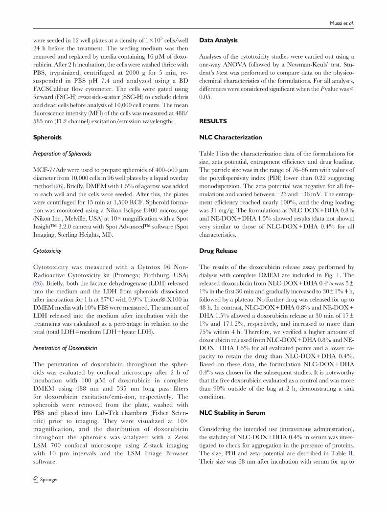
The results of the doxorubicin release assay performed bydialysis with complete DMEM are included in Fig. 1. Thereleased doxorubicin from NLC-DOX+DHA 0.4% was 5±1% in the first 30 min and gradually increased to 30±1% 4 h,followed by a plateau. No further drug was released for up to48 h. In contrast, NLC-DOX+DHA 0.8% and NE-DOX+DHA 1.5% allowed a doxorubicin release at 30 min of 17±1% and 17±2%, respectively, and increased to more than75% within 4 h. Therefore, we verified a higher amount ofdoxorubicin released fromNLC-DOX+DHA 0.8% andNE-DOX+DHA 1.5% for all evaluated points and a lower ca-pacity to retain the drug than NLC-DOX+DHA 0.4%.Based on these data, the formulation NLC-DOX+DHA0.4% was chosen for the subsequent studies. It is noteworthythat the free doxorubicin evaluated as a control and was morethan 90% outside of the bag at 2 h, demonstrating a sinkcondition.
NLC Stability in Serum
Considering the intended use (intravenous administration),the stability of NLC-DOX+DHA 0.4% in serum was inves-tigated to check for aggregation in the presence of proteins.The size, PDI and zeta potential are described in Table II.Their size was 68 nm after incubation with serum for up to
Mussi et al.
24 h. The variation of PDI was very small (in the range of 0.15to 0.18). There was no significant difference in zeta potential(−32 to −37 mV). Thus, the NLC-DOX+DHA 0.4% wasconsidered very stable after incubation with serum and sug-gests that this formulation will not aggregate after intravenousadministration.
Adherent Monolayer Cells
Cytotoxicity
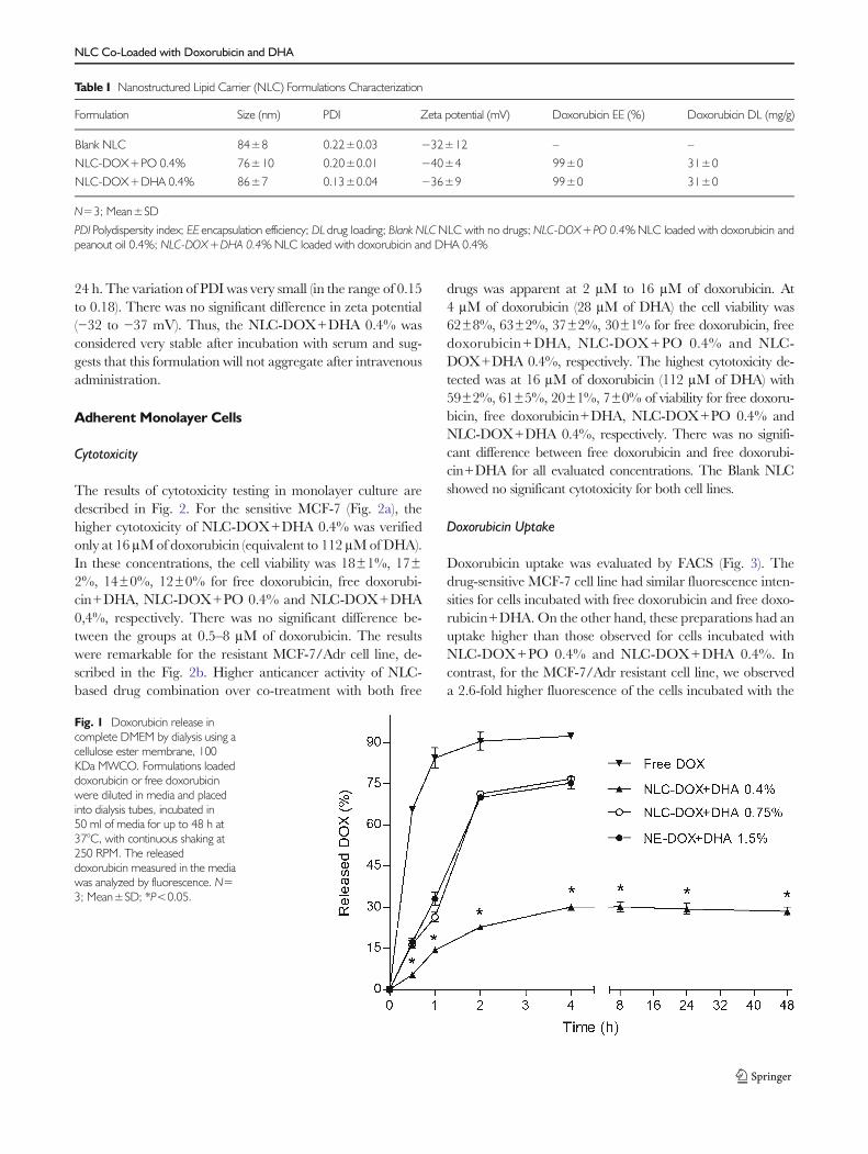
The results of cytotoxicity testing in monolayer culture aredescribed in Fig. 2. For the sensitive MCF-7 (Fig. 2a), thehigher cytotoxicity of NLC-DOX+DHA 0.4% was verifiedonly at 16 μMof doxorubicin (equivalent to 112 μMofDHA).In these concentrations, the cell viability was 18±1%, 17±2%, 14±0%, 12±0% for free doxorubicin, free doxorubi-cin+DHA, NLC-DOX+PO 0.4% and NLC-DOX+DHA0,4%, respectively. There was no significant difference be-tween the groups at 0.5–8 μM of doxorubicin. The resultswere remarkable for the resistant MCF-7/Adr cell line, de-scribed in the Fig. 2b. Higher anticancer activity of NLC-based drug combination over co-treatment with both free
drugs was apparent at 2 μM to 16 μM of doxorubicin. At4 μM of doxorubicin (28 μM of DHA) the cell viability was62±8%, 63±2%, 37±2%, 30±1% for free doxorubicin, freedoxorubicin+DHA, NLC-DOX+PO 0.4% and NLC-DOX+DHA 0.4%, respectively. The highest cytotoxicity de-tected was at 16 μM of doxorubicin (112 μM of DHA) with59±2%, 61±5%, 20±1%, 7±0% of viability for free doxoru-bicin, free doxorubicin+DHA, NLC-DOX+PO 0.4% andNLC-DOX+DHA 0.4%, respectively. There was no signifi-cant difference between free doxorubicin and free doxorubi-cin+DHA for all evaluated concentrations. The Blank NLCshowed no significant cytotoxicity for both cell lines.
Doxorubicin Uptake
Doxorubicin uptake was evaluated by FACS (Fig. 3). Thedrug-sensitive MCF-7 cell line had similar fluorescence inten-sities for cells incubated with free doxorubicin and free doxo-rubicin+DHA.On the other hand, these preparations had anuptake higher than those observed for cells incubated withNLC-DOX+PO 0.4% and NLC-DOX+DHA 0.4%. Incontrast, for the MCF-7/Adr resistant cell line, we observeda 2.6-fold higher fluorescence of the cells incubated with the
Table I Nanostructured Lipid Carrier (NLC) Formulations Characterization
Formulation Size (nm) PDI Zeta potential (mV) Doxorubicin EE (%) Doxorubicin DL (mg/g)
Blank NLC 84±8 0.22±0.03 −32±12 – –
NLC-DOX+PO 0.4% 76±10 0.20±0.01 −40±4 99±0 31±0
NLC-DOX+DHA 0.4% 86±7 0.13±0.04 −36±9 99±0 31±0
N=3; Mean±SD
PDI Polydispersity index; EE encapsulation efficiency; DL drug loading; Blank NLCNLC with no drugs; NLC-DOX+PO 0.4%NLC loaded with doxorubicin andpeanout oil 0.4%; NLC-DOX+DHA 0.4%NLC loaded with doxorubicin and DHA 0.4%
Fig. 1 Doxorubicin release incomplete DMEM by dialysis using acellulose ester membrane, 100KDa MWCO. Formulations loadeddoxorubicin or free doxorubicinwere diluted in media and placedinto dialysis tubes, incubated in50 ml of media for up to 48 h at37°C, with continuous shaking at250 RPM. The releaseddoxorubicin measured in the mediawas analyzed by fluorescence. N=3; Mean±SD; *P<0.05.
NLC Co-Loaded with Doxorubicin and DHA
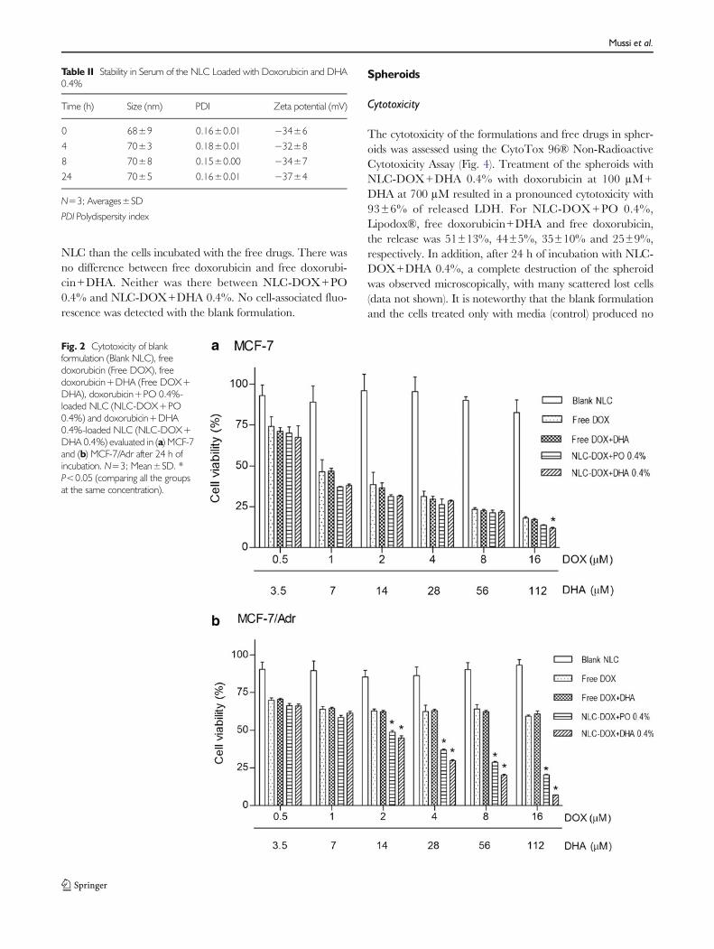
NLC than the cells incubated with the free drugs. There wasno difference between free doxorubicin and free doxorubi-cin+DHA. Neither was there between NLC-DOX+PO0.4% and NLC-DOX+DHA 0.4%. No cell-associated fluo-rescence was detected with the blank formulation.
Spheroids
Cytotoxicity
The cytotoxicity of the formulations and free drugs in spher-oids was assessed using the CytoTox 96® Non-RadioactiveCytotoxicity Assay (Fig. 4). Treatment of the spheroids withNLC-DOX+DHA 0.4% with doxorubicin at 100 μM+DHA at 700 μM resulted in a pronounced cytotoxicity with93±6% of released LDH. For NLC-DOX+PO 0.4%,Lipodox®, free doxorubicin+DHA and free doxorubicin,the release was 51±13%, 44±5%, 35±10% and 25±9%,respectively. In addition, after 24 h of incubation with NLC-DOX+DHA 0.4%, a complete destruction of the spheroidwas observed microscopically, with many scattered lost cells(data not shown). It is noteworthy that the blank formulationand the cells treated only with media (control) produced no
Table II Stability in Serum of the NLC Loaded with Doxorubicin and DHA0.4%
Time (h) Size (nm) PDI Zeta potential (mV)
0 68±9 0.16±0.01 −34±6
4 70±3 0.18±0.01 −32±8
8 70±8 0.15±0.00 −34±7
24 70±5 0.16±0.01 −37±4
N=3; Averages±SD
PDI Polydispersity index
Fig. 2 Cytotoxicity of blankformulation (Blank NLC), freedoxorubicin (Free DOX), freedoxorubicin+DHA (Free DOX+DHA), doxorubicin+PO 0.4%-loaded NLC (NLC-DOX+PO0.4%) and doxorubicin+DHA0.4%-loaded NLC (NLC-DOX+DHA 0.4%) evaluated in (a) MCF-7and (b) MCF-7/Adr after 24 h ofincubation. N=3; Mean±SD. *P<0.05 (comparing all the groupsat the same concentration).
Mussi et al.
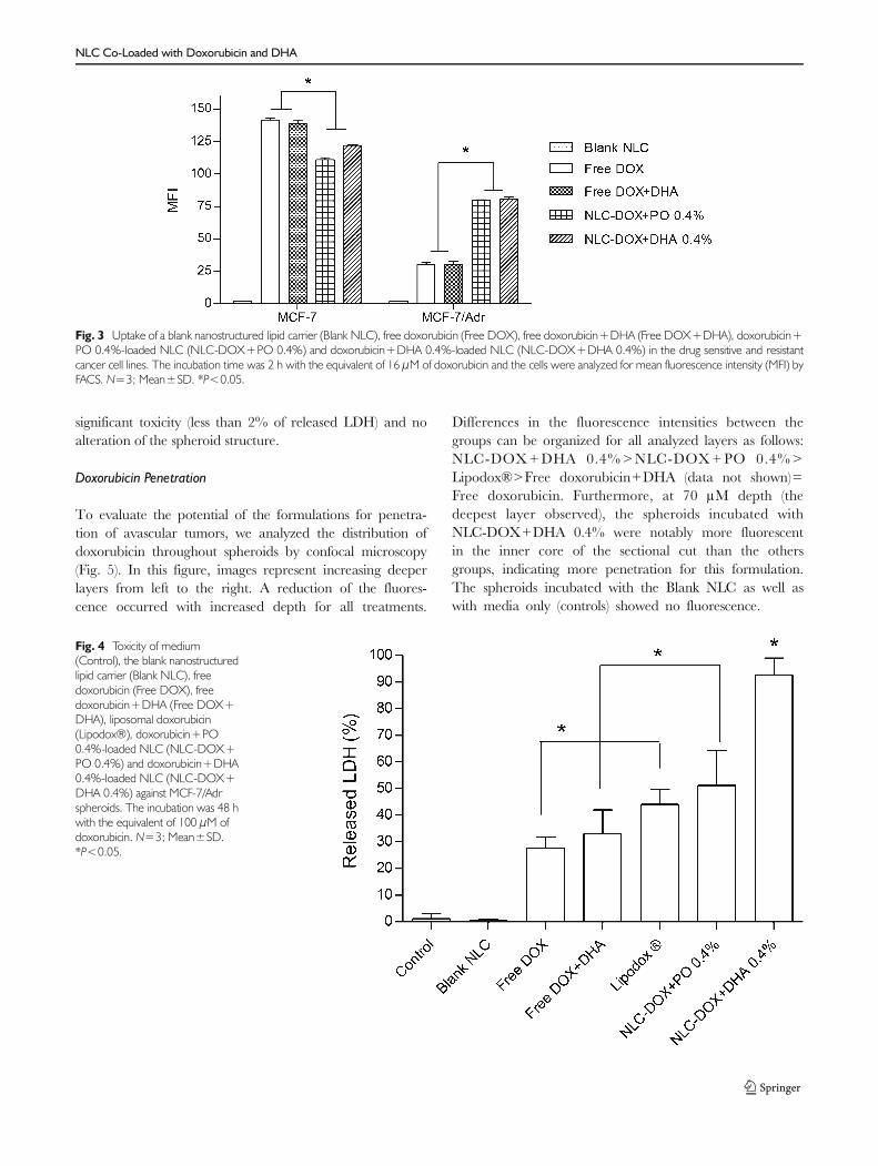
significant toxicity (less than 2% of released LDH) and noalteration of the spheroid structure.
Doxorubicin Penetration
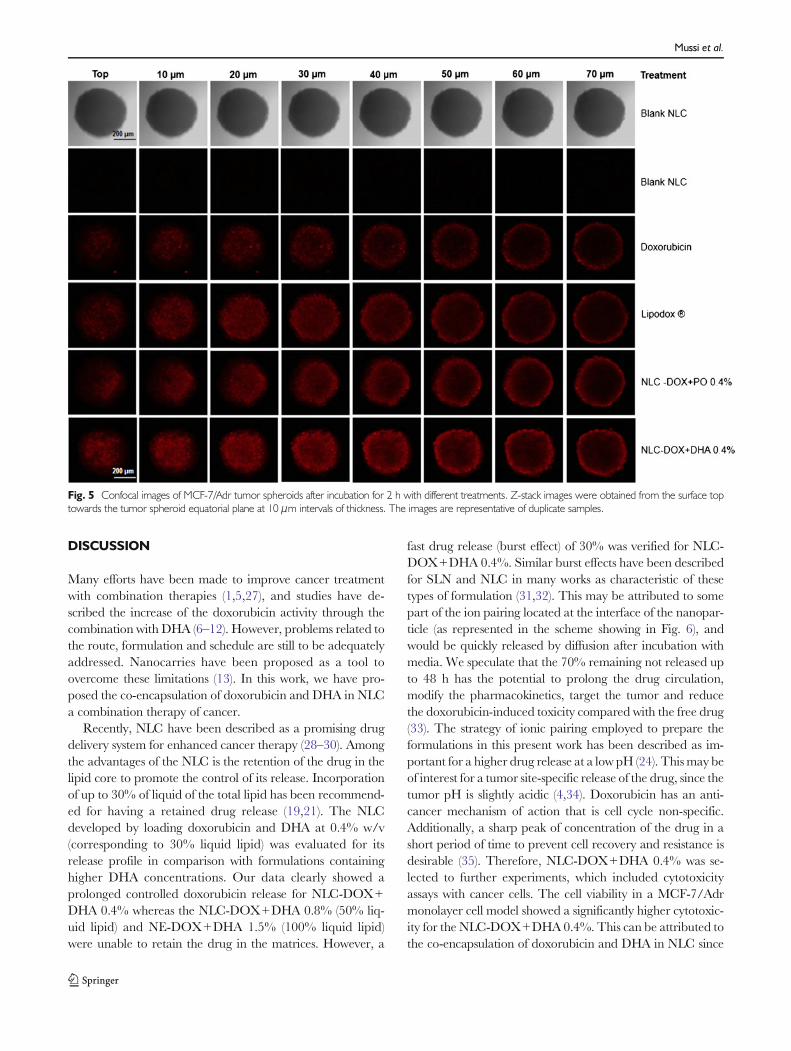
To evaluate the potential of the formulations for penetra-tion of avascular tumors, we analyzed the distribution ofdoxorubicin throughout spheroids by confocal microscopy(Fig. 5). In this figure, images represent increasing deeperlayers from left to the right. A reduction of the fluores-cence occurred with increased depth for all treatments.
Differences in the fluorescence intensities between thegroups can be organized for all analyzed layers as follows:NLC-DOX+DHA 0.4%>NLC-DOX+PO 0.4%>Lipodox®>Free doxorubicin+DHA (data not shown)=Free doxorubicin. Furthermore, at 70 μM depth (thedeepest layer observed), the spheroids incubated withNLC-DOX+DHA 0.4% were notably more fluorescentin the inner core of the sectional cut than the othersgroups, indicating more penetration for this formulation.The spheroids incubated with the Blank NLC as well aswith media only (controls) showed no fluorescence.
Fig. 3 Uptake of a blank nanostructured lipid carrier (Blank NLC), free doxorubicin (Free DOX), free doxorubicin+DHA (Free DOX+DHA), doxorubicin+PO 0.4%-loaded NLC (NLC-DOX+PO 0.4%) and doxorubicin+DHA 0.4%-loaded NLC (NLC-DOX+DHA 0.4%) in the drug sensitive and resistantcancer cell lines. The incubation time was 2 h with the equivalent of 16 μM of doxorubicin and the cells were analyzed for mean fluorescence intensity (MFI) byFACS. N=3; Mean±SD. *P<0.05.
Fig. 4 Toxicity of medium(Control), the blank nanostructuredlipid carrier (Blank NLC), freedoxorubicin (Free DOX), freedoxorubicin+DHA (Free DOX+DHA), liposomal doxorubicin(Lipodox®), doxorubicin+PO0.4%-loaded NLC (NLC-DOX+PO 0.4%) and doxorubicin+DHA0.4%-loaded NLC (NLC-DOX+DHA 0.4%) against MCF-7/Adrspheroids. The incubation was 48 hwith the equivalent of 100 μM ofdoxorubicin. N=3; Mean±SD.*P<0.05.
NLC Co-Loaded with Doxorubicin and DHA
DISCUSSION
Many efforts have been made to improve cancer treatmentwith combination therapies (1,5,27), and studies have de-scribed the increase of the doxorubicin activity through thecombination with DHA (6–12). However, problems related tothe route, formulation and schedule are still to be adequatelyaddressed. Nanocarries have been proposed as a tool toovercome these limitations (13). In this work, we have pro-posed the co-encapsulation of doxorubicin and DHA in NLCa combination therapy of cancer.
Recently, NLC have been described as a promising drugdelivery system for enhanced cancer therapy (28–30). Amongthe advantages of the NLC is the retention of the drug in thelipid core to promote the control of its release. Incorporationof up to 30% of liquid of the total lipid has been recommend-ed for having a retained drug release (19,21). The NLCdeveloped by loading doxorubicin and DHA at 0.4% w/v(corresponding to 30% liquid lipid) was evaluated for itsrelease profile in comparison with formulations containinghigher DHA concentrations. Our data clearly showed aprolonged controlled doxorubicin release for NLC-DOX+DHA 0.4% whereas the NLC-DOX+DHA 0.8% (50% liq-uid lipid) and NE-DOX+DHA 1.5% (100% liquid lipid)were unable to retain the drug in the matrices. However, a
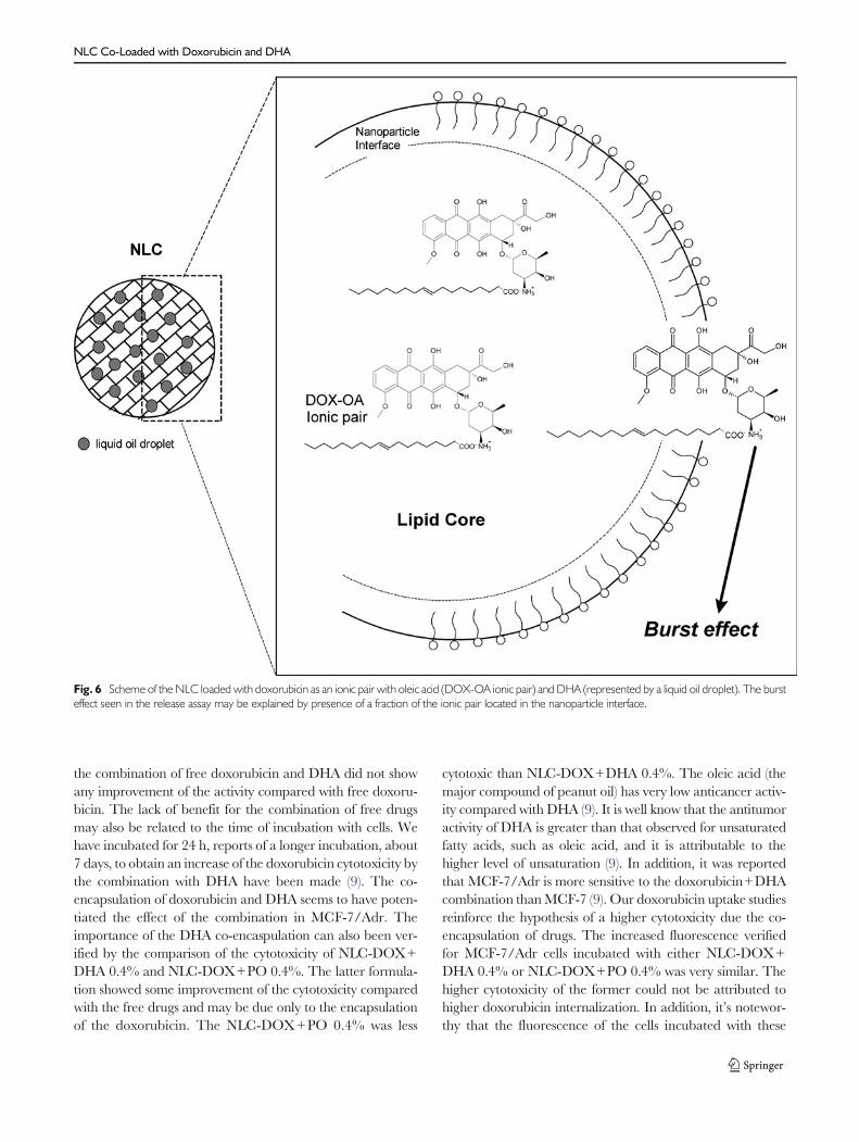
fast drug release (burst effect) of 30% was verified for NLC-DOX+DHA 0.4%. Similar burst effects have been describedfor SLN and NLC in many works as characteristic of thesetypes of formulation (31,32). This may be attributed to somepart of the ion pairing located at the interface of the nanopar-ticle (as represented in the scheme showing in Fig. 6), andwould be quickly released by diffusion after incubation withmedia. We speculate that the 70% remaining not released upto 48 h has the potential to prolong the drug circulation,modify the pharmacokinetics, target the tumor and reducethe doxorubicin-induced toxicity compared with the free drug(33). The strategy of ionic pairing employed to prepare theformulations in this present work has been described as im-portant for a higher drug release at a low pH (24). This may beof interest for a tumor site-specific release of the drug, since thetumor pH is slightly acidic (4,34). Doxorubicin has an anti-cancer mechanism of action that is cell cycle non-specific.Additionally, a sharp peak of concentration of the drug in ashort period of time to prevent cell recovery and resistance isdesirable (35). Therefore, NLC-DOX+DHA 0.4% was se-lected to further experiments, which included cytotoxicityassays with cancer cells. The cell viability in a MCF-7/Adrmonolayer cell model showed a significantly higher cytotoxic-ity for the NLC-DOX+DHA 0.4%. This can be attributed tothe co-encapsulation of doxorubicin and DHA in NLC since
Fig. 5 Confocal images of MCF-7/Adr tumor spheroids after incubation for 2 h with different treatments. Z-stack images were obtained from the surface toptowards the tumor spheroid equatorial plane at 10 μm intervals of thickness. The images are representative of duplicate samples.
Mussi et al.
the combination of free doxorubicin and DHA did not showany improvement of the activity compared with free doxoru-bicin. The lack of benefit for the combination of free drugsmay also be related to the time of incubation with cells. Wehave incubated for 24 h, reports of a longer incubation, about7 days, to obtain an increase of the doxorubicin cytotoxicity bythe combination with DHA have been made (9). The co-encapsulation of doxorubicin and DHA seems to have poten-tiated the effect of the combination in MCF-7/Adr. Theimportance of the DHA co-encaspulation can also been ver-ified by the comparison of the cytotoxicity of NLC-DOX+DHA 0.4% and NLC-DOX+PO 0.4%. The latter formula-tion showed some improvement of the cytotoxicity comparedwith the free drugs and may be due only to the encapsulationof the doxorubicin. The NLC-DOX+PO 0.4% was less
cytotoxic than NLC-DOX+DHA 0.4%. The oleic acid (themajor compound of peanut oil) has very low anticancer activ-ity compared with DHA (9). It is well know that the antitumoractivity of DHA is greater than that observed for unsaturatedfatty acids, such as oleic acid, and it is attributable to thehigher level of unsaturation (9). In addition, it was reportedthat MCF-7/Adr is more sensitive to the doxorubicin+DHAcombination thanMCF-7 (9). Our doxorubicin uptake studiesreinforce the hypothesis of a higher cytotoxicity due the co-encapsulation of drugs. The increased fluorescence verifiedfor MCF-7/Adr cells incubated with either NLC-DOX+DHA 0.4% or NLC-DOX+PO 0.4% was very similar. Thehigher cytotoxicity of the former could not be attributed tohigher doxorubicin internalization. In addition, it’s notewor-thy that the fluorescence of the cells incubated with these
Fig. 6 Scheme of the NLC loaded with doxorubicin as an ionic pair with oleic acid (DOX-OA ionic pair) and DHA (represented by a liquid oil droplet). The bursteffect seen in the release assay may be explained by presence of a fraction of the ionic pair located in the nanoparticle interface.
NLC Co-Loaded with Doxorubicin and DHA
formulations was higher than for those incubated with freedrugs. It seems that both formulations bypassed P-glycoprotein efflux pump, since we verified its overexpressionin this cell line (data not shown). Our uptake data are inaccordance with previous works that indicated higher uptakein MCF-7/Adr of doxorubicin encapsulated in lipid nanopar-ticle than with the free drug (29,36).
The higher cytotoxicity of NLC-DOX+DHA 0.4% wasalso verified in the MCF-7/Adr spheroid model and theseresults are promising. Spheroids have been proposed as a toolfor negative selection to evaluate drug candidates becausemost therapeutic approaches were found to be less active inthis model than cell monolayer cultures (37). Nevertheless,they have also been described as a more relevant in vitro tumormodel for use in assessment of drug combinations (38) andmimic in vivo tumors in many ways, including the architecture,gradients of pH and PO2 and vascularization (37). Due tothese characteristics, this model is also more suitable for drugpenetration studies in vitro than to monolayered cell cultures(39). Our data showed a deeper doxorubicin penetration intridimensional structure of spheroids with NLC-DOX+DHA0.4% that seems to overcome the binding barrier. The pres-ence of this barrier, characterized by a limited drug penetra-tion of the tumor structure, was confirmed since the freedoxorubicin was more confined to the periphery of the spher-oids as previously reported (26,40). The penetration of doxo-rubicin with NLC-DOX+DHA 0.4% was higher than thatobserved with NLC-DOX+PO 0.4%, the commercial lipo-somal formulation (Lipodox®) and with free drugs. Thesefinding also support the importance of the presence of DHAand its co-encapsulation with doxorubicin in NLC.
Thus, NLC-DOX+DHA 0.4% has potentially useful an-titumor formulation characteristics. Its activity was evidentlyhigh in drug resistant MCF-7/Adr when compared with thetwo commercially available forms of doxorubicin (free hydro-chloride salt and liposomal). We suggest that this newly devel-oped nanocarrier for codelivery of doxorubicin/DHA with apotential for effective combination cancer therapy and over-coming drug resistance.
CONCLUSION
Our nanostructured lipid carrier (NLC) loaded with doxoru-bicin and DHA showed size, zeta potential and serum stabilityadequate for parental administration and a controlled releaseof doxorubicin. The in vitro antitumor activity of such NLCloaded with doxorubicin and DHA 0.4% in monolayermodels was significant higher for MCF-7/Adr compared tothe free drugs and the entrapped drug not combined withDHA. These findings in cell monolayers were supported bystudies with spheroids that showed an enhanced penetrationinto the tumor structure. Thus, the association of doxorubicin
and DHA and their co-encapsulation in NLC improved thecytotoxicity and overcame the drug resistance associated withP-glycoprotein activity in MCF-7/Adr. Thus, the NLC load-ed with doxorubicin and DHA represents a promising alter-native for combination cancer therapy.
ACKNOWLEDGMENTS AND DISCLOSURES
This study was supported by “Minas Gerais State Agency forResearch and Development” (FAPEMIG, Brazil) and by theBrazilian agencies. The authors wish to kindly thank Dr.William Hartner for his helpful advice in editing this manu-script. The authors state no conflict of interest and havereceived no payment in preparation of this manuscript. Allauthors have approved the final article.
REFERENCES
1. Livi L,Meattini I, Cardillo Cde L,Mangoni M, Greto D, Petrucci A,et al. Non-pegylated liposomal doxorubicin in combination withcyclophosphamide or docetaxel as first-line therapy inmetastaticbreast cancer: a retrospective analysis. Tumori. 2009;95(4):422–6.
2. Shi Y, Moon M, Dawood S, McManus B, Liu PP. Mechanisms andmanagement of doxorubicin cardiotoxicity. Herz. 2011;36(4):296–305.
3. Primeau AJ, Rendon A, Hedley D, Lilge L, Tannock IF. Thedistribution of the anticancer drug doxorubicin in relation to bloodvessels in solid tumors. Clin Cancer Res. 2005;11(24 Pt 1):8782–8.
4. Trédan O, Galmarini CM, Patel K, Tannock IF. Drug resistanceand the solid tumor microenvironment. J Natl Cancer Inst.2007;99(19):1441–54.
5. Jassem J, Pieńkowski T, Płuzańska A, Jelic S, Gorbunova V, Mrsic-Krmpotic Z, et al. Doxorubicin and paclitaxel versus fluorouracil,doxorubicin, and cyclophosphamide as first-line therapy for womenwith metastatic breast cancer: final results of a randomized phase IIImulticenter trial. J Clin Oncol. 2001;19(6):1707–15.
6. Germain E, Chajès V, Cognault S, Lhuillery C, Bougnoux P.Enhancement of doxorubicin cytotoxicity by polyunsaturated fattyacids in the human breast tumor cell lineMDA-MB-231: relationshipto lipid peroxidation. Int J Cancer. 1998;75(4):578–83.
7. Colas S,MahéoK, Denis F, Goupille C,Hoinard C, Champeroux P,et al. Sensitization by dietary docosahexaenoic acid of rat mammarycarcinoma to anthracycline: a role for tumor vascularization. ClinCancer Res. 2006;12(19):5879–86.
8. Siddiqui RA, Harvey KA, Xu Z, Bammerlin EM, Walker C,Altenburg JD. Docosahexaenoic acid: a natural powerful adjuvantthat improves efficacy for anticancer treatment with no adverseeffects. Biofactors. 2011;37(6):399–412.
9. Mahéo K, Vibet S, Steghens JP, Dartigeas C, Lehman M, BougnouxP, et al. Differential sensitization of cancer cells to doxorubicin by DHA:a role for lipoperoxidation. Free Radic Biol Med. 2005;39(6):742–51.
10. HardmanWE, Avula CP, Fernandes G, Cameron IL. Three percentdietary fish oil concentrate increased efficacy of doxorubicin againstMDA-MB 231 breast cancer xenografts. Clin Cancer Res. 2001;7(7):2041–9.
11. Hajjaji N, Besson P, Bougnoux P. Tumor and non-tumor tissuesdifferential oxidative stress response to supplemental DHA and che-motherapy in rats. Cancer Chemother Pharmacol. 2012;70(1):17–23.
Mussi et al.
12. Bougnoux P, Hajjaji N, Ferrasson MN, Giraudeau B, Couet C, LeFloch O. Improving outcome of chemotherapy of metastatic breastcancer by docosahexaenoic acid: a phase II trial. Br J Cancer.2009;101(12):78–85.
13. Parhi P, Mohanty C, Sahoo SK. Nanotechnology-based combina-tional drug delivery: an emerging approach for cancer therapy. DrugDiscov Today. 2012;17(17–18):1044–52.
14. Okuda T, Kidoaki S. Multidrug delivery systems with single formu-lation—current status and future perspective. J BiomaterNanobiotechnol. 2012;3(1):50–60.
15. Maeda H, Wu J, Sawa T, Matsumura Y, Hori K. Tumor vascularpermeability and the EPR effect in macromolecular therapeutics: areview. J Control Release. 2000;65(1–2):271–84.
16. Torchilin V. Tumor delivery of macromolecular drugs based on theEPR effect. Adv Drug Deliv Rev. 2011;63(3):131–5.
17. Li Y, Wang J, Wientjes MG, Au JL. Delivery of nanomedicines toextracellular and intracellular compartments of a solid tumor. AdvDrug Deliv Rev. 2012;64(1):29–39.
18. Mehnert W, Mäder K. Solid lipid nanoparticles: production, char-acterization and applications. Adv Drug Deliv Rev. 2001;47(2–3):165–96.
19. Müller RH, Radtke M, Wissing SA. Nanostructured lipidmatrices for improved microencapsulation of drugs. Int JPharm. 2002;242(1–2):121–8.
20. Wissing SA, Kayser O, Müller RH. Solid lipid nanoparticles forparenteral drug delivery. Adv Drug Deliv Rev. 2004;56(9):1257–72.
21. Radtke M, Müller RH. NLC-nanostructured lipid carriers: the newgeneration of lipid drug carriers. New Drugs. 2001;2:48–52.
22. Serpe L, SatalanoMG, Cavalli R, Ugazio E, Bosco O, Canaparo R,et al. Cytotoxicity of anticancer drugs incorporated in solid lipidnanoparticles on HT-29 colorectal cancer cell line. Eur J PharmBiopharm. 2004;58(3):673–80.
23. Wong HL, Rauth AM, Bendayan R, Manias JL, Ramaswamy M,Liu Z, et al. A new polymer-lipid hybrid nanoparticle system increasescytotoxicity of doxorubicin against multidrug-resistant human breastcancer cells. Pharm Res. 2006;23(7):1574–85.
24. Mussi SV, Silva RC, Oliveira MC, Lucci CM, Azevedo RB, FerreiraLA. New approach to improve encapsulation and antitumor activityof doxorubicin loaded in solid lipid nanoparticles. Eur J Pharm Sci.2013;48(1–2):282–90.
25. Elbayoumi T, Torchilin VP. Tumor-targeted immuno-liposomes forcancer therapy and imaging. J Pharm Innov. 2008;3:51–8.
26. Perche F, Patel NR, Torchilin VP. Accumulation and toxicity ofantibody-targeted doxorubicin-loaded PEG-PE micelles in ovariancancer cell spheroid model. J Control Release. 2012;164(1):95–102.
27. Flaherty KT, Infante JR, Daud A, Gonzalez R, Kefford RF,Sosman J, et al. Combined BRAF and MEK inhibition in
melanoma with BRAF V600 mutations. N Engl J Med.2012;367(18):1694–703.
28. Joshi MD, Müller RH. Lipid nanoparticles for parenteral delivery ofactives. Eur J Pharm Biopharm. 2009;71(2):161–72.
29. Zhang X-g, Miao J, Dai Y-Q, Du Y-Z, Yuan H, Hu F-Q. Reversalactivity of nanostructured lipid carriers loading cytotoxic drug inmulti-drug resistant cancer cells. Int J Pharm. 2008;361(1–2):239–44.
30. Yang XY, Li YX, Li M, Zhang L, Feng LX, Zhang N. Hyaluronicacid-coated nanostructured lipid carriers for targeting paclitaxel tocancer. Cancer Lett. 2013;334(2):338–45.
31. Bunjes H. Lipid nanoparticles for the delivery of poorly water-solubledrugs. J Pharm Pharmacol. 2010;62(11):1637–45.
32. Rosenblatt KM, Douroumis D, Bunjes H. Drug release from differ-ently structured monoolein/poloxamer nanodispersions studied withdifferential pulse polarography and ultrafiltration at low pressure. JPharm Sci. 2007;96(6):1564–75.
33. Souto EB, Müller RH. Lipid nanoparticles: effect on bioavailabilityand pharmacokinetic changes. Handb Exp Pharmacol. 2010;197:115–41.
34. Subedi RK, KangKW, Choi H. Preparation and characterization ofsolid lipid nanoparticles loaded with doxorubicin. Eur J Pharm Sci.2009;37(3–4):508–13.
35. Needham D, Ponce A. Nanoscale drug delivery vehicles for solidtumors: a new paradigm for localized drug delivery using tempera-ture sensitive liposomes. In: Amiji MM, editor. Nanotechnology forcancer therapy. Boca Raton: CRCPress, LLC (a subsidiary of Taylorand Francis); 2007. p. 677–719.
36. Miao J, Du YZ, Yuan H, Zhang XG, Hu FQ. Drug resistancereversal activity of anticancer drug loaded solid lipid nanoparticlesin multi-drug resistant cancer cells. Colloids Surf B: Biointerfaces.2013;110:74–80.
37. Hirschhaeuser F, Menne H, Dittfeld C, West J, Mueller-Klieser W,Kunz-Schughart LA. Multicellular tumor spheroids: anunderestimated tool is catching up again. J Biotechnol. 2010;148(1):3–15.
38. Perche F, Torchilin VP. Cancer cell spheroids as a model toevaluate chemotherapy protocols. Cancer Biol Ther.2012;13(12):1205–13.
39. Dufau I, Frongia C, Sicard F, Dedieu L, Cordelier P, Ausseil F, et al.Multicellular tumor spheroid model to evaluate spatio-temporaldynamics effect of chemotherapeutics: application to thegemcitabine/CHK1 inhibitor combination in pancreatic cancer.BMC Cancer. 2012;12(15):1–11.
40. Kim TH, Mount CW, Gombotz WR, Pun SH. The delivery ofdoxorubicin to 3-D multicellular spheroids and tumors in a murinexenograft model using tumor-penetrating triblock polymeric mi-celles. Biomaterials. 2010;31(28):7386–97.
NLC Co-Loaded with Doxorubicin and DHA
Copyright © 2022 FDOKUMEN