Knockdown of MADD and c-FLIP overcomes resistance to TRAIL-induced apoptosis in ovarian cancer cells
20
Knockdown of MADD and c-FLIP overcomes resistance to TRAIL-induced apoptosis in ovarian cancer cells Liang-Cheng LI, Ph.D, Shankar JAYARAM, Ph.D, Lakshmy GANESH, MS., Lixia QIAN, BS., Jacob ROTMENSCH, MD + , Ajay V. MAKER, MD., and Bellur S. PRABHAKAR, Ph.D * Department of Microbiology and Immunology, College of Medicine, University of Illinois at Chicago + Department of Obstetrics and Gynecology, Rush University-Medical Center, Chicago, IL 60612, USA Abstract OBJECTIVE—The clinical utility of TRAIL in the treatment of established human malignancies is limited by the development of resistance to TRAIL. We hypothesized that knockdown of MADD, a TRAIL-resistance factor, may overcome TRAIL resistance in ovarian cancer cells. STUDY DESIGN—MADD expression in resected ovarian cancer specimens and cell lines was quantified using q-RT-PCR. Sensitivity of ovarian cancer cell lines to TRAIL, with or without MADD knockdown was assessed. RESULTS—MADD is expressed at relatively higher levels in human malignant ovarian cancer tissues and cell lines compared to normal ovarian tissues. The cell lines OVCA429 and OVCAR3 were susceptible, and CAOV-3 and SKOV-3 were resistant to TRAIL. MADD knockdown in CAOV-3 cells, but not in SKOV-3 cells, conferred TRAIL sensitivity. Knockdown of c-FLIP in SKOV-3 cells increased spontaneous and TRAIL-induced apoptosis, which was further increased upon MADD knockdown. CONCLUSION—MADD/c-FLIP L knockdown can render TRAIL-resistant ovarian cancer cells susceptible to TRAIL. Keywords Ovarian Cancer; MADD; IG20; shRNA; c-FLIP; apoptosis Introduction Ovarian cancer is a very common malignancy, with an estimated incidence of 21,880 new cases in 2010, as well as the most fatal gynecological malignancy and fifth leading cause of cancer-related mortality in women, with an estimated 13,850 deaths annually in the United States 1 . When possible, the best outcomes for advanced disease are obtained utilizing a © 2011 Mosby, Inc. All rights reserved. * Corresponding author: Bellur S. Prabhakar, Department of Microbiology and Immunology, College of Medicine, University of Illinois at Chicago, Chicago, IL 60612, USA; Tel: 312-996-4945; Fax: 312-996-6415; [email protected]. DISCLOSURE: None of the authors have a conflict of interest. Publisher's Disclaimer: This is a PDF file of an unedited manuscript that has been accepted for publication. As a service to our customers we are providing this early version of the manuscript. The manuscript will undergo copyediting, typesetting, and review of the resulting proof before it is published in its final citable form. Please note that during the production process errors may be discovered which could affect the content, and all legal disclaimers that apply to the journal pertain. NIH Public Access Author Manuscript Am J Obstet Gynecol. Author manuscript; available in PMC 2012 October 1. Published in final edited form as: Am J Obstet Gynecol. 2011 October ; 205(4): 362.e12–362.e25. doi:10.1016/j.ajog.2011.05.035. NIH-PA Author Manuscript NIH-PA Author Manuscript NIH-PA Author Manuscript
-
Upload
independent -
Category
Documents
-
view
4 -
download
0
Transcript of Knockdown of MADD and c-FLIP overcomes resistance to TRAIL-induced apoptosis in ovarian cancer cells
Knockdown of MADD and c-FLIP overcomes resistance toTRAIL-induced apoptosis in ovarian cancer cells
Liang-Cheng LI, Ph.D, Shankar JAYARAM, Ph.D, Lakshmy GANESH, MS., Lixia QIAN, BS.,Jacob ROTMENSCH, MD+, Ajay V. MAKER, MD., and Bellur S. PRABHAKAR, Ph.D*
Department of Microbiology and Immunology, College of Medicine, University of Illinois atChicago+ Department of Obstetrics and Gynecology, Rush University-Medical Center, Chicago, IL 60612,USA
AbstractOBJECTIVE—The clinical utility of TRAIL in the treatment of established human malignanciesis limited by the development of resistance to TRAIL. We hypothesized that knockdown ofMADD, a TRAIL-resistance factor, may overcome TRAIL resistance in ovarian cancer cells.
STUDY DESIGN—MADD expression in resected ovarian cancer specimens and cell lines wasquantified using q-RT-PCR. Sensitivity of ovarian cancer cell lines to TRAIL, with or withoutMADD knockdown was assessed.
RESULTS—MADD is expressed at relatively higher levels in human malignant ovarian cancertissues and cell lines compared to normal ovarian tissues. The cell lines OVCA429 and OVCAR3were susceptible, and CAOV-3 and SKOV-3 were resistant to TRAIL. MADD knockdown inCAOV-3 cells, but not in SKOV-3 cells, conferred TRAIL sensitivity. Knockdown of c-FLIP inSKOV-3 cells increased spontaneous and TRAIL-induced apoptosis, which was further increasedupon MADD knockdown.
CONCLUSION—MADD/c-FLIPL knockdown can render TRAIL-resistant ovarian cancer cellssusceptible to TRAIL.
KeywordsOvarian Cancer; MADD; IG20; shRNA; c-FLIP; apoptosis
IntroductionOvarian cancer is a very common malignancy, with an estimated incidence of 21,880 newcases in 2010, as well as the most fatal gynecological malignancy and fifth leading cause ofcancer-related mortality in women, with an estimated 13,850 deaths annually in the UnitedStates1. When possible, the best outcomes for advanced disease are obtained utilizing a
© 2011 Mosby, Inc. All rights reserved.*Corresponding author: Bellur S. Prabhakar, Department of Microbiology and Immunology, College of Medicine, University ofIllinois at Chicago, Chicago, IL 60612, USA; Tel: 312-996-4945; Fax: 312-996-6415; [email protected]: None of the authors have a conflict of interest.Publisher's Disclaimer: This is a PDF file of an unedited manuscript that has been accepted for publication. As a service to ourcustomers we are providing this early version of the manuscript. The manuscript will undergo copyediting, typesetting, and review ofthe resulting proof before it is published in its final citable form. Please note that during the production process errors may bediscovered which could affect the content, and all legal disclaimers that apply to the journal pertain.
NIH Public AccessAuthor ManuscriptAm J Obstet Gynecol. Author manuscript; available in PMC 2012 October 1.
Published in final edited form as:Am J Obstet Gynecol. 2011 October ; 205(4): 362.e12–362.e25. doi:10.1016/j.ajog.2011.05.035.
NIH
-PA Author Manuscript
NIH
-PA Author Manuscript
NIH
-PA Author Manuscript
combination of surgery and neoadjuvant or adjuvant chemotherapy. Initial response rates tochemotherapy vary from 60–80%, however, most patients with persistent or recurrentdisease requiring chronic chemotherapy will develop drug resistance and eventuallyexperience progression of disease2. For these patients, new therapeutic strategies are needed.
TRAIL (TNF-related apoptosis-inducing ligand) is a member of the tumor necrosis factorsuper family of cell death-inducing ligands3. TRAIL can induce programmed cell death orapoptosis in a variety of tumor cell lines and not in normal or non-transformed cells4.TRAIL induces apoptosis by activating the extrinsic apoptosis pathway by interacting withthe death receptors (DRs), DR4 and DR5. TRAIL binding to DRs induces receptoroligomerization resulting in the formation of a death inducing signaling complex (DISC),which contains the Fas associated death domain (FADD) and pro-caspase-8 and leads toactivation of caspase-8. Activation of caspase-8 activates caspase-3 and induces apoptosis5.Alternatively, activated caspase-8 can cleave Bid, a pro-apoptotic protein, resulting intruncated Bid (tBid) that can translocate to mitochondria and initiate intrinsic apoptosis6–8.
While higher levels of expression of DRs can render cells more susceptible to TRAILinduced apoptosis, increased levels of expression of decoy death receptors (DcR1 andDcR2) and anti-apoptotic proteins (c-FLIP, x-IAP, Survivin, BCL2, MCL1 and MADD) canconfer significant resistance to TRAIL-induced apoptosis9–15. The two decoy receptorsDcR1 and DcR2 lack a functional death domain and compete with DR4 and DR5 for cellsurface TRAIL binding but cannot transmit the apoptotic signal9. The anti-apoptotic proteinc-FLIP, an inhibitor of death ligand induced apoptosis, comes in two isoforms -- a long form(c-FLIPL) and a short form (c-FLIPS). The c-FLIPL, a 55-kDa protein, contains two DEDsand a caspase-like domain, whereas c-FLIPS, a 26-kDa protein consists only of two DEDs10.Both isoforms are recruited to the DISC via binding to FADD, and they preventprocaspase-8 recruitment and thus block DR-mediated apoptosis 16, 17. The c-FLIPL is overexpressed in a number of different tumors and is often associated with TRAILresistance 11, 12. The Bcl2 family proteins (Bcl2 and MCL1) act as mitochondrialintracellular checkpoints12–14 and resist apoptosis. Execution of the apoptotic program isalso controlled by inhibitor of apoptosis proteins (IAP, x-IAP and Survivin), which canselectively bind and inhibit caspases-3, -7, and -9, thereby prevent cell death in response tomultiple stimuli15.
We have previously identified and characterized MADD as a TRAIL resistance factor.MADD is an isoform of the IG20 (Insulinoma-Glucagonoma 20) gene18 and is expressed atvery low levels in most healthy tissues but is expressed at significantly higher levels in manyhuman tumors and tumor cell lines15, 18, 19. Knockdown of MADD expression results inenhanced spontaneous and TRAIL-induced apoptosis in cells derived from cervical cancer,neuroblastoma and thyroid cancer 20,21,22. Further, expression of exogenous MADD, andnot other IG20 splice variants, in the absence of all endogenous isoforms can rescue thesecells from undergoing apoptosis20, 23. These findings indicated that only MADD isoform ofthe IG20 gene can promote cancer cell survival20, 23 The current study was initiated todetermine if ovarian cancer tissues and cells express MADD at higher levels and whether itcontributes to TRAIL resistance in ovarian cancer cells.
Materials and methodsCell culture
OVCA429, OVCAR-3, CAOV-3 and SKOV-3 ovarian cancer cells were purchased fromATCC and cultured according their instructions. Briefly, OVCA429 and SKOV-3 cells werecultured in RPMI 1640 (Invitrogen, CA, USA) supplemented with 10% fetal bovine serum(FBS). OVCAR-3 cells were cultured in RPMI 1640 containing 20% FBS plus 0.01mg/ml
LI et al. Page 2
Am J Obstet Gynecol. Author manuscript; available in PMC 2012 October 1.
NIH
-PA Author Manuscript
NIH
-PA Author Manuscript
NIH
-PA Author Manuscript
bovine insulin. CAOV-3 cells were cultured in DMEM (Invitrogen, CA, USA) with 10%FBS. Culture media were also supplemented with 100 units/ml of penicillin, and 100 μg/mlof streptomycin. The cell lines were maintained at 37° C in a humidified chamber with 5%CO2.
AntibodiesAntibody to FLIPL (NF6) was purchased from Enzo life science Inc. (Farmingdale, NY).The preparation of anti-MADD exon 13L (anti-13L) specific antibodies has been reportedearlier 24. The goat anti-mouse IgG1 peroxidase-conjugated secondary antibody wasobtained from Caltag Laboratories (Burlingame, CA) and the anti-rabbit peroxidase-conjugated polyclonal secondary antibody was purchased from GE Healthcare (Piscataway,NJ). Antibodies against DR4, DR5, DcR1 and DcR2 were purchased from Ebioscience (SanDiego, CA).
Tissue samples and RNA preparationSnap-frozen normal, benign and malignant ovarian cancer tissues (Supplementary Table 1)were collected as per the protocol approved by the institutional review board of theUniversity of Illinois at Chicago. Snap frozen tissue samples were obtained fromCooperative Human Tissue Network Midwestern Division. Frozen tissues (100mg) wereimmersed in liquid nitrogen, were ground into fine powder and solubilized in TRIZOL®
reagent (Invitrogen Life Technologies, CA, USA). Total RNA was extracted from ovariantissues or from ovarian cancer cells according to the manufacturer’s instructions.
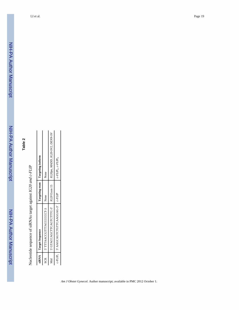
Design of small inhibitory RNAsThe nucleotide sequences of various shRNAs used in this study are shown in supplementaryTable 2. The shRNAs targeting exon 15 of MADD (Mid) and the SCR (negative control) areidentical to those previously described 20, 21. The siRNA targeting c-FLIP was designedusing OligoEngine Workstation 2 and purchased from OligoEngine, Inc. (Seattle, WA).These siRNAs were screened in OVCA429 cells and the most efficient one was used toconstruct the cFLIP- shRNA lentivirus.
Plasmid constructionThe siRNAs were cloned into the pSUPER vector using BgI II and HindIII sites 25 togenerate pSup-cFLIP plasmids. The shRNA cassettes (including the H1 RNA promoter andthe shRNA) were excised from pSup-cFLIP using XbaI and ClaI sites and ligated into thepNL-SIN-CMV-GFP vector to generate cFLIP lentivirus constructs (c-FLIPi). The pcTat,pcRev and pHIT/G were gifts from Dr. B.R. Cullen (Duke University Medical Center) andDr. T.J. Hope (Northwestern University, Department of Cell & Molecular Biology).
Preparation of Lentivirus stocksLentivirus stocks were prepared as described previously 25. Briefly, sub-confluent 293FTcells grown in 100 mm plates were co-transfected with 10.8 μg of lentivirus vector(containing either SCR, MID or cFLIP shRNA), 1 μg pcRev, 1 μg of pcTat and 0.5 μg ofpHIT/G using calcium phosphate. Culture medium was replaced after 16 h, and thesupernatant was harvested at 40 h and filtered using a 0.22 mm filter. The optimal viral titerfor each cell type was determined as the least amount of viral supernatant required totransduce at least 80% of target cells without apparent cytotoxicity.
Quantitative Real-Time PCRQuantitative real-time RT-PCR (q-RT-PCR) was carried out using TaqMan® one-step RT-PCR Master Mix reagents (Applied Biosystems, Carlsbad, CA) according to the
LI et al. Page 3
Am J Obstet Gynecol. Author manuscript; available in PMC 2012 October 1.
NIH
-PA Author Manuscript
NIH
-PA Author Manuscript
NIH
-PA Author Manuscript
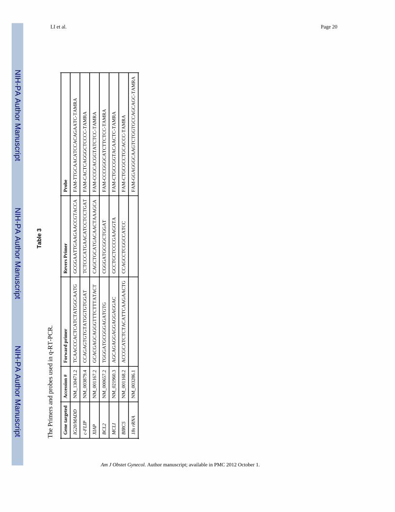
manufacturer’s protocol. Briefly, one microgram RNA was added into 25 μl of reactionmixture containing 12.5 μl of Master mix, 0.75 μl of 40X MultiScribe, RNase inhibitor mix,0.75 nmol/L each primer and 0.25 nmol/L probe. The primer and probe sequences are listedin Table 3. The RT-PCR reaction was performed as follows: reverse transcription at 48° Cfor 30min, denaturation at 95° C for 10 min followed by 40 cycles of denaturation at 95° Cfor 15 sec, annealing at 60° C for 1 min. TaqMan® 18s Ribosomal RNA control reagents(Applied Biosystems, Carlsbad, CA) were used for amplifying the endogenous control.Delta–delta CT method was used to calculate the relative expression levels of tested genesversus to 18s rRNA. Data were analyzed using q-gene program 26.
Reverse transcription–polymerase chain reactionWe used one μg of RNA for reverse transcription-polymerase chain reaction (RT-PCR)using the Super-Script III One-Step RT-PCR system (Invitrogen Life Technologies,Carlsbad, CA, USA). Briefly, the cDNAs were synthesized at 50° C for 30 minutes followedby incubation at 94° C for 2 minutes. Subsequently, 30 cycles of PCR were carried out withdenaturation at 94° C for 50 seconds, annealing at 55° C for 50 seconds and extension at 72°C for 1min followed by a final incubation at 72° C for 7 min. For amplifying regionsflanking exons 13L and 16, F-1 and B-1 primer pairs (5′-CGG GAC TCT GAC TCC GAACCT AC-3′ and 5′-GCG GTT CAG CTT GCT CAG GAC-3′, respectively) were used.GAPDH was used as a loading control and was amplified using. GAPDH primers that havebeen previously published 27. The PCR products were then separated on a 5%polyacrylamide gel.
Immunofluorescence stainingTotal of 3 × 10⁵ cells were placed into 60-mm culture dishes with cover glasses, andcultured overnight. The cells were fixed with acetone and permeabilized with 0.01% TritonX-100 and subsequently blocked with 1% BSA for 30 min at RT. Cells were incubatedovernight with 13L antibody 25 at 4 °C, washed and probed with a biotinylated anti-rabbitantibody (Caltag Laboratories, CA, USA) and streptavidin-FITC (BD PharMingen). Normalrabbit anti-serum was used as a negative control. The image was visualized and capturedwith a LSM 510 META confocal microscope.
Flow cytometry to detect death (DRs) and decoy (DcRs) receptorsCell surface expression of death/decoy receptors was evaluated by flow cytometry aspreviously described 27. For each cell line, 1 × 10⁶ cells were incubated with 5.0 μg of theappropriate anti-death/decoy receptor Ab (goat polyclonal antisera for DR4, DR5, DcR1,and rabbit antiserum for DcR2) or control (goat or rabbit IgG, respectively) for 45 minutes.Cells were then washed with PBS and incubated for 45 minutes with 2.0 μg of theappropriate FITC-conjugated secondary antibody (donkey anti-goat or goat anti-rabbit IgG,respectively) (Jackson Immunoresearch Laboratories, West Grove, PA). Cells were thenwashed, fixed with 1% formaldehyde-PBS, and analyzed using a Cyan ADP flow cytometer(CyAn ADP, Beckman Coulter).
Apoptosis assayPE (Phycoerythrin) conjugated antibody against activated caspase-3 (CloneC92-605) wasused to label caspase-3 according to the manufacturer’s instructions (BD PharMingen) andsamples were analyzed by flow cytometry. Briefly, ovarian cancer cells (1~2 ×10⁵) wereplated into six-well plates. Twenty-four hours later, cells were treated overnight withdifferent shRNA-expressing lentiviruses, washed and replenished with fresh warm medium.72hrs post-transduction, the cells were either treated or un-treated with 25ng/ml or 50ng/mlof TRAIL for 4 hrs, trypsinized and washed twice with cold PBS. The cells were re-
LI et al. Page 4
Am J Obstet Gynecol. Author manuscript; available in PMC 2012 October 1.
NIH
-PA Author Manuscript
NIH
-PA Author Manuscript
NIH
-PA Author Manuscript
suspended in BD Cytofix/Cytoperm™ solution (BD PharMingen) and incubated on ice for20 min. The cells were washed with 1X BD Perm/Wash™ buffer (BD PharMingen) andlabeled with PE conjugated anti-activated caspase-3 antibody at room temperature for 30minutes before analysis by flow cytometry (CyAn ADP, Beckman Coulter).
Western Blot AnalysisDifferent shRNA-expressing, lentivirus-transduced ovarian cancer cells were trypsinizedand washed with phosphate-buffered saline and lysed at 0 °C for 30 min in a lysis buffer (20mM Hepes, pH 7.4, 2 mM EGTA, 420mM NaCL, 1% Triton X-100, 10% glycerol, 1 mMdithiothreitol, 1 mM phenylmethylsulfonyl fluoride, 10 μg/ml leupeptin, 10 μ/ml aprotinin, 1mM Na3VO4, and 5 mM NaF). The protein content was determined using a dye-bindingmicroassay (Bio-Rad), and after boiling the samples for 5 min in a 1X SDS protein samplebuffer, 50 μg of protein per lane was loaded and separated on 10% SDS-polyacrylamide gel.The proteins were blotted onto Hybond ECL membranes (Amersham Biosciences), blockedwith Tris-buffered saline with Tween-20 (TBST 10 mM Tris-HCl, pH 7.4, 150 mM NaCl,0.1% Tween-20) containing 5% milk. Blots were incubated overnight with antibodiesagainst MADD protein or c-FLIP diluted in TBST buffer containing 5% BSA. Themembranes were then washed, incubated with a anti-rabbit secondary antibody (1:5,000) ina blocking buffer for 1 h, and repeatedly washed. Proteins were detected using an enhancedchemiluminescence plus western blotting detection system (Amersham, UK). HRP labeledanti-beta-actin (Sigma-Aldrich) antibodies were used to detect beta-actin, which served as aloading control.
Statistical analysisAll results are expressed as mean ± SE. Student’s t test was used to determine P valuesusing Microsoft Excel Software (version 2003). P-values less than or equal to 0.05 wereconsidered significant.
ResultsExpression of IG20/MADD in ovarian cancer tissues and cell lines
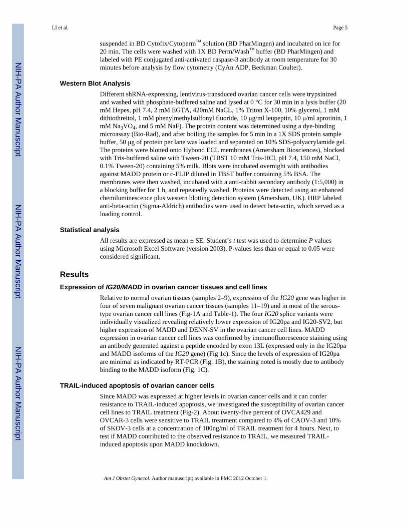
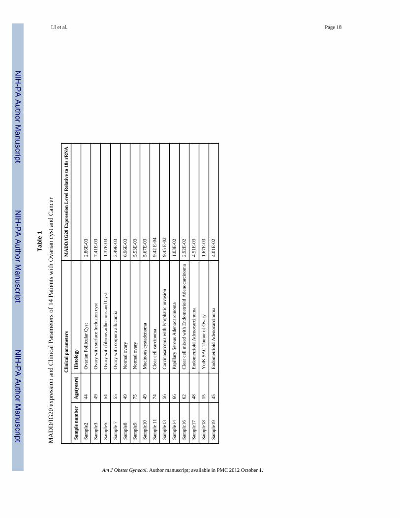
Relative to normal ovarian tissues (samples 2–9), expression of the IG20 gene was higher infour of seven malignant ovarian cancer tissues (samples 11–19) and in most of the serous-type ovarian cancer cell lines (Fig-1A and Table-1). The four IG20 splice variants wereindividually visualized revealing relatively lower expression of IG20pa and IG20-SV2, buthigher expression of MADD and DENN-SV in the ovarian cancer cell lines. MADDexpression in ovarian cancer cell lines was confirmed by immunofluorescence staining usingan antibody generated against a peptide encoded by exon 13L (expressed only in the IG20paand MADD isoforms of the IG20 gene) (Fig 1c). Since the levels of expression of IG20paare minimal as indicated by RT-PCR (Fig. 1B), the staining noted is mostly due to antibodybinding to the MADD isoform (Fig. 1C).
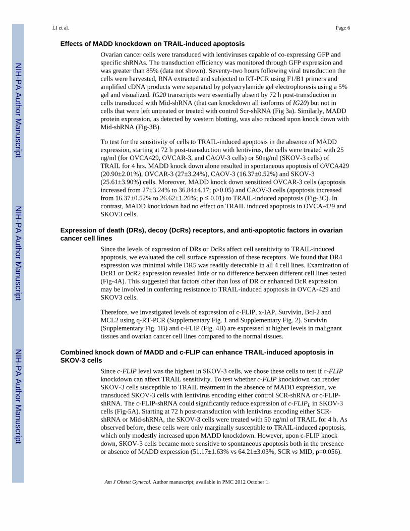
TRAIL-induced apoptosis of ovarian cancer cellsSince MADD was expressed at higher levels in ovarian cancer cells and it can conferresistance to TRAIL-induced apoptosis, we investigated the susceptibility of ovarian cancercell lines to TRAIL treatment (Fig-2). About twenty-five percent of OVCA429 andOVCAR-3 cells were sensitive to TRAIL treatment compared to 4% of CAOV-3 and 10%of SKOV-3 cells at a concentration of 100ng/ml of TRAIL treatment for 4 hours. Next, totest if MADD contributed to the observed resistance to TRAIL, we measured TRAIL-induced apoptosis upon MADD knockdown.
LI et al. Page 5
Am J Obstet Gynecol. Author manuscript; available in PMC 2012 October 1.
NIH
-PA Author Manuscript
NIH
-PA Author Manuscript
NIH
-PA Author Manuscript
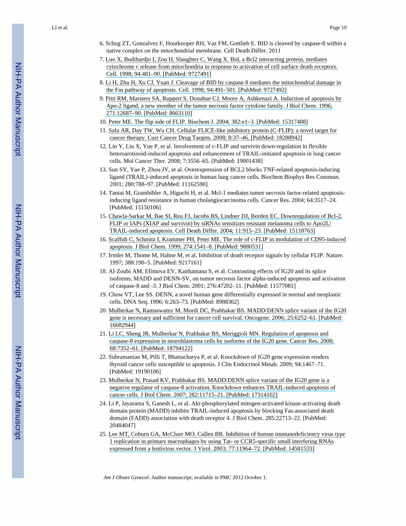
Effects of MADD knockdown on TRAIL-induced apoptosisOvarian cancer cells were transduced with lentiviruses capable of co-expressing GFP andspecific shRNAs. The transduction efficiency was monitored through GFP expression andwas greater than 85% (data not shown). Seventy-two hours following viral transduction thecells were harvested, RNA extracted and subjected to RT-PCR using F1/B1 primers andamplified cDNA products were separated by polyacrylamide gel electrophoresis using a 5%gel and visualized. IG20 transcripts were essentially absent by 72 h post-transduction incells transduced with Mid-shRNA (that can knockdown all isoforms of IG20) but not incells that were left untreated or treated with control Scr-shRNA (Fig 3a). Similarly, MADDprotein expression, as detected by western blotting, was also reduced upon knock down withMid-shRNA (Fig-3B).
To test for the sensitivity of cells to TRAIL-induced apoptosis in the absence of MADDexpression, starting at 72 h post-transduction with lentivirus, the cells were treated with 25ng/ml (for OVCA429, OVCAR-3, and CAOV-3 cells) or 50ng/ml (SKOV-3 cells) ofTRAIL for 4 hrs. MADD knock down alone resulted in spontaneous apoptosis of OVCA429(20.90±2.01%), OVCAR-3 (27±3.24%), CAOV-3 (16.37±0.52%) and SKOV-3(25.61±3.90%) cells. Moreover, MADD knock down sensitized OVCAR-3 cells (apoptosisincreased from 27±3.24% to 36.84±4.17; p>0.05) and CAOV-3 cells (apoptosis increasedfrom 16.37±0.52% to 26.62±1.26%; p ≤ 0.01) to TRAIL-induced apoptosis (Fig-3C). Incontrast, MADD knockdown had no effect on TRAIL induced apoptosis in OVCA-429 andSKOV3 cells.
Expression of death (DRs), decoy (DcRs) receptors, and anti-apoptotic factors in ovariancancer cell lines
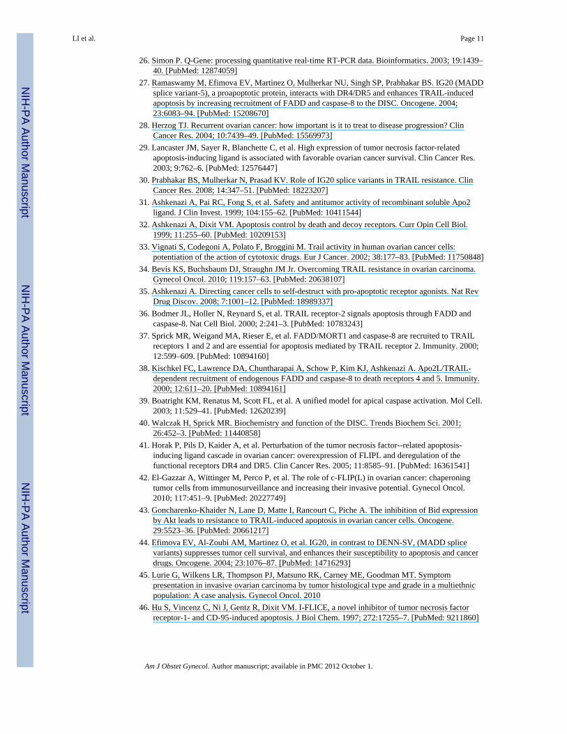
Since the levels of expression of DRs or DcRs affect cell sensitivity to TRAIL-inducedapoptosis, we evaluated the cell surface expression of these receptors. We found that DR4expression was minimal while DR5 was readily detectable in all 4 cell lines. Examination ofDcR1 or DcR2 expression revealed little or no difference between different cell lines tested(Fig-4A). This suggested that factors other than loss of DR or enhanced DcR expressionmay be involved in conferring resistance to TRAIL-induced apoptosis in OVCA-429 andSKOV3 cells.
Therefore, we investigated levels of expression of c-FLIP, x-IAP, Survivin, Bcl-2 andMCL2 using q-RT-PCR (Supplementary Fig. 1 and Supplementary Fig. 2). Survivin(Supplementary Fig. 1B) and c-FLIP (Fig. 4B) are expressed at higher levels in malignanttissues and ovarian cancer cell lines compared to the normal tissues.
Combined knock down of MADD and c-FLIP can enhance TRAIL-induced apoptosis inSKOV-3 cells
Since c-FLIP level was the highest in SKOV-3 cells, we chose these cells to test if c-FLIPknockdown can affect TRAIL sensitivity. To test whether c-FLIP knockdown can renderSKOV-3 cells susceptible to TRAIL treatment in the absence of MADD expression, wetransduced SKOV-3 cells with lentivirus encoding either control SCR-shRNA or c-FLIP-shRNA. The c-FLIP-shRNA could significantly reduce expression of c-FLIPL in SKOV-3cells (Fig-5A). Starting at 72 h post-transduction with lentivirus encoding either SCR-shRNA or Mid-shRNA, the SKOV-3 cells were treated with 50 ng/ml of TRAIL for 4 h. Asobserved before, these cells were only marginally susceptible to TRAIL-induced apoptosis,which only modestly increased upon MADD knockdown. However, upon c-FLIP knockdown, SKOV-3 cells became more sensitive to spontaneous apoptosis both in the presenceor absence of MADD expression (51.17±1.63% vs 64.21±3.03%, SCR vs MID, p=0.056).
LI et al. Page 6
Am J Obstet Gynecol. Author manuscript; available in PMC 2012 October 1.
NIH
-PA Author Manuscript
NIH
-PA Author Manuscript
NIH
-PA Author Manuscript

This was further enhanced upon TRAIL treatment only in cells with MADD knockdown as(58.85±3.3% vs 77.13±3.03%, SCR vs MID p<0.01) (Fig-5B).
DiscussionMany ovarian cancer patients will experience a significant recurrence free interval aftersurgical cytoreduction and adjuvant chemotherapy, however, the majority will have diseaserecurrence within two years and only ~20% will survive 5 years28. As a result, new targetedtherapeutic strategies are needed. Through a better understanding of the molecular pathwayspresent in ovarian cancer, new combination therapies may be designed to target multiplepathways, and possibly improve therapeutic outcomes without significantly increasingtoxicity to normal cells. It has been shown that Tumor necrosis factor (TNF) relatedapoptosis-inducing ligand (TRAIL) is expressed at a 10-fold higher level in ovariancarcinoma compared to normal tissue29 and can selectively induce apoptosis in cancer cellswith little or no effect on normal cells30–32. Therefore, it is an attractive potential therapeuticagent for ovarian cancer. However, many ovarian cancers are resistant to TRAIL inducedapoptosis33, 34.
TRAIL induces apoptosis by binding to death receptor 4 (DR4) and DR5 35. Binding to theDRs results in the recruitment of Fas-associated death domain (FADD) and procaspase-8 tothe death-inducing signaling complex36–38, and leads to activation of caspase-8 39, andsubsequently initiates the apoptosis-executing caspase cascade40. The cellular FLICEinhibitory protein (c-FLIP), which is homologous to caspase-8 but lacks the enzymaticactivity, can also associate with the DISC, blocking activation of caspase-8 throughcompetition for binding sites. In addition, the intrinsic apoptotic pathway can be activated bycaspase-8 through cleavage of the BH3-only protein Bid that triggers perturbation of themitochondria by Bax and Bak and finally activation of caspase-9 and effector caspases 8. Anumber of mechanisms underlying TRAIL resistance have been investigated in ovariancancer, including absence of death receptor expression41, down-regulation of caspase-8expression 42 or over expression of c-FLIP 41, leading to inhibition of the extrinsic apoptoticpathway. Furthermore, down-regulation of Bid expression in ovarian cancer 43 has beenshown to block activation of the intrinsic pathway. Investigating mechanisms and thestrategies to overcome TRAIL resistance will generate new insight into the development ofeffective treatment strategies for improving ovarian cancer therapy; we have shown thatIG20 appears to play a critical role.
Our early studies showed that MADD is highly expressed in different cancers and cancercell lines and can confer TRAIL resistance20, 22, therefore, we investigated MADDexpression and function in ovarian cancer tissues and found that it was expressed at higherlevels in tissues and cell lines. Specifically, we found that MADD and DENN-SV wereexpressed in all ovarian cancer cell lines tested and showed MADD protein expression inthese cells using an antibody that specifically reacts only with an amino acid sequence inIG20pa and MADD isoforms (Fig. 1). This is of significance because our earlier studiesdemonstrated that the MADD splice variant of the IG20 gene is a cancer cell pro-survivalfactor 20, 22.
The IG20 gene can encode 6 different isoforms, namely IG20pa, MADD, DENN-SV, IG20-SV2, KIAA0358 and IG20-SV4. The KIAA0358 and IG20-SV4 isoforms are enriched onlyin select neuronal cells21 and thus are not pertinent to this study. Of the other 4 isoforms, theIG20pa and the IG20-SV2 may be expressed at very low levels or may not be expressed, andthus are not required for critical cellular functions including cell survival. However, theMADD and the DENN-SV isoforms are ubiquitously expressed in most normal tissues atvery low levels but are expressed at much higher levels in cancer cells and tissues18, 22, 44.
LI et al. Page 7
Am J Obstet Gynecol. Author manuscript; available in PMC 2012 October 1.
NIH
-PA Author Manuscript
NIH
-PA Author Manuscript
NIH
-PA Author Manuscript

Selective knockdown of MADD using exon 13 specific siRNA renders cancer cellssusceptible to spontaneous as well as induced apoptosis23. Moreover, re-expression ofsiRNA resistant MADD and not the other isoforms of the IG20 gene, in the absence of allendogenous isoforms due to siRNA mediated knockdown, can confer resistance tospontaneous as well as TRAIL-induced apoptosis in a variety of cancer cells22, 23.Therefore, we tested the sensitivity of ovarian cancer cells to TRAIL treatment in thepresence or absence of MADD expression.
We selected four epithelial serous subtype ovarian cancer cell lines, OVCA-429, OVCAR-3,CAOV-3 and SKOV-3 because epithelial serous type is a major histological type of ovariancancer45 and is usually diagnosed at an advanced stage of the disease. These types of tumorsoften develop drug resistance and therefore, there is a significant need for newer modalitiesof treatment for ovarian cancer.
We found that while OVCA-429 and OVCAR-3 were moderately sensitive, CAOV-3 andSKOV-3 cells were less sensitive to TRAIL treatment (Fig-2). To evaluate the potentialcontribution of MADD to the observed TRAIL resistance, we transduced these cells withSCR or MID-shRNA carrying lentivirus, MID-shRNA effectively down-modulated themRNA and protein expression (Fig. 3A and -3B). Although down-modulation of MADDresulted in spontaneous apoptosis of all four types of cells (Fig. 3C), it further sensitizedonly CAOV-3 cells to TRAIL induced apoptosis (Fig. 3C). These data suggested that whileMADD may not contribute to TRAIL resistance in OVCA-429 and OVCAR-3 cells, it coulddo so in CAOV-3 cells. These data also indicated that other factors may be contributing tothe TRAIL resistance of SKOV-3 cells.
Known inhibitors that can block TRAIL-induced apoptosis include expression of lowerlevels of DRs or higher levels of DcRs, or anti-apoptotic factors such as cFLIP, x-IAP,Survivin, Bcl-2 and MCL19–15, 17,30. Therefore, we tested for the levels of expression ofthese molecules. Our results showed little or no difference in the levels of expression of DRsand DcRs (Fig. 4A) and indicated that factors other than the levels of expression of thesemolecules were most likely responsible for the differential susceptibility to TRAIL.
We found much higher levels of c-FLIP and survivin transcripts in all four cell lines andsome of the malignant tissues relative to normal tissues. However, the levels of transcriptsfor XIAP, Bcl2 and MCL1 were comparable in both normal and tumor tissues with theexception of few ovarian cancer tissues which expressed much higher levels (Fig. 4B).Survivin levels were comparable in all four cell lines and suggested that it may not be acritical TRAIL resistance factor in these cells, however, c-FLIP levels varied between thecell lines and was the highest in SKOV-3 cells.
To determine if c-FLIP was associated with TRAIL resistance, we knocked down either c-FLIP or MADD, or both and tested for TRAIL-induced apoptosis of SKOV-3 cells.Knockdown of c-FLIPL sensitized SKOV-3 cells to spontaneous apoptosis and also TRAIL-induced apoptosis but only in the absence of MADD expression.
c-FLIP binds to FADD and prevents pro-caspase-8 recruitment thereby conferring resistanceto TRAIL-induced apoptosis17, 46. Although MADD knockdown might have resulted inincreased FADD recruitment to DRs, high levels of c-FLIP in these cells likely preventedprocaspase-8 recruitment to FADD and thus prevented apoptosis in MADD knockdowncells treated with TRAIL. This notion is supported by the increased spontaneous apoptosisof cells with c-FLIP knockdown. Cells with c-FLIP knockdown showed increased TRAIL–induced apoptosis, but only upon MADD knockdown (Fig. 5). This likely resulted fromincreased DR oligomerization and FADD recruitment to DRs upon MADD knockdown, andprocaspase-8 recruitment to FADD upon c-FLIP knockdown.
LI et al. Page 8
Am J Obstet Gynecol. Author manuscript; available in PMC 2012 October 1.
NIH
-PA Author Manuscript
NIH
-PA Author Manuscript
NIH
-PA Author Manuscript

Collectively, these results infer that either reduction in FADD recruitment to DR by MADD,or procaspase-8 recruitment to FADD by c-FLIP can prevent TRAIL-induced apoptosis, anda combined knockdown of these proteins might restore TRAIL susceptibility. This raises thepossibility of using TRAIL in conjunction with MADD and c-FLIP inhibitors as a potentialtherapeutic approach to treating certain ovarian cancers, particularly in patients who havedeveloped resistance to conventional therapies due to the expression of different resistancefactors. Though a clinically available antagonistic antibody has not been made specificallyagainst c-FLIP, its expression has been shown to be down-regulated by multiple candidatetherapeutics including the cox-2 inhibitor, celecoxib47, pigment epithelial-derivedfactor 48, 49, rocaglamide50 and lupeol51. Testing these cell lines in the presence and absenceof these c-FLIP antagonists are the subject of our future research. In regards to MADD,there are no known therapeutic inhibitors of its expression and this is a topic of our currenthigh-throughput screening research.
Supplementary MaterialRefer to Web version on PubMed Central for supplementary material.
AcknowledgmentsThis work was supported in part by the grant RO1 CA107506 to B.S.P. from the National Institutes of Health,Bethesda, Maryland, and by a grant from Elsa U. Pardee Foundation to L-C L.
Abbreviations
IG-20 Insulinoma Glucagonoma clone-20
MADD Map-kinase Activating Death Domain containing protein
DENN Differentially Expressed in Normal and Neoplastic tissues
SVs Splice Variants
DR4/5 Death receptor4 and 5
TRAIL Tumor necrosis factor-related apoptosis inducing ligand
c-FLIP Cellular FLICE-inhibitory protein
x-IAP x-linked Inhibitor of Apoptosis Protein
Bcl-2 B-cell lymphoma 2
MCL1 Myeloid Cell Leukemia sequence 1
References1. Jemal A, Siegel R, Xu J, Ward E. Cancer statistics, 2010. CA Cancer J Clin. 2010; 60:277–300.
[PubMed: 20610543]2. Qazi F, McGuire WP. The treatment of epithelial ovarian cancer. CA Cancer J Clin. 1995; 45:88–
101. [PubMed: 7889393]3. Wiley SR, Schooley K, Smolak PJ, et al. Identification and characterization of a new member of the
TNF family that induces apoptosis. Immunity. 1995; 3:673–82. [PubMed: 8777713]4. Walczak H, Miller RE, Ariail K, et al. Tumoricidal activity of tumor necrosis factor-related
apoptosis-inducing ligand in vivo. Nat Med. 1999; 5:157–63. [PubMed: 9930862]5. Okada H, Mak TW. Pathways of apoptotic and non-apoptotic death in tumour cells. Nat Rev
Cancer. 2004; 4:592–603. [PubMed: 15286739]
LI et al. Page 9
Am J Obstet Gynecol. Author manuscript; available in PMC 2012 October 1.
NIH
-PA Author Manuscript
NIH
-PA Author Manuscript
NIH
-PA Author Manuscript
6. Schug ZT, Gonzalvez F, Houtkooper RH, Vaz FM, Gottlieb E. BID is cleaved by caspase-8 within anative complex on the mitochondrial membrane. Cell Death Differ. 2011
7. Luo X, Budihardjo I, Zou H, Slaughter C, Wang X. Bid, a Bcl2 interacting protein, mediatescytochrome c release from mitochondria in response to activation of cell surface death receptors.Cell. 1998; 94:481–90. [PubMed: 9727491]
8. Li H, Zhu H, Xu CJ, Yuan J. Cleavage of BID by caspase 8 mediates the mitochondrial damage inthe Fas pathway of apoptosis. Cell. 1998; 94:491–501. [PubMed: 9727492]
9. Pitti RM, Marsters SA, Ruppert S, Donahue CJ, Moore A, Ashkenazi A. Induction of apoptosis byApo-2 ligand, a new member of the tumor necrosis factor cytokine family. J Biol Chem. 1996;271:12687–90. [PubMed: 8663110]
10. Peter ME. The flip side of FLIP. Biochem J. 2004; 382:e1–3. [PubMed: 15317488]11. Safa AR, Day TW, Wu CH. Cellular FLICE-like inhibitory protein (C-FLIP): a novel target for
cancer therapy. Curr Cancer Drug Targets. 2008; 8:37–46. [PubMed: 18288942]12. Lin Y, Liu X, Yue P, et al. Involvement of c-FLIP and survivin down-regulation in flexible
heteroarotinoid-induced apoptosis and enhancement of TRAIL-initiated apoptosis in lung cancercells. Mol Cancer Ther. 2008; 7:3556–65. [PubMed: 19001438]
13. Sun SY, Yue P, Zhou JY, et al. Overexpression of BCL2 blocks TNF-related apoptosis-inducingligand (TRAIL)-induced apoptosis in human lung cancer cells. Biochem Biophys Res Commun.2001; 280:788–97. [PubMed: 11162590]
14. Taniai M, Grambihler A, Higuchi H, et al. Mcl-1 mediates tumor necrosis factor-related apoptosis-inducing ligand resistance in human cholangiocarcinoma cells. Cancer Res. 2004; 64:3517–24.[PubMed: 15150106]
15. Chawla-Sarkar M, Bae SI, Reu FJ, Jacobs BS, Lindner DJ, Borden EC. Downregulation of Bcl-2,FLIP or IAPs (XIAP and survivin) by siRNAs sensitizes resistant melanoma cells to Apo2L/TRAIL-induced apoptosis. Cell Death Differ. 2004; 11:915–23. [PubMed: 15118763]
16. Scaffidi C, Schmitz I, Krammer PH, Peter ME. The role of c-FLIP in modulation of CD95-inducedapoptosis. J Biol Chem. 1999; 274:1541–8. [PubMed: 9880531]
17. Irmler M, Thome M, Hahne M, et al. Inhibition of death receptor signals by cellular FLIP. Nature.1997; 388:190–5. [PubMed: 9217161]
18. Al-Zoubi AM, Efimova EV, Kaithamana S, et al. Contrasting effects of IG20 and its spliceisoforms, MADD and DENN-SV, on tumor necrosis factor alpha-induced apoptosis and activationof caspase-8 and -3. J Biol Chem. 2001; 276:47202–11. [PubMed: 11577081]
19. Chow VT, Lee SS. DENN, a novel human gene differentially expressed in normal and neoplasticcells. DNA Seq. 1996; 6:263–73. [PubMed: 8988362]
20. Mulherkar N, Ramaswamy M, Mordi DC, Prabhakar BS. MADD/DENN splice variant of the IG20gene is necessary and sufficient for cancer cell survival. Oncogene. 2006; 25:6252–61. [PubMed:16682944]
21. Li LC, Sheng JR, Mulherkar N, Prabhakar BS, Meriggioli MN. Regulation of apoptosis andcaspase-8 expression in neuroblastoma cells by isoforms of the IG20 gene. Cancer Res. 2008;68:7352–61. [PubMed: 18794122]
22. Subramanian M, Pilli T, Bhattacharya P, et al. Knockdown of IG20 gene expression rendersthyroid cancer cells susceptible to apoptosis. J Clin Endocrinol Metab. 2009; 94:1467–71.[PubMed: 19190106]
23. Mulherkar N, Prasad KV, Prabhakar BS. MADD/DENN splice variant of the IG20 gene is anegative regulator of caspase-8 activation. Knockdown enhances TRAIL-induced apoptosis ofcancer cells. J Biol Chem. 2007; 282:11715–21. [PubMed: 17314102]
24. Li P, Jayarama S, Ganesh L, et al. Akt-phosphorylated mitogen-activated kinase-activating deathdomain protein (MADD) inhibits TRAIL-induced apoptosis by blocking Fas-associated deathdomain (FADD) association with death receptor 4. J Biol Chem. 285:22713–22. [PubMed:20484047]
25. Lee MT, Coburn GA, McClure MO, Cullen BR. Inhibition of human immunodeficiency virus type1 replication in primary macrophages by using Tat- or CCR5-specific small interfering RNAsexpressed from a lentivirus vector. J Virol. 2003; 77:11964–72. [PubMed: 14581533]
LI et al. Page 10
Am J Obstet Gynecol. Author manuscript; available in PMC 2012 October 1.
NIH
-PA Author Manuscript
NIH
-PA Author Manuscript
NIH
-PA Author Manuscript
26. Simon P. Q-Gene: processing quantitative real-time RT-PCR data. Bioinformatics. 2003; 19:1439–40. [PubMed: 12874059]
27. Ramaswamy M, Efimova EV, Martinez O, Mulherkar NU, Singh SP, Prabhakar BS. IG20 (MADDsplice variant-5), a proapoptotic protein, interacts with DR4/DR5 and enhances TRAIL-inducedapoptosis by increasing recruitment of FADD and caspase-8 to the DISC. Oncogene. 2004;23:6083–94. [PubMed: 15208670]
28. Herzog TJ. Recurrent ovarian cancer: how important is it to treat to disease progression? ClinCancer Res. 2004; 10:7439–49. [PubMed: 15569973]
29. Lancaster JM, Sayer R, Blanchette C, et al. High expression of tumor necrosis factor-relatedapoptosis-inducing ligand is associated with favorable ovarian cancer survival. Clin Cancer Res.2003; 9:762–6. [PubMed: 12576447]
30. Prabhakar BS, Mulherkar N, Prasad KV. Role of IG20 splice variants in TRAIL resistance. ClinCancer Res. 2008; 14:347–51. [PubMed: 18223207]
31. Ashkenazi A, Pai RC, Fong S, et al. Safety and antitumor activity of recombinant soluble Apo2ligand. J Clin Invest. 1999; 104:155–62. [PubMed: 10411544]
32. Ashkenazi A, Dixit VM. Apoptosis control by death and decoy receptors. Curr Opin Cell Biol.1999; 11:255–60. [PubMed: 10209153]
33. Vignati S, Codegoni A, Polato F, Broggini M. Trail activity in human ovarian cancer cells:potentiation of the action of cytotoxic drugs. Eur J Cancer. 2002; 38:177–83. [PubMed: 11750848]
34. Bevis KS, Buchsbaum DJ, Straughn JM Jr. Overcoming TRAIL resistance in ovarian carcinoma.Gynecol Oncol. 2010; 119:157–63. [PubMed: 20638107]
35. Ashkenazi A. Directing cancer cells to self-destruct with pro-apoptotic receptor agonists. Nat RevDrug Discov. 2008; 7:1001–12. [PubMed: 18989337]
36. Bodmer JL, Holler N, Reynard S, et al. TRAIL receptor-2 signals apoptosis through FADD andcaspase-8. Nat Cell Biol. 2000; 2:241–3. [PubMed: 10783243]
37. Sprick MR, Weigand MA, Rieser E, et al. FADD/MORT1 and caspase-8 are recruited to TRAILreceptors 1 and 2 and are essential for apoptosis mediated by TRAIL receptor 2. Immunity. 2000;12:599–609. [PubMed: 10894160]
38. Kischkel FC, Lawrence DA, Chuntharapai A, Schow P, Kim KJ, Ashkenazi A. Apo2L/TRAIL-dependent recruitment of endogenous FADD and caspase-8 to death receptors 4 and 5. Immunity.2000; 12:611–20. [PubMed: 10894161]
39. Boatright KM, Renatus M, Scott FL, et al. A unified model for apical caspase activation. Mol Cell.2003; 11:529–41. [PubMed: 12620239]
40. Walczak H, Sprick MR. Biochemistry and function of the DISC. Trends Biochem Sci. 2001;26:452–3. [PubMed: 11440858]
41. Horak P, Pils D, Kaider A, et al. Perturbation of the tumor necrosis factor--related apoptosis-inducing ligand cascade in ovarian cancer: overexpression of FLIPL and deregulation of thefunctional receptors DR4 and DR5. Clin Cancer Res. 2005; 11:8585–91. [PubMed: 16361541]
42. El-Gazzar A, Wittinger M, Perco P, et al. The role of c-FLIP(L) in ovarian cancer: chaperoningtumor cells from immunosurveillance and increasing their invasive potential. Gynecol Oncol.2010; 117:451–9. [PubMed: 20227749]
43. Goncharenko-Khaider N, Lane D, Matte I, Rancourt C, Piche A. The inhibition of Bid expressionby Akt leads to resistance to TRAIL-induced apoptosis in ovarian cancer cells. Oncogene.29:5523–36. [PubMed: 20661217]
44. Efimova EV, Al-Zoubi AM, Martinez O, et al. IG20, in contrast to DENN-SV, (MADD splicevariants) suppresses tumor cell survival, and enhances their susceptibility to apoptosis and cancerdrugs. Oncogene. 2004; 23:1076–87. [PubMed: 14716293]
45. Lurie G, Wilkens LR, Thompson PJ, Matsuno RK, Carney ME, Goodman MT. Symptompresentation in invasive ovarian carcinoma by tumor histological type and grade in a multiethnicpopulation: A case analysis. Gynecol Oncol. 2010
46. Hu S, Vincenz C, Ni J, Gentz R, Dixit VM. I-FLICE, a novel inhibitor of tumor necrosis factorreceptor-1- and CD-95-induced apoptosis. J Biol Chem. 1997; 272:17255–7. [PubMed: 9211860]
LI et al. Page 11
Am J Obstet Gynecol. Author manuscript; available in PMC 2012 October 1.
NIH
-PA Author Manuscript
NIH
-PA Author Manuscript
NIH
-PA Author Manuscript
47. Liu X, Yue P, Schonthal AH, Khuri FR, Sun SY. Cellular FLICE-inhibitory protein down-regulation contributes to celecoxib-induced apoptosis in human lung cancer cells. Cancer Res.2006; 66:11115–9. [PubMed: 17145853]
48. Lai LJ, Ho TC. Pigment Epithelial-derived Factor Inhibits c-FLIP Expression and AssistsCiglitazone-induced Apoptosis in Hepatocellular Carcinoma. Anticancer Res. 31:1173–80.[PubMed: 21508362]
49. Lee SJ, Noh HJ, Sung EG, et al. Berberine sensitizes TRAIL-induced apoptosis throughproteasome-mediated downregulation of c-FLIP and Mcl-1 proteins. Int J Oncol. 2011; 38:485–92.[PubMed: 21170508]
50. Bleumink M, Kohler R, Giaisi M, Proksch P, Krammer PH, Li-Weber M. Rocaglamide breaksTRAIL resistance in HTLV-1-associated adult T-cell leukemia/lymphoma by translationalsuppression of c-FLIP expression. Cell Death Differ. 18:362–70. [PubMed: 20706274]
51. Murtaza I, Saleem M, Adhami VM, Hafeez BB, Mukhtar H. Suppression of cFLIP by lupeol, adietary triterpene, is sufficient to overcome resistance to TRAIL-mediated apoptosis inchemoresistant human pancreatic cancer cells. Cancer Res. 2009; 69:1156–65. [PubMed:19176377]
LI et al. Page 12
Am J Obstet Gynecol. Author manuscript; available in PMC 2012 October 1.
NIH
-PA Author Manuscript
NIH
-PA Author Manuscript
NIH
-PA Author Manuscript
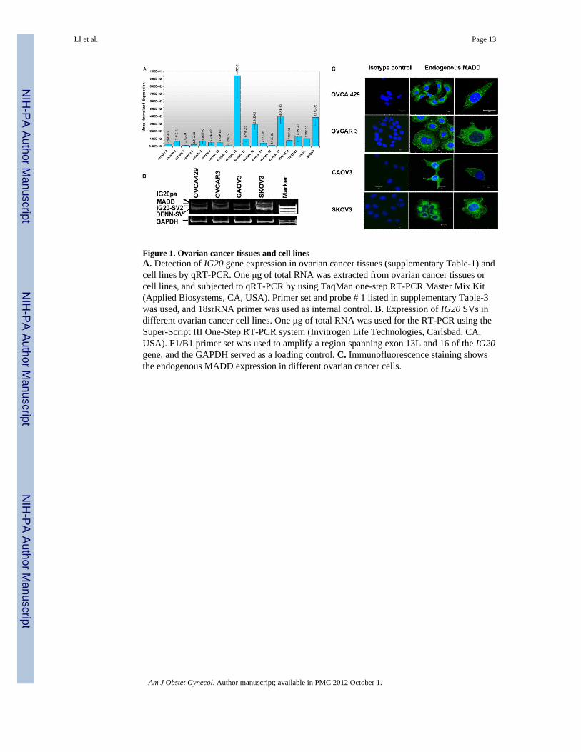
Figure 1. Ovarian cancer tissues and cell linesA. Detection of IG20 gene expression in ovarian cancer tissues (supplementary Table-1) andcell lines by qRT-PCR. One μg of total RNA was extracted from ovarian cancer tissues orcell lines, and subjected to qRT-PCR by using TaqMan one-step RT-PCR Master Mix Kit(Applied Biosystems, CA, USA). Primer set and probe # 1 listed in supplementary Table-3was used, and 18srRNA primer was used as internal control. B. Expression of IG20 SVs indifferent ovarian cancer cell lines. One μg of total RNA was used for the RT-PCR using theSuper-Script III One-Step RT-PCR system (Invitrogen Life Technologies, Carlsbad, CA,USA). F1/B1 primer set was used to amplify a region spanning exon 13L and 16 of the IG20gene, and the GAPDH served as a loading control. C. Immunofluorescence staining showsthe endogenous MADD expression in different ovarian cancer cells.
LI et al. Page 13
Am J Obstet Gynecol. Author manuscript; available in PMC 2012 October 1.
NIH
-PA Author Manuscript
NIH
-PA Author Manuscript
NIH
-PA Author Manuscript

Figure 2. Susceptibility of different ovarian cancer cell lines to TRAIL- induced apoptosisApproximately, 4 × 10⁵ of OVCA-429, OVCAR-3, CAOV-3 and SKOV-3 ovarian cancercells were cultured in 6-well plates, and treated with different doses of TRAIL for 4 h.Subsequently, cells were fixed, permeabilized and stained with PE-conjugated anti-caspase-3 antibody for 30 min at room temperature and subjected to FACS analysis. ** P<0.01 in OVCA-429 vs control; + P< 0.05 in OVCAR-3 vs control; ## P<0.01 in SKOV-3 vscontrol. Representative data are shown from three independent experiments.
LI et al. Page 14
Am J Obstet Gynecol. Author manuscript; available in PMC 2012 October 1.
NIH
-PA Author Manuscript
NIH
-PA Author Manuscript
NIH
-PA Author Manuscript
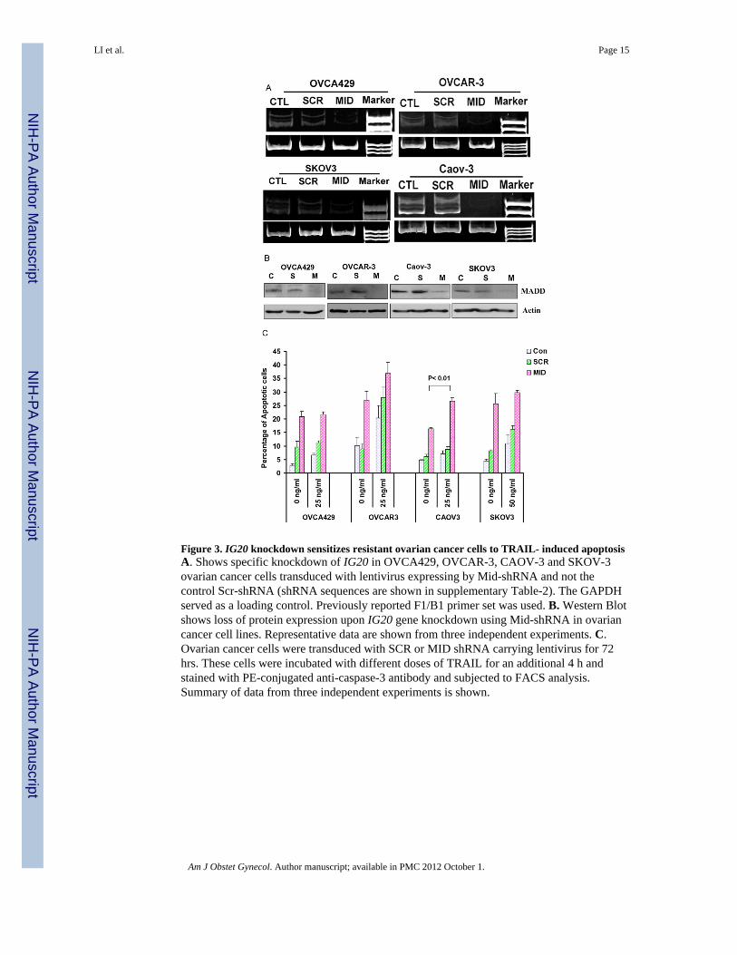
Figure 3. IG20 knockdown sensitizes resistant ovarian cancer cells to TRAIL- induced apoptosisA. Shows specific knockdown of IG20 in OVCA429, OVCAR-3, CAOV-3 and SKOV-3ovarian cancer cells transduced with lentivirus expressing by Mid-shRNA and not thecontrol Scr-shRNA (shRNA sequences are shown in supplementary Table-2). The GAPDHserved as a loading control. Previously reported F1/B1 primer set was used. B. Western Blotshows loss of protein expression upon IG20 gene knockdown using Mid-shRNA in ovariancancer cell lines. Representative data are shown from three independent experiments. C.Ovarian cancer cells were transduced with SCR or MID shRNA carrying lentivirus for 72hrs. These cells were incubated with different doses of TRAIL for an additional 4 h andstained with PE-conjugated anti-caspase-3 antibody and subjected to FACS analysis.Summary of data from three independent experiments is shown.
LI et al. Page 15
Am J Obstet Gynecol. Author manuscript; available in PMC 2012 October 1.
NIH
-PA Author Manuscript
NIH
-PA Author Manuscript
NIH
-PA Author Manuscript
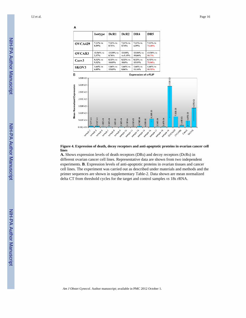
Figure 4. Expression of death, decoy receptors and anti-apoptotic proteins in ovarian cancer celllinesA. Shows expression levels of death receptors (DRs) and decoy receptors (DcRs) indifferent ovarian cancer cell lines. Representative data are shown from two independentexperiments. B. Expression levels of anti-apoptotic proteins in ovarian tissues and cancercell lines. The experiment was carried out as described under materials and methods and theprimer sequences are shown in supplementary Table-2. Data shown are mean normalizeddelta CT from threshold cycles for the target and control samples vs 18s rRNA.
LI et al. Page 16
Am J Obstet Gynecol. Author manuscript; available in PMC 2012 October 1.
NIH
-PA Author Manuscript
NIH
-PA Author Manuscript
NIH
-PA Author Manuscript
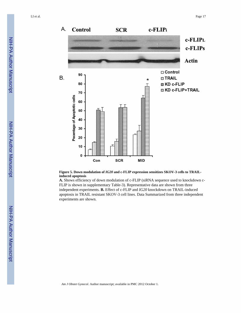
Figure 5. Down modulation of IG20 and c-FLIP expression sensitizes SKOV-3 cells to TRAIL-induced apoptosisA. Shows efficiency of down modulation of c-FLIP (siRNA sequence used to knockdown c-FLIP is shown in supplementary Table-3). Representative data are shown from threeindependent experiments. B. Effect of c-FLIP and IG20 knockdown on TRAIL-inducedapoptosis in TRAIL resistant SKOV-3 cell lines. Data Summarized from three independentexperiments are shown.
LI et al. Page 17
Am J Obstet Gynecol. Author manuscript; available in PMC 2012 October 1.
NIH
-PA Author Manuscript
NIH
-PA Author Manuscript
NIH
-PA Author Manuscript
NIH
-PA Author Manuscript
NIH
-PA Author Manuscript
NIH
-PA Author Manuscript
LI et al. Page 18
Tabl
e 1
MA
DD
/IG20
exp
ress
ion
and
Clin
ical
Par
amet
ers o
f 14
Patie
nts w
ith O
varia
n cy
st a
nd C
ance
r
Clin
ical
par
amet
ers
MA
DD
/IG
20 E
xpre
ssio
n L
evel
Rel
ativ
e to
18s
rR
NA
Sam
ple
num
ber
Age
(yea
rs)
His
tolo
gy
Sam
ple2
44O
varia
n Fo
llicu
lar C
yst
2.86
E-03
Sam
ple3
49O
vary
with
surf
ace
Incl
usio
n cy
st7.
41E-
03
Sam
ple5
54O
vary
with
fibr
ous a
dhes
ions
and
Cys
t1.
37E-
03
Sam
ple
755
Ova
ry w
ith c
orpo
ra a
lbic
antia
2.49
E-03
Sam
ple8
49N
orm
al o
vary
6.96
E-03
Sam
ple9
75N
orm
al o
vary
5.53
E-03
Sam
ple1
049
Muc
inou
s cys
tade
nom
a5.
67E-
03
Sam
ple
1174
Cle
ar c
ell c
arci
nom
a9.
42 E
-04
Sam
ple1
356
Car
cino
sarc
oma
with
lym
phat
ic in
vasi
on9.
45 E
-02
Sam
ple1
466
Papi
llary
Ser
ous A
deno
carc
inom
a1.
03E-
02
Sam
plc1
662
Cle
ar c
ell m
ixed
with
End
omet
rioid
Ade
noca
rcin
oma
2.92
E-02
Sam
ple1
748
Endo
met
rioid
Ade
noca
rcin
oma
4.51
E-03
Sam
ple1
815
Yoi
K S
AC
Tum
or o
f Ova
ry1.
67E-
03
Sam
ple1
945
Endo
met
rioid
Ade
noca
rcin
oma
4.01
E-02
Am J Obstet Gynecol. Author manuscript; available in PMC 2012 October 1.
NIH
-PA Author Manuscript
NIH
-PA Author Manuscript
NIH
-PA Author Manuscript
LI et al. Page 19
Tabl
e 2
Nuc
leot
ide
sequ
ence
of s
iRN
As t
arge
t aga
inst
IG20
and
c-F
LIP
siR
NA
Tar
get S
eque
nce
Tar
getin
g ex
onT
arge
ting
isof
orm
SCR
5′ T
TTA
AC
CG
TTTA
CC
CC
CC
T-3
Non
eN
one
Mid
5′ G
TAC
CA
GC
TTC
AG
TCTT
TC-3′
IG20
Exo
n 15
IG20
pa, M
ADD
, IG
20-S
V2, D
ENN
-SV
c-FL
IPi
5′ A
AG
CA
GTC
TGTT
CA
AG
GA
G-3′
c-FL
IPc-
FLIP
L, c-
FLIP
S
Am J Obstet Gynecol. Author manuscript; available in PMC 2012 October 1.
NIH
-PA Author Manuscript
NIH
-PA Author Manuscript
NIH
-PA Author Manuscript
LI et al. Page 20
Tabl
e 3
The
Prim
ers a
nd p
robe
s use
d in
q-R
T-PC
R.
Gen
e ta
rget
edA
cces
sion
#Fo
rwar
d pr
imer
Rev
ers P
rim
erPr
obe
IG20
/MAD
DN
M_1
3047
1.2
TCA
AC
CC
AC
TCA
TCTA
TGG
CA
ATG
GC
GG
AA
TTG
AA
GA
AC
CG
TAC
CA
FAM
-TTG
CA
AC
ATC
CA
CA
GA
ATC
-TA
MR
A
c-FL
IPN
M_0
0387
9.4
CC
AG
AG
TGTG
TATG
GTG
TGG
AT
TCTC
CC
ATG
AA
CA
TCC
TCC
TGA
TFA
M-C
AC
TCA
GG
GC
TCC
CC
-TA
MR
A
XIAP
NM
_001
167.
2G
CA
CG
AG
CA
GG
GTT
TCTT
TATA
CT
CA
GC
TGC
ATG
AC
AA
CTA
AA
GC
AFA
M-C
CG
CA
CG
GTA
TCTC
C-T
AM
RA
BCL2
NM
_000
657.
2TG
GG
ATG
CG
GG
AG
ATG
TGC
GG
GA
TGC
GG
CTG
GA
TFA
M-C
CC
GG
GC
ATC
TTC
TCC
-TA
MR
A
MC
LIN
M_0
2196
0.3
AG
CA
GA
GG
AG
GA
GG
AG
GA
CG
CC
TGC
TCC
CG
AA
GG
TAFA
M-C
TGC
CG
GTA
CA
AC
TC-T
AM
RA
BIRC
5N
M_0
0116
8.2
AC
CG
CA
TCTC
TAC
ATT
CA
AG
AA
CTG
CC
AG
CC
TCG
GC
CA
TCC
FAM
-CTG
CG
CC
TGC
AC
CC
-TA
MR
A
18s r
RNA
NM
_003
286.
1FA
M-G
GA
GG
GC
AA
GTC
TGG
TGC
CA
GC
AG
C-T
AM
RA
Am J Obstet Gynecol. Author manuscript; available in PMC 2012 October 1.